
Mesoporous Silica Nanoparticles in Chemical Detection: From Small Species to Large Bio-Molecules


Abstract
:1. Introduction
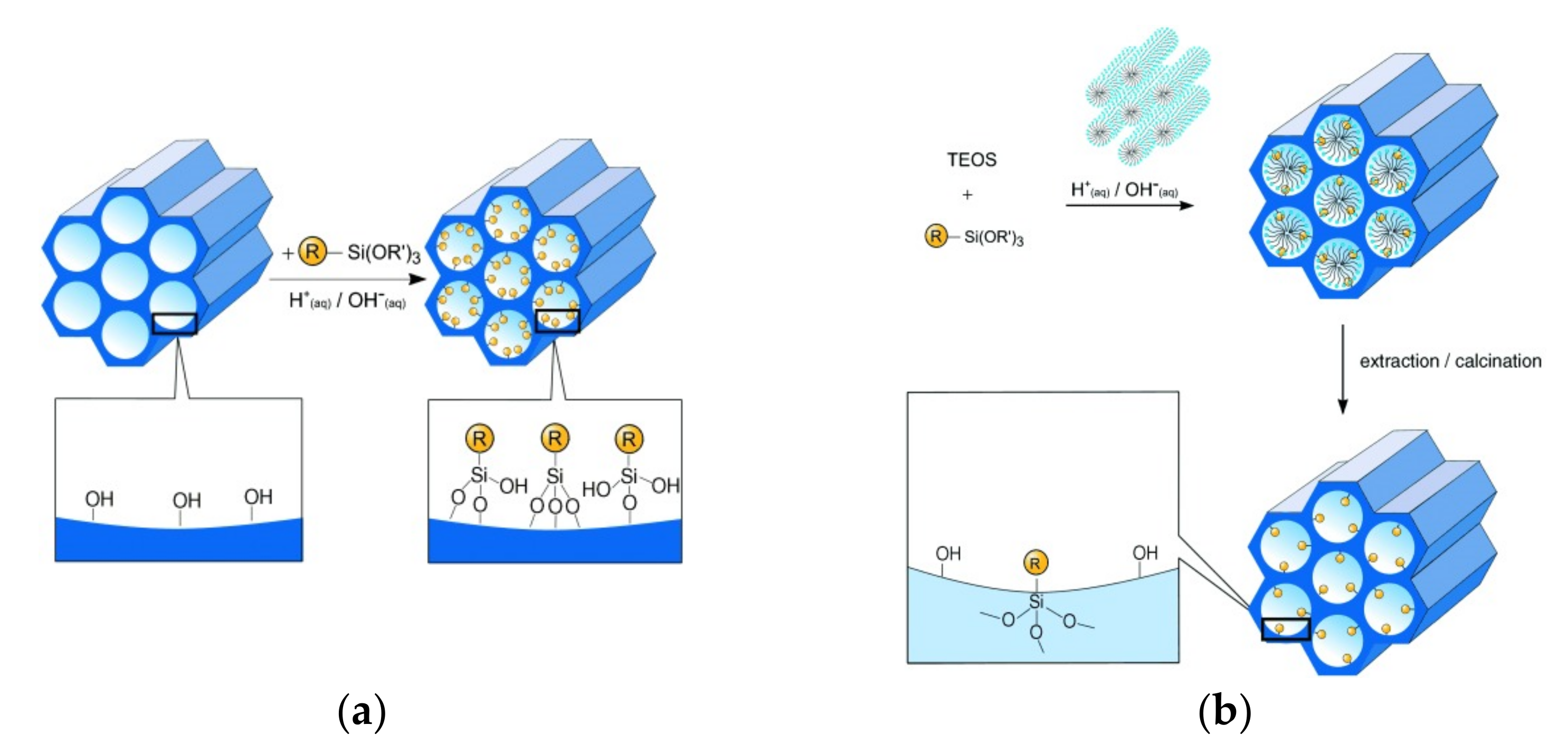
- (1)
- Probes are loaded inside the pores of the material to preserve or modify their optical or binding properties.
- (2)
- Chemosensors or probes are covalently bound to the surface of the material that is acting as a solid support.
- (3)
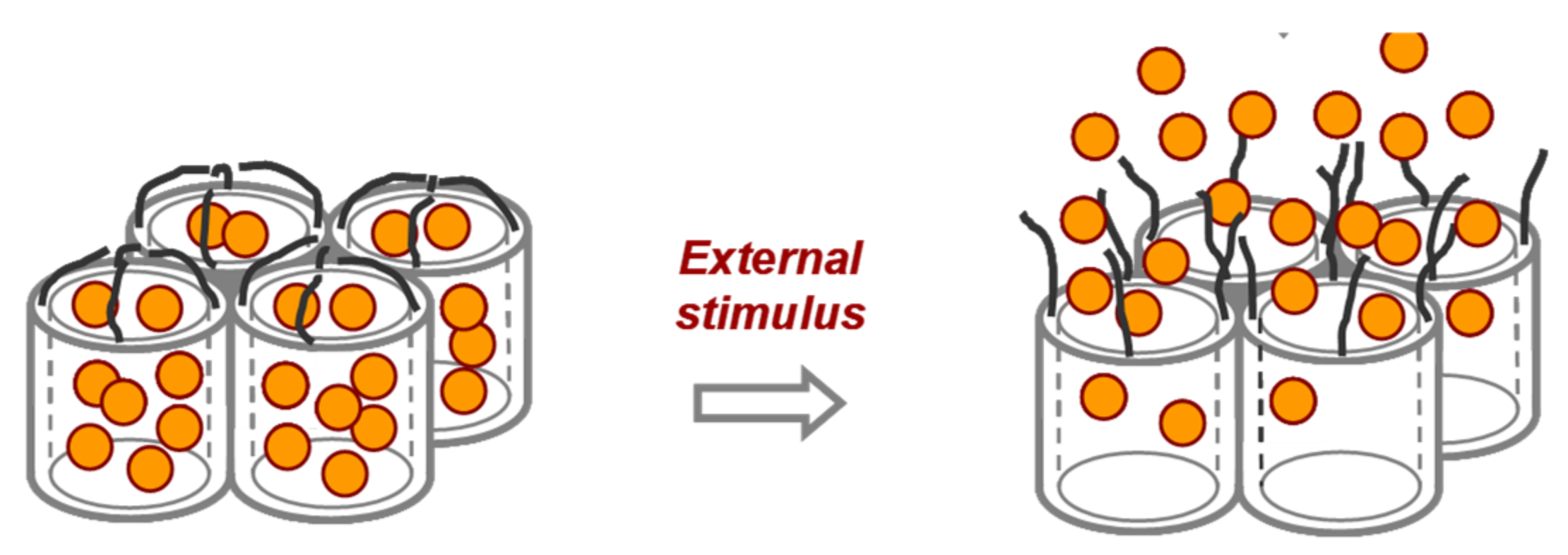
- Dyes are loaded inside the pores of the nanomaterials, and these pores are closed with molecular gates. These molecular gates retain the cargo inside the pores and are only opened in the presence of the analyte with the concomitant release of the dye.
2. Detection Studies
2.1. Detection of Cations
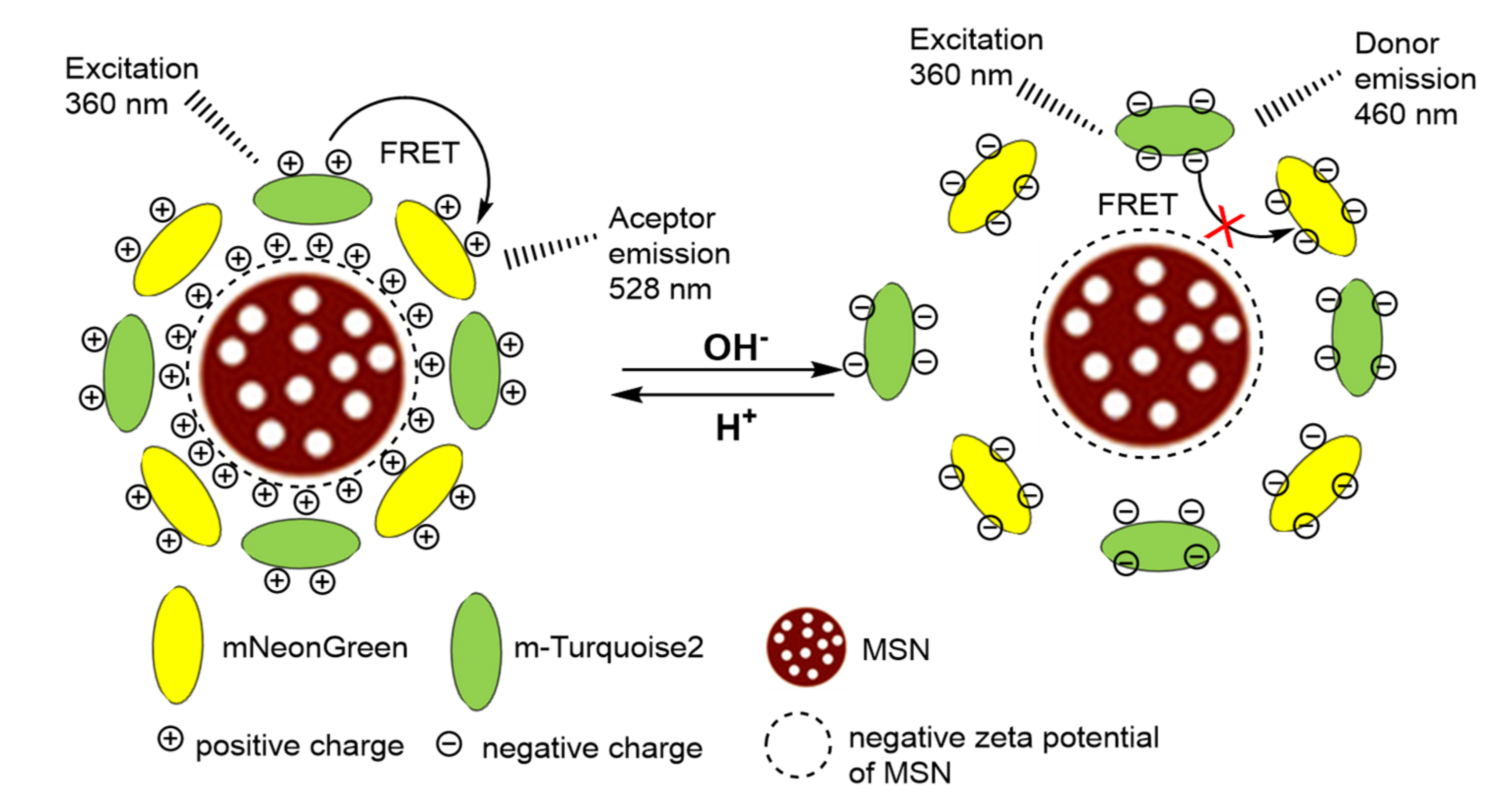
2.2. pH Measurements
2.3. Detection of Radicals
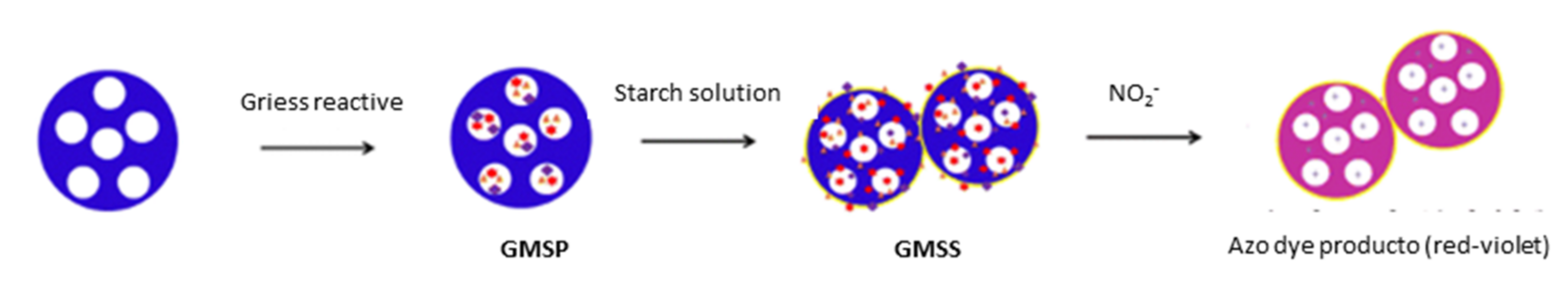
2.4. Detection of Anions
2.5. Detection of Neutral Analytes
2.5.1. Mesoporous Silica Nanoparticles in Gas Sensing
2.5.2. Mesoporous Silica Nanoparticles in the Sensing of Other Neutral Molecules
2.5.3. Mesoporous Silica Nanoparticles in Sensing of Biogenic Analytes
2.6. Detection of Miscellaneous Compounds
3. Challenges and Perspectives
4. Conclusions
Author Contributions
Funding
Acknowledgments
Conflicts of Interest
References
- Aznar, E.; Oroval, M.; Pascual, L.; Murguía, J.R.; Martínez-Máñez, R.; Sancenón, F. Gated Materials for On-Command Release of Guest Molecules. Chem. Rev. 2016, 116, 561–718. [Google Scholar] [CrossRef] [PubMed]
- Sun, Z.; Cui, G.; Li, H.; Liu, Y.; Tian, Y.; Yan, S. Multifunctional optical sensing probes based on organic—Inorganic hybrid composites. J. Mater. Chem. B 2016, 4, 5194–5216. [Google Scholar] [CrossRef] [PubMed]
- Alberti, S.; Soler-Illia, G.J.A.A.; Azzaroni, O. Gated supramolecular chemistry in hybrid mesoporous silica nanoarchitectures: Controlled delivery and molecular transport in response to chemical, physical and biological stimuli. Chem. Commun. 2015, 51, 6050–6075. [Google Scholar] [CrossRef] [PubMed] [Green Version]
- Visakh, P.M.; Martínez Morlanes, M.J. Nanomaterials and Nanocomposites; Wiley-VCH: Weinheim, Germany, 2016. [Google Scholar]
- Kresge, C.T.; Leonowicz, M.E.; Roth, W.J.; Vartuli, J.C.; Beck, J.S. Ordered mesoporous molecular sieves synthesized by a liquid-crystal template mechanism. Nature 1992, 359, 710–712. [Google Scholar] [CrossRef]
- Pal, N.; Lee, J.-H.; Cho, E.-B. Recent Trends in Morphology-Controlled Synthesis and Application of Mesoporous Silica Nanoparticles. Nanomaterials 2020, 10, 2122. [Google Scholar] [CrossRef]
- Singh, D.; Saini, R.K.; Bhagwan, S. Emerging Photovoltaic Materials: Silicon and Beyond; Kurinec, S.K., Ed.; Wiley: Hoboken, NJ, USA, 2019; Chapter 12; pp. 443–486. [Google Scholar]
- Manj, R.Z.A.; Chen, X.; Rehman, W.U.; Zhu, G.; Luo, W.; Yang, J. Big Potential from Silicon-Based Porous Nanomaterials: In Field of Energy Storage and Sensors. Front. Chem. 2018, 6, 359. [Google Scholar] [CrossRef] [Green Version]
- Mehmood, Y.; Khan, I.U.; Shahzad, Y.; Khan, R.U.; Iqbal, M.S.; Khan, H.A.; Khalid, I.; Yousaf, A.M.; Khalid, S.H.; Asghar, S.; et al. In-Vitro and In-Vivo Evaluation of Velpatasvir- Loaded Mesoporous Silica Scaffolds. A Prospective Carrier for Drug Bioavailability Enhancement. Pharmaceutics 2020, 12, 307. [Google Scholar] [CrossRef] [Green Version]
- Huang, R.; Shen, Y.-W.; Guan, Y.-Y.; Jiang, Y.-X.; Wu, Y.; Rahman, K.; Zhang, L.-J.; Liu, H.-J.; Luan, X. Mesoporous silica nanoparticles: Facile surface functionalization and versatile biomedical applications in oncology. Acta Biomater. 2020, 116, 1–15. [Google Scholar] [CrossRef]
- García-Fernández, A.; Aznar, E.; Martínez-Máñez, R.; Sancenón, F. New Advances in In Vivo Applications of Gated Mesoporous Silica as Drug Delivery Nanocarriers. Small 2020, 16, 1902242. [Google Scholar] [CrossRef]
- Pontón, I.; Martí del Rio, A.; Gómez Gómez, M.; Sánchez-García, D. Preparation and Applications of Organo-Silica Hybrid Mesoporous Silica Nanoparticles for the Co-Delivery of Drugs and Nucleic Acids. Nanomaterials 2020, 10, 2466. [Google Scholar] [CrossRef]
- Chen, L.; Liu, M.; Zhou, Q.; Li, X. Recent developments of mesoporous silica nanoparticles in biomedicine. Emergent Mater. 2020, 3, 381–405. [Google Scholar] [CrossRef]
- Castillo, R.R.; Baeza, A.; Vallet-Regí, M. Recent applications of the combination of mesoporous silica nanoparticles with nucleic acids: Development of bioresponsive devices, carriers and sensors. Biomater. Sci. 2017, 5, 353–377. [Google Scholar] [CrossRef]
- Walcarius, A. Silica-based electrochemical sensors and biosensors: Recent trends. Curr. Opin. Electrochem. 2018, 10, 88–97. [Google Scholar] [CrossRef]
- Bagheri, E.; Ansari, L.; Abnous, K.; Taghdisi, S.; Seyed, M.; Ramezani, P.; Ramezani, M.; Alibolandi, M. Silica–Quantum Dot Nanomaterials as a Versatile Sensing Platform. Crit. Rev. Anal. Chem. 2021, 51, 687–708. [Google Scholar] [CrossRef]
- Gastaldi, L.; Ugazio, E.; Sapino, S.; Iliade, P.; Miletto, I.; Berlier, G. Mesoporous silica as a carrier for topical application: The Trolox case study. Phys. Chem. Chem. Phys. 2012, 14, 11318. [Google Scholar] [CrossRef]
- Yang, H.; Zheng, K.; Zhang, Z.; Shi, W.; Jing, S.; Wang, L.; Zheng, W.; Zhao, D.; Xu, J.; Zhang, P. Adsorption and protection of plasmid DNA on mesoporous silica nanoparticles modified with various amounts of organosilane. J. Colloid Interface Sci. 2012, 369, 317. [Google Scholar] [CrossRef]
- Zimny, K.; Blin, J.L.; Stébé, M.J. Ordered mesoporous silica templated by nonionic fluorinated liquid crystals. J. Phys. Chem. C 2009, 113, 11285. [Google Scholar] [CrossRef]
- Wirnsberger, G.; Stucky, G.D. Artificial Noses Sniff DNA. ChemPhysChem 2000, 1, 89–90. [Google Scholar]
- Scott, B.J.; Wirnsberger, G.; Stucky, G.D. Mesoporous and Mesostructured Materials for Optical Applications. Chem. Mater. 2001, 13, 3140–3150. [Google Scholar] [CrossRef]
- Stein, A.; Melde, B.J.; Schroden, R.C. Hybrid Inorganic–Organic Mesoporous Silicates—Nanoscopic Reactors Coming of Age. Adv. Mater. 2000, 12, 1403–1419. [Google Scholar] [CrossRef]
- De Juan, F.; Ruiz-Hitzky, E. Selective Functionalization of Mesoporous Silica. Adv. Mater. 2000, 12, 430–432. [Google Scholar] [CrossRef]
- Lim, M.H.; Stein, A. Comparative Studies of Grafting and Direct Syntheses of Inorganic−Organic Hybrid Mesoporous Materials. Chem. Mater. 1999, 11, 3285–3295. [Google Scholar] [CrossRef]
- Climent, E.; Bernardos, A.; Martínez-Máñez, R.; Maquieira, A.; Marcos, M.D.; Pastor-Navarro, N.; Puchades, R.; Sancenón, F.; Soto, J.; Amorós, P. Controlled Delivery Systems Using Antibody-Capped Mesoporous Nanocontainers. J. Am. Chem. Soc. 2009, 131, 14075. [Google Scholar] [CrossRef]
- Climent, E.; Martínez-Máñez, R.; Sancenón, F.; Marcos, M.D.; Soto, J.; Maquieira, A.; Amorós, P. Controlled Delivery Using Oligonucleotide-Capped Mesoporous Silica Nanoparticles. Angew. Chem. Int. Ed. 2010, 49, 7281–7283. [Google Scholar] [CrossRef]
- Coll, C.; Aznar, E.; Martínez-Máñez, R.; Marcos, M.D.; Sancenón, F.; Soto, J.; Amorós, P.; Cano, J.; Ruiz, E. Fatty Acid Carboxylate- and Anionic Surfactant-Controlled Delivery Systems That Use Mesoporous Silica Supports. Chem. Eur. J. 2010, 16, 10048–10061. [Google Scholar] [CrossRef]
- Candel, I.; Bernardos, A.; Climent, E.; Marcos, M.D.; Martínez-Máñez, R.; Sancenón, F.; Soto, J.; Costero, A.M.; Gil, S.; Parra, M. Selective opening of nanoscopic capped mesoporous inorganic materials with nerve agent simulants; an application to design chromo-fluorogenic probes. Chem. Commun. 2011, 47, 8313–8315. [Google Scholar] [CrossRef]
- Choudhury, N.; Saha, B.; De, P. Recent progress in polymer-based optical chemosensors for Cu2+ and Hg2+ Ions: A comprehensive review. Eur. Polym. J. 2021, 145, 110233. [Google Scholar] [CrossRef]
- Ferreira Andrade, G.; Ferreir Soares, D.C.; Gouvêados Santos, R.; Martins Barros Sousa, E. Mesoporous silica SBA-16 nanoparticles: Synthesis, physicochemical characterization, release profile, and in vitro cytocompatibility studies. Microporous Mesoporous Mater. 2013, 168, 102–110. [Google Scholar] [CrossRef]
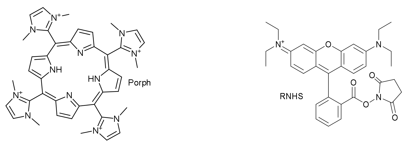
- Marcelo, G.A.; Mota, J.P.; Lodeiro, C.; Oliveira, E. New dual colorimetric/fluorimetric probes for Hg2+ detection & extraction based on mesoporous SBA-16 nanoparticles containing porphyrin or rhodamine chromophores. Dye. Pigment. 2019, 161, 427–437. [Google Scholar]
- Chen, F.; Xiao, F.; Zhang, W.; Lin, C.; Wu, Y. Highly Stable and NIR Luminescent Ru–LPMSN Hybrid Materials for Sensitive Detection of Cu2+ in Vivo. ACS Appl. Mater. Interfaces 2018, 10, 26964–26971. [Google Scholar] [CrossRef]
- Knežević, N.Ž.; Durand, J.-O. Large pore mesoporous silica nanomaterials for application in delivery of biomolecules. Nanoscale 2015, 7, 2199. [Google Scholar] [CrossRef] [PubMed]
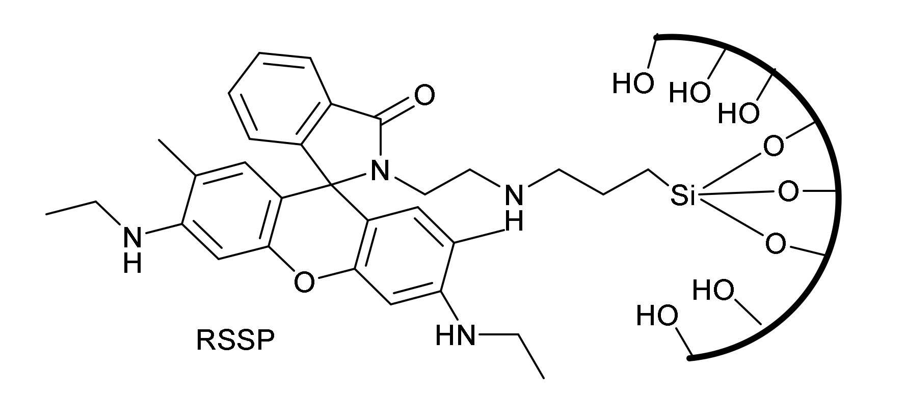
- Kim, H.; Rao, B.A.; Jeong, J.; Angupillai, S.; Choi, J.S.; Nam, J.-O.; Lee, C.-S.; Son, Y.-A. A rhodamine scaffold immobilized onto mesoporous silica as a fluorescent probe for the detection of Fe (III) and applications in bio-imaging and microfluidic chips. Sens. Actuators B Chem. 2016, 224, 404–412. [Google Scholar] [CrossRef]
- Gai, F.; Zhou, T.; Chu, G.; Li, Y.; Liu, Y.; Huo, Q.; Akhtar, F. Mixed anionic surfactant-templated mesoporous silica nanoparticles for fluorescence detection of Fe3+. Dalton Trans. 2016, 45, 508–514. [Google Scholar] [CrossRef] [PubMed] [Green Version]
- Wu, H.; Jia, J.; Xu, Y.; Qian, X.; Zhu, W. A reusable bifunctional fluorescent sensor for the detection and removal of silver ions in aqueous solutions. Sens. Actuators B Chem. 2018, 265, 59–66. [Google Scholar] [CrossRef]
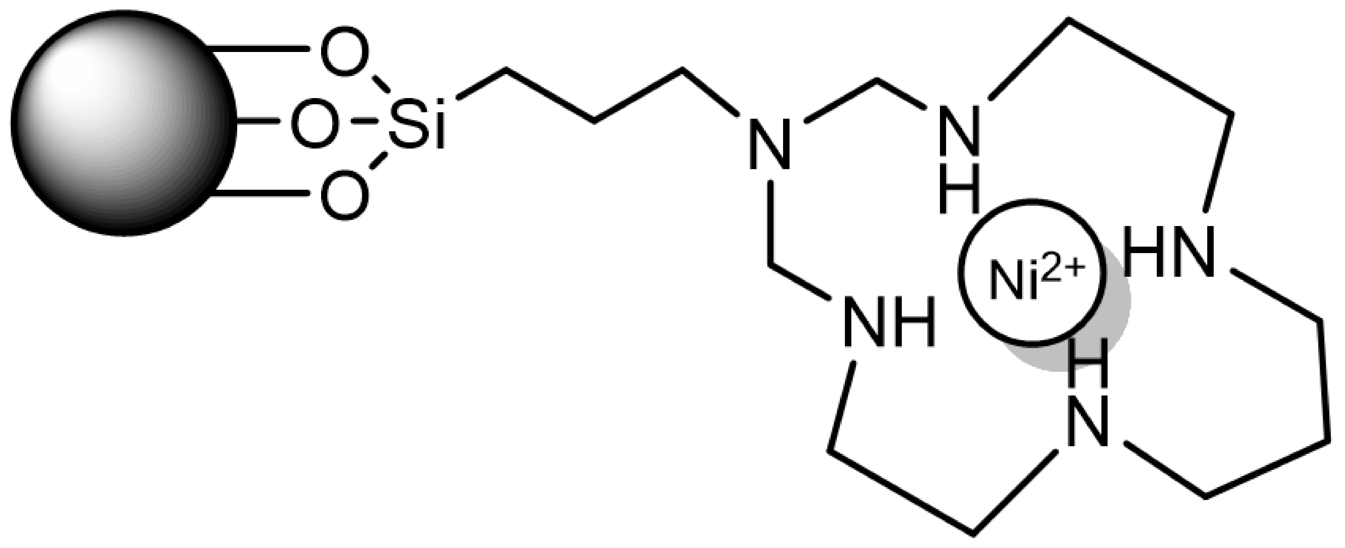
- Jimenez-Falcao, S.; Villalonga, A.; Parra-Nieto, J.; Llopis-Lorente, A.; Martinez-Ruiz, P.; Martínez-Máñez, R.; Villalonga, R. Dithioacetal-mechanized mesoporous nanosensor for Hg(II) determination. Microporous Mesoporous Mater. 2020, 297, 110054. [Google Scholar] [CrossRef]
- Bell, J.; Climent, E.; Hecht, M.; Buurman, M.; Rurack, K. Combining a Droplet-Based Microfluidic Tubing System with Gated Indicator Releasing Nanoparticles for Mercury Trace Detection. ACS Sens. 2016, 1, 334–338. [Google Scholar] [CrossRef]
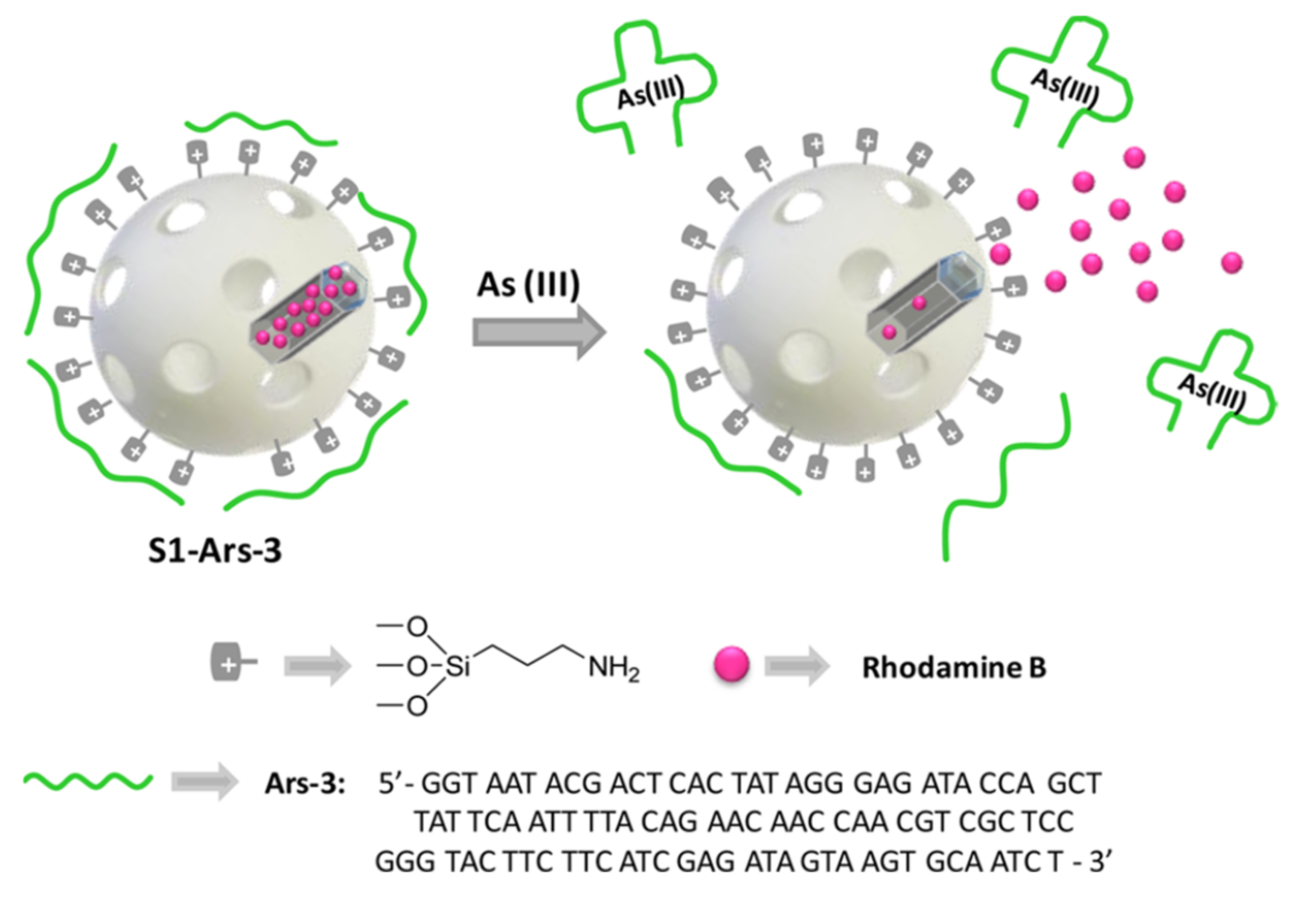
- Oroval, M.; Coll, C.; Bernardos, A.; Marcos, M.D.; Martínez-Mañez, R.; Shchukin, D.G.; Sancenón, F. Selective Fluorogenic Sensing of As(III) Using Aptamer-Capped Nanomaterials. ACS Appl. Mater. Interfaces 2017, 9, 11332–11336. [Google Scholar] [CrossRef] [Green Version]
- Yu, H.; Chen, C.; Cao, X.; Liu, Y.; Zhou, S.; Wang, P. Ratiometric fluorescent pH nanoprobes based on in situ assembling of fluorescence resonance energy transfer between fluorescent proteins. Anal. Bioanal. Chem. 2017, 409, 5073–5080. [Google Scholar] [CrossRef]
- Cadenas, E.; Davies, K.J.A. Mitochondrial free radical generation, oxidative stress, and aging. Free Rad. Biol. Med. 2000, 29, 222–230. [Google Scholar] [CrossRef]
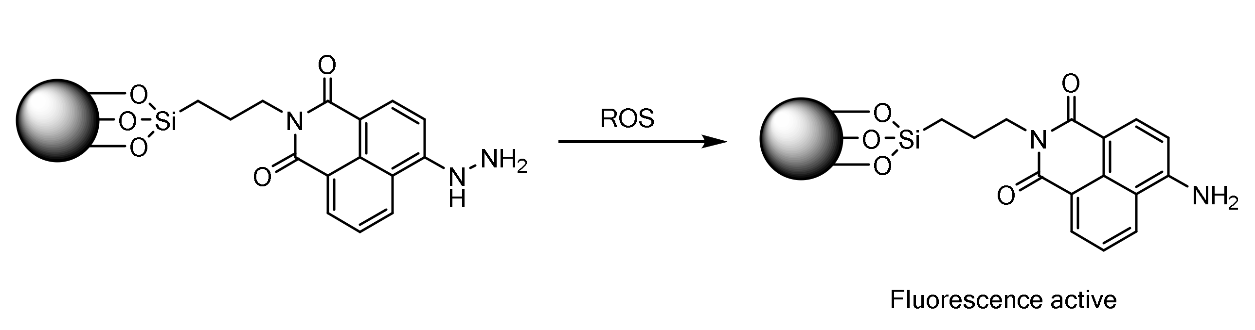
- Jha, G.; Roy, S.; Sahu, P.K.; Banerjee, S.; Anoop, N.; Rahaman, A.; Sarkar, M. Free-radical sensing by using naphthalimide based mesoporous silica (MCM-41) nanoparticles: A combined fluorescence and cellular imaging study. Chem. Phys. Lett. 2018, 692, 324–332. [Google Scholar] [CrossRef]
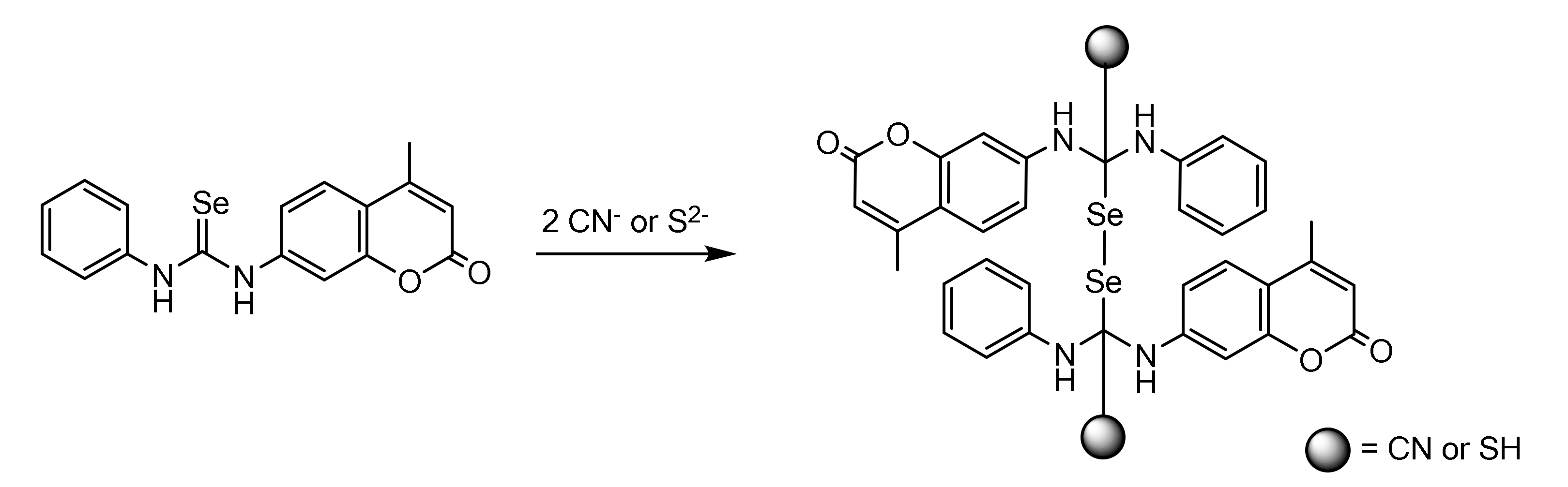
- Casula, A.; Llopis-Lorente, A.; Garau, A.; Isaia, F.; Kubicki, M.; Lippolis, V.; Sancenón, F.; Martínez-Máñez, R.; Owczarzak, A.; Santi, C.; et al. A new class of silica-supported chromo-fluorogenic chemosensors for anion recognition based on a selenourea scaffold. Chem. Commun. 2017, 53, 3729–3732. [Google Scholar] [CrossRef]
- Hussain, R.A.; Badshah, A.; Tahir, M.N.; Hassan, T.U.; Bano, A. Synthesis, Chemical Characterization, DNA Binding, Antioxidant, Antibacterial, and Antifungal Activities of Ferrocence Incorporated Selenoureas. J. Biochem. Mol. Toxicol. 2014, 28, 60–68. [Google Scholar] [CrossRef]
- Suzuki, H.; Iijima, K.; Moriya, A.; McElroy, K.; Scobie, G.; Fyfe, V.; McColl, K.E.L. Conditions for acid catalysed luminal nitrosation are maximal at the gastric cardia. Gut 2003, 52, 1095–1101. [Google Scholar] [CrossRef] [Green Version]
- Taweekarn, T.; Wongniramaikul, W.; Limsakul, W.; Sriprom, W.; Phawachalotorn, C.; Choodum, A. A novel colorimetric sensor based on modified mesoporous silica nanoparticles for rapid on-site detection of nitrite. Microchim. Acta 2020, 187, 643. [Google Scholar] [CrossRef]
- Ma, Q.; Lai, Y.; Gao, J.; Wang, Q. Assay of fluoride by a novel organic–inorganic mesoporous nano-sized sensor. Luminescence 2016, 31, 1125–1129. [Google Scholar] [CrossRef]
- El Sayed, S.; Licchelli, M.; Martinez-Mañez, R.; Sancenon, F. Capped Mesoporous Silica Nanoparticles for the Selective and Sensitive Detection of Cyanide. Chem. Asian J. 2017, 12, 2670–2674. [Google Scholar] [CrossRef]
- Pasparakis, M.; Vandenabeele, P. Necroptosis and its role in inflammation. Nature 2015, 517, 311–320. [Google Scholar] [CrossRef]
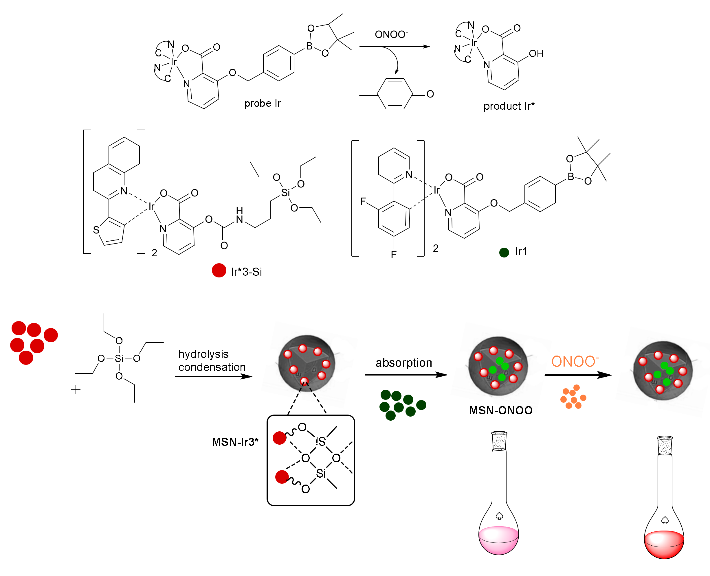
- Cheng, D.; Pan, Y.; Wang, L.; Zeng, Z.; Yuan, L.; Zhang, X.; Chang, Y.-T. Selective Visualization of the Endogenous Peroxynitrite in an Inflamed Mouse Model by a Mitochondria-Targetable Two-Photon Ratiometric Fluorescent Probe. J. Am. Chem. Soc. 2017, 139, 285–292. [Google Scholar] [CrossRef]
- Chen, Z.; Yan, P.; Zou, L.; Zhao, M.; Jiang, J.; Liu, S.; Zhang, K.Y.; Huang, W.; Zhao, Q. Using Ultrafast Responsive Phosphorescent Nanoprobe to Visualize Elevated Peroxynitrite In Vitro and In Vivo via Ratiometric and Time-Resolved Photoluminescence Imaging. Adv. Healthc. Mater. 2018, 7, 1800309. [Google Scholar] [CrossRef]
- Yang, J.; Cheng, F.; Zhu, Z.; Feng, J.; Xue, M.; Meng, Z.; Qiu, L. An enhanced gas sensor based on SiO2@mesoporous MCM-41 core–shell nanocomposites for SO2 visual detection. Analyst 2020, 145, 4352–4357. [Google Scholar] [CrossRef]
- Wang, L.; Zhang, H.; Zhou, X.; Liu, Y.; Lei, B. Preparation, characterization and oxygen sensing properties of luminescent carbon dots assembled mesoporous silica microspheres. J. Colloid Interface Sci. 2016, 478, 256–262. [Google Scholar] [CrossRef]
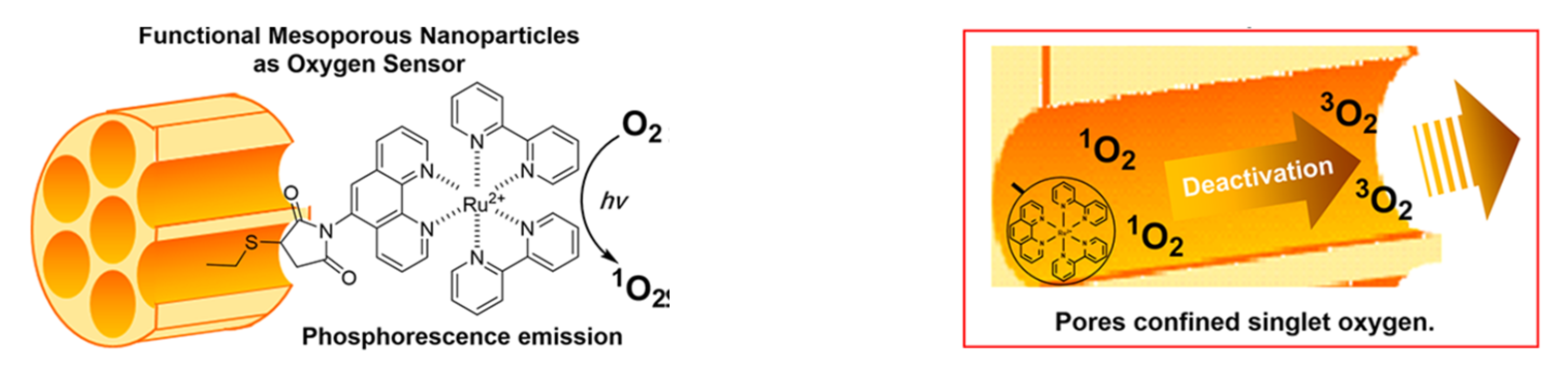
- Yang, Z.; Wen, J.; Wang, Q.; Li, Y.; Zhao, Y.; Tian, Y.; Wang, X.; Cao, X.; Zhang, Y.; Lu, G.; et al. Sensitive, Real-Time, and In-Vivo Oxygen Monitoring for Photodynamic Therapy by Multifunctional Mesoporous Nanosensors. ACS Appl. Mater. Interfaces 2019, 11, 187–194. [Google Scholar] [CrossRef]
- Kitajima, N.; Umehara, Y.; Son, A.; Kondo, T.; Tanabe, K. Confinement of Singlet Oxygen Generated from Ruthenium Complex-Based Oxygen Sensor in the Pores of Mesoporous Silica Nanoparticles. Bioconjugate Chem. 2018, 29, 4168–4175. [Google Scholar] [CrossRef]
- Gao, Z.; Qiao, M.; Tan, M.; Peng, H.; Ding, L. Surface functionalization of mesoporous silica nanoparticles with pyronine derivative for selective detection of hydrogen sulfide in aqueous solution. Colloids Surf. A 2020, 586, 124194. [Google Scholar] [CrossRef]
- El Sayed, S.; Pascual, L.; Licchelli, M.; Martínez-Mañez, R.; Gil, S.; Costero, A.M.; Sancenón, F. Chromogenic Detection of Aqueous Formaldehyde Using Functionalized Silica Nanoparticles. ACS Appl. Mater. Interfaces 2016, 8, 14318–14322. [Google Scholar] [CrossRef]
- Climent, E.; Biyikal, M.; Gawlitza, K.; Dropa, T.; Urban, M.; Costero, A.M.; Martínez-Máñez, R.; Rurack, K. Determination of the chemical warfare agents Sarin, Soman and Tabun in natural waters employing fluorescent hybrid silica materials. Sens. Actuators B 2017, 246, 1056–1065. [Google Scholar] [CrossRef]
- Climent, E.; Biyikal, M.; Gawlitza, K.; Dropa, T.; Urban, M.; Costero, A.M.; Martínez-Máñez, R.; Rurack, K. A Rapid and Sensitive Strip-Based Quick Test for Nerve Agents Tabun, Sarin, and Soman Using BODIPY-Modified Silica Materials. Chem. Eur. J. 2016, 22, 11138–11142. [Google Scholar] [CrossRef] [Green Version]
- Pascual, L.; El Sayed, S.; Martínez-Mañez, R.; Costero, A.M.; Gil, S.; Gaviña, P.; Sancenón, F. Acetylcholinesterase-Capped Mesoporous Silica Nanoparticles That Open in the Presence of Diisopropylfluorophosphate (a Sarin or Soman Simulant). Org. Lett. 2016, 18, 5548–5551. [Google Scholar] [CrossRef]
- Juárez, L.A.; Costero, A.M.; Parra, M.; Gaviña, P.; Gil, S.; Martínez-Máñez, R.; Sancenón, F. NO2-controlled cargo delivery from gated silica mesoporous nanoparticles. Chem. Commun. 2017, 53, 585–588. [Google Scholar] [CrossRef]
- Wang, C.; Li, Q.; Wang, B.; Li, D.; Yu, J. Fluorescent sensors based on AIEgen-functionalised mesoporous silica nanoparticles for the detection of explosives and antibiotics. Inorg. Chem. Front. 2018, 5, 2183–2188. [Google Scholar] [CrossRef]
- Kim, Y.; Min Lee, K.; Young Chang, J. Highly luminescent tetra(biphenyl-4-yl)ethene-grafted molecularly imprinted mesoporous silica nanoparticles for fluorescent sensing of diethylstilbestrol. Sens. Actuators B 2017, 242, 1296–1304. [Google Scholar] [CrossRef]
- Tan, S.Y.; Teh, C.; Yen Ang, C.; Li, M.; Li, P.; Korzh, V.; Zhao, Y. Responsive mesoporous silica nanoparticles for sensing of hydrogen peroxide and simultaneous treatment toward heart failure. Nanoscale 2017, 9, 2253–2261. [Google Scholar] [CrossRef] [PubMed] [Green Version]
- Liu, C.; Chen, W.; Qing, Z.; Zheng, J.; Xiao, Y.; Yang, S.; Wang, L.; Li, Y.; Yang, R. In Vivo Lighted Fluorescence via Fenton Reaction: Approach for Imaging of Hydrogen Peroxide in Living Systems. Anal. Chem. 2016, 88, 3998–4003. [Google Scholar] [CrossRef] [PubMed]
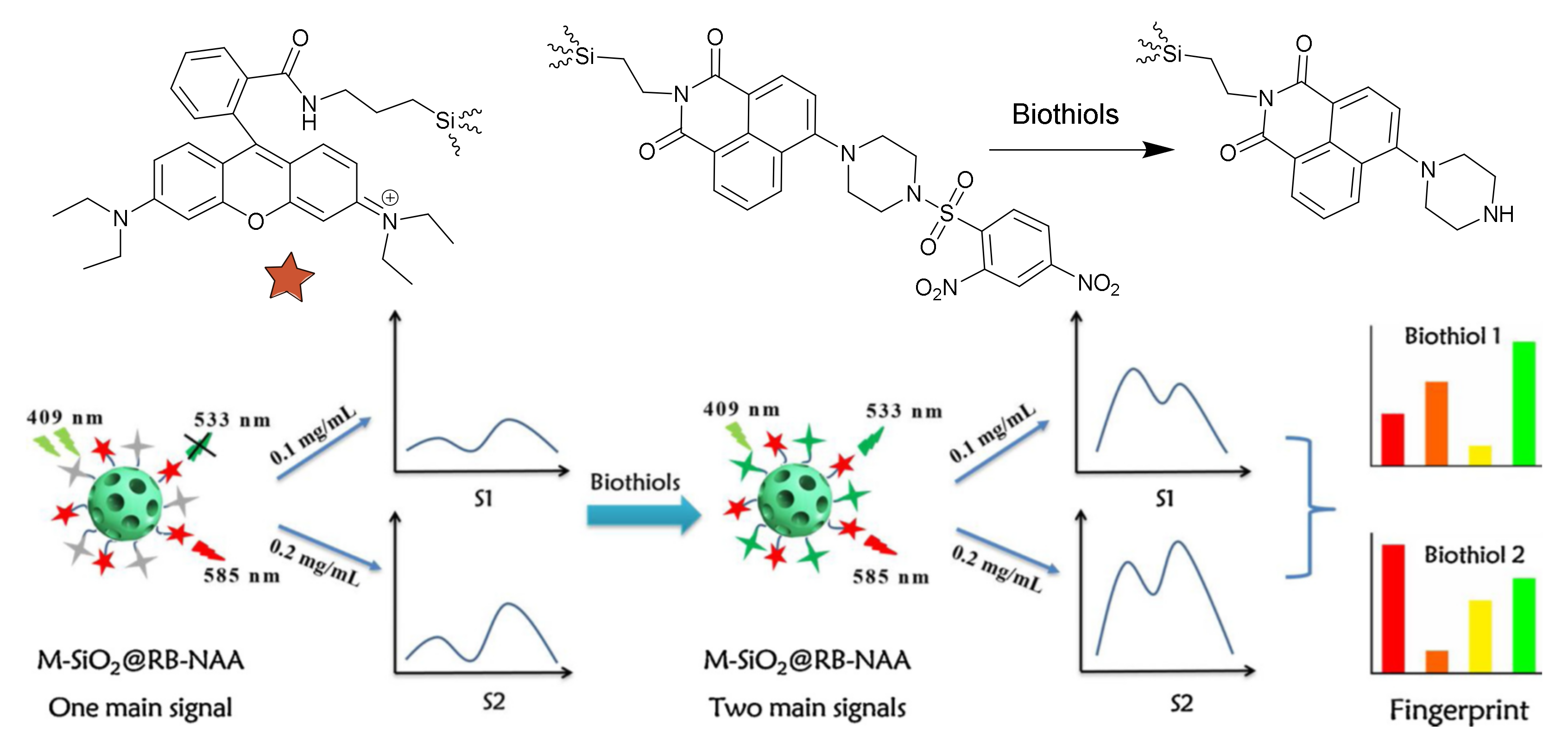
- Gao, Z.; Wang, Z.; Qiao, M.; Peng, H.; Ding, L.; Fang, Y. Mesoporous silica nanoparticles-based fluorescent mini sensor array with dual emission for discrimination of biothiols. Colloids Surf. A 2020, 606, 125433. [Google Scholar] [CrossRef]
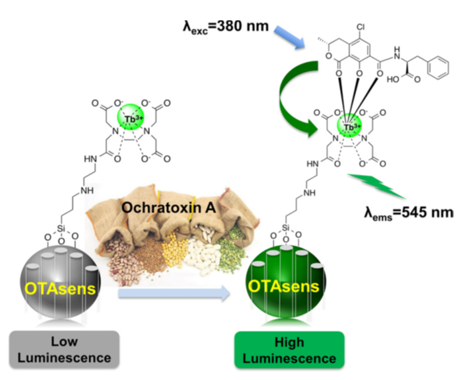
- Altunbasa, O.; Ozdasa, A.; Yilmaz, M.D. Luminescent detection of Ochratoxin A using terbium chelated mesoporous silica nanoparticles. J. Hazard. Mater. 2020, 382, 121049. [Google Scholar] [CrossRef] [PubMed]
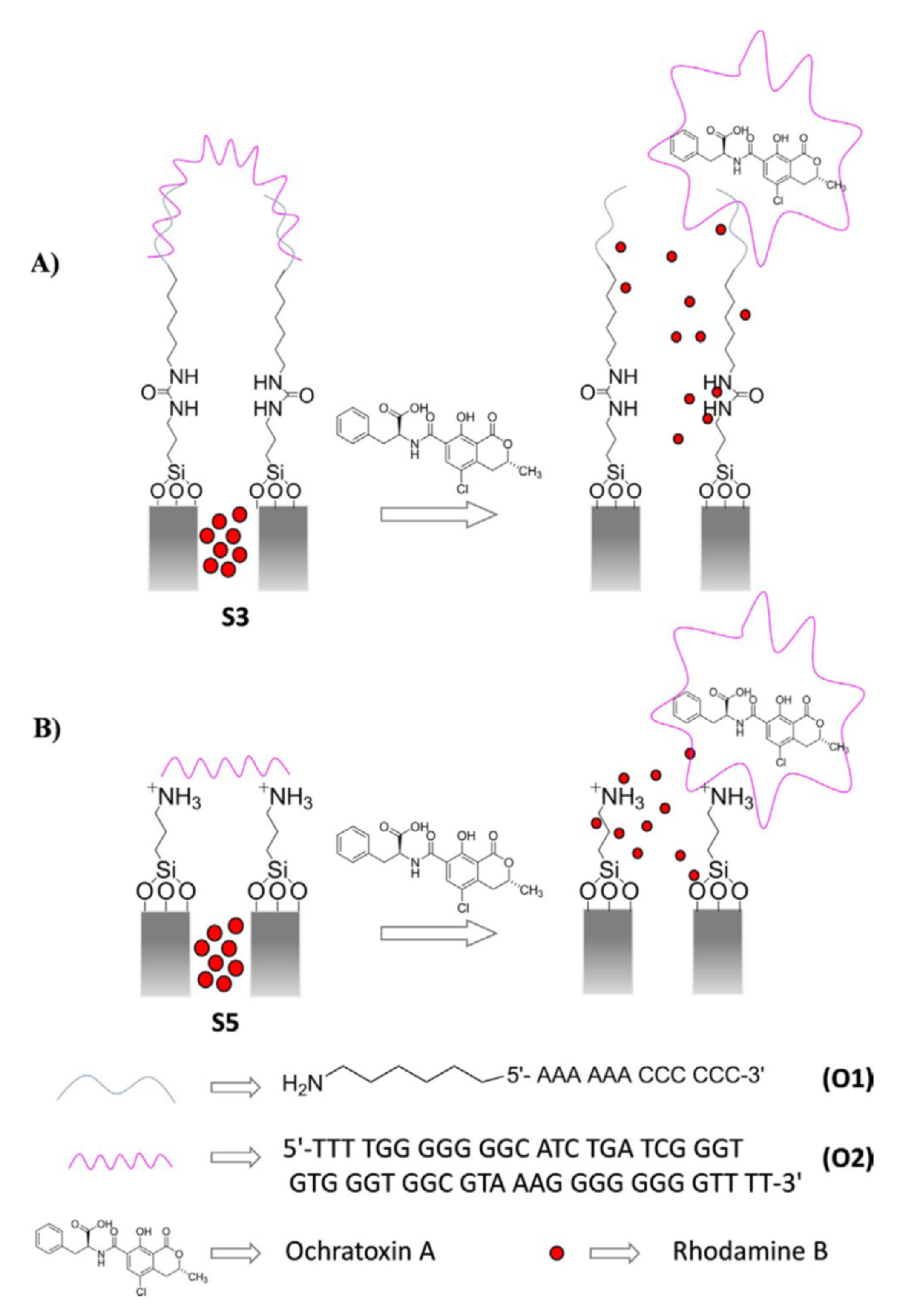
- Ribes, A.; Santiago-Felipe, S.; Bernardos, A.; Marcos, M.D.; Pardo, T.; Sancenón, F.; Martínez-Mañez, R.; Aznar, E. Two New Fluorogenic Aptasensors Based on Capped Mesoporous Silica Nanoparticles to Detect Ochratoxin A. ChemistryOpen 2017, 6, 653–659. [Google Scholar] [CrossRef] [PubMed]
- Ribes, A.; Aznar, E.; Bernardos, A.; Marcos, M.D.; Amorós, P.; Martínez-Máñez, R.; Sancenón, F. Fluorogenic Sensing of Carcinogenic Bisphenol A using Aptamer-Capped Mesoporous Silica Nanoparticles. Chem. Eur. J. 2017, 23, 8581–8584. [Google Scholar] [CrossRef]
- Costa, E.; Climent, E.; Gawlitza, K.; Wan, W.; Weller, M.G.; Rurack, K. Optimization of analytical assay performance of antibody-gated indicator-releasing mesoporous silica particles. J. Mater. Chem. B 2020, 8, 4950–4961. [Google Scholar] [CrossRef]
- Barros, M.; López-Carrasco, A.; Amorós, P.; Gil, S.; Gaviña, P.; Parra, M.; El Haskouri, J.; Terencio, M.C.; Costero, A.M. Chromogenic Chemodosimeter Based on Capped Silica Particles to Detect Spermine and Spermidine. Nanomaterials 2021, 11, 818. [Google Scholar] [CrossRef]
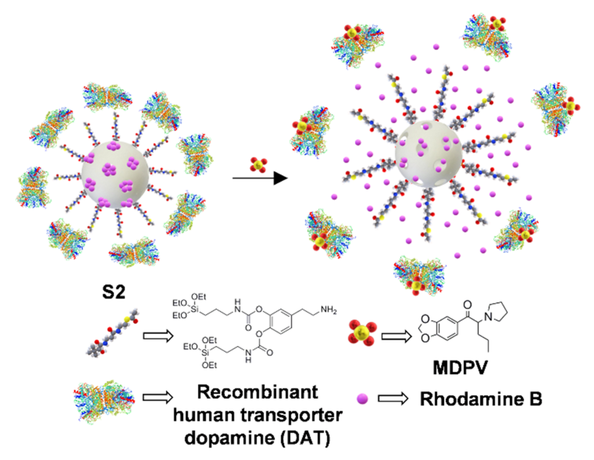
- Garrido, E.; Alfonso, M.; Díaz de Greñu, B.; Marcos, M.D.; Costero, A.M.; Gil, S.; Sancenón, F.; Martínez-Mañez, R. A Sensitive Nanosensor for the In Situ Detection of the Cannibal Drug. ACS Sens. 2020, 5, 2966–2972. [Google Scholar] [CrossRef]
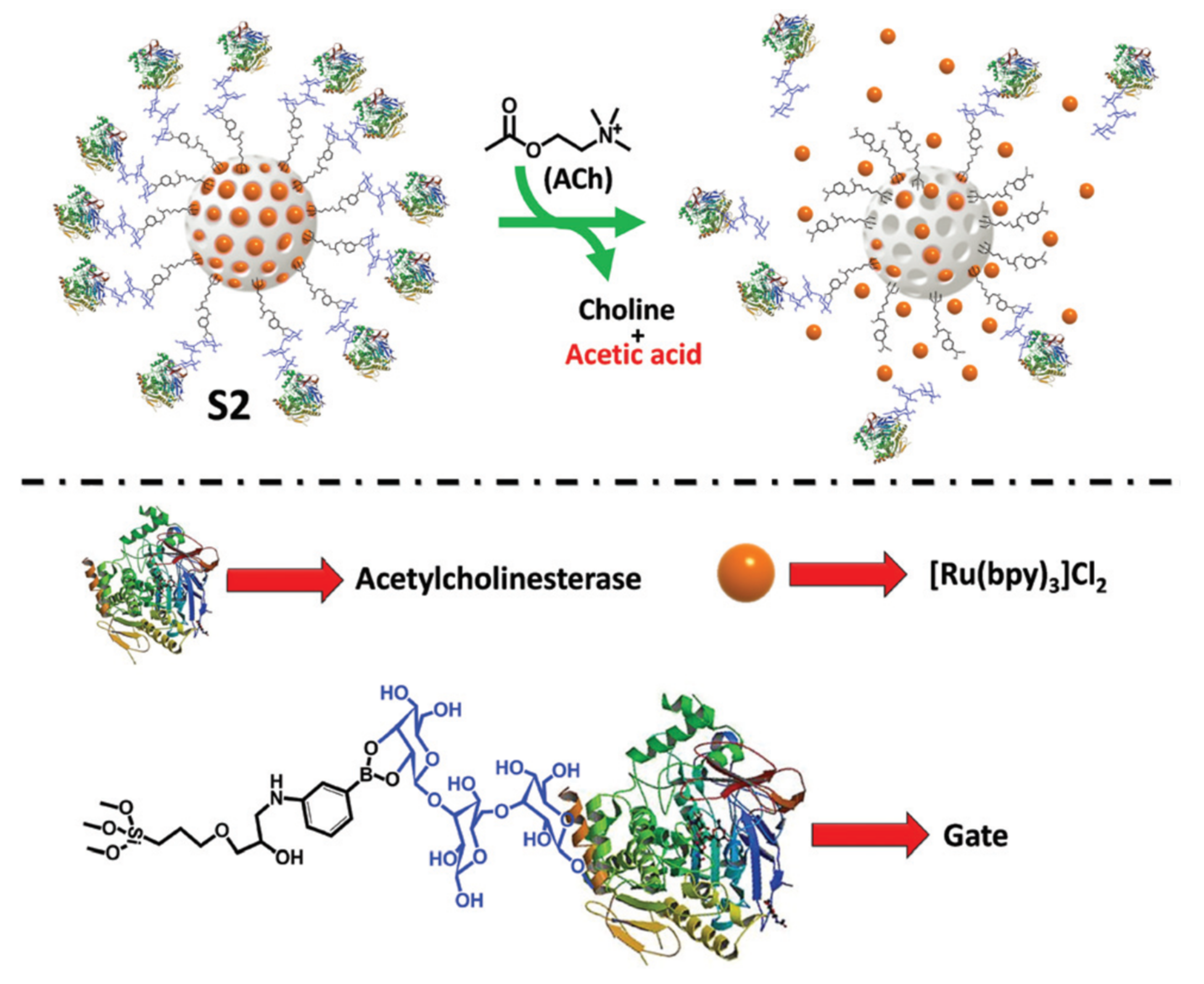
- Godoy-Reyes, T.M.; Llopis-Lorente, A.; García-Fernández, A.; Gaviña, P.; Costero, A.M.; Martínez-Máñez, R.; Sancenón, F. Acetylcholine-responsive cargo release using acetylcholinesterase-capped nanomaterials. Chem. Commun. 2019, 55, 5785–5788. [Google Scholar] [CrossRef]
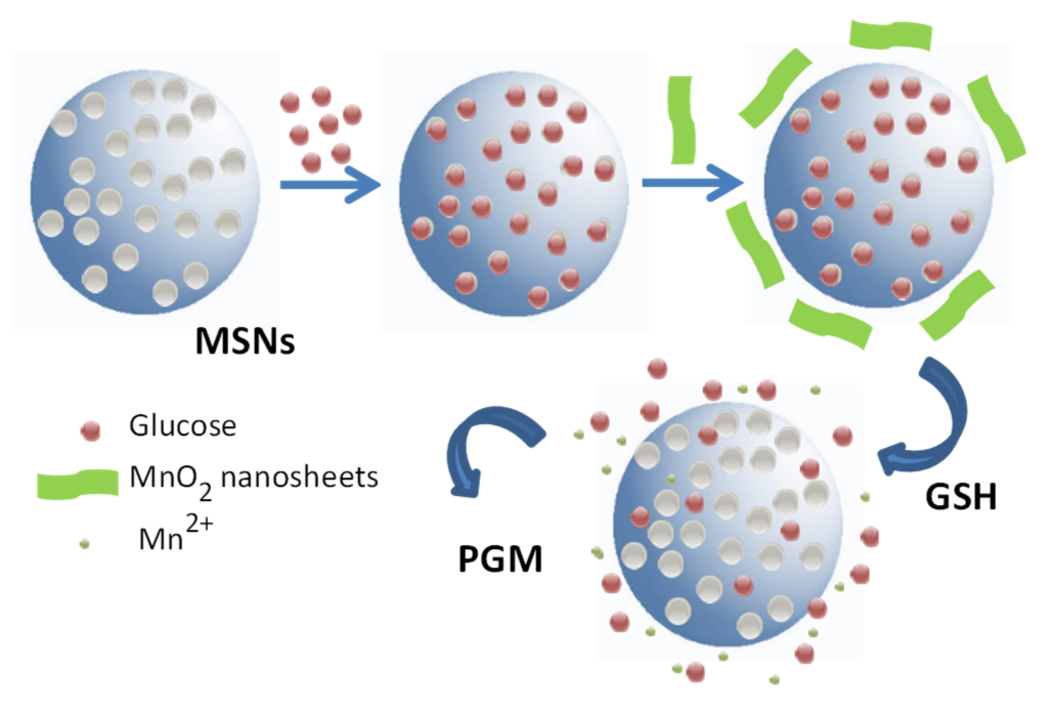
- Tan, Q.; Zhang, R.; Kong, R.; Kong, W.; Zhao, W.; Qu, F. Detection of glutathione based on MnO2 nanosheet-gated mesoporous silica nanoparticles and target induced release of glucose measured with a portable glucose meter. Microchim. Acta 2018, 185, 44. [Google Scholar] [CrossRef]
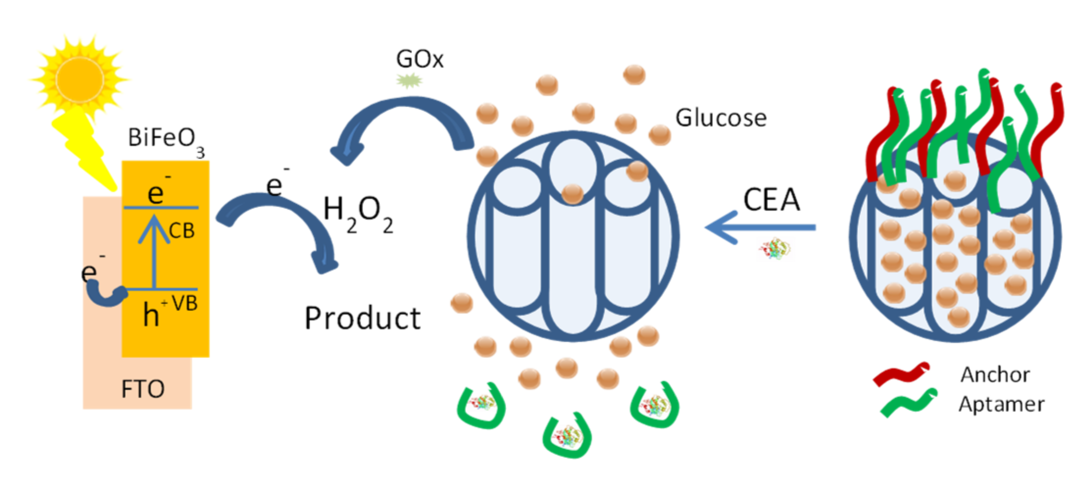
- Zhou, Q.; Lin, Y.; Lu, M.; Tang, D. Bismuth ferrite-based photoactive materials for the photoelectrochemical detection of disease biomarkers coupled with multifunctional mesoporous silica nanoparticles. J. Mater. Chem. B 2017, 5, 9600–9607. [Google Scholar] [CrossRef]
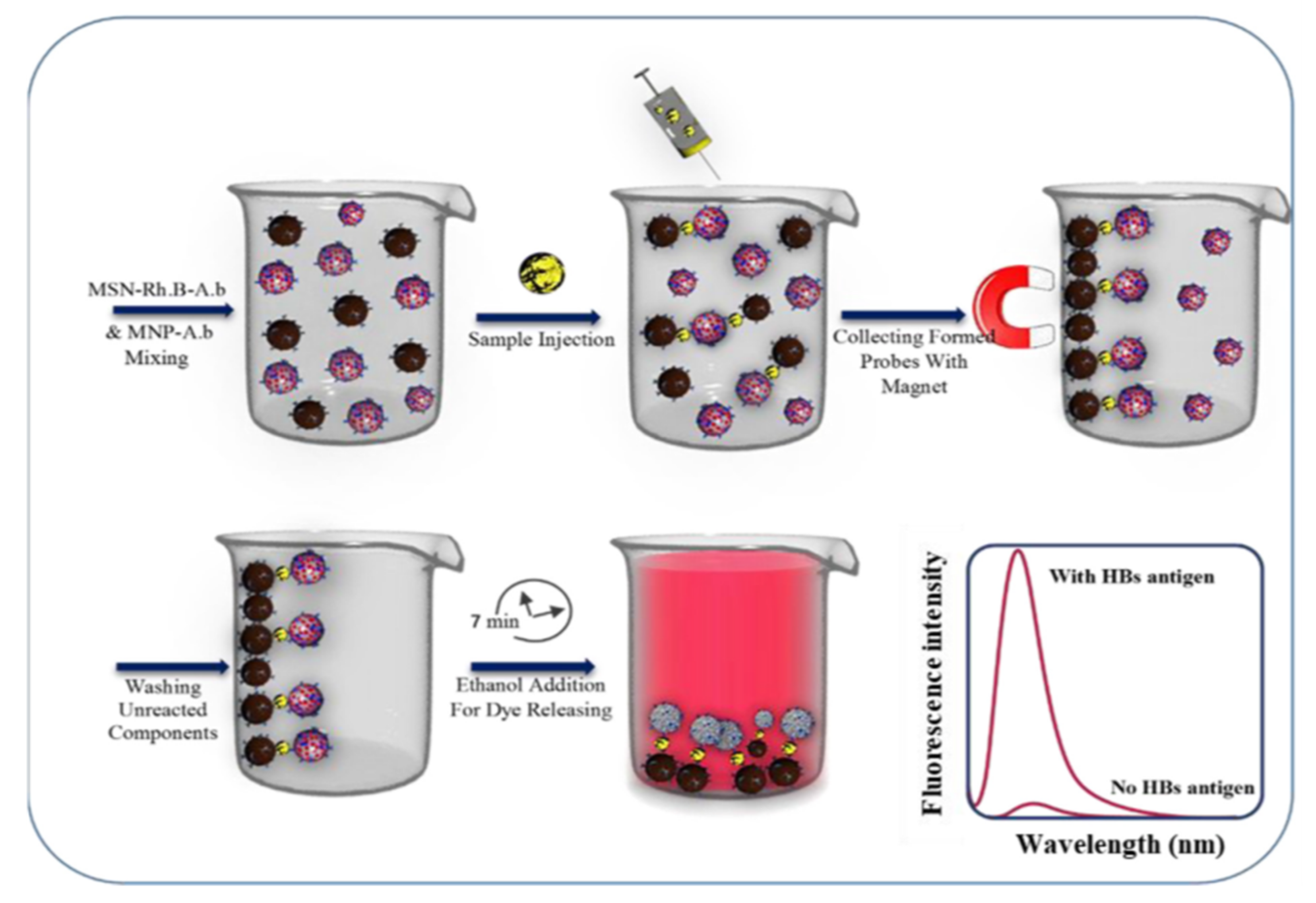
- Ghafary, Z.; Hallaj, R.; Salimi, A.; Mafakheri, S. Ultrasensitive fluorescence immunosensor based on mesoporous silica and magnetic nanoparticles: Capture and release strategy. Spectrochim. Acta Part. A Mol. Biomol. Spectrosc. 2021, 257, 119749. [Google Scholar] [CrossRef]
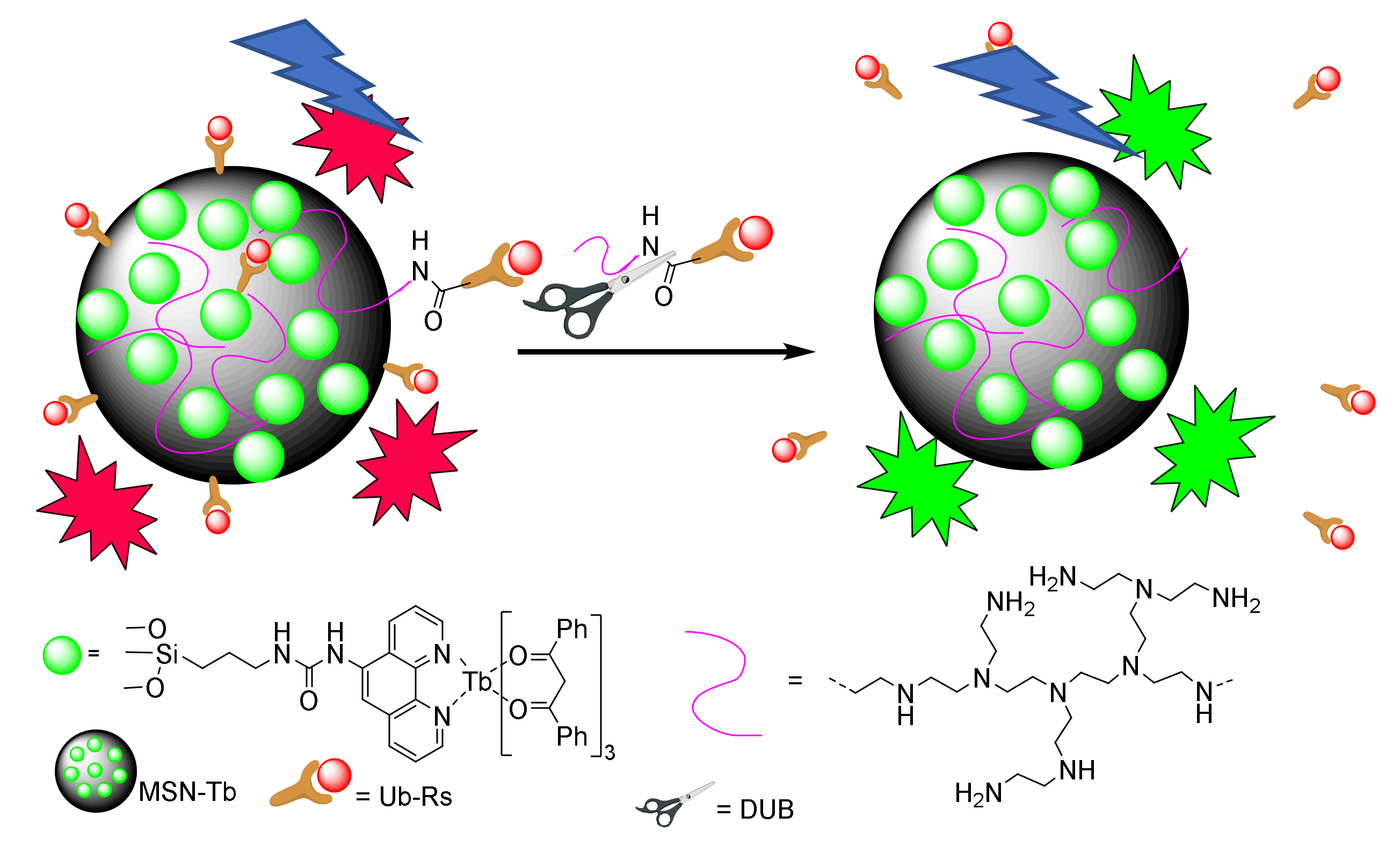
- Liang, Y.-Y.; Zhang, J.; Cui, H.; Shao, Z.-S.; Cheng, C.; Wang, Y.-B.; Wang, H.-S. Fluorescence resonance energy transfer (FRET)-based nanoarchitecture for monitoring deubiquitinating enzyme activity. Chem. Commun. 2020, 56, 3183–3186. [Google Scholar] [CrossRef]
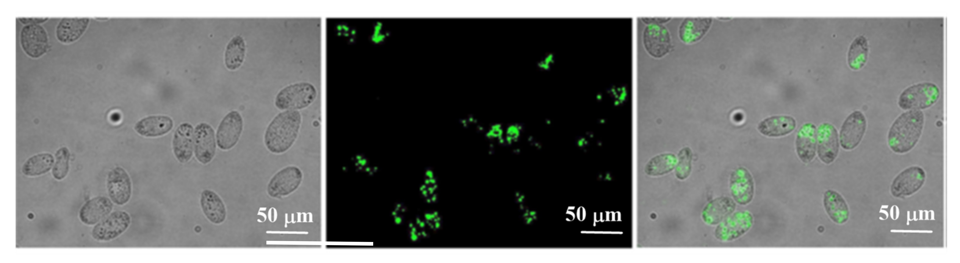
- Jimenez-Falcao, S.; Villalonga, A.; Arévalo-Villena, M.; Briones-Pérez, A.; Martínez-Máñez, R.; Martínez-Ruiz, P.; Villalonga, R. Enzyme-controlled mesoporous nanosensor for the detection of living Saccharomyces cerevisiae. Sens. Actuators B Chem. 2020, 303, 127197. [Google Scholar] [CrossRef]
- Zhou, Y.; Yang, S.; Guo, J.; Dong, H.; Yin, K.; Huang, W.T.; Yang, R. In Vivo Imaging of Hypoxia Associated with Inflammatory Bowel Disease by a Cytoplasmic Protein-Powered Fluorescence Cascade Amplifier. Anal. Chem. 2020, 92, 5787–5794. [Google Scholar] [CrossRef]
- Veiko, V.P.; Zakoldaev, R.A.; Sergeev, M.M.; Pavel, A.; Danilov, P.A.; Kudryashov, S.I.; Kostiuk, G.K.; Sivers, A.N.; Ionin, A.A.; Antropova, T.V.; et al. Direct laser writing of barriers with controllable permeability in porous glass. Opt. Express 2018, 26, 28150. [Google Scholar] [CrossRef]
- Lijing, Z.; Zakoldaev, R.A.; Sergeev, M.M.; Veiko, V.P. Fluorescent Bulk Waveguide Sensor in Porous Glass: Concept, Fabrication, and Testing. Nanomaterials 2020, 10, 2169. [Google Scholar] [CrossRef]
- Shen, H.; Abtahi, A.; Lussem, B.; Boudouris, B.W.; Mei, J. Device Engineering in Organic Electrochemical Transistors toward Multifunctional Applications. ACS Appl. Electron. Mater. 2021, 3, 2434–2448. [Google Scholar] [CrossRef]
- Hwang, C.; Park, N.; Kim, E.S.; Kim, M.; Kim, S.D.; Park, S.; Kim, N.Y.; Kim, J.H. Ultra-fast and recyclable DNA biosensor for point-of-care detection of SARS-CoV-2 (COVID-19). Biosens. Bioelectron. 2021, 185, 113177. [Google Scholar] [CrossRef]


| SDS (g) | SDBS (g) | H2O mL | HCl (g) | TEO (mL) | APTES (mL) | T (°C) | |
|---|---|---|---|---|---|---|---|
| MSN1 | 0.072 | 0 | 8.75 | 0.5 | 0.375 | 0.020 | 40 |
| MSN2 | 0.072 | 0 | 8.75 | 0.5 | 0.375 | 0.040 | 40 |
| MSN3 | 0.060 | 0.014 | 8.75 | 0.5 | 0.375 | 0.030 | 40 |
| MSN4 | 0.060 | 0.014 | 8.75 | 0.5 | 0.375 | 0.040 | 40 |
| MSN5 | 0.060 | 0.014 | 8.75 | 0.5 | 0.375 | 0.040 | 25 |
Publisher’s Note: MDPI stays neutral with regard to jurisdictional claims in published maps and institutional affiliations. |
© 2021 by the authors. Licensee MDPI, Basel, Switzerland. This article is an open access article distributed under the terms and conditions of the Creative Commons Attribution (CC BY) license (https://creativecommons.org/licenses/by/4.0/).
Share and Cite
Parra, M.; Gil, S.; Gaviña, P.; Costero, A.M. Mesoporous Silica Nanoparticles in Chemical Detection: From Small Species to Large Bio-Molecules. Sensors 2022, 22, 261. https://doi.org/10.3390/s22010261
Parra M, Gil S, Gaviña P, Costero AM. Mesoporous Silica Nanoparticles in Chemical Detection: From Small Species to Large Bio-Molecules. Sensors. 2022; 22(1):261. https://doi.org/10.3390/s22010261
Chicago/Turabian StyleParra, Margarita, Salvador Gil, Pablo Gaviña, and Ana M. Costero. 2022. "Mesoporous Silica Nanoparticles in Chemical Detection: From Small Species to Large Bio-Molecules" Sensors 22, no. 1: 261. https://doi.org/10.3390/s22010261