
Microbial Electrochemical Systems: Principles, Construction and Biosensing Applications


1
Nanoscience Program, University of Science and Technology (UST), Zewail City of Science and Technology, 6th October City, Giza 12578, Egypt
2
National Research Centre (NRC), Applied Organic Chemistry Department, El Bohouth st., Dokki, Giza 12622, Egypt
3
Institute of Biochemistry and Cell Biology, National Research Council (CNR), Via P. Castellino 111, 80131 Naples, Italy
4
Department of Chemistry and Biomolecular Science, Clarkson University, Potsdam, NY 13699-5810, USA
*
Author to whom correspondence should be addressed.
Sensors 2021, 21(4), 1279; https://doi.org/10.3390/s21041279
Submission received: 13 January 2021
/
Revised: 2 February 2021
/
Accepted: 4 February 2021
/
Published: 11 February 2021
(This article belongs to the Special Issue Advances in Optical, Fluorescent and Luminescent Biosensors)
Abstract
:Microbial electrochemical systems are a fast emerging technology that use microorganisms to harvest the chemical energy from bioorganic materials to produce electrical power. Due to their flexibility and the wide variety of materials that can be used as a source, these devices show promise for applications in many fields including energy, environment and sensing. Microbial electrochemical systems rely on the integration of microbial cells, bioelectrochemistry, material science and electrochemical technologies to achieve effective conversion of the chemical energy stored in organic materials into electrical power. Therefore, the interaction between microorganisms and electrodes and their operation at physiological important potentials are critical for their development. This article provides an overview of the principles and applications of microbial electrochemical systems, their development status and potential for implementation in the biosensing field. It also provides a discussion of the recent developments in the selection of electrode materials to improve electron transfer using nanomaterials along with challenges for achieving practical implementation, and examples of applications in the biosensing field.
1. Introduction
Microbial electrochemical system (MES) is a promising fast expanding technology that integrates microbial systems, electrochemistry and materials science to develop energy, environment and sensing devices [1]. MES exploits the biocatalytic activity of living microbes to harvest electrons from the biodegradable organic substances and therefore explore the interaction between living microbial cells (electron donor) and surface of electrodes (electron acceptor) [2]. A critical requirement for the development of MESs is to achieve effective integration and facilitate electron transfer between microorganisms (e.g., having ability to accept or donate electrons to and from electrodes) and the electrode surface, which are fundamental to their performance.
Based on the nature of these interactions, MESs can use processes that involve: (i) extracellular electron transfer (primary MES) in which the electrode potential lies within the physiological range of the microorganisms and rely primarily on Faraday processes (e.g., microbial electrocatalysis through extracellular electron transfer) and, (ii) indirect interactions in which the microbial environment (metabolite, pH, oxygen pressure, etc.) is controlled by electrochemical processes [3]. For these processes to take place, the microbial system should be in close vicinity of the electrochemical system and the system needs to be integrated into a reactor that ideally would require low operation and maintenance cost. The electrode surface, microbial kinetics, reactor configurations and the electrogenic microorganisms used to construct the device all play a critical role in controlling these processes. In the past several years, there have been advancements in the development of new materials for bioelectrodes, engineered microbes, substrate types and interspecies electron processes to improve interfacial electron transfer and the microbial/electrode interface [4]. While these new developments have improved performance, several challenges still exist that hampered the implementation of these systems in real world applications. This article reviews the principles of MES, their development status and promise for implementation in the biosensing field.
2. Principles of Microbial Electrochemical Systems
Microbial electron transport chain (METC) represents the most important compartment in the living systems, since the oxidation of degradable organic substrates is the main energy source of live microbial cells [5]. Therefore, measuring the efficiency of microbial respiration and the activity of the electron transport chain are considered main indicators of cellular activity, as they are essential for the replication and proliferation of aerobic organisms [6]. Hence, earlier efforts were made to employ the measurement of dissolved oxygen consumption by living cells as a direct measure of the microbial survival [7]. Consequently, the electron transfer process from living-microorganisms towards electrodes in MES is exploited in microbial fuel cells [8,9] or diagnostic tools for rapid assessment of microbial activity [10,11,12]. In these regards, many MES approaches were designed and tested for biological purposes [13,14,15,16]. The electrical current value generated by the MESs is directly proportional to the number of viable microbial cells that are incorporated in MESs. On the other hand, non-viable or non-cultivable living cells do not have electrochemical contribution, and thus, they do not generate electrochemical signals. Thus, the resulting bioelectrochemical responses reflect the extent of anodic respiration, intracellular redox reactions (e.g., intracellular enzyme activities) and/or other biological interactions [17,18]. Since the bioelectrochemical responses can be linked to microbial processes, the design of high performance MESs has gained increasing attention due to their many promising applications in the environment, energy and biomedical fields.
2.1. Extracellular Electron Transfer
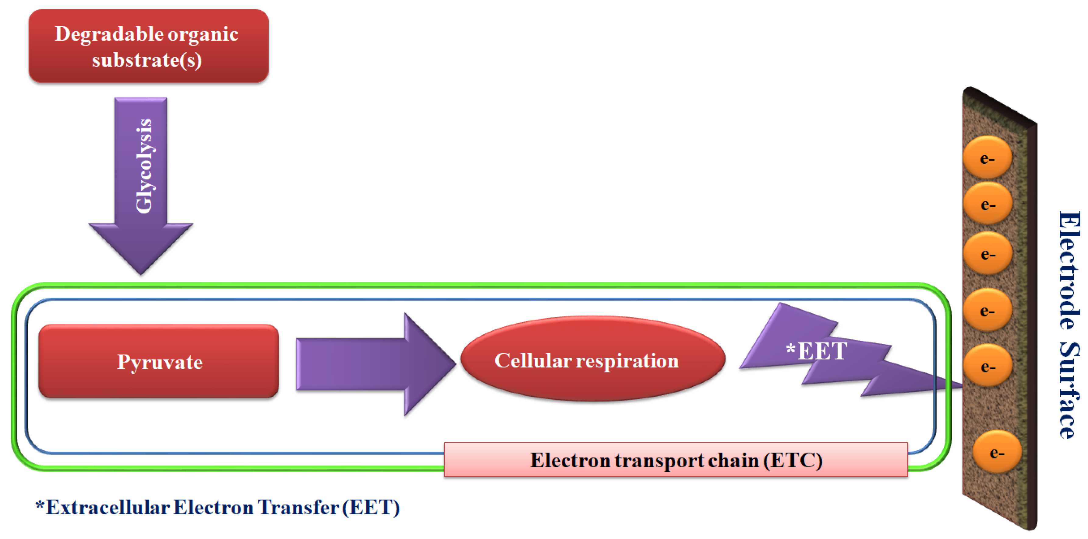
The operation of primary MES involves extracellular electron transfer (EET), directly from the cell to the acceptor, or indirectly mediated by electron shuttles. An example of bioelectrochemical signals generated in the MES systems is shown in Figure 1, illustrating conversion of degradable organic substrates to pyruvate via the glycolysis process, which is the central precursor of generating bioenergy.
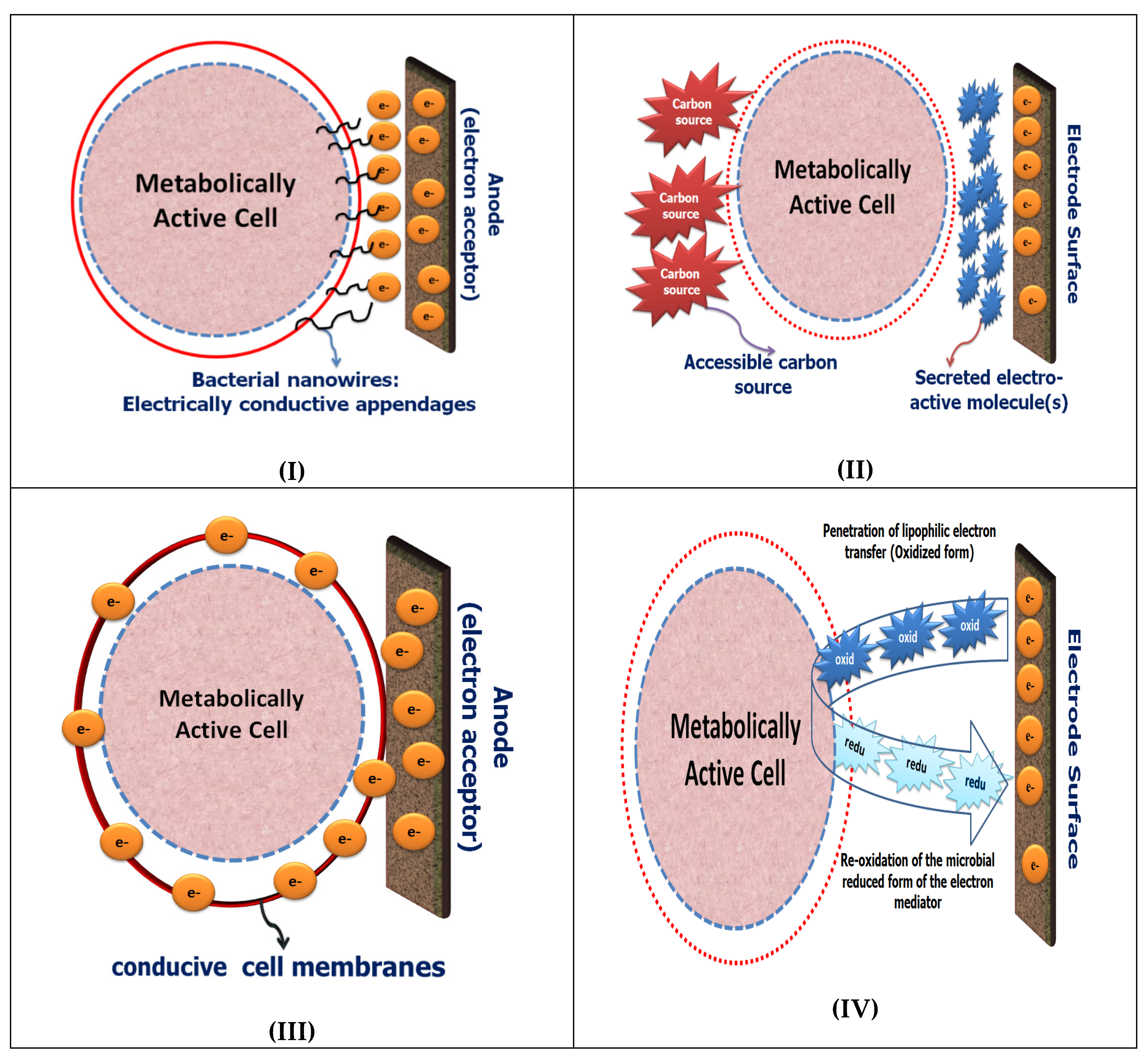
In the absence of oxygen, as the final electron acceptor, anodic respiration is the main regulating factor for the electron transfer from the living cells to the electrode surface. Through the classical aerobic respiration pathway, two electrons liberated by the enzymatic oxidation of NADH or NADPH via the first enzymatic complex NADH dehydrogenase and transferred to coenzyme Q (CoQ), to ubisemiquinone, and then to ubiquinol (the natural electron shuttles). Subsequently, the electrons from ubiquinol are transferred through the electron transport chain (ETC) to complex III (bc1 complex), cytochrome c, complex IV (cytochrome c oxidase), and finally to oxygen (as the final electron acceptor) to produce H2O. The energy that is released, due to the electrons flow down the electron transport chain, is used to pump protons out through complexes I, III, and IV. This creates a proton electrochemical gradient [19]. Keeping in mind that the electron transport chain is physically separated from the outside environment by cytoplasmic membranes with additional layers, such as cell walls, peptidoglycans, or outer membranes the physical transfer of the biologically liberated/created electrons to the surface of the electrode also plays an important critical role [20,21]. Mechanisms for transferring electrons from the microbial intracellular compartments to the surface of the electrode have been studied predominantly in bacterial systems; thus the use of bacteria is more common in the construction of microbial fuel cell technology. As shown in Figure 2, two different mechanisms can be used for connecting the living microbial cells with the electrode surface through direct electron transfer (DET) or mediated electron transfer (MET). The full description of each mechanism is given in the next section.
2.1.1. Direct Electron Transfer via Electroactive Microbes
Through anodic respiration, certain microbial species, known as Exoelectrogens or Electroactive microorganisms, have been identified as electroactive organisms [22]. The Exoelectrogens usually refers to microorganisms that have the capability to transfer electrons extracellularly to conductive materials directly without mediators [23,24]. These electroactive microbes are also called anode respiring bacteria, electrochemically active bacteria, and electricigens [25]. Direct electron transfer (DET), which requires physical contact between the surface of electrode and the redox centers of microbes [26], can be achieved directly (mediated-less) via the electroactive species, e.g., cell-wall containing cytochromes, of the adhered organism [27,28,29,30,31]. Alternatively, a conductive layer in outer-membranes can form to conduct electrons [32]. For example, bacterial nanowires (Pili) were created by Shewanella oneidensis MR-1 in its outer membrane that facilitate extracellular electron transport [33,34,35,36]. Secretion of electroactive metabolite(s) in the extracellular matrix was also identified as an alternative mechanism to provide self-mediation of electron transport [26,37,38,39]. For example, microbial natural-electron mediators (water-soluble compounds with low molecular weight), which act as quorum sensing molecules, produced by Shewanella putrefaciens into the extracellular matrix, have shown the ability to mediate the electron transfer [40,41,42,43]. Phenazines, as the quorum sensing (QS) molecules, produced by Pseudomonas aeruginosa modulated the current production as a result of the anodic-respiration [44]. The DET is not limited to bacteria. Some eukaryotic microorganisms were also identified as potential candidates for that purpose, as illustrated by a mediated-less bioelectrochemical approach for studying the intracellular level of Candida albicans [45]. The electron transfer capacity was strongly controlled by the mitochondrial respiratory chain efficiency, since inhibition of the respiratory chain, either chemically using specific enzyme inhibitors, or genetically by knocking out specific genes from respiratory chain complexes, led to a decrease in the generated bioelectrochemical response [45].
2.1.2. Mediated Electron Transfer
Unlike the exoelectrogens, many microorganisms are electrochemically inactive which means they are unable to transfer their electrons to the electrode surfaces without the use of soluble chemical redox mediators [46,47]. Hence, artificial redox mediators (exogenous electron shuttles) are required to enable the microbe-electrode interaction(s) through a process which is known as mediated electron transfer (MET) [43,48]. The MET is commonly used in the developments of MESs, and its redox reaction mechanisms are generally well understood; therefore, MES has been used for the rapid detection of cell viability and cytotoxicity [49,50,51,52]. For example, 2,6-dichlorophenolindophenol (DCIP) has been exploited as a traveling electron mediator (a membrane permeable molecule) for monitoring the cell viability of Candida and Saccharomyces species [20]. The specific action of DCIP, i.e., accepting the liberated electrons from the complex I of the respiratory chain system (mitochondrial NADH-dehydrogenases), which is not provided in the respiratory chain system of the S. cerevisiae, was a potential indicator for the selective assessing of complex I activity in the C. albicans. In another study, a double mediator approach using a mixture of 2,3,5,6-Tetramethyl-p-phenylenediamine, or menadione in a combination with the hexacyanoferrate(III) (ferricyanide or FCN), was used to probe the intracellular redox activity and tracking the metabolic pathway activity of Chinese hamster ovary cells or S. cerevisiae [53,54]. As the double mediator systems were applied, fast electron transfer along with higher sensitivity were achieved when they were compared with the responses of the single mediator system. Accordingly, another double-mediated approach using DCIP/FCN has been developed and applied for probing the intracellular redox activity of Staphylococcus aureus and its pathway stimulation with different organic compounds such as glucose of acetate as a sole source of electrons. When only a single hydrophilic mediator such as the FCN was used, bio-electrochemical responses were not generated, indicating that the living bacterial cells are not communicating with the electrode surface. On the other hand, when the lipophilic mediator (DCIP) was used with the FCN, strong bio-electrochemical signals were generated, showing the possibility of wiring the living cells with the solid conductive surfaces. The double-mediator system was used to amplify the electric-current response from the intracellular NAD(P)H:quinone acceptor oxidoreductase (NQO) activity [34]. Therefore, the current magnification enhanced the assay performance [33]. A short summary of the direct and mediated ET between living microorganisms and the electrode surface is sketched in Figure 2.
3. Bioelectrochemistry of Biofilms
Basically, in the non-mediated microbial electrochemical systems, metabolically active microorganisms and electrodes are directly communicating together through the formation of electro-active biofilms [4,11,55,56]. In fact, the biofilms are a mixture of heterogeneous communities of microbial cells surrounded by a condensed layer of exopolysaccharides matrix (EPSs), and are strongly adhered to living tissues or solid surfaces [57,58]. In most biofilms, the living cells represent less than 10% of the total content in prevalence represented by the matrix (about 90%). In general, the biofilm matrices contain components such as polysaccharides, proteins and extracellular DNA, but their content is dependent on the bacterial species and the environmental conditions [59]. Moreover, it provides high mechanical stability, the EPS environment mediates the cell-cell adhesion to the solid surfaces and forms a cohesive three-dimensional network that interconnects biofilm cells.
The process of biofilm formation is of high importance in many industrial applications such as the biodegradation of chemical contaminants in wastewater, biocatalysis, and microbial fuel cells [18,60,61]. However, other biofilms may have serious implications on public health and environment [62]. For instance, microbial contamination on metal implants and prosthetic biomedical devices causing biofilm formation can be life-threatening, leading to chronic infections, device failure, and high mortality rates [63,64]. In the microbial electrochemical systems different biofilms could be characterized as electrochemically active or inactive [65]. The electrochemically active biofilms are defined as a community of microorganisms interacting with a conductive surface by either transferring to (anodic behavior) or removing electrons from the electrode (cathodic behavior) [66,67,68].
The nature of the biofilm assisted-bioelectrochemical signals was reported as physical connections through the bacterial appendages, microbial nanowires, cyt-c and ferric iron in some microorganisms such as Thiobacillus denitrificans, Shewanella oneidensis and Geobacter sulfurreducens [69,70]. O’Toole et al. reported the important role of flagella, and/or motility, in the initial cell-to-surface signal transduction [71]. They showed that defective flagella mutants (flgK) did not develop microcolonies on solid substrates over the course of the experiments. This observation supported the role of intact flagella to the microcolony formation.
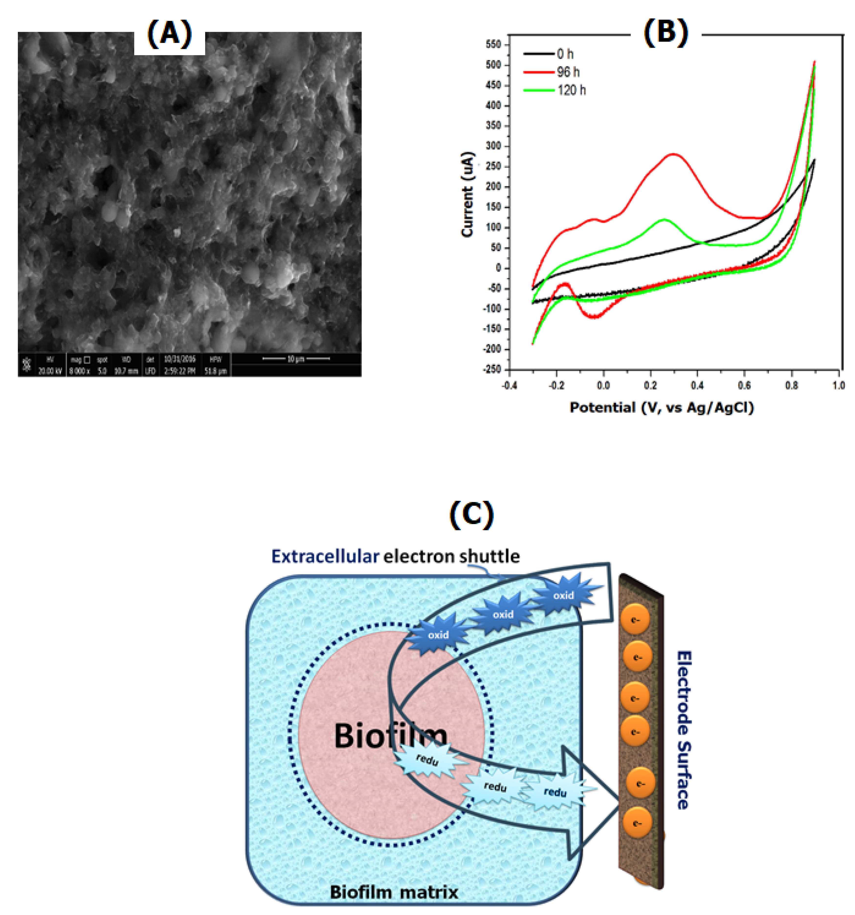
MESs were shown to be the most effective way for studying and understanding the role of the conductive solid surfaces and the environment in biofilm formation [4,72,73]. Bioelectrochemical analysis of the biofilm formation at different electrode modifiers was conducted to understand the influence of the electrode materials on the biofilm progression [11,74]. In addition to morphological characterization using microscopic techniques, cyclic voltammetry (CV) is another technique that can be used for monitoring the online formation of biofilms and their electrochemical activity [75,76]. Several redox peaks were observed over different incubation times with the bacterial culture, indicating a direct electron transfer from the outer-redox layer of the bacteria to the electrode surface. CV can be used in conjunction with physical characterization tools, e.g., scanning electron microscopy, to confirm the mature biofilm formation at the electrode surface as shown in Figure 3A,B.
On the other hand, electrochemically inactive biofilms could be integrated with electrode surfaces via extracellular electron receptors/transmitters, or electron mediators. Electron transmitters in the oxidized form can penetrate the cell-wall, as well as the cell membranes to capture the intracellular electrons. Afterwards, the reduced electron mediators are released to convey the accepted-electrons via redox reactions taking place at the electrode surface (as shown in Figure 3C). The use of soluble redox mediators in the MES bioreactor is an effective way to improve the extracellular electron transfer [77].
4. Microbial Electrochemical Devices
4.1. Half-Cell Based MESs
Construction and evaluation of the performance of MESs could be achieved by using CV, electrochemical impedance spectroscopy (EIS), or by amperometric measurements [78,79]. Those electrochemical techniques can be used to obtain mechanistic or kinetic information of extracellular electron transfer from living cells to the electrode surface and further the basic understanding of the microbe-electrode interaction [80]. Particular interest is always given to CV, since this method provides information about the formation of biofilms and their electrochemical and electro-catalytic activities, extracted from the real-time recording of voltammograms [81]. CV was used to characterize the anaerobic growth of E. coli and its secreted mediators and evaluate their role in the functioning of the cell, after the formation of a biofilm on the surface of the platinized titanium mesh electrodes [82]. Moreover, the biofilm formation and microbial adherence to the electrode surface could be effectively measured by using the impedimetric signal [79]. Additionally, monitoring the microbial cell number or microbial responses to different stresses could be determined from amperometric measurements [83]. Internal resistance variation during bacterial growth at electrode surface was assessed using EIS, whereas the equivalent circuit-based analysis revealed that the initial internal resistance of the cell has been internally reduced by around 50 % over an eight hour period of microbes-electrode incubation [84,85]. EIS is among one of the most powerful material characterization techniques for analyzing microbial-electrochemical reactions, monitoring biofilm progression, and evaluating microbial immobilization on different electrode materials. It can also be used for assessing the electrode properties and for studying the mass transfer resistances and evaluating the diffusion limitations of the reactants [86].
4.2. Microbial Fuel Cells
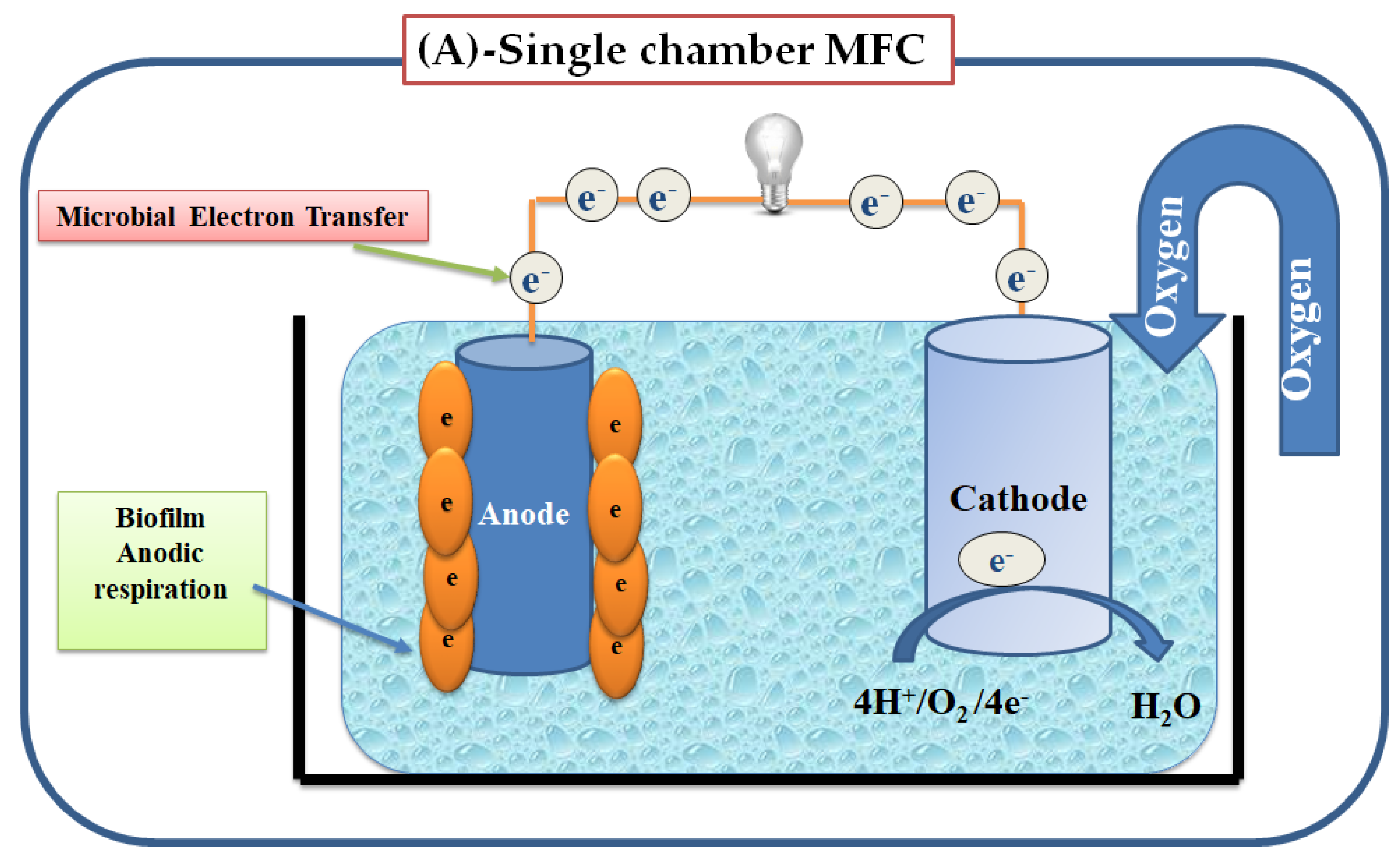
Microbial fuel cells (MFCs) are common microbial electrochemical systems that exploit the activity of living-microorganisms to convert chemical energy through the oxidation of organic substrates to electricity [87]. Typical MFCs consists of an anode and a cathode both of which are incubated with a liquid culture of living microbes (electrogenic or electroactive organisms). For the anodic reaction, living cells consume the degradable organic substrate (electron donors) that are transferred from the bulk solution and through the cellular metabolism to the anode surface [6]. Two different designs (i.e., single or double-chambers) of MFCs have been used, illustrated in Figure 4. In the double-chambers, an anaerobic anode chamber and an aerobic cathode chamber, are generally separated by a Proton Exchange Membrane (PEM) such as Nafion or sulfonated poly-(ether-ether-ketone) (SPEEK) as a kind of the non-fluorinated membranes [88]. Under hypoxic conditions, the anodic respiration followed by biofilm formation at the MFC electrode [89]. A mixed culture or single microbial strain could be used to build a MFC with high performance [90,91]. Regulator factors, biofilm structure adaptation, and/or the type of the microbial community have a great impact on the electrochemical characteristics. The number and dimensions of the biofilm pores affect the transfer of metabolites to the electrode surface while the transfer of protons, substrate and metabolites between the electrode surface and the bulk solution has an effect on the current generation [92]. The current generated by MFCs arises from the transfer of electrons received by the anode when live-bacteria oxidize organic materials, which are then transferred to the cathode via the external circuit, as shown in Figure 4.
Typically, MFCs are classified into two types depending on how extracellular electrons are delivered from the attached microorganisms to the anode; (i) Mediator-based MFCs, in which electro-active secreted metabolites, or artificial redox compounds are used to shuttle the electrons [93]; (ii) mediated-less fuel cells which does not require the addition of electroactive metabolites to transfer the electrons, but it relies mainly on the presence of electro-active organisms such as Shewanella [94,95], Rhodoferax [96] and Geobacteraceae [97]. These organisms transfer electrons directly to the anode via molecular nanowires and electrochemically active redox enzymes in their outer membrane. Wastewater treatments coupled with clean energy production are the most important practical applications of the MFCs [98]. The power generated by MFCs depends on various factors such as the electron transfer rate from the bacteria to anode, the diffusion of substrate into the biofilm, the ohmic resistance of the electrolyte and the electrochemical kinetics. The maximum power generated by the MFC also depends on the total internal resistance of the system [99,100]. The reviews of the basic MFCs technology, challenges and applications can be found in several articles [101,102].
5. Biosensing Applications of MESs for Microbial Detection
5.1. State of Art Pathogenic Microorganism’s Detection
Microbial infections have significantly increased over the past few decades and are now considered as one of the most critical global challenges. In particular, pathogenic microorganisms are responsible for bacterial infection which significantly affects the health of human, animals and plants. The detection of pathogens, including contaminants and other important harmful bio-molecules, such as toxins, plays a crucial role in the prevention of microbial infections. Rapid and sensitive detection with selective identification of microorganisms, including pathogens, are exceedingly important in clinical microbiology, microbial forensics, food and environmental analysis. However, the limited availability of efficient diagnostic tools is actually an impediment for rapid detection and treatment [103].
Classical microbiological methods widely used for the detection of pathogens, including culturing, plating techniques, microscopy, and serology, are insufficient for today’s needs. In particular, microscopy, which is a simple and relatively easy technique to be handled, lacks sensitivity. On the other hand, complicated steps, such as pre-enrichment, selective plating, biochemical screening and serological confirmation, are needed for the culture and plating methods [104]. Since such methods depend on the ability of microbes to grow and form colonies, its use is often time-consuming, tedious and unsuccessful [105]. Another widely used test for the microbial detection is the Enzyme-linked Immunosorbent assay (ELISA) that relies on the specific binding of antibodies to their specific antigens. ELISA allows the detection of very small quantities of antigens, such as molecules (hormones, toxins, etc.) and macromolecules (peptides, proteins, etc.) through a series of binding events [106]. Although largely used, this method requires intensive labor and, besides being expensive, is affected by false positives due to non-specific antigen-antibody reactions. To overcome these limitations, other molecular techniques, including DNA sequencing, Polymerase Chain Reaction (PCR) or microarray analysis, have been implemented, allowing a more sensitive and specific detection of microbes [107,108]. In spite of advantages in sensitivity, these molecular techniques still have limitations, related to the use of complicated protocols (requiring skilled workers to carry out the tests and their data interpretation) and of expensive materials (i.e., fluorescent probes for labeling). Moreover, all molecular methods cannot distinguish between viable and nonviable organisms which may lead to overestimation of infection [109].
5.2. Electrochemical Biosensing Devices
Biosensors have been proposed as alternatives to the current analytical methods, offering higher sensitivities, lower cost and portability for on-site analysis and multiplexing. Biosensors have attracted significant interest due to their potential importance for clinical, diagnostic, environmental, and bio-security applications [110,111]. Biosensors integrate biologically active species (biological recognition elements) with a physical transducer which converts the biological responses to measurable signals. These signals can be detected optically, acoustically, mechanically, calorimetrically, or electrochemically [112]. Electrochemical biosensors are the most common biosensing techniques due to their high specificity and sensitivity, and their ease of use. Also, their use allows the on-line detection of a broad spectrum of analytes in complex matrices (e.g., blood, serum, urine or food) [113,114]. Moreover, the advanced miniaturization of modern microelectronics allows building micro/and nano-electrodes, which are suited for detection of very small volumes of samples (microliters to nano-liters) [115]. The low cost and the possibility for large-scale production of electrochemical sensors, with pocket and portable devices, are other reasons, which make the electrochemical approaches more appealing for high-throughput analysis [85]. Several factors should be taken in consideration when developing biosensors to achieve high performances comparable or better than the other affordable methods:
- Selection of electrode materials and surface modification before using as support for the immobilization of the bio-recognition element (i.e., enzymes, antibodies, peptides, aptamers, phages or whole cells) [116].
- The selection of the electrochemical method (e.g., potentiometric, voltammetric, amperometric, or impedimetric method).
Electrochemical immunosensors are among the most common platforms for microbial detection and identification [117,118]. The signal is usually obtained through the detection changes of an immobilized recognition element (i.e., antigen or antibody) on the electrode surface. Two possible mechanisms can be used to develop an electrochemical immunosensor, based on a competitive or a sandwich organization. When dealing with competitive immune-sensors, two approaches could be considered: (1) Immobilizing antibodies, which react with free antigens from the sample in competition with labeled antigens, returning the amount of unreacted antibodies and indirectly the amount of sample antigens; and (2) immobilizing antigens, and using labeled antibodies to measure the amount of unreacted antigens. This last approach is useful to prevent problems related to antibody immobilization (loss of affinity, orientation of the immobilized protein) [118]. In a sandwich assay, using immobilized antibodies, labeled secondary antibodies (directed toward a second binding site of the antigen) are added. At this point, antigen is “sandwiched” between two antibodies. In spite of the high sensitivity and selectivity, antibodies have limited stability and tend to denature in conditions outside the physiological range. Among different alternative approaches, biomimetic artificial receptors having superior stability have been explored [119,120]. Molecularly imprinted polymers (MIPs), defined as artificial recognition elements, are of growing interest for applications in several sectors of life science for detecting molecules of specific interest MIPs can be developed to have high bio-recognition capability, mechanical and chemical stability. Also, they are relatively easy to prepare and have low cost, which make them superior over natural recognition reagents [121].
5.3. Biosensing Applications of MESs
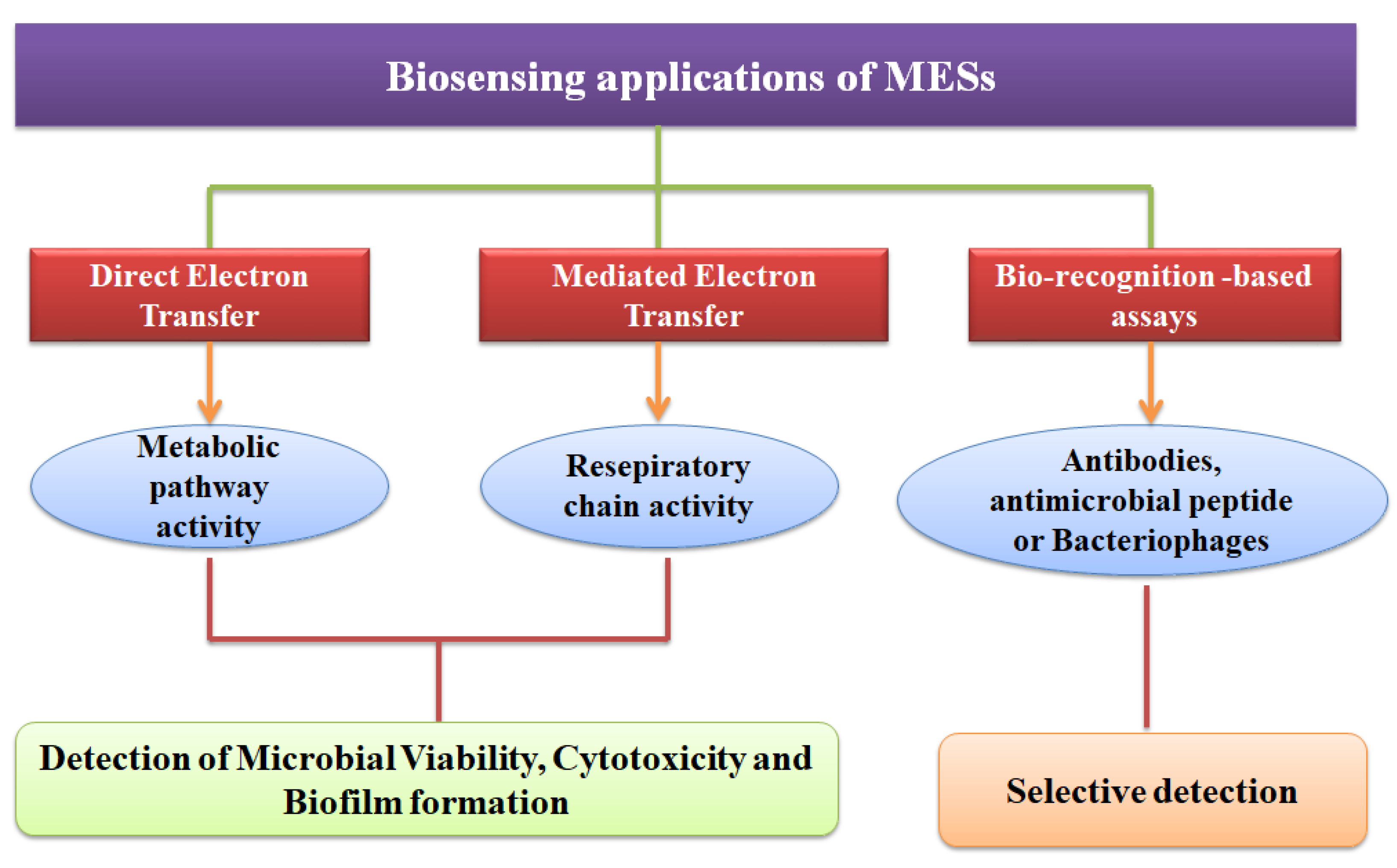
Instead of applying biological recognition elements, such as antibodies, the online-measuring of cell viability with the ability to distinguish living from the dead cells can be achieved through the use of MESs [1,12]. MESs could be used effectively to distinguish between live and dead microbial cells, monitor the intracellular redox functions of living cells, and characterize electrochemically active biofilms. Because the construction of MESs involves the effective integration of microbiology with electrochemistry and material sciences, major improvements in the operation of MESs can be achieved by optimizing the electrode materials to improve the electron transfer at the microorganism-electrode interface [122]. A summary of the most common biosensing applications of the MESs is provided in Figure 5, and details were expanded in Table 1.
5.4. Impact of the Electrode Materials on the Performance of MESs
Conventional MESs were built using carbon materials, such as graphite granules, graphite felt, carbon paper, and carbon cloth [128,129]. Inherent drawbacks of these materials due to the limited surface area have been addressed by exploring the benefit of nanostructured materials, which can provide enhancements of the performance of MESs by virtue of their specific high surface areas, conductivity and catalytic activity. In particular, carbon-based materials, including carbon fibers, activated carbon, graphene, carbon nanotubes (CNTs), and fullerene exhibit high electrochemically active surface area [130,131] that can be used as electrode materials for enhancing the detection limits, and advance the development of new MESs-based detection technologies [132,133,134].
In MESs, the microbial kinetics such as oxidation of the extracellular secretions or the metabolic activities of microorganism are affected by the reactions taking place at the electrode surface [135]. A wide range of materials have been used as electrode materials, e.g., carbon paper, cloth, graphite foil, rods, metal or metal nanoparticles to modify working electrodes. The material used can significantly alter the electrocatalytic functions, accelerate the biofilm formation, or catalyze the secreted biomolecules, which can facilitate the electron transfer process [136]. By integrating nanomaterials, more effective microbial sensors were developed, demonstrating promise of this technology for real applications [137].
Nanostructured materials, such as metal and metal oxide nanoparticles (NPs), or nano-hybrids have been employed in the construction of MESs [138]. Carbon nanostructures have shown promising performance as electrode material, due to their high electrical conductivity, biocompatibility, and electrocatalytic properties [139]. Owing to its large surface area, rich electronic states, and good mechanical properties, graphene-based materials have played an important role in the development of MESs in recent years. Graphene offers a large surface area for bacterial colonization, facilitates direct electron transfer and improves the electron transfer efficiency [140]. Graphene has been used in the manufacturing of MESs due to its physicochemical properties, biocompatibility, mechanical strength and flexibility [141]. A significant enhancement for the bacteria-electrode interactions was observed at graphene-modified electrodes and thus the measurement time has been reduced and the assay sensitivity has been improved [142]. Graphene stimulated the microbial production of phenazine, a well-known electron shuttling quorum sensing molecule for the extracellular electron transfer process in Pseudomonas aeruginosa [143]. The use of a macroporous monolithic MFC anode based on polyaniline hybridized three-dimensional (3D) graphene was effective at addressing problems related to the low bacterial loading capacity and low extracellular electron transfer efficiency between the bacteria and the electrodes [144,145]. Flexible 3D reduced graphene oxide–nickel (rGO–Ni) foam was used to increase the power generation of microbial fuel cells [146]. Other carbon materials such as CNTs and multi-walled carbon nanotubes (MWCNTs), especially when functionalized with chemical groups like amines or carboxylates were shown to lower the over-voltage and facilitate detection and fast electron transfer rates, leading to higher sensitivity of microbial detection [147]. For example, a MWCNTs/CPE sensor exhibited better electro-catalytic performance and direct electron transfer capability for detection of metabolically active bacterial cells [148]. In addition, it has been reported that CNTs provided a suitable platform for a cell adhesion, cell attachment and growth. Tsai et al. coated CNTs over a carbon cloth to form a highly conductive MFC anode with a large surface area and found that the maximum power density improved by 250 percent [149]. A dispersion of nanocrystalline platinum anchored CNTs in water was used in a two-chambers MFC to accelerate the mediated electron transfer [150]. In a report by Sharma et al. the electron transfer efficiency of the E. coli-based bioelectrochemical system was enhanced by electrode modification with the CNTs [151].
In addition to carbon-based nanomaterials, metal NPs (MNPs) have also shown promise for the construction of MESs due to their superior conductivity, large surface area and high catalytic activity. Therefore, the decoration of electrodes with metal nanostructures is an effective way to enhance direct electron transfer and the catalytic activity of the working electrodes [123]. In 2014, Lieber group showed that biogenic NPs can serve as “bridges” to facilitate efficient extracellular electron transfer from Shewanella cells to electrode surfaces and also between interconnected cell networks [152]. Au–Ag core–shell NPs were used by Ding and colleagues to form aggregates for simultaneous bacterial imaging and synergistic antibacterial activity [125]. In addition, the power generation of MFC was improved with AuNPs modified carbon paper [153]. On the other hand, metal oxide nanostructures were utilized in mi sensors especially in the form of nanocomposites with MNPs or other nanomaterials [154]. For example, ZnO formed nanocomposite with AuNPs, graphene, MWCNTs, and these hybrids were used for the construction of high performance MESs [100,143].
Owing to their unique characteristics such as high surface-to-volume ratio, electrocatalytic activity, electrical and mechanical properties, nanomaterials have intensively studied for use in MESs particularly for accelerating the electron transfer [11,155,156] and promote microbial-electrode adherence and biofilm formation [138,157].
6. Conclusions and Future Perspectives
In this article, the use of MESs was reviewed as a potential non-destructive monitoring tool for assessing microbial cell viability, biofilm formation and intracellular redox functions. A comprehensive discussion of the developments of the first and second generation of MESs was provided along with an overview highlighting recent developments in the field such as the use of nanostructured materials to improve electrodes performance and the integration of MESs with spectroscopy techniques. Due to the high conductivity, large surface area, and low interfacial charge resistance provided by most of the most nanostructured electrodes, significant improvements in the microbe-electrode interactions and in the extracellular electron transfer process can be achieved using nanomaterials as electrode materials. To achieve practical implementation, several future developments are needed. First, there is a need to improve the overall performance (e.g., power generation, stability and component integration) and reduce the cost of MESs for these to provide a competitive solution to existing technologies. Second, MESs need to be scaled up and efforts need to be dedicated to integrate the existing configurations to address current needs and reach out detection limits that are of practical relevance. Third, performance of these devices in practical scenarios and side-by-side comparison with accepted methods should be evaluated to demonstrate feasibility for implementation. From a fundamental perspective, integration of MESs with other techniques, such as scanning tunneling microscopy (STM) and/or atomic force microscopy (AFM) may provide greater insight about the structural behavior of the microbes at electrode surfaces and other electroactive (intra or extracellular) species. This could help achieve better understanding of the molecular structures, electron transfer mechanisms, and the interactions between microbes and electrodes.
Author Contributions
Conceptualization, writing—original draft preparation, writing—review and editing, R.Y.A.H., F.F. and S.A. All authors have read and agreed to the published version of the manuscript.
Funding
The project (Development of new disposable nano-biosensor platforms for the detection of neurotoxic chemicals) was supported by a Bilateral Project ASRT (Egypt)—CNR (Italy) 2018–2019 to R.Y.A.H. and F.F.
Institutional Review Board Statement
Not applicable.
Informed Consent Statement
Not applicable.
Conflicts of Interest
The authors declare no conflict of interest.
References
- Selim, H.M.M.; Kamal, A.M.; Ali, D.M.M.; Hassan, R.Y.A. Bioelectrochemical Systems for Measuring Microbial Cellular Functions. Electroanalysis 2017, 29, 1498–1505. [Google Scholar] [CrossRef]
- Khater, D.Z.; El-Khatib, K.M.; Hazaa, M.M.; Hassan, R.Y. Development of bioelectrochemical system for monitoring the biodegradation performance of activated sludge. Appl. Biochem. Biotechnol. 2015, 175, 3519–3530. [Google Scholar] [CrossRef] [PubMed]
- Hassan, R.Y.; Bilitewski, U. Direct electrochemical determination of Candida albicans activity. Biosens. Bioelectron. 2013, 49, 192–198. [Google Scholar] [CrossRef]
- Mahmoud, R.H.; Samhan, F.A.; Ibrahim, M.K.; Ali, G.H.; Hassan, R.Y.A. Formation of electroactive biofilms derived by nanostructured anodes surfaces. Bioprocess Biosyst. Eng. 2021. [Google Scholar] [CrossRef]
- Wesolowski, J.; Hassan, R.Y.; Hodde, S.; Bardroff, C.; Bilitewski, U. Sensing of oxygen in microtiter plates: A novel tool for screening drugs against pathogenic yeasts. Anal. Bioanal. Chem. 2008, 391, 1731–1737. [Google Scholar] [CrossRef]
- Santoro, C.; Arbizzani, C.; Erable, B.; Ieropoulos, I. Microbial fuel cells: From fundamentals to applications. A review. J. Power Sources 2017, 356, 225–244. [Google Scholar] [CrossRef] [PubMed]
- Holder, S.L.; Lee, C.H.; Popuri, S.R. Simultaneous wastewater treatment and bioelectricity production in microbial fuel cells using cross-linked chitosan-graphene oxide mixed-matrix membranes. Environ. Sci. Pollut. Res. 2017, 24, 13782–13796. [Google Scholar] [CrossRef] [PubMed]
- Ng, I.S.; Hsueh, C.-C.; Chen, B.-Y. Electron transport phenomena of electroactive bacteria in microbial fuel cells: A review of Proteus hauseri. Bioresour. Bioprocess. 2017, 4, 53. [Google Scholar] [CrossRef] [Green Version]
- Katuri, K.P.; Enright, A.-M.; O’Flaherty, V.; Leech, D. Microbial analysis of anodic biofilm in a microbial fuel cell using slaughterhouse wastewater. Bioelectrochemistry 2012, 87, 164–171. [Google Scholar] [CrossRef] [PubMed]
- Cui, S.; Li, M.; Hassan, R.Y.A.; Heintz-Buschart, A.; Wang, J.; Bilitewski, U. Inhibition of Respiration of Candida albicans by Small Molecules Increases Phagocytosis Efficacy by Macrophages. mSphere 2020, 5, e00016-20. [Google Scholar] [CrossRef] [Green Version]
- Mahmoud, R.H.; Abdo, S.M.; Samhan, F.A.; Ibrahim, M.K.; Ali, G.H.; Hassan, R.Y. Biosensing of algal-photosynthetic productivity using nanostructured bioelectrochemical systems. J. Chem. Technol. Biotechnol. 2020, 95, 1028–1037. [Google Scholar] [CrossRef]
- Hassan, R.Y.A.; Wollenberger, U. Direct Determination of Bacterial Cell Viability Using Carbon Nanotubes Modified Screen-printed Electrodes. Electroanalysis 2019, 31, 1112–1117. [Google Scholar] [CrossRef]
- Sedki, M.; Hassan, R.Y.A.; Hefnawy, A.; El-Sherbiny, I.M. Sensing of bacterial cell viability using nanostructured bioelectrochemical system: rGO-hyperbranched chitosan nanocomposite as a novel microbial sensor platform. Sens. Actuators B Chem. 2017, 252, 191–200. [Google Scholar] [CrossRef]
- Šefčovičová, J.; Tkac, J. Application of nanomaterials in microbial-cell biosensor constructions. Chem. Pap. 2015, 69, 42–53. [Google Scholar] [CrossRef]
- Peixoto, L.; Min, B.; Martins, G.; Brito, A.G.; Kroff, P.; Parpot, P.; Angelidaki, I.; Nogueira, R. In situ microbial fuel cell-based biosensor for organic carbon. Bioelectrochemistry 2011, 81, 99–103. [Google Scholar] [CrossRef] [PubMed]
- Abrevaya, X.C.; Sacco, N.J.; Bonetto, M.C.; Hilding-Ohlsson, A.; Cortón, E. Analytical applications of microbial fuel cells. Part I: Biochemical oxygen demand. Biosens. Bioelectron. 2015, 63, 580–590. [Google Scholar] [CrossRef] [PubMed]
- Chouler, J.; Cruz-Izquierdo, Á.; Rengaraj, S.; Scott, J.L.; Di Lorenzo, M. A screen-printed paper microbial fuel cell biosensor for detection of toxic compounds in water. Biosens. Bioelectron. 2018, 102, 49–56. [Google Scholar] [CrossRef] [PubMed]
- Sedki, M.; Hassan, R.Y.A.; Andreescu, S.; El-Sherbiny, I.M. Online-monitoring of biofilm formation using nanostructured electrode surfaces. Mater. Sci. Eng. C 2019, 100, 178–185. [Google Scholar] [CrossRef] [PubMed]
- Matsumae, Y.; Takahashi, Y.; Ino, K.; Shiku, H.; Matsue, T. Electrochemical monitoring of intracellular enzyme activity of single living mammalian cells by using a double-mediator system. Anal. Chim. Acta 2014, 842, 20–26. [Google Scholar] [CrossRef] [Green Version]
- Hassan, R.Y.A.; Bilitewski, U. A viability assay for Candida albicans based on the electron transfer mediator 2,6-dichlorophenolindophenol. Anal. Biochem. 2011, 419, 26–32. [Google Scholar] [CrossRef] [Green Version]
- Zhao, J.; Li, F.; Cao, Y.; Zhang, X.; Chen, T.; Song, H.; Wang, Z. Microbial extracellular electron transfer and strategies for engineering electroactive microorganisms. Biotechnol. Adv. 2020, 107682. [Google Scholar] [CrossRef]
- Logan, B.E.; Rossi, R.; Ragab, A.; Saikaly, P.E. Electroactive microorganisms in bioelectrochemical systems. Nat. Rev. Microbiol. 2019, 17, 307–319. [Google Scholar] [CrossRef]
- Yang, Y.; Xu, M.; Guo, J.; Sun, G. Bacterial extracellular electron transfer in bioelectrochemical systems. Process Biochem. 2012, 47, 1707–1714. [Google Scholar] [CrossRef]
- Kumar, R.; Singh, L.; Zularisam, A.W. Exoelectrogens: Recent advances in molecular drivers involved in extracellular electron transfer and strategies used to improve it for microbial fuel cell applications. Renew. Sustain. Energy Rev. 2016, 56, 1322–1336. [Google Scholar] [CrossRef] [Green Version]
- Kracke, F.; Vassilev, I.; Krömer, J.O. Microbial electron transport and energy conservation—The foundation for optimizing bioelectrochemical systems. Front. Microbiol. 2015, 6, 575. [Google Scholar] [CrossRef] [PubMed] [Green Version]
- Shi, L.; Dong, H.; Reguera, G.; Beyenal, H.; Lu, A.; Liu, J.; Yu, H.-Q.; Fredrickson, J.K. Extracellular electron transfer mechanisms between microorganisms and minerals. Nat. Rev. Microbiol. 2016, 14, 651–662. [Google Scholar] [CrossRef]
- Carmona-Martinez, A.A.; Harnisch, F.; Fitzgerald, L.A.; Biffinger, J.C.; Ringeisen, B.R.; Schröder, U. Cyclic voltammetric analysis of the electron transfer of Shewanella oneidensis MR-1 and nanofilament and cytochrome knock-out mutants. Bioelectrochemistry 2011, 81, 74–80. [Google Scholar] [CrossRef] [PubMed]
- Carmona-Martínez, A.A.; Harnisch, F.; Kuhlicke, U.; Neu, T.R.; Schröder, U. Electron transfer and biofilm formation of Shewanella putrefaciens as function of anode potential. Bioelectrochemistry 2013, 93, 23–29. [Google Scholar] [CrossRef] [PubMed]
- Xu, S.; Barrozo, A.; Tender, L.M.; Krylov, A.I.; El-Naggar, M.Y. Multiheme Cytochrome Mediated Redox Conduction through Shewanella oneidensis MR-1 Cells. J. Am. Chem. Soc. 2018, 140, 10085–10089. [Google Scholar] [CrossRef] [PubMed]
- Subramanian, P.; Pirbadian, S.; El-Naggar, M.Y.; Jensen, G.J. Ultrastructure of Shewanella oneidensis MR-1 nanowires revealed by electron cryotomography. Proc. Natl. Acad. Sci. USA 2018, 115, E3246–E3255. [Google Scholar] [CrossRef] [Green Version]
- Wu, X.; Zou, L.; Huang, Y.; Qiao, Y.; Long, Z.E.; Liu, H.; Li, C.M. Shewanella putrefaciens CN32 outer membrane cytochromes MtrC and UndA reduce electron shuttles to produce electricity in microbial fuel cells. Enzym. Microb. Technol. 2018, 115, 23–28. [Google Scholar] [CrossRef] [PubMed]
- Myers, C.R.; Myers, J.M. Localization of cytochromes to the outer membrane of anaerobically grown Shewanella putrefaciens MR-1. J. Bacteriol. 1992, 174, 3429–3438. [Google Scholar] [CrossRef] [PubMed] [Green Version]
- Pirbadian, S.; Barchinger, S.E.; Leung, K.M.; Byun, H.S.; Jangir, Y.; Bouhenni, R.A.; Reed, S.B.; Romine, M.F.; Saffarini, D.A.; Shi, L.; et al. Shewanella oneidensis MR-1 nanowires are outer membrane and periplasmic extensions of the extracellular electron transport components. Proc. Natl. Acad. Sci. USA 2014, 111, 12883. [Google Scholar] [CrossRef] [PubMed] [Green Version]
- Leung, K.M.; Wanger, G.; El-Naggar, M.Y.; Gorby, Y.; Southam, G.; Lau, W.M.; Yang, J. Shewanella oneidensis MR-1 Bacterial Nanowires Exhibit p-Type, Tunable Electronic Behavior. Nano Lett. 2013, 13, 2407–2411. [Google Scholar] [CrossRef]
- Malvankar, N.S.; Lovley, D.R. Microbial nanowires for bioenergy applications. Curr. Opin. Biotechnol. 2014, 27, 88–95. [Google Scholar] [CrossRef] [PubMed]
- Reguera, G.; McCarthy, K.D.; Mehta, T.; Nicoll, J.S.; Tuominen, M.T.; Lovley, D.R. Extracellular electron transfer via microbial nanowires. Nature 2005, 435, 1098–1101. [Google Scholar] [CrossRef] [PubMed]
- Marsili, E.; Baron, D.B.; Shikhare, I.D.; Coursolle, D.; Gralnick, J.A.; Bond, D.R. Shewanella secretes flavins that mediate extracellular electron transfer. Proc. Natl. Acad. Sci. USA 2008, 105, 3968. [Google Scholar] [CrossRef] [Green Version]
- Hu, Y.; Yang, Y.; Katz, E.; Song, H. Programming the quorum sensing-based AND gate in Shewanella oneidensis for logic gated-microbial fuel cells. Chem. Commun. 2015, 51, 4184–4187. [Google Scholar] [CrossRef]
- Newman, D.K.; Kolter, R. A role for excreted quinones in extracellular electron transfer. Nature 2000, 405, 94–97. [Google Scholar] [CrossRef]
- Yong, Y.C.; Yu, Y.Y.; Li, C.M.; Zhong, J.J.; Song, H. Bioelectricity enhancement via overexpression of quorum sensing system in Pseudomonas aeruginosa-inoculated microbial fuel cells. Biosens. Bioelectron. 2011, 30, 87–92. [Google Scholar] [CrossRef]
- Tamboli, E.; Eswari, J.S. Chapter 3.2—Microbial Fuel Cell Configurations: An Overview. In Microbial Electrochemical Technology; Mohan, S.V., Varjani, S., Pandey, A., Eds.; Elsevier: Amsterdam, The Netherlands, 2019; pp. 407–435. [Google Scholar]
- von Canstein, H.; Ogawa, J.; Shimizu, S.; Lloyd, J.R. Secretion of flavins by Shewanella species and their role in extracellular electron transfer. Appl. Environ. Microbiol. 2008, 74, 615. [Google Scholar] [CrossRef] [PubMed] [Green Version]
- Wu, Y.; Liu, T.; Li, X.; Li, F. Exogenous Electron Shuttle-Mediated Extracellular Electron Transfer of Shewanella putrefaciens 200: Electrochemical Parameters and Thermodynamics. Environ. Sci. Technol. 2014, 48, 9306–9314. [Google Scholar] [CrossRef] [PubMed]
- Venkataraman, A.; Rosenbaum, M.; Arends, J.B.A.; Halitschke, R.; Angenent, L.T. Quorum sensing regulates electric current generation of Pseudomonas aeruginosa PA14 in bioelectrochemical systems. Electrochem. Commun. 2010, 12, 459–462. [Google Scholar] [CrossRef]
- Hassan, R.Y.; Mekawy, M.; Ramnani, P.; Mulchandani, A. Monitoring of microbial cell viability using nanostructured electrodes modified with Graphene/Alumina nanocomposite. Biosens. Bioelectron. 2017, 91, 857–862. [Google Scholar] [CrossRef] [PubMed]
- Hernandez, C.A.; Osma, J.F. Microbial Electrochemical Systems: Deriving Future Trends From Historical Perspectives and Characterization Strategies. Front. Environ. Sci. 2020, 8, 44. [Google Scholar] [CrossRef]
- Pankratova, G.; Gorton, L. Electrochemical communication between living cells and conductive surfaces. Curr. Opin. Electrochem. 2017, 5, 193–202. [Google Scholar] [CrossRef]
- Schmitz, S.; Rosenbaum, M.A. Boosting mediated electron transfer in bioelectrochemical systems with tailored defined microbial cocultures. Biotechnol. Bioeng. 2018, 115, 2183–2193. [Google Scholar] [CrossRef]
- Hassan, R.Y.A.; Wollenberger, U. Mediated bioelectrochemical system for biosensing the cell viability of Staphylococcus aureus. Anal. Bioanal. Chem. 2016, 408, 579–587. [Google Scholar] [CrossRef]
- Fang, D.; Gao, G.; Yang, Y.; Wang, Y.; Gao, L.; Zhi, J. Redox Mediator-Based Microbial Biosensors for Acute Water Toxicity Assessment: A Critical Review. ChemElectroChem 2020, 7, 2513–2526. [Google Scholar] [CrossRef]
- Kwon, J.; Cho, E.-M.; Nandhakumar, P.; Yang, S.I.; Yang, H. Rapid and Sensitive Detection of Aspergillus niger Using a Single-Mediator System Combined with Redox Cycling. Anal. Chem. 2018, 90, 13491–13497. [Google Scholar] [CrossRef]
- Li, J.; Yu, Y.; Wang, Y.; Qian, J.; Zhi, J. The benzoquinone-mediated electrochemical microbial biosensor for water biotoxicity assay. Electrochim. Acta 2013, 97, 52–57. [Google Scholar] [CrossRef]
- Rawson, F.J.; Downard, A.J.; Baronian, K.H. Electrochemical detection of intracellular and cell membrane redox systems in Saccharomyces cerevisiae. Sci. Rep. 2014, 4, 5216. [Google Scholar] [CrossRef] [Green Version]
- Zhao, J.; Wang, M.; Yang, Z.; Wang, Z.; Wang, H.; Yang, Z. The different behaviors of three oxidative mediators in probing the redox activities of the yeast Saccharomyces cerevisiae. Anal. Chim. Acta 2007, 597, 67–74. [Google Scholar] [CrossRef]
- Mahmoud, R.H.; Samhan, F.A.; Ali, G.H.; Ibrahim, M.K.; Hassan, R.Y.A. Assisting the biofilm formation of exoelectrogens using nanostructured microbial fuel cells. J. Electroanal. Chem. 2018, 824, 128–135. [Google Scholar] [CrossRef]
- Cong, Y.; Xu, Q.; Feng, H.; Shen, D. Efficient electrochemically active biofilm denitrification and bacteria consortium analysis. Bioresour. Technol. 2013, 132, 24–27. [Google Scholar] [CrossRef]
- Saratale, G.D.; Saratale, R.G.; Shahid, M.K.; Zhen, G.; Kumar, G.; Shin, H.-S.; Choi, Y.-G.; Kim, S.-H. A comprehensive overview on electro-active biofilms, role of exo-electrogens and their microbial niches in microbial fuel cells (MFCs). Chemosphere 2017, 178, 534–547. [Google Scholar] [CrossRef]
- Flemming, H.-C.; Wingender, J. The biofilm matrix. Nat. Rev. Microbiol. 2010, 8, 623–633. [Google Scholar] [CrossRef] [PubMed]
- Azeredo, J.; Azevedo, N.F.; Briandet, R.; Cerca, N.; Coenye, T.; Costa, A.R.; Desvaux, M.; Di Bonaventura, G.; Hébraud, M.; Jaglic, Z.; et al. Critical review on biofilm methods. Crit. Rev. Microbiol. 2017, 43, 313–351. [Google Scholar] [CrossRef] [PubMed] [Green Version]
- Li, X.G.; Wang, T.; Sun, J.S.; Huang, X.; Kong, X.S. Biodegradation of high concentration phenol containing heavy metal ions by functional biofilm in bioelectro-reactor. J. Environ. Sci. 2006, 18, 639–643. [Google Scholar]
- Atci, E.; Babauta, J.T.; Sultana, S.T.; Beyenal, H. Microbiosensor for the detection of acetate in electrode-respiring biofilms. Biosens. Bioelectron. 2016, 81, 517–523. [Google Scholar] [CrossRef] [Green Version]
- Barsoumian, A.E.; Mende, K.; Sanchez, C.J.; Beckius, M.L.; Wenke, J.C.; Murray, C.K.; Akers, K.S. Clinical infectious outcomes associated with biofilm-related bacterial infections: A retrospective chart review. BMC Infect. Dis. 2015, 15, 223. [Google Scholar] [CrossRef] [PubMed] [Green Version]
- Veerachamy, S.; Yarlagadda, T.; Manivasagam, G.; Yarlagadda, P.K. Bacterial adherence and biofilm formation on medical implants: A review. Proc. Inst. Mech. Eng. Part H J. Eng. Med. 2014, 228, 1083–1099. [Google Scholar] [CrossRef]
- Magana, M.; Sereti, C.; Ioannidis, A.; Mitchell, C.A.; Ball, A.R.; Magiorkinis, E.; Chatzipanagiotou, S.; Hamblin, M.R.; Hadjifrangiskou, M.; Tegos, G.P. Options and Limitations in Clinical Investigation of Bacterial Biofilms. Clin. Microbiol. Rev. 2018, 31, e00084-16. [Google Scholar] [CrossRef] [Green Version]
- Halan, B.; Buehler, K.; Schmid, A. Biofilms as living catalysts in continuous chemical syntheses. Trends Biotechnol. 2012, 30, 453–465. [Google Scholar] [CrossRef] [PubMed]
- Babauta, J.; Renslow, R.; Lewandowski, Z.; Beyenal, H. Electrochemically active biofilms: Facts and fiction. A review. Biofouling 2012, 28, 789–812. [Google Scholar] [CrossRef] [Green Version]
- Prévoteau, A.; Rabaey, K. Electroactive Biofilms for Sensing: Reflections and Perspectives. ACS Sens. 2017, 2, 1072–1085. [Google Scholar] [CrossRef] [PubMed]
- Erable, B.; Duţeanu, N.M.; Ghangrekar, M.M.; Dumas, C.; Scott, K. Application of electro-active biofilms. Biofouling 2010, 26, 57–71. [Google Scholar] [CrossRef] [PubMed] [Green Version]
- Reguera, G.; Nevin, K.P.; Nicoll, J.S.; Covalla, S.F.; Woodard, T.L.; Lovley, D.R. Biofilm and nanowire production leads to increased current in Geobacter sulfurreducens fuel cells. Appl. Environ. Microbiol. 2006, 72, 7345. [Google Scholar] [CrossRef] [Green Version]
- Zhou, G.; Yuan, J.; Gao, H. Regulation of biofilm formation by BpfA, BpfD, and BpfG in Shewanella oneidensis. Front. Microbiol. 2015, 6, 790. [Google Scholar] [CrossRef] [PubMed]
- O’Toole, G.A.; Kolter, R. Flagellar and twitching motility are necessary for Pseudomonas aeruginosa biofilm development. Mol. Microbiol. 1998, 30, 295–304. [Google Scholar] [CrossRef] [PubMed]
- Pinck, S.; Ostormujof, L.M.; Teychené, S.; Erable, B. Microfluidic Microbial Bioelectrochemical Systems: An Integrated Investigation Platform for a More Fundamental Understanding of Electroactive Bacterial Biofilms. Microorganisms 2020, 8, 1841. [Google Scholar] [CrossRef] [PubMed]
- Qi, X.; Liu, P.; Liang, P.; Hao, W.; Li, M.; Li, Q.; Zhou, Y.; Huang, X. Biofilm’s morphology design for high sensitivity of bioelectrochemical sensor: An experimental and modeling study. Sci. Total Environ. 2020, 729, 138908. [Google Scholar] [CrossRef] [PubMed]
- Cornejo, J.A.; Lopez, C.; Babanova, S.; Santoro, C.; Artyushkova, K.; Ista, L.; Schuler, A.J.; Atanassov, P. Surface Modification for Enhanced Biofilm Formation and Electron Transport in Shewanella Anodes. J. Electrochem. Soc. 2015, 162, H597–H603. [Google Scholar] [CrossRef] [Green Version]
- Li, G.; Wu, Y.; Hong, Y.; Zhao, X.; Reyes, P.I.; Lu, Y. Magnesium Zinc Oxide Nanostructure-Modified Multifunctional Sensors for Full-Scale Dynamic Monitoring of Pseudomonas aeruginosa Biofilm Formation. ECS J. Solid State Sci. Technol. 2020, 9, 115026–115032. [Google Scholar] [CrossRef]
- Strycharz, S.M.; Malanoski, A.P.; Snider, R.M.; Yi, H.; Lovley, D.R.; Tender, L.M. Application of cyclic voltammetry to investigate enhanced catalytic current generation by biofilm-modified anodes of Geobacter sulfurreducens strain DL1 vs. variant strain KN400. Energy Environ. Sci. 2011, 4, 896–913. [Google Scholar] [CrossRef] [Green Version]
- Guo, Y.; Wang, G.; Zhang, H.; Wen, H.; Li, W. Effects of biofilm transfer and electron mediators transfer on Klebsiella quasipneumoniae sp. 203 electricity generation performance in MFCs. Biotechnol. Biofuels 2020, 13, 162. [Google Scholar] [CrossRef]
- Herrero-Hernandez, E.; Greenfield, D.; Smith, T.J.; Akid, R. Evaluation of the Performance of a Mediatorless Microbial Fuel Cell by Electrochemical Impedance Spectroscopy. Electroanalysis 2019, 31, 1189–1194. [Google Scholar] [CrossRef]
- Bharatula, L.D.; Marsili, E.; Kwan, J.J. Impedimetric detection of Pseudomonas aeruginosa attachment on flexible ITO-coated polyethylene terephthalate substrates. Electrochim. Acta 2020, 332, 135390. [Google Scholar] [CrossRef]
- Steidl, R.J.; Lampa-Pastirk, S.; Reguera, G. Mechanistic stratification in electroactive biofilms of Geobacter sulfurreducens mediated by pilus nanowires. Nat. Commun. 2016, 7, 12217. [Google Scholar] [CrossRef] [Green Version]
- Elmogy, S.; Ismail, M.A.; Hassan, R.Y.A.; Noureldeen, A.; Darwish, H.; Fayad, E.; Elsaid, F.; Elsayed, A. Biological Insights of Fluoroaryl-2,2’-Bichalcophene Compounds on Multi-Drug Resistant Staphylococcus aureus. Molecules 2020, 26, 139. [Google Scholar] [CrossRef]
- Astorga, S.E.; Hu, L.X.; Marsili, E.; Huang, Y. Electrochemical Signature of Escherichia coli on Nickel Micropillar Array Electrode for Early Biofilm Characterization. ChemElectroChem 2019, 6, 4674–4680. [Google Scholar] [CrossRef]
- Huynh, D.T.N.; Kim, A.Y.; Kim, Y.R. Identification of Pathogenic Factors in Klebsiella pneumoniae Using Impedimetric Sensor Equipped with Biomimetic Surfaces. Sensors 2017, 17, 1406. [Google Scholar] [CrossRef]
- Hidalgo, D.; Sacco, A.; Hernández, S.; Tommasi, T. Electrochemical and impedance characterization of Microbial Fuel Cells based on 2D and 3D anodic electrodes working with seawater microorganisms under continuous operation. Bioresour. Technol. 2015, 195, 139–146. [Google Scholar] [CrossRef] [PubMed] [Green Version]
- Muñoz, J.; Montes, R.; Baeza, M. Trends in electrochemical impedance spectroscopy involving nanocomposite transducers: Characterization, architecture surface and bio-sensing. TrAC Trends Anal. Chem. 2017, 97, 201–215. [Google Scholar] [CrossRef]
- Yellappa, M.; Sravan, J.S.; Sarkar, O.; Reddy, Y.V.R.; Mohan, S.V. Modified conductive polyaniline-carbon nanotube composite electrodes for bioelectricity generation and waste remediation. Bioresour. Technol. 2019, 284, 148–154. [Google Scholar] [CrossRef] [PubMed]
- Dolch, K.; Danzer, J.; Kabbeck, T.; Bierer, B.; Erben, J.; Förster, A.H.; Maisch, J.; Nick, P.; Kerzenmacher, S.; Gescher, J. Characterization of microbial current production as a function of microbe–electrode-interaction. Bioresour. Technol. 2014, 157, 284–292. [Google Scholar] [CrossRef]
- Shabani, M.; Younesi, H.; Pontié, M.; Rahimpour, A.; Rahimnejad, M.; Zinatizadeh, A.A. A critical review on recent proton exchange membranes applied in microbial fuel cells for renewable energy recovery. J. Clean. Prod. 2020, 264, 121446. [Google Scholar] [CrossRef]
- Pasternak, G.; Greenman, J.; Ieropoulos, I. Dynamic evolution of anodic biofilm when maturing under different external resistive loads in microbial fuel cells. Electrochemical perspective. J. Power Sources 2018, 400, 392–401. [Google Scholar] [CrossRef]
- Khater, D.Z.; El-khatib, K.M.; Hassan, R.Y.A. Exploring the Bioelectrochemical Characteristics of Activated Sludge Using Cyclic Voltammetry. Appl. Biochem. Biotechnol. 2018, 184, 92–101. [Google Scholar] [CrossRef]
- Yi, Y.; Xie, B.; Zhao, T.; Liu, H. Comparative analysis of microbial fuel cell based biosensors developed with a mixed culture and Shewanella loihica PV-4 and underlying biological mechanism. Bioresour. Technol. 2018, 265, 415–421. [Google Scholar] [CrossRef]
- Katuri, K.P.; Kamireddy, S.; Kavanagh, P.; Mohammad, A.; Conghaile, P.Ó.; Kumar, A.; Saikaly, P.E.; Leech, D. Electroactive biofilms on surface functionalized anodes: The anode respiring behavior of a novel electroactive bacterium, Desulfuromonas acetexigens. Water Res. 2020, 185, 116284. [Google Scholar] [CrossRef]
- Li, T.; Yang, X.-L.; Chen, Q.-L.; Song, H.-L.; He, Z.; Yang, Y.-L. Enhanced Performance of Microbial Fuel Cells with Electron Mediators from Anthraquinone/Polyphenol-Abundant Herbal Plants. ACS Sustain. Chem. Eng. 2020, 8, 11263–11275. [Google Scholar] [CrossRef]
- Nourbakhsh, F.; Mohsennia, M.; Pazouki, M. Nickel oxide/carbon nanotube/polyaniline nanocomposite as bifunctional anode catalyst for high-performance Shewanella-based dual-chamber microbial fuel cell. Bioprocess Biosyst. Eng. 2017, 40, 1669–1677. [Google Scholar] [CrossRef]
- Jain, A.; Zhang, X.; Pastorella, G.; Connolly, J.O.; Barry, N.; Woolley, R.; Krishnamurthy, S.; Marsili, E. Electron transfer mechanism in Shewanella loihica PV-4 biofilms formed at graphite electrode. Bioelectrochemistry 2012, 87, 28–32. [Google Scholar] [CrossRef] [Green Version]
- Liu, Z.D.; Du, Z.W.; Lian, J.; Zhu, X.Y.; Li, S.H.; Li, H.R. Improving energy accumulation of microbial fuel cells by metabolism regulation using Rhodoferax ferrireducens as biocatalyst. Lett. Appl. Microbiol. 2007, 44, 393–398. [Google Scholar] [CrossRef]
- Holmes, D.E.; Nicoll, J.S.; Bond, D.R.; Lovley, D.R. Potential role of a novel psychrotolerant member of the family Geobacteraceae, Geopsychrobacter electrodiphilus gen. nov., sp. nov., in electricity production by a marine sediment fuel cell. Appl. Environ. Microbiol. 2004, 70, 6023–6030. [Google Scholar] [CrossRef] [PubMed] [Green Version]
- Logan, B.E. Exoelectrogenic bacteria that power microbial fuel cells. Nat. Rev. Microbiol. 2009, 7, 375–381. [Google Scholar] [CrossRef] [PubMed]
- Cao, Y.; Mu, H.; Liu, W.; Zhang, R.; Guo, J.; Xian, M.; Liu, H. Electricigens in the anode of microbial fuel cells: Pure cultures versus mixed communities. Microb. Cell Factories 2019, 18, 39. [Google Scholar] [CrossRef] [PubMed] [Green Version]
- Dessie, Y.; Tadesse, S.; Eswaramoorthy, R. Review on manganese oxide based biocatalyst in microbial fuel cell: Nanocomposite approach. Mater. Sci. Energy Technol. 2020, 3, 136–149. [Google Scholar] [CrossRef]
- Li, W.-W.; Yu, H.-Q.; He, Z. Towards sustainable wastewater treatment by using microbial fuel cells-centered technologies. Energy Environ. Sci. 2014, 7, 911–924. [Google Scholar] [CrossRef] [Green Version]
- Wei, J.; Liang, P.; Huang, X. Recent progress in electrodes for microbial fuel cells. Bioresour. Technol. 2011, 102, 9335–9344. [Google Scholar] [CrossRef]
- Quek, S.B.; Cheng, L.; Cord-Ruwisch, R. Microbial fuel cell biosensor for rapid assessment of assimilable organic carbon under marine conditions. Water Res. 2015, 77, 64–71. [Google Scholar] [CrossRef] [Green Version]
- Franco-Duarte, R.; Černáková, L.; Kadam, S.; Kaushik, K.S.; Salehi, B.; Bevilacqua, A.; Corbo, M.R.; Antolak, H.; Dybka-Stępień, K.; Leszczewicz, M.; et al. Advances in Chemical and Biological Methods to Identify Microorganisms-From Past to Present. Microorganisms 2019, 7, 130. [Google Scholar] [CrossRef] [Green Version]
- Rajapaksha, P.; Elbourne, A.; Gangadoo, S.; Brown, R.; Cozzolino, D.; Chapman, J. A review of methods for the detection of pathogenic microorganisms. Analyst 2019, 144, 396–411. [Google Scholar] [CrossRef]
- Cavaiuolo, M.; Paramithiotis, S.; Drosinos, E.H.; Ferrante, A. Development and optimization of an ELISA based method to detect Listeria monocytogenes and Escherichia coli O157 in fresh vegetables. Anal. Methods 2013, 5, 4622–4627. [Google Scholar] [CrossRef]
- Galluzzi, L.; Magnani, M.; Saunders, N.; Harms, C.; Bruce, I.J. Current molecular techniques for the detection of microbial pathogens. Sci. Prog. 2007, 90 Pt 1, 29–50. [Google Scholar] [CrossRef]
- Taponen, S.; Salmikivi, L.; Simojoki, H.; Koskinen, M.T.; Pyörälä, S. Real-time polymerase chain reaction-based identification of bacteria in milk samples from bovine clinical mastitis with no growth in conventional culturing. J. Dairy Sci. 2009, 92, 2610–2617. [Google Scholar] [CrossRef] [PubMed] [Green Version]
- Wesolowski, J.; Hassan, R.Y.A.; Reinhardt, K.; Hodde, S.; Bilitewski, U. Antifungal compounds redirect metabolic pathways in yeasts: Metabolites as indicators of modes of action. J. Appl. Microbiol. 2010, 108, 462–471. [Google Scholar] [CrossRef]
- Hassan, R.Y.A.; Kamel, A.M.; Hashem, M.S.; Hassan, H.N.A.; Abd El-Ghaffar, M.A. A new disposable biosensor platform: Carbon nanotube/poly(o-toluidine) nanocomposite for direct biosensing of urea. J. Solid State Electrochem. 2018, 22, 1817–1823. [Google Scholar] [CrossRef]
- Mustafa, F.; Hassan, R.Y.A.; Andreescu, S. Multifunctional Nanotechnology-Enabled Sensors for Rapid Capture and Detection of Pathogens. Sensors 2017, 17, 2121. [Google Scholar] [CrossRef] [PubMed] [Green Version]
- Hussein, H.A.; Hassan, R.Y.A.; Chino, M.; Febbraio, F. Point-of-Care Diagnostics of COVID-19: From Current Work to Future Perspectives. Sensors 2020, 20, 4289. [Google Scholar] [CrossRef]
- Hassan, R.Y.A.; Sultan, M.A.; El-Alamin, M.M.A.; Atia, M.A.; Aboul-Enein, H.Y. A Disposable Carbon Nanotubes-screen Printed Electrode (CNTs-SPE) for Determination of the Antifungal Agent Posaconazole in Biological Samples. Electroanalysis 2017, 29, 843–849. [Google Scholar] [CrossRef]
- Kimmel, D.W.; LeBlanc, G.; Meschievitz, M.E.; Cliffel, D.E. Electrochemical Sensors and Biosensors. Anal. Chem. 2012, 84, 685–707. [Google Scholar] [CrossRef] [PubMed] [Green Version]
- Sin, M.L.Y.; Mach, K.E.; Wong, P.K.; Liao, J.C. Advances and challenges in biosensor-based diagnosis of infectious diseases. Expert Rev. Mol. Diagn. 2014, 14, 225–244. [Google Scholar] [CrossRef] [Green Version]
- Parlak, O.; Richter-Dahlfors, A. Bacterial Sensing and Biofilm Monitoring for Infection Diagnostics. Macromol. Biosci. 2020, 20, 2000129. [Google Scholar] [CrossRef] [PubMed]
- Cesewski, E.; Johnson, B.N. Electrochemical biosensors for pathogen detection. Biosens. Bioelectron. 2020, 159, 112214. [Google Scholar] [CrossRef]
- Patris, S.; Vandeput, M.; Kauffmann, J.-M. Antibodies as target for affinity biosensors. TrAC Trends Anal. Chem. 2016, 79, 239–246. [Google Scholar] [CrossRef]
- Gui, R.; Jin, H.; Guo, H.; Wang, Z. Recent advances and future prospects in molecularly imprinted polymers-based electrochemical biosensors. Biosens. Bioelectron. 2018, 100, 56–70. [Google Scholar] [CrossRef]
- Gupta, N.; Bhardwaj, K.; Vikrant, K.; Kim, K.-H.; Bhardwaj, N. Advances in nanomaterial-based electrochemical biosensors for the detection of microbial toxins, pathogenic bacteria in food matrices. J. Hazard. Mater. 2021, 401, 123379–123392. [Google Scholar]
- Golabi, M.; Kuralay, F.; Jager, E.W.H.; Beni, V.; Turner, A.P.F. Electrochemical bacterial detection using poly(3-aminophenylboronic acid)-based imprinted polymer. Biosens. Bioelectron. 2017, 93, 87–93. [Google Scholar] [CrossRef] [Green Version]
- Qi, X.; Wang, S.; Li, T.; Wang, X.; Jiang, Y.; Zhou, Y.; Zhou, X.; Huang, X.; Liang, P. An electroactive biofilm-based biosensor for water safety: Pollutants detection and early-warning. Biosens. Bioelectron. 2021, 173, 112822. [Google Scholar] [CrossRef]
- Dige, I.; Nilsson, H.; Kilian, M.; Nyvad, B. In situ identification of streptococci and other bacteria in initial dental biofilm by confocal laser scanning microscopy and fluorescence in situ hybridization. Eur. J. Oral Sci. 2007, 115, 459–467. [Google Scholar] [CrossRef]
- Järvinen, A.K.; Laakso, S.; Piiparinen, P.; Aittakorpi, A.; Lindfors, M.; Huopaniemi, L.; Piiparinen, H.; Mäki, M. Rapid identification of bacterial pathogens using a PCR- and microarray-based assay. BMC Microbiol. 2009, 9, 161. [Google Scholar] [CrossRef] [Green Version]
- Morkvenaite-Vilkonciene, I.; Ramanaviciene, A.; Ramanavicius, A. 9,10-Phenanthrenequinone as a redox mediator for the imaging of yeast cells by scanning electrochemical microscopy. Sens. Actuators B Chem. 2016, 228, 200–206. [Google Scholar] [CrossRef]
- Yeor-Davidi, E.; Zverzhinetsky, M.; Krivitsky, V.; Patolsky, F. Real-time monitoring of bacterial biofilms metabolic activity by a redox-reactive nanosensors array. J. Nanobiotechnol. 2020, 18, 81. [Google Scholar] [CrossRef] [PubMed]
- Zhang, X.; Jiang, X.; Yang, Q.; Wang, X.; Zhang, Y.; Zhao, J.; Qu, K.; Zhao, C. Online Monitoring of Bacterial Growth with an Electrical Sensor. Anal. Chem. 2018, 90, 6006–6011. [Google Scholar] [CrossRef]
- Sharma, M.; Alvarez-Gallego, Y.; Achouak, W.; Pant, D.; Sarma, P.M.; Dominguez-Benetton, X. Electrode material properties for designing effective microbial electrosynthesis systems. J. Mater. Chem. A 2019, 7, 24420–24436. [Google Scholar] [CrossRef]
- Zhou, M.; Chi, M.; Luo, J.; He, H.; Jin, T. An overview of electrode materials in microbial fuel cells. J. Power Sources 2011, 196, 4427–4435. [Google Scholar] [CrossRef]
- Qiao, Y.; Li, C.M.; Bao, S.-J.; Bao, Q.-L. Carbon nanotube/polyaniline composite as anode material for microbial fuel cells. J. Power Sources 2007, 170, 79–84. [Google Scholar] [CrossRef]
- Sharma, T.; Mohana Reddy, A.L.; Chandra, T.S.; Ramaprabhu, S. Development of carbon nanotubes and nanofluids based microbial fuel cell. Int. J. Hydrog. Energy 2008, 33, 6749–6754. [Google Scholar] [CrossRef]
- Yazdi, A.A.; D’Angelo, L.; Omer, N.; Windiasti, G.; Lu, X.; Xu, J. Carbon nanotube modification of microbial fuel cell electrodes. Biosens. Bioelectron. 2016, 85, 536–552. [Google Scholar] [CrossRef]
- Hamed, M.S.; Majdi, H.S.; Hasan, B.O. Effect of Electrode Material and Hydrodynamics on the Produced Current in Double Chamber Microbial Fuel Cells. ACS Omega 2020, 5, 10339–10348. [Google Scholar] [CrossRef]
- Mustakeem, M. Electrode materials for microbial fuel cells: Nanomaterial approach. Mater. Renew. Sustain. Energy 2015, 4, 22. [Google Scholar] [CrossRef] [Green Version]
- Olabi, A.G.; Wilberforce, T.; Sayed, E.T.; Elsaid, K.; Rezk, H.; Abdelkareem, M.A. Recent progress of graphene based nanomaterials in bioelectrochemical systems. Sci. Total Environ. 2020, 749, 141225. [Google Scholar] [CrossRef] [PubMed]
- Kumar, A.; Hsu, L.H.-H.; Kavanagh, P.; Barrière, F.; Lens, P.N.L.; Lapinsonnière, L.; Lienhard, V.J.H.; Schröder, U.; Jiang, X.; Leech, D. The ins and outs of microorganism–electrode electron transfer reactions. Nat. Rev. Chem. 2017, 1, 0024. [Google Scholar] [CrossRef]
- Ball, A.S.; Patil, S.; Soni, S. Introduction into nanotechnology and microbiology. Methods Microbiol. 2019, 46, 1–18. [Google Scholar]
- Liu, Y.; Zhang, X.; Zhang, Q.; Li, C. Microbial Fuel Cells: Nanomaterials Based on Anode and Their Application. Energy Technol. 2020, 8, 2000206. [Google Scholar] [CrossRef]
- Roh, S.H. Layer-by-layer self-assembled carbon nanotube electrode for microbial fuel cells application. J. Nanosci. Nanotechnol. 2013, 13, 4158–4161. [Google Scholar] [CrossRef]
- Wang, H.; Wang, G.; Ling, Y.; Qian, F.; Song, Y.; Lu, X.; Chen, S.; Tong, Y.; Li, Y. High power density microbial fuel cell with flexible 3D graphene-nickel foam as anode. Nanoscale 2013, 5, 10283–10290. [Google Scholar] [CrossRef] [PubMed]
- Zhou, S.; Lin, M.; Zhuang, Z.; Liu, P.; Chen, Z. Biosynthetic graphene enhanced extracellular electron transfer for high performance anode in microbial fuel cell. Chemosphere 2019, 232, 396–402. [Google Scholar] [CrossRef]
- Palanisamy, G.; Jung, H.-Y.; Sadhasivam, T.; Kurkuri, M.D.; Kim, S.C.; Roh, S.-H. A comprehensive review on microbial fuel cell technologies: Processes, utilization, and advanced developments in electrodes and membranes. J. Clean. Prod. 2019, 221, 598–621. [Google Scholar] [CrossRef]
- Zhao, S.; Li, Y.; Yin, H.; Liu, Z.; Luan, E.; Zhao, F.; Tang, Z.; Liu, S. Three-dimensional graphene/Pt nanoparticle composites as freestanding anode for enhancing performance of microbial fuel cells. Sci. Adv. 2015, 1, e1500372. [Google Scholar] [CrossRef] [PubMed] [Green Version]
- Yong, Y.C.; Dong, X.C.; Chan-Park, M.B.; Song, H.; Chen, P. Macroporous and monolithic anode based on polyaniline hybridized three-dimensional graphene for high-performance microbial fuel cells. ACS Nano 2012, 6, 2394–2400. [Google Scholar] [CrossRef] [PubMed]
- Hou, J.; Liu, Z.; Yang, S.; Zhou, Y. Three-dimensional macroporous anodes based on stainless steel fiber felt for high-performance microbial fuel cells. J. Power Sources 2014, 258, 204–209. [Google Scholar] [CrossRef]
- Singh, S.; Pophali, A.; Omar, R.; Kumar, R.; Kumar, P.; Mondal, D.; Pant, D.; Verma, N. A nickel oxide-decorated in situ grown 3-D graphitic forest engrained carbon foam electrode for microbial fuel cells. Chem Commun. 2020, 57, 879–882. [Google Scholar] [CrossRef]
- Lin, X.Q.; Li, Z.L.; Liang, B.; Nan, J.; Wang, A.J. Identification of biofilm formation and exoelectrogenic population structure and function with graphene/polyanliline modified anode in microbial fuel cell. Chemosphere 2019, 219, 358–364. [Google Scholar] [CrossRef] [PubMed]
- Hassan, R.Y.A.; Hassan, H.N.A.; Abdel-Aziz, M.S.; Khaled, E. Nanomaterials-based microbial sensor for direct electrochemical detection of Streptomyces Spp. Sens. Actuators B Chem. 2014, 203, 848–853. [Google Scholar] [CrossRef]
- Tsai, H.-Y.; Wu, C.-C.; Lee, C.-Y.; Shih, E.P. Microbial fuel cell performance of multiwall carbon nanotubes on carbon cloth as electrodes. J. Power Sources 2009, 194, 199–205. [Google Scholar] [CrossRef]
- Thepsuparungsikul, N.; Phonthamachai, N.; Ng, H.Y. Multi-walled carbon nanotubes as electrode material for microbial fuel cells. Water Sci. Technol. 2012, 65, 1208–1214. [Google Scholar] [CrossRef]
- Zheng, T.; Li, J.; Ji, Y.; Zhang, W.; Fang, Y.; Xin, F.; Dong, W.; Wei, P.; Ma, J.; Jiang, M. Progress and Prospects of Bioelectrochemical Systems: Electron Transfer and Its Applications in the Microbial Metabolism. Front. Bioeng. Biotechnol. 2020, 8, 10. [Google Scholar] [CrossRef] [Green Version]
- Jiang, X.; Hu, J.; Lieber, A.M.; Jackan, C.S.; Biffinger, J.C.; Fitzgerald, L.A.; Ringeisen, B.R.; Lieber, C.M. Nanoparticle Facilitated Extracellular Electron Transfer in Microbial Fuel Cells. Nano Lett. 2014, 14, 6737–6742. [Google Scholar] [CrossRef] [PubMed] [Green Version]
- Hsu, L.; Deng, P.; Zhang, Y.; Jiang, X. Core/Shell Bacterial Cables: A One-Dimensional Platform for Probing Microbial Electron Transfer. Nano Lett. 2018, 18, 4606–4610. [Google Scholar] [CrossRef]
- Zhang, Y.; Hsu, L.H.-H.; Jiang, X. Living electronics. Nano Res. 2020, 13, 1205–1213. [Google Scholar] [CrossRef]
- Di Lorenzo, M.; Scott, K.; Curtis, T.P.; Head, I.M. Effect of increasing anode surface area on the performance of a single chamber microbial fuel cell. Chem. Eng. J. 2010, 156, 40–48. [Google Scholar] [CrossRef]
- Gajda, I.; Greenman, J.; Ieropoulos, I.A. Recent advancements in real-world microbial fuel cell applications. Curr. Opin. Electrochem. 2018, 11, 78–83. [Google Scholar] [CrossRef] [PubMed]
- Sonawane, J.M.; Yadav, A.; Ghosh, P.C.; Adeloju, S.B. Recent advances in the development and utilization of modern anode materials for high performance microbial fuel cells. Biosens. Bioelectron. 2017, 90, 558–576. [Google Scholar] [CrossRef]
Figure 1.
Basic concept of microbe-electrode interactions in common microbial electrochemical systems. Adhered living microbes transfer the biological produced electrons to the electrode surface to generate bioelectrochemical signals.
Figure 1.
Basic concept of microbe-electrode interactions in common microbial electrochemical systems. Adhered living microbes transfer the biological produced electrons to the electrode surface to generate bioelectrochemical signals.

Figure 2.
Illustration of the Extracellular electron transfer in the microbial electrochemical systems. The direct microbial electro transfer (DET) is enabled through the growing of nanowires (I), or by the formation of a conductive layer at the microbial cell wall (III). The mediated electron transfer (MET) was conducted either via the naturally secreted mediators (II) or by the chemically utilized electron redox-shuttles (IV).

Figure 3.
Characterization of biofilm formation at screen-printed electrodes using scanning electron microscopy and cyclic voltammetry. (A) Biofilm formation of bacterial mixed culture at screen printed electrodes modified with Multiwalled Carbon Nanotubes. (B) Direct bioelectrochemical signals of the formed biofilm at the SPE. (C) Mediated electron transfer used to monitor the bio-electrochemical responses of proliferated biofilms at electrode surfaces.

Figure 4.
(A): Design of single chamber MFC. (B): Design of double chamber MFC separated by a PEM membrane. PEM stands for proton exchange membrane.
Figure 4.
(A): Design of single chamber MFC. (B): Design of double chamber MFC separated by a PEM membrane. PEM stands for proton exchange membrane.


Figure 5.
Application of microbial electrochemical systems based on monitoring of microbial cell viability, intracellular activities, biofilm formation.
Figure 5.
Application of microbial electrochemical systems based on monitoring of microbial cell viability, intracellular activities, biofilm formation.


Table 1.
Comparison of classical methods and MES-based techniques for microbial detection.
| Classical Microbiological Methods | |||||
| Techniques | Test Based on | Detection | Advantages | Disadvantages | References |
| Plating techniques | Culturing | Coloration Fluorescence Selective plating | direct detection | time-consuming serological confirmation fail to detect non-cultivable bacteria | [104] |
| Microscopy | Optical | Coloration Fluorescence | simple and relatively easy techniques | lack sensitivity | [123] |
| Serology | Antibodies/Antigens | Precipitation Coloration Fluorescence | detection of very small quantities | requires intensive labor expensive affected by false positives | |
| PCR/DNA sequencing | Genetic—Molecular | Sanger methodology Dye labeled nucleotides | high sensitivity | complicated protocols require skilled workers expensive materials overestimation of infection | [107,108] |
| Microarray | Molecular | Fluorescence | high sensitivity | complicated protocols require skilled workers expensive materials overestimation of infection | [124] |
| MES Biosensors | |||||
| Organisms | Test Based on | Detection | Applications | Advantages | |
| Candida albicans | DCIP 1 | Electrochemically Photometrically | measure cell viability | distinguish between viable and nonviable organisms | [20] |
| Staphylococcus aureus | FCN 2 DCIP 1 | Electrochemically | monitor cell proliferation estimate the cell number measure cell viability probe redox centers | rapid detection distinguish between viable and nonviable organisms | [125] |
| Saccharomyces cerevisiae | FCN 2 | Electrochemically | locate cellular source of electrons | study cell pathways monitor the intracellular redox functions of living cells | [53] |
| Saccharomyces cerevisiae | Menadione 3 DCIP 1 TMPD 4 | Electrochemically | biotoxicity assay | monitor the intracellular redox functions of living cells | [53,54] |
| Pseudomonas aeruginosa |
| Electrochemically | monitor microbial attachment monitor biofilm formation | monitor the intracellular redox functions of living cells characterize electrochemically active biofilms | [18,126] |
| Bacterial Growth |
| Electrochemically | Monitoring of Bacterial Growth monitor chemical oxygen demand | Online Monitoring of Bacterial Growth | [127] |
1 2,6-dichlorophenolindophenol; via oxidation reactions of DCIP; 2 ferricyanide; 3 2-methyl-1,4-naphthalenedione; 4 N,N,N′,N′-tetramethyl-p-phenylenediamine. 5 DET is standing for the direct electrochemical testing.
Publisher’s Note: MDPI stays neutral with regard to jurisdictional claims in published maps and institutional affiliations. |
© 2021 by the authors. Licensee MDPI, Basel, Switzerland. This article is an open access article distributed under the terms and conditions of the Creative Commons Attribution (CC BY) license (http://creativecommons.org/licenses/by/4.0/).
Share and Cite
MDPI and ACS Style
Hassan, R.Y.A.; Febbraio, F.; Andreescu, S. Microbial Electrochemical Systems: Principles, Construction and Biosensing Applications. Sensors 2021, 21, 1279. https://doi.org/10.3390/s21041279
AMA Style
Hassan RYA, Febbraio F, Andreescu S. Microbial Electrochemical Systems: Principles, Construction and Biosensing Applications. Sensors. 2021; 21(4):1279. https://doi.org/10.3390/s21041279
Chicago/Turabian StyleHassan, Rabeay Y.A., Ferdinando Febbraio, and Silvana Andreescu. 2021. "Microbial Electrochemical Systems: Principles, Construction and Biosensing Applications" Sensors 21, no. 4: 1279. https://doi.org/10.3390/s21041279
Note that from the first issue of 2016, this journal uses article numbers instead of page numbers. See further details here.