
Deep Learning Methods for Remote Heart Rate Measurement: A Review and Future Research Agenda

 , ,
, ,
Abstract
:1. Introduction
2. End-to-End Deep Learning Methods
2.1. 2D Convolutional Neural Network (2D CNN)
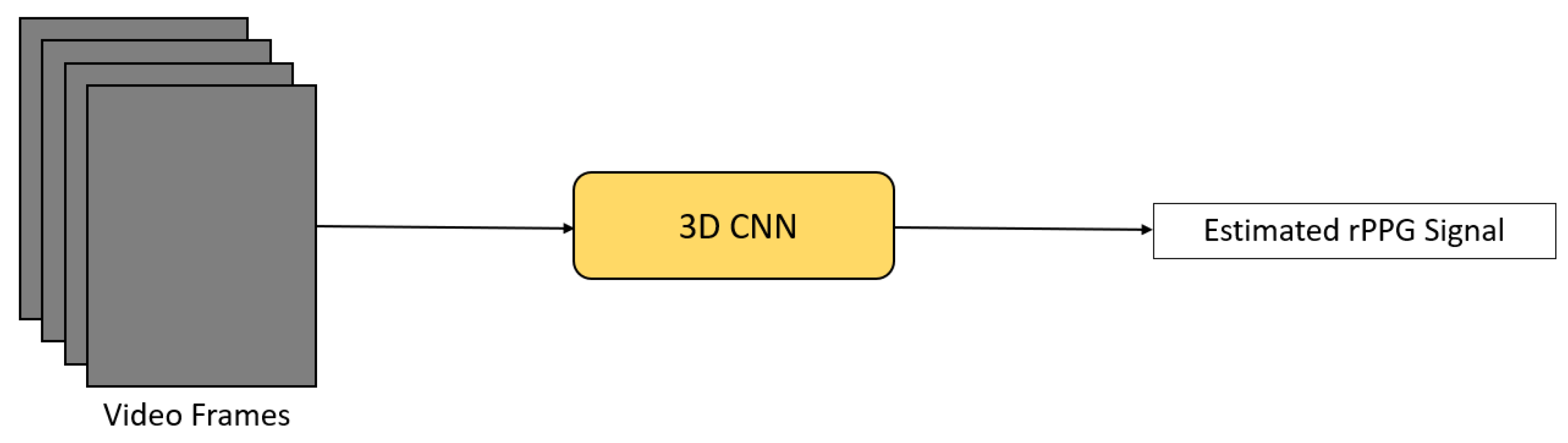
2.2. Spatio-Temporal Network—3D Convolutional Neural Network (3D CNN)
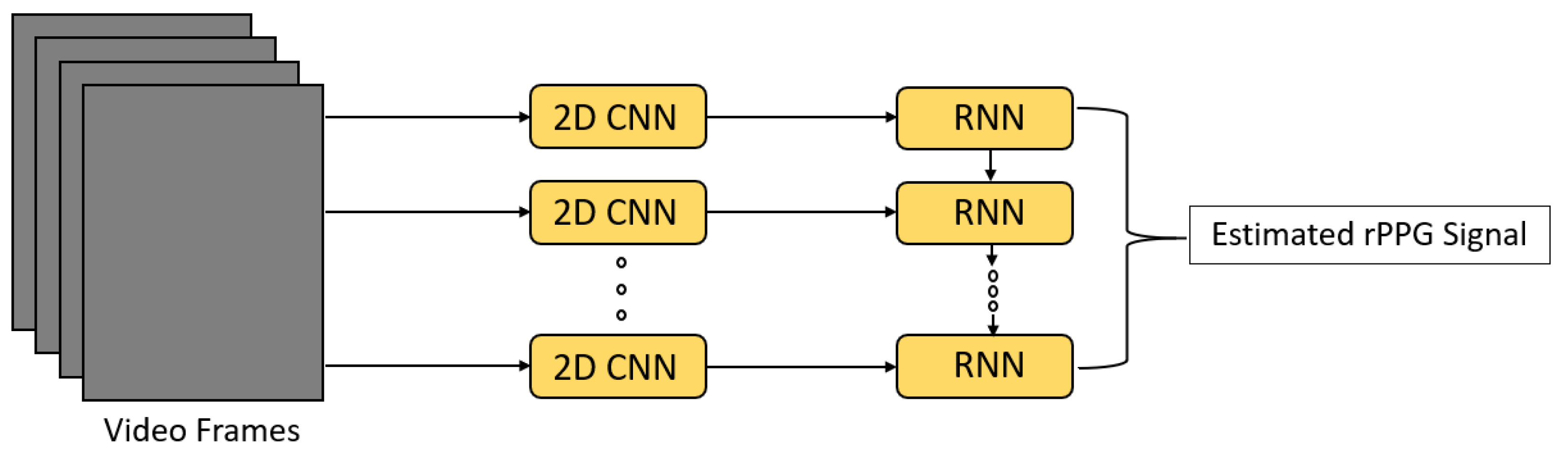
2.3. Spatio-Temporal Network—2D Convolutional Neural Network + Recurrent Neural Network (2D CNN + RNN)
3. Hybrid Deep Learning Methods
3.1. Deep Learning for Signal Optimization
3.2. Deep Learning for Signal Extraction
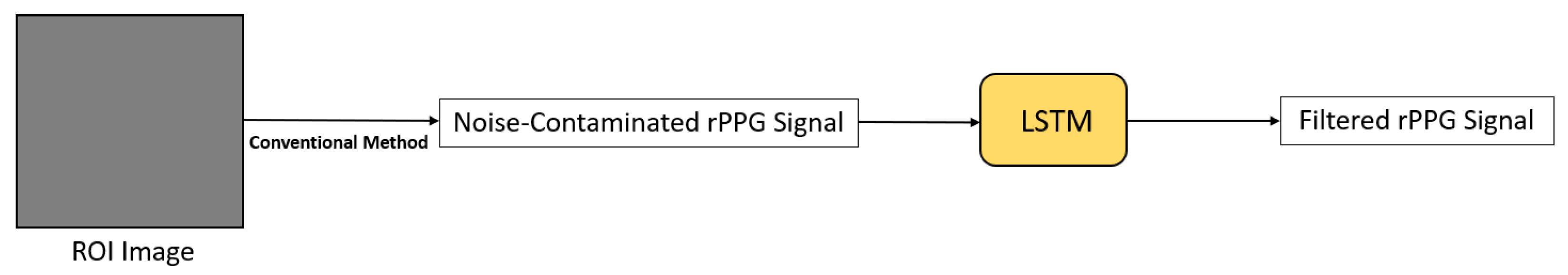
3.2.1. Long Short-Term Memory (LSTM)
3.2.2. 2D Convolutional Neural Network (2D CNN)
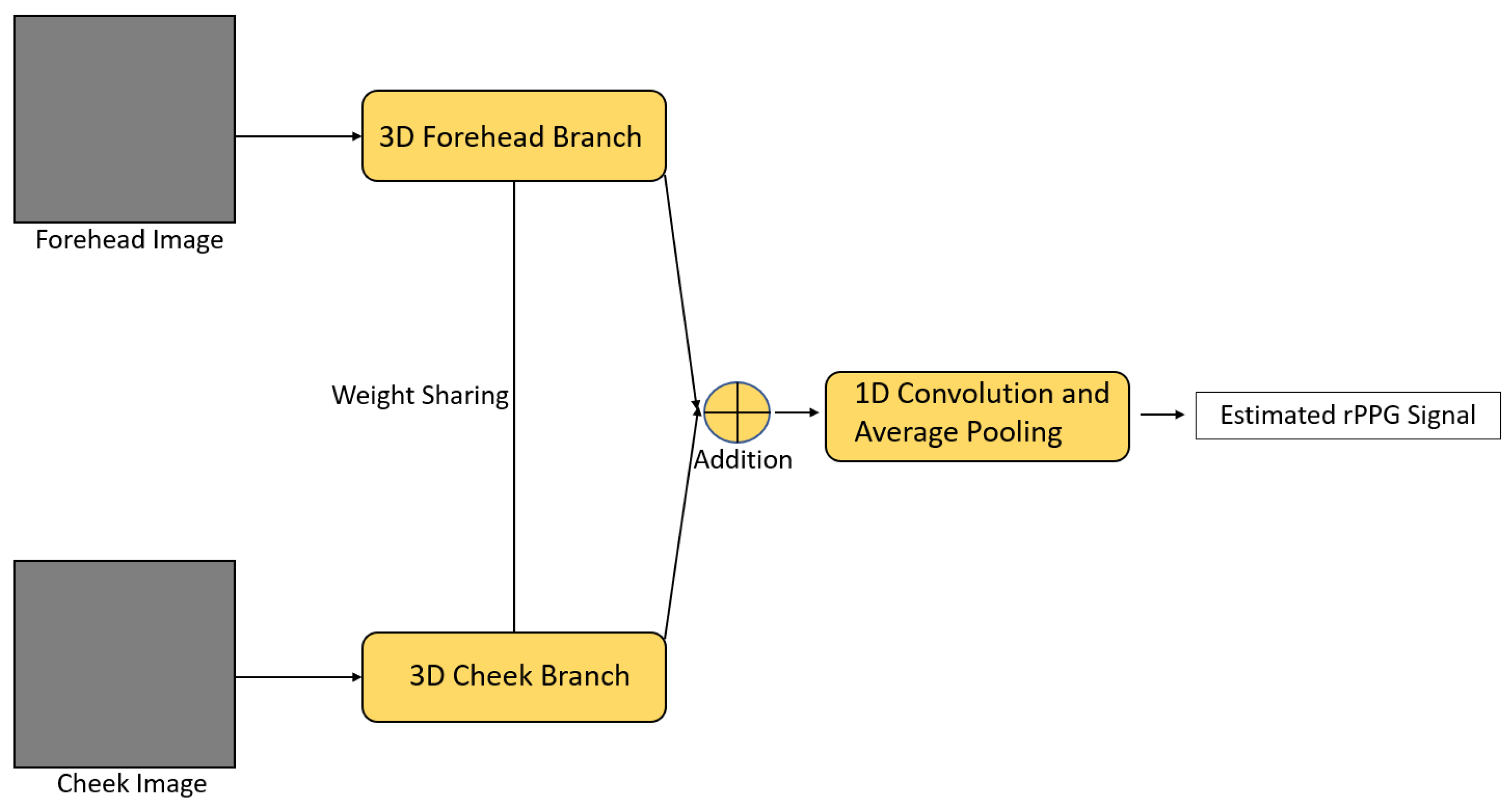
3.2.3. Spatio-Temporal Network—3D Convolutional Neural Network (3D CNN)
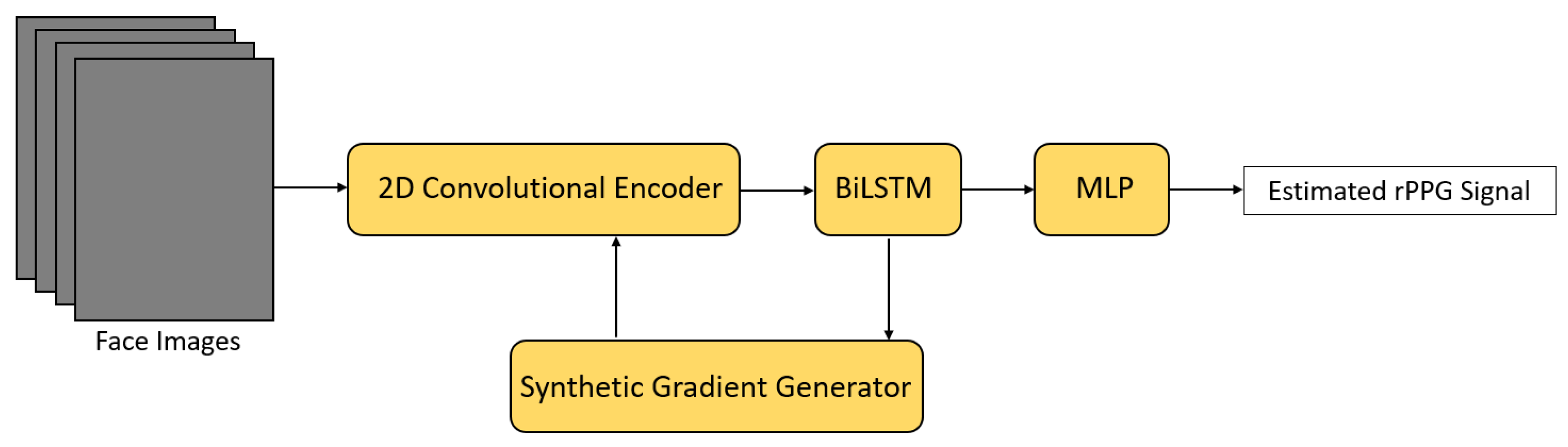
3.2.4. Spatio-Temporal Network—2D Convolutional Neural Network + Recurrent Neural Network (2D CNN + RNN)
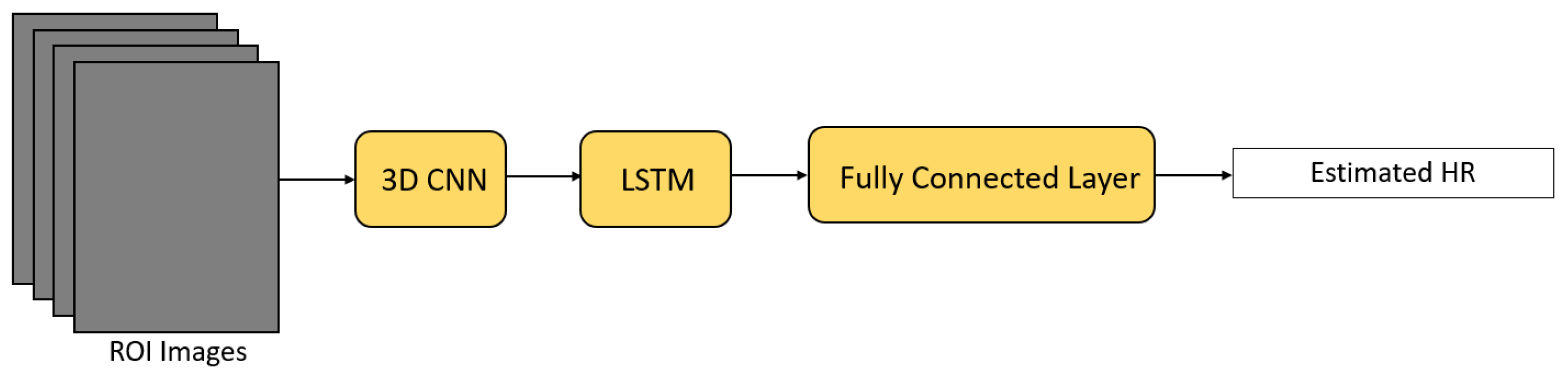
3.2.5. 3D Convolutional Neural Network + Recurrent Neural Network (3D CNN + RNN)
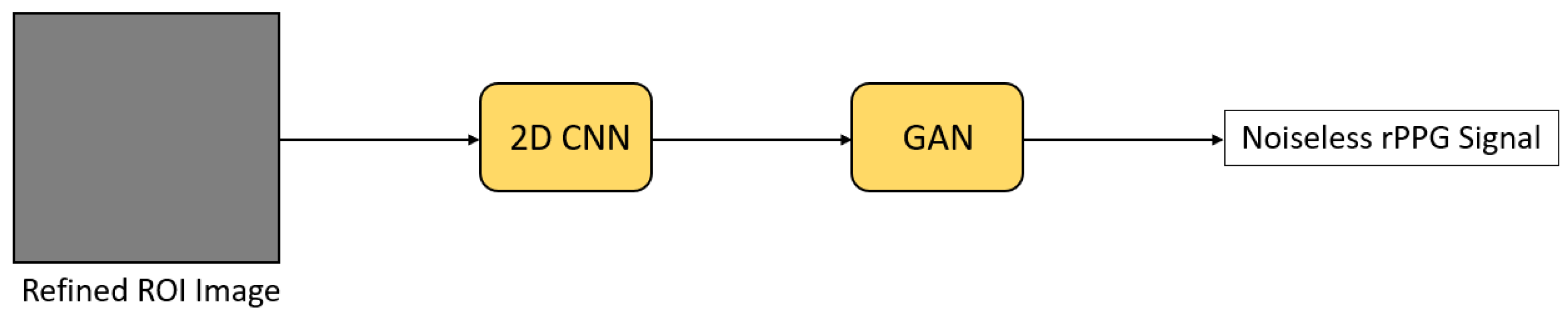
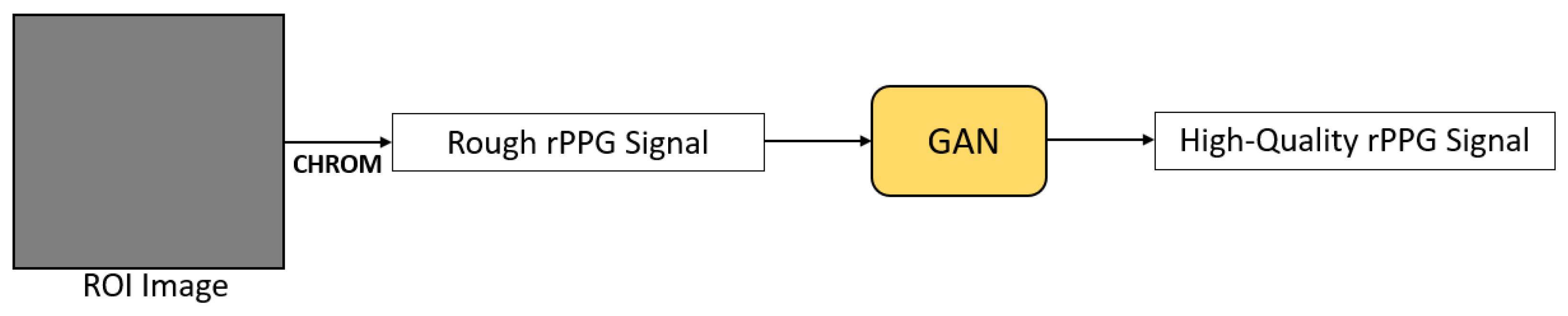
3.2.6. Generative Adversarial Network (GAN)
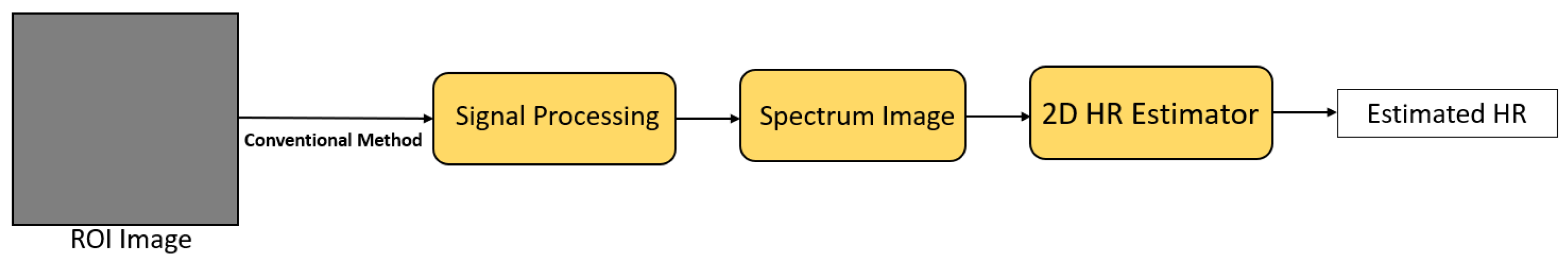
3.3. Deep Learning for Heart Rate Estimation
4. Applications
4.1. Affective Computing
4.2. Pandemic Control
4.3. Deepfake Detection
4.4. Telehealth
4.5. Face Anti-Spoofing
4.6. Driving Condition Monitoring
4.7. Searching for Survivors during Natural Disasters
4.8. Neonatal Monitoring
4.9. Fitness Tracking
5. Resources
5.1. Toolboxes
5.2. Datasets
5.3. Open Challenge on Remote Physiological Signal Sensing
6. Research Gaps
6.1. Influencing Factors
6.2. Measuring Other Vital Signs
6.3. Datasets
6.4. Performance on Different Heart Rate Ranges
6.5. Understanding of Deep Learning-Based Methods
7. Conclusions
Author Contributions
Funding
Institutional Review Board Statement
Informed Consent Statement
Data Availability Statement
Acknowledgments
Conflicts of Interest
References
- Jeong, I.; Finkelstein, J. Introducing Contactless Blood Pressure Assessment Using a High Speed Video Camera. J. Med. Syst. 2016, 40, 1–10. [Google Scholar] [CrossRef]
- Bal, U. Non-contact estimation of heart rate and oxygen saturation using ambient light. Biomed. Opt. Express 2015, 6, 86–97. [Google Scholar] [CrossRef] [PubMed] [Green Version]
- Massaroni, C.; Nicolò, A.; Sacchetti, M.; Schena, E. Contactless Methods For Measuring Respiratory Rate: A Review. IEEE Sens. J. 2021, 21, 12821–12839. [Google Scholar] [CrossRef]
- Iozzia, L.; Cerina, L.; Mainardi, L. Relationships between heart-rate variability and pulse-rate variability obtained from video-PPG signal using ZCA. Physiol. Meas. 2016, 37, 1934–1944. [Google Scholar] [CrossRef] [PubMed]
- Scalise, L. Non contact heart monitoring. Adv. Electrocardiogr. Methods Anal. 2012, 4, 81–106. [Google Scholar]
- Shao, D.; Liu, C.; Tsow, F. Noncontact Physiological Measurement Using a Camera: A Technical Review and Future Directions. ACS Sens. 2021, 6, 321–334. [Google Scholar] [CrossRef] [PubMed]
- Sun, Y.; Thakor, N. Photoplethysmography Revisited: From Contact to Noncontact, From Point to Imaging. IEEE Trans. Biomed. Eng. 2016, 63, 463–477. [Google Scholar] [CrossRef] [PubMed] [Green Version]
- Swinehart, D.F. The Beer-Lambert Law. J. Chem. Educ. 1962, 39, 333. [Google Scholar] [CrossRef]
- Aarts, L.; Jeanne, V.; Cleary, J.P.; Lieber, C.; Nelson, J.; Bambang-Oetomo, S.; Verkruysse, W. Non-contact heart rate monitoring utilizing camera photoplethysmography in the neonatal intensive care unit—A pilot study. Early Hum. Dev. 2013, 89. [Google Scholar] [CrossRef]
- Rouast, P.; Adam, M.; Chiong, R.; Cornforth, D.; Lux, E. Remote heart rate measurement using low-cost RGB face video: A technical literature review. Front. Comput. Sci. 2018, 12, 858–872. [Google Scholar] [CrossRef]
- Maurya, L.; Kaur, P.; Chawla, D.; Mahapatra, P. Non-contact breathing rate monitoring in newborns: A review. Comput. Biol. Med. 2021, 132, 104321. [Google Scholar] [CrossRef]
- McDuff, D.J.; Estepp, J.R.; Piasecki, A.M.; Blackford, E.B. A survey of remote optical photoplethysmographic imaging methods. In Proceedings of the 2015 37th Annual International Conference of the IEEE Engineering in Medicine and Biology Society (EMBC), Milan, Italy, 25–29 August 2015; pp. 6398–6404. [Google Scholar] [CrossRef]
- Wang, W.; den Brinker, A.C.; Stuijk, S.; de Haan, G. Algorithmic Principles of Remote PPG. IEEE Trans. Biomed. Eng. 2017, 64, 1479–1491. [Google Scholar] [CrossRef] [Green Version]
- Khanam, F.T.Z.; Al-Naji, A.; Chahl, J. Remote Monitoring of Vital Signs in Diverse Non-Clinical and Clinical Scenarios Using Computer Vision Systems: A Review. Appl. Sci. 2019, 9, 4474. [Google Scholar] [CrossRef] [Green Version]
- Verkruysse, W.; Svaasand, L.O.; Nelson, J.S. Remote plethysmographic imaging using ambient light. Opt. Express 2008, 16, 21434–21445. [Google Scholar] [CrossRef] [Green Version]
- Poh, M.Z.; McDuff, D.J.; Picard, R.W. Non-contact, automated cardiac pulse measurements using video imaging and blind source separation. Opt. Express 2010, 18, 10762–10774. [Google Scholar] [CrossRef]
- Lewandowska, M.; Rumiński, J.; Kocejko, T.; Nowak, J. Measuring pulse rate with a webcam—A non-contact method for evaluating cardiac activity. In Proceedings of the 2011 Federated Conference on Computer Science and Information Systems (FedCSIS), Szczecin, Poland, 19–21 September 2011; pp. 405–410. [Google Scholar]
- de Haan, G.; Jeanne, V. Robust Pulse Rate From Chrominance-Based rPPG. IEEE Trans. Biomed. Eng. 2013, 60, 2878–2886. [Google Scholar] [CrossRef]
- Tominaga, S. Dichromatic reflection models for a variety of materials. Color Res. Appl. 1994, 19, 277–285. [Google Scholar] [CrossRef]
- de Haan, G.; van Leest, A. Improved motion robustness of remote-PPG by using the blood volume pulse signature. Physiol. Meas. 2014, 35, 1913–1926. [Google Scholar] [CrossRef] [PubMed]
- Wang, W.; Stuijk, S.; de Haan, G. A Novel Algorithm for Remote Photoplethysmography: Spatial Subspace Rotation. IEEE Trans. Biomed. Eng. 2016, 63, 1974–1984. [Google Scholar] [CrossRef] [PubMed]
- Dasari, A.; Arul Prakash, S.K.; Jeni, L.; Tucker, C. Evaluation of biases in remote photoplethysmography methods. Npj Digit. Med. 2021, 4. [Google Scholar] [CrossRef]
- Chen, X.; Cheng, J.; Song, R.; Liu, Y.; Ward, R.; Wang, Z.J. Video-Based Heart Rate Measurement: Recent Advances and Future Prospects. IEEE Trans. Instrum. Meas. 2019, 68, 3600–3615. [Google Scholar] [CrossRef]
- Viola, P.; Jones, M. Rapid object detection using a boosted cascade of simple features. In Proceedings of the 2001 IEEE Computer Society Conference on Computer Vision and Pattern Recognition, CVPR 2001, Kauai, HI, USA, 8–14 December 2001; Volume 1, p. I. [Google Scholar] [CrossRef]
- Zhan, Q.; Wang, W.; de Haan, G. Analysis of CNN-based remote-PPG to understand limitations and sensitivities. Biomed. Opt. Express 2020, 11, 1268–1283. [Google Scholar] [CrossRef] [PubMed]
- Spetlik, R.; Franc, V.; Cech, J.; Matas, J. Visual Heart Rate Estimation with Convolutional Neural Network. In Proceedings of the British Machine Vision Conference (BMVC), Newcastle, UK, 2–6 September 2018. [Google Scholar]
- Chen, W.; McDuff, D. DeepPhys: Video-Based Physiological Measurement Using Convolutional Attention Networks. In Proceedings of the European Conference on Computer Vision (ECCV), Munich, Germany, 8–14 September 2018. [Google Scholar]
- Liu, X.; Fromm, J.; Patel, S.; McDuff, D. Multi-Task Temporal Shift Attention Networks for On-Device Contactless Vitals Measurement. arXiv 2021, arXiv:2006.03790v2. [Google Scholar]
- Yu, Z.; Li, X.; Zhao, G. Remote Photoplethysmograph Signal Measurement from Facial Videos Using Spatio-Temporal Networks. In Proceedings of the British Machine Vision Conference (BMVC), Cardiff, UK, 9–12 September 2019. [Google Scholar]
- Yu, Z.; Peng, W.; Li, X.; Hong, X.; Zhao, G. Remote Heart Rate Measurement From Highly Compressed Facial Videos: An End-to-End Deep Learning Solution with Video Enhancement. In Proceedings of the 2019 IEEE/CVF International Conference on Computer Vision (ICCV), Seoul, Korea, 27–28 October 2019; pp. 151–160. [Google Scholar] [CrossRef] [Green Version]
- Yu, Z.; Li, X.; Niu, X.; Shi, J.; Zhao, G. AutoHR: A Strong End-to-End Baseline for Remote Heart Rate Measurement with Neural Searching. IEEE Signal Process. Lett. 2020, 27, 1245–1249. [Google Scholar] [CrossRef]
- Hu, M.; Qian, F.; Wang, X.; He, L.; Guo, D.; Ren, F. Robust Heart Rate Estimation with Spatial-Temporal Attention Network from Facial Videos. IEEE Trans. Cogn. Dev. Syst. 2021, 1. [Google Scholar] [CrossRef]
- Zhang, P.; Li, B.; Peng, J.; Jiang, W. Multi-hierarchical Convolutional Network for Efficient Remote Photoplethysmograph Signal and Heart Rate Estimation from Face Video Clips. arXiv 2021, arXiv:2104.02260. [Google Scholar]
- Hu, M.; Qian, F.; Guo, D.; Wang, X.; He, L.; Ren, F. ETA-rPPGNet: Effective Time-Domain Attention Network for Remote Heart Rate Measurement. IEEE Trans. Instrum. Meas. 2021, 70, 1–12. [Google Scholar] [CrossRef]
- Hu, M.; Guo, D.; Wang, X.; Ge, P.; Chu, Q. A Novel Spatial-Temporal Convolutional Neural Network for Remote Photoplethysmography. In Proceedings of the 2019 12th International Congress on Image and Signal Processing, BioMedical Engineering and Informatics (CISP-BMEI), Suzhou, China, 19–21 October 2019; pp. 1–6. [Google Scholar] [CrossRef]
- Lin, J.; Gan, C.; Han, S. TSM: Temporal Shift Module for Efficient Video Understanding. In Proceedings of the IEEE International Conference on Computer Vision, Seoul, Korea, 27–28 October 2019. [Google Scholar]
- Liu, H.; Simonyan, K.; Yang, Y. DARTS: Differentiable Architecture Search. In Proceedings of the International Conference on Learning Representations, New Orleans, LA, USA, 6–9 May 2019. [Google Scholar]
- Xu, Y.; Xie, L.; Zhang, X.; Chen, X.; Qi, G.J.; Tian, Q.; Xiong, H. PC-DARTS: Partial Channel Connections for Memory-Efficient Architecture Search. In Proceedings of the International Conference on Learning Representations, New Orleans, LA, USA, 6–9 May 2019. [Google Scholar]
- Qiu, Z.; Yao, T.; Mei, T. Learning Spatio-Temporal Representation with Pseudo-3D Residual Networks. In Proceedings of the 2017 IEEE International Conference on Computer Vision (ICCV), Venice, Italy, 22–29 October 2017; pp. 5534–5542. [Google Scholar] [CrossRef] [Green Version]
- Shi, X.; Chen, Z.; Wang, H.; Yeung, D.Y.; Wong, W.K.; Woo, W.C. Convolutional LSTM Network: A Machine Learning Approach for Precipitation Nowcasting. In Proceedings of the 28th International Conference on Neural Information Processing Systems—Volume 1; NIPS’15; MIT Press: Cambridge, MA, USA, 2015; pp. 802–810. [Google Scholar]
- Li, X.; Alikhani, I.; Shi, J.; Seppanen, T.; Junttila, J.; Majamaa-Voltti, K.; Tulppo, M.; Zhao, G. The OBF Database: A Large Face Video Database for Remote Physiological Signal Measurement and Atrial Fibrillation Detection. In Proceedings of the 2018 13th IEEE International Conference on Automatic Face Gesture Recognition (FG 2018), Xi’an, China, 15–19 May 2018; pp. 242–249. [Google Scholar] [CrossRef] [Green Version]
- Lempe, G.; Zaunseder, S.; Wirthgen, T.; Zipser, S.; Malberg, H. ROI Selection for Remote Photoplethysmography. In Bildverarbeitung für die Medizin 2013; Meinzer, H.P., Deserno, T.M., Handels, H., Tolxdorff, T., Eds.; Springer: Berlin/Heidelberg, Germany, 2013; pp. 99–103. [Google Scholar]
- Tang, C.; Lu, J.; Liu, J. Non-contact Heart Rate Monitoring by Combining Convolutional Neural Network Skin Detection and Remote Photoplethysmography via a Low-Cost Camera. In Proceedings of the 2018 IEEE/CVF Conference on Computer Vision and Pattern Recognition Workshops (CVPRW), Salt Lake City, UT, USA, 18–22 June 2018; pp. 1309–1315. [Google Scholar] [CrossRef]
- Paracchini, M.; Marcon, M.; Villa, F.; Zappa, F.; Tubaro, S. Biometric Signals Estimation Using Single Photon Camera and Deep Learning. Sensors 2020, 20, 6102. [Google Scholar] [CrossRef]
- Sabokrou, M.; Pourreza, M.; Li, X.; Fathy, M.; Zhao, G. Deep-HR: Fast Heart Rate Estimation from Face Video Under Realistic Conditions. arXiv 2020, arXiv:2002.04821. [Google Scholar]
- Liu, S.; Huang, D.; Wang, Y. Receptive Field Block Net for Accurate and Fast Object Detection. In Computer Vision—ECCV 2018; Ferrari, V., Hebert, M., Sminchisescu, C., Weiss, Y., Eds.; Springer International Publishing: Cham, Switzerland, 2018; pp. 404–419. [Google Scholar]
- Bian, M.; Peng, B.; Wang, W.; Dong, J. An Accurate LSTM Based Video Heart Rate Estimation Method. In Pattern Recognition and Computer Vision; Lin, Z., Wang, L., Yang, J., Shi, G., Tan, T., Zheng, N., Chen, X., Zhang, Y., Eds.; Springer International Publishing: Cham, Switzerland, 2019; pp. 409–417. [Google Scholar]
- Botina-Monsalve, D.; Benezeth, Y.; Macwan, R.; Pierrart, P.; Parra, F.; Nakamura, K.; Gomez, R.; Miteran, J. Long Short-Term Memory Deep-Filter in Remote Photoplethysmography. In Proceedings of the 2020 IEEE/CVF Conference on Computer Vision and Pattern Recognition Workshops (CVPRW), Seattle, WA, USA, 14–19 June 2020; pp. 1242–1249. [Google Scholar] [CrossRef]
- Liu, X.; Jiang, Z.; Fromm, J.; Xu, X.; Patel, S.; McDuff, D. MetaPhys: Few-Shot Adaptation for Non-Contact Physiological Measurement. In Proceedings of the Conference on Health, Inference, and Learning; Association for Computing Machinery: New York, NY, USA, 2021; pp. 154–163. [Google Scholar]
- Bousefsaf, F.; Pruski, A.; Maaoui, C. 3D Convolutional Neural Networks for Remote Pulse Rate Measurement and Mapping from Facial Video. Appl. Sci. 2019, 9, 4364. [Google Scholar] [CrossRef] [Green Version]
- Tsou, Y.Y.; Lee, Y.A.; Hsu, C.T.; Chang, S.H. Siamese-RPPG Network: Remote Photoplethysmography Signal Estimation from Face Videos. In Proceedings of the 35th Annual ACM Symposium on Applied Computing; SAC’20; Association for Computing Machinery: New York, NY, USA, 2020; pp. 2066–2073. [Google Scholar] [CrossRef] [Green Version]
- Perepelkina, O.; Artemyev, M.; Churikova, M.; Grinenko, M. HeartTrack: Convolutional neural network for remote video-based heart rate monitoring. In Proceedings of the 2020 IEEE/CVF Conference on Computer Vision and Pattern Recognition Workshops (CVPRW), Seattle, WA, USA, 14–19 June 2020; pp. 1163–1171. [Google Scholar] [CrossRef]
- Tsou, Y.Y.; Lee, Y.A.; Hsu, C.T. Multi-task Learning for Simultaneous Video Generation and Remote Photoplethysmography Estimation. In Computer Vision—ACCV 2020; Ishikawa, H., Liu, C.L., Pajdla, T., Shi, J., Eds.; Springer International Publishing: Cham, Switzerland, 2021; pp. 392–407. [Google Scholar]
- Liu, S.Q.; Yuen, P.C. A General Remote Photoplethysmography Estimator with Spatiotemporal Convolutional Network. In Proceedings of the 2020 15th IEEE International Conference on Automatic Face and Gesture Recognition (FG 2020), Buenos Aires, Argentina, 18–22 May 2020; pp. 481–488. [Google Scholar] [CrossRef]
- Wang, Z.K.; Kao, Y.; Hsu, C.T. Vision-Based Heart Rate Estimation Via A Two-Stream CNN. In Proceedings of the 2019 IEEE International Conference on Image Processing (ICIP), Taipei, Taiwan, 22–25 September 2019; pp. 3327–3331. [Google Scholar] [CrossRef]
- Huang, B.; Chang, C.M.; Lin, C.L.; Chen, W.; Juang, C.F.; Wu, X. Visual Heart Rate Estimation from Facial Video Based on CNN. In Proceedings of the 2020 15th IEEE Conference on Industrial Electronics and Applications (ICIEA), Kristiansand, Norway, 9–13 November 2020; pp. 1658–1662. [Google Scholar] [CrossRef]
- Lee, E.; Chen, E.; Lee, C.Y. Meta-rPPG: Remote Heart Rate Estimation Using a Transductive Meta-learner. In Computer Vision—ECCV 2020; Vedaldi, A., Bischof, H., Brox, T., Frahm, J.M., Eds.; Springer International Publishing: Cham, Switzerland, 2020; pp. 392–409. [Google Scholar]
- Huang, B.; Lin, C.L.; Chen, W.; Juang, C.F.; Wu, X. A novel one-stage framework for visual pulse rate estimation using deep neural networks. Biomed. Signal Process. Control 2021, 66, 102387. [Google Scholar] [CrossRef]
- Song, R.; Chen, H.; Cheng, J.; Li, C.; Liu, Y.; Chen, X. PulseGAN: Learning to Generate Realistic Pulse Waveforms in Remote Photoplethysmography. IEEE J. Biomed. Health Inform. 2021, 25, 1373–1384. [Google Scholar] [CrossRef]
- Finn, C.; Abbeel, P.; Levine, S. Model-Agnostic Meta-Learning for Fast Adaptation of Deep Networks. In Proceedings of the 34th International Conference on Machine Learning, Sydney, Australia, 6–11 August 2017; pp. 1126–1135. [Google Scholar]
- Newell, A.; Yang, K.; Deng, J. Stacked Hourglass Networks for Human Pose Estimation. In Computer Vision—ECCV 2016; Leibe, B., Matas, J., Sebe, N., Welling, M., Eds.; Springer International Publishing: Cham, Switzerland, 2016; pp. 483–499. [Google Scholar]
- Hu, S.X.; Moreno, P.G.; Xiao, Y.; Shen, X.; Obozinski, G.; Lawrence, N.; Damianou, A. Empirical Bayes Transductive Meta-Learning with Synthetic Gradients. In Proceedings of the International Conference on Learning Representations, Glasgow, UK, 23–28 August 2020. [Google Scholar]
- Mirza, M.; Osindero, S. Conditional Generative Adversarial Nets. arXiv 2014, arXiv:1411.1784. [Google Scholar]
- Yang, W.; Li, X.; Zhang, B. Heart Rate Estimation from Facial Videos Based on Convolutional Neural Network. In Proceedings of the 2018 International Conference on Network Infrastructure and Digital Content (IC-NIDC), Guiyang, China, 22–24 August 2018; pp. 45–49. [Google Scholar] [CrossRef]
- He, K.; Zhang, X.; Ren, S.; Sun, J. Deep Residual Learning for Image Recognition. In Proceedings of the 2016 IEEE Conference on Computer Vision and Pattern Recognition (CVPR), Las Vegas, NV, USA, 27–30 June 2016; pp. 770–778. [Google Scholar] [CrossRef] [Green Version]
- Niu, X.; Han, H.; Shan, S.; Chen, X. SynRhythm: Learning a Deep Heart Rate Estimator from General to Specific. In Proceedings of the 2018 24th International Conference on Pattern Recognition (ICPR), Beijing, China, 20–24 August 2018; pp. 3580–3585. [Google Scholar] [CrossRef]
- Niu, X.; Zhao, X.; Han, H.; Das, A.; Dantcheva, A.; Shan, S.; Chen, X. Robust Remote Heart Rate Estimation from Face Utilizing Spatial-temporal Attention. In Proceedings of the 2019 14th IEEE International Conference on Automatic Face Gesture Recognition (FG 2019), Lille, France, 14–18 May 2019; pp. 1–8. [Google Scholar] [CrossRef] [Green Version]
- Niu, X.; Shan, S.; Han, H.; Chen, X. RhythmNet: End-to-End Heart Rate Estimation From Face via Spatial-Temporal Representation. IEEE Trans. Image Process. 2020, 29, 2409–2423. [Google Scholar] [CrossRef] [PubMed] [Green Version]
- Niu, X.; Yu, Z.; Han, H.; Li, X.; Shan, S.; Zhao, G. Video-based remote physiological measurement via cross-verified feature disentangling. In European Conference on Computer Vision; Springer: Berlin/Heidelberg, Germany, 2020; pp. 295–310. [Google Scholar]
- Song, R.; Zhang, S.; Li, C.; Zhang, Y.; Cheng, J.; Chen, X. Heart Rate Estimation From Facial Videos Using a Spatiotemporal Representation with Convolutional Neural Networks. IEEE Trans. Instrum. Meas. 2020, 69, 7411–7421. [Google Scholar] [CrossRef]
- Lu, H.; Han, H. NAS-HR: Neural architecture search for heart rate estimation from face videos. Virtual Real. Intell. Hardw. 2021, 3, 33–42. [Google Scholar] [CrossRef]
- Meziatisabour, R.; Benezeth, Y.; De Oliveira, P.; Chappe, J.; Yang, F. UBFC-Phys: A Multimodal Database For Psychophysiological Studies of Social Stress. IEEE Trans. Affect. Comput. 2021, 1. [Google Scholar] [CrossRef]
- McDuff, D.; Gontarek, S.; Picard, R. Remote measurement of cognitive stress via heart rate variability. In Proceedings of the 2014 36th Annual International Conference of the IEEE Engineering in Medicine and Biology Society, Chicago, IL, USA, 26–30 August 2014; pp. 2957–2960. [Google Scholar] [CrossRef]
- Gupta, P.; Bhowmick, B.; Pal, A. Exploring the Feasibility of Face Video Based Instantaneous Heart-Rate for Micro-Expression Spotting. In Proceedings of the 2018 IEEE/CVF Conference on Computer Vision and Pattern Recognition Workshops (CVPRW), Salt Lake City, UT, USA, 18–22 June 2018; pp. 1316–1323. [Google Scholar] [CrossRef]
- Monkaresi, H.; Bosch, N.; Calvo, R.A.; D’Mello, S.K. Automated Detection of Engagement Using Video-Based Estimation of Facial Expressions and Heart Rate. IEEE Trans. Affect. Comput. 2017, 8, 15–28. [Google Scholar] [CrossRef]
- Kessler, V.; Thiam, P.; Amirian, M.; Schwenker, F. Pain recognition with camera photoplethysmography. In Proceedings of the 2017 Seventh International Conference on Image Processing Theory, Tools and Applications (IPTA), Montreal, QC, Canada, 28 November–1 December 2017; pp. 1–5. [Google Scholar] [CrossRef]
- Huang, D.; Feng, X.; Zhang, H.; Yu, Z.; Peng, J.; Zhao, G.; Xia, Z. Spatio-Temporal Pain Estimation Network with Measuring Pseudo Heart Rate Gain. IEEE Trans. Multimed. 2021, 1. [Google Scholar] [CrossRef]
- Yang, R.; Guan, Z.; Yu, Z.; Zhao, G.; Feng, X.; Peng, J. Non-contact Pain Recognition from Video Sequences with Remote Physiological Measurements Prediction. arXiv 2021, arXiv:2105.08822. [Google Scholar]
- Mitra, B.; Luckhoff, C.; Mitchell, R.D.; O’Reilly, G.M.; Smit, D.V.; Cameron, P.A. Temperature screening has negligible value for control of COVID-19. Emerg. Med. Australas. 2020, 32, 867–869. [Google Scholar] [CrossRef]
- Vilke, G.M.; Brennan, J.J.; Cronin, A.O.; Castillo, E.M. Clinical Features of Patients with COVID-19: Is Temperature Screening Useful? J. Emerg. Med. 2020, 59, 952–956. [Google Scholar] [CrossRef] [PubMed]
- Stave, G.M.; Smith, S.E.; Hymel, P.A.; Heron, R.J.L. Worksite Temperature Screening for COVID-19. J. Occup. Environ. Med. 2021, 63, 638–641. [Google Scholar]
- Lippi, G.; Mattiuzzi, C.; Henry, B. Is Body Temperature Mass Screening a Reliable and Safe Option for Preventing COVID-19 Spread? SSRN Electron. J. 2021. [Google Scholar] [CrossRef]
- Natarajan, A.; Su, H.W.; Heneghan, C. Assessment of physiological signs associated with COVID-19 measured using wearable devices. medRxiv 2020. [Google Scholar] [CrossRef]
- Pavri, B.; Kloo, J.; Farzad, D.; Riley, J. Behavior of the PR Interval with Increasing Heart Rate in Patients with COVID-19. Heart Rhythm 2020, 17. [Google Scholar] [CrossRef]
- Gawałko, M.; Kapłon-Cieślicka, A.; Hohl, M.; Dobrev, D.; Linz, D. COVID-19 associated atrial fibrillation: Incidence, putative mechanisms and potential clinical implications. Int. J. Cardiol. Heart Vasc. 2020, 30, 100631. [Google Scholar] [CrossRef] [PubMed]
- Stone, E.; Kiat, H.; McLachlan, C.S. Atrial fibrillation in COVID-19: A review of possible mechanisms. FASEB J. 2020, 34, 11347–11354. [Google Scholar] [CrossRef]
- Schnaubelt, S.; Breyer, M.K.; Siller-Matula, J.; Domanovits, H. Atrial fibrillation: A risk factor for unfavourable outcome in COVID-19? A case report. Eur. Heart J. Case Rep. 2020, 4, 1–6. [Google Scholar] [CrossRef]
- Shi, J.; Alikhani, I.; Li, X.; Yu, Z.; Seppänen, T.; Zhao, G. Atrial Fibrillation Detection From Face Videos by Fusing Subtle Variations. IEEE Trans. Circuits Syst. Video Technol. 2020, 30, 2781–2795. [Google Scholar] [CrossRef] [Green Version]
- Zhu, G.; Li, J.; Meng, Z.; Yu, Y.; Li, Y.; Tang, X.; Dong, Y.; Sun, G.; Zhou, R.; Wang, H.; et al. Learning from Large-Scale Wearable Device Data for Predicting the Epidemic Trend of COVID-19. Discret. Dyn. Nat. Soc. 2020, 2020, 1–8. [Google Scholar] [CrossRef]
- Mishra, T.; Wang, M.; Metwally, A.A.; Bogu, G.K.; Brooks, A.W.; Bahmani, A.; Alavi, A.; Celli, A.; Higgs, E.; Dagan-Rosenfeld, O.; et al. Pre-symptomatic detection of COVID-19 from smartwatch data. Nat. Biomed. Eng. 2020, 4, 1208–1220. [Google Scholar] [CrossRef] [PubMed]
- Suwajanakorn, S.; Seitz, S.M.; Kemelmacher-Shlizerman, I. Synthesizing Obama: Learning Lip Sync from Audio. ACM Trans. Graph. 2017, 36. [Google Scholar] [CrossRef]
- Nguyen, T.; Nguyen, C.; Nguyen, D.; Nguyen, D.; Nahavandi, S. Deep Learning for Deepfakes Creation and Detection. arXiv 2019, arXiv:1909.11573. [Google Scholar]
- Korshunov, P.; Marcel, S. DeepFakes: A New Threat to Face Recognition? Assessment and Detection. arXiv 2018, arXiv:1812.08685. [Google Scholar]
- Westerlund, M. The Emergence of Deepfake Technology: A Review. Technol. Innov. Manag. Rev. 2019, 9, 40–53. [Google Scholar] [CrossRef]
- Fernandes, S.; Raj, S.; Ortiz, E.; Vintila, I.; Salter, M.; Urosevic, G.; Jha, S. Predicting Heart Rate Variations of Deepfake Videos using Neural ODE. In Proceedings of the 2019 IEEE/CVF International Conference on Computer Vision Workshop (ICCVW), Seoul, Korea, 27–28 October 2019; pp. 1721–1729. [Google Scholar] [CrossRef]
- Ciftci, U.; Demir, I.; Yin, L. FakeCatcher: Detection of Synthetic Portrait Videos using Biological Signals. IEEE Trans. Pattern Anal. Mach. Intell. 2020, 1. [Google Scholar] [CrossRef] [PubMed]
- Tuckson, R.V.; Edmunds, M.; Hodgkins, M.L. Telehealth. N. Engl. J. Med. 2017, 377, 1585–1592. [Google Scholar] [CrossRef]
- Song, R.; Li, J.; Wang, M.; Cheng, J.; Li, C.; Chen, X. Remote Photoplethysmography with an EEMD-MCCA Method Robust Against Spatially Uneven Illuminations. IEEE Sens. J. 2021, 21, 13484–13494. [Google Scholar] [CrossRef]
- Zhao, F.; Li, M.; Qian, Y.; Tsien, J.Z. Remote Measurements of Heart and Respiration Rates for Telemedicine. PLoS ONE 2013, 8, e71384. [Google Scholar] [CrossRef] [Green Version]
- Zhou, X.; Snoswell, C.L.; Harding, L.E.; Bambling, M.; Edirippulige, S.; Bai, X.; Smith, A.C. The Role of Telehealth in Reducing the Mental Health Burden from COVID-19. Telemed. E-Health 2020, 26, 377–379. [Google Scholar] [CrossRef] [Green Version]
- Alsaadi, I. Physiological Biometric Authentication Systems, Advantages, Disadvantages And Future Development: A Review. Int. J. Sci. Technol. Res. 2015, 4, 285–289. [Google Scholar]
- Kumar, S.; Singh, S.; Kumar, J. A comparative study on face spoofing attacks. In Proceedings of the 2017 International Conference on Computing, Communication and Automation (ICCCA), Greater Noida, India, 5–6 May 2017; pp. 1104–1108. [Google Scholar] [CrossRef]
- Yu, Z.; Qin, Y.; Li, X.; Zhao, C.; Lei, Z.; Zhao, G. Deep learning for face anti-spoofing: A survey. arXiv 2021, arXiv:2106.14948. [Google Scholar]
- Liu, S.Q.; Lan, X.; Yuen, P.C. Remote Photoplethysmography Correspondence Feature for 3D Mask Face Presentation Attack Detection. In Proceedings of the European Conference on Computer Vision (ECCV), Munich, Germany, 8–14 September 2018. [Google Scholar]
- Li, X.; Komulainen, J.; Zhao, G.; Yuen, P.C.; Pietikäinen, M. Generalized face anti-spoofing by detecting pulse from face videos. In Proceedings of the 2016 23rd International Conference on Pattern Recognition (ICPR); IEEE: New York, NY, USA, 2016; pp. 4244–4249. [Google Scholar]
- Liu, S.; Yuen, P.C.; Zhang, S.; Zhao, G. 3D Mask Face Anti-spoofing with Remote Photoplethysmography. In Computer Vision—ECCV 2016; Leibe, B., Matas, J., Sebe, N., Welling, M., Eds.; Springer International Publishing: Cham, Switzerland, 2016; pp. 85–100. [Google Scholar]
- Yu, Z.; Li, X.; Wang, P.; Zhao, G. TransRPPG: Remote Photoplethysmography Transformer for 3D Mask Face Presentation Attack Detection. arXiv 2021, arXiv:2104.07419. [Google Scholar]
- Lin, B.; Li, X.; Yu, Z.; Zhao, G. Face liveness detection by rppg features and contextual patch-based cnn. In Proceedings of the 2019 3rd International Conference on Biometric Engineering and Applications, Stockholm, Sweden, 29–31 May 2019; pp. 61–68. [Google Scholar]
- Kuo, J.; Koppel, S.; Charlton, J.L.; Rudin-Brown, C.M. Evaluation of a video-based measure of driver heart rate. J. Saf. Res. 2015, 54, 55–59. [Google Scholar] [CrossRef] [PubMed] [Green Version]
- Zhang, Q.; Xu, G.Q.; Wang, M.; Zhou, Y.; Feng, W. Webcam based non-contact real-time monitoring for the physiological parameters of drivers. In Proceedings of the 4th Annual IEEE International Conference on Cyber Technology in Automation, Control and Intelligent, Hong Kong, China, 4–7 June 2014; pp. 648–652. [Google Scholar] [CrossRef]
- Lee, K.; Han, D.; Ko, H. Video Analytic Based Health Monitoring for Driver in Moving Vehicle by Extracting Effective Heart Rate Inducing Features. J. Adv. Transp. 2018, 2018, 1–9. [Google Scholar] [CrossRef]
- Zhang, Q.; Zhou, Y.; Song, S.; Liang, G.; Ni, H. Heart Rate Extraction Based on Near-Infrared Camera: Towards Driver State Monitoring. IEEE Access 2018, 6, 33076–33087. [Google Scholar] [CrossRef]
- Wu, B.F.; Chu, Y.W.; Huang, P.W.; Chung, M.L. Neural Network Based Luminance Variation Resistant Remote-Photoplethysmography for Driver’s Heart Rate Monitoring. IEEE Access 2019, 7, 57210–57225. [Google Scholar] [CrossRef]
- Huang, P.W.; Wu, B.J.; Wu, B.F. A Heart Rate Monitoring Framework for Real-World Drivers Using Remote Photoplethysmography. IEEE J. Biomed. Health Inform. 2021, 25, 1397–1408. [Google Scholar] [CrossRef] [PubMed]
- Tsai, Y.C.; Lai, P.W.; Huang, P.W.; Lin, T.M.; Wu, B.F. Vision-Based Instant Measurement System for Driver Fatigue Monitoring. IEEE Access 2020, 8, 67342–67353. [Google Scholar] [CrossRef]
- Magdalena Nowara, E.; Marks, T.K.; Mansour, H.; Veeraraghavan, A. SparsePPG: Towards Driver Monitoring Using Camera-Based Vital Signs Estimation in Near-Infrared. In Proceedings of the IEEE Conference on Computer Vision and Pattern Recognition (CVPR) Workshops, Salt Lake City, UT, USA, 18–22 June 2018. [Google Scholar]
- Wu, B.F.; Chu, Y.W.; Huang, P.W.; Chung, M.L.; Lin, T.M. A Motion Robust Remote-PPG Approach to Driver’s Health State Monitoring. In Computer Vision—ACCV 2016 Workshops; Chen, C.S., Lu, J., Ma, K.K., Eds.; Springer International Publishing: Cham, Switzerland, 2017; pp. 463–476. [Google Scholar]
- Hernandez-Ortega, J.; Nagae, S.; Fierrez, J.; Morales, A. Quality-Based Pulse Estimation from NIR Face Video with Application to Driver Monitoring. In Pattern Recognition and Image Analysis; Morales, A., Fierrez, J., Sánchez, J.S., Ribeiro, B., Eds.; Springer International Publishing: Cham, Switzerland, 2019; pp. 108–119. [Google Scholar]
- Al-Naji, A.A.; Perera, A.; Chahl, J. Remote measurement of cardiopulmonary signal using an unmanned aerial vehicle. In Proceedings of the IOP Conference Series: Materials Science and Engineering, Bangkok, Thailand, 24–26 February 2018; Volume 405, p. 012001. [Google Scholar] [CrossRef] [Green Version]
- Al-Naji, A.A.; Perera, A.; Chahl, J. Remote monitoring of cardiorespiratory signals from a hovering unmanned aerial vehicle. Biomed. Eng. Online 2017, 16. [Google Scholar] [CrossRef] [Green Version]
- Al-Naji, A.; Perera, A.G.; Mohammed, S.L.; Chahl, J. Life Signs Detector Using a Drone in Disaster Zones. Remote Sens. 2019, 11, 2441. [Google Scholar] [CrossRef] [Green Version]
- Al-Naji, A.; Chahl, J. Remote Optical Cardiopulmonary Signal Extraction with Noise Artifact Removal, Multiple Subject Detection Long-Distance. IEEE Access 2018, 6, 11573–11595. [Google Scholar] [CrossRef]
- Klaessens, J.H.; van den Born, M.; van der Veen, A.; van de Kraats, J.S.; van den Dungen, F.A.; Verdaasdonk, R.M. Development of a baby friendly non-contact method for measuring vital signs: First results of clinical measurements in an open incubator at a neonatal intensive care unit. In Advanced Biomedical and Clinical Diagnostic Systems XII; Vo-Dinh, T., Mahadevan-Jansen, A., Grundfest, W.S., Eds.; SPIE: Bellingham, WA, USA, 2014; pp. 257–263. [Google Scholar]
- Villarroel, M.; Guazzi, A.; Jorge, J.; Davis, S.; Watkinson, P.; Green, G.; Shenvi, A.; McCormick, K.; Tarassenko, L. Continuous non-contact vital sign monitoring in neonatal intensive care unit. Healthc. Technol. Lett. 2014, 1, 87–91. [Google Scholar] [CrossRef] [PubMed] [Green Version]
- Scalise, L.; Bernacchia, N.; Ercoli, I.; Marchionni, P. Heart rate measurement in neonatal patients using a webcamera. In Proceedings of the 2012 IEEE International Symposium on Medical Measurements and Applications Proceedings, Budapest, Hungary, 18–19 May 2012; pp. 1–4. [Google Scholar] [CrossRef]
- Cobos-Torres, J.C.; Abderrahim, M.; Martínez-Orgado, J. Non-Contact, Simple Neonatal Monitoring by Photoplethysmography. Sensors 2018, 18, 4362. [Google Scholar] [CrossRef] [PubMed] [Green Version]
- Gibson, K.; Al-Naji, A.; Fleet, J.A.; Steen, M.; Chahl, J.; Huynh, J.; Morris, S. Noncontact Heart and Respiratory Rate Monitoring of Preterm Infants Based on a Computer Vision System: Protocol for a Method Comparison Study. JMIR Res. Protoc. 2019, 8, e13400. [Google Scholar] [CrossRef] [PubMed]
- Mestha, L.K.; Kyal, S.; Xu, B.; Lewis, L.E.; Kumar, V. Towards continuous monitoring of pulse rate in neonatal intensive care unit with a webcam. In Proceedings of the 2014 36th Annual International Conference of the IEEE Engineering in Medicine and Biology Society, Chicago, IL, USA, 26–30 August 2014; pp. 3817–3820. [Google Scholar] [CrossRef]
- Van Gastel, M.; Balmaekers, B.; Oetomo, S.B.; Verkruysse, W. Near-continuous non-contact cardiac pulse monitoring in a neonatal intensive care unit in near darkness. In Proceedings of the Optical Diagnostics and Sensing XVIII: Toward Point-of-Care Diagnostics; Coté, G.L., Ed.; SPIE: Bellingham, WA, USA, 2018; pp. 230–238. [Google Scholar]
- Malafaya, D.; Domingues, S.; Oliveira, H.P. Domain Adaptation for Heart Rate Extraction in the Neonatal Intensive Care Unit. In Proceedings of the 2020 IEEE International Conference on Bioinformatics and Biomedicine (BIBM), Seoul, Korea, 16–19 December 2020; pp. 1082–1086. [Google Scholar] [CrossRef]
- Antink, C.H.; Ferreira, J.C.M.; Paul, M.; Lyra, S.; Heimann, K.; Karthik, S.; Joseph, J.; Jayaraman, K.; Orlikowsky, T.; Sivaprakasam, M.; et al. Fast body part segmentation and tracking of neonatal video data using deep learning. Med. Biol. Eng. Comput. 2020, 58, 3049–3061. [Google Scholar] [CrossRef]
- Chaichulee, S.; Villarroel, M.; Jorge, J.; Arteta, C.; McCormick, K.; Zisserman, A.; Tarassenko, L. Cardio-respiratory signal extraction from video camera data for continuous non-contact vital sign monitoring using deep learning. Physiol. Meas. 2019, 40, 115001. [Google Scholar] [CrossRef] [Green Version]
- Wang, W.; den Brinker, A.C.; Stuijk, S.; de Haan, G. Robust heart rate from fitness videos. Physiol. Meas. 2017, 38, 1023–1044. [Google Scholar] [CrossRef] [Green Version]
- Chang, C.M.; Hung, C.C.; Zhao, C.; Lin, C.L.; Hsu, B.Y. Learning-based Remote Photoplethysmography for Physiological Signal Feedback Control in Fitness Training. In Proceedings of the 2020 15th IEEE Conference on Industrial Electronics and Applications (ICIEA), Kristiansand, Norway, 9–13 November 2020; pp. 1663–1668. [Google Scholar] [CrossRef]
- Zhao, C.; Lin, C.L.; Chen, W.; Chen, M.K.; Wang, J. Visual heart rate estimation and negative feedback control for fitness exercise. Biomed. Signal Process. Control 2020, 56, 101680. [Google Scholar] [CrossRef]
- Xie, K.; Fu, C.H.; Liang, H.; Hong, H.; Zhu, X. Non-contact Heart Rate Monitoring for Intensive Exercise Based on Singular Spectrum Analysis. In Proceedings of the 2019 IEEE Conference on Multimedia Information Processing and Retrieval (MIPR), San Jose, CA, USA, 28–30 March 2019; pp. 228–233. [Google Scholar] [CrossRef]
- McDuff, D.; Blackford, E. iPhys: An Open Non-Contact Imaging-Based Physiological Measurement Toolbox. In Proceedings of the 2019 41st Annual International Conference of the IEEE Engineering in Medicine and Biology Society (EMBC), Berlin, Germany, 23–27 July 2019; pp. 6521–6524. [Google Scholar] [CrossRef] [Green Version]
- van der Kooij, K.M.; Naber, M. An open-source remote heart rate imaging method with practical apparatus and algorithms. Behav. Res. Methods 2019, 51, 2106–2119. [Google Scholar] [CrossRef]
- Boccignone, G.; Conte, D.; Cuculo, V.; D’Amelio, A.; Grossi, G.; Lanzarotti, R. An Open Framework for Remote-PPG Methods and Their Assessment. IEEE Access 2020, 8, 216083–216103. [Google Scholar] [CrossRef]
- Estepp, J.R.; Blackford, E.B.; Meier, C.M. Recovering pulse rate during motion artifact with a multi-imager array for non-contact imaging photoplethysmography. In Proceedings of the 2014 IEEE International Conference on Systems, Man, and Cybernetics (SMC), San Diego, CA, USA, 5–8 October 2014; pp. 1462–1469. [Google Scholar] [CrossRef]
- Heusch, G.; Anjos, A.; Marcel, S. A Reproducible Study on Remote Heart Rate Measurement. arXiv 2017, arXiv:1709.00962. [Google Scholar]
- Soleymani, M.; Lichtenauer, J.; Pun, T.; Pantic, M. A Multimodal Database for Affect Recognition and Implicit Tagging. IEEE Trans. Affect. Comput. 2012, 3, 42–55. [Google Scholar] [CrossRef] [Green Version]
- Zhang, Z.; Girard, J.M.; Wu, Y.; Zhang, X.; Liu, P.; Ciftci, U.; Canavan, S.; Reale, M.; Horowitz, A.; Yang, H.; et al. Multimodal Spontaneous Emotion Corpus for Human Behavior Analysis. In Proceedings of the 2016 IEEE Conference on Computer Vision and Pattern Recognition (CVPR), Las Vegas, NV, USA, 27–30 June 2016; pp. 3438–3446. [Google Scholar] [CrossRef]
- Stricker, R.; Müller, S.; Gross, H.M. Non-contact video-based pulse rate measurement on a mobile service robot. In Proceedings of the 23rd IEEE International Symposium on Robot and Human Interactive Communication, Edinburgh, UK, 25–29 August 2014; pp. 1056–1062. [Google Scholar] [CrossRef]
- Bobbia, S.; Macwan, R.; Benezeth, Y.; Mansouri, A.; Dubois, J. Unsupervised skin tissue segmentation for remote photoplethysmography. Pattern Recognit. Lett. 2019, 124, 82–90. [Google Scholar] [CrossRef]
- Niu, X.; Han, H.; Shan, S.; Chen, X. VIPL-HR: A Multi-modal Database for Pulse Estimation from Less-constrained Face Video. In Asian Conference on Computer Vision 2018; Jawahar, C., Li, H., Mori, G., Schindler, K., Eds.; Springer International Publishing: Cham, Switzerland, 2018; pp. 562–576. [Google Scholar]
- Russakovsky, O.; Deng, J.; Su, H.; Krause, J.; Satheesh, S.; Ma, S.; Huang, Z.; Karpathy, A.; Khosla, A.; Bernstein, M.; et al. Imagenet large scale visual recognition challenge. Int. J. Comput. Vis. 2015, 115, 211–252. [Google Scholar] [CrossRef] [Green Version]
- Li, X.; Han, H.; Lu, H.; Niu, X.; Yu, Z.; Dantcheva, A.; Zhao, G.; Shan, S. The 1st Challenge on Remote Physiological Signal Sensing (RePSS). In Proceedings of the 2020 IEEE/CVF Conference on Computer Vision and Pattern Recognition Workshops (CVPRW), Seattle, WA, USA, 14–19 June 2020; pp. 1274–1281. [Google Scholar] [CrossRef]
- Nowara, E.M.; McDuff, D.; Veeraraghavan, A. Systematic analysis of video-based pulse measurement from compressed videos. Biomed. Opt. Express 2021, 12, 494–508. [Google Scholar] [CrossRef] [PubMed]
- Kenzaka, T.; Okayama, M.; Kuroki, S.; Fukui, M.; Yahata, S.; Hayashi, H.; Kitao, A.; Sugiyama, D.; Kajii, E.; Hashimoto, M. Importance of vital signs to the early diagnosis and severity of sepsis: Association between vital signs and sequential organ failure assessment score in patients with sepsis. Intern. Med. 2012, 51, 871–876. [Google Scholar] [CrossRef] [Green Version]
- Chalari, E.; Intas, G.; Stergiannis, P.; Vezyridis, P.; Paraskevas, V.; Fildissis, G. The importance of vital signs in the triage of injured patients. Crit. Care Nurs. Q. 2012, 35, 292–298. [Google Scholar] [CrossRef]
- Manta, C.; Jain, S.S.; Coravos, A.; Mendelsohn, D.; Izmailova, E.S. An Evaluation of Biometric Monitoring Technologies for Vital Signs in the Era of COVID-19. Clin. Transl. Sci. 2020, 13, 1034–1044. [Google Scholar] [CrossRef]
- Pimentel, M.A.; Redfern, O.C.; Hatch, R.; Young, J.D.; Tarassenko, L.; Watkinson, P.J. Trajectories of vital signs in patients with COVID-19. Resuscitation 2020, 156, 99–106. [Google Scholar] [CrossRef] [PubMed]
- Djeldjli, D.; Bousefsaf, F.; Maaoui, C.; Bereksi-Reguig, F.; Pruski, A. Remote estimation of pulse wave features related to arterial stiffness and blood pressure using a camera. Biomed. Signal Process. Control 2021, 64, 102242. [Google Scholar] [CrossRef]
- Luo, H.; Yang, D.; Barszczyk, A.; Vempala, N.; Wei, J.; Wu, S.J.; Zheng, P.P.; Fu, G.; Lee, K.; Feng, Z.P. Smartphone-based blood pressure measurement using transdermal optical imaging technology. Circ. Cardiovasc. Imaging 2019, 12, e008857. [Google Scholar] [CrossRef] [PubMed] [Green Version]
- Fan, X.; Ye, Q.; Yang, X.; Choudhury, S.D. Robust blood pressure estimation using an RGB camera. J. Ambient Intell. Humaniz. Comput. 2020, 11, 4329–4336. [Google Scholar] [CrossRef]
- Casalino, G.; Castellano, G.; Zaza, G. A mHealth solution for contact-less self-monitoring of blood oxygen saturation. In Proceedings of the 2020 IEEE Symposium on Computers and Communications (ISCC), Rennes, France, 7–10 July 2020; pp. 1–7. [Google Scholar] [CrossRef]
- Shao, D.; Liu, C.; Tsow, F.; Yang, Y.; Du, Z.; Iriya, R.; Yu, H.; Tao, N. Noncontact Monitoring of Blood Oxygen Saturation Using Camera and Dual-Wavelength Imaging System. IEEE Trans. Biomed. Eng. 2016, 63, 1091–1098. [Google Scholar] [CrossRef] [PubMed]
- Kong, L.; Zhao, Y.; Dong, L.; Jian, Y.; Jin, X.; Li, B.; Feng, Y.; Liu, M.; Liu, X.; Wu, H. Non-contact detection of oxygen saturation based on visible light imaging device using ambient light. Opt. Express 2013, 21, 17464–17471. [Google Scholar] [CrossRef] [PubMed]
- Nowara, E.M.; McDuff, D.; Veeraraghavan, A. A meta-analysis of the impact of skin tone and gender on non-contact photoplethysmography measurements. In Proceedings of the IEEE/CVF Conference on Computer Vision and Pattern Recognition Workshops, Seattle, WA, USA, 14–19 June 2020; pp. 284–285. [Google Scholar]
- Hassan, M.; Malik, A.; Fofi, D.; Karasfi, B.; Meriaudeau, F. Towards health monitoring using remote heart rate measurement using digital camera: A feasibility study. Measurement 2020, 149, 106804. [Google Scholar] [CrossRef]
- Blackford, E.B.; Estepp, J.R. Measurements of pulse rate using long-range imaging photoplethysmography and sunlight illumination outdoors. In Proceedings of the Optical Diagnostics and Sensing XVII: Toward Point-of-Care Diagnostics, San Diego, CA, USA, 6–10 August 2017; Volume 10072, p. 100720S. [Google Scholar]


| Ref. | Year | 2D CNN | 3D CNN | 2D CNN + RNN | NAS | Attention |
|---|---|---|---|---|---|---|
| [26] | 2018 | ✓ | ||||
| [27] | 2018 | ✓ | ✓ | |||
| [28] | 2020 | ✓ | ✓ | |||
| [29] | 2019 | ✓ | ✓ | |||
| [30] | 2019 | ✓ | ✓ | |||
| [31] | 2020 | ✓ | ✓ | |||
| [32] | 2021 | ✓ | ✓ | |||
| [33] | 2021 | ✓ | ||||
| [34] | 2021 | ✓ | ✓ | |||
| [35] | 2019 | ✓ | ✓ |
| 3D CNN-Based | LSTM Variant | BiLSTM Variant | ConvLSTM Variant |
|---|---|---|---|
| RMSE = 2.048, R = 0.989 | RMSE = 3.139, R = 0.975 | RMSE = 4.595, R = 0.945 | RMSE = 2.937, R = 0.977 |
| Ref. | Year | LSTM | 2D CNN | 3D CNN | 2D CNN + RNN | 3D CNN + RNN | GAN |
|---|---|---|---|---|---|---|---|
| [47] | 2019 | ✓ | |||||
| [48] | 2020 | ✓ | |||||
| [45] | 2020 | ✓ | ✓ | ||||
| [49] | 2021 | ✓ | |||||
| [50] | 2019 | ✓ | |||||
| [51] | 2020 | ✓ | |||||
| [52] | 2020 | ✓ | |||||
| [53] | 2020 | ✓ | |||||
| [54] | 2020 | ✓ | |||||
| [55] | 2019 | ✓ | |||||
| [56] | 2020 | ✓ | |||||
| [57] | 2020 | ✓ | |||||
| [58] | 2021 | ✓ | |||||
| [59] | 2021 | ✓ |
| Ref. | Year | End-to-End/Hybrid | Description |
|---|---|---|---|
| [26] | 2018 | End-to-End | End-to-end HR estimation with an extractor and an estimator |
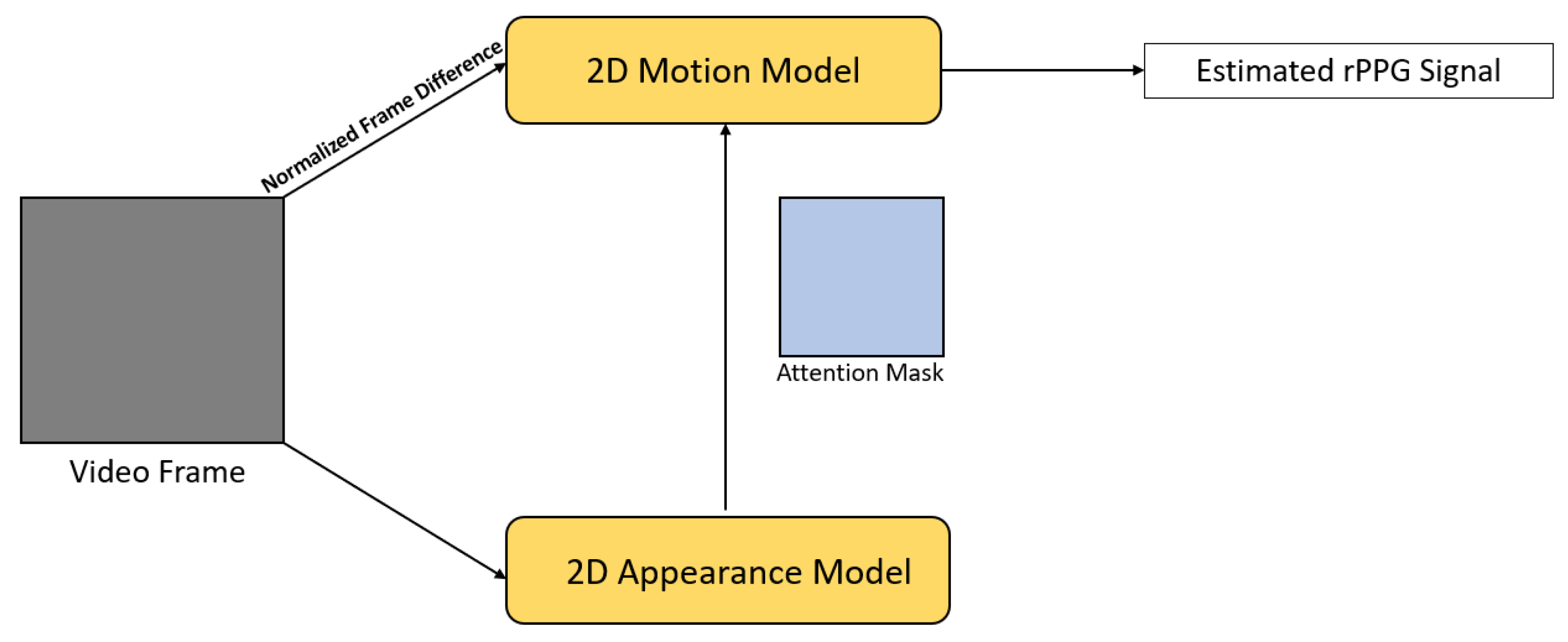
| [27] | 2018 | End-to-End | Normalized frame difference as motion representation, |
| attention mechanism was used to guide the motion model, | |||
| visualization of spatio-temporal distribution of physiological signals | |||
| [43] | 2018 | Hybrid | 2D CNN network for skin detection |
| [64] | 2018 | Hybrid | Spectrum images were used for HR estimation |
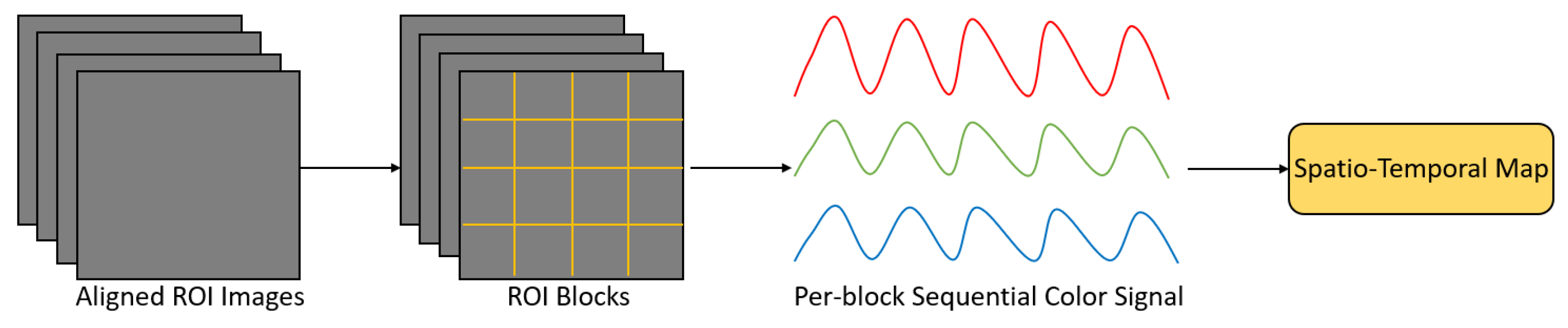
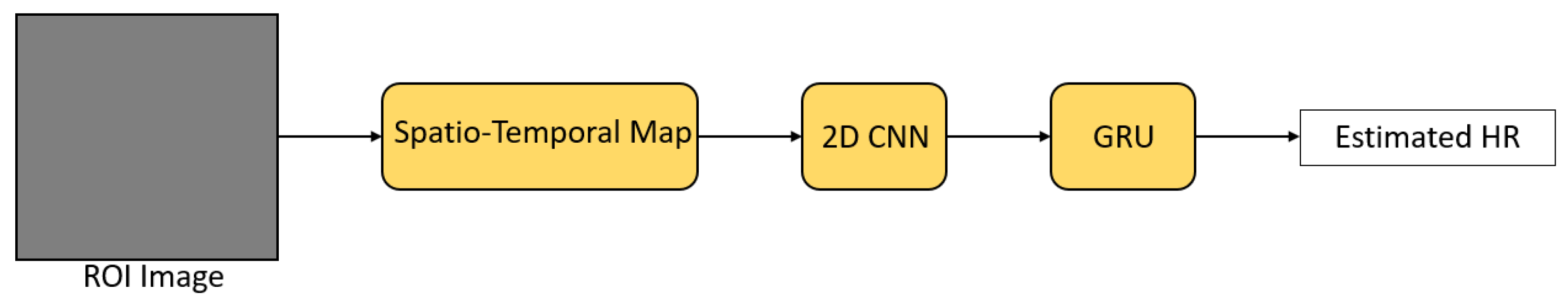
| [66] | 2018 | Hybrid | Spatio-temporal maps were used for HR estimation, |
| transfer learning approach to deal with data shortage | |||
| [29] | 2019 | End-to-End | Compared 3D CNN-based and RNN-based spatio-temporal network, |
| can estimate HR and HRV accurately | |||
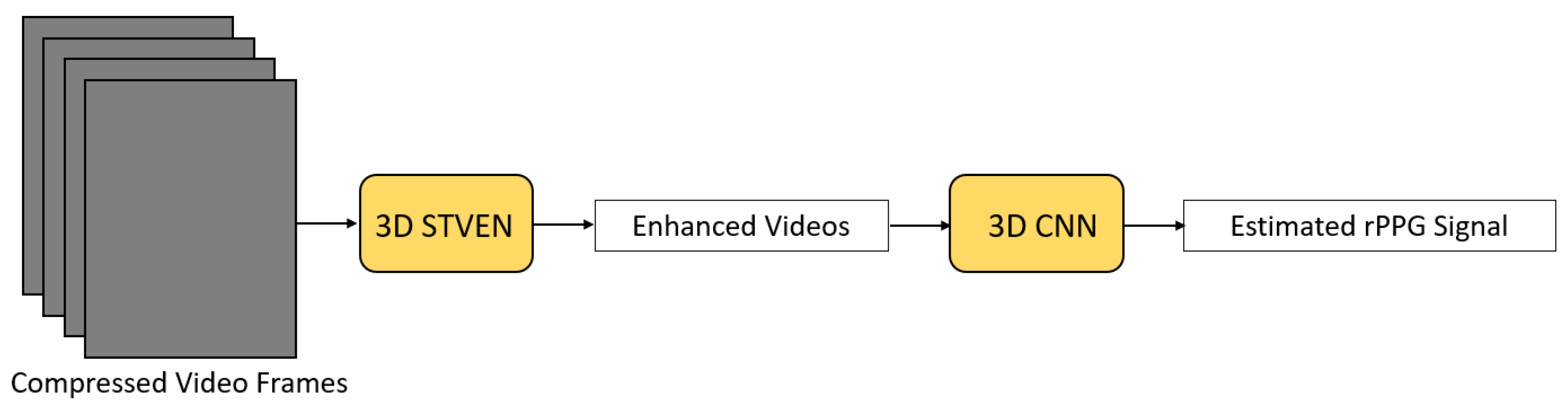
| [30] | 2019 | End-to-End | Enhancing video quality to deal with highly compressed videos, |
| can estimate HR and HRV accurately | |||
| [35] | 2019 | End-to-End | Attention mechanism was used to guide the trunk branch for signal extraction |
| [47] | 2019 | Hybrid | LSTM network for signal filtering, |
| transfer learning approach to deal with data shortage | |||
| [50] | 2019 | Hybrid | 3D CNN for signal extraction, |
| data augmentation method for generating videos with synthetic rPPG signals, | |||
| multilayer perceptron for HR estimation | |||
| [55] | 2019 | Hybrid | 2D CNN-based two-stream approach for signal extraction, |
| and LSTM network for signal refining | |||
| [67] | 2019 | Hybrid | Spatio-temporal maps were used for HR estimation, |
| attention mechanism was applied to remove noise | |||
| [28] | 2020 | End-to-End | Temporal shift module to model temporal information, |
| attention mechanism was applied to guide the motion model, | |||
| able to estimate HR and RR simultaneously by one network | |||
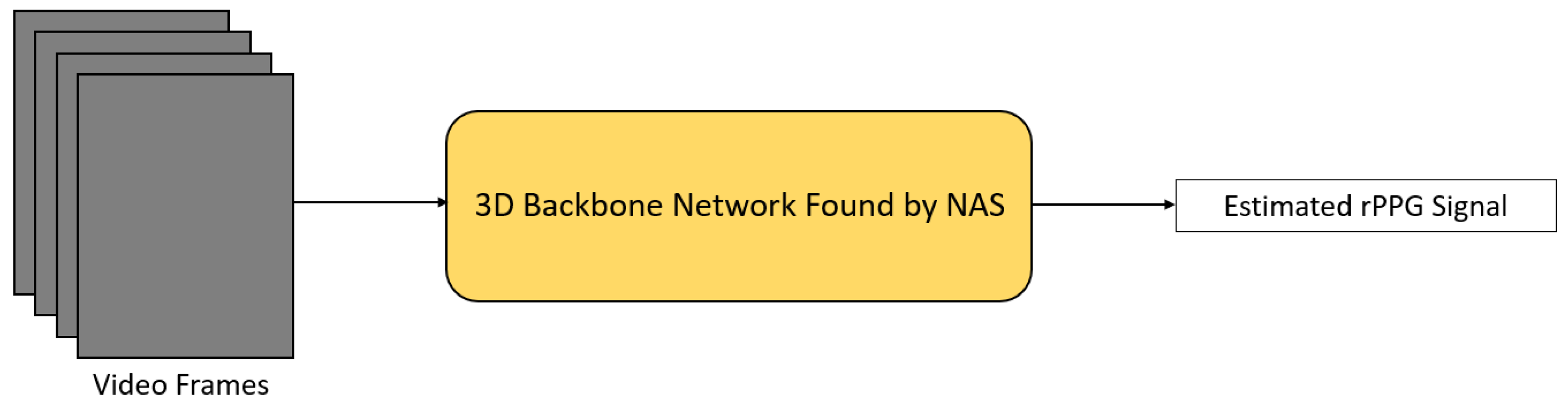
| [31] | 2020 | End-to-End | Used NAS to find a well-suited network for HR estimation |
| [44] | 2020 | Hybrid | 2D CNN encoder-decoder model for skin detection, |
| transfer learning approach to deal with data shortage | |||
| [45] | 2020 | Hybrid | Two GAN-style modules to enhance the detected ROI and remove noise, |
| 2D CNN for signal extraction | |||
| [48] | 2020 | Hybrid | LSTM network for signal filtering |
| [51] | 2020 | Hybrid | Siamese 3D CNN for signal extraction |
| [52] | 2020 | Hybrid | 3D CNN with attention mechanism for signal extraction, |
| feedforward neural network for HR estimation | |||
| [54] | 2020 | Hybrid | 3D CNN that can take different skin regions for signal extraction |
| [56] | 2020 | Hybrid | 2D CNN + LSTM spatio-temporal network for signal extraction |
| [57] | 2020 | Hybrid | 2D CNN + BiLSTM spatio-temporal network for signal extraction, |
| meta-learning approach for fast adaptation | |||
| [68] | 2020 | Hybrid | Spatio-temporal maps were used for HR estimation |
| [69] | 2020 | Hybrid | Spatio-temporal maps were used for HR estimation |
| [70] | 2020 | Hybrid | Spatio-temporal maps were used for HR estimation, |
| transfer learning approach to deal with data shortage | |||
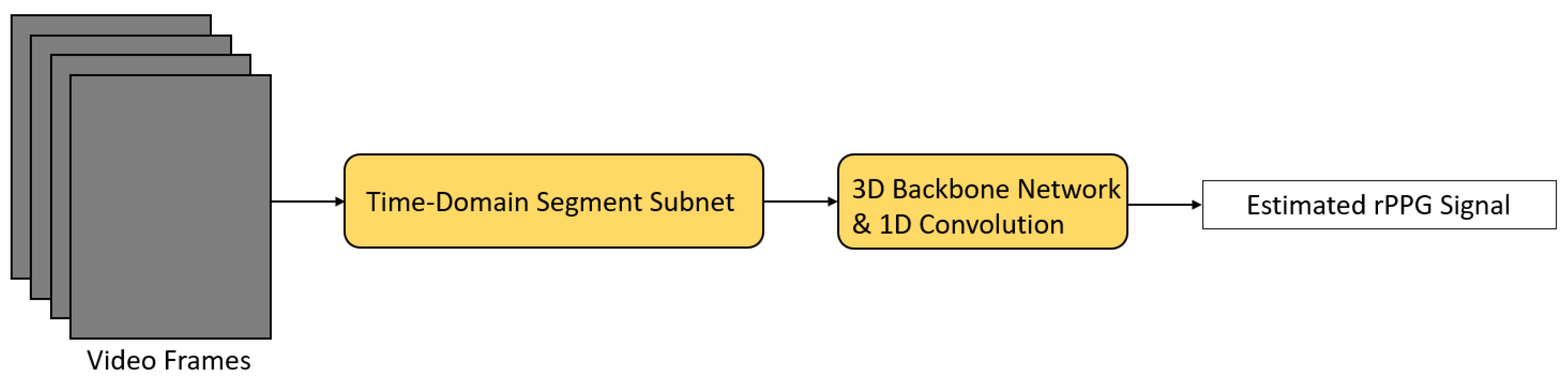
| [32] | 2021 | End-to-End | Avoid extracting redundant information from video segments, |
| attention mechanism was applied to deal with different noise | |||
| [33] | 2021 | End-to-End | An efficient framework for performing HR estimation quickly |
| [34] | 2021 | End-to-End | Dealt with the problem of extracting redundant video information, |
| attention mechanism was applied to learn important features and eliminate noise | |||
| [49] | 2021 | Hybrid | TS-CAN from another paper was utilized for signal extraction, |
| meta-learning approach for fast adaptation | |||
| [53] | 2021 | Hybrid | Multi-task framework for simultaneous signal extraction and data augmentation |
| [58] | 2021 | Hybrid | 3D CNN + LSTM spatio-temporal network for signal extraction |
| [59] | 2021 | Hybrid | GAN for generating high-quality rPPG signal from rough rPPG signal |
| [71] | 2021 | Hybrid | Spatio-temporal maps were used for HR estimation, |
| NAS was used to find a CNN for mapping spatio-temporal maps into HR |
| Dataset | Subjects | Description |
|---|---|---|
| AFRL [140] | 25 | 9 RGB cameras with 120 fps, resolution is 658 × 492, ECG, PPG, RR are recorded |
| COHFACE [141] | 40 | 1 RGB webcam with 20 fps, resolution is 640 × 480, BVP, RR are recorded |
| MAHNOB-HCI [142] | 27 | 1 RGB camera with 60 fps, 5 monochrome cameras with 60 fps, both resolution are 780 × 580, ECG, RR are recorded |
| MMSE-HR [143] | 140 | 1 3D stereo imaging sensor with 25 fps, 1 2D video sensor with 25 fps, 1 thermal sensor with 25 fps, RGB sensor resolution is 1040 × 1392, thermal sensor resolution is 640 × 480, HR, RR, BP are recorded |
| OBF [41] | 106 (6 with atrial fibrillation) | 1 RGB camera with 60 fps, 1 NIR camera with 30 fps, RGB camera resolution is 1920 × 1080, NIR camera resolution is 640 × 480, ECG, BVP, RR are recorded |
| PURE [144] | 10 | 1 RGB camera with 30 fps, resolution is 640 × 480, HR, SpO2, PPG are recorded |
| UBFC-RPPG [145] | 42 | 1 RGB webcam with 30 fps, resolution is 640 × 480, HR, PPG are recorded |
| VIPL-HR [146] | 107 | 1 RGB webcam with 25 fps, 1 RGB-D camera with 30 fps, 1 smartphone camera with 30 fps, RGB webcam resolution is 960 × 720, RGB-D NIR camera resolution is 640 × 480, RGB-D RGB camera resolution is 1920 × 1080, smartphone camera resolution is 1920 × 1080, HR, SpO2, BVP are recorded |
| Methods | AFRL | COHFACE | MAHNOB-HCI | MMSE-HR | OBF | PURE | UBFC-RPPG | VIPL-HR |
|---|---|---|---|---|---|---|---|---|
| [26] | X | RMSE = 10.78 | RMSE = 9.24 | X | X | RMSE = 2.37 | X | X |
| MAE = 8.10 | MAE = 7.25 | MAE = 1.84 | ||||||
| R = 0.29 | R = 0.51 | R = 0.98 | ||||||
| [27] | MAE = 2.45 | X | MAE = 4.57 | X | X | X | X | X |
| SNR = 4.65 | SNR = −8.98 | |||||||
| [64] | X | X | RMSE = 4.26 | X | X | X | X | X |
| R = 0.81 | ||||||||
| [66] | X | X | RMSE = 4.49 | RMSE = 6.83 | X | X | X | X |
| [29] | X | X | RMSE = 7.88 | X | RMSE = 1.812 | X | X | X |
| MAE = 5.96 | R = 0.992 | |||||||
| R = 0.76 | ||||||||
| [30] | X | X | RMSE = 5.93 | X | RMSE = 1.8 | X | X | X |
| MAE = 4.03 | R = 0.992 | |||||||
| R = 0.88 | ||||||||
| [35] | X | RMSE = 11.88 | X | X | X | RMSE = 1.58 | X | X |
| MAE = 7.31 | MAE = 0.88 | X | X | |||||
| R = 0.36 | R = 0.99 | X | X | |||||
| SNR = −1.93 | SNR = 9.18 | X | X | |||||
| [47] | X | X | X | RMSE = 3.187 | X | X | X | X |
| MAE = 4.35 | ||||||||
| R = 0.8254 | ||||||||
| [50] | X | X | X | X | X | X | RMSE = 8.64 | X |
| MAE = 5.45 | X | |||||||
| [55] | X | RMSE = 9.96 | X | X | X | X | X | X |
| MAE = 8.09 | ||||||||
| R = 0.40 | ||||||||
| [67] | X | X | X | RMSE = 10.10 | X | X | X | RMSE = 7.99 |
| R = 0.64 | MAE = 5.40 | |||||||
| R = 0.66 | ||||||||
| [28] | RMSE = 3.72 | X | X | RMSE = 5.66 | X | X | X | X |
| MAE = 1.45 | MAE = 3.00 | |||||||
| R = 0.94 | R = 0.92 | |||||||
| SNR = 8.64 | SNR = 2.37 | |||||||
| [31] | X | X | RMSE = 5.10 | RMSE = 5.87 | X | X | X | RMSE = 8.68 |
| MAE = 3.78 | R = 0.89 | MAE = 5.68 | ||||||
| R = 0.86 | R = 0.72 | |||||||
| [45] | X | X | RMSE = 3.41 | X | X | X | X | X |
| R = 0.92 | ||||||||
| [48] | X | X | X | MAE = 1.31 | X | X | X | X |
| SNR = 9.44 | X | X | X | X | ||||
| [51] | X | RMSE = 1.29 | X | X | X | RMSE = 1.56 | RMSE = 0.97 | X |
| MAE = 0.70 | MAE = 0.51 | MAE = 0.48 | ||||||
| R = 0.73 | R = 0.83 | |||||||
| [52] | X | X | X | X | X | X | RMSE = 3.368 | X |
| MAE = 2.412 | ||||||||
| R = 0.983 | ||||||||
| [54] | X | RMSE = 7.06 | RMSE = 6.26 | X | X | RMSE = 0.43 | X | X |
| MAE = 3.07 | MAE = 4.81 | MAE = 0.28 | ||||||
| R = 0.86 | R = 0.79 | R = 0.999 | ||||||
| [57] | X | X | RMSE = 3.68 | X | X | X | RMSE = 7.42 | X |
| MAE = 3.01 | MAE = 5.97 | |||||||
| R = 0.85 | R = 0.53 | |||||||
| [68] | X | X | RMSE = 3.99 | RMSE = 5.49 | X | X | X | RMSE = 8.14 |
| R = 0.87 | R = 0.84 | MAE = 5.30 | ||||||
| R = 0.76 | ||||||||
| [69] | X | X | X | RMSE = 6.04 | RMSE = 1.26 | X | X | RMSE = 7.97 |
| R = 0.84 | R = 0.996 | MAE = 5.02 | ||||||
| R = 0.796 | ||||||||
| [70] | X | X | RMSE = 3.23 | X | X | X | X | X |
| MAE = 1.53 | ||||||||
| R = 0.97 | ||||||||
| [32] | X | RMSE = 7.52 | X | X | X | RMSE = 1.21 | X | X |
| MAE = 5.19 | MAE = 0.74 | |||||||
| R = 0.68 | R = 1.00 | |||||||
| [33] | X | RMSE = 9.50 | X | X | X | X | RMSE = 3.82 | X |
| MAE = 5.57 | MAE = 2.15 | |||||||
| R = 0.75 | R = 0.97 | |||||||
| [34] | X | RMSE = 6.65 | X | RMSE = 5.84 | X | RMSE = 0.77 | RMSE = 3.97 | X |
| MAE = 4.67 | R = 0.85 | MAE = 0.34 | MAE = 1.46 | |||||
| R = 0.77 | R = 0.99 | R = 0.93 | ||||||
| [49] | X | X | X | RMSE = 3.12 | X | X | RMSE = 3.12 | X |
| MAE = 1.87 | MAE = 2.46 | |||||||
| R = 0.89 | R = 0.96 | |||||||
| [53] | X | RMSE = 1.65 | X | X | X | RMSE = 1.07 | RMSE = 2.09 | X |
| MAE = 0.68 | MAE = 0.40 | MAE = 0.47 | ||||||
| R = 0.72 | R = 0.92 | |||||||
| [58] | X | X | RMSE = 6.42 | X | X | X | RMSE = 7.24 | X |
| MAE = 5.01 | MAE = 5.29 | |||||||
| [59] | X | X | RMSE = 6.53 | X | X | RMSE = 4.29 | RMSE = 2.10 | X |
| MAE = 4.15 | MAE = 2.28 | MAE = 1.19 | ||||||
| R = 0.71 | R = 0.99 | R = 0.98 | ||||||
| [71] | X | X | X | X | X | RMSE = 2.02 | X | RMSE = 8.01 |
| MAE = 1.65 | MAE = 5.12 | |||||||
| R = 0.99 | R = 0.79 |
Publisher’s Note: MDPI stays neutral with regard to jurisdictional claims in published maps and institutional affiliations. |
© 2021 by the authors. Licensee MDPI, Basel, Switzerland. This article is an open access article distributed under the terms and conditions of the Creative Commons Attribution (CC BY) license (https://creativecommons.org/licenses/by/4.0/).
Share and Cite
Cheng, C.-H.; Wong, K.-L.; Chin, J.-W.; Chan, T.-T.; So, R.H.Y. Deep Learning Methods for Remote Heart Rate Measurement: A Review and Future Research Agenda. Sensors 2021, 21, 6296. https://doi.org/10.3390/s21186296
Cheng C-H, Wong K-L, Chin J-W, Chan T-T, So RHY. Deep Learning Methods for Remote Heart Rate Measurement: A Review and Future Research Agenda. Sensors. 2021; 21(18):6296. https://doi.org/10.3390/s21186296
Chicago/Turabian StyleCheng, Chun-Hong, Kwan-Long Wong, Jing-Wei Chin, Tsz-Tai Chan, and Richard H. Y. So. 2021. "Deep Learning Methods for Remote Heart Rate Measurement: A Review and Future Research Agenda" Sensors 21, no. 18: 6296. https://doi.org/10.3390/s21186296