
A Review of Deep Learning-Based Contactless Heart Rate Measurement Methods


Abstract
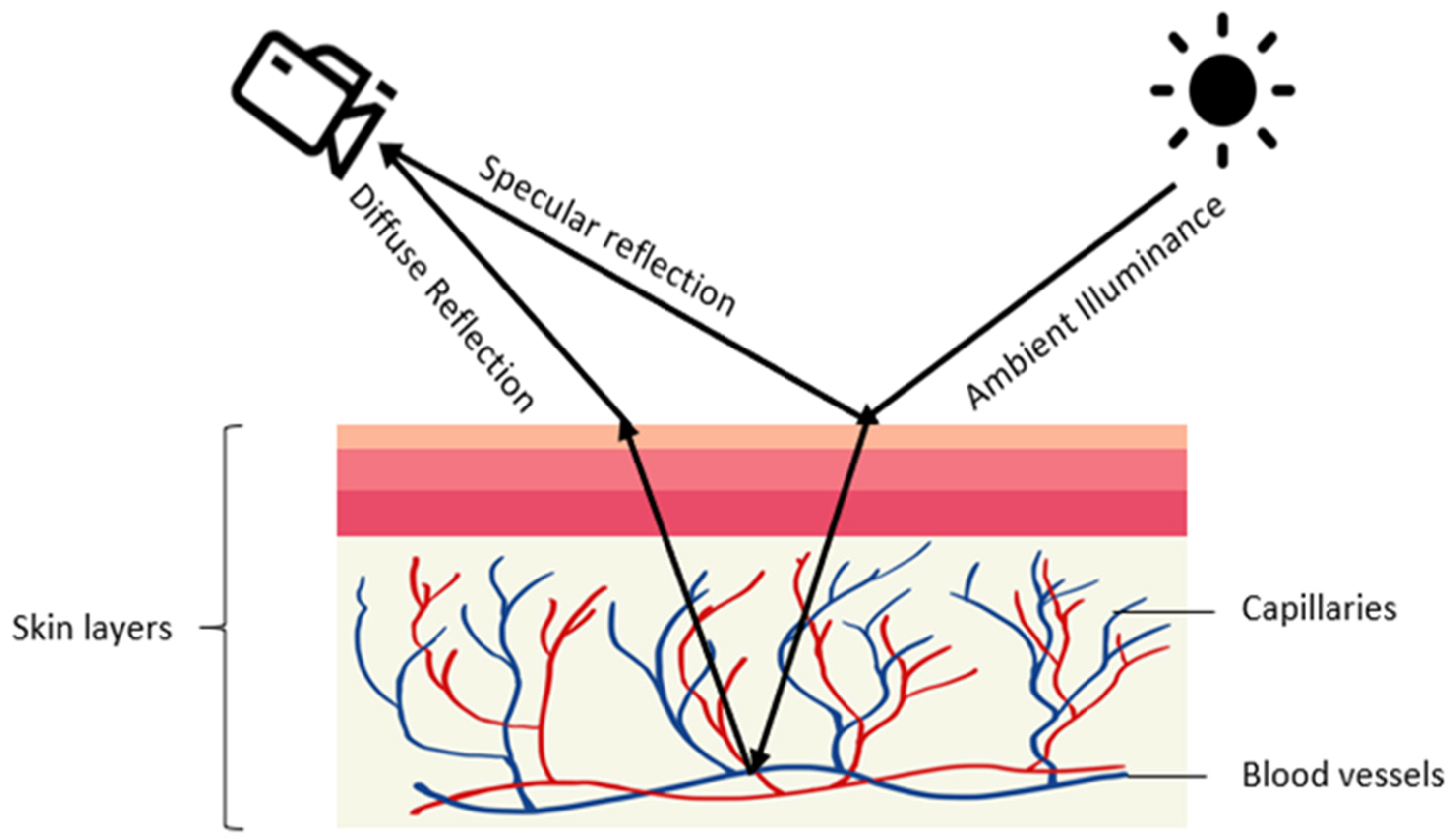
:1. Introduction

| Emphasis | Ref | Year | Task |
|---|---|---|---|
| Contactless | [66] | 2018 | Provides typical components of rPPG and notes the main challenges; groups published studies by their choice of algorithm. |
| Contactless | [67] | 2012 | Covers three main stages of monitoring physiological measurements based on photoplethysmographic imaging: image acquisition, data collection, and parameter extraction. |
| Contactless and contact | [68] | 2016 | States review of contact-based PPG and its limitations; introduces research activities on wearable and non-contact PPG. |
| Contactless and contact | [69] | 2009 | Reviews photoplethysmographic measurement techniques from contact sensing placement to non-contact sensing placement, and from point measurement to imaging measurement. |
| Contactless newborn infants | [28] | 2013 | Investigates the feasibility of camera-based PPG for contactless HR monitoring in newborn infants with ambient light. |
| Contactless newborn infants | [30] | 2016 | Comparative analysis to benchmark state-of-the-art video and image-guided noninvasive pulse rate (PR) detection. |
| Contactless and contact | [70] | 2017 | Heart rate measurement using facial videos based on photoplethysmography and ballistocardiography. |
| Contactless and contact | [71] | 2014 | Covers methods of non-contact HR measurement with capacitively coupled ECG, Doppler radar, optical vibrocardiography, thermal imaging, RGB camera, and HR from speech. |
| Contactless RR and contact | [72] | 2011 | Discusses respiration monitoring approaches (both contact and non-contact). |
| Contactless newborn infants | [31] | 2019 | Addresses HR measurement in babies. |
| Contactless | [73] | 2019 | Examines challenges associated with illumination variations and motion artifacts. |
| Contactless | [74] | 2017 | Covers HR measurement techniques including camera-based photoplethysmography, reflectance pulse oximetry, laser Doppler technology, capacitive sensors, piezoelectric sensors, electromyography, and a digital stethoscope. |
| Contactless Main challenges | [75] | 2015 | Covers issues in motion and ambient lighting tolerance, image optimization (including multi-spectral imaging), and region of interest optimization. |
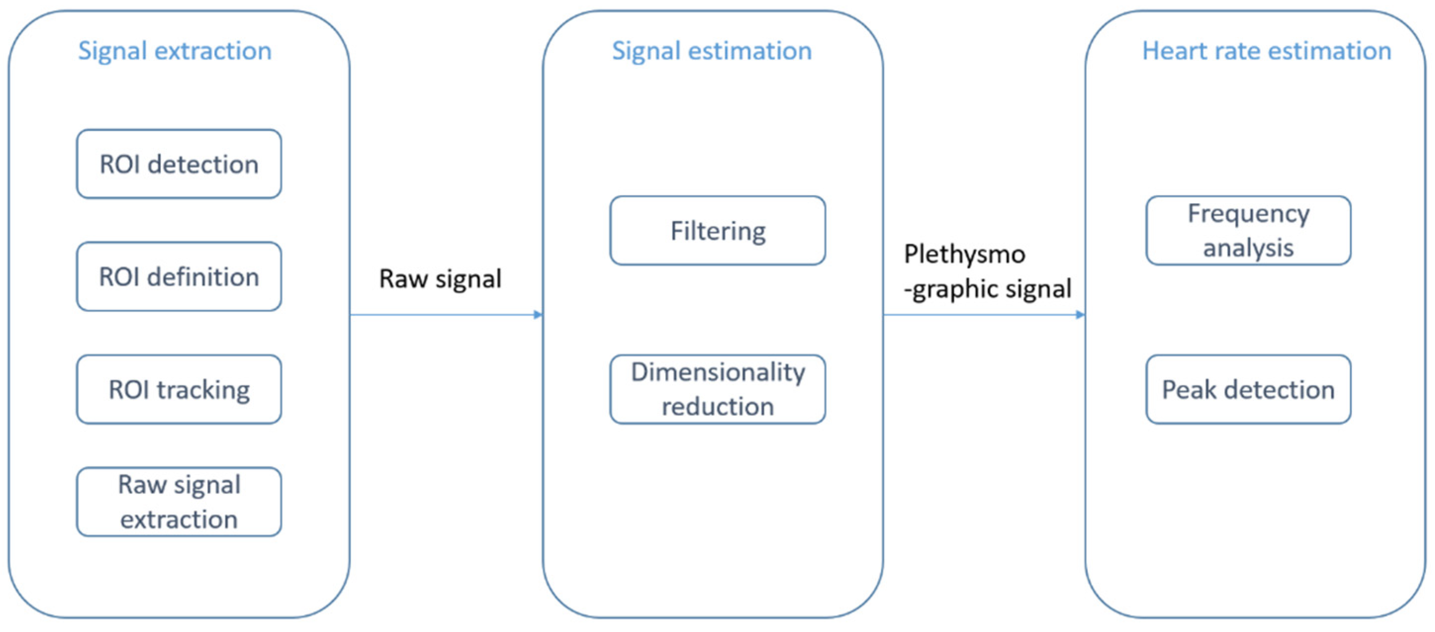
2. Contactless PPG Methods Based on Deep Learning
2.1. Combination of Conventional and Deep Learning Methods
2.1.1. Deep Learning Methods for Signal Estimation
2.1.2. Deep Learning Methods for Signal Extraction
2.2. End-to-End Deep Learning Methods
2.2.1. VGG-Style CNN
2.2.2. CNN-LSTM Network
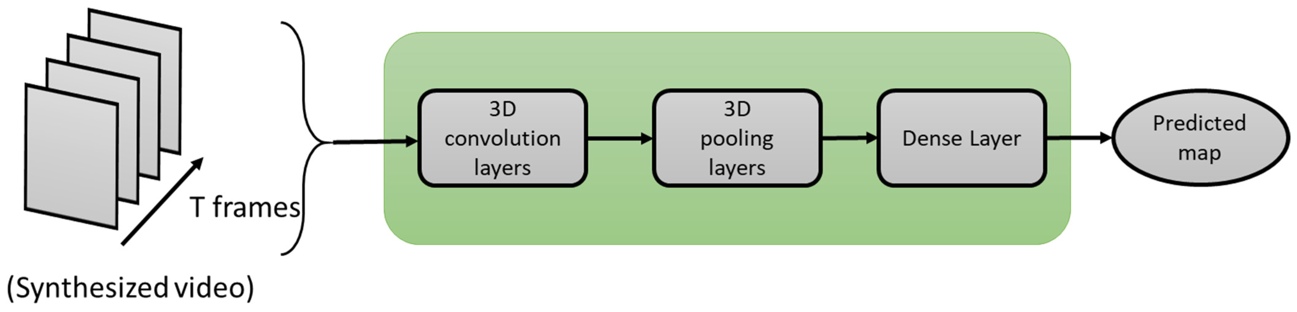
2.2.3. 3D-CNN Network
3. Selected Deep Learning Models for Comparison
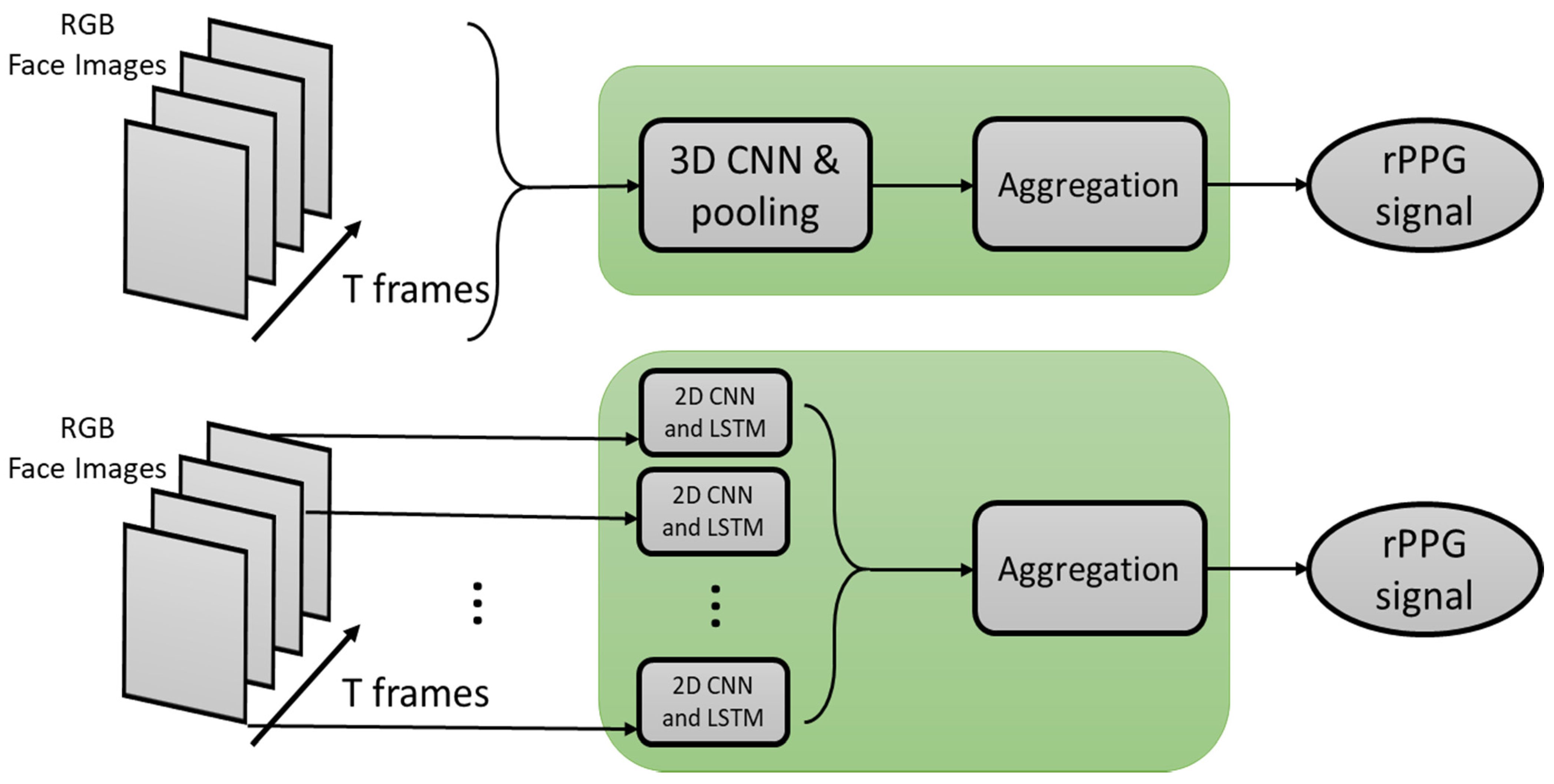
3.1. STVEN-rPPGNet
3.2. IPPG-3D-CNN
3.3. PhysNet
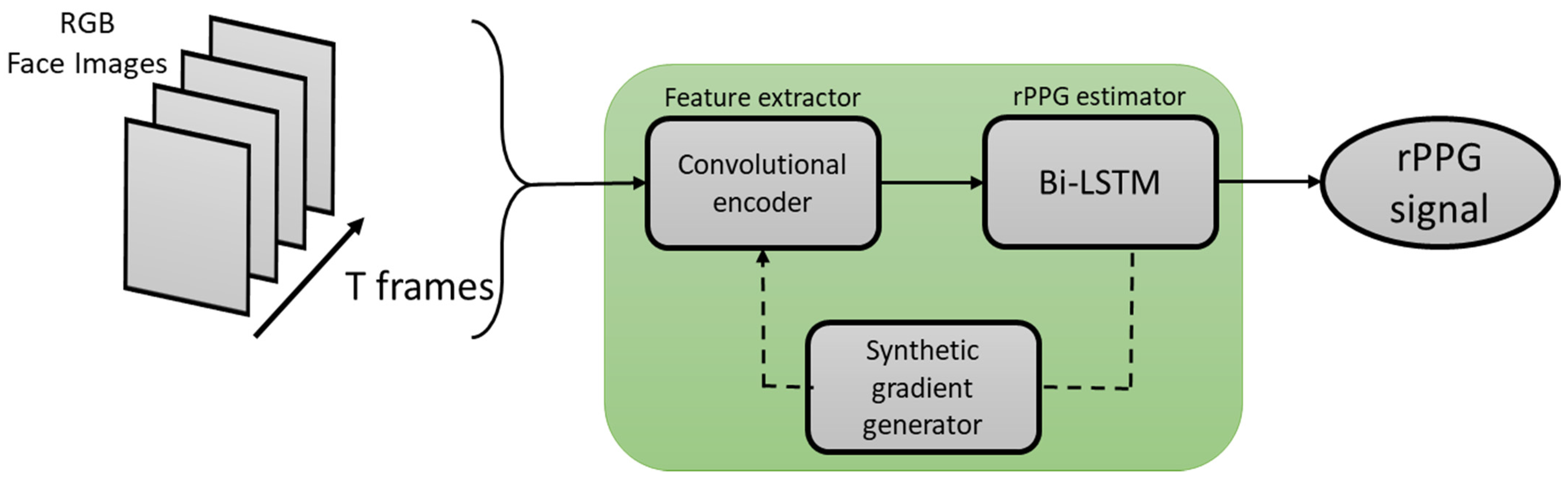
3.4. Meta-rPPG
4. Comparison Results and Discussion
4.1. Dataset
4.2. Experimental Setup
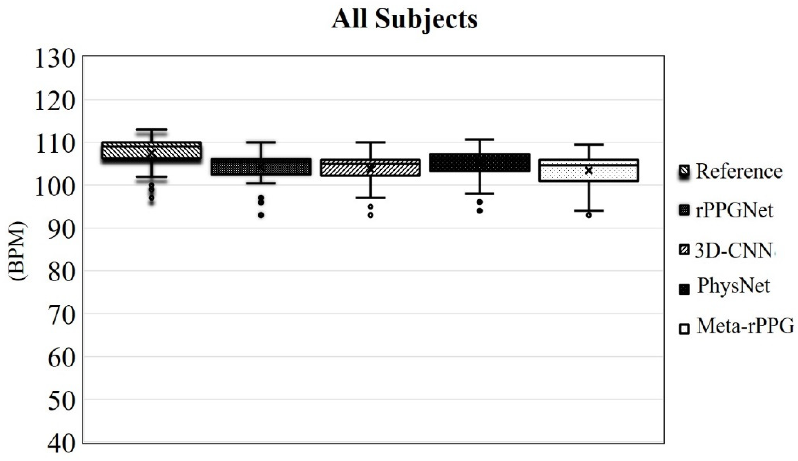
4.3. Results and Discussion
5. Conclusions
Author Contributions
Funding
Institutional Review Board Statement
Informed Consent Statement
Data Availability Statement
Conflicts of Interest
References
- Challoner, A.V.; Ramsay, C.A. A photoelectric plethysmograph for the measurement of cutaneous blood flow. Phys. Med. Biol. 1974, 19, 317–328. [Google Scholar] [CrossRef]
- Alian, A.A.; Shelley, K.H. Photoplethysmography. Best Pract. Res. Clin. Anaesthesiol. 2014, 28, 395–406. [Google Scholar] [CrossRef]
- Shelley, K.H. Photoplethysmography: Beyond the calculation of arterial oxygen saturation and heart rate. Anesth. Analg. 2007, 105, S31–S36. [Google Scholar] [CrossRef] [PubMed] [Green Version]
- Madhav, K.V.; Ram, M.R.; Krishna, E.H.; Reddy, K.N.; Reddy, K.A. Estimation of respiratory rate from principal components of photoplethysmographic signals. In Proceedings of the IEEE EMBS Conference on Biomedical Engineering and Sciences (IECBES), Kuala Lumpur, Malaysia, 30 November 2010; pp. 311–314. [Google Scholar] [CrossRef]
- Karlen, W.; Raman, S.; Ansermino, J.M.; Dumont, G.A. Multiparameter respiratory rate estimation from the photoplethysmogram. IEEE Transact. Biomed. Eng. 2013, 60, 1946–1953. [Google Scholar] [CrossRef]
- Yousefi, R.; Nourani, M. Separating arterial and venous-related components of photoplethysmographic signals for accurate extraction of oxygen saturation and respiratory rate. IEEE J. Biomed. Health Inf. 2014, 19, 848–857. [Google Scholar]
- Kinnunen, H.; Rantanen, A.; Kenttä, T.; Koskimäki, H. Feasible assessment of recovery and cardiovascular health: Accuracy of nocturnal HR and HRV assessed via ring PPG in comparison to medical grade ECG. Physiol. Meas. 2020, 41, 04NT01. [Google Scholar] [CrossRef]
- Orphanidou, C. Derivation of respiration rate from ambulatory ECG and PPG using ensemble empirical mode decomposition: Comparison and fusion. Comput. Biol. Med. 2017, 81, 45–54. [Google Scholar] [CrossRef]
- Clifton, D.A.; Meredith, D.; Villarroel, M.; Tarassenko, L. Home monitoring: Breathing rate from PPG and ECG. Inst. Biomed. Eng. 2012. Available online: http://www.robots.ox.ac.uk/~davidc/pubs/WT2012.pdf (accessed on 26 May 2021).
- Madhav, K.V.; Raghuram, M.; Krishna, E.H.; Komalla, N.R.; Reddy, K.A. Extraction of respiratory activity from ECG and PPG signals using vector autoregressive model. In Proceedings of the 2012 IEEE International Symposium on Medical Measurements and Applications Proceedings, Budapest, Hungary, 18–19 May 2012; pp. 1–4. [Google Scholar]
- Gu, W.B.; Poon, C.C.; Leung, H.K.; Sy, M.Y.; Wong, M.Y.; Zhang, Y.T. A novel method for the contactless and continuous measurement of arterial blood pressure on a sleeping bed. In Proceedings of the 2009 Annual International Conference of the IEEE Engineering in Medicine and Biology Society, Minneapolis, MN, USA, 3–6 September 2009; pp. 6084–6086. [Google Scholar]
- Wang, L.; Lo BP, L.; Yang, G.Z. Multichannel reflective PPG earpiece sensor with passive motion cancellation. IEEE Transact. Biomed. Circ. Syst. 2007, 1, 235–241. [Google Scholar] [CrossRef] [PubMed]
- Kabiri Ameri, S.; Ho, R.; Jang, H.; Tao, L.; Wang, Y.; Wang, L.; Schnyer, D.M.; Akinwande, D.; Lu, N. Graphene electronic tattoo sensors. ACS Nano 2017, 11, 7634–7641. [Google Scholar] [CrossRef] [PubMed]
- Nardelli, M.; Vanello, N.; Galperti, G.; Greco, A.; Scilingo, E.P. Assessing the Quality of Heart Rate Variability Estimated from Wrist and Finger PPG: A Novel Approach Based on Cross-Mapping Method. Sensors 2020, 20, 3156. [Google Scholar] [CrossRef] [PubMed]
- Phan, D.; Siong, L.Y.; Pathirana, P.N. Smartwatch: Performance evaluation for long-term heart rate monitoring. In Proceedings of the 2015 International Symposium on Bioelectronics and Bioinformatics (ISBB), Beijing, China, 14–17 October 2015; pp. 144–147. [Google Scholar]
- Wong, M.Y.; Leung, H.K.; Pickwell-MacPherson, E.; Gu, W.B.; Zhang, Y.T. Contactless recording of photoplethysmogram on a sleeping bed. In Proceedings of the 2009 Annual International Conference of the IEEE Engineering in Medicine and Biology Society, Beijing, China, 14–17 October 2009; pp. 907–910. [Google Scholar]
- Allen, J. Photoplethysmography and its application in clinical physiological measurement. Physiol. Meas. 2007, 28, R1. [Google Scholar] [CrossRef] [PubMed] [Green Version]
- Charlton, P.H.; Birrenkott, D.A.; Bonnici, T.; Pimentel, M.A.; Johnson, A.E.; Alastruey, J.; Tarassenko, L.; Watkinson, P.J.; Beale, R.; Clifton, D.A. Breathing rate estimation from the electrocardiogram and photoplethys-mogram: A review. IEEE Rev. Biomed. Eng. 2017, 11, 2–20. [Google Scholar] [CrossRef] [Green Version]
- Schäfer, A.; Vagedes, J. How accurate is pulse rate variability as an estimate of heart rate variability?: A review on studies comparing photoplethysmographic technology with an electrocardiogram. Int. J. Cardiol. 2013, 166, 15–29. [Google Scholar] [CrossRef]
- Biswas, D.; Simoes-Capela, N.; Van Hoof, C.; Van Helleputte, N. Heart Rate Estimation From Wrist-Worn Photoplethysmography: A Review. IEEE Sens. J. 2019, 19, 6560–6570. [Google Scholar] [CrossRef]
- Castaneda, D.; Esparza, A.; Ghamari, M.; Soltanpur, C. A review on wearable photoplethysmography sensors and their potential future applications in health care. Int. J. Biosensors Bioelectron. 2018, 4, 195. [Google Scholar]
- Pereira, T.; Tran, N.; Gadhoumi, K.; Pelter, M.M.; Do, D.H.; Lee, R.J.; Colorado, R.; Meisel, K.; Hu, X. Photoplethysmography based atrial fibrillation detection: A review. npj Digit. Med. 2020, 3, 1–12. [Google Scholar] [CrossRef] [Green Version]
- Nye, R.; Zhang, Z.; Fang, Q. Continuous non-invasive blood pressure monitoring using photoplethysmography: A review. In Proceedings of the 2015 International Symposium on Bioelectronics and Bioinformatics (ISBB), Beijing, China, 14–17 October 2015; pp. 176–179. [Google Scholar]
- Johansson, A. Neural network for photoplethysmographic respiratory rate monitoring. Med. Biol. Eng. Comput. 2003, 41, 242–248. [Google Scholar] [CrossRef]
- Panwar, M.; Gautam, A.; Biswas, D.; Acharyya, A. PP-Net: A Deep Learning Framework for PPG-Based Blood Pressure and Heart Rate Estimation. IEEE Sens. J. 2020, 20, 10000–10011. [Google Scholar] [CrossRef]
- Biswas, D.; Everson, L.; Liu, M.; Panwar, M.; Verhoef, B.-E.; Patki, S.; Kim, C.H.; Acharyya, A.; Van Hoof, C.; Konijnenburg, M.; et al. CorNET: Deep Learning Framework for PPG-Based Heart Rate Estimation and Biometric Identification in Ambulant Environment. IEEE Trans. Biomed. Circ. Syst. 2019, 13, 282–291. [Google Scholar] [CrossRef]
- Chang, X.; Li, G.; Xing, G.; Zhu, K.; Tu, L. DeepHeart. ACM Trans. Sens. Netw. 2021, 17, 1–18. [Google Scholar] [CrossRef]
- Aarts, L.A.; Jeanne, V.; Cleary, J.P.; Lieber, C.; Nelson, J.S.; Oetomo, S.B.; Verkruysse, W. Non-contact heart rate monitoring utilizing camera photoplethysmography in the neonatal intensive care unit—A pilot study. Early Hum. Dev. 2013, 89, 943–948. [Google Scholar] [CrossRef]
- Villarroel, M.; Chaichulee, S.; Jorge, J.; Davis, S.; Green, G.; Arteta, C.; Zisserman, A.; McCormick, K.; Watkinson, P.; Tarassenko, L. Non-contact physiological monitoring of preterm infants in the Neonatal Intensive Care Unit. NPJ Digit. Med. 2019, 2, 128. [Google Scholar] [CrossRef] [Green Version]
- Sikdar, A.; Behera, S.K.; Dogra, D.P. Computer-vision-guided human pulse rate estimation: A review. IEEE Rev. Bio Med. Eng. 2016, 9, 91–105. [Google Scholar] [CrossRef]
- Anton, O.; Fernandez, R.; Rendon-Morales, E.; Aviles-Espinosa, R.; Jordan, H.; Rabe, H. Heart Rate Monitoring in Newborn Babies: A Systematic Review. Neonatology 2019, 116, 199–210. [Google Scholar] [CrossRef] [PubMed]
- Fernández, A.; Carús, J.L.; Usamentiaga, R.; Alvarez, E.; Casado, R. Unobtrusive health monitoring system using video-based physiological in-formation and activity measurements. IEEE 2013, 89, 943–948. [Google Scholar]
- Haque, M.A.; Irani, R.; Nasrollahi, K.; Moeslund, T.B. Heartbeat rate measurement from facial video. IEEE Intell. Syst. 2016, 31, 40–48. [Google Scholar] [CrossRef] [Green Version]
- Kumar, M.; Veeraraghavan, A.; Sabharwal, A. DistancePPG: Robust non-contact vital signs monitoring using a camera. Biomed. Opt. Express 2015, 6, 1565–1588. [Google Scholar] [CrossRef] [PubMed] [Green Version]
- Liu, S.Q.; Lan, X.; Yuen, P.C. Remote photoplethysmography correspondence feature for 3D mask face presentation attack detection. In Proceedings of the European Conference on Computer Vision, ECCV Papers, Munich, Germany, 8–14 September 2018; pp. 558–573. [Google Scholar] [CrossRef]
- Gudi, A.; Bittner, M.; Lochmans, R.; van Gemert, J. Efficient real-time camera based estimation of heart rate and its variability. In Proceedings of the IEEE International Conference on Computer Vision Workshops, Seoul, Korea, 27–28 October 2019. [Google Scholar]
- Verkruysse, W.; Svaasand, L.O.; Nelson, J.S. Remote plethysmographic imaging using ambient light. Opt. Express 2008, 16, 21434–21445. [Google Scholar] [CrossRef] [PubMed] [Green Version]
- Balakrishnan, G.; Durand, F.; Guttag, J. Detecting Pulse from Head Motions in Video. In Proceedings of the 2013 IEEE Conference on Computer Vision and Pattern Recognition, Portland, OR, USA, 25–27 June 2013; pp. 3430–3437. [Google Scholar]
- Lewandowska, M.; Rumiński, J.; Kocejko, T.; Nowak, J. Measuring pulse rate with a webcam—A non-contact method for evaluating cardiac activity. In Proceedings of the 2011 Federated Conference on Computer Science and Information Systems (FedCSIS), Szczecin, Poland, 18–21 September 2011; pp. 405–410. [Google Scholar]
- De Haan, G.; Jeanne, V. Robust pulse rate from chrominance-based rPPG. IEEE Transact. Biomed. Eng. 2013, 60, 2878–2886. [Google Scholar] [CrossRef]
- Tasli, H.E.; Gudi, A.; den Uyl, M. Remote PPG based vital sign measurement using adaptive facial regions. In Proceedings of the 2014 IEEE International Conference on Image Processing (ICIP), Paris, France, 27–30 October 2014; pp. 1410–1414. [Google Scholar]
- Yu, Y.P.; Kwan, B.H.; Lim, C.L.; Wong, S.L. Video-based heart rate measurement using short-time Fourier transform. In Proceedings of the 2013 International Symposium on Intelligent Signal Processing and Communication Systems, Naha, Japan, 12–15 November 2013; pp. 704–707. [Google Scholar]
- Monkaresi, H.; Hussain, M.S.; Calvo, R.A. Using Remote Heart Rate Measurement for Affect Detection. In Proceedings of the FLAIRS Conference, Sydney, Australia, 3 May 2014. [Google Scholar]
- Monkaresi, H.; Calvo, R.A.; Yan, H. A Machine Learning Approach to Improve Contactless Heart Rate Monitoring Using a Webcam. IEEE J. Biomed. Health Inf. 2013, 18, 1153–1160. [Google Scholar] [CrossRef]
- Viola, P.; Jones, M.J.C. Rapid object detection using a boosted cascade of simple features. In Proceedings of the 2001 IEEE Computer Society Conference on Computer Vision and Pattern Recognition (CVPR 2001), Kauai, HI, USA, 8–14 December 2001; Volume 1, p. 3. [Google Scholar]
- Špetlík, R.; Franc, V.; Matas, J. Visual heart rate estimation with convolutional neural network. In Proceedings of the British Machine Vision Conference, Newcastle, UK, 3–6 September 2018; pp. 3–6. [Google Scholar]
- Wei, L.; Tian, Y.; Wang, Y.; Ebrahimi, T.; Huang, T. Automatic webcam-based human heart rate measurements using laplacian eigenmap. In Proceedings of the Asian Conference on Computer Vision, Daejeon, Korea, 5–9 November 2012; Springer: Berlin/Heidelberg, Germany, 2012; pp. 281–292. [Google Scholar]
- Xu, S.; Sun, L.; Rohde, G.K. Robust efficient estimation of heart rate pulse from video. Biomed. Opt. Express 2014, 5, 1124–1135. [Google Scholar] [CrossRef] [Green Version]
- Hsu, Y.C.; Lin, Y.L.; Hsu, W. Learning-based heart rate detection from remote photoplethysmography features. In Proceedings of the 2014 IEEE International Conference on Acoustics, Speech and Signal Processing (ICASSP), Florence, Italy, 4–9 May 2014; pp. 4433–4437. [Google Scholar]
- Lee, K.Z.; Hung, P.C.; Tsai, L.W. Contact-free heart rate measurement using a camera. In Proceedings of the 2012 Ninth Conference on Computer and Robot Vision, Toronto, ON, Canada, 28–30 May 2012; pp. 147–152. [Google Scholar]
- Poh, M.Z.; McDuff, D.J.; Picard, R.W. Non-contact, automated cardiac pulse measurements using video imaging and blind source separation. Opt. Express 2010, 18, 10762–10774. [Google Scholar] [CrossRef]
- Poh, M.Z.; McDuff, D.J.; Picard, R.W. Advancements in noncontact, multiparameter physiological measurements using a webcam. IEEE Transact. Biomed. Eng. 2010, 58, 7–11. [Google Scholar] [CrossRef] [Green Version]
- Li, X.; Chen, J.; Zhao, G.; Pietikainen, M. Remote heart rate measurement from face videos under realistic situations. In Proceedings of the IEEE Conference on Computer Vision and Pattern Recognition, Columbus, OH, USA, 23–28 June 2014; pp. 4264–4271. [Google Scholar]
- Lee, K.; Lee, J.; Ha, C.; Han, M.; Ko, H. Video-Based Contactless Heart-Rate Detection and Counting via Joint Blind Source Separation with Adaptive Noise Canceller. Appl. Sci. 2019, 9, 4349. [Google Scholar] [CrossRef] [Green Version]
- Kwon, S.; Kim, H.; Park, K.S. Validation of heart rate extraction using video imaging on a built-in camera system of a smartphone. In Proceedings of the 2012 Annual International Conference of the IEEE Engineering in Medicine and Biology Society, San Diego, CA, USA, 28 August–1 September 2012; pp. 2174–2177. [Google Scholar]
- Datcu, D.; Cidota, M.; Lukosch, S.; Rothkrantz, L. Noncontact automatic heart rate analysis in visible spectrum by specific face regions. In Proceedings of the 14th International Conference on Computer Systems and Technologies, Ruse, Bulgaria, 28–29 June 2013; pp. 120–127. [Google Scholar]
- Holton, B.D.; Mannapperuma, K.; Lesniewski, P.J.; Thomas, J.C. Signal recovery in imaging photoplethysmography. Physiol. Meas. 2013, 34, 1499. [Google Scholar] [CrossRef] [PubMed]
- Irani, R.; Nasrollahi, K.; Moeslund, T.B. Improved pulse detection from head motions using DCT. In Proceedings of the 2014 International Conference on Computer Vision Theory and Applications (VISAPP), Lisbon, Portugal, 5–8 January 2014; Volume 3, pp. 118–124. [Google Scholar]
- Wang, W.W.; Stuijk, S.S.; De Haan, G.G. Exploiting Spatial Redundancy of Image Sensor for Motion Robust rPPG. IEEE Trans. Biomed. Eng. 2015, 62, 415–425. [Google Scholar] [CrossRef] [PubMed] [Green Version]
- Feng, L.; Po, L.M.; Xu, X.; Li, Y. Motion artifacts suppression for remote imaging photoplethysmography. In Proceedings of the 2014 19th International Conference on Digital Signal Processing, Hong Kong, China, 20–23 August 2014; pp. 18–23. [Google Scholar]
- Tran, D.N.; Lee, H.; Kim, C. A robust real time system for remote heart rate measurement via camera. In Proceedings of the 2015 IEEE International Conference on Multimedia and Expo (ICME), Turin, Italy, 29 June–3 July 2015; pp. 1–6. [Google Scholar]
- McDuff, D. Deep super resolution for recovering physiological information from videos. In Proceedings of the IEEE Conference on Computer Vision and Pattern Recognition Workshops, Salt Lake City, UT, USA, 18–22 June 2018; pp. 1367–1374. [Google Scholar]
- Hu, J.; Niu, H.; Carrasco, J.; Lennox, B.; Arvin, F. Voronoi-Based Multi-Robot Autonomous Exploration in Unknown Environments via Deep Reinforcement Learning. IEEE Trans. Veh. Technol. 2020, 69, 14413–14423. [Google Scholar] [CrossRef]
- Ciregan, D.; Meier, U.; Schmidhuber, J. Multi-column deep neural networks for image classification. In Proceedings of the 2012 IEEE Conference on Computer Vision and Pattern Recognition, Providence, RI, USA, 16–21 June 2012; pp. 3642–3649. [Google Scholar]
- Krizhevsky, A.; Sutskever, I.; Hinton, G.E. Imagenet classification with deep convolutional neural networks. Commun. ACM 2012, 60, 1097–1105. [Google Scholar] [CrossRef]
- Rouast, P.V.; Adam MT, P.; Chiong, R.; Cornforth, D.; Lux, E. Remote heart rate measurement using low-cost RGB face video: A technical lit-erature review. Front. Comput. Sci. 2018, 12, 858–872. [Google Scholar] [CrossRef]
- Liu, H.; Wang, Y.; Wang, L. A review of non-contact, low-cost physiological information measurement based on photople-thysmographic imaging. In Proceedings of the 2012 Annual International Conference of the IEEE Engineering in Medicine and Biology Society, San Diego, CA, USA, 28 August–1 September 2012; pp. 2088–2091. [Google Scholar]
- Sun, Y.; Thakor, N. Photoplethysmography Revisited: From Contact to Noncontact, From Point to Imaging. IEEE Trans. Biomed. Eng. 2016, 63, 463–477. [Google Scholar] [CrossRef] [Green Version]
- Hu, S.; Peris, V.A.; Echiadis, A.; Zheng, J.; Shi, P. Development of effective photoplethysmographic measurement techniques: From contact to non-contact and from point to imaging. In Proceedings of the 2009 Annual International Conference of the IEEE Engineering in Medicine and Biology Society, Minneapolis, MN, USA, 3–6 September 2009; pp. 6550–6553. [Google Scholar]
- Hassan, M.A.; Malik, A.S.; Fofi, D.; Saad, N.; Karasfi, B.; Ali, Y.S.; Meriaudeau, F. Heart rate estimation using facial video: A review. Biomed. Signal Process. Control 2017, 38, 346–360. [Google Scholar] [CrossRef]
- Kranjec, J.; Beguš, S.; Geršak, G.; Drnovšek, J. Non-contact heart rate and heart rate variability measurements: A review. Biomed. Signal Process. Control 2014, 13, 102–112. [Google Scholar] [CrossRef]
- AL-Khalidi, F.Q.; Saatchi, R.; Burke, D.; Elphick, H.; Tan, S. Respiration rate monitoring methods: A review. Pediatr. Pulmonol. 2011, 46, 523–529. [Google Scholar] [CrossRef] [Green Version]
- Chen, X.; Cheng, J.; Song, R.; Liu, Y.; Ward, R.; Wang, Z.J. Video-Based Heart Rate Measurement: Recent Advances and Future Prospects. IEEE Trans. Instrum. Meas. 2019, 68, 3600–3615. [Google Scholar] [CrossRef]
- Kevat, A.C.; Bullen DV, R.; Davis, P.G.; Kamlin, C.O.F. A systematic review of novel technology for monitoring infant and newborn heart rate. Acta Paediatr. 2017, 106, 710–720. [Google Scholar] [CrossRef] [PubMed]
- McDuff, D.J.; Estepp, J.R.; Piasecki, A.M.; Blackford, E.B. A survey of remote optical photoplethysmographic imaging methods. In Proceedings of the 2015 37th Annual International Conference of the IEEE Engineering in Medicine and Biology Society (EMBC), Milan, Italy, 25–29 August 2015; pp. 6398–6404. [Google Scholar]
- Li, P.; Hu, Y.; Liu, Z.-P. Prediction of cardiovascular diseases by integrating multi-modal features with machine learning methods. Biomed. Signal Process. Control 2021, 66, 102474. [Google Scholar] [CrossRef]
- Qiu, Y.; Liu, Y.; Arteaga-Falconi, J.; Dong, H.; El Saddik, A. EVM-CNN: Real-Time Contactless Heart Rate Estimation From Facial Video. IEEE Trans. Multimedia 2018, 21, 1778–1787. [Google Scholar] [CrossRef]
- Ren, S.; Cao, X.; Wei, Y.; Sun, J. Face alignment at 3000 fps via regressing local binary features. In Proceedings of the IEEE Conference on Computer Vision and Pattern Recognition, Columbus, OH, USA, 24–27 June 2014; pp. 1685–1692. [Google Scholar]
- Luguev, T.; Seuß, D.; Garbas, J.U. Deep Learning based Affective Sensing with Remote Photoplethysmography. In Proceedings of the 2020 54th Annual Conference on Information Sciences and Systems (CISS), Princeton, NJ, USA, 18–20 March 2020; pp. 1–4. [Google Scholar]
- Paracchini, M.; Marcon, M.; Villa, F.; Zappa, F.; Tubaro, S. Biometric Signals Estimation Using Single Photon Camera and Deep Learning. Sensors 2020, 20, 6102. [Google Scholar] [CrossRef]
- Zhan, Q.; Wang, W.; De Haan, G. Analysis of CNN-based remote-PPG to understand limitations and sensitivities. Biomed. Opt. Express 2020, 11, 1268–1283. [Google Scholar] [CrossRef]
- Chen, W.; McDuff, D. Deepphys: Video-based physiological measurement using convolutional attention networks. In Proceedings of the European Conference on Computer Vision (ECCV), Munich, Germany, 8–14 September2018; pp. 349–365. [Google Scholar]
- Simonyan, K.; Zisserman, A. Very deep convolutional networks for large-scale image recognition. arXiv 2014, arXiv:1409.1556. [Google Scholar]
- Reiss, A.; Indlekofer, I.; Schmidt, P.; Van Laerhoven, K. Deep PPG: Large-Scale Heart Rate Estimation with Convolutional Neural Networks. Sensors 2019, 19, 3079. [Google Scholar] [CrossRef] [Green Version]
- Available online: https://archive.ics.uci.edu/ml/datasets/PPG-DaLiA (accessed on 26 May 2021).
- Fernandez, A.; Bunke RB, H.; Schmiduber, J. A novel connectionist system for improved unconstrained handwriting recog-nition. IEEE Transact. Pattern Anal. Mach. Intell. 2009, 31. [Google Scholar] [CrossRef] [Green Version]
- Sak, H.; Senior, A.W.; Beaufays, F. Long Short-Term Memory Recurrent Neural Network Architectures for Large Scale Acoustic Modeling. Available online: https://storage.googleapis.com/pub-tools-public-publication-data/pdf/43905.pdf (accessed on 26 May 2021).
- Li, X.; Wu, X. Constructing long short-term memory based deep recurrent neural networks for large vocabulary speech recognition. In Proceedings of the 2015 IEEE International Conference on Acoustics, Speech and Signal Processing (ICASSP), South Brisbane, Australia, 19–24 April 2015; pp. 4520–4524. [Google Scholar]
- Lee, E.; Chen, E.; Lee, C.Y. Meta-rppg: Remote heart rate estimation using a transductive meta-learner. arXiv 2020, arXiv:2007.06786. [Google Scholar]
- Tran, D.; Bourdev, L.; Fergus, R.; Torresani, L.; Paluri, M. Learning spatiotemporal features with 3d convolutional networks. In Proceedings of the IEEE International Conference on Computer Vision, Santiago, Chile, 7–13 December 2015; pp. 4489–4497. [Google Scholar]
- Yu, Z.; Peng, W.; Li, X.; Hong, X.; Zhao, G. Remote Heart Rate Measurement from Highly Compressed Facial Videos: An End-to-End Deep Learning Solution with Video Enhancement. In Proceedings of the 2019 IEEE/CVF International Conference on Computer Vision (ICCV), Seoul, Korea, 27 October–2 November 2019; pp. 151–160. [Google Scholar]
- Yu, Z.; Li, X.; Zhao, G. Remote photoplethysmograph signal measurement from facial videos using spatio-temporal networks. arXiv 2019, arXiv:1905.02419. [Google Scholar]
- Perepelkina, O.; Artemyev, M.; Churikova, M.; Grinenko, M. HeartTrack: Convolutional Neural Network for Remote Video-Based Heart Rate Monitoring. In Proceedings of the IEEE/CVF Conference on Computer Vision and Pattern Recognition Workshops, Seattle, WA, USA, 14–19 June 2020; pp. 288–289. [Google Scholar]
- Bousefsaf, F.; Pruski, A.; Maaoui, C. 3D Convolutional Neural Networks for Remote Pulse Rate Measurement and Mapping from Facial Video. Appl. Sci. 2019, 9, 4364. [Google Scholar] [CrossRef] [Green Version]
- Liu, S.-Q.; Yuen, P.C. A General Remote Photoplethysmography Estimator with Spatiotemporal Convolutional Network. In Proceedings of the 2020 15th IEEE International Conference on Automatic Face and Gesture Recognition (FG 2020), Buenos Aires, Argentina, 16–20 November 2020; pp. 481–488. [Google Scholar]
- Bobbia, S.; Macwan, R.; Benezeth, Y.; Mansouri, A.; Dubois, J. Unsupervised skin tissue segmentation for remote photoplethysmography. Pattern Recognit. Lett. 2019, 124, 82–90. [Google Scholar] [CrossRef]
- Song, R.; Zhang, S.; Li, C.; Zhang, Y.; Cheng, J.; Chen, X. Heart rate estimation from facial videos using a spa-tiotemporal representation with convolutional neural networks. IEEE Trans. Instrum. Meas. 2000, 69, 7411–7421. [Google Scholar] [CrossRef]
- Huang, B.; Lin, C.-L.; Chen, W.; Juang, C.-F.; Wu, X. A novel one-stage framework for visual pulse rate estimation using deep neural networks. Biomed. Signal Process. Control 2021, 66, 102387. [Google Scholar] [CrossRef]
- McDuff, D.; Blackford, E. iPhys: An Open Non-Contact Imaging-Based Physiological Measurement Toolbox. In Proceedings of the 2019 41st Annual International Conference of the IEEE Engineering in Medicine and Biology Society (EMBC), Berlin, Germany, 23–27 July 2019; pp. 6521–6524. [Google Scholar]
- Available online: https://www.idiap.ch/software/bob/docs/bob/docs/stable/index.html# (accessed on 26 May 2021).
- Tsou, Y.Y.; Lee, Y.A.; Hsu, C.T.; Chang, S.H. Siamese-rPPG network: Remote photoplethysmography signal estimation from face videos. In Proceedings of the 35th Annual ACM Symposium on Applied Computing, Brno, Czech Republic, 30 March–3 April 2020; pp. 2066–2073. [Google Scholar]
- Wang, Z.-K.; Kao, Y.; Hsu, C.-T. Vision-Based Heart Rate Estimation via a Two-Stream CNN. In Proceedings of the 2019 IEEE International Conference on Image Processing (ICIP), Taipei, Taiwan, 22–25 September 2019; pp. 3327–3331. [Google Scholar]
- Jaiswal, K.B.; Meenpal, T. Continuous Pulse Rate Monitoring from Facial Video Using rPPG. In Proceedings of the 2020 11th International Conference on Computing, Communication and Networking Technologies (ICCCNT), Kharagpur, India, 1–3 July 2020; pp. 1–5. [Google Scholar]


| Emphasis | Ref | Year | Task |
|---|---|---|---|
| Contact | [17] | 2007 | Basic principle of PPG operation, pulse wave analysis, clinical applications |
| Contact ECG and PPG | [18] | 2018 | Breathing rate (BR) estimation from ECG and PPG, BR algorithms and its assessment |
| Contact | [22] | 2020 | Approaches for PPG-based atrial fibrillation detection |
| Contact Wearable device | [20] | 2019 | PPG acquisition, HR estimation algorithms, developments on wrist PPG applications, biometric identification |
| Contact ECG and PPG | [19] | 2012 | Accuracy of pulse rate variability (PRV) as an estimate of HRV |
| Contact Wearable device | [21] | 2018 | Current developments and challenges of wearable PPG-based monitoring technologies |
| Contact Blood pressure | [23] | 2015 | Approaches involving PPG for continuous and non-invasive monitoring of blood pressure |
| Focus | Ref | Year | Feature | Dataset |
|---|---|---|---|---|
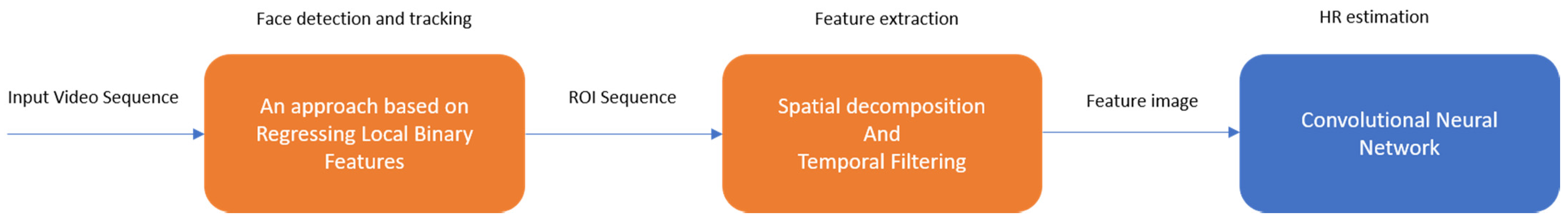
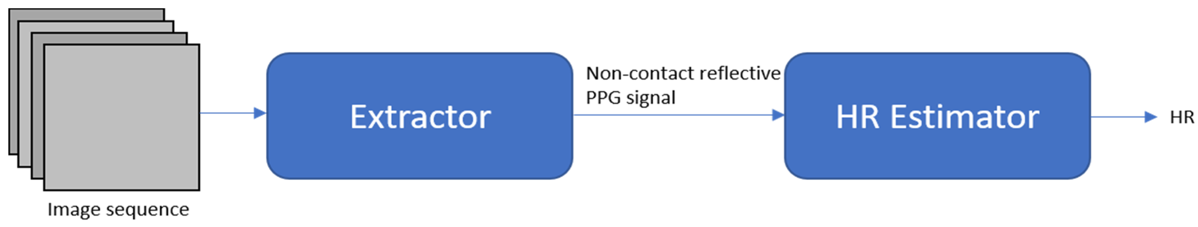
| End-to-end system Robust to illumination changes and subject’s motion | [46] | 2018 | A two-step convolutional neural network composed of an extractor and HR estimator | COHFACE PURE MAHNOB-HCI |
| Signal estimation enhancement | [77] | 2019 | Eulerian video magnification (EVM) to extract face color changes and using CNN to estimate heart rate | MMSE-HR |
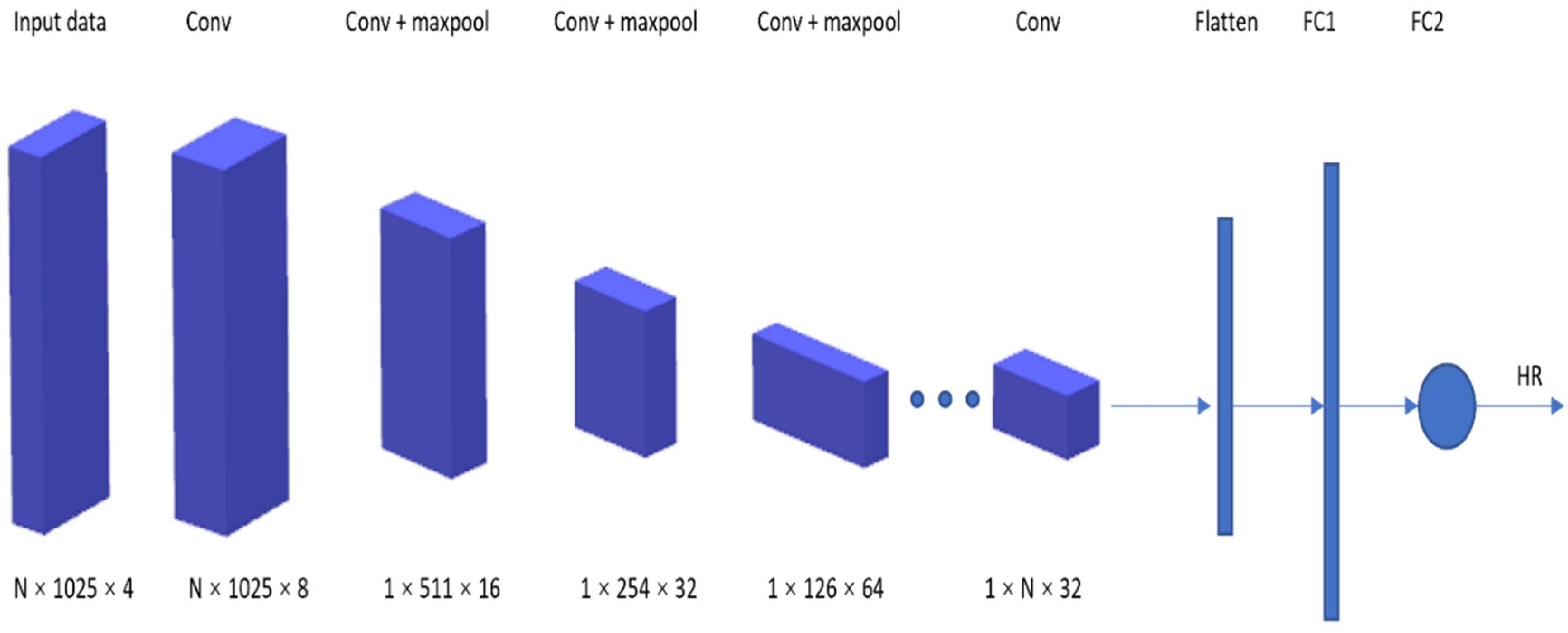
| 3D-CNN for signal extraction | [79] | 2020 | Using deep spatiotemporal networks for contactless HRV measurements from raw facial videos; employing data augmentation | MAHNOB-HCI |
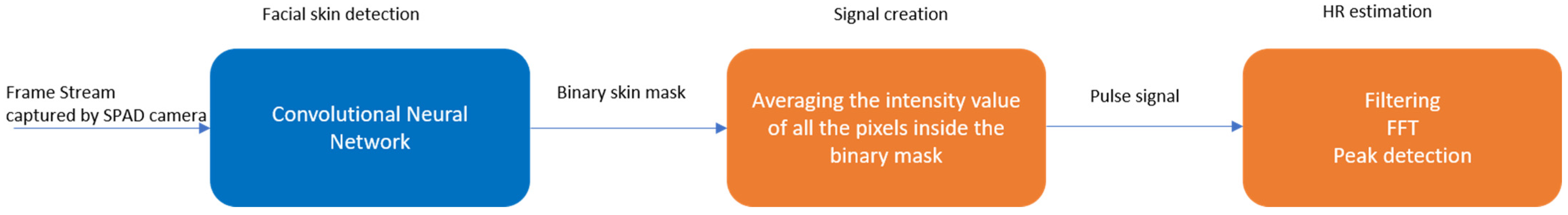
| Single-photon camera | [80] | 2020 | Neural network for skin detection | N/A |
| Understanding of CNN-based PPG methods | [81] | 2020 | Analysis of CNN-based remote PPG to understand limitations and sensitivities | HNU PURE |
| End-to-end system Attention mechanism | [82] | 2018 | Robust measurement under heterogeneous lighting and motions | MAHNOB-HCI |
| End-to-end system Real-life conditions dataset | [84] | 2019 | Major shortcoming of existing datasets: dataset size, small number of activities, data recording in laboratory setting | PPG-DaLiA |
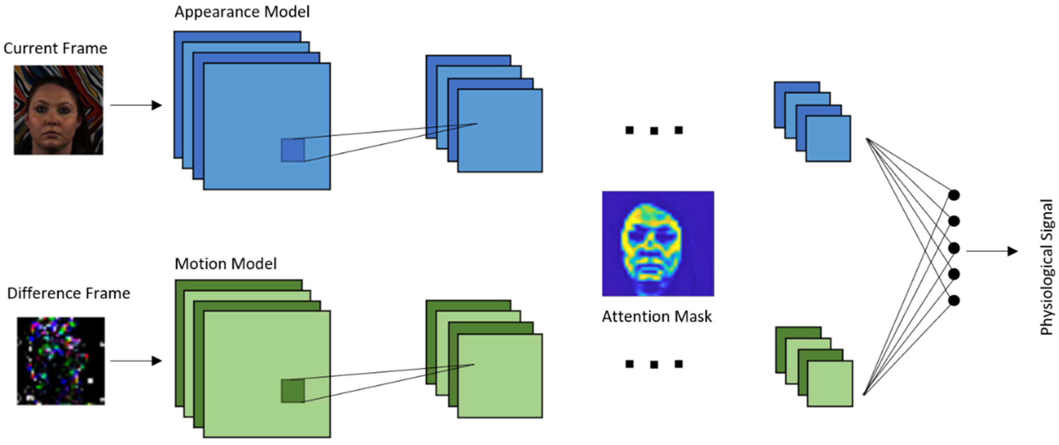
| Synthetic training data Attention mechanism | [93] | 2020 | CNN training with synthetic data to accurately estimate HR in different conditions | UBFC-RPPG MoLi-ppg-1 MoLi-ppg-2 |
| Synthetic training data | [94] | 2019 | Automatic 3D-CNN training process with synthetic data with no image processing | UBFC-RPPG |
| End-to-end supervised learning approach Meta-learning | [89] | 2017 | Meta-rPPG for abundant training data with a distribution not deviating too much from distribution of testing data | MAHNOB-HCI UBFC-RPPG |
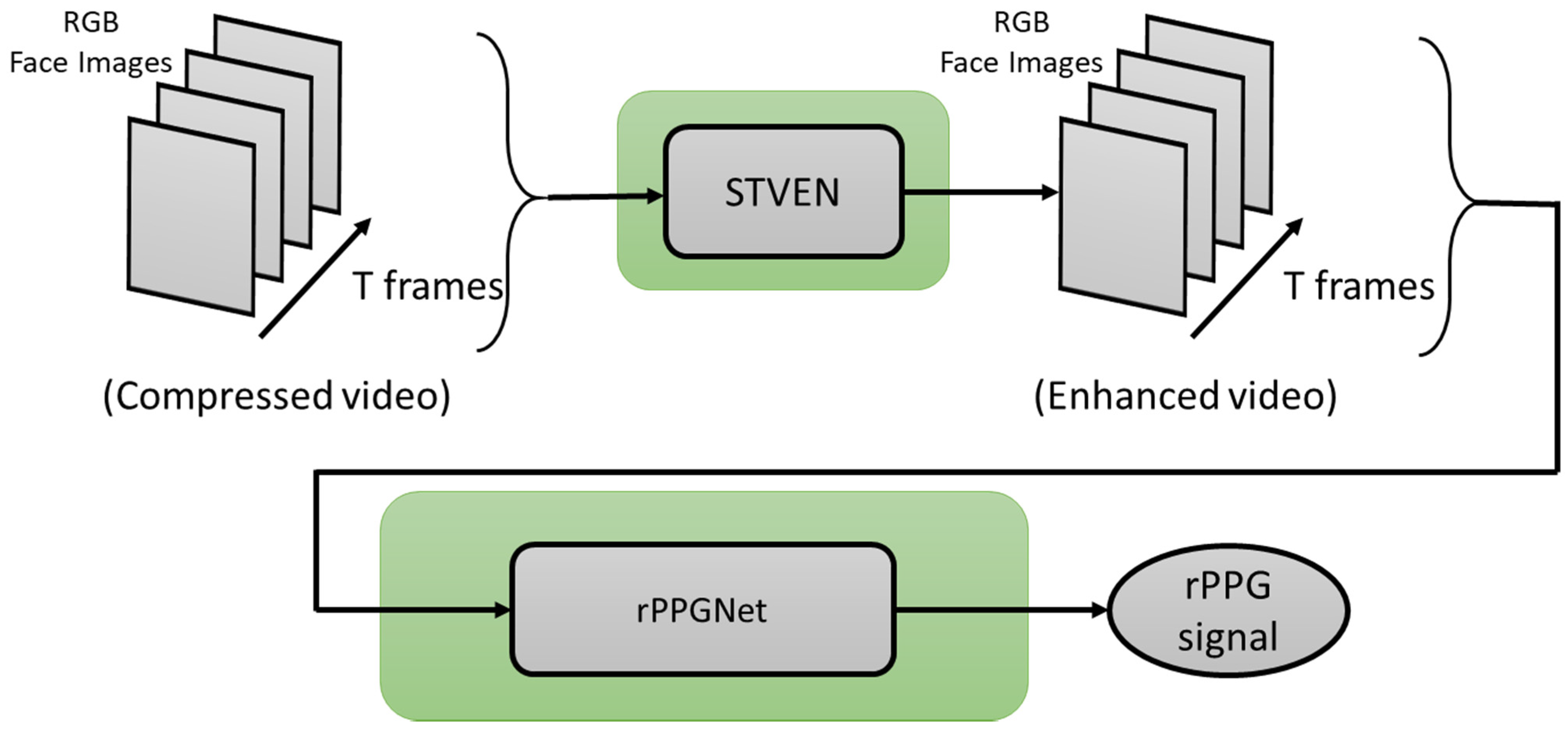
| Counter video compression loss | [91] | 2019 | STEVEN for video quality enhancement rPPGNet for signal recovery | MAHNOB-HCI |
| Spatiotemporal network | [92] | 2019 | Measuring rPPG signal from raw facial video; taking temporal context into account | MAHNOB-HCI |
| Spatiotemporal network | [95] | 2020 | Spatiotemporal convolution network, different types of input skin | MAHNOB-HCI PURE |
| Method | Network Architecture | ||
|---|---|---|---|
| STVEN-rPPGNet | Module | Layer | Kernel |
| STVEN | Convolution 1 | 3 × 3 × 7 | |
| Convolution 2 | 3 × 4 × 4 | ||
| Convolution 3 | 4 × 4 × 4 | ||
| Spatiotemporal block | [3 × 3 × 3] × 6 | ||
| Deconvolution 1 | 4 × 4 × 4 | ||
| Deconvolution 2 | 1 × 4 × 4 | ||
| Deconvolution 3 | 1 × 7 × 7 | ||
| rPPGNet | Convolution 1 | 1 × 5 × 5 | |
| Spatiotemporal block | [3 × 3 × 3] × 4 | ||
| Spatial global average pooling | 1 × 16 × 16 | ||
| Deconvolution 1 | 1 × 1 × 1 | ||
| iPPG-3 DCNN | Convolution 1 | 58 × 20 × 20 | |
| Max pooling | 2 × 2 × 2 | ||
| Dense | 512 | ||
| Dense | 76 | ||
| PhysNet | Convolution 1 | 1 × 5 × 5 | |
| Max pooling | 1 × 2 × 2 | ||
| Convolution 2 | 3 × 3 × 3 | ||
| Convolution 3 | 3 × 3 × 3 | ||
| Spatial global average pooling | |||
| Convolution 4 | 1 × 1 × 1 | ||
| Meta-rPPG | Convolution 1 | 3 × 3 | |
| Convolution 2 | 3 × 3 | ||
| Convolution 3 | 3 × 3 | ||
| Convolutional Encoder | Convolution 4 | 3 × 3 | |
| Convolution 5 | 3 × 3 | ||
| Average pooling | 2 × 2 | ||
| rPPG Estimator | Bidirectional LSTM | --- | |
| Linear | --- | ||
| Ordinal | --- | ||
| Synthetic Gradient Generator | Convolution 1 | 3 × 3 | |
| Convolution 2 | 3 × 3 | ||
| Convolution 3 | 3 × 3 | ||
| Convolution 4 | 3 × 3 | ||
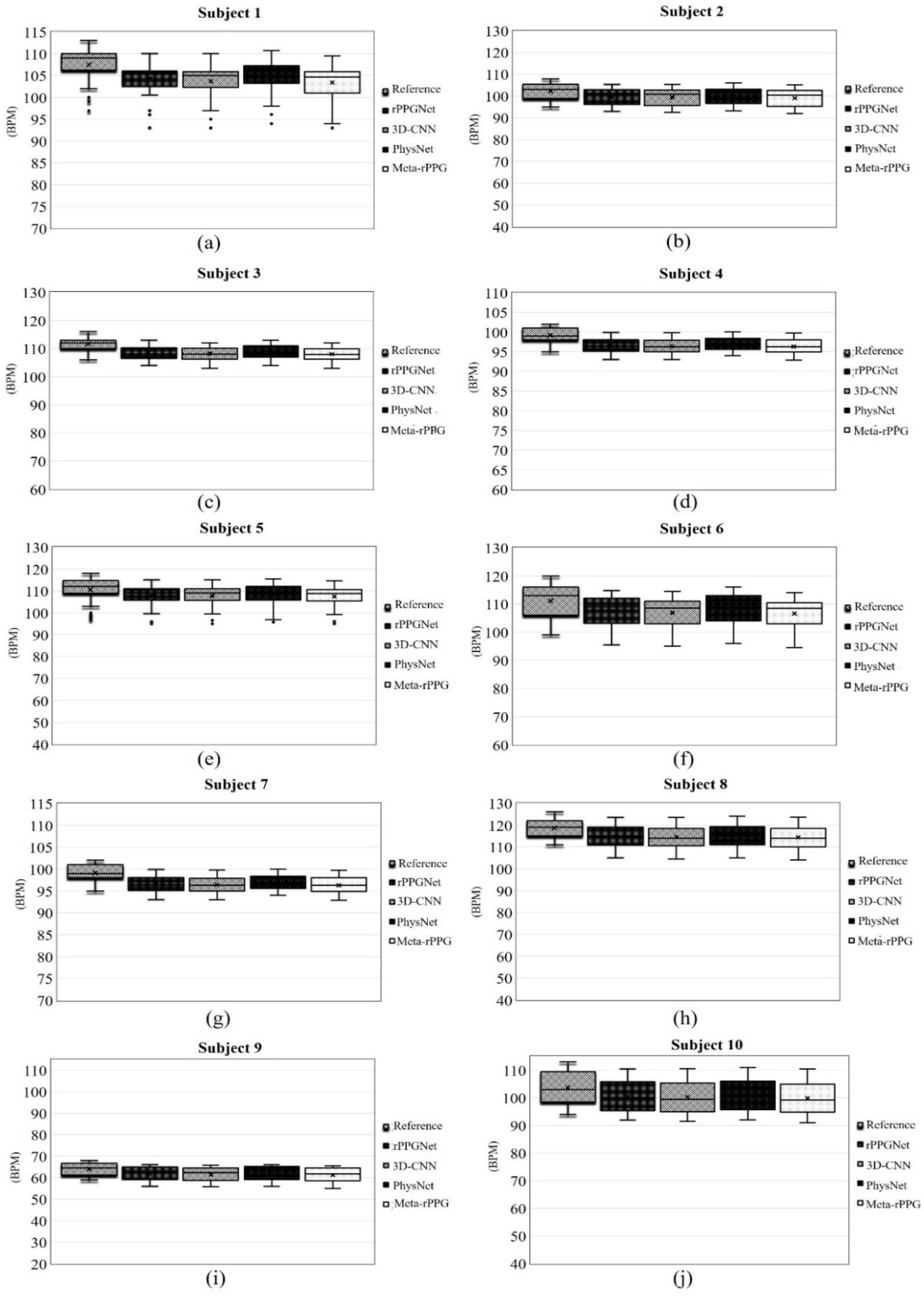
| Subject # | Method | HR (bpm) | ||
|---|---|---|---|---|
| MAE | MSE | SD | ||
| Subject 1 | rPPGNet | 3.22 | 11.41 | 3.93 |
| 3D-CNN | 3.75 | 14.92 | 3.86 | |
| PhysNet | 2.53 | 7.31 | 3.96 | |
| Meta-rPPG | 4.09 | 17.67 | 3.95 | |
| Subject 2 | rPPGNet | 2.72 | 7.82 | 3.82 |
| 3D-CNN | 2.87 | 8.81 | 3.93 | |
| PhysNet | 2.25 | 5.47 | 3.79 | |
| Meta-rPPG | 3.18 | 10.71 | 4.01 | |
| Subject 3 | rPPGNet | 3.12 | 11.14 | 2.32 |
| 3D-CNN | 3.43 | 13.28 | 2.33 | |
| PhysNet | 2.74 | 8.74 | 2.42 | |
| Meta-rPPG | 3.63 | 14.78 | 2.36 | |
| Subject 4 | rPPGNet | 2.63 | 7.79 | 1.79 |
| 3D-CNN | 2.74 | 8.42 | 1.74 | |
| PhysNet | 2.14 | 5.48 | 1.75 | |
| Meta-rPPG | 2.83 | 8.96 | 1.77 | |
| Subject 5 | rPPGNet | 2.82 | 8.90 | 5.48 |
| 3D-CNN | 2.96 | 9.72 | 5.50 | |
| PhysNet | 2.38 | 6.66 | 5.54 | |
| Meta-rPPG | 3.22 | 11.37 | 5.48 | |
| Subject 6 | rPPGNet | 3.76 | 15.09 | 5.71 |
| 3D-CNN | 4.21 | 18.91 | 5.66 | |
| PhysNet | 2.93 | 9.26 | 5.95 | |
| Meta-rPPG | 4.56 | 22.34 | 5.63 | |
| Subject 7 | rPPGNet | 3.42 | 12.40 | 8.79 |
| 3D-CNN | 3.85 | 15.78 | 8.66 | |
| PhysNet | 2.91 | 9.04 | 8.94 | |
| Meta-rPPG | 4.01 | 17.02 | 8.72 | |
| Subject 8 | rPPGNet | 3.66 | 14.51 | 4.87 |
| 3D-CNN | 3.93 | 16.82 | 4.92 | |
| PhysNet | 3.18 | 11.21 | 4.92 | |
| Meta-rPPG | 4.20 | 19.07 | 4.96 | |
| Subject 9 | rPPGNet | 2.24 | 5.49 | 3.47 |
| 3D-CNN | 2.52 | 6.76 | 3.47 | |
| PhysNet | 2.04 | 4.76 | 3.55 | |
| Meta-rPPG | 2.78 | 8.13 | 3.58 | |
| Subject 10 | rPPGNet | 3.14 | 10.74 | 5.65 |
| 3D-CNN | 3.36 | 12.34 | 5.63 | |
| PhysNet | 2.60 | 7.63 | 5.77 | |
| Meta-rPPG | 3.67 | 14.60 | 5.62 | |
| Averaged across all subjects | rPPGNet | 3.07 | 10.53 | 4.58 |
| 3D-CNN | 2.98 | 12.58 | 4.57 | |
| PhysNet | 2.57 | 7.56 | 4.66 | |
| Meta-rPPG | 3.62 | 14.47 | 4.61 | |
| Reference value | 0 | 0 | 0 | |
| Method | rPPGNet | 3D-CNN | PhysNet | Meta-rPPG |
|---|---|---|---|---|
| Time | 1.12 (s) | 0.74 (s) | 1.19 (s) | 1.7 (s) |
Publisher’s Note: MDPI stays neutral with regard to jurisdictional claims in published maps and institutional affiliations. |
© 2021 by the authors. Licensee MDPI, Basel, Switzerland. This article is an open access article distributed under the terms and conditions of the Creative Commons Attribution (CC BY) license (https://creativecommons.org/licenses/by/4.0/).
Share and Cite
Ni, A.; Azarang, A.; Kehtarnavaz, N. A Review of Deep Learning-Based Contactless Heart Rate Measurement Methods. Sensors 2021, 21, 3719. https://doi.org/10.3390/s21113719
Ni A, Azarang A, Kehtarnavaz N. A Review of Deep Learning-Based Contactless Heart Rate Measurement Methods. Sensors. 2021; 21(11):3719. https://doi.org/10.3390/s21113719
Chicago/Turabian StyleNi, Aoxin, Arian Azarang, and Nasser Kehtarnavaz. 2021. "A Review of Deep Learning-Based Contactless Heart Rate Measurement Methods" Sensors 21, no. 11: 3719. https://doi.org/10.3390/s21113719