Article

Expanding Actinomycetota Diversity in the TBRC Culture Collection through Metabarcoding and Simulated In Situ Cultivation of Thailand’s Mekong River Microbiota

by
Supattra Kitikhun
1,
Paopit Siriarchawattana
1,
Suwanee Chunhametha
1,
Chanwit Suriyachadkun
1,
Pattaraporn Rattanawaree
1,
Chitwadee Phithakrotchanakoon
1,
Piyanun Harnpicharnchai
1,
Lily Eurwilaichitr
2,* and
Supawadee Ingsriswang
1,*
1
Thailand Bioresource Research Center (TBRC), National Center for Genetic Engineering and Biotechnology, National Science and Technology Development Agency, Pathum Thani 12120, Thailand
2
National Energy Technology Center, National Science and Technology Development Agency, Pathum Thani 12120, Thailand
*
Authors to whom correspondence should be addressed.
Diversity 2023, 15(5), 663; https://doi.org/10.3390/d15050663
Submission received: 15 March 2023 / Revised: 8 May 2023 / Accepted: 12 May 2023 / Published: 13 May 2023
(This article belongs to the Special Issue Feature Papers in Microbial Diversity and Culture Collections)

Abstract

:
Culture-independent and culture-dependent approaches were employed to investigate the taxonomic diversity and biosynthetic gene cluster potential of Actinomycetota in the Mekong River. Through 16S rRNA gene metabarcoding, 21,103 OTUs were revealed to represent 190 genera and at least 595 species of Actinomycetota, including putatively novel taxa. Conventional and in situ cultivation (IC) methods provided 75 Actinomycetota isolates representing 72 species from 21 genera. Of these, 45 species in 4 genera were new to the Thailand Bioresource Research Center (TBRC), a collection of 20,079 Actinomycetota strains from 660 species. Applying both culture-independent and culture-dependent approaches to the same sample revealed greater diversity among the Actinomycetota in the Mekong River than one approach alone.

1. Introduction

Actinomycetota constitutes a diverse group of Gram-positive bacteria noted for their abilities to produce antibiotics and other bioactive metabolites; almost one-fourth of such compounds discovered to date have been produced by Actinomycetota [1]. Actinomycetota also have potential uses in many other applications, including biocontrol [2,3,4,5,6], plant growth promotion [7,8,9,10,11], and bioremediation [12,13,14]. To foster and support such scientific research, the Thailand Bioresource Research Center (TBRC) has maintained 20,079 strains (660 species in 121 genera) of Actinomycetota. These comprise approximately 65% of the TBRC’s bacterial collection, and 21% of the entire TBRC microbial collection. Approximately 90% of the Actinomycetota in the collection were acquired from several TBRC biodiversity surveys and research projects to tap the potential of Actinomycetota for various applications. Based on the prediction of biosynthetic gene clusters in the genome using AntiSMASH 6.0 [15], Actinomycetota species in the Actinomadura, Actinoplanes, Amycolatopsis, Dactylosporangium, Kitasatospora, Kutzneria, Micromonospora, Nocardia, Nonomuraea, Streptomyces, and Streptosporangium genera exhibit biosynthetic potential, as each of these genera contains more than 20 biosynthetic gene clusters (BGCs) with >60% homology to known clusters (Table 1). The species in these genera were predicted to contain 20–54 BGCs per genome.
The most abundant Actinomycetota taxa in the TBRC collection (89%) belong to the families Micromonosporaceae, Nocardiaceae, Pseudonocardiaceae, Streptomycetaceae, Streptosporangiaceae, and Thermomonosporaceae, with >80 strains for each of these families. The other 30 families comprise 1–80 strains each. Although Actinomycetota can be found in a variety of natural environments [16], most of those (98%) in the TBRC collection were obtained from terrestrial habitats, and 92% of these were from soils. Since Actinomycetota from environments other than soil also have a high potential for producing valuable compounds, it is advantageous to expand surveys and research to include other habitats. Aquatic environments have emerged as an important habitat for Actinomycetota with bioactivities [17,18,19,20]. Studies of bioactive compound-producing Actinomycetota from freshwater have lagged behind those of bioactive compound-producing Actinomycetota from soil ecosystems [20], most likely because of a perceived difficulty in isolating Actinomycetota from freshwater sources, such as because freshwater Actinomycetota are typically less abundant than in soils [21]. Secondary metabolites produced in freshwater habitats are thought to be highly potent in order to compensate for their dilution in water [1,19,22]. As improvements in culturing methods and advancements in DNA technology have revealed a rich diversity of Actinomycetota in aquatic environments [23,24], exploration of this habitat will likely yield valuable novel species and increase the number of the poorly represented taxa in the TBRC collection.
The Mekong River shows great potential as a source of novel freshwater Actinomycetota. As the third longest transboundary river in Asia, the river lies at the heart of the Indo-Burma biodiversity hotspot and is considered one of the richest areas of biodiversity in the world [25]. To explore freshwater Actinomycetota in the Mekong River, a combination of culture-independent and culture-dependent approaches was employed by a TBRC research team. An in situ cultivation (IC) method developed for the isolation and cultivation of bacteria in a simulated environment was also used to target for isolation novel strains and species of Actinomycetota.

2. Materials and Methods

2.1. Sample Collection

A total of 50 water samples, comprising 25 surface water samples (collected at 0.30 m below the surface) and 25 suspended-particle water samples (collected at 0.30 m above the riverbed), was collected along the Mekong River in Thailand at the five hydrology stations of the Department of Water Resources. These are located in Chiang Saen (CS), Chiang Khan (CK), Nong Khai (NK), Mukdahan (MD), and Khong Chiam (KC) Districts (Figure 1 and Table 2). Each hydrology station contained five sampling sites. The exact locations of the sampling sites were systematically determined using information from Acoustic Doppler Current Profiler (ADCP) machine. In addition, 15 sediment samples were collected from the riverbed, and 15 soil samples were collected on the riverbank. Furthermore, the temperature and pH values were measured at each sampling site. Water samples were stored in plastic bottles, while sediment and soil samples were placed in separate Ziploc® bags. All of the samples were kept at 4 °C during transportation from the sampling sites to the laboratory. After arriving at the laboratory, 1–10 L of each water sample was filtered prior to preparation for metabarcoding. A total of 1 L of the water sample was used for the analysis of physicochemical characteristics. The remainder of each sample was kept at 4 °C prior to processing for microbial cultivation using the IC and standard plate methods (Section 2.3).

2.2. DNA Extraction and 16S-Amplicon Sequencing

Between 1 and 10 L of each water sample was vacuum filtered successively through polyethersulfone (PES) membranes (0.8-, 0.4-, and 0.22-µm pore size (PALL, New York, NY, USA). DNA was extracted from cells on the membranes in the DNeasy PowerWater kit (QIAGEN, Hilden, Germany) according to the manufacturer’s protocol. Sediment and soil samples were extracted according to the protocol described by Zhou et al. [26]. DNA concentration and purity were assessed by absorbance at 280 nm and by the 260/280 nm absorbance ratio, respectively, in a NanoDropTM spectrophotometer 2000 (Thermo Scientific, Waltham, MA, USA). DNA whose 260/280 nm absorbance ratio was between 1.8 and 2.0 was retained for library construction. Metagenomic DNA from water, sediment, and soil samples was used as the template in polymerase chain reactions (PCRs) to amplify the 16S rRNA gene taxonomic marker. The resulting amplicons were sequenced on the Illumina MiSeq platform. The amplicon sequencing library was prepared using the bacteria-specific primers 341F (5′-CCTACGGGNGGCWGCAG-3′) and 805R (5′-GACTACHVGGGTATCTAATCC-3′) targeting the V3–V4 region of the 16S rRNA gene [27].

2.3. Isolation and Cultivation

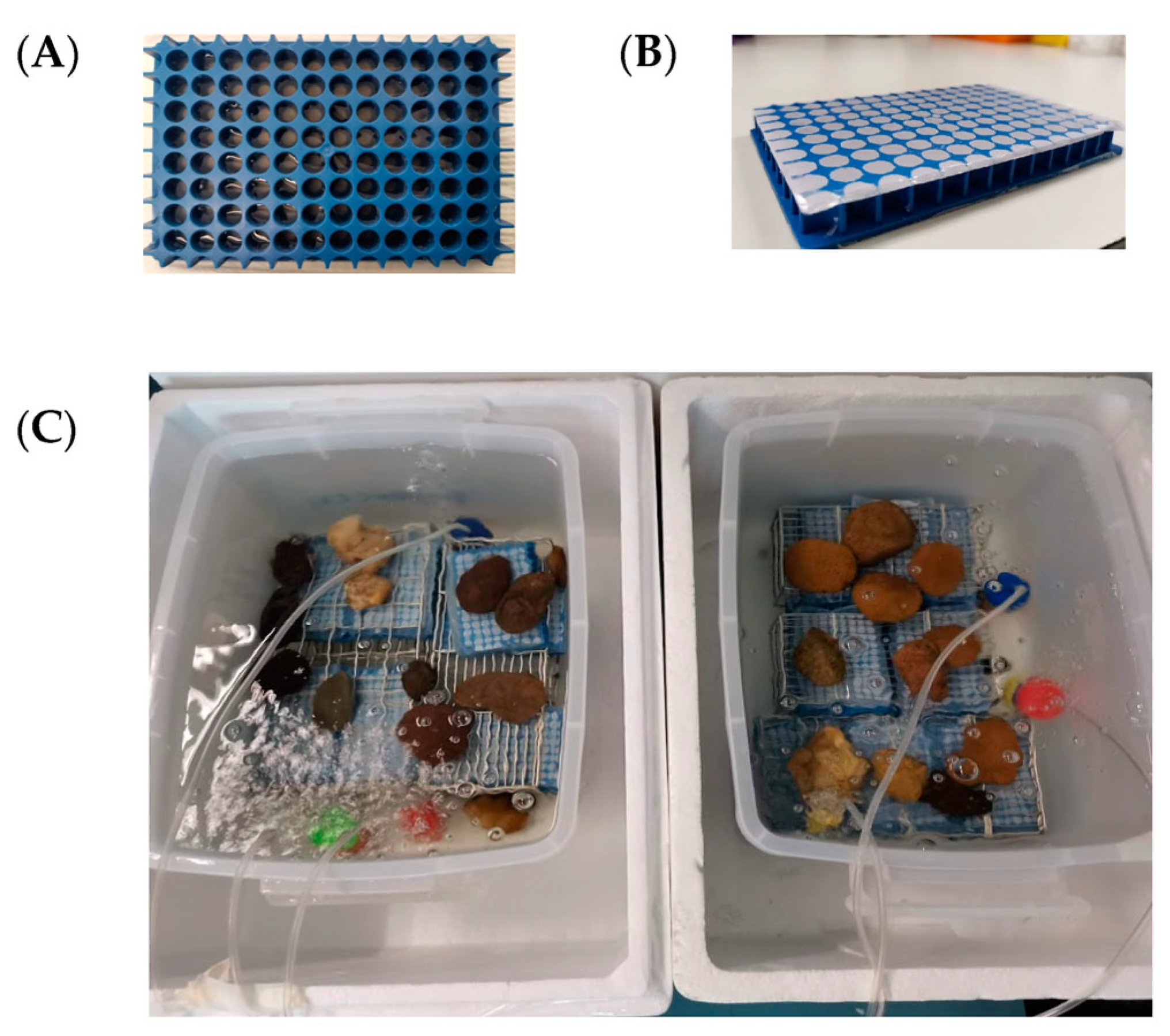
2.3.1. Design and Development of the IC Plate

The IC plate method was designed according to Nichols et al. (2010) [28], with modifications. Briefly, an empty 200 µL-pipette tip rack (BioRobotix, Waltham, MA, USA) was polished by a polishing machine to smooth the bottom surface, and the resulting polished rack was used as the IC plate with 96 through-holes. After cleaning with water, a thin layer of silicone glue was applied to the bottom of the IC plate. The bottom of the IC plate was then covered by a 0.03 µm-pore size polycarbonate membrane (Whatman, Dassel, Germany). After letting the glue dry overnight, the IC plate was sterilized by autoclaving. Thereafter, each well was filled with 150 µL 1.5% (w/v) sterile agar. Cell suspension (1 µL) was pipetted onto the agar in each well to trap the bacterial cells. A second piece of sterile 0.03 µm-pore size polycarbonate membrane was carefully attached to the top part of the IC plate in order to cover the IC plate before the glue was left to dry overnight (Figure 2).

2.3.2. Isolation of Actinomycetota

All of the water samples from the Mekong River collected at each of the hydrology stations were mixed at a 1:1 (v/v) ratio, i.e., five samples from five sampling points at each station were mixed together in equal volume. The mixed samples were serially diluted with sterile water. The IC and conventional plate methods were performed simultaneously in this study. A volume of 180 µL of diluted cell suspension (10−4) was plated onto soil extract agar [29] containing 50 mg/L cycloheximide and soil extract agar containing 50 mg/L cyclohexamide, 25 mg/L nalidixic acid, and 1 mg/L terbinafine. For the IC method, a volume of 180 µL cell suspension (10−4) was diluted with sterile water to a final volume of 960 µL prior to use as an IC inoculum. Then, 1 µL of the inoculum was inoculated into each of the 96 wells of the IC plates, for a total of 10 plates. Thereafter, the IC plates were incubated in a chamber (Figure 2C) containing water collected from the Mekong River, with an air pump to draw oxygen into that water. IC plates containing cell suspension were incubated at room temperature for four weeks. After that, all of the cultured microbes from the IC plates were simultaneously transferred to (a) soil extract agar [29] containing 50 mg/L cycloheximide and (b) soil extract agar containing 50/L cyclohexamide, 25 mg/L nalidixiacid, and 1 mg/L terbinafine in 96-well agar plates. Finally, all of the agar plates were incubated at 30 ± 2 °C for 2–4 weeks to allow for growth of the Actinomycetota.

2.3.3. Identification and Dereplication of Actinomycetota Isolates

Putative colonies of both mycelial and non-mycelial Actinomycetota were observed by the unaided eye and under a light microscope (model CX 31; Olympus, Tokyo, Japan) with a 50×X long working distance objective lens (model SLMPLN50×; Olympus). Colonies of mycelial Actinomycetota were expected to be slow-growing, folded, and of chalky (opaque) or leathery appearance, and contain aerial and substrate mycelia of different colors [30]. Colonies with different morphological appearances were picked and subcultured on soil extract agar and yeast extract–malt extract agar medium (ISP-2) (BD DifcoTM, Franklin Lakes, NJ, USA) to obtain pure colonies. Pure cultures were preserved in 20% (v/v) glycerol at −80 °C.
Taxonomic identification of the isolates was based, first, on sequencing their 16S rRNA gene: purified genomic DNA was used as a DNA template for the amplification of the 16S rRNA gene using universal primers for Bacteria, BSF8/20 (5′-AGAGTTTGATCCTGGCTCAG-3′) and REVB (5′-GGTTACCTTGTTACGACTT-3′) [31]. Each PCR (50 µL) contained 1X Phusion HF buffer (Thermo Scientific, Waltham, MA, USA), 0.5 µM each of forward and reverse primers, 0.2 mM each of dNTPs, 1 U of Phusion High-Fidelity DNA polymerase (Thermo Scientific, Waltham, MA, USA), and 25 ng of DNA template. The amplification conditions were 98 °C for 30 s, then 35 cycles of 98 °C for 10 s, 57.7 °C for 30 s, and 72 °C for 45 s; and a final extension at 72 °C for 10 min. PCR products were purified and sequenced by Macrogen, Seoul, Korea. The assembled sequences were compared with others in the EzBioCloud database [32] using Blastn ver. 2.12.0+ [33] to identify the closest matching sequences of type strains.

2.4. Analysis of Microbial Community Diversity

Sequences of the V3–V4 region of the bacterial 16S rRNA genes from the water, sediment, and soil samples were subjected to quality control using FastQC [34]. Operational taxonomic units (OTUs) were constructed by sequence clustering using CD-HIT (ver. 4.8.1) [35] with a 0.97 sequence identity threshold and word length of 10. Chimeric sequences were removed using VSEARCH version 2.18.0 [36]. Taxonomy assignment of bacterial OTUs was performed on the representative OTU sequences using QIIME2’s classify-sklearn v. 2020.2.0 [37] with a confidence of 0.7 and k-mer of 7 against multiple 16S rRNA gene databases, including EzBioCloud [32], GTDB release 95 [38], and SILVA v. 132 [39]. Shannon and Chao1 diversity of each sample was calculated using vegan [40] and fossil R [41] packages.

3. Results

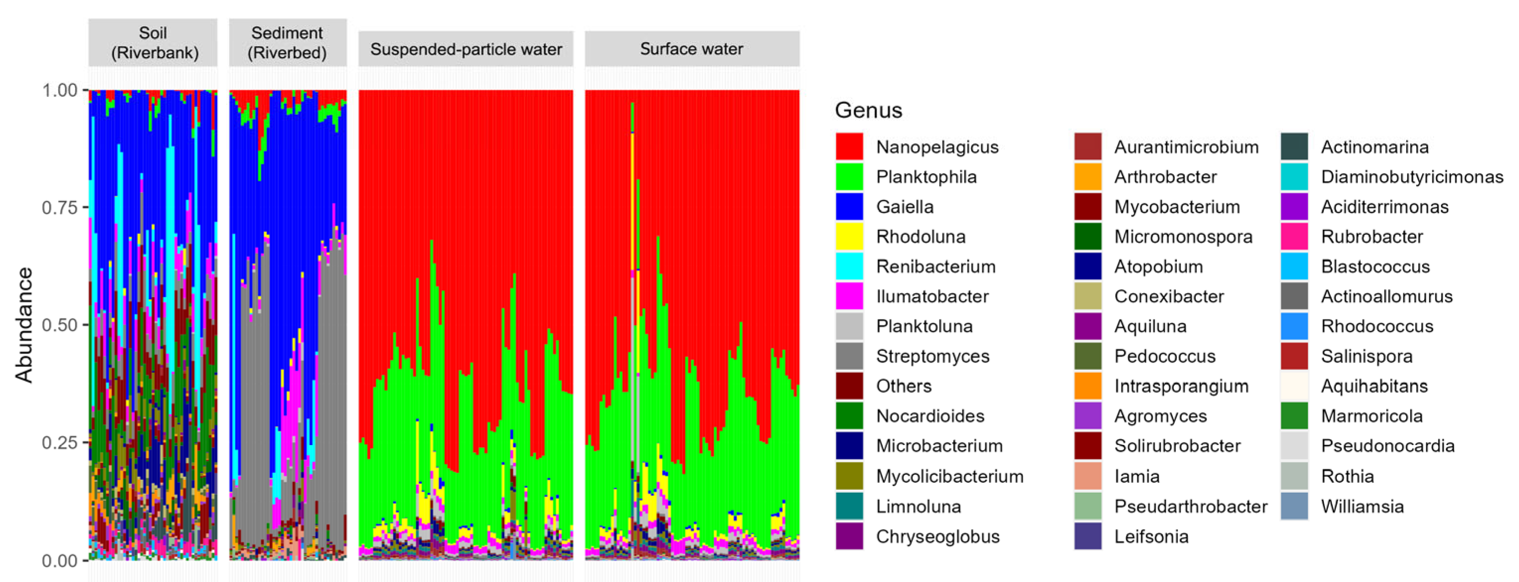
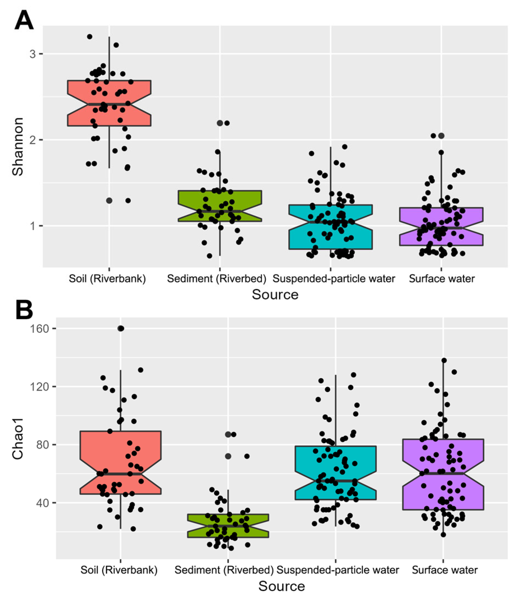
3.1. Culture-Independent, Metabarcoding-Based Diversity, and Composition of Actinomycetota in the Mekong River

The taxonomic distribution of Actinomycetota diversity in the Mekong River samples based on 16S rRNA gene metabarcoding is shown in Figure 3. Sequencing of the V3–V4 region of the 16S rRNA gene resulted in 7,030,770 reads. After quality filtering, 1,337,379 reads remained for analysis, and 21,103 OTUs of Actinomycetota were obtained based on >97% sequence similarity to reference sequences in the SILVA, EzBioCloud, and GTDB databases.
A total of 190 Actinomycetota genera was identified from all of the Mekong River samples. The bacterial composition of water samples differed markedly from that of the sediment and soil samples. The dominant Actinomycetota in the surface water and suspended-particle water samples were Nanopelagicus (62%), Planktophila (28%), and Rhodoluna (2%). In the riverbed sediment samples, the dominant Actinomycetota genera were Gaiella (45%), Streptomyces (34%), and Ilumatobacter (6%), whereas the dominant genera in riverbank soil samples were Gaiella (31%), Renibacterium (11%), and Nocardioides (9%) (Figure 3). From these 190 Actinomycetota genera, 110 were not yet in the TBRC collection. Of these, 11 genera, including Acidiferrimicrobium, Acidothermus, Actinomarina, Flaviluna, Gaiella, Limnoluna, Nanopelagicus, Planktoluna, Planktophila, Raoultibacter, and Thermoleophilum, are in the List of Prokaryotic names with Standing in Nomenclature (LPSN). However, Actinomarina, Flaviluna, Limnoluna, Nanopelagicus, Planktoluna, and Planktophila were categorized as “Candidatus” genera in the LPSN, indicating that cultivated type species have not yet been validly published under the International Code of Nomenclature of Prokaryotes (ICNP) [42].
To investigate variation in Actinomycetota compositions of the Mekong River water, sediment, and soil samples, the alpha-diversity of the OTUs was analyzed using Shannon’s and Chao 1 diversity indices at the genus level (Figure 4). The two types of water samples (surface water and suspended-particle water) exhibited similar levels of genus richness and evenness (Figure 4A,B), while sediment and soil samples comprised different levels of diversity. The riverbank soil showed the highest richness and evenness levels of Actinomycetota compared to other samples, while the riverbed sediment samples contained the lowest level of Actinomycetota genus diversity.

3.2. Culture-Dependent Isolation of Actinomycetota in the Mekong River

Actinomycetota in the Mekong River were also recovered by culture-dependent methods using the standard plate and IC techniques. After the initial colony isolation, 75 Actinomycetota-like isolates were obtained. The 16S rRNA gene in each was amplified and sequenced. The 75 isolates were confirmed to be Actinomycetota through EZBioCloud searches, with ≥98% identity. These 75 isolates were affiliated with 72 species in 21 different genera, including Actinotalea, Aeromicrobium, Agrococcus, Agromyces, Arthrobacter, Asanoa, Brevibacterium, Cellulomonas, Geodermatophilus, Kribbella, Microbacterium, Microbispora, Micromonospora, Mycolicibacterium, Nocardia, Nocardioides, Nonomuraea, Pseudarthrobacter, Rhodococcus, Streptomyces, and Williamsia. Among the isolated species, 45 species belonging to 18 genera were new to the TBRC collection. These species were members of the Actinotalea, Aeromicrobium, Agrococcus, Agromyces, Brevibacterium, Cellulomonas, Geodermatophilus, Kribbella, Microbacterium, Micromonospora, Mycolicibacterium, Nocardia, Nocardioides, Nonomuraea, Pseudarthrobacter, Rhodococcus, Streptomyces, and Williamsia genera (Table 3).
Remarkably, the genera Actinotalea, Aeromicrobium, Agrococcus, and Williamsia were entirely new genera to the TBRC collection.

4. Discussion

Since aquatic Actinomycetota have not been as extensively exploited as their terrestrial counterparts [1], Actinomycetota from freshwater ecosystems have now garnered increased interest as a promising source of novel bioactive compounds of pharmaceutical and biotechnological importance [43,44]. Our metabarcoding results confirmed a higher relative abundance of Actinomycetota (in relation to other bacteria) in water samples from the Mekong River compared to sediment and soil samples. In the Mekong River’s surface and suspended-particle water samples, the Actinomycetota families exhibiting the highest abundance were the unicellular, free-living Nanopelagicaceae and Ilumatobacteraceae. The family Nanopelagicaceae has been found ubiquitously in freshwater systems [45]. Whole-genome sequences of Nanopelagicaceae and several other Actinomycetota suggested that they underwent evolutionary genome reduction events, resulting in highly streamlined genomes, possibly to help limit energy consumption (sensu Morris et al., 2012) [46]. Nanopelagicaceae are unable to synthesize various vitamins and amino acids, and require reduced sulphur, implying their dependency on these nutrients and highlighting a difficulty in the long-term cultivation of Nanopelagicaceae [45]. Additionally, the free-living Ilumatobacteraceae are part of particle-associated bacterial communities in aquatic ecosystems and play important roles in organic matter decomposition [47]. The high abundance of these families in the water samples and their relatively low abundance in the sediment and soil samples imply that they are capable of active growth and reproduction in aquatic environments, rather than being simply transients from terrestrial environments [48].
In addition to the culture-independent metabarcoding approach, our investigation using the culture-dependent approach yielded 75 Actinomycetota strains. Most of these isolates belonged to the Streptomycetaceae and Micromonosporaceae genera, which hosted more than 50% of the Actinomycetota isolates. These two families also account for approximately half of the Actinomycetota strains in the TBRC collection, which were mostly isolated from various terrestrial sources. We provided 45 species in 19 genera to the TBRC collection, including Actinotalea, Aeromicrobium, Agrococcus, and Williamsia, all of which were entirely new genera to the collection. Nanopelagicus, Planktophila, and Gaiella, as well as Streptomyces, were dominant genera identified from the Mekong River with the culture-independent approach. However, no representatives of these genera were cultivated here. Improvement or modification of culturing techniques may be required to isolate these Actinomycetota.
Our study showed that the IC methods could be applied simultaneously with the standard plate method to recover Actinomycetota from the Mekong River, as a total of eight species (Actinotalea fermentans, Cellulomonas fimi, Cellulomonas oligotrophica, Mycolicibacterium fluoranthenivorans, Mycolicibacterium tokaiense, Nocardioides aquiterrae, Pseudarthrobacter niigatensis, and Rhodococcus cerastii) were isolated by the IC technique but not by the standard plate technique. The success of the IC technique was most likely due to the ability to allow the Actinomycetota to grow in conditions that mimicked their natural habitat [28,49,50]. To obtain more diverse Actinomycetota species from the Mekong River, a variety of isolation media suitable for Actinomycetota can be developed. For example, it was found that the fastidious Nanopelagicaceae found in freshwater environments could be isolated and maintained in the laboratory using a catalase-supplement method [51]. Moreover, from genome analysis, Nanopelagicaceae was categorized as auxotrophic for heme, and it was cultured by adding heme and riboflavin to the growth medium [52]. Some rare or slow-growing Actinomycetota in the environment cannot be easily isolated by the agar-plating method due to competition with fast-growing microbe [4,43]. Thus, a recent array of culturing strategies with high-throughput capability has uncovered these Actinomycetota. For example, microfluidic streak plate methods, which allow single-cell cultivation in a droplet, cultivated individual slow-growing Actinomycetota [53]. Additionally, culturomics tools employing multiple culture conditions in combination with proteomics analysis, e.g., MALDI-TOF mass spectra profiling, will be beneficial in the recovery and identification of previously uncultured or rare Actinomycetota [54]. Using these techniques can effectively expand the richness of Actinomycetota in the TBRC collection and increase opportunities to discover microorganisms capable of producing new bioactive compounds.
Several reports have investigated the diversity of Actinomycetota in freshwater ecosystems [17,23,53,55], but the characterization of bioactive compounds produced by isolated Actinomycetota lags behind that of terrestrial and marine Actinomycetota [1,18,19,20]. In addition, it has been postulated that a large fraction of Actinomycetota genomes contain still-unexplored gene clusters with high potential for the production of secondary metabolites [56,57]. Further characterization of the BGC potentials of freshwater Actinomycetota identified in this work will be valuable in uncovering strains with the capability to produce secondary metabolites. The availability of whole genome sequence data and genome mining algorithms can assist the identification and annotation of BGCs. However, the accuracy of the algorithms would still depend largely on the diversity and quality of genome data in databases. It has been observed that the numbers of BGCs vary among different genera according to genetic diversity. The less well-studied genera of Actinomycetota should be a primary source for unraveling important BGCs that may be related to their evolution [58]. There was also a high number of non-homologous BGCs, based on the prediction results shown in (Table 1). The predicted non-homologous BGCs should be further explored to identify bioactive metabolite production capabilities. Thus, it is advantageous to identify some cryptic or silent BGCs to facilitate the discovery of new secondary metabolites. Identifying new biosynthetic gene clusters from Actinomycetota will require multidisciplinary approaches to avoid rediscovering the same molecules. Genome mining with the development of synthetic biology can be used to design an expression system in heterologous hosts that enables the effective and rapid finding of secondary metabolites [55]. Together with analytical metabolomics tools, they are key to unraveling hidden pathways and discovering novel bioactive compounds [59].
Our work showed that employing both culture-independent and culture-dependent approaches simultaneously is beneficial in providing a more complete picture of Actinomycetota species in the Mekong River and for augmenting existing Actinomycetota strains in the TBRC collection. With the potential of BGC prediction, Actinomycetota exhibit high potential for the production of bioactive compounds. Such studies will enhance the capability of the TBRC collection in terms of microbial diversity and utilization, which will effectively serve the research and industrial communities.

Author Contributions

Conceptualization, L.E. and S.I.; Methodology, S.K., C.S., P.R., C.P., P.S. and S.I.; Investigation, S.K., C.S., P.R., C.P., P.S. and S.I.; Validation, S.K., C.S., P.R. and P.S.; Formal Analysis, P.S. and S.I.; Data Curation, S.C. and P.S.; Resources, S.K., C.S. and P.R.; Writing—original draft preparation, S.K., S.C., P.H. and S.I.; Writing—review and editing, S.K., S.C., P.H. and S.I.; Visualization, S.K., S.C., P.H. and P.S.; Supervision, L.E. and S.I.; Project Administration, S.I.; Funding acquisition, L.E. and S.I. All authors have read and agreed to the published version of the manuscript.

Funding

This research was funded by the Lancang-Mekong Cooperation Special Fund (P2052658) and the CAS-NSTDA Joint Research Program (P2051809).

Data Availability Statement

Metagenomic datasets generated during the study are deposited and are publicly available at the NCBI Sequence Read Archive under BioProject PRJNA848859.

Acknowledgments

The authors would like to thank Lei Cai and Junmin Liang from the Institute of Microbiology of the Chinese Academy of Sciences (IMCAS) for their advice on in situ culturing techniques. In addition, we greatly appreciate the effort of the World Federation for Culture Collections (WFCC) in supporting culture collection activities. Finally, we are grateful for the cooperation and assistance of the Department of Water Resources (Ministry of Natural Resources and Environment, Thailand) during sampling.

Conflicts of Interest

The authors declare no conflict of interest. The funders had no role in the design of the study; in the collection, analyses, or interpretation of data; in the writing of the manuscript; or in the decision to publish the results.

References

  1. Sibanda, T.; Mabinya, L.V.; Mazomba, N.; Akinpelu, D.A.; Bernard, K.; Olaniran, A.O.; Okoh, A.I. Antibiotic Producing Potentials of Three Freshwater Actinomycetes Isolated from the Eastern Cape Province of South Africa. Int. J. Mol. Sci. 2010, 11, 2612–2623. [Google Scholar] [CrossRef]
  2. Allali, K.; Goudjal, Y.; Zamoum, M.; Bouznada, K.; Sabaou, N.; Zitouni, A. Nocardiopsis dassonvillei strain MB22 from the Algerian Sahara promotes wheat seedlings growth and potentially controls the common root rot pathogen Bipolaris sorokiniana. J. Plant Pathol. 2019, 101, 1115–1125. [Google Scholar] [CrossRef]
  3. Chaiharn, M.; Chunhaleuchanon, S.; Lumyong, S. Screening siderophore producing bacteria as potential biological control agent for fungal rice pathogens in Thailand. World J. Microbiol. Biotechnol. 2009, 25, 1919–1928. [Google Scholar] [CrossRef]
  4. El-Tarabily, K.A.; Soliman, M.H.; Nassar, A.H.; Al-Hassani, H.A.; Sivasithamparam, K.; McKenna, F.; Hardy, G.E.S.J. Biological control of Sclerotinia minor using a chitinolytic bacterium and actinomycetes. Plant Pathol. 2000, 49, 573–583. [Google Scholar] [CrossRef]
  5. Errakhi, R.; Bouteau, F.; Lebrihi, A.; Barakate, M. Evidences of biological control capacities of Streptomyces spp. against Sclerotium rolfsii responsible for damping-off disease in sugar beet (Beta vulgaris L.). World J. Microbiol. Biotechnol. 2007, 23, 1503–1509. [Google Scholar] [CrossRef]
  6. Li, X.; Jing, T.; Zhou, D.; Zhang, M.; Qi, D.; Zang, X.; Zhao, Y.; Li, K.; Tang, W.; Chen, Y.; et al. Biocontrol efficacy and possible mechanism of Streptomyces sp. H4 against postharvest anthracnose caused by Colletotrichum fragariae on strawberry fruit. Postharvest Biol. Technol. 2021, 175, 111401. [Google Scholar] [CrossRef]
  7. Chukwuneme, C.F.; Babalola, O.O.; Kutu, F.R.; Ojuederie, O.B. Characterization of actinomycetes isolates for plant growth promoting traits and their effects on drought tolerance in maize. J. Plant Interact. 2020, 15, 93–105. [Google Scholar] [CrossRef] [Green Version]
  8. Franco-Correa, M.; Quintana, A.; Duque, C.; Suarez, C.; Rodríguez, M.X.; Barea, J.-M. Evaluation of actinomycete strains for key traits related with plant growth promotion and mycorrhiza helping activities. Appl. Soil Ecol. 2010, 45, 209–217. [Google Scholar] [CrossRef]
  9. Gopalakrishnan, S.; Srinivas, V.; Vidya, M.S.; Rathore, A. Plant growth-promoting activities of Streptomyces spp. in sorghum and rice. SpringerPlus 2013, 2, 574. [Google Scholar] [CrossRef] [Green Version]
  10. Goudjal, Y.; Toumatia, O.; Sabaou, N.; Barakate, M.; Mathieu, F.; Zitouni, A. Endophytic actinomycetes from spontaneous plants of Algerian Sahara: Indole-3-acetic acid production and tomato plants growth promoting activity. World J. Microbiol. Biotechnol. 2013, 29, 1821–1829. [Google Scholar] [CrossRef] [Green Version]
  11. Qin, S.; Miao, Q.; Feng, W.-W.; Wang, Y.; Zhu, X.; Xing, K.; Jiang, J.-H. Biodiversity and plant growth promoting traits of culturable endophytic actinobacteria associated with Jatropha curcas L. growing in Panxi dry-hot valley soil. Appl. Soil Ecol. 2015, 93, 47–55. [Google Scholar] [CrossRef]
  12. Álvarez, A.; Yañez, M.L.; Benimeli, C.S.; Amoroso, M.J. Maize plants (Zea mays) root exudates enhance lindane removal by native Streptomyces strains. Int. Biodeterior. Biodegrad. 2012, 66, 14–18. [Google Scholar] [CrossRef]
  13. Arya, R.; Mishra, N.K.; Sharma, A.K. Brevibacillus borstelensis and Streptomyces albogriseolus have roles to play in degradation of herbicide, sulfosulfuron. 3 Biotech 2016, 6, 246. [Google Scholar] [CrossRef] [PubMed] [Green Version]
  14. Lin, Q.S.; Chen, S.H.; Hu, M.Y.; Haq, M.R.U.; Yang, L.; Li, H. Biodegradation of Cypermethrin by a newly isolated actinomycetes HU-S-01 from wastewater sludge. Int. J. Environ. Sci. Technol. 2011, 8, 45–56. [Google Scholar] [CrossRef] [Green Version]
  15. Blin, K.; Shaw, S.; Kloosterman, A.M.; Charlop-Powers, Z.; van Wezel, G.P.; Medema, M.H.; Weber, T. antiSMASH 6.0: Improving cluster detection and comparison capabilities. Nucleic Acids Res. 2021, 49, W29–W35. [Google Scholar] [CrossRef]
  16. Goodfellow, M.; Williams, S.T. Ecology of Actinomycetes. Annu. Rev. Microbiol. 1983, 37, 189–216. [Google Scholar] [CrossRef]
  17. Jiang, C.; Xu, L. Diversity of aquatic actinomycetes in lakes of the middle plateau, yunnan, china. Appl. Environ. Microbiol. 1996, 62, 249–253. [Google Scholar] [CrossRef] [Green Version]
  18. Rifaat, H.M. The biodiversity of Actinomycetes in the River Nile exhibiting antifungal activity. J. Mediterr. Ecol. 2003, 4, 5–8. [Google Scholar]
  19. Shrestha, B.; Nath, D.K.; Maharjan, A.; Poudel, A.; Pradhan, R.N.; Aryal, S. Isolation and Characterization of Potential Antibiotic-Producing Actinomycetes from Water and Soil Sediments of Different Regions of Nepal. Int. J. Microbiol. 2021, 2021, 5586165. [Google Scholar] [CrossRef]
  20. Zothanpuia; Passari, A.K.; Leo, V.V.; Chandra, P.; Kumar, B.; Nayak, C.; Hashem, A.; Allah, E.F.A.; Alqarawi, A.A.; Singh, B.P. Bioprospection of actinobacteria derived from freshwater sediments for their potential to produce antimicrobial compounds. Microb. Cell Factories 2018, 17, 68. [Google Scholar] [CrossRef]
  21. Eccleston, G.P.; Brooks, P.R.; Kurtböke, D.I. The occurrence of bioactive micromonosporae in aquatic habitats of the Sunshine Coast in Australia. Mar. Drugs 2008, 6, 243–261. [Google Scholar] [CrossRef] [PubMed]
  22. Zhang, L.; An, R.; Wang, J.; Sun, N.; Zhang, S.; Hu, J.; Kuai, J. Exploring novel bioactive compounds from marine microbes. Curr. Opin. Microbiol. 2005, 8, 276–281. [Google Scholar] [CrossRef] [PubMed]
  23. Farrell, M.J.; Govender, D.; Hajibabaei, M.; van der Bank, M.; Davies, T.J. Bacterial diversity in the waterholes of the Kruger National Park: An eDNA metabarcoding approach (1). Genome 2019, 62, 229–242. [Google Scholar] [CrossRef] [PubMed] [Green Version]
  24. Liang, D.; Xia, J.; Song, J.; Sun, H.; Xu, W. Using eDNA to Identify the Dynamic Evolution of Multi-Trophic Communities Under the Eco-Hydrological Changes in River. Front. Environ. Sci. 2022, 10, 853. [Google Scholar] [CrossRef]
  25. Coates, D.P.O.; Suntornratana, U.; Tung, N.T.; Viravong, S. Biodiversity and fisheries in the Lower Mekong Basin; Phnom Penh: Mekong development series. Mekong River Comm. Phnom Penh Cambodia 2003, 2, 30. [Google Scholar]
  26. Zhou, J.; Bruns, M.A.; Tiedje, J.M. DNA recovery from soils of diverse composition. Appl. Environ. Microbiol. 1996, 62, 316–322. [Google Scholar] [CrossRef] [PubMed] [Green Version]
  27. Klindworth, A.; Pruesse, E.; Schweer, T.; Peplies, J.; Quast, C.; Horn, M.; Glöckner, F.O. Evaluation of general 16S ribosomal RNA gene PCR primers for classical and next-generation sequencing-based diversity studies. Nucleic Acids Res. 2013, 41, e1. [Google Scholar] [CrossRef]
  28. Nichols, D.; Cahoon, N.; Trakhtenberg, E.M.; Pham, L.; Mehta, A.; Belanger, A.; Kanigan, T.; Lewis, K.; Epstein, S.S. Use of ichip for high-throughput in situ cultivation of “uncultivable” microbial species. Appl. Environ. Microbiol. 2010, 76, 2445–2450. [Google Scholar] [CrossRef] [Green Version]
  29. Suriyachadkun, C.; Chunhametha, S.; Thawai, C.; Tamura, T.; Potacharoen, W.; Kirtikara, K.; Sanglier, J.J. Planotetraspora thailandica sp. nov., isolated from soil in Thailand. Int. J. Syst. Evol. Microbiol. 2009, 59, 992–997. [Google Scholar] [CrossRef] [Green Version]
  30. Drechsler, C. Morphology of the Genus Actinomyces. II. Bot. Gaz. 1919, 67, 147–168. [Google Scholar] [CrossRef]
  31. Kanokratana, P.; Chanapan, S.; Pootanakit, K.; Eurwilaichitr, L. Diversity and abundance of Bacteria and Archaea in the Bor Khlueng Hot Spring in Thailand. J. Basic Microbiol. 2004, 44, 430–444. [Google Scholar] [CrossRef] [PubMed]
  32. Yoon, S.H.; Ha, S.M.; Kwon, S.; Lim, J.; Kim, Y.; Seo, H.; Chun, J. Introducing EzBioCloud: A taxonomically united database of 16S rRNA gene sequences and whole-genome assemblies. Int. J. Syst. Evol. Microbiol. 2017, 67, 1613–1617. [Google Scholar] [CrossRef] [PubMed]
  33. Camacho, C.; Coulouris, G.; Avagyan, V.; Ma, N.; Papadopoulos, J.; Bealer, K.; Madden, T.L. BLAST+: Architecture and applications. BMC Bioinform. 2009, 10, 421. [Google Scholar] [CrossRef] [PubMed] [Green Version]
  34. Andrews, S. FastQC: A Quality Control Tool for High Throughput Sequence Data; Babraham Bioinformatics, Babraham Institute: Cambridge, UK, 2010. [Google Scholar]
  35. Fu, L.; Niu, B.; Zhu, Z.; Wu, S.; Li, W. CD-HIT: Accelerated for clustering the next-generation sequencing data. Bioinformatics 2012, 28, 3150–3152. [Google Scholar] [CrossRef]
  36. Rognes, T.; Flouri, T.; Nichols, B.; Quince, C.; Mahé, F. VSEARCH: A versatile open source tool for metagenomics. PeerJ 2016, 4, e2584. [Google Scholar] [CrossRef] [Green Version]
  37. Bolyen, E.; Rideout, J.R.; Dillon, M.R.; Bokulich, N.A.; Abnet, C.C.; Al-Ghalith, G.A.; Alexander, H.; Alm, E.J.; Arumugam, M.; Asnicar, F.; et al. Reproducible, interactive, scalable and extensible microbiome data science using QIIME 2. Nat. Biotechnol. 2019, 37, 852–857. [Google Scholar] [CrossRef]
  38. Parks, D.H.; Chuvochina, M.; Rinke, C.; Mussig, A.J.; Chaumeil, P.-A.; Hugenholtz, P. GTDB: An ongoing census of bacterial and archaeal diversity through a phylogenetically consistent, rank normalized and complete genome-based taxonomy. Nucleic Acids Res. 2022, 50, D785–D794. [Google Scholar] [CrossRef]
  39. Quast, C.; Pruesse, E.; Yilmaz, P.; Gerken, J.; Schweer, T.; Yarza, P.; Peplies, J.; Glöckner, F.O. The SILVA ribosomal RNA gene database project: Improved data processing and web-based tools. Nucleic Acids Res. 2013, 41, D590–D596. [Google Scholar] [CrossRef]
  40. Oksanen, J.; Simpson, G.; Blanchet, F.G.; Kindt, R.; Legendre, P.; Minchin, P.; Hara, R.; Solymos, P.; Stevens, H.; Szöcs, E.; et al. Vegan Community Ecology Package. Version 2.6. 2 April 2022. [Google Scholar]
  41. Vavrek, M. Fossil: Palaeoecological and Palaeogeographical Analysis Tools. Palaeontol. Electron. 2011, 14, 16. [Google Scholar]
  42. Whitman, W.B.; Sutcliffe, I.C.; Rossello-Mora, R. Proposal for changes in the International Code of Nomenclature of Prokaryotes: Granting priority to Candidatus names. Int. J. Syst. Evol. Microbiol. 2019, 69, 2174–2175. [Google Scholar] [CrossRef]
  43. Ezeobiora, C.E.; Igbokwe, N.H.; Amin, D.H.; Enwuru, N.V.; Okpalanwa, C.F.; Mendie, U.E. Uncovering the biodiversity and biosynthetic potentials of rare actinomycetes. Future J. Pharm. Sci. 2022, 8, 23. [Google Scholar] [CrossRef]
  44. Zothanpuia; Passari, A.K.; Chandra, P.; Leo, V.V.; Mishra, V.K.; Kumar, B.; Singh, B.P. Production of Potent Antimicrobial Compounds from Streptomyces cyaneofuscatus Associated with Fresh Water Sediment. Front. Microbiol. 2017, 8, 68. [Google Scholar] [CrossRef] [PubMed] [Green Version]
  45. Neuenschwander, S.M.; Ghai, R.; Pernthaler, J.; Salcher, M.M. Microdiversification in genome-streamlined ubiquitous freshwater Actinobacteria. ISME J. 2018, 12, 185–198. [Google Scholar] [CrossRef] [PubMed] [Green Version]
  46. Morris, J.J.; Lenski, E.L.; Zinser, R.E. The Black Queen Hypothesis: Evolution of Dependencies through Adaptive Gene Loss. mBio 2012, 3, e00036-12. [Google Scholar] [CrossRef] [Green Version]
  47. Bashenkhaeva, M.V.; Galachyants, Y.P.; Khanaev, I.V.; Sakirko, M.V.; Petrova, D.P.; Likhoshway, Y.V.; Zakharova, Y.R. Comparative analysis of free-living and particle-associated bacterial communities of Lake Baikal during the ice-covered period. J. Great Lakes Res. 2020, 46, 508–518. [Google Scholar] [CrossRef]
  48. Zaitlin, B.; Watson, S.B. Actinomycetes in relation to taste and odour in drinking water: Myths, tenets and truths. Water Res. 2006, 40, 1741–1753. [Google Scholar] [CrossRef]
  49. Kaeberlein, T.; Lewis, K.; Epstein, S.S. Isolating “Uncultivable” Microorganisms in Pure Culture in a Simulated Natural Environment. Science 2002, 296, 1127–1129. [Google Scholar] [CrossRef] [Green Version]
  50. dos Santos, J.D.; João, S.A.; Martín, J.; Vicente, F.; Reyes, F.; Lage, O.M. iChip-Inspired Isolation, Bioactivities and Dereplication of Actinomycetota from Portuguese Beach Sediments. Microorganisms 2022, 10, 1471. [Google Scholar] [CrossRef] [PubMed]
  51. Kim, S.; Kang, I.; Seo, J.H.; Cho, J.C. Culturing the ubiquitous freshwater actinobacterial acI lineage by supplying a biochemical ‘helper’ catalase. ISME J. 2019, 13, 2252–2263. [Google Scholar] [CrossRef]
  52. Kim, S.; Kang, I.; Lee, J.-W.; Jeon, C.O.; Giovannoni, S.J.; Cho, J.-C. Heme auxotrophy in abundant aquatic microbial lineages. Proc. Natl. Acad. Sci. USA 2021, 118, e2102750118. [Google Scholar] [CrossRef]
  53. Jiang, C.Y.; Dong, L.; Zhao, J.K.; Hu, X.; Shen, C.; Qiao, Y.; Zhang, X.; Wang, Y.; Ismagilov, R.F.; Liu, S.J.; et al. High-Throughput Single-Cell Cultivation on Microfluidic Streak Plates. Appl. Environ. Microbiol. 2016, 82, 2210–2218. [Google Scholar] [CrossRef] [PubMed] [Green Version]
  54. Fonkou, M.D.M.; Mailhe, M.; Ndongo, S.; Ricaboni, D.; Morand, A.; Cornu, F.; Alou, M.T.; Bilen, M.; Andrieu, C.; Levasseur, A.; et al. Noncontiguous finished genome sequences and descriptions of Actinomyces ihuae, Actinomyces bouchesdurhonensis, Actinomyces urinae, Actinomyces marseillensis, Actinomyces mediterranea and Actinomyces oralis sp. nov. identified by culturomics. New Microbes New Infect. 2018, 25, 30–44. [Google Scholar] [CrossRef] [PubMed]
  55. Zothanpuia; Passari, A.K.; Gupta, V.K.; Singh, B.P. Detection of antibiotic-resistant bacteria endowed with antimicrobial activity from a freshwater lake and their phylogenetic affiliation. PeerJ 2016, 4, e2103. [Google Scholar] [CrossRef] [PubMed] [Green Version]
  56. Alam, K.; Mazumder, A.; Sikdar, S.; Zhao, Y.-M.; Hao, J.; Song, C.; Wang, Y.; Sarkar, R.; Islam, S.; Zhang, Y.; et al. Streptomyces: The biofactory of secondary metabolites. Front. Microbiol. 2022, 13, 968053. [Google Scholar] [CrossRef] [PubMed]
  57. Kurtböke, D.İ.; Grkovic, T.; Quinn, R.J. Marine Actinomycetes in Biodiscovery. In Springer Handbook of Marine Biotechnology; Kim, S.-K., Ed.; Springer: Berlin/Heidelberg, Germany, 2015; pp. 663–676. [Google Scholar]
  58. Doroghazi, J.R.; Metcalf, W.W. Comparative genomics of actinomycetes with a focus on natural product biosynthetic genes. BMC Genom. 2013, 14, 611. [Google Scholar] [CrossRef] [Green Version]
  59. Avalon, N.E.; Murray, A.E.; Baker, B.J. Integrated Metabolomic-Genomic Workflows Accelerate Microbial Natural Product Discovery. Anal. Chem. 2022, 94, 11959–11966. [Google Scholar] [CrossRef]
Figure 1. Locations of the five hydrology stations of the Department of Water Resources, in Chiang Saen (CS), Chiang Khan (CK), Nong Khai (NK), Mukdahan (MD), and Khong Chiam (KC) Districts, along the Mekong River, Thailand.
Diversity 15 00663 g001
Figure 2. Design and workflow of the IC plate method. The IC plate contains 96 wells filled with 1.5% (w/v) sterile agar medium. Water samples were added onto the agar medium in each well (A). Then, the inoculated plate was covered by sterile 0.03 µm-pore size polycarbonate membranes (B), followed by incubation in a water chamber connected to an aquarium-type air pump (C).
Diversity 15 00663 g002
Figure 3. Relative abundance of Actinomycetota at the genus level in water samples (surface water and suspended-particle water), sediment, and soil samples from the Mekong River. y-axis indicates relative abundance level of Actinomycetota genera identified; x-axis indicates different samples.
Diversity 15 00663 g003
Figure 4. Boxplot of alpha-diversity of Actinomycetota genera identified from the Mekong samples. (A) Shannon and (B) Chao1 indices reflect the abundances in the indicated samples. The boxplot for soil (riverbank), sediment (riverbed), suspended-particle water, and surface water samples are shown in orange, green, blue, and purple, respectively. Black dots show the actual index values for each sample.
Diversity 15 00663 g004
Table 1. Putative BGCs of some species in Actinomycetota genera predicted by AntiSMASH 6.0.
GenusNumber of SpeciesNumber of GenomesNumber of Species Containing BGCs with >60% Similarity Number of >60% Homologous BGCsNumber of Non-Homologous BGCsNumber of BGCs Per Genome *
Acrocarpospora44495335.25
Actinoallomurus11151834
Actinocatenispora1112418
Actinocorallia22272832.5
Actinokineospora1111832
Actinomadura1415144914034.67
Actinophytocola1114844
Actinoplanes2324215915820.08
Allokutzneria11161653
Amycolatopsis1516156817435.69
Asanoa555134017.4
Catellatospora33383627.33
Dactylosporangium91083310629.9
Frankia11121629
Gordonia998127216.33
Herbidospora33392330
Kibdelosporangium222122750
Kineosporia1112821
Kitasatospora666438541.67
Kutzneria5653410354.5
Lentzea11181240
Marinitenerispora1113416
Microbispora888258024.75
Micromonospora46484613630220.92
Microtetraspora33372825
Mycobacterium22271315
Nocardia2929299447737.9
Nocardiopsis1115623
Nonomuraea1414144013929
Phytohabitans555123527.8
Phytomonospora1112517
Planobispora464186331.33
Planomonospora444173828.5
Planosporangium33351515
Plantactinospora22231019.5
Polymorphospora11141133
Prauserella343121420
Pseudonocardia555143622
Pseudosporangium1112421
Rhodococcus55553016.2
Saccharopolyspora444192325.5
Sinosporangium232142934.33
Sphaerimonospora22261618.5
Sphaerisporangium888227127.5
Streptacidiphilus22291824
Streptomyces1391401371401119936.31
Streptosporangium111211419129.33
Thermomonospora1113720
Tsukamurella1111816
Virgisporangium33393732.67
Yinghuangia1116824
* The value in this column will represent the average number of BGCs if the number of genomes is >1.
Table 2. Latitude and longitude of each hydrology station location.
Hydrology StationLatitudeLongitude
Chiang Saen20.25691100.101
Chiang Khan17.90517101.6752
Nong Khai17.88286102.7312
Mukdahan16.58983104.7398
Khong Chiam15.33025105.4863
Table 3. Summary of 75 Actinomycetota isolates obtained from the Mekong River.
GenusNumber of IsolatesOrderFamilySpecies New to TBRC
Actinotalea2MicrococcalesCellulomonadaceaeActinotalea fermentans
Aeromicrobium1PropionibacterialesNocardioidaceaeAeromicrobium erythreum
Agrococcus1MicrococcalesMicrobacteriaceaeAgrococcus terreus
Agromyces1MicrococcalesMicrobacteriaceaeAgromyces indicus
Arthrobacter1MicrococcalesMicrococcaceae-
Asanoa1MicromonosporalesMicromonosporaceae-
Brevibacterium1MicrococcalesBrevibacteriaceaeBrevibacterium frigoritolerans
Cellulomonas2MicrococcalesCellulomonadaceaeCellulomonas fimi, Cellulomonas oligotrophica
Geodermatophilus1GeodermatophilalesGeodermatophilaceaeGeodermatophilus normandii
Kribbella1PropionibacterialesKribbellaceaeKribbella speibonae
Microbacterium2MicrococcalesMicrobacteriaceaeMicrobacterium invictum
Microbispora1StreptosporangialesStreptosporangiaceae-
Micromonospora18MicromonosporalesMicromonosporaceaeMicromonospora rifamycinica
Mycolicibacterium4CorynebacterialesMycobacteriaceaeMycolicibacterium anyangense, Mycolicibacterium fluoranthenivorans, Mycolicibacterium pallens, Mycolicibacterium tokaiense
Nocardia3CorynebacterialesNocardiaceaeNocardia grenadensis, Nocardia higoensis, Nocardia niwae
Nocardioides2PropionibacterialesNocardioidaceaeNocardioides aquiterrae
Nonomuraea2StreptosporangialesStreptosporangiaceaeNonomuraea helvata, Nonomuraea lycopersici
Pseudarthrobacter2MicrococcalesMicrococcaceaePseudarthrobacter niigatensis, Pseudarthrobacter oxydans
Rhodococcus2CorynebacterialesNocardiaceaeRhodococcus cerastii, Rhodococcus pedocola
Streptomyces26StreptomycetalesStreptomycetaceaeStreptomyces actinomycinicus, Streptomyces aurantiacus, Streptomyces badius, Streptomyces brasiliensis, Streptomyces durhamensis, Streptomyces echinatus, Streptomyces globisporus, Streptomyces griseoruber, Streptomyces mauvecolor, Streptomyces naganishii, Streptomyces nigra, Streptomyces panaciradicis, Streptomyces pluricolorescens, Streptomyces prasinopilosus, Streptomyces reticuliscabiei, Streptomyces rubiginosohelvolus, Streptomyces sannanensis, Streptomyces sindenensis, Streptomyces turgidiscabies,
Williamsia1CorynebacterialesNocardiaceaeWilliamsia muralis
Disclaimer/Publisher’s Note: The statements, opinions and data contained in all publications are solely those of the individual author(s) and contributor(s) and not of MDPI and/or the editor(s). MDPI and/or the editor(s) disclaim responsibility for any injury to people or property resulting from any ideas, methods, instructions or products referred to in the content.

