Article

First Record of Beauveria varroae, Cordyceps blackwelliae, and Purpureocillium lavendulum from Greece and Their Pathogenicity against Thaumetopoea pityocampa

by
Ioannis Lagogiannis
1,2,*,
Spiridon Mantzoukas
3,*,
Panagiotis A. Eliopoulos
4 and
Konstantinos Poulas
1
1
Department of Pharmacy, University of Patras, 26504 Patras, Greece
2
ELGO-Demeter, Plant Protection Division of Patras, 26442 Patras, Greece
3
Department of Agriculture, University of Ioannina, 47100 Arta, Greece
4
Laboratory of Plant Health Management, Department of Agrotechnology, University of Thessaly, Geopolis, 45100 Larissa, Greece
*
Authors to whom correspondence should be addressed.
Diversity 2023, 15(3), 312; https://doi.org/10.3390/d15030312
Submission received: 22 December 2022 / Revised: 12 February 2023 / Accepted: 16 February 2023 / Published: 21 February 2023

Abstract

:
Entomopathogenic fungi (EPF) consist of a wide range of fungi that can be used as pest control agents, endophytes, and plant growth promoters. In this study of EPF in suburban soils from Achaia, Greece, we used adult beetles as baits for trapping fungal isolates. According to the macroscopic and microscopic traits of the collected isolates, three species corresponded to Beauveria varroae Vuill. (Hypocreales: Cordycipitaceae); Purpureocillium lavendulum Perdomo, Gené, Cano & Guarro (Hypocreales: Ophiocordycipitaceae); and Cordyceps blackwelliae Mongkolsamrit, Noisripoom, Thanakitpipattana, Spatafora & Luangsaard (Hypocreales: Claviceptaceae). Their taxonomic identity was established by ITS-rDNA sequence amplification and sequencing, molecular database comparisons, and phylogenetic analysis. The application of these new EPF species clearly demonstrated remarkable insecticidal action on Thaumetopoea pityocampa (Lepidoptera, Notodontidae) larvae, which increased with the application dose. Our findings are important based on the enhancement of the application of new EPF species as biocontrol agents within the framework of eco-friendly pest management.

1. Introduction

Entomopathogenic fungi (EPF) are a wide category of fungi that are not specifically attached to an insect host. During the previous centuries there has been a continuous parallel evolution between insect defenses and fungal strategies to invade them [1]. EPF can interact with their hosts innocuously via mutualistic endosymbiosis [2] or act as a necessary food source for insects [3]. They often act as disease-causing organisms, and more than half of insect diseases are estimated to be caused by them [4]. Therefore, EPF can also be used as pest control agents [4,5,6]. Although they pose a great danger to their insect hosts, they rarely harm humans or other non-target animals. Their interactions with plant organisms are beneficial for both [6,7,8]. The interaction of EPF with insect hosts can be a valuable tool to collect EPF using insect species as baits [9,10,11].
The effect of the evolutionary procedure on EPF, in cooperation with their short generation time, has led to heterogeneous taxa with different lifestyles and morphological variations [12]. As a result, insect pathogen species can survive in many different habitats [13]. Such differences refer to the soil type [14], agricultural or natural lands [15], and the plant species [9,16] or the way these are cultivated in a crop [17,18]. Their preference for the conditions of the environment they inhabit and their low tolerance to changes can be a useful sign of the existing conditions [13].
One of the most widely known genera of insect-pathogenic fungi is the cosmopolitan genus Beauveria Vuill. (Cordycipitaceae: Hypocreales) [19,20]. Beauveria species are well studied worldwide and have been isolated from different hosts and regions [21,22,23,24]. Apart from their morphological features, they can also be identified through molecular methods [25,26,27]. Even though some of its species are common (i.e., B. bassiana), others, such as B. varroae, can be very rare [28].
Contrary to the genus Beauveria, Purpureocillium consists of a relatively novel genus that includes only two species that are known today, P. lilacinum (Thom) Luangsaard, Houbraken, Hywel-Jones & Samson (Hypocreales: Ophiocordycipitaceae) and P. lavendulum [29]. The major differences between the two species include variations in the shape and morphology of the conidia. They also differ significantly in that P. lavendulum is more effective against plant nematodes since it reaches its growth limit at 35 °C [30,31]. Although P. lilacinum has been studied at a decent level [32], research on P. lavendulum is still poor.
The genus Cordyceps also includes species that have not yet been sufficiently studied. Like the other genera, it belongs to the category of insect-pathogenic fungal species that have similarities in their life cycles but show great morphological differences, as well as differences in the hosts they prefer, their location, and the way they are cultured [33]. Fungi of this genus produce chemical components with high therapeutic, protective, and nutritional value [34,35,36]. Cordyceps sinensis (Bark) Sacc. Link (Claviceptaceae) is one of the most cited species of the genus [34].
Apart from identification and taxonomy, collected EPF isolates were evaluated for their virulence against the pine processionary moth Thaumetopoea pityocampa (Denis & Schiffermüller) (Lepidoptera: Notodontidae), one of the most devastating forest pests. It is not only the most important defoliator of the pine tree [37] but it can also cause serious allergic reactions in humans and domestic animals due to larval setae [38]. Microbial biocontrol is one of the most promising alternatives for non-chemical pest management, especially in forest ecosystems where chemicals are most of the time totally forbidden.
The present study aimed to collect and identify strains of EPF species not previously recorded in Greece and evaluate their insecticidal activity. Our results are discussed within the framework of enrichment of the arsenal of biological control with undiscovered EPF species.

2. Materials and Methods
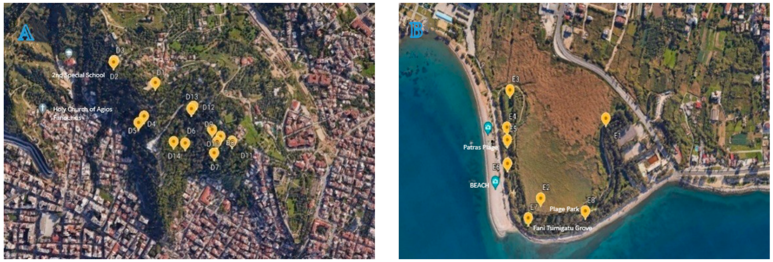
2.1. Insect Baits

The insect bait method has been applied to isolate local EPF strains from the soil. This method has been more efficient than collecting directly from soil [39]. Two stored product beetles, the confused flour beetle Tribolium confusum Jacquelin du Val (Coleoptera: Tenebrionidae) and the maize weevil Sitophilus zeamais (L.) (Coleoptera: Curculionidae) were used as baits. These insects are easily infected by EPF and their mass rearing in the lab is very simple and inexpensive.
Tribolium confusum was reared on whole wheat flour (+5% dried yeast), whereas Sitophilus zeamais was reared on hard wheat grain. All insects were maintained in cages at constant conditions of temperature (25 ± 1 °C), relative humidity (60–70%), and photoperiod (16L:8D).
Thaumetopoea pityocampa was reared after 1500 larvae were collected from five habitats in stands of Pinus nigra Arn. in Achaia, Patras between February and May 2019 (Figure 1). Several infested pine samples (50–60) were placed in sterile wet sand in plastic boxes with vented apertures and transferred to EMBIA Laboratory. The sand was moisturized once per week. Larvae were supplied with fresh maritime pine needles, the primary host species in the Achaia pine forest. Fresh twigs were provided every one or two days. All larvae were maintained at the same constant conditions with beetles.

2.2. Sampling

Seasonally, samples were collected at 22 locations (176 samples in total) in two suburban green spaces in Patras (Figure 1). Prior to sampling, we removed surface litter and plowed the soil up to a depth of 10 cm using a soil sampler. A kilo of soil was extracted from each location and kept in plastic bags at 4 °C until they were transported to the lab for additional processing. The samples were placed on rough cardboard for 24 h to reduce their humidity after being air-dried to prevent entomopathogenic nematode contamination. Following that, the soil was placed into Petri dishes after being sieved. Ten tests were performed on each soil sample; thus, 100 adults of each species were tested per sample. The samples were then stored for 14 days at 25 °C in dedicated dark chambers.

2.3. Isolation

To sterilize them, all dead adults were submerged in 6% NaOCl2 for three seconds (to prevent the growth of saprophytic fungus). Once mycelia formed, they were placed in 9 cm sterile Petri plates with ddH2O (double distilled water) impregnated Whatman paper. Conidia from the infected adults were put in 9 cm sterilized Petri plates on a layer of Sabouraud Dextrose Agar (SDA) to isolate entomopathogenic fungi. The Petri dishes were kept in a dark area (25 ± 1 °C, relative humidity 65 ± 5%) to promote fungal development. The fungi were segregated once more to stop infestation and obtain clear cultivation. Chosen isolates are stored in a deep freezer at −20 °C in our microorganism repository.

2.4. Morphological Observation

The fungal morphology was initially examined macroscopically by observing colony features (shape, color, hyphae, and size). To ensure purity and monosporic cultures, the isolates were subcultured repeatedly on plates with SDA.

2.5. Genomic DNA Extraction and Polymerase Chain Reaction (PCR)

The isolates were morphologically identified using a ZEISS Primo Star microscope (Carl Zeiss Microscopy GmbH, Jena, Germany) at 400× magnifications after being subcultured numerous times on plates with SDA to assure purity and monosporic cultures. Using a sterile loop, conidia were taken from the surfaces of dead cadavers and placed on Potato Dextrose Agar (PDA). Genomic DNA (gDNA) was isolated using the technique described by Rogers and Bendich [37,40].
A fragment of the ITS spacer region was extended using the universal primer sets ITS4 (5′ TCCTCCGCTTATTGATATGC-3′) and ITS5 (5′-GGAAGTAAAAGTCGTAAC AAGG-3′). Taq 2X Master Mix (M0270) (New England Biolabs GmbH, Frankfurt, Germany) was used to perform PCR reactions in a volume of 25 L. The reactions contained a working concentration of 1X Master Mix, 0.5 M of each primer, and 50 ng of the template gDNA. The PCR procedure for amplifying the ITS regions consisted of 33 cycles, each lasting 30 s at 95 °C, 40 s at 55 °C, and 1 min at 68 °C, with a final elongation step lasting 5 min at 68 °C. PCR products were stored at 4 °C. Gel electrophoresis was used to determine the quantity and quality of the PCR products. A 1% agarose gel was stained with SYBR Safe DNA Gel Stain (Invitrogen, Waltham, MA, USA), and the results were seen under a UV light (BIO-RAD, Molecular Imager Gel Doc XR System) (Bio-Rad Laboratory, Hercules, CA, USA).

2.6. Sequencing and Phylogenetic Analysis

The amplified products were purified and sequenced at Eurofins Genomics (Ebersberg, Germany). The Basic Local Alignment Search Tool (NCBI BLAST) was used to compare the similarity of the fungal DNA sequences in the current work with homologous sequences. Alignment of the chosen sequences was accomplished using Muscle. The Jukes–Cantor model in MEGAX was used to create a phylogenetic tree using the neighbor-joining method. Using a bootstrap analysis with 1000 repeats, branch support was calculated. The sequences were submitted to GenBank (Genbank accessions MZ047310, MZ047311).

2.7. Virulence Estimation against T. pityocampa Larvae

Conidial suspensions of B. varroae, P. lavendulum, and C. blackwelliae were prepared at a range of doses (103, 104, 105, 106, 107, and 108 conidia/mL) to assess their insecticidal potential. Ten surface sterilized T. pityocampa 3rd instar larvae were sprayed with 10 mL of each EPF conidial suspension and then placed in 9 cm sterile Petri dishes. The prepared suspensions were applied at 1 kgf cm−2 using a Potter spray tower (Burkard Manufacturing Co., Ltd., Rickmansworth, Hertfordshire, UK). Treated larvae were maintained with constant rearing conditions and supplied with fresh twigs ad libitum. Dead larvae were recorded every two days. Ten Petri dishes (repetitions) were used for each concentration. Untreated larvae sprayed with only with 10 mL of surfactant solution (Tergitol NP-9 0.05%) were used as controls. Dead larvae were removed and surface-sterilized with 2% sodium hypochlorite for a few seconds to avoid development of saprophytic fungi. Followingly, they were transferred and kept in the dark at 25 °C for 5–7 days; those that showed fungal growth were classified as infected. EPF species from each dead larva was initially identified under a microscope via observation of the conidia and hyphal growth and confirmed by PCR analysis.

2.8. Statistical Analysis

Mean values of larval mortality were compared using analysis of variance, with the main factors being EPF species, conidial dose, and exposure time. Where necessary, experimental data were arcsine transformed to meet the requirements of parametric analysis for equal variation among treatments. To find statistically significant differences between factors, the Tukey’s test was used with a significance level of 0.05. All statistical tests were performed using SPSS (SPSS, Inc., Chicago, IL, USA, version 23) [41].

3. Results

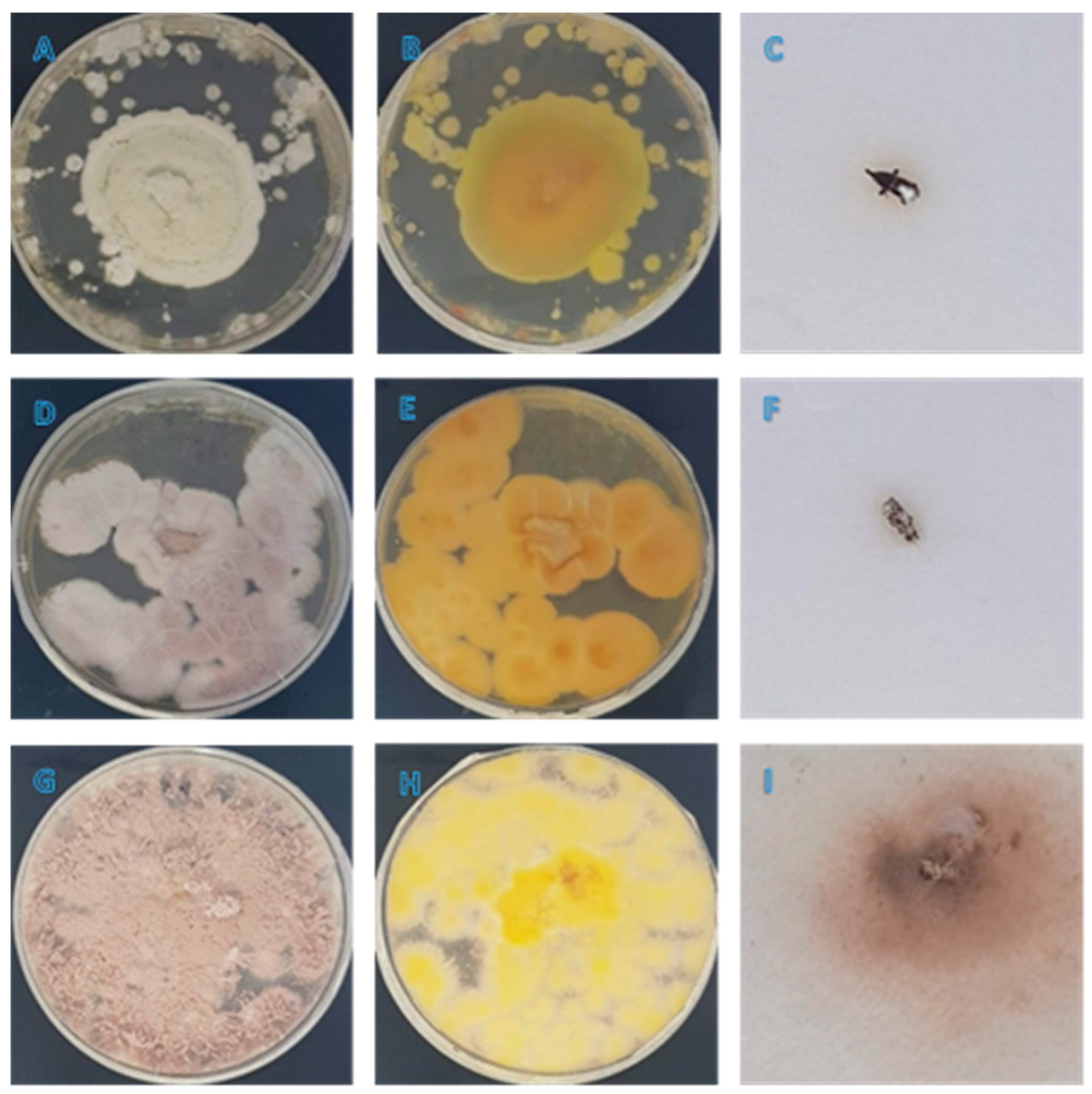
3.1. Morphological Observation

Isolates kept in Petri dishes (Figure 2) were morphologically identified using a microscope (ZEISS Primo Star, Carl Zeiss Microscopy GmbH, Jena, Germany) according to the morphological characteristics reported for B. varroae [24], P. lavendulum [29] and C. blackwelliae [42].

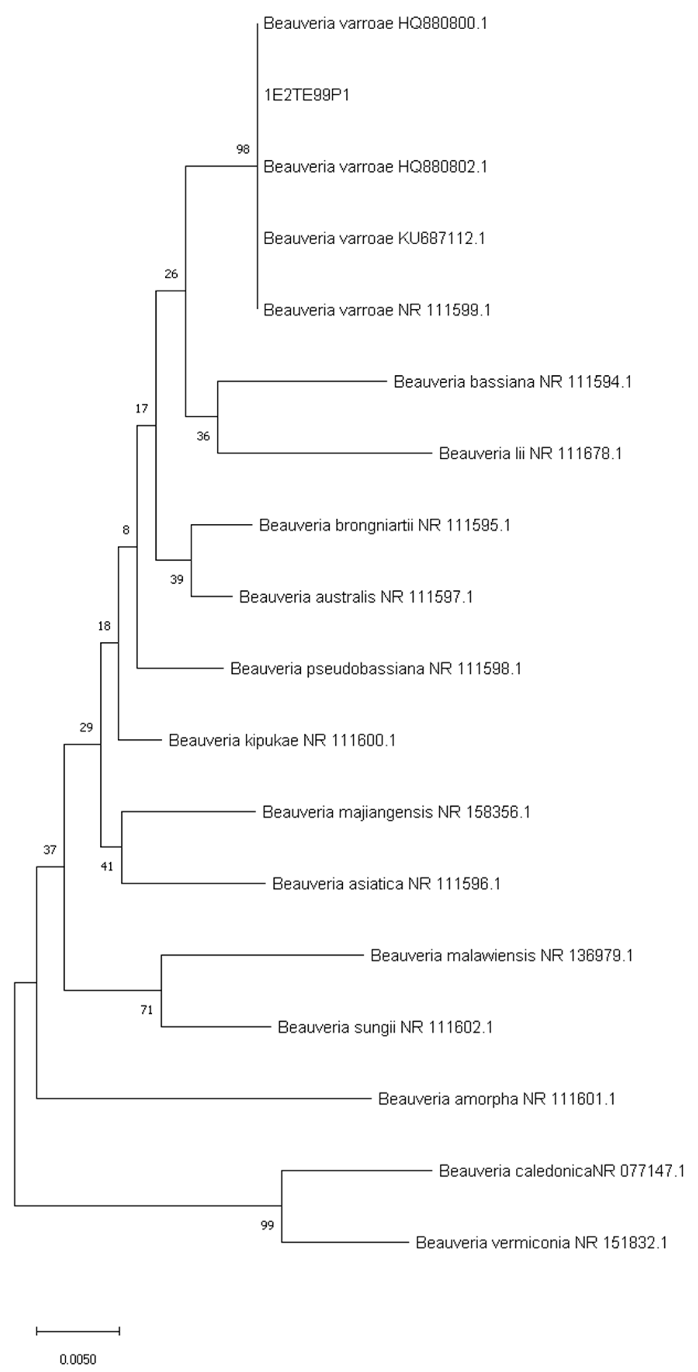
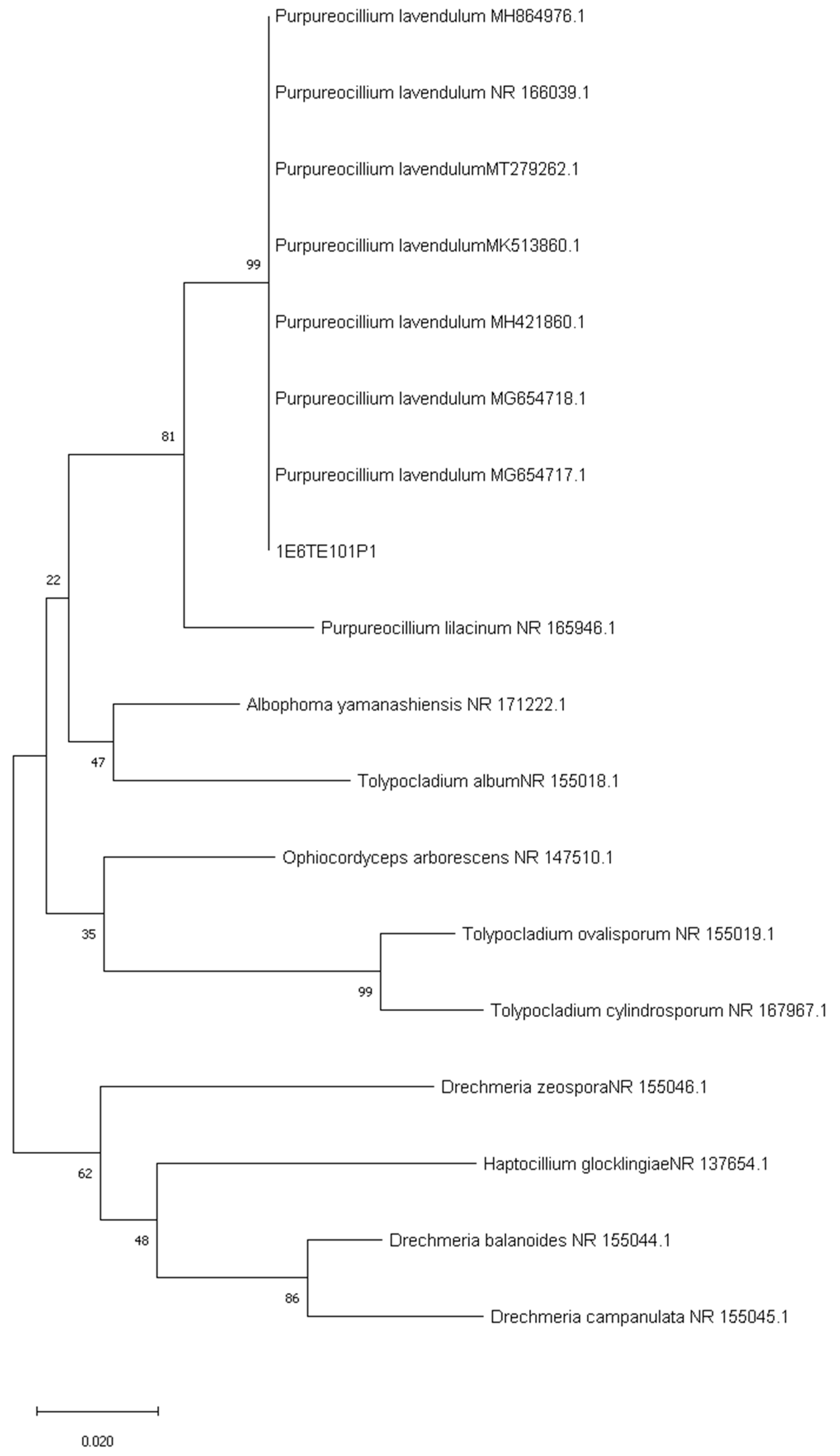
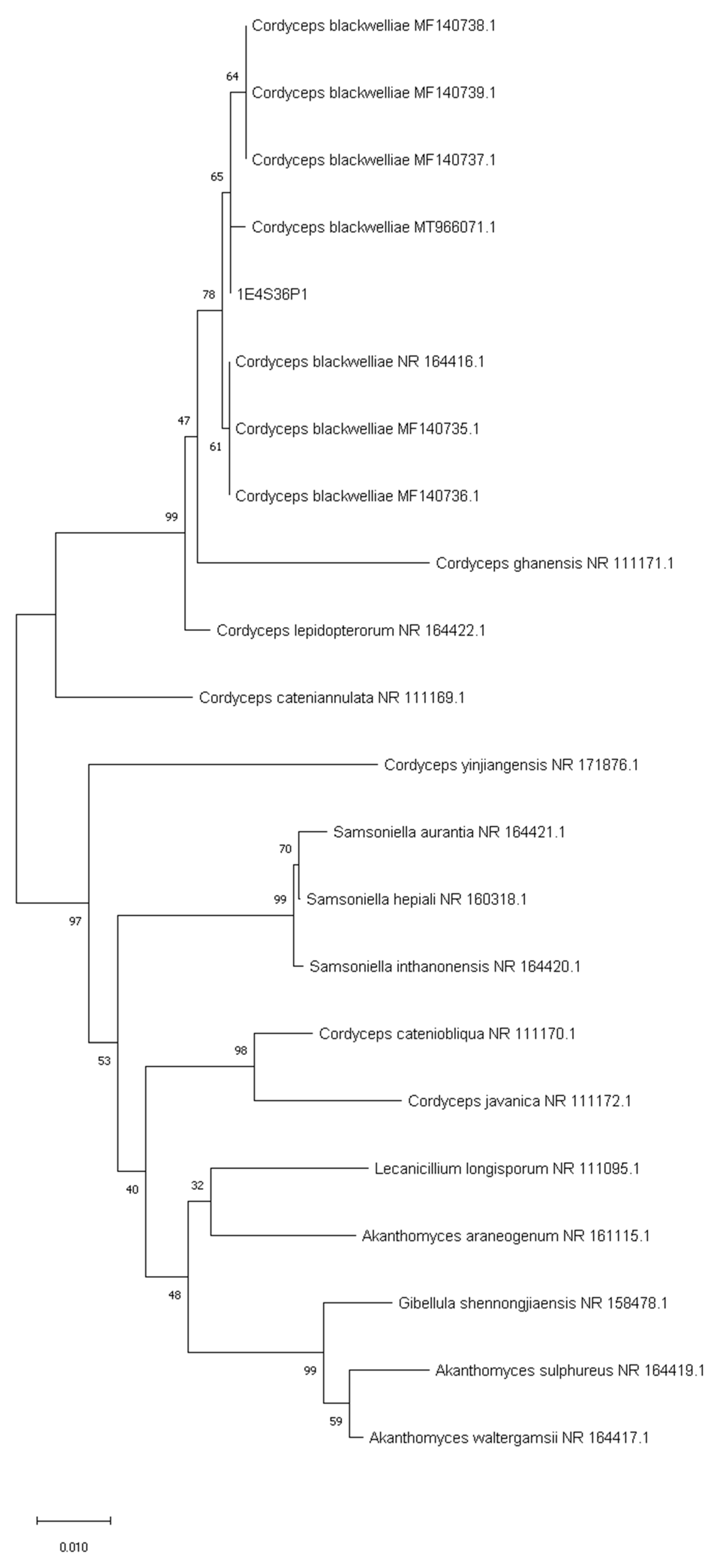
3.2. Sequencing of ITS and Phylogenetic Analysis

The E99 isolate ITS-rDNA length was 572 bp. A BLAST search revealed that the sequence of the isolate shared 99% homology with B. varroae species NR 111599.1, KU687112.1, HQ880802.1, and HQ880800.1 (Figure 3). Both morphological and molecular identification showed that our isolate was B. varroae.
The E101 isolate ITS -rDNA had a length of 589 bp. A BLAST search revealed that the sequence of the isolate had 99% homology with P. lavendulum species MG654717.1, MG654718.1, MH421860.1, MK513860.1, MT279262.1, NR 166039.1, and MH864976.1 (Figure 4). Both morphological and molecular identification showed that our isolate was P. lavendulum.
The ITS rDNA length of the D68 isolate was 585 bp. Using MEGAX for analyzing and conducting automatic and manual sequence alignment, the ITS dataset comprised 575 characters after the exclusion or replacement of ambiguous positions. A BLAST search revealed that the sequence of the isolate shared 99% homology with C. blackwelliae MT966071.1, MF140737.1, MF140738.1, MF140739.1, NR_164416.1, MF140735.1, and MF140736.1 (Figure 5). Based mainly on morphological and ancillary molecular identification, our isolate appeared to be C. blackwelliae.

3.3. Insecticidal Action of EPF on T. pityocampa Larvae

At the end of the experiment, the control mortality rate (H2O + Tergitol NP-9 0.05%) was 3.3%. However, the level of larval mortality caused by B. varroae varied according to the concentration applied (Table 1). After 144 h, the larval mortality varied from 60 to 91%. Significant main effects and interactions were found for each factor (Concentration: F = 1.107, df = 6686, p < 0.001; Exposure Time: F = 3.031, df = 2437, p < 0.001; Concentration × Exposure Time: F = 0.716, df = 151,127, p < 0.001).
After 144 h, P. lavendulum caused larval mortality rates ranging from 60 to 91%. The main effects and interactions of all factors were found to be significant (Table 2) (Concentration: F = 1.194, df = 6686, p < 0.001; Exposure Time: F = 3.017, df = 2437, p < 0.001; Concentration × Exposure Time: F = 1.506, df = 151,127, p < 0.001).
In addition, C. blackwelliae exhibited varying levels of larval mortality (Table 3). Larval mortality after 144 h varied from 43 to 77%. All factors’ main impacts and interactions were found to be significant. (Concentration: F = 1.386, df = 6686, p < 0.001; Exposure Time: F = 3.457, df = 2437, p < 0.001; Concentration × Exposure Time: F = 3.913, df = 151,127, p < 0.001).

4. Discussion

It is estimated that 750 different EPF species infect insects and mites. [4]. Although they target a variety of species, distinct fungal species have an extremely narrow target range. EPF are located in a variety of environments according to the insect host, habitat, and region, including insects and other arthropod pathogens, in soil and phylloplanes, and as endophytes [6,25,43,44,45,46].
To the best of our knowledge, this is the first study to report these EPF isolates of entomopathogenic fungi in Greece. The study depended not only on morphological characteristics but also on the molecular identification of the new isolates. The presence of EPF in the soil can be influenced by a variety of factors, including the type of soil [14], the species of cultivated plants [16], and agricultural techniques [15,17,18]. The level of soil colonization by these EPF can be a good predictor of their condition because of their low tolerance to environmental changes [13].
Our results indicate that this is the first time that B. varroae has been reported to occur in the soils of Greece. Only two soil isolates of B. varroae have been previously reported: one from Iraq [28] and the second from Portugal, which was isolated from hedgerow soil using mainly Quercus spp. and Pinus spp. [47]. In contrast, B. varroae was isolated from the varroa mite Varroa destructor Anderson & Trueman (Mesostigmata: Varroidae) in France and from weevils of Larinus sp. (Coleoptera: Curculionidae) in Switzerland [21]. It has also been isolated from insect pests of stored wheat and maize in Turkey [48] and from a lepidopteran insect found in beet fields in Iran [49]. In general, B. varroae is rarely found.
Moreover, this is the first time that C. blackwelliae has been reported to occur in and been isolated from Greek soils. Hosts of Cordyceps with Isaria-like morphs are reported on a wide range of insects such as coleopteran larvae and pupae, lepidopteran larvae or pupae, cicada nymphs, and white flies [42,50,51,52]. This EPF species seems difficult to isolate from soil.
Additionally, this is the first time that P. lavendulum has been found in Greece. It has also been isolated from soil in Caracas, Venezuela [29]. The conidia of the fungus are subglobose or lemon shaped and emit a yellowish pigment that diffuses throughout the culture [29]. In addition, Perdomo et al. [29] reported that P. lavendulum was isolated from bronchial wash in Reus, Spain and from Aethus sp. in Tafo, Ghana.
In the present study, the virulence and sporulation ability of three EPF species, recorded for the first time from Greece, were evaluated. Mortality varied with the species, reaching 71–91% at the highest dose after 144 h. These results show that the studied EPF species were able to kill the larvae of T. pityocampa in a controlled environment, and therefore, they could be suitable candidates for use as microbial biocontrol agents in future programs to reduce T. pityocampa populations.
Er et al. [53] showed that P. fumosoroseus and P. farinosus were also highly virulent against T. pityocampa larvae. Moreover, L. lecanii recorded only 54.6% mortality [53], whereas another isolate of the same fungus killed up to 100% of larvae 6 days after inoculation [54]. Sonmez et al. [55] found that the EPF M. brunneum and B. bassiana were highly pathogenic to the larvae of T. pityocampa. Although the susceptibility of the different instars varied, with the fourth instar being the least sensitive, mortality was dose dependent [55]. Other studies have also showed that isolates of B. bassiana, M. anisopliae, and Isaria farinosa (Holmsk.) (Hypocreales: Cordycipitaceae) could be very pathogenic against fourth instar larvae of T. pityocampa [56,57,58].
Differences between the current study and previous ones [53,54,55,56,57,58] are attributed to the different EPF isolates, host larval instars, application methods, temperatures, and relative humidities. It has been well documented that the EPF efficacy against T. pityocampa larvae is significantly affected by the larval instar, EPF isolate and applied doses, and EPF application method [55,58]. In addition, it is probable that EPF species have ecological suitability for use against pests because of their habitat and geographic location [59].
These findings demonstrate the discovery of novel, previously undiscovered local EPF species that may be more suitable and efficient for biological control in IPM programs than those currently available in Greece. Additional research should be carried out to harness this natural resource to detect and preserve variety and to incorporate the results into local crop protection strategies.

5. Conclusions

The present work presents information on EPF that could be associated with agricultural and forest environment interests in Greece. Three new native species of entomopathogenic fungi were isolated from suburban soil samples in Greece for the first time. According to their macroscopic and microscopic traits, the three species corresponded to B. varroae, C. blackwelliae, and P. lavendulum. The amplification and sequencing of the ITS-rDNA sequences, comparison of the molecular databases, and phylogenetic analyses confirmed the taxonomic identity of the species. All species proved to be highly virulent against third instar larvae of T. pityocampa. Their insecticidal ability deserves further research to evaluate its potential for use in biological control programs.

Author Contributions

Conceptualization, S.M.; methodology, I.L. and S.M.; software, S.M.; validation, I.L., S.M., P.A.E. and K.P.; formal analysis, I.L., S.M., P.A.E. and K.P.; investigation, I.L. and S.M.; resources, S.M.; data curation, S.M.; writing—original draft preparation, I.L. and S.M.; writing—review and editing, I.L., S.M. and K.P.; visualization, S.M.; supervision, K.P.; project administration, S.M. and K.P.; funding acquisition, K.P. All authors have read and agreed to the published version of the manuscript.

Funding

This research received no external funding.

Institutional Review Board Statement

Not applicable.

Informed Consent Statement

Not applicable.

Data Availability Statement

Not applicable.

Conflicts of Interest

The authors declare no conflict of interest.

References

  1. Qu, S.; Wang, S. Interaction of entomopathogenic fungi with the host immune system. Dev. Comp. Immunol. 2018, 83, 96–103. [Google Scholar] [CrossRef] [PubMed]
  2. Suh, S.O.; Noda, H.; Blackwell, M. Insect symbiosis: Derivation of yeast-like endosymbionts within an entomopathogenic filamentous lineage. Mol. Biol. Evol. 2001, 18, 995–1000. [Google Scholar] [CrossRef] [PubMed]
  3. Mueller, U.G.; Gerardo, N.M.; Aanen, D.K.; Six, D.L.; Schultz, T.R. The evolution of agriculture in insects. Annu. Rev. Ecol. Evol. Syst. 2005, 36, 563–595. [Google Scholar] [CrossRef]
  4. Mantzoukas, S.; Eliopoulos, P.A. Endophytic entomopathogenic fungi: A valuable biological control tool against plant pests. Appl. Sci. 2020, 10, 360. [Google Scholar] [CrossRef] [Green Version]
  5. Shah, P.A.; Pell, J.K. Entomopathogenic fungi as biological control agents. Appl. Microbiol. Biotechnol. 2003, 61, 413–423. [Google Scholar] [CrossRef] [PubMed]
  6. Vega, F.E.; Goettel, M.S.; Blackwell, M.; Chandler, D.; Jackson, M.A.; Keller, S.; Koike, M.; Maniania, N.K.; Monzón, A.; Ownley, B.H.; et al. Fungal entomopathogens: New insights on their ecology. Fungal Ecol. 2009, 2, 149–159. [Google Scholar] [CrossRef] [Green Version]
  7. Mantzoukas, S.; Lagogiannis, I.; Mpousia, D.; Ntoukas, A.; Karmakolia, K.; Eliopoulos, P.A.; Poulas, K. Beauveria bassiana endophytic strain as plant growth promoter: The case of the grape vine Vitis vinifera. J. Fungi 2021, 7, 142. [Google Scholar] [CrossRef]
  8. Aly, A.H.; Debbab, A.; Kjer, J.; Proksch, P. Fungal endophytes from higher plants: A prolific source of phytochemicals and other bioactive natural products. Fungal Divers. 2010, 41, 1–16. [Google Scholar] [CrossRef]
  9. Mantzoukas, S.; Pettas, I.; Lagogiannis, I. Stored product pests as models for trapping entomopathogenic fungi from olive tree orchards in Western Greece. J. Stored Prod. Res. 2020, 87, 101584. [Google Scholar] [CrossRef]
  10. Zimmermann, G. The Galleria bait method for detection of entomopathogenic fungi in soil. J. Appl. Entomol. 1986, 102, 213–215. [Google Scholar] [CrossRef]
  11. Mantzoukas, S.; Lagogiannis, I.; Ntoukas, A.; Eliopoulos, P.A.; Kouretas, D.; Karpouzas, D.G.; Poulas, K. Trapping entomopathogenic fungi from vine terroir soil samples with insect baits for controlling serious pests. Appl. Sci. 2020, 10, 3539. [Google Scholar] [CrossRef]
  12. Lovett, B.; St. Leger, R.J. The insect pathogens. Microbiol. Spectr. 2017, 5. [Google Scholar] [CrossRef] [PubMed]
  13. Meyling, N.V.; Eilenberg, J. Occurrence and distribution of soil borne entomopathogenic fungi within a single organic agroecosystem. Agric. Ecosyst. Environ. 2006, 113, 336–341. [Google Scholar] [CrossRef]
  14. Tkaczuk, C.; Mietkiewski, R. Occurrence of entomopathogenic fungi in different kinds of soil. Rocz. Nauk. Rol. Ser. E 1996, 25, 41–46. [Google Scholar]
  15. Quesada-Moraga, E.; Navas-Cortés, J.A.; Maranhao, E.A.A.; Ortiz-Urquiza, A.; Santiago-Álvarez, C. Factors affecting the occurrence and distribution of entomopathogenic fungi in natural and cultivated soils. Mycol. Res. 2007, 111, 947–966. [Google Scholar] [CrossRef]
  16. Krysa, A.; Ropek, D.; Kuzniar, T. The occurrence of entomopathogenic fungi depending on season in selected organic farm. J. Res. Appl. Agric. Eng. 2012, 57, 226–230. [Google Scholar]
  17. Oliveira, I.; Pereira, J.A.; Quesada-Moraga, E.; Lino-Neto, T.; Bento, A.; Baptista, P. Effect of soil tillage on natural occurrence of fungal entomopathogens associated to Prays oleae Bern (Lepidoptera, Plutellidae). Sci. Hortic. 2013, 159, 190–196. [Google Scholar] [CrossRef]
  18. Jabbour, R.; Barbercheck, M.E. Soil management effects on entomopathogenic fungi during the transition to organic agriculture in a feed grain rotation. Biol. Control 2009, 51, 435–443. [Google Scholar] [CrossRef]
  19. Roberts, D.W.; Hajek, A.E. Entomopathogenic fungi as bioinsecticides. In Frontiers in Industrial Mycology; Leatham, G.F., Ed.; Chapman & Hall: New York, NY, USA, 1992; pp. 144–159. [Google Scholar]
  20. Goettel, M.S.; Eilenberg, J.; Glare, T.R. Entomopathogenic fungi and their role in regulation of insect populations. In Comprehensive Molecular Insect Science; Gilbert, L.I., Iatrou, K., Gill, S., Eds.; Elsevier: Oxford, UK, 2005; Volume 6, pp. 361–406. [Google Scholar] [CrossRef]
  21. Rehner, S.A.; Minnis, A.M.; Sung, G.H.; Luangsa-ard, J.J.; Devotto, L.; Humber, R.A. Phylogeny and systematics of the anamorphic, entomopathogenic genus Beauveria. Mycologia 2011, 103, 1055–1073. [Google Scholar] [CrossRef]
  22. Wang, Y.; Tang, D.X.; Duan, D.E.; Wang, Y.B.; Yu, H. Morphology, molecular characterization, and virulence of Beauveria pseudobassiana isolated from different hosts. J. Invertebr. Pathol. 2020, 172, 107333. [Google Scholar] [CrossRef]
  23. Zhang, S.L.; He, L.M.; Chen, X.; Huang, B. Beauveria lii sp. nov. isolated from Henosepilachna vigintioctopunctata. Mycotaxon 2012, 121, 199–206. [Google Scholar] [CrossRef]
  24. Imoulan, A.; Hussain, M.; Kirk, P.M.; El Meziane, A.; Yao, Y.J. Entomopathogenic fungus Beauveria: Host specificity, ecology and significance of morpho-molecular characterization in accurate taxonomic classification. J. Asia Pac. Entomol. 2017, 20, 1204–1212. [Google Scholar] [CrossRef]
  25. Ghikas, D.V.; Kouvelis, V.N.; Typas, M.A. Phylogenetic and biogeographical implications inferred by mitochondrial intergenic region analyses and ITS1- 5.8S-ITS2 of the entomopathogenic fungi B. bassiana and B. brongniartii. BMC Microbiol. 2010, 10, 174–188. [Google Scholar] [CrossRef] [PubMed] [Green Version]
  26. Chen, X.; Huang, C.; He, L.; Zhang, S.; Li, Z. Molecular tracing of white muscardine in the silkworm, Bombyx mori (Linn.) II. Silkworm white muscardine is not caused by artificial release or natural epizootic of Beauveria bassiana in China. J. Invert. Path. 2015, 125, 16–22. [Google Scholar] [CrossRef] [PubMed]
  27. Coates, B.S.; Hellmich, R.L.; Lewis, L.C. Beauveria bassiana haplotype determination based on nuclear rDNA internal transcribed spacer PCR-RFLP. Mycol. Res. 2002, 106, 40–50. [Google Scholar] [CrossRef] [Green Version]
  28. Hassan, F.R.; Abdullah, S.K.; Assaf, L.H. First record of Βeauveria varroae from Iraq. Nov. Hedwigia 2019, 108, 427–433. [Google Scholar] [CrossRef]
  29. Perdomo, H.; Cano, J.; García, D.; Gené, J.; Hernández, M.; Guarro, J. Polyphasic analysis of Purpureocillium lilacinum isolates from different origins and proposal of the new species Purpureocillium lavendulum. Mycologia 2013, 105, 151–161. [Google Scholar] [CrossRef]
  30. Liu, L.; Cao, Y.R.; Zhang, C.C.; Fan, H.F.; Guo, Z.Y.; Yang, H.Y.; Chen, M.; Han, J.J.; Xu, J.; Zhang, K.Q.; et al. An efficient gene disruption system for the nematophagous fungus Purpureocillium lavendulum. Fungal Biol. 2019, 123, 274–282. [Google Scholar] [CrossRef]
  31. Liang, L.M.; Zhang, Y.; Xu, J.; Zhang, K.Q.; Cao, Y.R. Characterization of the complete mitochondrial genome of the nematophagous fungus Purpureocillium lavendulum. Mitochondrial DNA Part B Resour. 2021, 6, 33–35. [Google Scholar] [CrossRef]
  32. Luangsa-Ard, J.; Houbraken, J.; van Doorn, T.; Hong, S.B.; Borman, A.M.; Hywel-Jones, N.L.; Samson, R.A. Purpureocillium, a new genus for the medically important Paecilomyces lilacinus. FEMS Microbiol. Lett. 2011, 321, 141–149. [Google Scholar] [CrossRef]
  33. Olatunji, O.J.; Tang, J.; Tola, A.; Auberon, F.; Oluwaniyi, O.; Ouyang, Z. The genus Cordyceps: An extensive review of its traditional uses, phytochemistry and pharmacology. Fitoterapia 2018, 129, 293–316. [Google Scholar] [CrossRef]
  34. Panda, A.K.; Swain, K.C. Traditional uses and medicinal potential of Cordyceps sinensis of Sikkim. J. Ayurveda Integr. Med. 2011, 2, 9–13. [Google Scholar] [CrossRef] [Green Version]
  35. Holliday, J.C.; Cleaver, M.P. Medicinal value of the caterpillar fungi species of the genus Cordyceps (Fr.) Link (Ascomycetes). A review. Int. J. Med. Mushrooms 2008, 10, 219–234. [Google Scholar] [CrossRef]
  36. Zhou, X.; Gong, Z.; Su, Y.; Lin, J.; Tang, K. Cordyceps fungi: Natural products, pharmacological functions and developmental products. J. Pharm. Pharmacol. 2009, 61, 279–291. [Google Scholar] [CrossRef]
  37. Jacquet, J.-S.; Orazio, C.; Jactel, H. Defoliation by processionary moth significantly reduces tree growth, a quantitative review. Ann. Forest Sci. 2012, 69, 857–866. [Google Scholar] [CrossRef] [Green Version]
  38. Rodriguez-Mahillo, A.I.; Gonzalez-Muñoz, M.; Vega, J.M.; López, J.A.; Yart, A.; Kerdelhué, C.; Camafeita, E.; Ortiz, J.C.G.; Vogel, H.; Toffolo, E.P.; et al. Setae from the pine processionary moth (Thaumetopoea pityocampa) contain several relevant allergens. Contact Derm. 2012, 67, 367–374. [Google Scholar] [CrossRef]
  39. Chang, J.C.; Wu, S.S.; Liu, Y.C.; Yang, Y.H.; Tsai, Y.F.; Li, Y.H.; Tseng, C.T.; Tang, L.C.; Nai, Y.S. Construction and selection of an entomopathogenic fungal library from soil samples for controlling Spodoptera litura. Front. Sustain. Food Syst. 2021, 5, 596316. [Google Scholar] [CrossRef]
  40. Rogers, S.O.; Bendich, A.J. Extraction of DNA from milligram amounts of fresh, herbarium and mummified plant tissues. Plant. Mol. Biol. 1985, 5, 69–76. [Google Scholar] [CrossRef]
  41. IBM Corp. IBM SPSS Statistics for Windows; Version 27.0; IBM Corp: Armonk, NY, USA, 2020. [Google Scholar]
  42. Mongkolsamrit, S.; Noisripoom, W.; Thanakitpipattana, D.; Wutikhun, T.; Spatafora, J.W.; Luangsa-Ard, J. Disentangling cryptic species with isaria-like morphs in Cordycipitaceae. Mycologia 2018, 110, 230–257. [Google Scholar] [CrossRef] [PubMed]
  43. Rehner, S.A.; Posada, F.; Buckley, E.P.; Infante, F.; Castillo, A.; Vega, F.E. Phylogenetic origins of African and Neotropical Beauveria bassiana s.l. pathogens of the coffee berry borer, Hypothenemus hampei. J. Invertebr. Pathol. 2006, 93, 11–21. [Google Scholar] [CrossRef] [PubMed]
  44. Glare, T.R.; Reay, S.D.; Nelson, T.L.; Moore, R. Beauveria caledonica is a naturally occurring pathogen of forest beetles. Mycol. Res. 2008, 112, 352–360. [Google Scholar] [CrossRef] [PubMed]
  45. Aquino De Muro, M.; Elliott, S.; Moore, D.; Parker, B.L.; Skinner, M.; Reid, W.; El Bouhssini, M. Molecular characterisation of Beauveria bassiana isolates obtained from overwintering sites of Sunn Pests (Eurygaster and Aelia species). Mycol. Res. 2005, 109, 294–306. [Google Scholar] [CrossRef]
  46. Meyling, N.V.; Hajek, A.E. Principles from community and metapopulation ecology: Application to fungal entomopathogens. BioControl 2010, 55, 39–54. [Google Scholar] [CrossRef]
  47. Sharma, L.; Oliveira, I.; Torres, L.; Marques, G. Entomopathogenic fungi in Portuguese vineyards soils: Suggesting a ‘Galleria-Tenebrio-bait method’ as bait-insects Galleria and Tenebrio significantly underestimate the respective recoveries of Metarhizium robertsii and Beauveria bassiana. MycoKeys 2018, 38, 1–23. [Google Scholar] [CrossRef] [PubMed]
  48. Er, M.K.; Tunaz, H.; Ücük, C.; Bariş, C.; Işikber, A.A. Occurrence of entomopathogenic fungi on insect pests of stored wheat and maize in Central and South Anatolia in Turkey. Turk. Entomoloji Derg. 2016, 40, 249–263. [Google Scholar] [CrossRef] [Green Version]
  49. Darsouei, R.; Karimi, J.; Ghadamyari, M.; Hosseini, M. Natural Enemies of the Sugar Beet Army Worm, Spodoptera exigua (Lepidoptera: Noctuidae) in Northeast Iran. Entomolog. News 2018, 127, 446–464. [Google Scholar] [CrossRef]
  50. Samson, R.A. Paecilomyces and some allied hyphomycetes. Stud. Mycol. 1974, 6, 1–119. [Google Scholar]
  51. Sung, G.H.; Hywel-Jones, N.L.; Sung, J.M.; Luangsa-ard, J.J.; Shrestha, B.; Spatafora, J.W. Phylogenetic classification of Cordyceps and the clavicipitaceous fungi. Stud. Mycol. 2007, 57, 5–59. [Google Scholar] [CrossRef] [Green Version]
  52. Gallou, A.; Serna-Domínguez, M.G.; Berlanga-Padilla, A.M.; Ayala-Zermeño, M.A.; Mellín-Rosas, M.A.; Montesinos-Matías, R.; Arredondo-Bernal, H.C. Species clarification of Isaria isolates used as biocontrol agents against Diaphorina citri (Hemiptera: Liviidae) in Mexico. Fungal Biol. 2016, 120, 414–423. [Google Scholar] [CrossRef]
  53. Er, M.K.; Tunaz, H.; Gökçe, A. Pathogenicity of entomopathogenic fungi to Thaumetopoea pityocampa (Schiff.) (Lepidoptera: Thaumatopoeidae) larvae in laboratory conditions. J. Pest Sci. 2007, 80, 235–239. [Google Scholar] [CrossRef]
  54. Paparatti, B.; Fabozzi, R. A new pathogen of the pine processionary caterpillar (Thaumetopoea pityocampa Den. et Schif.), Lepidoptera: Thaumetopoeidae. Inf. Fitopatol. 1988, 38, 45–48. [Google Scholar]
  55. Sönmez, E.; Demir, İ.; Bull, J.C.; Butt, T.M.; Demirbağ, Z. Pine processionary moth (Thaumetopoea pityocampa, Lepidoptera: Thaumetopoeidae) larvae are highly susceptible to the entomopathogenic fungi Metarhizium brunneum and Beauveria bassiana. Biocontrol Sci. Tech. 2017, 27, 1168–1179. [Google Scholar] [CrossRef]
  56. Akinci, H.A.; Ozman-Sullivan, S.K.; Diler, H.; Celik, N.; Sullivan, G.T.; Karaca, G. Entomopathogenic fungi isolated from Thaumetopoea pityocampa and their efficacies against its larvae. Fresenius Environ. Bull. 2017, 26, 5251–5257. [Google Scholar]
  57. Ozdemir, I.O.; Kushiyev, R.; Erper, I.; Tuncer, C. Efficacy of entomopathogenic fungi Beauveria bassiana and Metarhizium anisopliae against Thaumetopoea pityocampa Shiff.(Lepidoptera: Thaumatopoeidae). Arch. Phytopathol. Plant Prot. 2019, 52, 470–480. [Google Scholar] [CrossRef]
  58. Sevim, A.; Demir, I.; Demirbağ, Z. Molecular characterization and virulence of Beauveria spp. from the pine processionary moth, Thaumetopoea pityocampa (Lepidoptera: Thaumetopoeidae). Mycopathologia 2010, 170, 269–277. [Google Scholar] [CrossRef] [PubMed]
  59. Sevim, A.; Demir, I.; Sönmez, E.; Kocacevik, S.; Demirbağ, Z. Evaluation of entomopathogenic fungi against the sycamore lace bug, Corythucha ciliata (Say) (Hemiptera: Tingidae). Turk. J. Agric. Forest. 2013, 37, 595–603. [Google Scholar] [CrossRef]
Figure 1. Suburban areas in the prefecture of Achaia where the soil samples were collected (A) DASYLIO (38°24′90.209″ N, 21°74′56.985″ E) and (B) ELOS (38°27′99.487″ N, 21°74′7.156″ E).
Diversity 15 00312 g001
Figure 2. Cultures of EPF species examined during the present study: B. varroae colony top (A) and bottom (B), B. varroae infecting S. zeamais. (C). P. lavendulum colony top (D) and bottom (E), P. lavendulum infecting S. zeamais. (F). C. blackwelliae colony top (G) and bottom (H), C. blackwelliae infecting S. zeamais (I).
Diversity 15 00312 g002
Figure 3. Phylogenetic tree of B. varroae with the new isolate from Greece (1E2T99P1 GenBank accession MZ047310) and related species from GenBank. The species names are followed by the GenBank accession numbers.
Diversity 15 00312 g003
Figure 4. Phylogenetic tree of P. lavendulum with the new isolate from Greece (1E6TE101P1 (GenBank accession MZ047311) and related species from GenBank. The species names are followed by the GenBank accession numbers.
Diversity 15 00312 g004
Figure 5. Phylogenetic tree of C. blackwelliae with the new isolate from Greece (1E4S36P1 RID: 8E2M17WB01R) and related species from GenBank. The species names are followed by the GenBank accession numbers.
Diversity 15 00312 g005
Table 1. Mean mortality (±SD) of T. pityocampa larvae. There is no significant difference between the means of the same column followed by the same letter (Tukey’s test, a = 0.05), HAT: Hours After Treatment.
TreatmentConcentrationExposure Time
48 HAT96 HAT144 HAT
B. varroae10320.0 ± 0.0 d47.0 ± 1.8 d60.0 ± 5.8 c
10436.3 ± 3.1 c53.0 ± 3.8 c63.0 ± 5.8 c
10543.3 ± 1.8 b60.0 ± 5.3 b67.0 ± 5.8 c
10646.4± 5.8 b63.0 ± 6.2 b80.0 ± 1.3 b
10750.0 ± 2.2 a73.0 ± 1.5 a89.0 ± 1.5 a
10853.0 ± 1.3 a83.0 ± 5.3 a91.0 ± 5.8 a
ControlH2O + Tergitol NP-90.0 ± 0.0 e3.7 ± 4.3 e4.7 ± 1.3 d
Table 2. Mean mortality (±SD) of T. pityocampa larvae. There is no significant difference between the mean of the same column followed by the same letter (Tukey’s test, a = 0.05), HAT: Hours After Treatment.
TreatmentConcentrationExposure Time
48 HAT96 HAT144 HAT
P. lavendulum10320.0 ± 2.0 e37.0 ± 3.8 c43.0 ± 6.1 c
10436.0 ± 1.0 d43.0 ± 4.8 c53.0 ± 2.8 b
10540.0 ± 0.0 c43.0 ± 1.3 c57.0 ± 2.8 b
10643.4 ± 2.8 b60.0 ± 1.0 b60.0 ± 2.1 b
10750.0 ± 3.1 a63.0 ± 3.5 b73.0 ± 1.5 a
10853.0 ± 5.3 a73.0 ± 5.3 a77.0 ± 2.1 a
ControlH2O + Tergitol NP-90.0 ± 0.0 f3.7± 4.3 d4.7 ± 1.3 d
Table 3. Mean mortality (±SD) of T. pityocampa larvae. There is no significant difference between the mean of the same column followed by the same letter (Tukey’s test, a = 0.05), HAT: Hours After Treatment.
TreatmentConcentrationExposure Time
48 HAT96 HAT144 HAT
Cordyceps Blackwelliae10312.1 ± 1.9 e13.3 ± 5.8 e20.0 ± 0.0 d
10420.0 ± 0.0 d33.0 ± 3.8 d43.0 ± 3.8 c
10536.0 ± 2.1 c43.0 ± 1.3 c47.0 ± 4.8 c
10643.4 ± 5.8 b50.0 ± 0.0 b60.0 ± 0.0 b
10750.0 ± 3.2 b53.0 ± 6.2 b67.0 ± 2.3 a
10863.0 ± 1.3 a63.0 ± 3.3 a71.0 ± 1.8 a
ControlH2O + Tergitol NP-90.0 ± 0.0 f3.7 ± 4.3 f4.7 ± 1.3 e
Disclaimer/Publisher’s Note: The statements, opinions and data contained in all publications are solely those of the individual author(s) and contributor(s) and not of MDPI and/or the editor(s). MDPI and/or the editor(s) disclaim responsibility for any injury to people or property resulting from any ideas, methods, instructions or products referred to in the content.

