Article

‘Unfinished’ Morphogenesis Hides Different Speciation Pathways in Charophytes: Evidence from the 190-Year-Old Original Material of Chara denudata (Charales, Charophyceae)

by
Roman E. Romanov
1,2,*,
Sophia S. Barinova
3,
Vyacheslav Yu. Nikulin
4 and
Andrey A. Gontcharov
4
1
Komarov Botanical Institute of the Russian Academy of Sciences, Professora Popova Str., 2, 197376 St. Petersburg, Russia
2
Institute for Water and Environmental Problems, Siberian Branch of the Russian Academy of Sciences, Molodezhnaya Str., 1, 656038 Barnaul, Russia
3
Institute of Evolution, University of Haifa, Abba Khoushi Ave, 199, Mount Carmel, Haifa 3498838, Israel
4
Federal Scientific Center of the East Asia Terrestrial Biodiversity, Far Eastern Branch of the Russian Academy of Sciences, 100-letiya Vladivostoka Ave, 159, 690022 Vladivostok, Russia
*
Author to whom correspondence should be addressed.
Diversity 2023, 15(2), 249; https://doi.org/10.3390/d15020249
Submission received: 24 November 2022 / Revised: 3 January 2023 / Accepted: 6 February 2023 / Published: 9 February 2023

Abstract

:
Several Chara L. species have ‘unfinished’ morphogenesis that is recognizable because of their imperfect stem and branchlet cortication compared to the perfectly corticated species. Chara denudata A. Braun, described from South Africa, is one of these species, assumed for a long time to be conspecific with C. dissoluta A. Braun ex Leonhardi, as described from Central Europe. An attempt to resolve this long-lasting uncertainty in the framework of integrative taxonomy is implemented here. The restudy of the original material of both species showed similarities but did not identify a hiatus in their morphological traits, which represents evidence for their placement in the subsection Chara R.D. Wood according to morphology. Bifid adaxial bract cells, a trait rarely encountered among charophytes, were found for the first time in C. dissoluta. According to the rbcL and matK sequences, C. denudata was unexpectedly placed within the section Grovesia R.D. Wood, far from the clusters of the section Chara with C. dissoluta. This is in obvious disagreement with the position of C. denudata according to morphology. Both species were distinct according to their biology, habitat preference, and distribution and were accepted as distinct species. Therefore, the ‘unfinished’ morphogenesis resulting in morphological similarity hides different speciation pathways in charophytes.

1. Introduction

Charophytes are easily recognizable plants because of their typical morphology and the general habit arising from repeating more or less similar modules. Many entities described as species are still debatable and not clearly outlined in individual clusters of macrospecies, according to the species concept in the last world monograph available [1,2]. The wide species concept implemented within it has been almost abandoned in regional treatments of charophytes, but uncertainties and enigmas remain among more than 400 extant species described to date. Some species have transitional, intermediate forms, leaving no space for a clear hiatus between extreme variants of the visible ‘continuum’.
The simplified morphology of some Chara L. species due to them not being fully developed, or, in other words, the incomplete cortication of individual internodes of the stem and branchlets, resulted in the descriptions of several species differing in the degree of cortex development. The species Chara brionica Stapf, C. denudata A. Braun, C. disjuncta Nordstedt, C. dissoluta A. Braun ex Leonhardi, C. howeana (Wood) Ling, Xie et Qiu, C. imperfecta A. Braun in Durieu, and C. scepusienis Fil. are the best examples of these cases. Some of them have been known for a long time from small areas or from only a few localities. Their morphology can be recognized as a result of ‘unfinished’ morphogenesis because of their imperfect stem and branchlet cortication compared to species with the complete cortication of the stem internodes and branchlet segments. It has been hypothesized that this morphology originates from the different growth rates of internodal and cortical cells during plant ontogenesis. All species with imperfect cortication have ‘counterparts’ with perfect cortication, e.g., C. contraria A. Braun ex Kützing for C. denudata and C. dissoluta, C. tomentosa L. for C. disjuncta, C. vulgaris L. for C. brionica, C. lipkinii R.E. Romanov et al. for C. imperfecta, and C. contraria or C. vulgaris for both C. howeana, and C. scepusiensis, which can be easily linked to these species with impoverished morphology but only from a morphological perspective. Both morphology and distribution are good bases for doubts about their species’ rank, raising questions about the reliability of these taxonomic decisions based on a limited number of studied plants whose morphology can be environmentally related.
The uncertainty associated with C. denudata and C. dissoluta [3,4] needs to be resolved in the framework of the ongoing project ‘The European Charophyte Monograph,’ which aims to cover all species known in Europe [5]. An integrative taxonomy, i.e., the combination of the results of morphological (both light and electron microscopy) and genetic studies with ecological and distributional traits, is the most fruitful for charophytes [6,7,8,9,10,11,12,13], although it is not helpful for all cases [14,15,16]. Here, we implemented a delineation between C. denudata, described from the shallow water bodies in South Africa, and C. dissoluta, described from the deep stratifying lakes in the Alps (for a long time, it was assumed that these were conspecific (cf. [17,18,19,20,21,22]; they were treated separately as forms, varieties or species from the subsection Chara R.D. Wood in only a few cases [1,2,4]).

2. Materials and Methods

2.1. Morphological Study

Specimens stored in the collections of the Komarov Botanical Institute of the Russian Academy of Sciences (LE), the Botanic Garden and Botanical Museum of Berlin-Dahlem (B), the Natural History Museum in Vienna (W), and the Papanin Institute for Biology of Inland Waters of the Russian Academy of Sciences (IBIW) and the living plants were studied at different magnifications without any treatment with the help of a Carl Zeiss Stereo Discovery V12 stereo microscope equipped with an AxioCam MRs-5 digital camera (Carl Zeiss AG, Oberkochen, Germany), stereo microscope Olympus SZ61, and microscope Olympus BH2 (Olympus Corporation, Shinjuku, Tokyo, Japan) equipped with Canon EOS80D digital camera (Canon Inc. Operations, Ohta-ku, Tokyo, Japan) and a Zeiss Stemi 305 stereo microscope (Carl Zeiss AG, Oberkochen, Germany), as well as a Dino–Lite Digital Microscope Pro (AnMo Electronics Corporation, Taiwan). Oospores taken from the original material of C. denudata (specimens LE A0001555 and LE A0001560) for scanning electron microscopy (SEM) were treated according to the method described by Romanov et al. [10]. The cleaned oospores were coated with gold and studied using a Zeiss EVO 40 scanning electron microscope (Carl Zeiss AG, Oberkochen, Germany).

Studied Specimens

Chara denudata
Lectotype [2]: hand-written inscription at the envelope: 8847 Chara denudata Alx. Br./Cap./no date/Drege. Label: Chara denudata Alex. Br./Cap. d. g. Hoff[nung]. Strombergen, in einem Fläche u[nd] Niederung/no date [XII 1832]/Drège (W 0207500).
Isolectotype: Chara 8847/[South Africa] Cap. b. sp. [Caput bonae spei; “Stormbergen, in einer Fläche und Niederung, 5000–6000 Fuss” [23]]/no date [XII 1832]/Drege (LE A0001555).—The plants are notably infected with disc-shaped colonies of Coleochaete sp.—The fragments were taken for SEM studies.
Isolectotype: Herb. Acad. Petrop. 8847 = Chara denudata Al. Braun.—Vidi Drege Append. Floram 1843. p. 172/Africa australis [South Africa, “Stormbergen, in einer Fläche und Niederung, 5000–6000 Fuss” [23]]/no date [XII 1832]/Drege (LE A0001560).—The fragments were taken for SEM and genetic studies.
Isolectotype: Chara denudata A. Braun/not indicated [South Africa]/no date [XII 1832]/not indicated [Drège] (LE A0001559).—This envelope seems to harbor a fragment of one of the samples listed above and fragments of female plants of triplostichous Chara (C. kraussii A. Braun ex Kützing?), but plants of C. denudata looks really similar to plants from LE A0001555 and have the same infestation pattern with Coleochaete. Therefore, it could be recognized as a part of specimen LE A0001555.
[South Africa] Cap. b. sp. [Caput bonae spei] / no date [XIX century]/Zeyher; distrib. Drege 1847 (Linnaea. XIX. XX) / original label: 4650 (LE A0001881).—This specimen consists of mostly Chara kraussii (?).
Plants from Oman/Oman: Oman, Dhofar: lower Wadi Hinna between the road Salalah-Mirbat and Atair, along the brook with running water, alt. 100–130 m, 17°02′ N, 54°36′ E. Chara is forming dense submerse mats, together with the fern Ceratopteris thalictroides./4 X 1998/Leg.: P. Hein & N. Kilian PH 5255/Krause: Algae 38653/Chara sp., det. H. Korsch, 18 X 2012; C. denudata, det. R. Romanov, 15 III 2017 (B 40 0040768).
The same label, Leg.: Hein 5255/Chara polyacantha A. Br., det. W. Krause, IV 1999; Seems to be C. socotrensioides (R.D. Wood) R.D. Wood, det. R. Romanov, 19 IV 2018 (W 2015-03597).—This specimen, a duplicate of B 40 0040768, contains upper fragments of plants with somewhat elongated upper stipulodes but no traceable cortex tubes.
Plants from Yemen. P. Hein, S. Achmed, S. Bahan, S. Ghoufaili, N. Kilian, S. Mohamed & S. Saad/Yemen, gov. Al Mahra, coastal mountains between Al Faydami and Hawf, track W of Jadib to the plateau with Anogeissus woodland, near the spring “Ain Ayn”, alt. 460 m, 16°38′ N, 52°57′ E.—Growing in shallow flowing water, with Potamogeton natans, Chara spec., Samolus valerandii and Ceratopteris thalictroides/23 XI 1999/Hein 6791/Seems to be C. socotrensioides (R.D. Wood) R.D. Wood, det. R. Romanov, 19 IV 2018 (W2015-03598).—This is a new species record for Yemen.
[Azerbaijan], Gusarsky Region [Qusar Region], settlement of Gaya-kend [Kayakend, Gayakend, ~41.407° N, 48.407° E], in water [it seems to be a river or small water body associated with river]/14 VI 1961/D.A. Aliev (LE).—This is a new species record for Azerbaijan and Caucasus.
Chara dissoluta
Lectotype [24]: [Switzerland] e lacu Neocomensi [Lake Neuchâtel, Neuenburgersee]/1854/Bulnheim (LE A0001500).—The plants are entangled with Nitella syncarpa (Thuill.) Chev.
[North] Makedonia, Lake Ohrid, north-western part, opposite of the town of Struga, depth 7 m/15 IX 2009/E. Chemeris/Chara, det. E. Chemeris (IBIW 54176).
[North Makedonia] Stenje, Prespasee, MK, N 40°56′13.5″ O 20°56′35.0″/15 VII 2011/A.Ch. Mrkvicka/5086. Herbarium A. Ch. Mkrvicka. Rasterfeld Kart. Fl. MEur./Chara sp., det. C. Ch. Mrkvicka/C. ohridana (Kostić) Krause nom. inv., det. R. Romanov 19 IV 2018 (W 2011-03650).
Slovakia, Prešov District, Spišské Pohradie, together with C. canescens Loisel. (iNat ID: https://www.inaturalist.org/observations?place_id=any&taxon_id=486259&user_id=fero&verifiable=any; accessed on 2 January 2023) and C. contraria (iNat ID: https://www.inaturalist.org/observations?nelat=49.00694713768931&nelng=20.718710668085457&place_id=any&swlat=49.005975916243145&swlng=20.71712280034864&taxon_id=180685&user_id=fero&verifiable=any; accessed on 2 January 2023), 49.00636° N, 20.717796° E, 400 m a.s.l./27 X 2022/F. Bednár, iNat ID: https://www.inaturalist.org/observations/140705386; accessed on 20 December 2022. This is a shallow water depression in a meadow, where highly calcified water runs from 4 active mineral springs forming travertine hill called Sivá Brada (Grey Beard) [25]. The travertine mound has a base diameter of about 500 m, a relative height of 25 m. This site was declared National Reserve in 1979. The location is managed, grazed, and mowed regularly.
Russia, Saint Petersburg, Kurortny District, vicinity of the settlement Molodyozhnoye, Gulf of Finland, at a spot with sparse vegetation sheltered from wind action with magnoliophytes, at sand, at the depth ca. 20 cm, together with C. aspera Willd. var. subinermis Kütz., rarely, 60.18919° N, 29.52842° E/27 VII 2020/R.E. Romanov (LE A0003221).—The fragments were taken for genetic studies. This is a new species record for Russia.
A map of distributional records was made with SimpleMappr (http://www.simplemappr.net; accessed on 20 December 2022) based on available records.

2.2. DNA Extraction, Amplification, and Sequencing

Total genomic DNA was extracted as described previously by Echt et al. [26] with some modifications [27]. Part of the rbcL gene was amplified as described previously [12]. The PCR products were purified with ExoSAP-IT PCR Product Cleanup Reagent (Affymetrix Inc., Santa Clara, CA, USA) and sequenced in both directions using an ABI 3500 genetic analyser (Applied Biosystems, Waltham, MA, USA) with a BigDye terminator v. 3.1 sequencing kit (Applied Biosystems, Waltham, MA, USA). Sequences were assembled with Staden Package v.1.4 [28] and aligned manually in the SeaView program [29]. The rbcL and matK sequences of C. denudata and C. dissoluta were deposited in GenBank under accession numbers ON502384 (C. denudata, LE A0001560, rbcL), OP919049 (C. dissoluta, LE A0003221, rbcL), OP946318 (C. dissoluta, LE A0003221, matK), OP946319 (C. denudata, LE A0001560, matK) (Supplementary Table S1).

2.3. Phylogenetic Analyses

The rbcL and matK datasets were assembled as described by Romanov et al. [12,13]. Maximum likelihood (ML) analyses were carried out using PAUP 4.0b10 [30]. Bayesian inference (BI) was performed using MrBayes 3.1.2 [31]. To determine the most appropriate DNA substitution models for our datasets, we used the Akaike information criterion (AIC; [32]) was applied with jModelTest 2.1.1 [33]. The GTR+I+G and TVM+G models were selected as the best fits for the rbcL and matK datasets, respectively. ML analyses were carried out using heuristic searches with a branch-swapping algorithm (tree bisection-reconnection). In BI, four parallel MCMC runs were carried out for 3 million generations. Sampling was carried out every 100 generations for a total of 30,000 samples. The convergence of the two chains was assessed, and stationarity was determined according to the ‘sump’ plot (the first 25% of the samples were discarded as ‘burn-in’). The posterior probabilities were calculated from the trees sampled during the stationary phase. The robustness of the trees was estimated by bootstrap percentages (BP; [34]) in ML and posterior probabilities (PP) in BI. A BP < 50% and PP < 0.95 were not considered. An ML-based bootstrap analysis was inferred using the web service RAxML version 7.7.1 (http://embnet.vitalit.ch/raxml-bb/; [35]; accessed on 1 October 2022).

3. Results

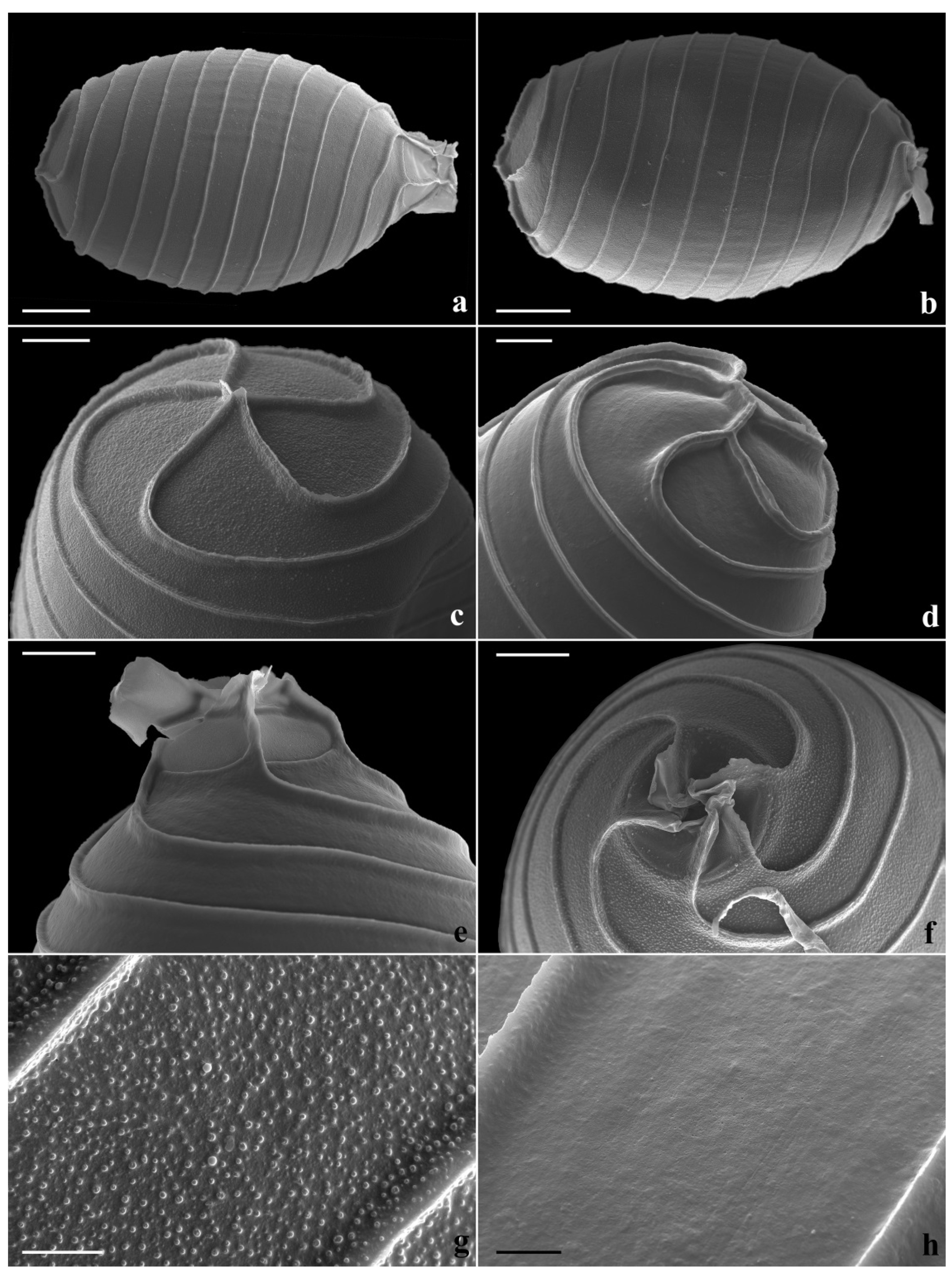
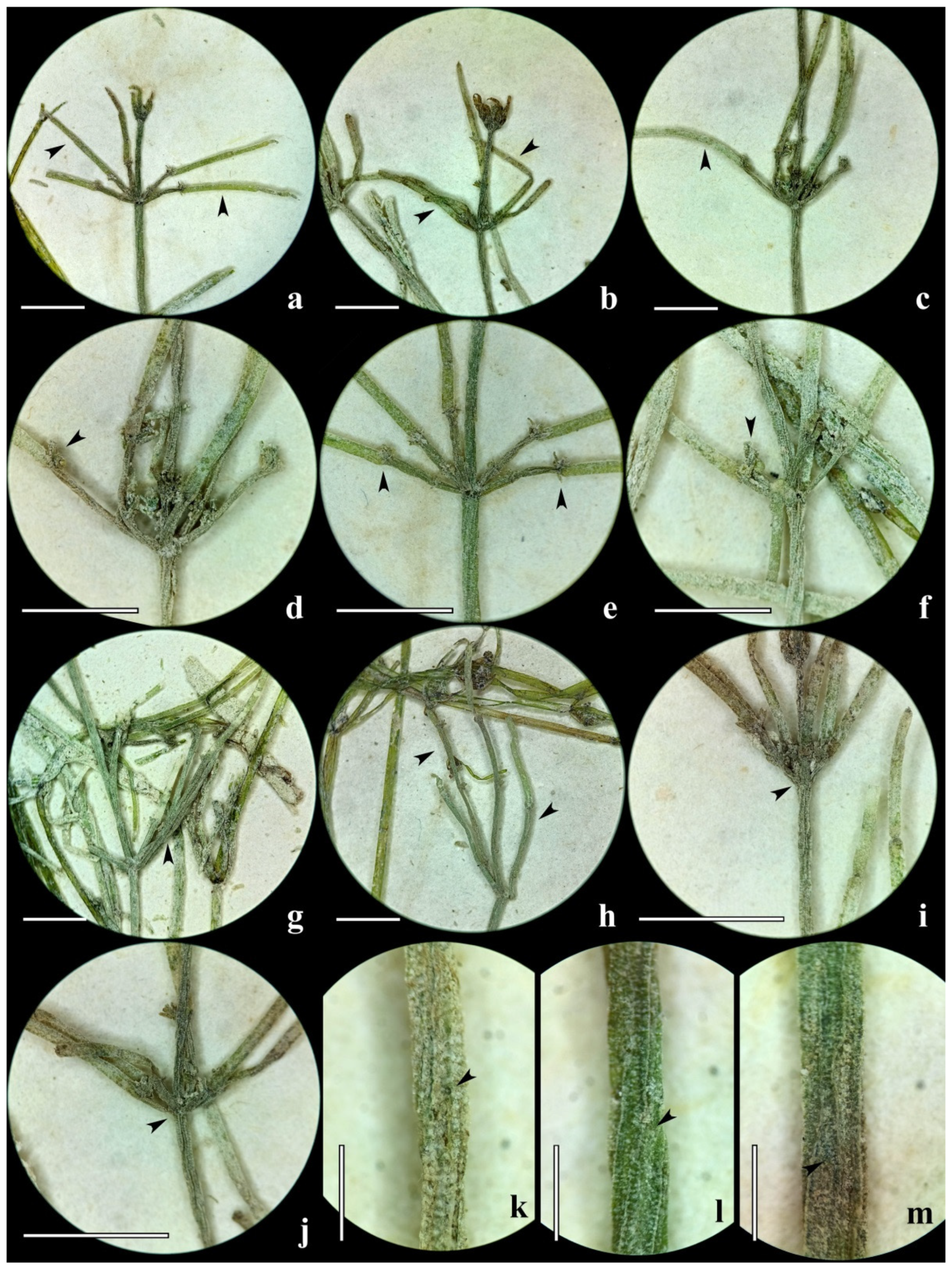
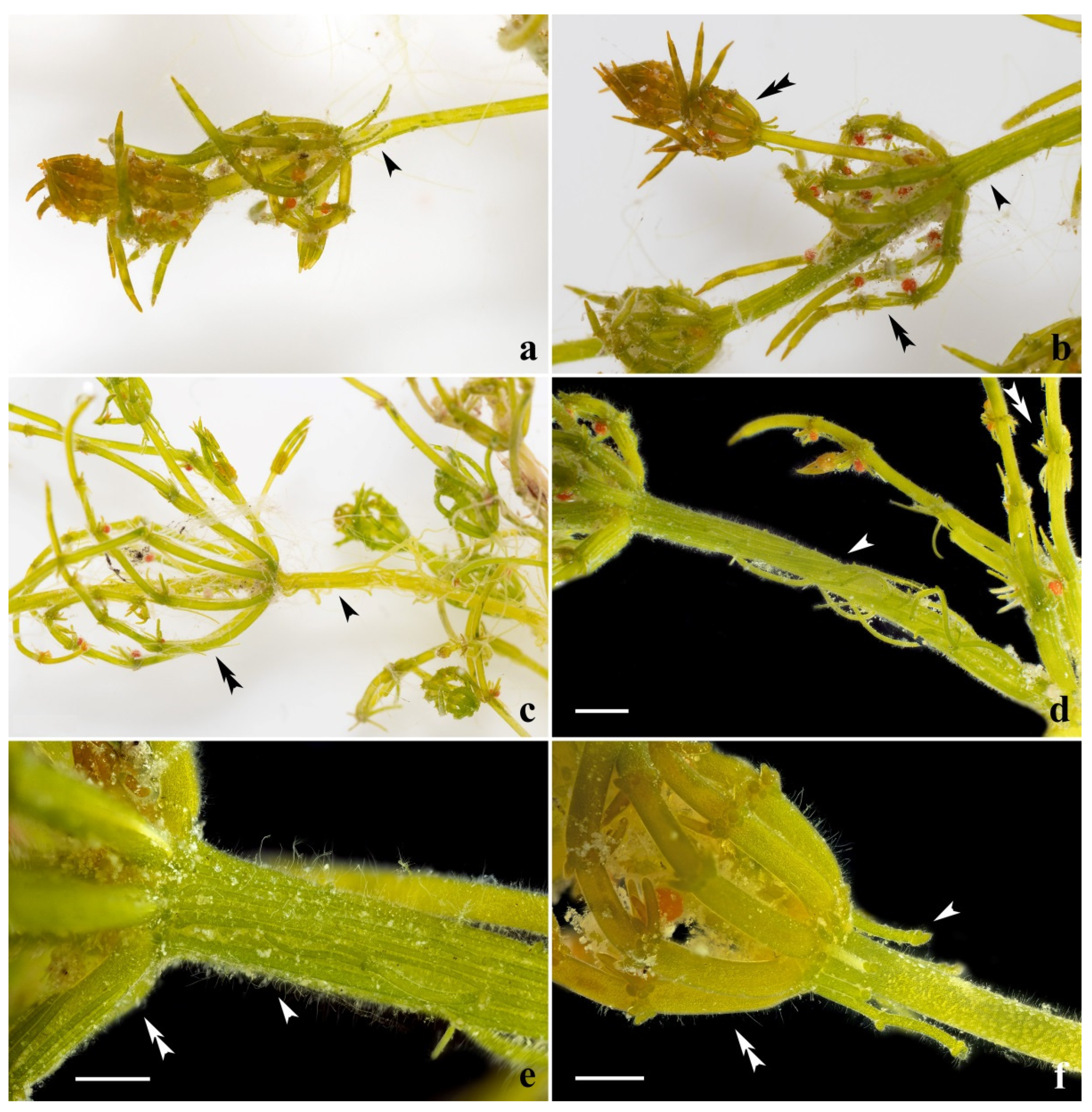
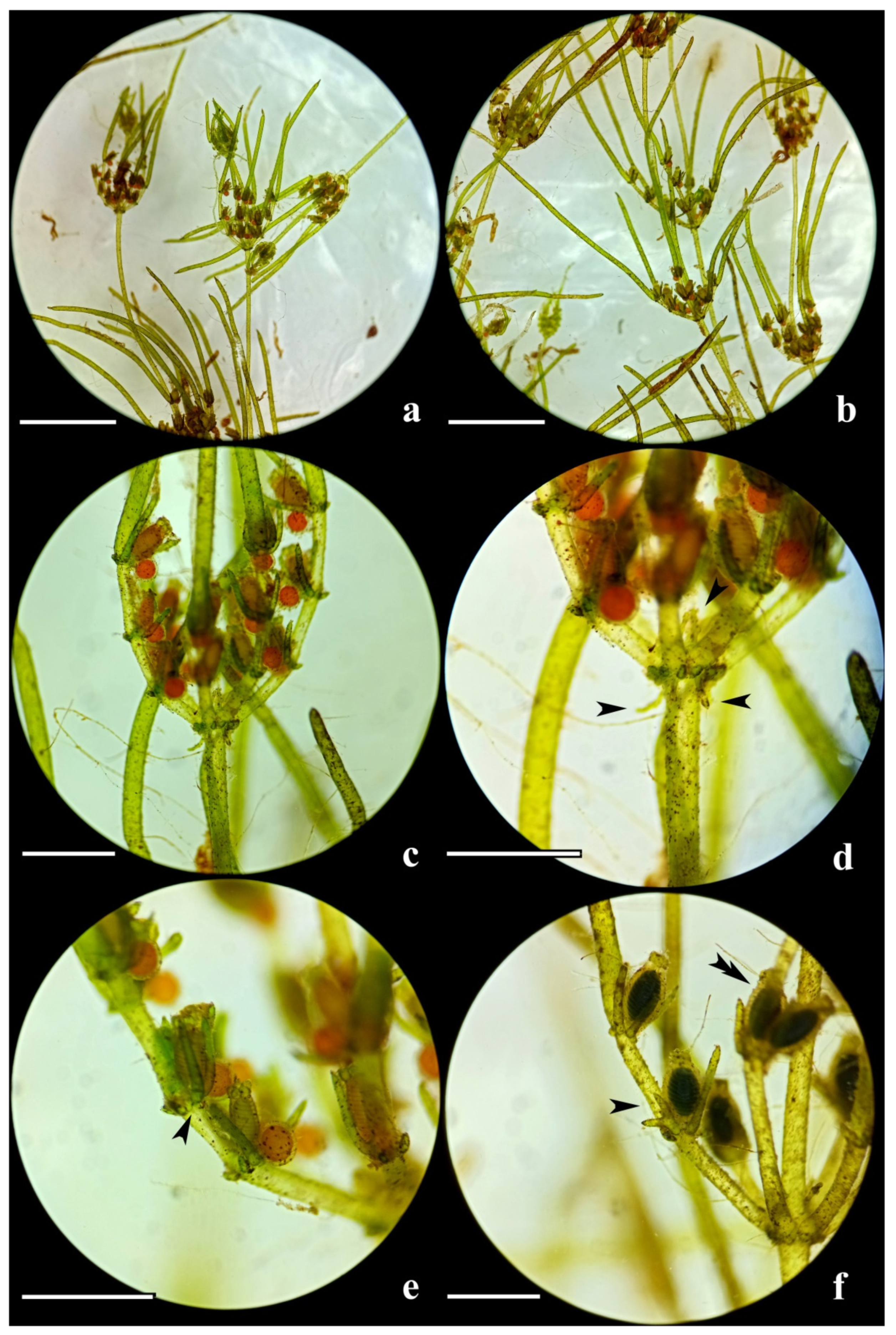
3.1. Description of the Specimens of Chara denudata

The plants are evenly and moderately encrusted with lime and grow in clumps or mats, not as solitary shoots. The older ecorticate parts are infected with numerous discoid thalli of Coleochaete sp. Ring-shaped bulbils are sometimes formed at the lowermost nodes (Figure 1). They consist of two dense rows of more or less isodiametric rounded cells of (70)90–200 μm in length with starch inside. The stem cortex is nearly absent to incomplete. It consists of only narrow primary tubes, with wide empty spaces (furrows) between them. Their length varies from almost invisible short initials to more or less elongated tubes. The ends of the short tubes can be slightly peeled off the stem (plants from Oman). The most developed cortex, which is not a frequent case, always consists of narrow primary tubes meeting in the center of the internode and having remarkably wide empty spaces between parallel tubes. The spine cells are solitary, small, and papillate and are barely recognizable. The stipulodes are bistipulate, diplostephanous, short, and obtuse, adpressed to the branchlets, and are up to 150 μm but are mostly much shorter than this. The cells in the upper rows can be slightly elongated, whereas, in the low rows, they can be hardly recognizable to almost invisible. The branchlets are ca. 10 in a whorl, somewhat or evidently longer than the internodes at the upper parts or from the near base of the plant up to the tip. They are sometimes several times longer, resulting, in this case, in the clasping of the branchlets from several apical whorls above the apex (plants from Oman). The branchlets consist of one–three ecorticate segments delineated with nodes and long, undifferentiated (or, in other words, nodeless) ecorticate three-celled end segments, comparable in length to differentiated part or longer. This can be broken at lower branchlets. Each or both segments can be abbreviated, constituting a negligible part of branchlet length (plants from Oman). Very narrow elongated initials of the branchlet cortex can be traced below the branchlet nodes in rare cases. The end cells of the branchlets are short, narrowly pointed, and neither acute nor mucronate. The adaxial (posterior) bract cells are short, bluntly conical, and mostly hardly recognizable. The abaxial (anterior) bract cells are cylindrical, narrowing mostly near their conical, shortly pointed tips, and are variable in length from two times shorter oogonia to several times longer than oogonia, slightly to evidently exceeding the length of adjacent branchlet cells (up to 2–2.3 times). The bracteoles are mostly and remarkably thinner and shorter than the adaxial bract cells. The plants are richly fertile, with solitary conjoined gametangia at one or two basal nodes of the branchlets. Oogonia can vertically geminate as an exception. The oospores are dark brown, with 11 or 12 low narrow ridges, together with (or without) a basal cage, having no empty spaces between the cage ribs (Figure 2). The oospore surface sculpture has two variants—granulated with prominent granules or roughened. The cage ribs can be irregularly flanged. Table 1 provides an overview of the measured morphometric data.

3.2. Description of the Specimens of Chara dissoluta

The plants are evenly and moderately encrusted with lime or completely incrusted, sparsely branching and growing in groups, being mostly lax to infrequently compact. The stem cortex is nearly absent to incomplete (Figure 3, Figure 4 and Figure 5). It mainly consists of only narrow primary tubes, with wide empty spaces (furrows) between them. Their length varies from almost invisible short initials to more or less elongated tubes that are visible above and below the branchlet whorl base (plants from the Gulf of Finland—GF and Slovakia). The ends of short tubes can be peeled off the stem (GF and Slovakia). The short solitary spine cells and the round initials of the secondary tubes are recognizable in the case of the elongated cortex tubes (GF and Slovakia). The secondary tubes can be evidently elongated, but the empty spaces between the cortex tubes are typical in these cases too. Both the primary and secondary tubes are not regularly associated with stem (Slovakia). The most developed cortex consists of narrow primary tubes meeting in the center of the internode and having evident, empty spaces between the parallel tubes (typical of the lectotype (Figure 3) and found in plants from Slovakia). The spine cells are solitary, small, and papillate and are hardly recognizable. The stipulodes are bistipulate, diplostephanous, short, obtuse, and adpressed to the branchlets. The cells in the upper rows can be slightly elongated, whereas, in the low rows, they can be hardly recognizable to almost invisible.
There are six–eight branchlets in a whorl (6(7?) in case of a lectotype), which are somewhat longer than the internodes at the upper parts of the plant and are 1.2–2.5 times shorter than the internodes in most parts of the plant. The branchlets consist of one–three ecorticate or corticated segments that are delineated with nodes and long, undifferentiated (or, in other words, nodeless) ecorticate two–four-celled end segments, which are longer than in the differentiated part. It can be broken at lower branchlets if it is incrusted. The number of cells in the undifferentiated segment is inversely correlated with the number of segments. All segments are usually abbreviated (lectotype and GF), constituting a small part of the branchlet length. The branchlet segments of a lectotype are corticated, with the cortex unclearly appearing in a dry state having narrow, empty longitudinal spaces between the tubes. The tubes from the upper and lower nodes meet each other at the center of the segment or reach the base of the branchlet at the basal segments. The narrow and elongated or usually rounded initials of the branchlet cortex can be traced below the branchlet nodes in some cases but not at all nodes (GF). The specimens from Slovakia have an intermediate expression of this trait (Figure 4). They have round initials or long tubes of branchlet cortex with empty spaces between them, which neither meet each other at the center of the segment nor reach the branchlet base. The end cells of the branchlets are short or somewhat elongated, narrowly pointed, and not clearly mucronate, yet the base is usually clearly narrower than the tip of the penultimate cell. The adaxial (posterior) bract cells are short and bluntly conical to conical. The abaxial (anterior) bract cells are more or less conical, with bluntly pointed tips, and are variable in length: from 0.6 of the oogonium length to mostly slightly shorter to slightly up to two times longer, and are evidently shorter (lectotype) to mostly slightly shorter and nearly equal to the length of the adjacent branchlet cells (GF; Figure 5). The bracteoles are similar in appearance to the adaxial bract cells. The plants are poorly fertile with unripe gametangia (lectotype) to richly fertile with unripe gametangia and clear protandry (Slovakia), with ripe oospores (GF), with solitary conjoined gametangia at one–three branchlet nodes. The oogonia can be laterally geminate in rare cases (GF). The oospores are black. Table 1 provides an overview of the measured morphometric data.

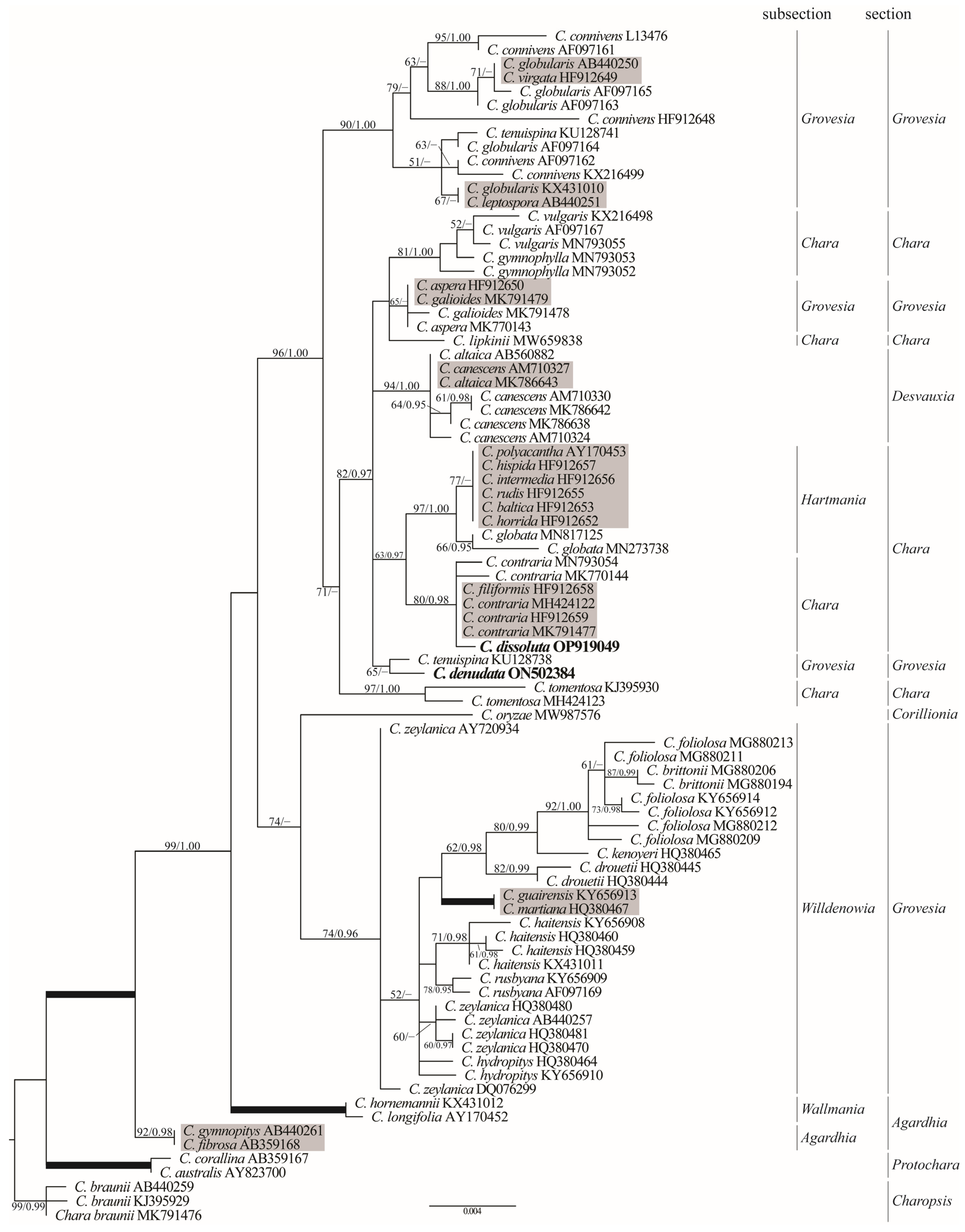
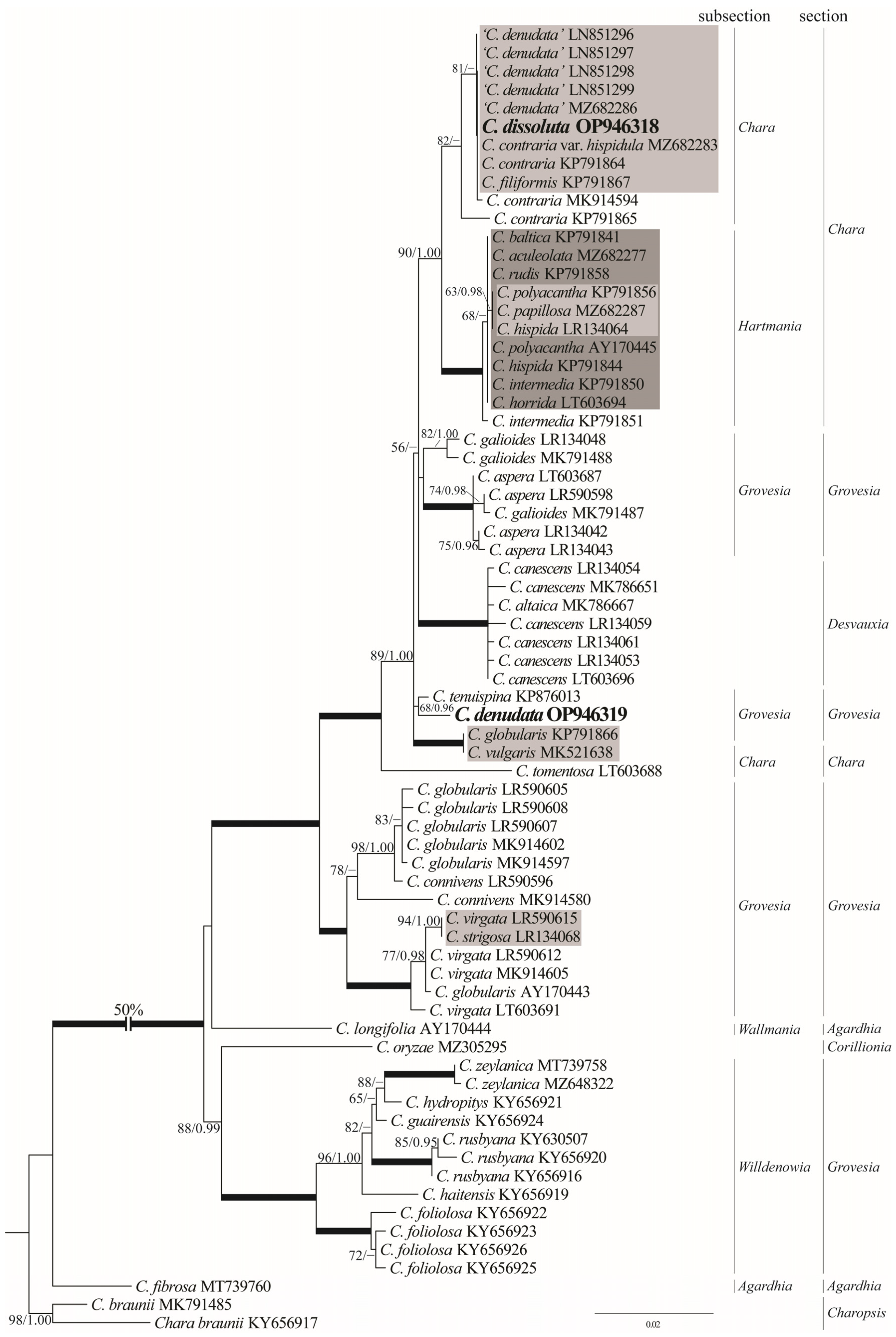
3.3. Phylogenetic Positions

The topologies of the ML and BI trees based on the rbcL dataset were similar to that of the BI tree, except for some differences in clade support (Figure 6). A total of 85 Characeae sequences were distributed between the 12 main clades. A sample of C. denudata placed with C. tenuispina A. Braun, with weak support (63 BP) in the subsection Grovesia R.D. Wood. This clade was part of a larger clade (composed of C. tomentosa L., C. contraria/C. filiformis A. Braun in Hertzsch, C. globata Mig., C. canescens/C. altaica A. Braun in A. Braun et Nordst., C. lipkinii Romanov et al., C. aspera Willd./C. galioides DC., C. vulgaris L./C. gymnophylla A. Braun, and complex clade (877/–), which included six identical sequences of six different species (C. polyacantha A. Braun in A. Braun, Rabenh. & Stizenb. nom. inv., C. hispida L., C. intermedia A. Braun nom. ill., C. rudis (A. Braun) Leonh. nom. ill., C. baltica (Hartm.) Bruzelius, C. horrida Wallman ex Wahlst.). The clades Hartmania R.D. Wood and Chara R.D. Wood were resolved as sister lineages (63/0.97).
Despite incongruent taxon sampling and marker length, the topology of the matK tree was similar to that obtained with rbcL, and it resolved the same subsections and section clades with a comparable significance (Figure 7). Chara denudata, again, was a sister to C. tenuispina, while C. dissoluta was placed distantly from it to C. contraria/C. filiformis subclade (82/–). Our C. denudata specimen had an identical matK sequence to that of four European “C. denudata” accessions: C. contraria, C. contraria var. hispidula A. Braun, and C. filiformis.

4. Discussion

4.1. Morphological Perspective

The restudy of the original material of the charophyte species described many years ago allows for the testing and improvement of existing species concepts (e.g., [1,2,37,38,39,40]) and may serve as an essential keystone for checking and confirming species’ distributional records. Integrative taxonomy is a fruitful approach, but its great age and sometimes a low amount of original material can limit the opportunities for adding new important data, e.g., nucleotide sequences and the scanning electron microscopy images of oospores. The storage of abundant original material in several collections can overcome these limitations, as was possible during this study.
The original material of Chara denudata was stored in at least three herbaria (B, LE, W). The part stored in B seemed to have been lost during the Second World War. The lectotype was selected by Wood [2] based on a specimen stored in W. Morphological descriptions and illustrations of plants from the original material, which has been published by several authors [1,2,4,17,41,42].
We believe that the three envelopes stored in LE and studied by us, namely LE 0001555, LE 0001559, and LE 0001560, belong to the original material of C. denudata and should be recognized as isolectotypes. They were collected by Drège in South Africa in December 1832 [23]. This material is not fragmented and is evidently more abundant and in better condition in comparison to the lectotype stored in W [2,4]. It revealed variability in plant arrangement, e.g., the length of the stem cortex tubes and the adaxial bract cells. Both parts were variable, from really short to invisible in the case of the stem cortex tubes, to very long within the same population.
The differences between the clumps of plants stored in LE 0001555 and LE 0001559 in comparison to ones from LE 0001560 were outlined, as the former consisted of more elongated, noncondensed plants with more elongated internodes, stem cortex tubes, and somewhat longer leaves with longer adaxial bract cells, which resulted in differences in the plant’s general appearance. It may be recognized as variability within the same population, reflecting the differences in the microenvironment in the same locality.
The morphological studies of the original material of C. denudata [1,2,4,17,41,42] described less variability than we found during this study. Few descriptions of this species based on materials other than the original material are known from Oman [43].
The morphology of the European “counterpart,” C. dissoluta, is well described and illustrated, relying on these references for comparison to C. denudata. Moreover, we implemented a restudy of a part of the original material of both species and some other specimens (LE, IBIW, W, and [24], this study). The great similarity in morphology between the European and South African plants is remarkable, and all differences that could be suggested in comparison to the original material for both species become insignificant if all ranges of variability that are described for the European plants are considered ([3,4,18,19,20,21,22,41,42,44,45,46]; see the discussion in [24]).
The compared species have different combinations of variable expressions of the same traits, resulting in their appearance as largely intergrading morphotypes. One morphological trait is remarkable. The adaxial bract cells of C. denudata were significantly longer than those of C. dissoluta. They were shorter, equal to, or somewhat longer than the oogonium length but were up to 2.5 times longer than the oogonium length in southeast-European populations of C. dissoluta [3,4,18,19,20,21,22,24,41,42,44,45,46]. The lower number of branchlets in the whorl is also notable, according to the original studied material (6(7?) vs. ca. 10), agreeing with some (e.g., [3,20,42]) but not all descriptions of the European plants. The size of the gametangia has overlapping values for the compared species, but C. dissoluta can produce smaller oospores and larger antheridia. The reliability of species delineation with the vertically geminating oogonia of C. denudata vs. the laterally geminating oogonia of C. dissoluta is unclear because of the rare occurrence of this trait in both cases.
A single case of bifid adaxial bract cell found in C. dissoluta from the Gulf of Finland is a rare morphological trait unknown for this species. A few examples of bifid cell occurrence have been reported for charophytes. The bifid cells found among the bracteoles, bract cells, and spine cells of C. aspera Willd. [47,48] and C. baltica (Hartm.) Bruzelius [2], the bract cells of Lychnothamnus barbatus (Meyen) Leonh. [2,41] and C. flaccida A. Braun [49], and the branchlet end cells of Nitellopsis obtusa (Desv. in Loisel.) J.Groves ([49], the identification of N. obtusa needs confirmation) are exceptional cases. This trait can be recognized as a rare charophyte abnormality, at least for a few species, but we still do not know the true scale and causes of this phenomenon. The bi-tripartite or bipartite branchlet end cells of Nitella bicornuta F.S. Han et W.Q. Chen, N. guangxiensis Y.J. Ling, C.Z. Deng et Z. Li, N. inaequipartita L.B. Liang [50], N. partita Nordst. [2,50], the bifurcate bracteoles, and the adaxial bract cells of C. kieneri E.F.K. Daily [51] have been suggested as key traits for only these species.
The oospores of the plants from the original material of C. denudata have two types of oospore surface: granulated and roughened (Figure 2). The granulated variant is similar to the pattern described for the specimen of this species from Tanganyika Plateau, Tanzania, i.e., granulated and roughened on the same surface [52,53]. The oospores of C. dissoluta var. ohridana Kostić have been described as smooth using SEM [52], which coincides with our observations [24]. The morphologically similar dioecious C. imperfecta has a roughened and minutely perforate oospore surface [52,53], i.e., overlapping with the variant found by us for C. denudata. However, even two of the patterns found in this study seemed to be not unique and sufficient for delineation with other species based on this trait if only species of Chara studied with SEM are considered (e.g., [52,53,54,55,56,57,58,59,60,61,62]).
The plants of C. dissoluta are usually poorly fertile or sterile [3,4,18,20,22,41,42,44,45,46], in contrast to the richly fertile plants of C. denudata, although this delineation seems to be not so crucial in the case of the plants of C. dissoluta growing in the shallows of large lakes and water bodies that are associated with them in the Balkans [24] or in the shallows of the Gulf of Finland and the shallow water depressions formed with the discharge of active mineral springs in Slovakia (this study).

4.2. Phylogenetic Perspective

The successful amplification of partial rbcL and matK sequences from a 190-year-old herbarium isolectotype specimen allowed us to assess the phylogenetic relationship of C. denudata with C. dissoluta and the rest of the genus. The overall topologies of our rbcL and matK trees (Figure 3 and Figure 4) were similar to those presented in the previous studies of the genus Chara [12,13,14] and generally congruently resolved the relationship between the species. According to the molecular data, C. denudata was placed in a large, supported assemblage that comprised sections Grovesia R.D. Wood, Chara R.D. Wood, and Desvauxia R.D. Wood and the traditional subsections of Chara R.D. Wood, Hartmania R.D. Wood, and Desvauxia R.D. Wood. Chara denudata was significantly positioned outside this lineage and showed close relationships with C. tenuispina in the subsection Grovesia. These species differed from each other in only one substitution in the rbcL sequence and eight substitutions in matK. Thus, our molecular data fully confirmed the distinctness of C. denudata and its distant relationship with other species of the subsection Chara. Therefore, C. contraria belongs to another group only distantly related to the group of the species associated with C. contraria, including the European populations earlier referred to as C. denudata.
Our results conflict with the phylogeny presented by Schneider et al. [14]. These authors analyzed the phylogenetic relationships between 327 charophyte accessions (including four accessions of European plants of C. dissoluta placed in GenBank under the name of ‘C. denudata’) using partial (518 bp) matK gene sequences. According to their results, European C. dissoluta (under the name ‘C. denudata’) was placed in the C. contraria cluster (C. imperfecta, C. filiformis, C. ohridana (Kostić) Krause nom. inv., C. contraria, C. arcadiensis [U. Raabe in] Schneider et al. nom. nud., C. gymnophylla) of the section Chara. This placement is in good agreement with [63] and mirrors the position of C. dissoluta in our tree. However, in the dataset studied by Schneider et al., ‘C. denudata’ was represented in European plants only [14] (i.e., under the name of species not reliably known from Europe [24]), whereas our specimens (isolectotypes of C. denudata) were collected in South Africa. The matK sequences of the C. contraria cluster also revealed limited genetic variation (identical for 17 individuals of three different species: C. contraria, C. filiformis and C. dissoluta placed in GenBank under ‘C. denudata’).
We concluded that South African and European plants usually refer to C. denudata (before our study) actually belong to different sections and, therefore, should be treated as a separate species despite their morphological similarity. This decision about their difference was implemented in the monograph covering charophytes in Germany [4] based on a morphological study of a lectotype of C. denudata, a small, fragmented specimen. The same decision was accepted in the monograph about European charophytes [24], and this study provides thorough evidence for it according to an integrative taxonomy.

4.3. Ecological Perspective
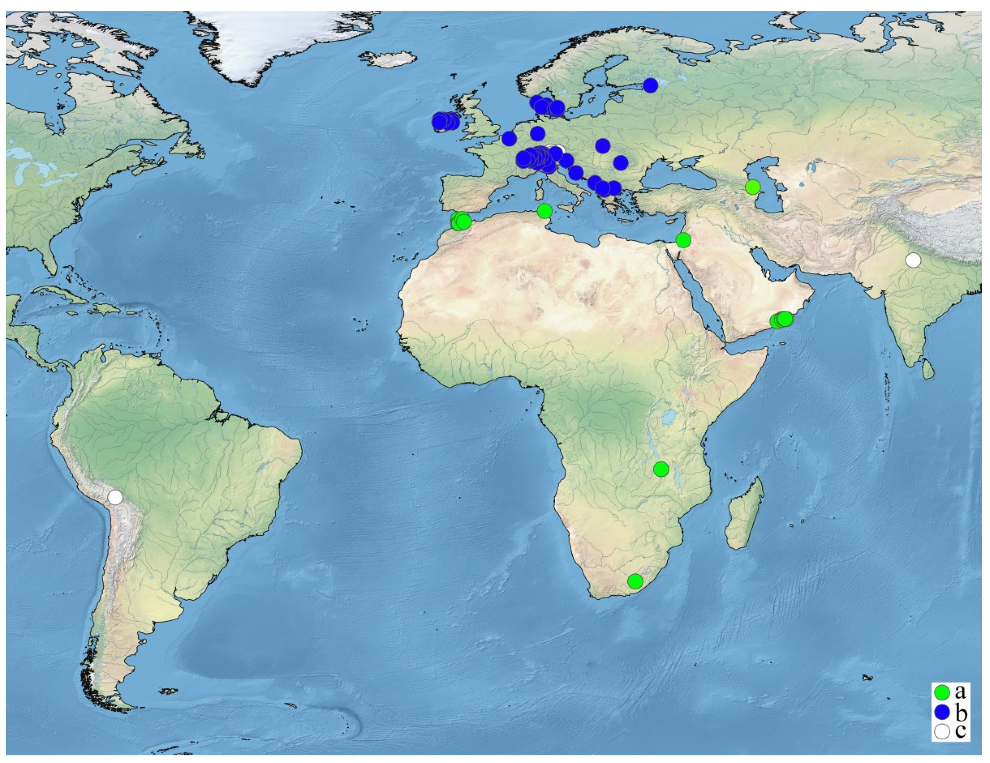
The short description of the type of locality for C. denudata [23] allows only a tentative outline of the environment [64,65,66]. It was situated in the Stormberg Mountains in Eastern Cape, an arid region with very hot summers and very cold winters. It seems to be a temporary, seasonal, or permanent wetland fed by both subsurface and surface water, only subsurface water inputs, or surface flow only. It could be due to an ephemeral, seasonally flowing stream, spring, seep, or the rock pools associated with them or the temporary pools in the depressions (the latter is locally called vleis or pans). The water in this habitat seems to be rich in total dissolved solids because of its high evaporation. The other habitats of C. denudata known from other regions are streams, rivers, wadis, and stagnant water bodies with alkaline waters ([43,67,68], studied specimens from B, W). The ecology of C. denudata looks strongly dissimilar to that of C. dissoluta. The latter is exclusively freshwater and mainly deep-water species of strongly alkaline stable standing water bodies, mainly deep transparent lakes on marl and limestone in temporal regions [69], and, rarely, the smaller water bodies associated with them [24,70]. New localities in Slovakia and Russia are remarkable because they belong to new types of species habitats, a minerotrophic wetland in Central Europe, and the shallows of the easternmost freshwater part of the Baltic Sea. However, when the species are compared, they seem to be ecologically distinct. Chara denudata can be recognized as a shallow water species of arid regions; it is usually richly fertile. Chara dissoluta is mostly a deep-water species of deep, stratified, transparent, temperate lakes; it has mostly low fertility.

4.4. Distribution

All distributional records from Africa and Eurasia are clearly or tentatively attributable to C. denudata ([2,17,23,41,43,52,67,68,71,72], studied specimens from LE, B, and W) and C. dissoluta ([24], this study) are summarized on the map (Figure 8). This allowed for a preliminary outline of the possible distribution range of C. denudata. One record from Tanzania, Tanganyika Plateau [52], could not be located exactly without specimen checking because not enough label data have been published. Moreover, it could actually be another species, and the study of its voucher is desirable. Another locality from the Middle East, cited as Kurdistan [73], also seems to belong to this species but cannot be depicted on the map because no exact locality is known.
The records from Lake Titikaka, South America, are based on deep water plants [75,76] and could belong to another species. The record from India seems to also be based on another species because its specimens were described as having remarkably long spine cells [74] that are different from the minute, hardly recognizable spine cells of C. denudata and C. dissoluta.
The sterile specimens fitting the description of monoecious C. denudata are actually impossible to delineate from dioecious C. imperfecta. At least one specimen illustrating this case was studied by the first author: Pflanzen der Türkey/C5 Nigde. Bolkar Daglari, ca. 100 m östl. Kara Göl. Bachlauf, ca. 20 cm Wassertiefe; 2630 m, 37°25′ N 034°37′ E/05 VIII [19]92/E. Raab-Straube Nr. KG 2-7/C. gymnophylla, det. W. Krause (B, Algae 38518).
Therefore, nearly all distributional records referring to C. denudata are situated in the tropical and subtropical arid regions of Africa and Asia (Figure 8). This is evidently dissimilar to C. dissoluta, which is mostly known from the temperate regions of Europe [4,20,24,41]. The lack of successful hybridization between the distant populations of the same morphospecies of Chara from the subsections Chara and Grovesia [77,78,79] could be an additional argument to attribute the morphologically similar, but geographically far distant taxa C. denudata and C. dissoluta to different species.

5. Conclusions

The study of the original material of Chara denudata in the framework of integrative taxonomy allowed for the clarification of its position and found its unexpected placement in the infrageneric division of the genus Chara according to rbcL sequences, which is in contrast to its position according to morphology. This proved the suggestions expressing that C. dissoluta and C. denudata are different entities [1,2,4]. The similarity and large overlap in the expression of the morphological traits of both species, as a result of the unfinished morphogenesis “stopped” at different stages, are making their clear separation impossible according to morphology only. It actually masks the different origins of C. denudata and C. dissoluta, as is evident from their phylogenetic affinity. This case indicates that the same incompletely developed or simplified morphology of charophytes could have been derived from different ancestors. There seems to be further evidence of a disagreement and the partial reflection of morphological infrageneric taxonomy of Chara with phylogenetic pattern [12]. Therefore, morphological similarity can lead to incorrect suggestions and taxonomic decisions, which can be avoided or at least softened in the framework of integrative taxonomy.

Supplementary Materials

The following are available online at https://www.mdpi.com/article/10.3390/d15020249/s1, Table S1: Species name, GenBank accession number, and the haplotypes for the taxa used in our analyses. The sequences obtained in this study are in bold. Shared haplotypes are highlighted in yellow.

Author Contributions

Conceptualization, R.E.R.; methodology, R.E.R., S.S.B., V.Y.N. and A.A.G.; software, V.Y.N.; validation R.E.R., S.S.B., V.Y.N. and A.A.G.; formal analysis, R.E.R., S.S.B., V.Y.N. and A.A.G.; investigation, R.E.R., S.S.B., V.Y.N. and A.A.G.; data curation, R.E.R., S.S.B., V.Y.N. and A.A.G.; writing—original draft preparation, R.E.R., V.Y.N.; writing—review and editing, R.E.R., S.S.B., V.Y.N. and A.A.G.; visualization, R.E.R. and V.Y.N.; funding acquisition, R.E.R., S.S.B., V.Y.N. and A.A.G. All authors have read and agreed to the published version of the manuscript.

Funding

This work was supported by the Russian Foundation for Basic Research, project no. 20-04-00280, and partly by the project “Flora and taxonomy of algae, lichens and bryophytes in Russia and phytogeographically important regions of the world” (no. 121021600184-6) of the Komarov Botanical Institute of the Russian Academy of Sciences, as part of the state task for IWEP SB RAS (registration no. 0306-2021-0001), the state assignment of the Ministry of Science and Higher Education of the Russian Federation (theme No. 121031000117-9), with the financial support the Ministry of Science and Higher Education of the Russian Federation under Agreement dated 28 September 2021, no. 075-15-2021-1056 (placement of the algal specimens in the LE collection) and the Israel Ministry of Aliyah and Integration.

Institutional Review Board Statement

Not applicable.

Informed Consent Statement

Not applicable.

Data Availability Statement

The data presented in this study are available on request from the corresponding author. In addition, the data that support the findings of this study are openly available in GenBank.

Acknowledgments

The curators of the herbaria listed above are kindly acknowledged for their encouragement and guidance, František Bednár for sharing his data about charophytes of Slovakia, specimens, and excellent photos of living plants, Tanja Schuster for sharing images of a lectotype of C. denudata, Klaus van de Weyer for the possibility of study of his specimens of C. dissoluta from Ireland and Germany, Vera Biberdžić and Ivana Trbojević for sharing images of C. dissoluta from the Balkans, Vera Biberdžić for possibility of study of C. dissoluta from the Balkans, Ilya Eremin and Anna Erst for their assistance in SEM studies, Ricky Taylor and Matthew Janks for really helpful clarification of environment in type locality of C. denudata, Elisabeth Lambert and Luc Denys for several inaccessible references, anonymous reviewers for important suggestions and improvements.

Conflicts of Interest

The authors declare no conflict of interest.

References

  1. Wood, R.D.; Imahori, K. Iconograph of the Characeae. In Monograph of the Characeae; Wood, R.D., Imahori, K., Eds.; Verlag von J. Cramer: Weinheim, Germany, 1964; pp. v–xv, 1–5, icons 1–395. [Google Scholar]
  2. Wood, R.D. A revision of the Characeae. In Monograph of the Characeae; Wood, R.D., Imahori, K., Eds.; Verlag von J. Cramer: Weinheim, Germany, 1965; pp. i–xxiv, 1–904. [Google Scholar]
  3. Jäger, D. Exemplare des Formenkreises Chara denudata A. Braun 1847 und Chara dissoluta A. Braun ex Leonhard 1864 aus dem Bodensee. Rostock. Meeresbiol. Beiträge 2010, 23, 29–39. [Google Scholar]
  4. Becker, R.; Blindow, I.; Doege, A.; Franke, T.; Gregor, T.; Hamann, U.; Jäger, D.; Jorda, C.; Kabus, T.; Korsch, H.; et al. Beschreibung der Characeen-Arten Deutschlands. In Armleuchteralgen. Die Characeen Deutschlands; Arbeitsgruppe Characeen Deutschlands Lehrstuhl für Ökologie der Universität Rostock, Ed.; Springer: Berlin/Heidelberg, Germany, 2016; pp. 209–572. [Google Scholar] [CrossRef]
  5. Nat, E.; Raabe, U.; Romanov, R.; Schubert, H.; Stewart, N. A book about the charophytes of Europe. IRGC News 2017, 28, 21–22. [Google Scholar]
  6. Sakayama, H. Taxonomy of Nitella (Charales, Charophyceae) based on comparative morphology of oospores and multiple DNA marker phylogeny using cultured material. Phycol. Res. 2008, 56, 202–215. [Google Scholar] [CrossRef]
  7. Casanova, M.T. An overview of Nitella in Australia (Characeae, Charophyta). Aust. Syst. Bot. 2009, 22, 192–218. [Google Scholar] [CrossRef]
  8. Sakayama, H.; Kasai, F.; Nozaki, H.; Watanabe, M.M.; Kawachi, M.; Shigyo, M.; Nishihiro, J.; Washitani, I.; Krienitz, L.; Ito, M. Taxonomic reexamination of Chara globularis (Charales, Charophyceae) from Japan based on oospore morphology and rbcL gene sequences, and the description of C. leptospora sp. nov. J. Phycol. 2009, 45, 917–927. [Google Scholar] [CrossRef] [PubMed]
  9. Pérez, W.; Hall, J.D.; McCourt, R.M.; Karol, K.G. Phylogeny of North American Tolypella (Charophyceae, Charophyta) based on plastid DNA sequences with a description of Tolypella ramosissima sp. nov. J. Phycol. 2014, 50, 776–789. [Google Scholar] [CrossRef] [PubMed]
  10. Romanov, R.E.; Gontcharov, A.A.; Barinova, S.S. Chara globata Mig. (Streptophyta: Charales): Rare species revised. Fottea Olomouc 2015, 15, 39–50. [Google Scholar] [CrossRef]
  11. Karol, K.G.; Alix, M.S.; Scribailo, R.W.; Skawinski, P.M.; Sleith, R.S.; Sardina, J.A.; Hall, J.D. New records of the rare North American endemic Chara brittonii (Characeae), with comments on its distribution. Brittonia 2018, 70, 277–288. [Google Scholar] [CrossRef]
  12. Romanov, R.E.; Barinova, S.S.; Nikulin, Y.V.; Gontcharov, A.A. Chara lipkinii (Charales, Charophyceae): A new dioecious Mediterranean species under risk of extinction in the wild and some implications for the taxonomy of the genus Chara. Fottea 2022, 22, 1–12. [Google Scholar] [CrossRef]
  13. Romanov, R.E.; Nikulin, A.Y.; Nikulin, V.Y.; Gontcharov, A.A. New species Chara oryzae and a new section Corillionia of Chara (Charales, Charophyceae) from European Mediterranean rice fields. Eur. J. Phycol. 2022, 57, 328–342. [Google Scholar] [CrossRef]
  14. Schneider, S.C.; Nowak, P.; von Ammon, U.; Ballot, A. Species differentiation in the genus Chara (Charophyceae): Considerable phenotypic plasticity occurs within homogenous genetic groups. Eur. J. Phycol. 2016, 51, 282–293. [Google Scholar] [CrossRef] [Green Version]
  15. Urbaniak, J.; Sakayama, H. Taxonomical analysis of closely related species of Chara L. section Hartmania (Streptophyta: Charales) based on morphological and molecular data. Fottea 2017, 17, 222–239. [Google Scholar] [CrossRef]
  16. Urbaniak, J.; Kwiatkowski, P. Taxonomic studies on the Chara section Hartmania in Poland based on morphological and molecular data. PhytoKeys 2019, 135, 71–90. [Google Scholar] [CrossRef] [PubMed]
  17. Braun, A. Die Characeen Afrika’s; Monatsberichte der Königlich preussischen Akademie der Wissenschaften zu Berlin: Berlin, Germany, 1868; Volume 1867, pp. 873–944. [Google Scholar]
  18. Groves, J.; Bullock-Webster, G.R. The British Charophyta; II. Charae; Ray Society: London, UK, 1924; pp. v–xi, 1–141, pl. 22–45. [Google Scholar]
  19. Hollerbach, M.M.; Krassavina, L.K. The Identification Manual of Freshwater Algae of the USSR. Iss. 14. The Charophytes—Charophyta; Nauka: Leningrad, Russia, 1983; pp. 1–190. (In Russian) [Google Scholar]
  20. Krause, W. Süßwasserflora von Mitteleuropa. Charales (Charophyceae), 18; Spektrum Akademischer Verlag: Heidelberg, Germany, 1997; pp. 1–202. [Google Scholar]
  21. Bailly, G.; Schaefer, O. Guide Illustré des Characées du Nord-Est de la France; Conservatoire Botanique National de Franche-Comté: Besançon, France, 2010; pp. 1–96. [Google Scholar]
  22. John, D.M.; Whitton, B.A.; Brook, A.J. (Eds.) Freshwater Algal Flora of the British Isles: An Identification Guide to Freshwater and Terrestrial Algae, 2nd ed.; Cambridge University Press: Cambridge, UK, 2011; pp. 1–878. [Google Scholar]
  23. Drège, J.F. Zwei pflanzengeographische Documente. Bes. Beigabe Zur Flora 1843, 26, 1–230. [Google Scholar]
  24. Romanov, R.; Stewart, N.; van de Weyer, K.; Roden, C.; Trbojević, I.; Biberdžić, V.; Trajanovska, S.; Alegro, A.; Kashta, L. Chara dissoluta. In Characeae of Europe; Schubert, H., Gregor, T., Blindow, I., Nat, E., Stewart, N., Romanov, R., van der Weyer, K., Denys, L., Korsch, H., Casanova, M.T., Eds.; Springer: Berlin/Heidelberg, Germany, 2023; in press. [Google Scholar]
  25. Marcin, D. Hydrogeologická štruktúra baldovce—Sivá Brada. Podzemná Voda 2000, VI, 114–121. [Google Scholar]
  26. Echt, C.S.; Erdahl, L.A.; McCoy, T.J. Genetic segregation of random amplified polymorphic DNA in diploid cultivated alfalfa. Genome 1992, 35, 84–87. [Google Scholar] [CrossRef]
  27. Kiselev, K.V.; Dubrovina, A.S.; Tyunin, A.P. The methylation status of plant genomic DNA influences PCR efficiency. J. Plant Physiol. 2015, 175, 59–67. [Google Scholar] [CrossRef]
  28. Bonfield, J.K.; Smith, K.F.; Staden, R. A new DNA sequence assembly program. Nucleic Acids Res. 1995, 23, 4992–4999. [Google Scholar] [CrossRef]
  29. Galtier, N.; Gouy, M.; Gautier, C. Seaview and phylo-win: Two graphic tools for sequence alignment and molecular phylogeny. Comput. Appl. Biosci. 1996, 12, 543–548. [Google Scholar] [CrossRef]
  30. Swofford, D.L. PAUP*. Phylogenetic Analysis using Parsimony (*and Other Methods). Version 4.0b10; Sinauer Associates: Sunder-land, MA, USA, 2002. [Google Scholar]
  31. Huelsenbeck, J.P.; Ronquist, F. MRBAYES: Bayesian inference of phylogenetic trees. Bioinformatics 2001, 17, 754–755. [Google Scholar] [CrossRef]
  32. Akaike, H. A new look at the statistical model identification. IEEE Trans. Autom. Control 1974, 19, 716–723. [Google Scholar] [CrossRef]
  33. Darriba, D.; Taboada, G.L.; Doallo, R.; Posada, D. jModelTest 2: More models, new heuristics and parallel computing. Nat. Methods 2012, 9, 772. [Google Scholar] [CrossRef] [PubMed]
  34. Stamatakis, A.; Hoover, P.; Rougemont, J. A rapid bootstrap algorithm for the RAxML web servers. Syst. Biol. 2008, 57, 758–771. [Google Scholar] [CrossRef] [PubMed]
  35. Kozlov, A.M.; Darriba, D.; Flouri, T.; Morel, B.; Stamatakis, A. RAxML-NG: A fast, scalable and user-friendly tool for maximum likelihood phylogenetic inference. Bioinformatics 2019, 35, 4453–4455. [Google Scholar] [CrossRef]
  36. Casanova, M.T.; Karol, K.G. A revision of Chara sect. Protochara, comb. et stat. nov. (Characeae: Charophyceae). Aust. Syst. Bot. 2014, 27, 23–37. [Google Scholar] [CrossRef]
  37. Proctor, V.W.; Griffin, D.G.; Hotchkiss, A.T. A synopsis of the genus Chara, series Gymnobasalia (subsection Willdenowia RDW). Am. J. Bot. 1971, 58, 894–901. [Google Scholar] [CrossRef]
  38. Casanova, M.T. Typification and circumscription of Nitella sonderi (Characeae, Charophyceae). Aust. Syst. Bot. 2007, 20, 464–472. [Google Scholar] [CrossRef]
  39. Casanova, M.T. Review of the species concepts Chara fibrosa and C. flaccida (Characeae, Charophyceae). Aust. Syst. Bot. 2013, 26, 291–297. [Google Scholar] [CrossRef]
  40. Milozza, A.; Abdelahad, N. The contribution of historical and morphological studies on herbarium specimens to a better definition of Chara pelosiana Avetta (Charales, Charophyceae). Plants 2021, 10, 2488. [Google Scholar] [CrossRef]
  41. Migula, W. Die Characeen Deutschlands, Oesterreichs und der Schweiz; Unter Berücksichtigung aller Arten Europas. Dr. L. Rabenhorst—Kryptogamenflora von Deutschland, Oesterreich und der Schweiz; Verlag Eduard Kummer: Leipzig, Germany, 1897; pp. VII, 1–765. [Google Scholar]
  42. Sluiter, C.P. Beiträge zur Kenntnis von Chara contraria A. Braun und Chara dissoluta A. Braun. Bot. Ztg. 1910, 68, 125–167. [Google Scholar]
  43. Hussain, M.I.; Victor, R.; Khoja, T.M. Charophytes of the Sultanate of Oman, Southern Arabia. Nova Hedwig. 2003, 77, 429–444. [Google Scholar] [CrossRef]
  44. Olsen, N.S. Danish Charophyta. Chorological, ecological and biological investigations. Det K. Dan. Vidensk. Selsk. Biol. Skr. 1944, 3, 1–240. [Google Scholar]
  45. Krause, W.; Krause, H. Exsikkate Europäischer Characeeen; 2. Aufl. 4 Bände, 1984–1986.
  46. Moore, J.A. Charophytes of Great Britain and Ireland; B.S.B.I. Handbook No. 5; Botanical Society of the British Isles: London, UK, 1986; pp. 1–144. [Google Scholar]
  47. Allen, G.O. Notes on charophytes from British Columbia. Proc. Linn. Soc. Lond. 1951, 162, 148–152. [Google Scholar] [CrossRef]
  48. Daily, F.K. The Characeae of Indiana. Butl. Univ. Bot. Stud. 1953, 11, 5–49. [Google Scholar]
  49. Naz, S.; Diba, N.J. Some morphological observations of charophytes (Characeae) from Bangladesh. J. Life Earth Sci. 2014, 7, 71–77. [Google Scholar] [CrossRef]
  50. Han, F.S.; Li, Y.Y. (Eds.) Flora Algarum Sinicarum Aquae Dulcis. Tomus 3. Charophyta; Science Press: Beijing, China, 1994; pp. 1–267. (In Chinese) [Google Scholar]
  51. Daily, F.K. Chara Kieneri, a new species from Nebraska. Butl. Univ. Bot. Stud. 1949, 9, 127–130. [Google Scholar]
  52. John, D.M.; Moore, J.A.; Green, D.R. Preliminary observations on the structure and ornamentation of the oosporangial wall in Chara (Charales, Chlorophyta). Br. Phycol. J. 1990, 25, 1–24. [Google Scholar] [CrossRef]
  53. Leitch, A.R.; John, D.M.; Moore, J.A. The oosporangium of the Characeae (Chlorophyta, Charales). Prog. Phycol. Res. 1990, 7, 213–268. [Google Scholar]
  54. Urbaniak, J.; Blaženčić, J. SEM study of oospore characteristics in endemic and endangered Balkan charophytes. Cryptogam. Algol. 2012, 33, 277–288. [Google Scholar] [CrossRef]
  55. Soulie–Märsche, I. Etude Comparée de Gyrogonites de Charophytes Actuelles et Fossiles et Phylogénie des Genres Actuels; (Thèse–ès–Sci. Univ. Montpellier 1979, Rev. Edit.); Imprimerie. des Tilleuls: Millau, France, 1989; pp. 1–237. [Google Scholar]
  56. Ray, S.; Pekkari, S.; Snoeijs, P. Oospore dimensions and wall ornamentation patterns in Swedish charophytes. Nord. J. Bot. 2001, 21, 207–224. [Google Scholar] [CrossRef]
  57. Mandal, D.K.; Blaženčić, J.; Ray, S. SEM study of compound oospore wall ornamentation of some members of Charales from Yugoslavia, Croatia and Slovenia. Arch. Biol. Sci. 2002, 54, 29–34. [Google Scholar] [CrossRef]
  58. Mandal, D.K.; Ray, S. Taxonomic significance of micromorphology and dimensions of oospores in the genus Chara L. (Charales, Chlorophyta). Arch. Biol. Sci. 2004, 56, 131–138. [Google Scholar] [CrossRef]
  59. Casanova, M.T. An overview of Chara L. in Australia (Characeae, Charophyta). Aust. Syst. Bot. 2005, 18, 25–39. [Google Scholar] [CrossRef]
  60. Urbaniak, J. A SEM and light microscopy study of the oospore wall ornamentation in Polish charophytes (Charales, Charophyceae)—Genus Chara. Nova Hedwig. 2011, 93, 1–28. [Google Scholar] [CrossRef]
  61. Ahmadi, A.; Riahi, H.; Sheidai, M.; van Raam, J.C. A study of the oospore characteristics in some charophytes (Characeae) of Iran. Nova Hedwig. 2012, 94, 487–504. [Google Scholar] [CrossRef]
  62. Romanov, R.E.; (Komarov Botanical Institute of the Russian Academy of Sciences, Saint-Petersburg, Russia). Personal communication, 2022. Unpublished Data.
  63. Mann, H.; Hanel, C.; Langangen, A.; Nowak, P. Chara contraria var. hispidula Braun (Charales) in Newfoundland, Canada a new variety described from North America. Bot. Lett. 2022, 169, 250–258. [Google Scholar] [CrossRef]
  64. Taylor, R.; (Independent Ecologist, Mtunzini, 3867, KwaZulu-Natal, South Africa). Personal communication, 2020.
  65. Janks, M.R.; (GroundTruth Wetlands cc, Hilton, Pietermaritzburg Area, South Africa). Personal communication, 2021.
  66. Janks, M.R. Montane Wetlands of the South African Great Escarpment: Plant Communities and Environmental Drivers. Master’s Thesis, Rhodes University, Makhanda, South Africa, December 2014. [Google Scholar]
  67. Rayss, T. Materiaux pour la Flore Algologique de la Palestine. II. Les Algues des Eaux Continentales. Palest. J. Bot. 1951, 5, 71–95. [Google Scholar]
  68. Guerlesquin, M. Nouvelle contribution à l’étude des Charophycées du Maroc nordoccidental (II). CNRS 1978, 249, 109–137. [Google Scholar]
  69. Stewart, N.F.; Church, J.M. Red Data Books of Britain and Ireland: Stoneworts; Joint Nature Conservation Committee: Peterborough, UK, 1992; pp. 1–144. [Google Scholar]
  70. Blaženčić, J.; Kashta, L.; Vesić, A.; Biberdžić, V.; Stevanović, B. Charophytes (Charales) of Lake Skadar/Shkodra: Ecology and Distribution. In The Skadar/Shkodra Lake Environment. The Handbook of Environmental Chemistry; Pešić, V., Karaman, G., Kostianoy, A., Eds.; Springer: Cham, Switzerland, 2018; Volume 80, pp. 169–202. [Google Scholar] [CrossRef]
  71. Corillion, R. Contribution à l’étude des Characées de Tunisie et bilan actuel de la flore charologique tunisienne. Bull. De La Société Phycol. De Fr. 1977, 22, 47–59. [Google Scholar]
  72. Barinova, S.; Smith, T. Flora of algae and cyanobacteria of continental waters of Israel in the XXI century: Taxonomy, autecology and water quality indicators. Diversity 2022, 14, 328. [Google Scholar] [CrossRef]
  73. Corillion, R. Sur la présence et la signification de formes à cortication incomplète du Chara contraria Kützing dans l’Est armoricain. Bull. De La Société Mayenne-Sci. 1960, 49–60. [Google Scholar]
  74. Pundhir, H.S. Vidyavati New karyomorphological observations on charophytes from Uttar Pradesh, India. Nucleus 1994, 37, 39–45. [Google Scholar]
  75. Allen, G.O. IX. Charophyta. Trans. Linn. Soc. Lond. 3rd Ser. 1940, 1, 155–160. [Google Scholar] [CrossRef]
  76. Guerlesquin, M. Charophytes. In Lake Titicaca: A Synthesys of Limnological Knowledge; Dejoux, C., Iltis, A., Eds.; Kluwer Academie Publishers: Dordrecht, The Netherlands; Boston, MA, USA; London, UK, 1992; pp. 232–240. [Google Scholar]
  77. Grant, M.C.; Proctor, V.W. Chara vulgaris and Chara contraria: Patterns of reproductive isolation for two cosmopolitan species complexes. Evolution 1972, 26, 267–281. [Google Scholar] [CrossRef] [PubMed]
  78. Proctor, V.W. Chara globularis Thuillier (=C. fragilis Desvaux): Breeding patterns within a cosmopolitan complex. Limnol. Oceanogr. 1971, 16, 422–436. [Google Scholar] [CrossRef]
  79. Proctor, V.W. Genetics of Charophyta. In The Genetics of Algae; Botanical Monographs. Vol. 12; Lewin, R.A., Ed.; Blackwell Scientific Publications: Oxford, UK, 1976; pp. 210–218. [Google Scholar]
Figure 1. Chara denudata: (a–m)—the isolectotype from South Africa (LE); (n,o)—a specimen from Oman (B); (a)—apical part with branchlets having long adaxial bract cells, the arrowhead indicates the unbroken long undifferentiated ecorticate end of a branchlet; (b)—differentiated part of the branchlet, arrowheads indicate long adaxial bract cells; (c,d)—apical part with short (c) and very short (d) adaxial bract cells, arrowheads indicate fertile branchlet node; (e)—upper part of plant after decalcification with acid and subsequent pressing, probably implemented by Hollerbach, arrowheads indicate conjoined gametangia; (f)—base of branchlet whorl, arrowhead indicates hardly recognizable short stipulodes; (g)—base of branchlet whorl and stem section below it with narrow elongated incompletely developed primary tubes of the stem cortex (arrowhead); (h)—base of branchlet whorl and stem section below it with narrow elongated primary tubes of the stem cortex with wide empty spaces between them, the arrowhead indicates slightly elongated diplostephanous stipulodes; (i)—base of branchlet whorl and stem section below it with narrow and long primary tubes of the stem cortex with wide empty spaces between them; (j)—stem sections showing narrow and long primary tubes of the stem cortex with wide empty spaces between them (arrowhead) and ecorticate internode (double arrowhead); (k)—stem sections showing narrow and long primary tubes of stem cortex with wide empty spaces between them; (l,m)—base of branchlet whorl with a ring-shaped nodal bulbil consisting of cells with starch grains (arrowhead at l); (n)—base of branchlet whorl with abbreviated ecorticate basal segments (arrowhead) below single fructifying branchlet nodes and long undifferentiated ecorticate branchlet segments; (o)—base of branchlet whorl and stem section below it with narrow elongated primary tubes of the stem cortex slightly peeling of the stem (arrowhead) with empty spaces between them; (a,b,fk)—LE A0001560; (ce,l,m)—LE A0001555; (n,o)—B 40 0040768. Scale: (ae,i,j)—2 mm, (fh)—0.5 mm, (k,m)—0.4 mm, (l)—0.2 mm, (n,o)—1 mm. Photos by R.E. Romanov.
Diversity 15 00249 g001
Figure 2. Oospores of the isolectotype of Chara denudata (LE), SEM: (a,b)—general appearance: (a)—oospore with basal cage having no open spaces between partly flanged ridges; (b)—oospore without basal cage; (c,d)—apical parts of oospores with different expression of surface sculpture; (e,f)—basal parts of oospores: (e)—basal cage without open spaces between ribs; (f)—basal part without evident basal cage, without open spaces between basal claws; (g)—granulated surface of oospore fossa and ribs; (h)—roughened surface of oospore fossa with indistinct granulations at oospore ribs; (ac,f,g)—LE A0001555; (d,e,h)—LE A0001560. Scale: (a,b)—100 μm, (cf)—50 μm, (g,h)—10 μm. Photos by R.E. Romanov.
Diversity 15 00249 g002
Figure 3. Lectotype of Chara dissoluta (LE A0001500): (a,b)—lax apical parts showing long ecorticate undifferentiated segments of branchlets (arrowheads); (c)—base of branchlet whorl, arrowhead indicates long ecorticate undifferentiated segment of branchlet; (df)—bases of branchlet whorls showing short corticated basal segments, short bracts and conjoined gametangia (arrowheads); (g,h)—bases of branchlet whorls showing long corticated basal segments (arrowheads); (i,j)—bases of branchlet whorls showing short hardly recognizable stipulodes (arrowheads); (km)—stem cortex, arrowheads indicate short barely visible spine cells (k) or empty spaces between primary cortical tubes (l,m). Scale: (aj)—5 mm, (km)—1 mm. Photos by R.E. Romanov.
Diversity 15 00249 g003
Figure 4. Chara dissoluta from minerotrophic wetland in Slovakia: (a,b)—apical and subapical parts showing short ecorticate undifferentiated segments of branchlets; (c)—middle part with arcuate branchlets and stem cortex peeling of the stem; (d)—stem cortex consisting of long primary tubes and short secondary tubes with empty spaces between them; (e)—base of branchlet whorl showing short stipulodes and cortication pattern of basal branchlet segment and stem; (f)—base of branchlet whorl with initials of branchlet cortex and short primary tubes of stem cortex peeling of the stem. Different length of tubes of stem cortex (arrowhead) and branchlet cortex (double arrowheads) is notable. Scale: (d,f)—1 mm, (e)—0.5 mm. Photos by F. Bednár.
Diversity 15 00249 g004
Figure 5. Chara dissoluta from the Gulf of Finland (LE A0003221): (a)—apical parts showing long ecorticate undifferentiated segments and abbreviated basal segments of branchlets; (b)—subapical part base of branchlet whorl, showing long ecorticate undifferentiated segments and abbreviated basal segments of branchlets; (c)—base of branchlet whorl showing short ecorticate basal segments, short bracts, and conjoined gametangia; (d)—base of branchlet whorl showing slightly elongated upper stipulodes, short primary tubes of stem cortex above and below of stem node peeling of the stem (arrowheads); (e)—branchlet nodes with conjoined gametangia, short bracts and short initial of branchlet cortex (arrowhead); (f)—branchlet nodes with ripe oogonia containing ripe black oospores, arrowhead indicates bifid bract cell. Scale: (a,b)—5 mm, (cf)—1 mm. Photos by R. Romanov.
Diversity 15 00249 g005
Figure 6. Maximum likelihood phylogenetic tree inferred in PAUP with the GTR+I+G nucleotide substitution model from 86 rbcL sequences of Chara. ML BP (>50%) and BI PP (>0.95). Branches received 100% BP and 1.00 PP support, and the newly obtained sequences are shown in bold. Sequences of the different species carrying one genotype are marked with grey. Chara sections and subsections are based on [2] with changes from [13,36].
Diversity 15 00249 g006
Figure 7. Maximum likelihood phylogenetic tree inferred in PAUP with the TVM+G nucleotide substitution model from 71 matK sequences of Chara. One branch was reduced by 50% in length. See the Figure 3 legend for details.
Diversity 15 00249 g007
Figure 8. Distribution area of the records tentatively referring to Chara denudata in Africa and Asia ([2,17,23,41,42,43,52,67,68,71,72], LE, B, W, studied specimens), distribution area of C. dissoluta in Europe ([24], this study), and published records associated with these species [24,74,75,76]: a—C. denudata; b—C. dissoluta; c—published records associated with these species, restudy of their vouchers is required.
Diversity 15 00249 g008
Table 1. Morphometry of the studied specimens of Chara denudata and C. dissoluta. All measurements were taken in a dry state from the pressed specimens of C. denudata (isolectotypes) and from the pressed (lectotype)/living plants of C. dissoluta.
Morphological TraitsValues
C. denudataC. dissoluta
Length of plant, cm7–8, 13–14>20/10–11
Diameter of stem, μm(486)513–945453–757/243–392, 653–720
Diameter of nodal bulbil, mm0.9–1.1–/–
Length of branchlet, mm7–1312–33/6–14
Length of adaxial bract cell, μm420–6380(233)286–960/(91)455–887
Length of oogonium without coronula, μm(456)651–810(427)495–785/455–721
Width of oogonium, μm419–513(214) 256–486/(232)405–450
Length of oogonium coronula, μm–/––/130–170
Width of oogonium coronula, μm–/––/208–263
Length of oospore without basal cage, μm538–723–/486–587
Width of oospore, μm350–423–/233–328
Length of basal cage, μm69–79–/–
Diameter of antheridium, μm(214)228–288(317)209–428 / 243–500(645)
Disclaimer/Publisher’s Note: The statements, opinions and data contained in all publications are solely those of the individual author(s) and contributor(s) and not of MDPI and/or the editor(s). MDPI and/or the editor(s) disclaim responsibility for any injury to people or property resulting from any ideas, methods, instructions or products referred to in the content.

