
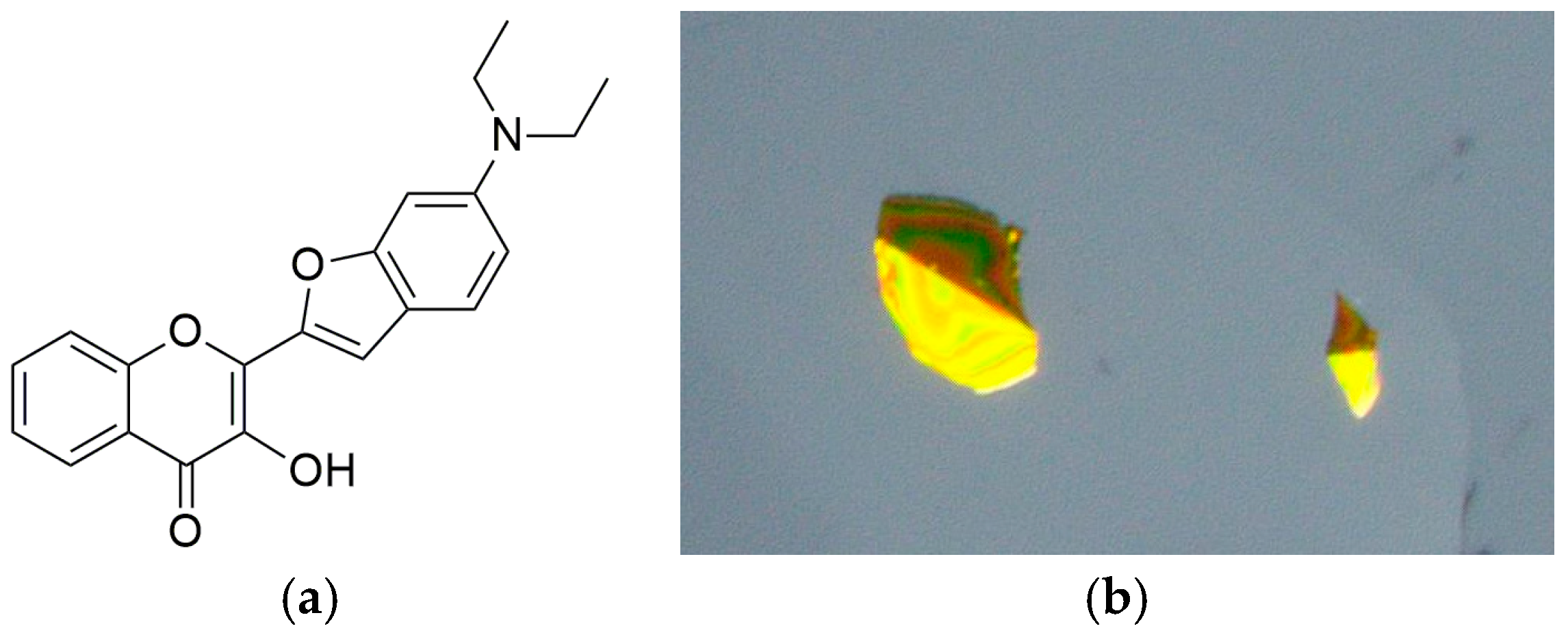
Structural Elucidation of 2-(6-(Diethylamino)benzofuran-2-yl)-3-hydroxy-4H-chromen-4-one and Labelling of Mycobacterium aurum Cells

Abstract
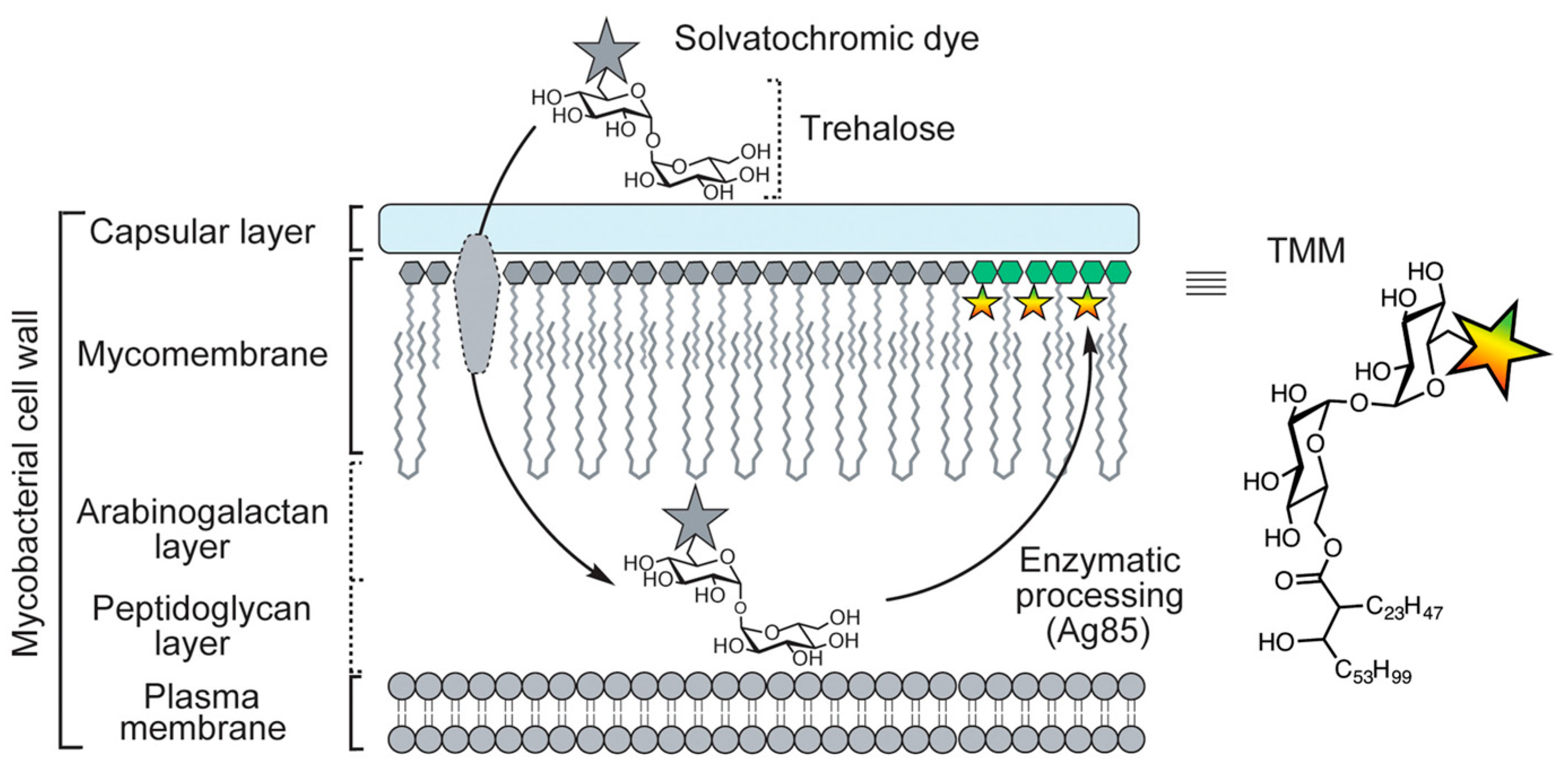
:1. Introduction
2. Results and Discussion
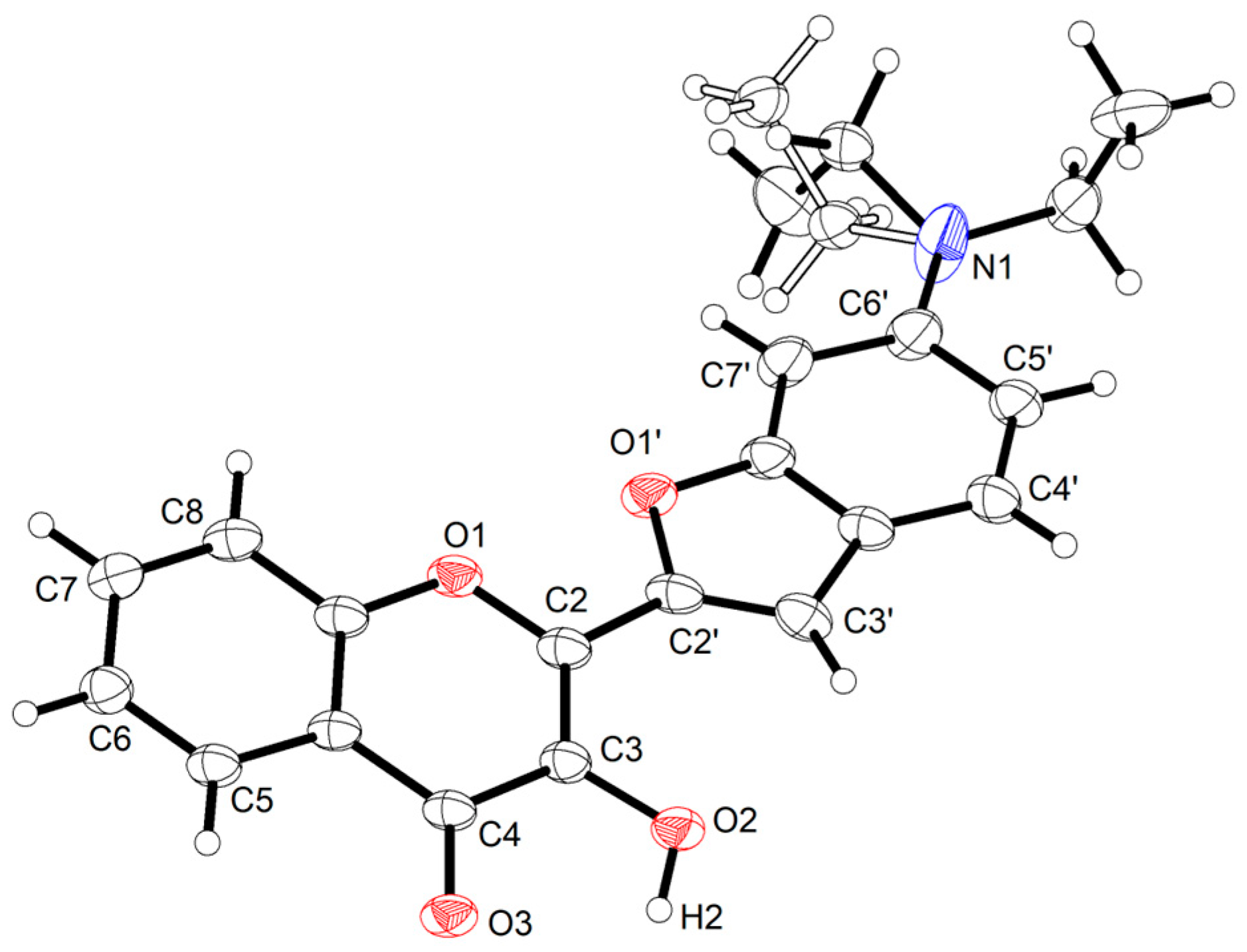
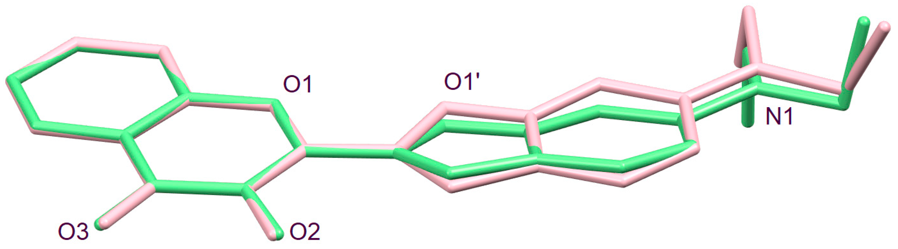
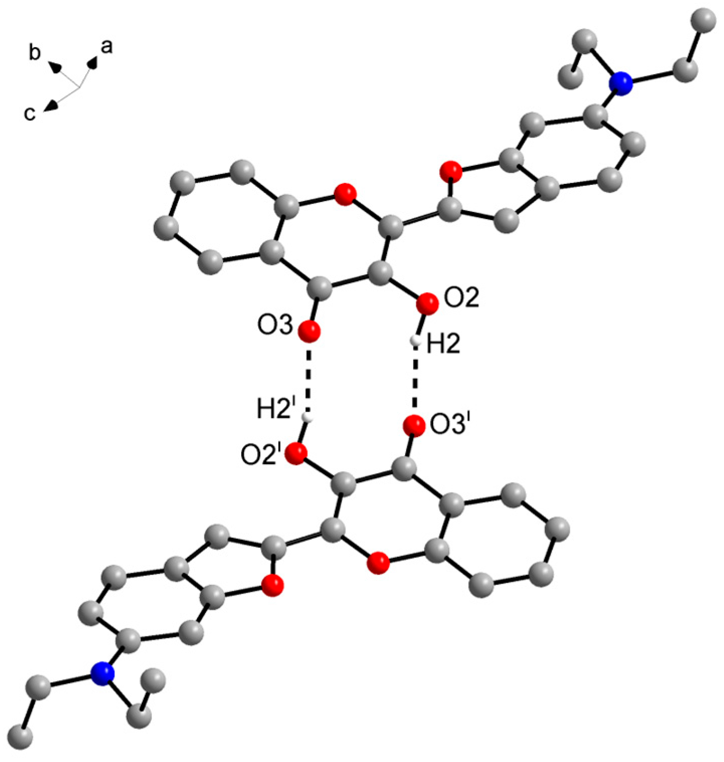
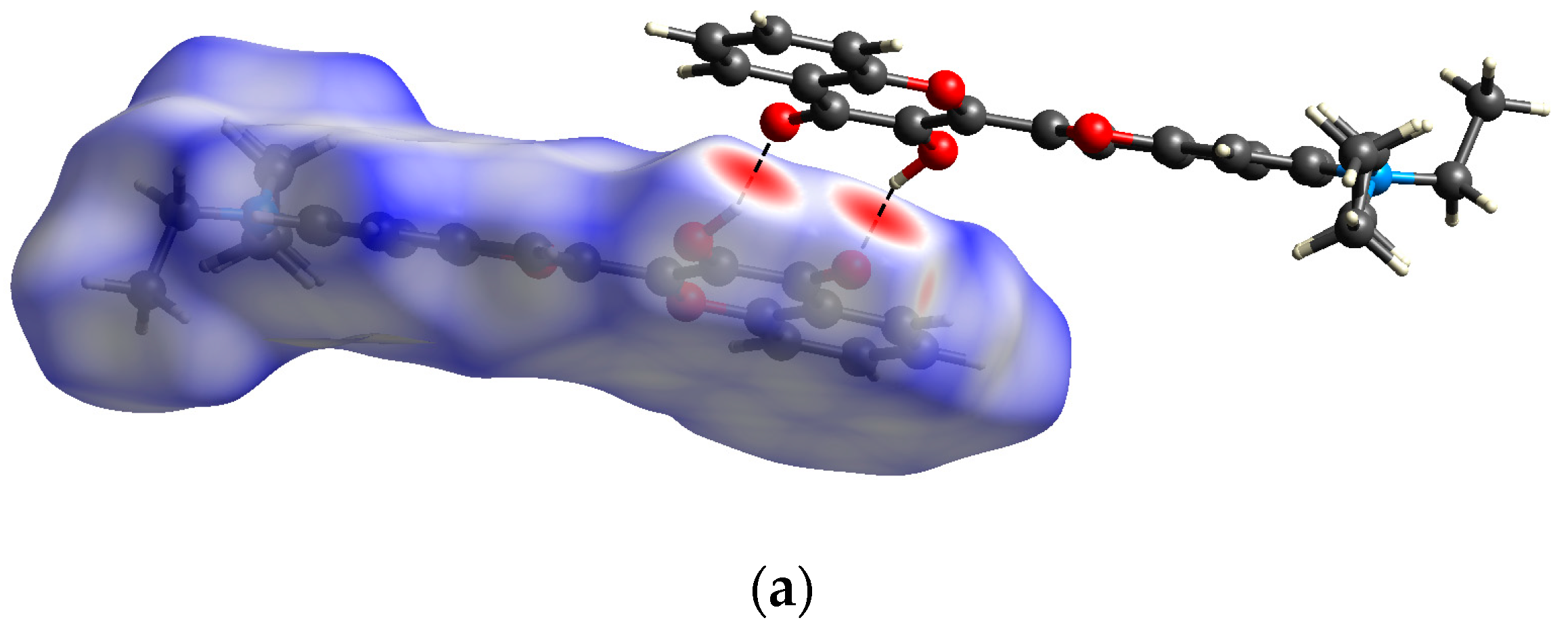
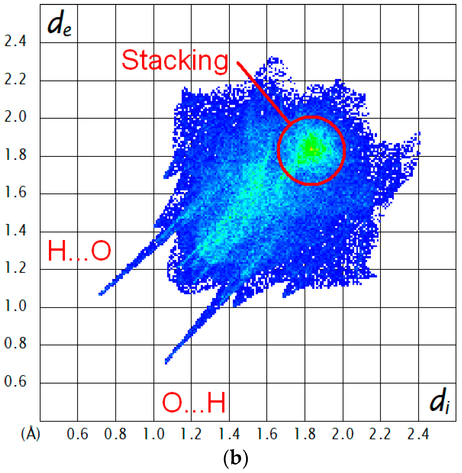
2.1. Structural Description of 3HC-2
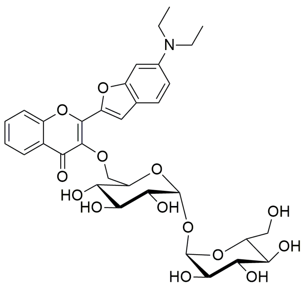
2.2. Labelling of Mycobacterium aurum Cells
3. Materials and Methods
3.1. General
3.2. X-ray Crystallography
3.3. Computational Methods
3.4. Microbiology
3.5. Fluorescence Microscopy
4. Conclusions
Supplementary Materials
Author Contributions
Funding
Data Availability Statement
Acknowledgments
Conflicts of Interest
Sample Availability
References
- World Health Organization. Global Tuberculosis Report 2022; World Health Organization: Geneva, Switzerland, 2022. [Google Scholar]
- To, K.; Cao, R.; Yegiazaryan, A.; Owens, J.; Venketaraman, V. General Overview of Nontuberculous Mycobacteria Opportunistic Pathogens: Mycobacterium avium and Mycobacterium abscessus. J. Clin. Med. 2020, 9, 2541. [Google Scholar] [CrossRef] [PubMed]
- Dahl, V.N.; Molhave, M.; Floe, A.; van Ingen, J.; Schon, T.; Lillebaek, T.; Andersen, A.B.; Wejse, C. Global trends of pulmonary infections with nontuberculous mycobacteria: A systematic review. Int. J. Infect. Dis. 2022, 125, 120–131. [Google Scholar] [CrossRef]
- Singhal, R.; Myneedu, V.P. Microscopy as a diagnostic tool in pulmonary tuberculosis. Int. J. Mycobacteriology 2015, 4, 1–6. [Google Scholar] [CrossRef] [PubMed]
- Kamariza, M.; Keyser, S.G.L.; Utz, A.; Knapp, B.D.; Ealand, C.; Ahn, G.; Cambier, C.J.; Chen, T.; Kana, B.; Huang, K.C.; et al. Toward Point-of-Care Detection of Mycobacterium tuberculosis: A Brighter Solvatochromic Probe Detects Mycobacteria within Minutes. JACS Au. 2021, 1, 1368–1379. [Google Scholar] [CrossRef] [PubMed]
- Tortoli, E.; Brown-Elliott, B.A.; Chalmers, J.D.; Cirillo, D.M.; Daley, C.L.; Emler, S.; Floto, R.A.; Garcia, M.J.; Hoefsloot, W.; Koh, W.-J.; et al. Same meat, different gravy: Ignore the new names of mycobacteria. Eur. Respir. J. 2019, 54, 1900795, Mycobacterium aurum has been reclassified into the new genus Mycolicibacterium, but the new names of mycobacteria have not widely been accepted. [Google Scholar] [CrossRef] [PubMed]
- Phelan, J.; Maitra, A.; McNerney, R.; Nair, M.; Gupta, A.; Coll, F.; Pain, A.; Bhakta, S.; Clark, T.G. The draft genome of Mycobacterium aurum, a potential model organism for investigating drugs against Mycobacterium tuberculosis and Mycobacterium leprae. Int. J. Mycobacteriology 2015, 4, 207–216. [Google Scholar] [CrossRef] [PubMed]
- Namouchi, A.; Cimino, M.; Favre-Rochex, S.; Charles, P.; Gicquel, B. Phenotypic and genomic comparison of Mycobacterium aurum and surrogate model species to Mycobacterium tuberculosis: Implications for drug discovery. BMC Genom. 2017, 18, 530. [Google Scholar] [CrossRef]
- Gupta, A.; Bhakta, S.; Kundu, S.; Gupta, M.; Srivastava, B.S.; Srivastava, R. Fast-growing, non-infectious and intracellularly surviving drug-resistant Mycobacterium aurum: A model for high-throughput antituberculosis drug screening. J. Antimicrob. Chemother. 2009, 64, 774–781. [Google Scholar] [CrossRef]
- Sood, S.; Yadav, A.; Shrivastava, R. Mycobacterium aurum is Unable to Survive Mycobacterium tuberculosis Latency Associated Stress Conditions: Implications as Non-suitable Model Organism. Indian J. Microbiol. 2016, 56, 198–204. [Google Scholar] [CrossRef]
- Honarvar, B.; Movahedan, H.; Mahmoodi, M.; Sheikholeslami, F.M.; Farnia, P. Mycobacterium aurum keratitis: An unusual etiology of a sight-threatening infection. Braz. J. Infect. Dis. 2012, 16, 204–208. [Google Scholar] [CrossRef]
- Madikizela, B.; Eckhardt, T.; Goddard, R.; Richter, A.; Lins, A.; Lehmann, C.; Imming, P.; Seidel, R.W. Synthesis, structural characterization and antimycobacterial evaluation of several halogenated non-nitro benzothiazinones. Med. Chem. Res. 2021, 30, 1523–1533. [Google Scholar] [CrossRef] [PubMed]
- Eckhardt, T.; Goddard, R.; Lehmann, C.; Richter, A.; Sahile, H.A.; Liu, R.; Tiwari, R.; Oliver, A.G.; Miller, M.J.; Seidel, R.W.; et al. Crystallographic evidence for unintended benzisothiazolinone 1-oxide formation from benzothiazinones through oxidation. Acta Crystallogr. Sect. C 2020, 76, 907–913. [Google Scholar] [CrossRef] [PubMed]
- Richter, A.; Seidel, R.W.; Goddard, R.; Eckhardt, T.; Lehmann, C.; Dörner, J.; Siersleben, F.; Sondermann, T.; Mann, L.; Patzer, M.; et al. BTZ-Derived Benzisothiazolinones with In Vitro Activity against Mycobacterium tuberculosis. ACS Med. Chem. Lett. 2022, 13, 1302–1310. [Google Scholar] [CrossRef] [PubMed]
- Klymchenko, A.S.; Ozturk, T.; Pivovarenko, V.G.; Demchenko, A.P. A 3-hydroxychromone with dramatically improved fluorescence properties. Tetrahedron Lett. 2001, 42, 7967–7970. [Google Scholar] [CrossRef]
- Klymchenko, A.S.; Pivovarenko, V.G.; Ozturk, T.; Demchenko, A.P. Modulation of the solvent-dependent dual emission in 3-hydroxychromones by substituents. New J. Chem. 2003, 27, 1336–1343. [Google Scholar] [CrossRef]
- Groom, C.R.; Bruno, I.J.; Lightfoot, M.P.; Ward, S.C. The Cambridge Structural Database. Acta Crystallogr. Sect. B 2016, 72, 171–179. [Google Scholar] [CrossRef]
- Thomas, I.R.; Bruno, I.J.; Cole, J.C.; Macrae, C.F.; Pidcock, E.; Wood, P.A. WebCSD: The online portal to the Cambridge Structural Database. J. Appl. Crystallogr. 2010, 43, 362–366. [Google Scholar] [CrossRef]
- Wera, M.; Pivovarenko, V.G.; Sikorski, A.; Lis, T.; Blazejowski, J. 2-(Furan-2-yl)-3-hydroxy-4H-chromen-4-one. Acta Crystallogr. Sect. E 2011, 67, o266. [Google Scholar] [CrossRef]
- Camargo, M.L.M.; dos Santos, F.A.; Pizzuti, L.; Abram, U.; Schwade, V.D. Complexes with Furyl-Substituted 3-Hydroxychromone: Synthesis, Characterization and Fluorescence Studies. J. Braz. Chem. Soc. 2021, 32, 1519–1530. [Google Scholar] [CrossRef]
- Camargo, M.L.M.; Schwalm, C.S.; Bortolotto, T.; de Freitas Daudt, N.; Rossi, G.G.; Anraku de Campos, M.M.; D’Oliveira, K.A.; Cuin, A.; Schwade, V.D. MII (M = Mn, Fe, Co, Ni and Cu) complexes with a chromone-derived neutral ligand: Synthesis, structural characterization, photocatalytic and mycobacterial activity studies. New J. Chem. 2022, 46, 2534–2545. [Google Scholar] [CrossRef]
- Tseng, H.-W.; Shen, J.-Y.; Kuo, T.-Y.; Tu, T.-S.; Chen, Y.-A.; Demchenko, A.P.; Chou, P.-T. Excited-state intramolecular proton-transfer reaction demonstrating anti-Kasha behavior. Chem. Sci. 2016, 7, 655–665. [Google Scholar] [CrossRef] [PubMed]
- Mughal, E.U.; Javid, A.; Sadiq, A.; Murtaza, S.; Zafar, M.N.; Khan, B.A.; Sumrra, S.H.; Tahir, M.N.; Kanwal; Khan, K.M. Synthesis, structure-activity relationship and molecular docking studies of 3-O-flavonol glycosides as cholinesterase inhibitors. Bioorganic Med. Chem. 2018, 26, 3696–3706. [Google Scholar] [CrossRef] [PubMed]
- Peters, E.-M.; Peters, K.; Meints, C.; Tochtermann, W. Crystal structure of (pM*,pM*)-(±)-bi-(dimethyl-3,6-decanooxepine-4,5-dicarboxylate), [C6HO(CH2)IO(COOCH3)2]2. Z. Für Krist.-New Cryst. Struct. 2001, 216, 315–316. [Google Scholar] [CrossRef]
- Alcaide, B.; Almendros, P.; Carrascosa, R.; Torres, M.R. Synthesis of a New Class of C2-Symmetrical Biheteroaryls by Ammonium Cerium(IV) Nitrate Mediated Dimerization of 2-(Furan-3-yl)pyrroles. Eur. J. Org. Chem. 2010, 2010, 823–826. [Google Scholar] [CrossRef]
- Fan, Y.-S.; Das, U.; Hsiao, M.-Y.; Liu, M.-H.; Lin, W. Chemoselective Intramolecular Wittig Reactions for the Synthesis of Oxazoles and Benzofurans. J. Org. Chem. 2014, 79, 11567–11582. [Google Scholar] [CrossRef]
- Mulay, S.V.; Bogoslavky, B.; Galanti, I.; Galun, E.; Gidron, O. Bifuran-imide: A stable furan building unit for organic electronics. J. Mater. Chem. C 2018, 6, 11951–11955. [Google Scholar] [CrossRef]
- Ookubo, Y.; Wakamiya, A.; Yorimitsu, H.; Osuka, A. Synthesis of a Library of Fluorescent 2-Aryl-3-trifluoromethylnaphthofurans from Naphthols by Using a Sequential Pummerer-Annulation/Cross-Coupling Strategy and their Photophysical Properties. Chem. A Eur. J. 2012, 18, 12690–12697. [Google Scholar] [CrossRef]
- Wang, C.-H.; Gao, Z.-C.; Sun, W.; Guo, X.; Zhang, F.-B. P⋯O noncovalent conformational locks for constructing highly planar Bis(diphenylphosphanyl) Bi(benzofurano). Dye. Pigment. 2021, 184, 108820. [Google Scholar] [CrossRef]
- Bernstein, J.; Davis, R.E.; Shimoni, L.; Chang, N.L. Patterns in Hydrogen Bonding: Functionality and Graph Set Analysis in Crystals. Angew. Chem. Int. Ed. 1995, 34, 1555–1573. [Google Scholar] [CrossRef]
- Thakuria, R.; Sarma, B.; Nangia, A. 7.03-Hydrogen Bonding in Molecular Crystals. In Comprehensive Supramolecular Chemistry II; Atwood, J.L., Ed.; Elsevier: Oxford, UK, 2017; pp. 25–48. [Google Scholar] [CrossRef]
- Binbuga, N.; Schultz, T.P.; Henry, W.P. Intra- and intermolecular hydrogen bonding in 3-hydroxy- and 5-hydroxychromone. Tetrahedron Lett. 2008, 49, 5762–5765. [Google Scholar] [CrossRef]
- T, J.A.S.; J, R.; Rajan, A.; Shankar, V. Features of the biochemistry of Mycobacterium smegmatis, as a possible model for Mycobacterium tuberculosis. J. Infect. Public Health 2020, 13, 1255–1264. [Google Scholar] [CrossRef] [PubMed]
- APEX4; Bruker AXS Inc.: Madison, WI, USA, 2017.
- SAINT; Bruker AXS Inc.: Madison, WI, USA, 2012.
- SADABS; Bruker AXS Inc.: Madison, WI, USA, 2012.
- Sheldrick, G.M. SHELXT-integrated space-group and crystal-structure determination. Acta Crystallogr. Sect. A 2015, 71, 3–8. [Google Scholar] [CrossRef] [PubMed]
- Sheldrick, G.M. Crystal structure refinement with SHELXL. Acta Crystallogr. Sect. C 2015, 71, 3–8. [Google Scholar] [CrossRef] [PubMed]
- Thorn, A.; Dittrich, B.; Sheldrick, G.M. Enhanced rigid-bond restraints. Acta Crystallogr. Sect. A 2012, 68, 448–451. [Google Scholar] [CrossRef]
- Brandenburg, K. Diamond; Crystal Impact GbR: Bonn, Germany, 2018. [Google Scholar]
- Spackman, P.R.; Turner, M.J.; McKinnon, J.J.; Wolff, S.K.; Grimwood, D.J.; Jayatilaka, D.; Spackman, M.A. CrystalExplorer: A program for Hirshfeld surface analysis, visualization and quantitative analysis of molecular crystals. J. Appl. Crystallogr. 2021, 54, 1006–1011. [Google Scholar] [CrossRef]
- Neese, F.; Wennmohs, F.; Becker, U.; Riplinger, C. The ORCA quantum chemistry program package. J. Chem. Phys. 2020, 152, 224108. [Google Scholar] [CrossRef]
- Becke, A.D. Density-functional thermochemistry. III. The role of exact exchange. J. Chem. Phys. 1993, 98, 5648–5652. [Google Scholar] [CrossRef]
- Hertwig, R.H.; Koch, W. On the parameterization of the local correlation functional. What is Becke-3-LYP? Chem. Phys. Lett. 1997, 268, 345–351. [Google Scholar] [CrossRef]
- Weigend, F.; Ahlrichs, R. Balanced basis sets of split valence, triple zeta valence and quadruple zeta valence quality for H to Rn: Design and assessment of accuracy. Phys. Chem. Chem. Phys. 2005, 7, 3297–3305. [Google Scholar] [CrossRef]
- Fletcher, R. Practical Methods of Optimization, 2nd ed.; John Wiley & Sons: Hoboken, NJ, USA, 2000. [Google Scholar]
- Macrae, C.F.; Sovago, I.; Cottrell, S.J.; Galek, P.T.A.; McCabe, P.; Pidcock, E.; Platings, M.; Shields, G.P.; Stevens, J.S.; Towler, M.; et al. Mercury 4.0: From visualization to analysis, design and prediction. J. Appl. Crystallogr. 2020, 53, 226–235. [Google Scholar] [CrossRef]


| D–H⋯A 1 | d(D–H) | d(H⋯A) | d(D⋯A) | <(DHA) |
|---|---|---|---|---|
| O2–H2⋯O3i | 0.857(14) | 1.902(15) | 2.7146(13) | 157.9(17) |
| Empirical Formula | C21H19NO4 |
| Mr | 349.37 |
| T (K) | 100(2) |
| λ (Å) | 0.71073 |
| Crystal system, space group | Triclinic, P-1 |
| a (Å) | 7.2022(5) |
| b (Å) | 8.4003(5) |
| c (Å) | 15.0621(10) |
| α (°) | 99.969(4) |
| β (°) | 94.673(4) |
| γ (°) | 111.188(2) |
| V (Å3) | 826.48(9) |
| Z, ρcalc (g cm−3) | 2, 1.404 |
| μcalc (mm−1) | 0.098 |
| F(000) | 368 |
| Crystal size (mm) | 0.236 × 0.112 × 0.020 |
| θ range (°) | 2.665–30.555 |
| Reflections collected/unique | 91202/5061 |
| Rint | 0.0553 |
| Observed reflections [I > 2σ(I)] | 3946 |
| Data/restraints/parameters | 5061/29/261 |
| Goodness-of-fit on F2 | 1.027 |
| R1 [I > 2σ(I)] | 0.0483 |
| wR2 (all data) | 0.1418 |
| Δρmax, Δρmin | 0.471/−0.556 |
Disclaimer/Publisher’s Note: The statements, opinions and data contained in all publications are solely those of the individual author(s) and contributor(s) and not of MDPI and/or the editor(s). MDPI and/or the editor(s) disclaim responsibility for any injury to people or property resulting from any ideas, methods, instructions or products referred to in the content. |
© 2023 by the authors. Licensee MDPI, Basel, Switzerland. This article is an open access article distributed under the terms and conditions of the Creative Commons Attribution (CC BY) license (https://creativecommons.org/licenses/by/4.0/).
Share and Cite
Richter, A.; Goddard, R.; Siersleben, F.; Mann, L.; Seidel, R.W. Structural Elucidation of 2-(6-(Diethylamino)benzofuran-2-yl)-3-hydroxy-4H-chromen-4-one and Labelling of Mycobacterium aurum Cells. Molbank 2023, 2023, M1647. https://doi.org/10.3390/M1647
Richter A, Goddard R, Siersleben F, Mann L, Seidel RW. Structural Elucidation of 2-(6-(Diethylamino)benzofuran-2-yl)-3-hydroxy-4H-chromen-4-one and Labelling of Mycobacterium aurum Cells. Molbank. 2023; 2023(2):M1647. https://doi.org/10.3390/M1647
Chicago/Turabian StyleRichter, Adrian, Richard Goddard, Fabienne Siersleben, Lea Mann, and Rüdiger W. Seidel. 2023. "Structural Elucidation of 2-(6-(Diethylamino)benzofuran-2-yl)-3-hydroxy-4H-chromen-4-one and Labelling of Mycobacterium aurum Cells" Molbank 2023, no. 2: M1647. https://doi.org/10.3390/M1647