Short Note

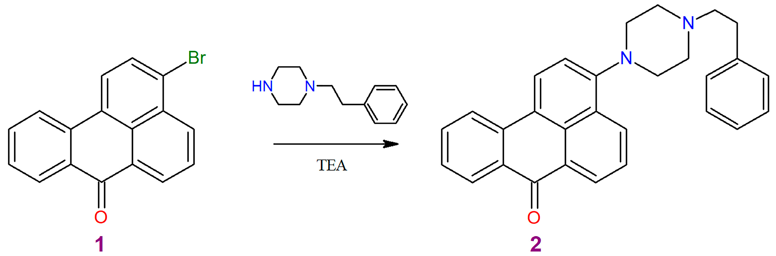
3-[4-(2-Phenylethyl)piperazin-1-yl]-7H-benzo[de]anthracen-7-one

1
Institute of Life Sciences and Technology, Daugavpils University, 1A Parades Street, LV-5401 Daugavpils, Latvia
2
Latvian Institute of Organic Synthesis, Aizkraukles Str. 21, LV-1006 Riga, Latvia
*
Author to whom correspondence should be addressed.
Molbank 2023, 2023(1), M1607; https://doi.org/10.3390/M1607
Submission received: 2 March 2023 / Revised: 17 March 2023 / Accepted: 20 March 2023 / Published: 23 March 2023
(This article belongs to the Section Organic Synthesis)

Abstract

:
The present work describes the synthesis of the new benzanthrone dye-3-[4-(2-phenylethyl)piperazin-1-yl]-7H-benzo[de]anthracen-7-one by bromine atom nucleophilic substitution reaction. The structure of the obtained benzanthrone derivative is characterized by NMR spectroscopy, mass spectrometry, and X-ray diffraction analysis. The photophysical properties of the target compound are investigated by means of UV-Vis and fluorescence spectroscopy in various organic solvents.

1. Introduction

Over the past few decades, great effort has been put into the design and synthesis of new rigid polyaromatic planar compounds with donor-π-acceptor architecture due to their interesting photophysical properties [1]. In these compounds, the electron-donating and electron-accepting groups are connected through a π-conjugated linker. Tuning different donor moiety or acceptor moiety in a D–A molecule would modify its physical and chemical properties. Due to the easy modification of donor, acceptor, and π spacers, many donor-acceptor compounds have been described for various applications [2].
Benzanthrone dyes are widely used among donor-acceptor type compounds as these substances have extended π-conjugated structures and good intramolecular charge transfer characteristics. The advantage of benzanthrone dyes over other classes of dyes is a wide range of donors capable of covering the spectrum from green to near infrared, and good photo- and thermal-stability. Due to these characteristics, benzanthrone dyes find potential applications in a variety of fields, such as sensors and markers [3,4,5,6,7,8].
Previously, heterocyclic amines of benzanthrone were synthesized and characterized showing interesting optical properties [9,10,11]. Herein, in continuation of our previous investigation we designed and synthesized novel donor-acceptor-type fluorescent benzanthrone heterocyclic amino derivative, which is found to show promising photophysical properties with high emission and remarkable fluorosolvatochromism. A new derivative was obtained from 3-bromobenzanthrone and monosubstituted piperazine. The obtained dye was fully characterized, including the determination of the structure by single crystal X-ray diffraction analysis and photophysical parameters in various organic solvents.

2. Results and Discussion

2.1. Synthesis

Organic materials based on the structure of the donor-acceptor type have always been the subject of research because the D-π-A structure of molecules has a great influence on their fluorescence properties, adjusting the energy level structure of molecules is important for developing efficient fluorescent probes. To introduce the donor group, we used the nucleophilic substitution of the halogen atom, since the chlorine or bromine atom in the benzanthrone nucleus is often used to synthesize benzanthrone derivatives [12]. 1-(2-Phenylethyl)piperazine was applied for reaction with 3-bromobenzanthrone accordingly to Scheme 1.
The obtained derivative 2 is a red crystalline compound. NMR spectroscopic study confirmed the chemical structure of this substance. In the 1H NMR spectra of the derivative 2, the multiple proton signals of piperazine methylene and ethylene groups were found from 2.70 ppm to 3.20 ppm. The signals of the aromatic protons of the benzanthrone core and phenyl group are located as the multiple signals at 7.12–8.70 ppm.
Study of the synthesized compound by chromatomass spectrometry shows presence of molecular ion (with M = 418), fragments without benzyl group (M = 327, 328), ion of unsubstituted benzanthrone (M = 228), and 1-phenylnaphthalene ion (M = 201) in the mass spectrum of the analyzed compound (see Figure S1).

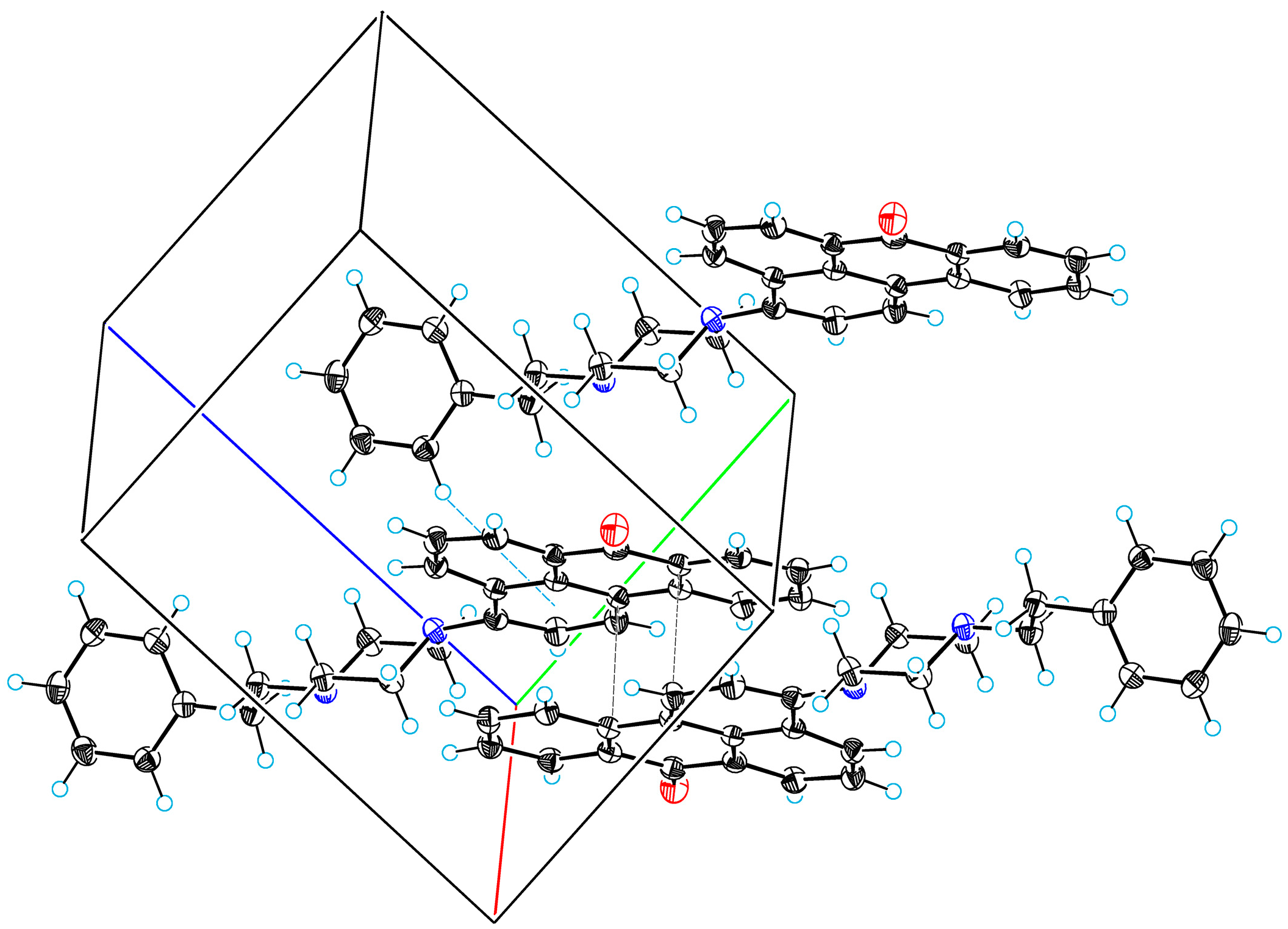
2.2. X-ray Crystallographic Study

Obtained compound 2 crystallizes from toluene in the form of red luminescent lamellar crystals, the structure of which was determined in this work by X-ray diffraction analysis of single crystals in this work (see Tables S1–S7).
Figure 1 shows a perspective view of molecule 2 with thermal ellipsoids and the atom-numbering scheme followed in the text. The molecular structure is characterized by planar benzanthrone system and piperazine cycle with chair conformation. The dihedral angle between the least squares plane of benzanthrone, the mean plane of piperazine cycle is equal 39.9(1)°. Table 1 lists selected torsion angles that characterize the molecular conformation of 2. The torsion angle of N21–C24–C25–C26 indicates that the phenethyl fragment has a fully staggered conformation. Between the mean plane of piperazine and the phenyl ring there is a dihedral angle of 77.0(1)°.
A common feature of benzanthrones is intermolecular π-π stacking interactions between benzanthrone systems. In the crystal structure of 2, there are these interactions with the shortest atom–atomic contact (intermolecular contact C13···C16) 3.421(2) Å (see Figure 2). In the crystal structure, molecules 2 associate to form centrosymmetric dimers by means of these π-π interactions. Additionally, a noteworthy feature is the weak intermolecular hydrogen bond of C–H···π type. The parameters of this bond are following: C31···π = 4.365(2) Å, H31···π = 3.49 Å, C31–H31···π = 153°, where π is centroid of C1–C2–C3–C17–C16–C15 cycle.

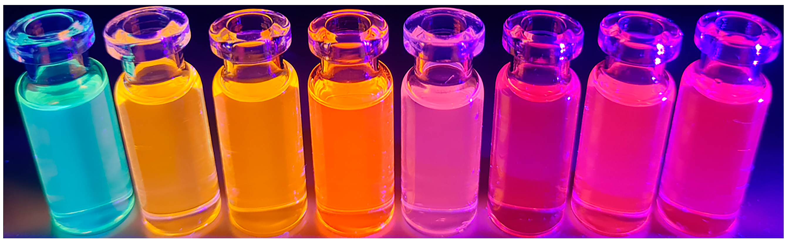
2.3. Spectroscopic Properties

The synthesized dye exhibits pronounced luminescent properties in the solid state and in various organic solvents. In this regard, the photophysical properties of the obtained derivative were evaluated, and the corresponding data are summarized in Table 2. The absorption and emission spectra were recorded in the eight organic solvents with a wide range of polarities (see Figure S2). The position of maximum absorption is situated from 426 nm in hexane to 457 nm in dimethylsulfoxide (DMSO), the bathochromic shift is 31 nm. Furthermore, obtained luminophore has high molar absorption coefficients.
The investigated dye luminesces at 530–665 nm, showing intense luminescence in hexane and significantly lower emission in polar solvents—acetone, ethanol, DMF, and DMSO (see Figure 3). The emission maxima undergo red shift from 537 nm to 664 nm when the solvent is changed from hexane to ethanol. The bathochromic shift of the absorption maximum is accompanied by the bathochromic shift of the fluorescence band. Comparing the results with the previously obtained spectroscopic data for 3-N′-substituted piperazino derivatives, it is found that both 3-(N′-phenyl)piperazino and 3-(N′-3-chlorophenyl)piperazino derivatives demonstrate negative solvatochromism, i.e., in methanol emission maxima shifts hy psochromically by ≈10–16 nm compared to ethyl acetate [10]. The bathochromic shift of the luminescence maximum of the synthesized compound upon passing from benzene to ethanol (68 nm) is larger than that of the unsubstituted amine, imines, and ethers described in the literature, and is 4–43 nm [13,14].
Compared to unsubstituted 3-aminobenzanthrone, the maximum absorption of the obtained compound is shifted towards short-wavelength side by 20–60 nm. However, the luminescence maximum is shifted bathochromically by 96 nm (in ethanol) [13,14]. 3-Substituted azomethines and ethers luminesce in the shorter wavelength range at 520–650 nm (in alcohol), and the luminescence maximum of the sulfur-containing substituted (556 nm) [15] is also hypsochromically shifted. Obviously, this is due to the donor properties of the substituent in the third position of benzanthrone, which affects the intramolecular charge transfer from the substituent to the carbonyl group. According to the study of the influence of the solvent on the absorption and luminescence spectra of benzanthrone derivatives [16], the synthesized dye 2 belongs to compounds in which both the singlet and triplet nπ* states exceed the ππ* levels. Such compounds fluoresce both in hydrocarbons and in alcohols.
The difference between the maximum emission and absorption spectra is called Stokes shift, which has a great importance for technological applications. Chromophores with pronounced Stokes shift are helpful to build sensors and biological probes to be used in optical microscopy [17,18,19]. The obtained dye 2 has relatively large Stokes shifts (up to 7311 cm−1 in ethanol). For 3-aminobenzanthrone, the Stokes shift is 3 times smaller (about 2100 cm in ethanol) than for compound 2 [14]. Therefore, developed dye can be used as a suitable sensitive probe for the labeling of biological objects and to determine the polarity of the environment.

3. Materials and Methods

3.1. Materials and Basic Measurements

All reagents were of analytical grade (Aldrich Chemical Company, Munich, Germany) and were used as received. The progress of the chemical reactions and the purity of products were monitored by TLC on silica gel plates (Fluka F60254, 20 × 10, 0.2 mm, ready-to-use), using with C6H6-CH3CN (3:1) as eluent, visualization under UV light. Column chromatography on silica gel was carried out on Merck Kieselgel (230–240 mesh) with dichloromethane as eluent. Melting points were determined on an MP70 Melting Point System apparatus and are not corrected.
1H NMR spectra were recorded on Bruker equipment, operating at 400 MHz in CDCl3 (with TMS as internal standard) at ambient temperature. Elemental analysis was performed on a Euro Vector EA-3000 CHNS-analyzer.
A Shimadzu GCMS-QP2010 system (Shimadzu Corporation, Kyoto, Japan) was used for the analysis and mass spectra were recording. The gas chromatograph was equipped with an electronically controlled split/splitless injection port. GC was carried out on a 5% diphenyl-/95% dimethylpolysiloxane fused-silica capillary column (Rtx-5SIL-MS, 30 m × 0.32 mm, 0.25 µm film thickness; Restek). Helium (99.999%) was used as the carrier gas at a constant flow of 1.6 mL min−1. The injection (injection volume of 1 µL) was performed at 250 °C in the split mode, with the split ratio 1:10. The oven temperature program was as follows: the temperature was held at 30 °C for 5 min, then 30–180 °C at the rate of 10 °C min−1, 180–300 °C at the rate of 15 °C min−1, and finally, held at 300 °C for 5 min. The mass spectrometer was operated in the electron ionization mode (ionization energy of 70 eV). The source and transfer line temperatures were 200 °C and 310 °C, respectively. Detection was carried out in scan mode: m/z 35–500.
The absorption spectra were obtained using the UV–visible spectrophotometer “Specord’s UV/VIS”. Fluorescence spectra were recorded on a FLSP920 (Edinburgh Instruments Ltd., Livingston, UK) spectrofluorometer in the visible range 450–800 nm. The studies were performed in quartz cuvettes with an absorbing layer thickness of 1 cm at a concentration of solutions in organic solvents of 10–5 mol/L.

3.2. Synthesis and Characterization

In a 25 mL round bottom flask a mixture of 3-bromobenzanthrone (0.31 g, 1 mmol), 1-(2-phenylethyl)piperazine (0.28 g, 1.5 mmol) and triethylamine (5 mL) was heated at reflux for 5–6 h. After cooling, the product was filtered off and dried. Purified by means of column chromatography on 1.5 × 30 cm column packed with silica gel 40/100 (eluent: dichloromethane) to obtain red compound in 65% yield with m.p. of 172–173 °C. Rf = 0.32 (in C6H6-CH3CN (3:1)).
1H NMR (400 MHz, CDCl3) δ 8.70 (dd, J = 7.3, 1.3 Hz, 1H), 8.51 (dd, J = 8.3, 1.4 Hz, 1H), 8.37–8.44 (m, 1H), 8.27 (d, J = 8.1 Hz, 1H), 8. 71 (dd, J = 8.1, 1.1 Hz, 1H), 7.68 (dd, J = 8.3, 7.3 Hz, 1H), 7.62 (ddd, J = 8.1, 7.1, 1.5 Hz, 1H), 7.41 (ddd, J = 8.1, 7.1, 1.0 Hz, 1H), 7.12–7.31 (m, 6H), 3.19 (br, 4H, pip), 2.83 (m, 2H, pipCH2), 2.77 (br, 4H, pip), 2.70 (m, 2H, CH2Ph). EI-MS m/z 418 [M]+ (9), 328(18), 327(76), 284(8), 256(7), 228(7), 201(6), 163(9), 70(100). Anal. calcd. for C29H26N2O: C, 83.22; H, 6.26; N, 6.69; found: C, 83.01; H, 6.09; N, 6.38.

3.3. Single Crystal X-ray Analysis

Diffraction data of compound 2 were collected at 120 K on a Rigaku, XtaLAB Synergy, Dualflex, HyPix diffractometer using CuKα radiation (λ = 1.54184 Å). The crystal structure was solved with the Superflip structure solution program using Charge Flipping [20] and refined by full-matrix least squares with the help of software package [21]. Crystal data: triclinic; a = 8.8957(1), b = 9.4174(1), c = 13.3599(2) Å, α = 75.459(1), β = 77.524(1), γ = 87.499(1)°; V = 1057.73(3) Å3, Z = 2, μ = 0.619 mm–1, Dcalc = 1.314 g·cm–3; space group is P1; R[F2>2σ(F2)] = 0.0396. For further details, see crystallographic data for compound 2 deposited at the Cambridge Crystallographic Data Centre as Supplementary Publication Number CCDC 2242887. Copies of the data can be obtained, free of charge, on application to CCDC, 12 Union Road, Cambridge CB2 1EZ, UK.

Supplementary Materials

The following are available online: Figure S1. Mass spectrum of 3-[4-(2-phenylethyl)piperazin-1-yl]-7H-benzo[de]anthracen-7-one. Table S1. Crystal data and structure refinement for 3-[4-(2-phenylethyl)piperazin-1-yl]-7H-benzo[de]anthracen-7-one. Table S2. Fractional Atomic Coordinates (×104) and Equivalent Isotropic Displacement Parameters (Å2 ×10³). Ueq is defined as 1/3 of the trace of the orthogonalized UIJ tensor. Table S3. Anisotropic Displacement Parameters (Å2 ×10³). The Anisotropic displacement factor exponent takes the form: –2π2[h2a*2U11+2hka*b*U12+…]. Table S4. Bond Lengths for 3-[4-(2-phenylethyl)piperazin-1-yl]-7H-benzo[de]anthracen-7-one. Table S5. Bond Angles for 3-[4-(2-phenylethyl)piperazin-1-yl]-7H-benzo[de]anthracen-7-one. Table S6. Torsion Angles for 3-[4-(2-phenylethyl)piperazin-1-yl]-7H-benzo[de]anthracen-7-one. Table S7. Hydrogen Atom Coordinates (Å ×104) and Isotropic Displacement Parameters (Å2 ×10³). Figure S2. The absorption and emission spectra of dye 2 in various organic solvents.

Author Contributions

R.F. and E.K. designed chemical synthesis, analyzed results, and wrote the manuscript. A.P. and S.O. performed spectroscopic experiments and analyzed results. S.B. performed single-crystal X-ray diffraction analysis and wrote the manuscript. All authors have read and agreed to the published version of the manuscript.

Funding

This work was supported by Daugavpils University Students Research Project. Project No.14-89/2022/11.

Data Availability Statement

The data presented in this study are available in this article and supplementary file.

Conflicts of Interest

The authors declare no conflict of interest.

References

  1. Hunger, K. (Ed.) Industrial Dyes, Chemistry, Properties, Applications; Wiley-VCH: Weinheim, Germany, 2003; pp. 574–589. [Google Scholar]
  2. Bureš, F. Fundamental aspects of property tuning in push–pull molecules. RSC Adv. 2014, 4, 58826–58851. [Google Scholar] [CrossRef] [Green Version]
  3. Tsiko, U.; Sych, G.; Volyniuk, D.; Bezvikonnyi, O.; Keruckiene, R.; Lazauskas, A.; Grazulevicius, J.V. Self-recovering mechanochromic luminescence of the derivatives of benzanthrone and carbazole: Towards damage-resistive information recording and security probes. Dye. Pigment. 2022, 199, 110082. [Google Scholar] [CrossRef]
  4. Kirilova, E.; Mickevica, I.; Mezaraupe, L.; Puckins, A.; Rubenina, I.; Osipovs, S.; Kokina, I.; Bulanovs, A.; Kirjusina, M.; Gavarane, I. Novel dye for detection of callus embryo by confocal laser scanning fluorescence microscopy. Luminescence 2019, 34, 353–359. [Google Scholar] [CrossRef] [PubMed]
  5. Gavarane, I.; Kirilova, E.; Rubeniņa, I.; Mežaraupe, L.; Osipovs, S.; Deksne, G.; Pučkins, A.; Kokina, I.; Bulanovs, A.; Kirjušina, M. A Simple and Rapid Staining Technique for Sex Determination of Trichinella Larvae Parasites by Confocal Laser Scanning Microscopy. Microsc. Microanal. 2019, 25, 1491–1497. [Google Scholar] [CrossRef] [PubMed]
  6. Staneva, D.; Vasileva-Tonkova, E.; Grabchev, I. pH sensor potential and antimicrobial activity of a new PPA dendrimer modified with benzanthrone fluorophores in solution and on viscose fabric. J. Photochem. Photobiol. A 2019, 375, 24–29. [Google Scholar] [CrossRef]
  7. Kalnina, I.; Klimkane, L.; Kirilova, E.; Toma, M.M.; Kizane, G.; Meirovics, I. Fluorescent Probe ABM for Screening Gastrointestinal Patient’s Immune State. J. Fluoresc. 2007, 17, 619–625. [Google Scholar] [CrossRef] [PubMed]
  8. Altaf, Y.; Ullah, S.; Khan, F.A.; Maalik, A.; Rubab, S.L.; Hashmi, M.A. Finding New Precursors for Light Harvesting Materials: A Computational Study of the Fluorescence Potential of Benzanthrone Dyes. ACS Omega 2021, 6, 32334–32341. [Google Scholar] [CrossRef] [PubMed]
  9. Kirilova, E.M.; Meirovics, I.A.; Belyakov, S.V. Preparation and Properties of Nitrogen Derivatives of Benzanthrone with Heterocyclic Fragments. Chem. Heterocycl. Compd. 2002, 38, 789. [Google Scholar] [CrossRef]
  10. Shivraj; Siddlingeshwar, B.; Thomas, A.; Kirilova, E.; Divakar, D.; Alkheraif, A. Experimental and theoretical insights on the effect of solvent polarity on the photophysical properties of a benzanthrone dye. Spectrochim. Acta A 2019, 218, 221–228. [Google Scholar] [CrossRef] [PubMed]
  11. Tarabara, U.; Kirilova, E.; Kirilov, G.; Vus, K.; Zhytniakivska, O.; Trusova, V.; Gorbenko, G. Benzanthrone dyes as mediators of cascade energy transfer in insulin amyloid fibrils. J. Mol. Liq. 2021, 324, 115102. [Google Scholar] [CrossRef]
  12. Krasovitskii, B.M.; Bolotin, B.M. Organic Luminescent Materials; Wiley-VCH: New York, NY, USA, 1988; pp. 149–152. [Google Scholar]
  13. Kapusta, P.; Machalický, O.; Hrdina, R.; Nepraš, M.; Zimmt, M.B.; Fidler, V. Photophysics of 3-Substituted Benzanthrones: Substituent and Solvent Control of Intersystem Crossing. J. Phys. Chem. A 2003, 107, 9740–9746. [Google Scholar] [CrossRef]
  14. Grabchev, I.; Moneva, I.; Wolarz, E.; Bauman, D.; Stoyanov, S. Spectral Properties of 3-Benzanthrone Derivative Dyes in Isotropic Solvents, Polymer Film and Liquid Crystal. Z. Naturforsch. 2001, 56, 291–296. [Google Scholar] [CrossRef]
  15. Wolfbeis, O.S.; Carlini, F.M. Long-wavelength fluorescent indicators for the determination of oxygen partial pressures. Anal. Chim. Acta 1984, 160, 301–304. [Google Scholar] [CrossRef]
  16. Bhujle, V.V.; Padhye, M.R. Effect of solvents on absorption and fluorescence-spectra of some substituted benzanthrones. Indian J. Chem. 1971, 9, 1405–1406. [Google Scholar]
  17. Weihs, F.; Dacres, H. Red-shifted bioluminescence Resonance Energy Transfer: Improved tools and materials for analytical in vivo approaches. Trends Anal. Chem. 2019, 116, 61–73. [Google Scholar] [CrossRef]
  18. Sednev, M.V.; Belov, V.N.; Hell, S.W. Fluorescent dyes with large Stokes shifts for super-resolution optical microscopy of biological objects: A review. Methods Appl. Fluoresc. 2015, 3, 042004. [Google Scholar] [CrossRef] [PubMed] [Green Version]
  19. Jun, J.V.; Chenoweth, D.M.; Petersson, E.J. Rational design of small molecule fluorescent probes for biological applications. Org. Biomol. Chem. 2020, 18, 5747–5763. [Google Scholar] [CrossRef] [PubMed]
  20. Palatinus, L.; Prathapa, S.J.; van Smaalen, S. EDMA: A computer program for topological analysis of discrete electron densities. J. Appl. Cryst. 2012, 45, 575–580. [Google Scholar] [CrossRef]
  21. Sheldrick, G.M. Crystal structure refinement with SHELXL. Acta Cryst. 2015, C71, 3–8. [Google Scholar] [CrossRef] [Green Version]
Scheme 1. Synthesis of 3-[4-(2-phenylethyl)piperazin-1-yl]-7H-benzo[de]anthracen-7-one (2).
Molbank 2023 m1607 sch001
Figure 1. ORTEP diagram molecule 2.
Molbank 2023 m1607 g001
Figure 2. A fragment of the packing of molecules 2, showing π-π stacking and C–H···π interactions.
Molbank 2023 m1607 g002
Figure 3. Compound 2 under ultraviolet light in organic solvents (hexane, benzene, ethyl acetate, chloroform, acetone, ethanol, DMF, and DMSO).
Molbank 2023 m1607 g003
Table 1. Selected torsion angles (°) for 2.
C2–C3–N18–C19 117.1(1)
C2–C3–N18–C23 −11.2(2)
C3–N18–C19–C20 167.19(8)
C3–N18–C23–C22 −167.70(8)
C17–C3–N18–C19 −65.8(1)
C17–C3–N18–C23 165.83(9)
N18–C19–C20–N21 59.8(1)
C19–N18–C23–C22 60.2(1)
C19–C20–N21–C22 −56.9(1)
C19–C20–N21–C24 −179.99(8)
C20–N21–C22–C23 56.8(1)
C20–N21–C24–C25 −168.23(9)
N21–C22–C23–N18 −59.7(1)
N21–C24–C25–C26 −176.77(8)
C22–N21–C24–C25 70.4(1)
C23–N18–C19–C20 −60.4(1)
C24–N21–C22–C23 178.22(8)
C24–C25–C26–C27 61.8(1)
C24–C25–C26–C31 −118.8(1)
Table 2. Photophysical parameters of compound 2 in various solvents (concentration is 10−5 mol l−1).
SolventAbsorption λabs, nmExtinction Coefficient lgεFluorescence λem, nmStokes Shift, cm−1
Hexane4264.055374852
Benzene4434.115965795
Ethyl acetate4404.155955921
CHCl34474.156156111
Acetone4474.086416771
DMF4524.096316276
DMSO4574.086436330
Ethanol4474.016647311
Disclaimer/Publisher’s Note: The statements, opinions and data contained in all publications are solely those of the individual author(s) and contributor(s) and not of MDPI and/or the editor(s). MDPI and/or the editor(s) disclaim responsibility for any injury to people or property resulting from any ideas, methods, instructions or products referred to in the content.

