Article

Upregulation of Inflammatory Mediators in Peripheral Blood CD40+ Cells in Children with Autism Spectrum Disorder

1
Department of Pharmacology and Toxicology, College of Pharmacy, King Saud University, Riyadh 11451, Saudi Arabia
2
Department of Physiology, College of Medicine, King Saud University, Riyadh 11451, Saudi Arabia
3
Department of Pharmaceutics, College of Pharmacy, King Saud University, Riyadh 11451, Saudi Arabia
4
Department of Botany and Microbiology, College of Science, King Saud University, Riyadh 11451, Saudi Arabia
*
Author to whom correspondence should be addressed.
Int. J. Mol. Sci. 2023, 24(8), 7475; https://doi.org/10.3390/ijms24087475
Submission received: 19 March 2023 / Revised: 11 April 2023 / Accepted: 13 April 2023 / Published: 19 April 2023

Abstract

:
Autism spectrum disorder (ASD) is a common and severe neurodevelopmental disorder in early childhood, defined as social and communication deficits and repetitive and stereotypic behaviours. The aetiology is unknown in most cases. However, several studies have identified immune dysregulation as potentially promoting ASD. Among the numerous immunological findings in ASD, reports of increased pro-inflammatory markers remain the most consistently observed. C-C chemokine receptor type 1 (CCR1) activation is pro-inflammatory in several neurological disorders. Previous evidence has implied that the expression of chemokine receptors, inflammatory mediators, and transcription factors play a pivotal role in several neuroinflammatory disorders. There have also been reports on the association between increased levels of proinflammatory cytokines and ASD. In this study, we aimed to investigate the possible involvement of CCR1, inflammatory mediators, and transcription factor expression in CD40+ cells in ASD compared to typically developing controls (TDC). Flow cytometry analysis was used to determine the levels of CCR1-, IFN-γ-, T-box transcription factor (T-bet-), IL-17A-, retinoid-related orphan receptor gamma t (RORγt-), IL-22- and TNF-α-expressing CD40 cells in PBMCs in children with ASD and the TDC group. We further examined the mRNA and protein expression levels of CCR1 using real-time PCR and western blot analysis. Our results revealed that children with ASD had significantly increased numbers of CD40+CCR1+, CD40+IFN-γ+, CD40+T-bet+, CD40+IL-17A+, CD40+RORγt+, CD4+IL-22+, and CD40+TNF-α+ cells compared with the TDC group. Furthermore, children with ASD had higher CCR1 mRNA and protein expression levels than those in the TDC group. These results indicate that CCR1, inflammatory mediators, and transcription factors expressed in CD40 cells play vital roles in disease progression.

1. Introduction

Autism spectrum disorder (ASD) is a complex heterogeneous neurodevelopmental disorder characterised by social interaction, communication impairments, and several social and behavioural abnormalities [1]. The aetiological conditions of ASD are still being explored, but previous studies have suggested multifactorial aetiopathogenesis resulting from interactions between genomic, genetic, and environmental factors associated with its development [2,3]. The immune system plays an important role in neurodevelopment, and immune abnormalities have been observed in patients with ASD [4,5]. Cytokine profiles are associated with changes in behavioural symptoms in a subset of individuals with ASD [6]. Previous studies have shown that abnormal cytokine production in individuals diagnosed with ASD is associated with abnormal cytokine levels [7]. Elevated plasma cytokine levels in ASD provide evidence of immune dysfunction and are associated with impaired behavioural outcomes [8]. We previously demonstrated immune aberrations through an imbalance in pro- and anti-inflammatory cytokines in children with ASD [9]. We have further reported the dysregulation of Th1, Th2, Th17, and T regulatory cell-related transcription factor signalling in children with ASD [10]. We also reported that the upregulation of the JAK-STAT signalling pathways in children with ASD could play a critical role in immune dysfunction in ASD [11]. The upregulation of peripheral chemokines and chemokine receptor expression is associated with immune dysregulation in children with ASD [12]. However, the exact underlying mechanism requires further investigation.
C-C chemokine receptor type 1 (CCR1) is a member of the beta chemokine receptor family [13]. Chemokine receptors are associated with several behavioural impairments in individuals with ASD [14]. Several studies have shown that chemokine receptor expression is higher in the brain tissue of patients with ASD [15,16]. Chemokine receptors are expressed on neurons, upregulated in neurological disorders [17], and are functional mediators of neuroinflammatory disorders [18]. Higher levels of chemokine receptors have been found in astrocytes, the anterior cingulate gyrus, the cerebellum, and the brain of patients with autism [19]. The activation of CCR1 is pro-inflammatory in neurological diseases, and it is expressed in various cell types in the brain, including smooth muscle cells, astrocytes, and neurons [20,21]. CCR1 activation contributes to the blood−brain barrier (BBB) damage associated with various neuroinflammatory diseases [22]. CD40 is a TNF receptor (TNFR) superfamily member mainly expressed in APCs such as B cells, macrophages, and dendritic cells [23]. CD40 is presented on the surface of B and T-cells, respectively, which, upon ligation, activates B-cells for antibody isotype switching and upregulates the production of pro-inflammatory cytokines. While most neurological diseases have several factors contributing to pathogenesis, aberrant neuroinflammation mediated by CD40 increases BBB permeability, exacerbates edema and neuronal and glial cell damage, and promotes the formation of occlusive microthrombi [24]. The CD40 signalling pathway has been well documented as an immune checkpoint and humoral and cellular immune stimulator. The CD40 signalling pathway in APCs contributes to numerous cellular functions such as Th17 cell polarisation, proinflammatory cytokine release, and immunoglobulin isotype switching [25]. Several studies have demonstrated the involvement of CD40 cells in the pathogenesis of systemic lupus erythematosus and BTBR mouse model of ASD [26,27]. A recent study showed that CD40 cells significantly increased in the BTBR mouse model of ASD [28]. Therefore, this study explored the overall inflammatory potential of APCs expressing CD40 in children with ASD.
Abnormal IFN-γ production is associated with several immune diseases. Previous studies have shown an increased production of inflammatory mediators in the brains of individuals with ASD [29]. Studies of blood from individuals with ASD showed that IFN-γ was significantly increased compared to controls [30]. A high level of prenatal IFN-γ increases the risk of having an ASD child [31]. Previous results revealed the unexpected role of IFN-γ in regulating neuronal connectivity and social behaviour [32]. T-box transcription factor (T-bet) is essential for differentiating Th1 cells [33]. T-bet expression is important in inflammatory responses and is critical in autoimmune diseases [34]. In humans, T-bet expression is induced in B cells [35], and a recent study has suggested that T-bet-expressing B cells are recruited to the CNS [36]. T-bet increases IL-17A levels in the CNS and T-cell infiltration, concomitant with neuroinflammation [37].
IL-17A is a key regulator of ASD development [38], and elevated IL-17 and IL-17A have been observed in children with ASD [39,40]. It has also been reported that autism-like symptoms in the offspring of immune-activated maternal mice may be due to IL-17A signalling [41]. The enrichment of IL-17 genes has been reported in individuals with ASD, indicating a possible role for this cytokine in the pathophysiology of this condition [42]. Retinoid-related orphan receptor gamma t (RORγt), a master transcription factor for Th17, is critical in mediating many autoimmune diseases [43]. Interleukin (IL)-22 is a potent mediator of inflammatory responses. Th22 cells are a major source of IL-22 and play an important role in several neurological disorders by promoting leukocyte infiltration in the brain [44]. IL-22 also regulates chemo-attractant production by microvascular endothelial cells in the BBB [45], plays a role in several human diseases, and its upregulation is associated with lymphocyte activation in the CNS [46]. TNF-α is a central regulator of inflammation and is elevated in the cerebrospinal fluid of children with ASD [47]. In addition, TNF-α expression is elevated in ASD, suggesting a dysregulated immune response [48]. Previous studies have shown that a high level of TNF-α is correlated with the severity of ASD symptoms [8,15,49]. These results indicate the potential role of inflammatory mediators in neuroimmune dysfunction. We hypothesised that CD40 expression promotes an immune imbalance in children with ASD. Thus, the restoration of CD40 may be considered a treatment strategy for immune abnormalities in children with ASD.

2. Results

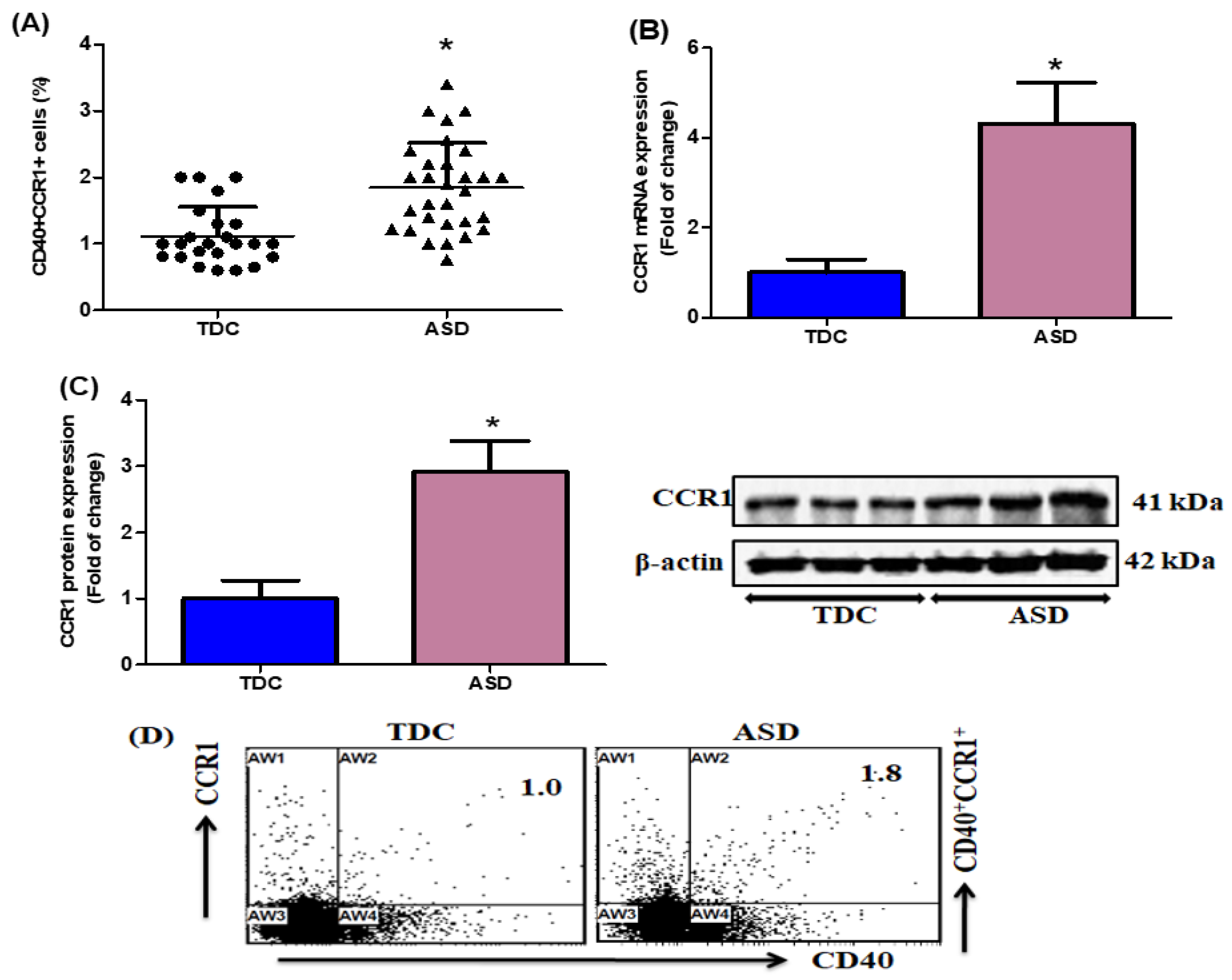
2.1. Upregulation of CCR1-Expressing CD40+ Cells in Children with ASD

Flow cytometry was performed to evaluate the number of CCR1-expressing CD40+ cells in the ASD and TDC groups. The number of CCR1-expressing CD40+ PBMCs was elevated in the children with ASD compared to that in the TDC group (Figure 1A). We also evaluated the gene expression of CCR1 from the ASD and TDC groups. The children with ASD showed a significant increase in CCR1 mRNA expression compared to the TDC group (Figure 1B). We further evaluated CCR1 protein expression levels in PBMCs. The protein expression level of CCR1 was upregulated in the children with ASD compared to the TDC group (Figure 1C). Our results suggest an association between chemokine receptors and ASD development.

2.2. Elevation of IFN-γ- and T-Bet-Expressing CD40+ Cells in Children with ASD

We evaluated the number of CCR1-expressing CD40+ cells in PBMCs from the children with ASD and the TDC group. Our results showed that the number of IFN-γ-expressing CD40+ cells was significantly higher in the children with ASD than in the TDC group (Figure 2A). We further evaluated the number of T-bet-expressing CD40+ cells in the PBMCs. We observed increased numbers of T-bet-expressing CD40+ cells in the children with ASD compared with those in the TDC group (Figure 2B). These results show that increased levels of inflammatory cytokines and transcription factors could be linked to the severity of immune dysfunction in children with ASD.

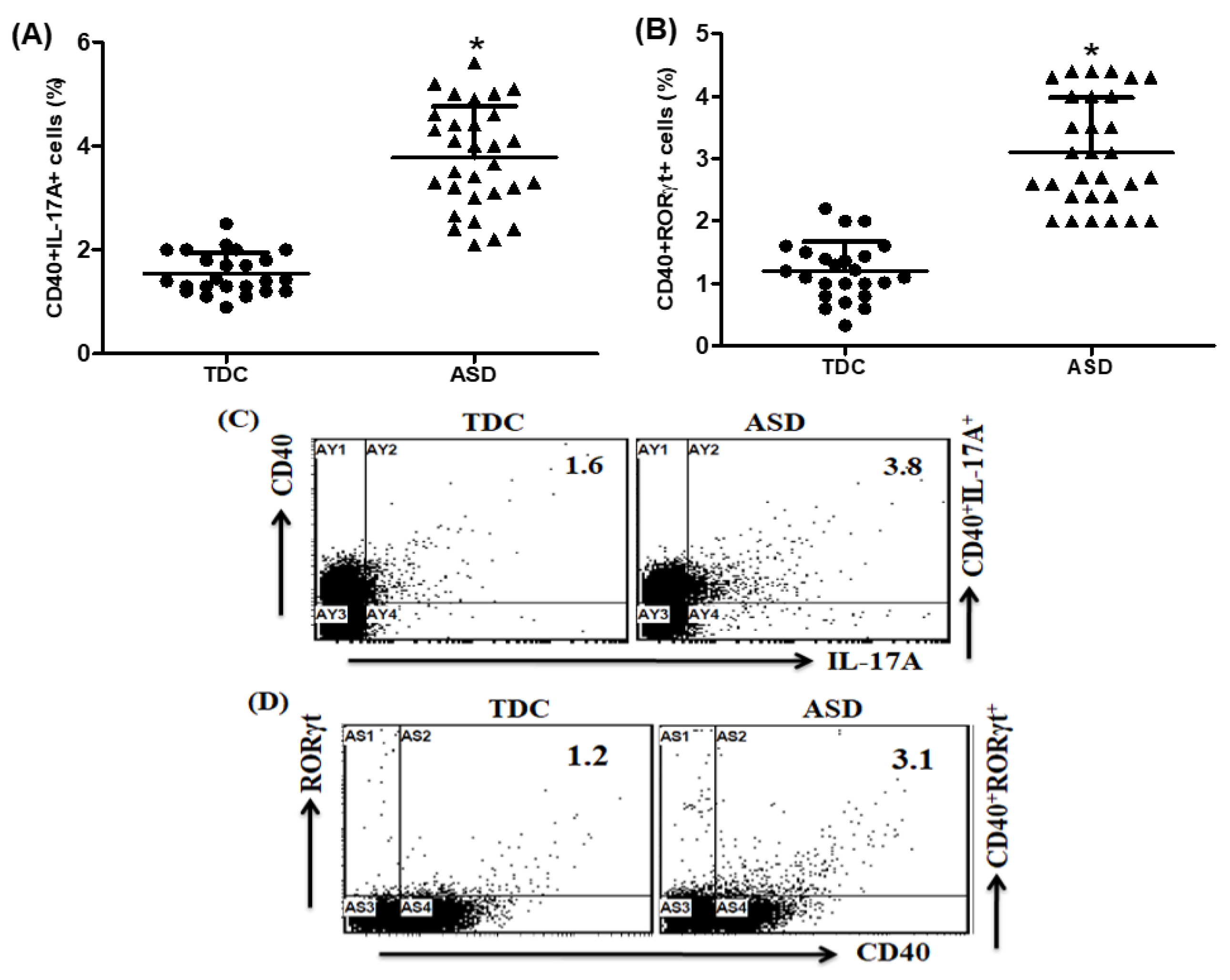
2.3. Upregulation of IL-17A- and RORγt-Expressing CD40+ Cells in Children with ASD

This study aimed to understand the contributions of cytokines and transcription factors in children with ASD. Our results showed that several IL-17A-expressing CD40+ cells were significantly upregulated in the children with ASD compared with those in the TDC group (Figure 3A). Additionally, our results showed that the number of RORγt-expressing CD40+ cells was higher in the children with ASD as compared with TDC group (Figure 3B). Based on these results, ASD development may be associated with alterations in cytokine and transcription factor signalling.

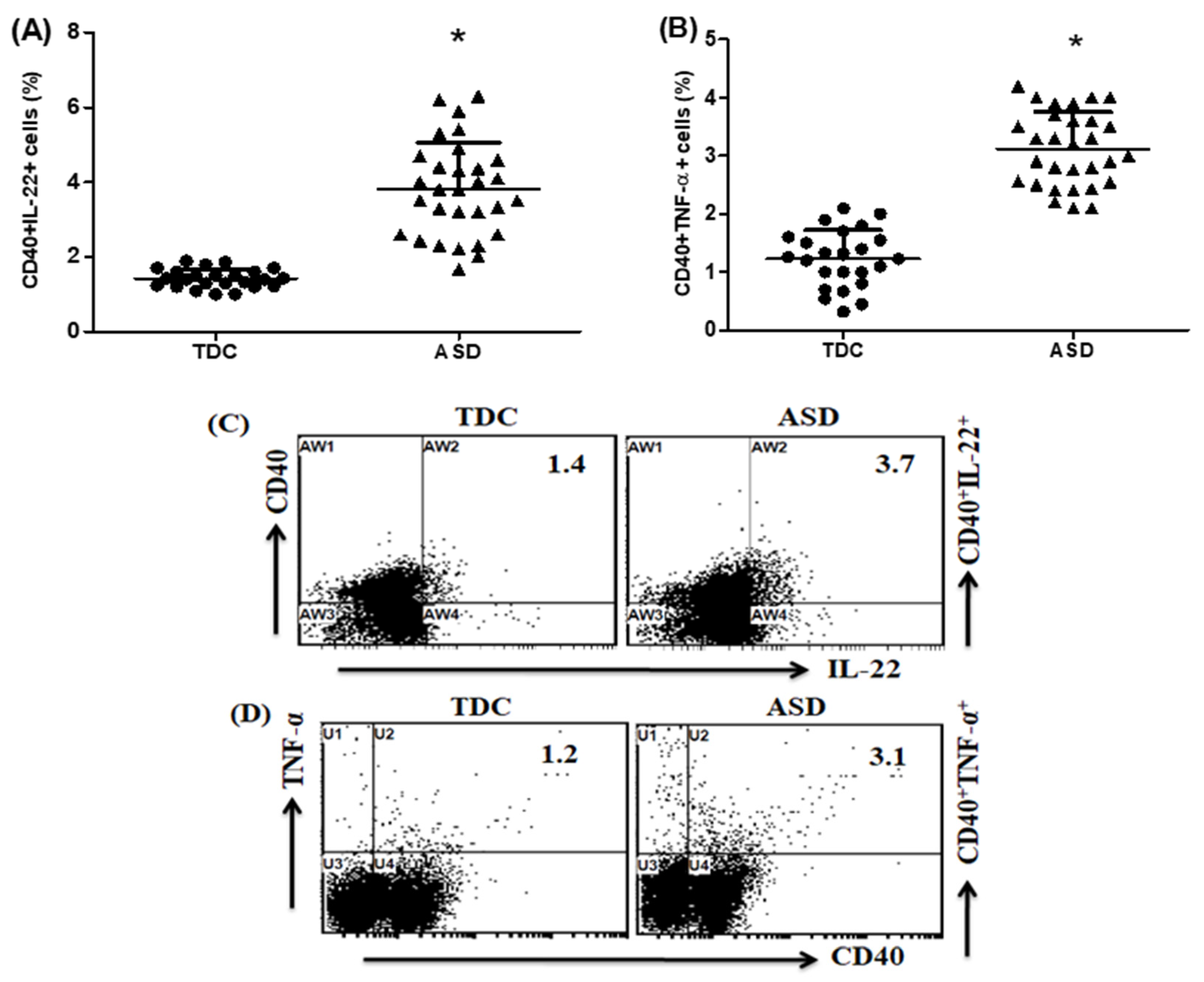
2.4. Upregulation of IL-22- and TNF-α-Expressing CD40+ Cells in Children with ASD

Our results further defined the number of IL-22- and TNF-α-expressing CD40+ cells in PBMCs from the children with ASD and the TDC group. We demonstrated that the number of IL-22-expressing CD40+ cells was higher in the children with ASD than in the TDC group (Figure 4A). Furthermore, our results showed that the children with ASD had significantly increased TNF-α-expressing CD40+ cells as compared to the TDC group (Figure 4B). Our results provide evidence for the upregulation of IL-22- and TNF-α expression in CD40 cells in children with ASD. According to the Pearson correlation coefficient, there was no correlation between the severity of symptoms and different immune parameters.

3. Discussion

Emerging evidence suggests that altered communication between the nervous system and inflammatory pathways is associated with multiple diseases, including ASD. The nervous and immune systems constantly communicate [50]. Multiple lines of evidence have recently pinpointed the key contribution of B lymphocytes to ASD pathogenesis. Altered immune responses commonly occur in individuals genetically susceptible to ASD [51,52]. Several studies have reported the involvement of immune dysregulation in the pathophysiology of ASD [53]. It has also been suggested that immune alterations contribute to behavioural effects in neurodevelopmental disorders, including ASD [54,55]. Evidence shows that dysregulation of the immune balance is a high risk factor for neurodevelopmental defects in ASD [56]. Several dysregulated cytokines and transcription factors in ASD have also been correlated with symptom severity and performance in ASD diagnostic tests [1,9]. Several chemokines recruit other immune cells to sites of tissue damage or infection. A previous study indicated an association between impaired behaviour and elevated chemokine levels in ASD [15]. Thus, cytokine, chemokine receptor, and transcription factor dysregulation could have important biological effects on neuronal development and activity that adversely affect behaviour.
It is well known that chemokines and their receptors play an important role in the immune system. Accumulating evidence has revealed that chemokine receptor expression, distribution, and function are involved in the pathogenesis of neurodegenerative diseases [57,58]. Chemokine receptors have been identified as regulators of peripheral immune cell trafficking and are expressed in the CNS [59,60]. Several studies have reported the expression of CCR1 in neurons, microglia, and astrocytes [61,62]. CCR1 mRNA and protein levels are increased in the brain, spinal cord, peripheral lymphoid organs, and blood plasma [63]. CCR1 promotes the entry of immune cells into the brain during neuroinflammation [64]. In the present study, we analysed CCR1 expression, an important proinflammatory chemokine among the CC chemokines, in children with ASD. Our results showed that CCR1-expressing CD40+ cells were significantly increased in the children with ASD compared with those in the TDC control group. Moreover, the children with ASD had significantly elevated CCR1 mRNA and protein expression compared to the TDC controls, indicating elevated levels of CCR1 expression. Our study provides strong evidence of the role of chemokine receptors in children with ASD. Therefore, chemokine receptors may be clinically useful disease markers for ASD. These observations may be highly relevant for children with ASD and other neuroimmune disorders.
A recent study described that the IFN-γ level was increased in the ASD brain [65]. It has been shown that IFN-γ level is elevated in children with ASD [9,28]. Consistent with the role of IFN-γ expression in ASD, the children with ASD had significantly increased IFN-γ expression levels [66]. Previous studies showed that prenatal IFN-γ imbalances could be linked to autism [31]. Early reports suggested a strong association with high levels of IFN-γ in patients diagnosed with ASD [67,68], which have been implicated in the pathophysiology of these neurobehavioural diseases [69]. Higher levels of cytokines, including IFN-γ, are demonstrated in ASD [8,70]. T-bet regulates Th1 and Th17 lymphocytes and infiltrates the T cells associated with CNS neuroinflammation [71,72]. T-bet plays an important role in disease development and is expressed in the CNS-infiltrating T cells [73,74]. T-bet-expressing cells are encephalitogenic in the CNS, and their infiltration is associated with neuroinflammation [37,75]. Thus, cytokine dysregulation could have important biological effects on neuronal development, adversely affecting behaviour. Our study showed that IFN-γ- and T-bet-expressing CD40+ cells were increased in children with ASD. In our study, CD40+IFN-γ+ and CD40+T-bet+ cells appeared more highly represented in aggressive ASD children. The exact mechanism by which IFN-γ- and T-bet-expressing CD40+ cells are involved in neuroinflammation remains to be explored. Further studies are needed to link these factors with disease severity. Therefore, increased cytokine and transcription factor levels may be associated with impaired communication and aberrant behaviour. These observations are highly relevant to children with ASD and other neuroimmune disorders. Therefore, further studies are needed to elucidate the association between proinflammatory cytokines and transcription factors in patients with ASD.
A recent study showed that IL-17A expression is significantly upregulated in the peripheral immune cells of children with ASD [9]. IL-17A is associated with behavioural impairments in ASD, suggesting that peripheral inflammation influences neuronal development [8,76]. Previous studies have shown that increased levels of IL-17A are associated with the severity of ASD behavioural symptoms [38,54]. In murine models, it has been shown that IL-17A plays a significant role in the induction of autism-like symptoms in the offspring of immune-activated mothers [16,77]. These parallel malformations are abnormalities found in the brain development of children with ASD [78,79]. RORγt, the key Th17 cell transcriptional regulator, is associated with neurodegeneration [80]. It has also been shown that RORγt expression is significantly upregulated in children with ASD and BTBR mice [9,40]. RORγt expression also correlates with IL-17 production, and the impact of suppressing RORγt could serve as a more effective treatment for neuroinflammation [81]. Recent findings suggest that pathogenic CD4+RORγt+ cells contribute to brain inflammation and neurobehavioural disorders [82]. Therefore, IL-17A dysregulation may play a central role in the development of ASD. Our results indicate that IL-17A- and RORγt-expressing CD40+ are upregulated in the children with ASD compared to the TDC control group. Therefore, we hypothesised that proinflammatory mediators and their transcription factors are involved in behavioural aggravation and neuroimmune dysfunction in children with ASD. These results provide evidence that IL-17A/RORγt expression in CD40 cells could be associated with immune and neuronal dysfunction in ASD, and further study is warranted. These observations suggest that dysfunctional immune responses may affect the core features of ASD and its associated behaviours in children.
IL-22 is a potent proinflammatory cytokine. IL-22 plays a critical role in human diseases, and its overexpression is associated with lymphocyte activation in the brain [83]. Previous studies have suggested that IL-22 overexpression is associated with immune dysfunction in children with ASD [84]. Another study has suggested that increased IL-22 expression promotes leukocyte infiltration into the brain [85], and increased cytokine IL-22 levels have been confirmed to elevate neurodegenerative disorders [86]. A more recent report noted that an increase in TNF-α level is a potentially important biomarker in ASD [87,88]. Previous studies have also demonstrated that TNF-α increases in various tissues of patients with ASD [89], and TNF-α expression is elevated in children with ASD [90]. It is also interesting to note that several studies also demonstrated increased TNF-α production in children with ASD [91,92]. Importantly, TNF-α crosses from the peripheral blood into the brain, directly affecting brain function and behaviour [93,94]. To our knowledge, there have been no investigations of IL-22- and TNF-α-expression CD40+ cells in ASD; therefore, our current study examined IL-22/TNF-α expression of CD40++ cells in children with ASD. Recent results have also shown that the number of IL-22-expressing CD40+ PBMCs is significantly higher in children with ASD [95]. In the present study, we observed that the children with ASD exhibited increased IL-22 and TNF-α expression in CD40+ cells. Therefore, dysfunctional immune defences and inflammatory reactions in children with ASD may be important precipitating factors that trigger this disorder. We speculate that IL-22/TNF-α expression could be used as a clinical marker for ASD, although this requires further study. Our findings suggest that these factors may serve as diagnostic markers and therapeutic targets for diagnosing and treating ASD.

4. Materials and Methods

4.1. Study Participants

All of the children were assessed by trained ASD clinicians. A total of 30 male children were diagnosed with ASD (mean ± SD = 6.5 ± 2.8 years) based on the Diagnostic and Statistical Manual of Mental Disorders, 5th edition [96]. Children with ASD were enrolled at the Autism Research Treatment Center of King Saud University, Saudi Arabia. The patients included in this study had no associated neurological disorders (such as seizures and tuberous sclerosis) or metabolic disorders (such as phenylketonuria), and patients with known medical conditions were excluded from the study because these comorbidities associated with ASD may have influenced the results. Additionally, the included patients were not receiving any medications.
A total of 24 healthy male children (mean ± SD = 6.1 ± 3.1 years) were enrolled as typically developing controls (TDCs), who attended a routine follow-up at the Well Baby Clinic, College of Medicine, King Khalid Hospital. The TDC children demonstrated no clinical findings suggestive of neuropsychiatric disorders. None of the participants had a history of a recent infection or fever. All of the processes complied with the National Institutes of Health guidelines and the legal requirements of King Saud University for studies involving human subjects. Additionally, the participants’ parents or legal guardians signed an informed written consent form in order for participation.

4.2. Study Measurements

Clinical evaluations of the children with ASD were performed based on their history, as obtained from clinical and neuropsychiatric examinations. The severity of the disease was assessed using the Childhood Autism Rating Scale [97], which rates children on a scale from one to four in each of 16 areas: relating to people, listening response, verbal and non-verbal communication, emotional and visual responses, consistency of intellectual response, fear or nervousness, touch and smell responses, imitation, adaptation to change, body use, object use, taste, activity level, and general impressions.

4.3. Chemicals and Antibodies

Fluorescein isothiocyanate, phycoerythrin; allophycocyanin; allophycocyanin-Cy7 and PE/Dazzle labelled CD40, CCR1, IFN-γ, T-bet, IL-17A, IL-22, and TNF-α anti-human monoclonal antibodies; and red blood cell lysing, fixation, and permeabilizing buffers were purchased from BioLegend (San Diego, CA, USA). FcR blocking reagent was purchased from Miltenyi Biotech (Bergisch Gladbach, Germany). RORγt, GolgiStop, and acid−citrate−dextrose vacutainer tubes were purchased from BD Biosciences (San Diego, CA, USA). The primers used were purchased from GenScript (Piscataway, NJ, USA). TRIzol reagent was obtained from Life Technologies (Grand Island, NY, USA). The SYBR Green PCR Master Mix and High-Capacity cDNA Reverse Transcription kit was purchased from Applied Biosystems (Paisley, UK). The primary antibodies against CCR1 and secondary anti-human antibodies used for Western blotting were purchased from Santa Cruz Biotechnology, Inc. (Dallas, TX, USA). Phorbol 12-myristate 13-acetate (PMA), ionomycin, phosphate-buffered saline (PBS), RPMI-1640 medium, Ficoll-Paque, and Hanks’ Balanced Salt Solution (HBSS) were purchased from Sigma-Aldrich (St. Louis, MO, USA). Nitrocellulose membranes were obtained from Bio-Rad Laboratories (Hercules, CA, USA), and a chemiluminescence Western blot detection kit was purchased from GE Healthcare Life Sciences (Piscataway, NJ, USA).

4.4. Preparation of PBMCs

Peripheral blood was obtained from the children with ASD and the TDC children in acid−citrate−dextrose vacutainer tubes (BD Biosciences, San Jose, CA, USA). As described previously, PBMCs were isolated using the Ficoll-Paque (specific gravity 1.077; Sigma-Aldrich, St. Louis, MO, USA) gradient density method [9,98]. The peripheral blood samples were centrifuged at 850× g for 10 min to remove the plasma. Blood cells were diluted with PBS and centrifuged in a Ficoll-Paque discontinuous gradient at 420× g for 30 min. The PBMC layer was collected and washed with cold distilled water and 10 × HBSS to remove red blood cells. As previously described, the cells were resuspended at 2 × 10⁶ cells/mL in RPMI-1640 medium [9,26].

4.5. Flow Cytometric Analysis

Flow cytometric analysis was used to measure the number of CCR1-, IFN-γ-, T-bet-, IL-17A-, RORγt, IL-22-, and TNF-α-expressing CD40+ cells in the PMBCs. Briefly, PBMCs were stimulated with PMA/ionomycin (Sigma-Aldrich, St. Louis, MO, USA) in the presence of a Golgi stop (BD Bioscience, San Jose, USA), as previously described [12,98]. PBMCs were washed and surface-stained with anti-CCR1 and anti-CD40 antibodies (BioLegend, San Diego, CA, USA). For the staining of cytokines and transcription factors, the cells were fixed, permeabilised, and stained for anti-IFN-γ, anti-T-bet, anti-IL-17A, anti-RORγt, anti-IL-22, and anti-TNF-α (BioLegend, San Diego, CA, USA). Human lymphocytes were isolated from the other immune cells (monocytes and granulocytes) using a conventional gating strategy based on their physical properties (forward and side scatter) to determine different immune markers in the lymphocytes. Chemokine receptors, inflammatory mediators, and transcription factors were identified based on the immunofluorescence characteristics of the antibody-labelled cells in the lymphocyte gate. The proportions of CD40+CCR1+, CD40+IFN-γ+, CD40+T-bet+, CD40+IL-17A+, CD40+RORγt+, CD40+IL-22+, and CD40+TNF-α+, cells were determined in the lymphocyte gate. Flow cytometry was conducted using an FC500 flow cytometer with CXP software (Beckman Coulter, Brea, CA, USA).

4.6. Gene Expression

Real time-PCR (RT-PCR) was performed as described previously [99]. The total RNA was extracted from PBMCs using TRIzol reagent (Invitrogen, Life Technologies, Waltham, MA, USA) and quantified as previously described [12,98]. cDNA synthesis was performed using a high-capacity cDNA reverse transcription kit (Applied Biosystems, Waltham, MA, USA), as previously described [11]. Quantitative analysis of the mRNA expression was performed using RT-PCR. The primers used in this study were purchased from GenScript (Piscataway, NJ, USA). CCR1 Forward: 5’-AATGTAATGGTGGCCTGGGG-3’; Reverse: 5′-TCCTCCCAACCCCCTATCAG-3’; GAPDH Forward: 5’-CTTTGCAGCAATGCCTCCTG-3’; GAPDH Reverse: 5’-ACCATGAGTCCTTCCACGAT-3’. Relative changes in the gene expression were determined using the 2−ΔΔCT method [100] with GAPDH as the reference gene.

4.7. Western Blotting

The total cellular protein was extracted from PBMCs using a previously described method [12,26]. Briefly, 40 µg of the isolated protein from PBMC was separated using 7% SDS-PAGE, followed by transfer to the nitrocellulose membrane (Bio-Rad, Hercules, CA, USA). The membrane was blocked in a blocking solution overnight at 4 °C, followed by incubation with a primary CCR1 antibody and secondary peroxidase-conjugated antibody (Santa Cruz Biotech, Dallas, TX, USA) at room temperature for 2 h. The CCR1 and β-actin bands were visualised using a Western blotting luminol reagent (Santa Cruz Biotechnology, Inc., Dallas, TX, USA) and quantified relative to the β-actin bands. Images were obtained using a ChemiDoc Imaging System (Bio-Rad, Hercules, CA, USA).

4.8. Statistical Analysis

The Student’s t-test was used to compare the two groups. Statistical analyses were performed using GraphPad Prism. The data were expressed as mean ± SD. Statistical significance was set at p < 0.05. Pearson’s correlation coefficient ‘r’ was used to assess the relationships between different immune parameters and CARS scores in ASD patients.

5. Conclusions

This is the first study demonstrating that CD40 expresses CCR1, proinflammatory cytokines, and other transcription factors in children with ASD. These findings suggest that CCR1, proinflammatory cytokines, and transcription factors may be associated with behavioural disturbances and disease severity in children with ASD. Therefore, the description of immunological parameters in ASD has important implications for diagnosis and should be considered when designing therapeutic strategies to treat the core symptoms and behavioural impairments in ASD.

Author Contributions

Conceptualization, A.A.A., M.A.A., A.N., S.M.A., S.A.B., L.Y.A.-A., M.M.A., M.S., M.Y.A., M.H.H. and S.F.A.; data curation, M.A.A., S.A.B., M.M.A., M.S. and S.F.A.; formal analysis, A.A.A., M.A.A., A.N., S.A.B., L.Y.A.-A., M.M.A., M.S., M.Y.A. and S.F.A.; funding acquisition, S.F.A.; investigation, M.A.A., A.N., S.M.A., S.A.B., L.Y.A.-A., M.M.A. and S.F.A.; methodology, A.A.A., M.A.A., A.N., S.M.A., S.A.B., L.Y.A.-A., M.M.A., M.S., M.H.H. and S.F.A.; project administration, M.A.A. and S.F.A.; resources, M.A.A., S.M.A., M.M.A., M.S., M.Y.A., M.H.H. and S.F.A.; software, A.A.A., S.M.A., S.A.B., L.Y.A.-A., M.M.A., M.S., M.Y.A., M.H.H. and S.F.A.; supervision, A.A.A., M.A.A., A.N., L.Y.A.-A. and S.F.A.; validation, A.A.A., M.A.A., A.N., S.M.A., L.Y.A.-A., M.M.A., M.S., M.Y.A., M.H.H. and S.F.A.; visualization, M.A.A., S.M.A. and S.F.A.; writing—original draft, A.A.A., M.A.A., A.N., S.M.A., S.A.B., L.Y.A.-A. and S.F.A.; writing—review and editing, A.A.A., A.N., S.M.A., S.A.B., L.Y.A.-A. and S.F.A. All authors have read and agreed to the published version of the manuscript.

Funding

Deputyship for Research and Innovation, Ministry of Education in Saudi Arabia: (IFKSURG-2-1471).

Institutional Review Board Statement

The local ethics committee of the College of Medicine, King Saud University, Riyadh, Saudi Arabia, approved this human study (Approval# E-10-220).

Informed Consent Statement

Informed written consent for participation in the study was signed by the subjects’ parents or legal guardians.

Data Availability Statement

All data presented in this study are available upon reasonable request from the corresponding author.

Acknowledgments

The authors extend their appreciation to the Deputyship for Research & Innovation, Ministry of Education in Saudi Arabia for funding this research work through the project no. (IFKSURG-2-1471).

Conflicts of Interest

The authors declare no conflict of interest.

Abbreviations

Autism spectrum disorders (ASD); central nervous system (CNS); phycoerythrin (PE); fluoroisothiocyanate (FITC); allophycocyanin (APC); cluster of differentiation (CD); interferon-gamma (IFN-γ); peripheral blood mononuclear cells (PBMCs); standard deviation (SD); T-box transcription factor (T-bet); C-C chemokine receptor type 1 (CCR1); typically developing controls (TDC); Roswell Park Memorial Institute (RPMI) 1640; phorbol 12-myristate 13-acetate (PMA); cluster of differentiation 40 (CD40); retinoid-related orphan receptor gamma t (RORγt); antigen-presenting cells (APCs); blood-brain barrier (BBB); tumour necrosis factor alpha (TNF-α); signal transducer and activator of transcription 3 (STAT3); retinoic acid-related orphan receptor gamma t (RORγt); interleukin 17 (IL-17); glyceraldehyde 3-phosphate dehydrogenase (GAPDH); messenger RNA (mRNA); complementary DNA (cDNA); reverse transcription polymerase chain reaction (RT-PCR).

References

  1. Masi, A.; DeMayo, M.M.; Glozier, N.; Guastella, A.J. An Overview of Autism Spectrum Disorder, Heterogeneity and Treatment Options. Neurosci. Bull. 2017, 33, 183–193. [Google Scholar] [CrossRef]
  2. Liu, X.; Takumi, T. Genomic and genetic aspects of autism spectrum disorder. Biochem. Biophys. Res. Commun. 2014, 452, 244–253. [Google Scholar] [CrossRef]
  3. Posar, A.; Visconti, P. Autism in 2016: The need for answers. J. Pediatr. (Rio. J.) 2017, 93, 111–119. [Google Scholar] [CrossRef] [PubMed]
  4. Gładysz, D.; Krzywdzińska, A.; Hozyasz, K.K. Immune Abnormalities in Autism Spectrum Disorder-Could They Hold Promise for Causative Treatment? Mol. Neurobiol. 2018, 55, 6387–6435. [Google Scholar] [CrossRef] [PubMed]
  5. Meltzer, A.; Van de Water, J. The role of the immune system in autism spectrum disorder. Neuropsychopharmacology 2017, 42, 284–298. [Google Scholar] [CrossRef]
  6. Jyonouchi, H.; Geng, L.; Davidow, A.L. Cytokine profiles by peripheral blood monocytes are associated with changes in behavioral symptoms following immune insults in a subset of ASD subjects: An inflammatory subtype? J. Neuroinflamm. 2014, 11, 187. [Google Scholar] [CrossRef] [PubMed]
  7. Ross, H.E.; Guo, Y.; Coleman, K.; Ousley, O.; Miller, A.H. Association of IL-12p70 and IL-6:IL-10 ratio with autism-related behaviors in 22q11.2 deletion syndrome: A preliminary report. Brain Behav. Immun. 2013, 31, 76–81. [Google Scholar] [CrossRef] [PubMed]
  8. Ashwood, P.; Krakowiak, P.; Hertz-Picciotto, I.; Hansen, R.; Pessah, I.; Van de Water, J. Elevated plasma cytokines in autism spectrum disorders provide evidence of immune dysfunction and are associated with impaired behavioral outcome. Brain Behav. Immun. 2011, 25, 40–45. [Google Scholar] [CrossRef]
  9. Ahmad, S.F.; Nadeem, A.; Ansari, M.A.; Bakheet, S.A.; Attia, S.M.; Zoheir, K.M.; Al-Ayadhi, L.Y.; Alzahrani, M.Z.; Alsaad, A.M.; Alotaibi, M.R.; et al. Imbalance between the anti- and pro-inflammatory milieu in blood leukocytes of autistic children. Mol. Immunol. 2017, 82, 57–65. [Google Scholar] [CrossRef]
  10. Ahmad, S.F.; Zoheir, K.M.A.; Ansari, M.A.; Nadeem, A.; Bakheet, S.A.; Al-Ayadhi, L.Y.; Alzahrani, M.Z.; Al-Shabanah, O.A.; Al-Harbi, M.M.; Attia, S.M. Dysregulation of Th1, Th2, Th17, and T regulatory cell-related transcription factor signaling in children with autism. Mol. Neurobiol. 2017, 54, 4390–4400. [Google Scholar] [CrossRef] [PubMed]
  11. Ahmad, S.F.; Nadeem, A.; Ansari, M.A.; Bakheet, S.A.; Al-Ayadhi, L.Y.; Attia, S.M. Upregulation of IL-9 and JAK-STAT signaling pathway in children with autism. Prog. Neuropsychopharmacol. Biol. Psychiatry 2017, 79 Pt B, 472–480. [Google Scholar] [CrossRef]
  12. Ahmad, S.F.; Ansari, M.A.; Nadeem, A.; Bakheet, S.A.; Al-Ayadhi, L.Y.; Attia, S.M. Upregulation of peripheral CXC and CC chemokine receptor expression on CD4+ T cells is associated with immune dysregulation in children with autism. Prog. Neuropsychopharmacol. Biol. Psychiatry 2018, 81, 211–220. [Google Scholar] [CrossRef] [PubMed]
  13. Gilliland, C.T.; Salanga, C.L.; Kawamura, T.; Trejo, J.; Handel, T.M. The chemokine receptor CCR1 is constitutively active, which leads to G protein-independent, β-arrestin-mediated internalization. J. Biol. Chem. 2013, 288, 32194–32210. [Google Scholar] [CrossRef]
  14. Abdallah, M.W.; Larsen, N.; Grove, J.; Nørgaard-Pedersen, B.; Thorsen, P.; Mortensen, E.L.; Hougaard, D.M. Amniotic fluid chemokines and autism spectrum disorders: An exploratory study utilizing a Danish Historic Birth Cohort. Brain Behav. Immun. 2012, 26, 170–176. [Google Scholar] [CrossRef]
  15. Ashwood, P.; Krakowiak, P.; Hertz-Picciotto, I.; Hansen, R.; Pessah, I.N.; Van de Water, J. Associations of impaired behaviors with elevated plasma chemokines in autism spectrum disorders. J. Neuroimmunol. 2011, 232, 196–199. [Google Scholar] [CrossRef] [PubMed]
  16. Choi, J.E.; Widjaja, F.; Careaga, M.; Bent, S.; Ashwood, P.; Hendren, R.L. Change in plasma cytokine levels during risperidone treatment in children with autism. J. Child. Adolesc. Psychopharmacol. 2014, 24, 586–589. [Google Scholar] [CrossRef]
  17. Jorda, A.; Cauli, O.; Santonja, J.M.; Aldasoro, M.; Aldasoro, C.; Obrador, E.; Vila, J.M.; Mauricio, M.D.; Iradi, A.; Guerra-Ojeda, S.; et al. Changes in Chemokines and Chemokine Receptors Expression in a Mouse Model of Alzheimer’s Disease. Int. J. Biol. Sci. 2019, 15, 453–463. [Google Scholar] [CrossRef] [PubMed]
  18. Karpus, W.J.; Fife, B.T.; Kennedy, K.J. Immunoneutralization of chemokines for the prevention and treatment of central nervous system autoimmune disease. Methods 2003, 29, 362–368. [Google Scholar] [CrossRef]
  19. Vargas, D.L.; Nascimbene, C.; Krishnan, C.; Zimmerman, A.W.; Pardo, C.A. Neuroglial activation and neuroinflammation in the brain of patients with autism. Ann. Neurol. 2005, 57, 67–81. [Google Scholar] [CrossRef] [PubMed]
  20. Cowell, R.M.; Xu, H.; Galasso, J.M.; Silverstein, F.S. Hypoxic-ischemic injury induces macrophage inflammatory protein-1alpha expression in immature rat brain. Stroke 2002, 33, 795–801. [Google Scholar] [CrossRef] [PubMed]
  21. Liu, H.; Ning, H.; Men, H.; Hou, R.; Fu, M.; Zhang, H.; Liu, J. Regulation of CCL5 expression in smooth muscle cells following arterial injury. PLoS ONE 2012, 7, e30873. [Google Scholar] [CrossRef]
  22. Ubogu, E.E.; Callahan, M.K.; Tucky, B.H.; Ransohoff, R.M. Determinants of CCL5-driven mononuclear cell migration across the blood-brain barrier Implications for therapeutically modulating neuroinflammation. J. Neuroimmunol. 2006, 179, 132–144. [Google Scholar] [CrossRef]
  23. Schönbeck, U.; Libby, P. The CD40/CD154 receptor/ligand dyad. Cell Mol. Life Sci. 2001, 58, 4–43. [Google Scholar]
  24. Ots, H.D.; Tracz, J.A.; Vinokuroff, K.E.; Musto, A.E. CD40-CD40L in Neurological Disease. Int. J. Mol. Sci. 2022, 23, 4115. [Google Scholar] [CrossRef] [PubMed]
  25. Iezzi, G.; Sonderegger, I.; Ampenberger, F.; Schmitz, N.; Marsland, B.J.; Kopf, M. CD40-CD40L cross-talk integrates strong antigenic signals and microbial stimuli to induce development of IL-17-producing CD4+ T cells. Proc. Natl. Acad. Sci. USA 2009, 106, 876–881. [Google Scholar] [CrossRef]
  26. Chen, Y.C.; Tsai, W.J.; Wu, M.H.; Lin, L.C.; Kuo, Y.C. Suberosin inhibits proliferation of human peripheral blood mononuclear cells through the modulation of the transcription factors NF-AT and NF-kappaB. Br. J. Pharmacol. 2007, 150, 298–312. [Google Scholar] [CrossRef]
  27. Wu, C.J.; Guo, J.; Luo, H.C.; Wei, C.D.; Wang, C.F.; Lan, Y.; Wei, Y.S. Association of CD40 polymorphisms and haplotype with risk of systemic lupus erythematosus. Rheumatol. Int. 2016, 36, 45–52. [Google Scholar] [CrossRef]
  28. Alomar, H.A.; Ansari, M.A.; Nadeem, A.; Attia, S.M.; Bakheet, S.A.; Al-Mazroua, H.A.; Hussein, M.H.; Alqarni, S.A.; Ahmad, S.F. A potent and selective CXCR2 antagonist improves neuroimmune dysregulation through the inhibition of NF-κB and notch inflammatory signaling in the BTBR mouse model of autism. J. Neuroimmunol. 2023, 377, 578069. [Google Scholar] [CrossRef] [PubMed]
  29. Croonenberghs, J.; Bosmans, E.; Deboutte, D.; Kenis, G.; Maes, M. Activation of the inflammatory response system in autism. Neuropsychobiology 2002, 45, 1–6. [Google Scholar] [CrossRef]
  30. Rose, D.; Ashwood, P. Potential cytokine biomarkers in autism spectrum disorders. Biomark Med. 2014, 8, 1171–1181. [Google Scholar] [CrossRef] [PubMed]
  31. Goines, P.E.; Croen, L.A.; Braunschweig, D.; Yoshida, C.K.; Grether, J.; Hansen, R.; Kharrazi, M.; Ashwood, P.; Van de Water, J. Increased midgestational IFN-γ, IL-4 and IL-5 in women bearing a child with autism: A case-control study. Mol. Autism. 2011, 2, 13. [Google Scholar] [CrossRef]
  32. Filiano, A.J.; Xu, Y.; Tustison, N.J.; Marsh, R.L.; Baker, W.; Smirnov, I.; Overall, C.C.; Gadani, S.P.; Turner, S.D.; Weng, Z.; et al. Unexpected role of interferon-γ in regulating neuronal connectivity and social behaviour. Nature 2016, 535, 425–429. [Google Scholar] [CrossRef]
  33. Frisullo, G.; Angelucci, F.; Caggiula, M.; Nociti, V.; Iorio, R.; Patanella, A.K.; Sancricca, C.; Mirabella, M.; Tonali, P.A.; Batocchi, A.P. pSTAT1, pSTAT3, and T-bet expression in peripheral blood mononuclear cells from relapsing-remitting multiple sclerosis patients correlates with disease activity. J. Neurosci. Res. 2006, 84, 1027–1036. [Google Scholar] [CrossRef]
  34. Wang, J.; Fathman, J.W.; Lugo-Villarino, G.; Scimone, L.; von Andrian, U.; Dorfman, D.M.; Glimcher, L.H. Transcription factor T-bet regulates inflammatory arthritis through its function in dendritic cells. J. Clin. Investig. 2006, 116, 414–421. [Google Scholar] [CrossRef]
  35. Knox, J.J.; Myles, A.; Cancro, M.P. T-Bet+ Memory B Cells: Generation, Function, and Fate. Immunol. Rev. 2019, 288, 149–160. [Google Scholar] [CrossRef] [PubMed]
  36. van Langelaar, J.; Rijvers, L.; Janssen, M.; Wierenga-Wolf, A.F.; Melief, M.J.; Siepman, T.A.; de Vries, H.E.; Unger, P.A.; van Ham, S.M.; Hintzen, R.Q.; et al. Induction of Brain-Infiltrating T-Bet-Expressing B Cells in Multiple Sclerosis. Ann. Neurol. 2019, 86, 264–278. [Google Scholar] [CrossRef] [PubMed]
  37. Spath, S.; Becher, B. T-bet or not T-bet: Taking the last bow on the autoimmunity stage. Eur. J. Immunol. 2013, 43, 2810–2813. [Google Scholar] [CrossRef]
  38. Al-Ayadhi, L.Y.; Mostafa, G.A. Elevated serum levels of interleukin-17A in children with autism. J. Neuroinflamm. 2012, 9, 158. [Google Scholar] [CrossRef]
  39. Suzuki, K.; Matsuzaki, H.; Iwata, K.; Kameno, Y.; Shimmura, C.; Kawai, S.; Yoshihara, Y.; Wakuda, T.; Takebayashi, K.; Takagai, S.; et al. Plasma Cytokine Profiles in Subjects with High-Functioning Autism Spectrum Disorders. PLoS ONE 2011, 6, e20470. [Google Scholar] [CrossRef] [PubMed]
  40. Nadeem, A.; Ahmad, S.F.; Attia, S.M.; Al-Ayadhi, L.Y.; Bakheet, S.A.; Al-Harbi, N.O. Oxidative and inflammatory mediators are upregulated in neutrophils of autistic children: Role of IL-17A receptor signaling. Prog. Neuropsychopharmacol. Biol. Psychiatry 2019, 90, 204–211. [Google Scholar] [CrossRef] [PubMed]
  41. Choi, G.B.; Yim, Y.S.; Wong, H.; Kim, S.; Kim, H.; Kim, S.V.; Hoeffer, C.A.; Littman, D.R.; Huh, J.R. The maternal interleukin-17a pathway in mice promotes autism-like phenotypes in offspring. Science 2016, 351, 933–939. [Google Scholar] [CrossRef] [PubMed]
  42. van der Zwaag, B.; Franke, L.; Poot, M.; Hochstenbach, R.; Spierenburg, H.A.; Vorstman, J.A.; van Daalen, E.; de Jonge, M.V.; Verbeek, N.E.; Brilstra, E.H.; et al. Gene-Network Analysis Identifies Susceptibility Genes Related to Glycobiology in Autism. PLoS ONE 2009, 4, e5324. [Google Scholar] [CrossRef]
  43. Pantelyushin, S.; Haak, S.; Ingold, B.; Kulig, P.; Heppner, F.L.; Navarini, A.A.; Becher, B. Rorγt+ innate lymphocytes and γδ T cells initiate psoriasiform plaque formation in mice. J. Clin. Investig. 2012, 122, 2252–2256. [Google Scholar] [CrossRef]
  44. Rolla, S.; Bardina, V.; De Mercanti, S.; Quaglino, P.; De Palma, R.; Gned, D.; Brusa, D.; Durelli, L.; Novelli, F.; Clerico, M. Th22 cells are expanded in multiple sclerosis and are resistant to IFN-β. J. Leukoc. Biol. 2014, 96, 1155–1164. [Google Scholar] [CrossRef]
  45. Wang, P.; Bai, F.; Zenewicz, L.A.; Dai, J.; Gate, D.; Cheng, G.; Yang, L.; Qian, F.; Yuan, X.; Montgomery, R.R.; et al. IL-22 Signaling Contributes to West Nile Encephalitis Pathogenesis. PLoS ONE 2012, 7, e44153. [Google Scholar] [CrossRef]
  46. Sonnenberg, G.F.; Fouser, L.A.; Artis, D. Functional biology of the IL-22-IL-22R pathway in regulating immunity and inflammation at barrier surfaces. Adv. Immunol. 2010, 107, 1–29. [Google Scholar]
  47. Chez, M.G.; Dowling, T.; Patel, P.B.; Khanna, P.; Kominsky, M. Elevation of tumor necrosis factor-alpha in cerebrospinal fluid of autistic children. Pediatr. Neurol. 2007, 36, 361–365. [Google Scholar] [CrossRef]
  48. Ricci, S.; Businaro, R.; Ippoliti, F.; Lo Vasco, V.R.; Massoni, F.; Onofri, E.; Troili, G.M.; Pontecorvi, V.; Morelli, M.; Rapp Ricciardi, M.; et al. Altered cytokine and BDNF levels in autism spectrum disorder. Neurotox. Res. 2013, 24, 491–501. [Google Scholar] [CrossRef]
  49. Xie, J.; Huang, L.; Li, X.; Li, H.; Zhou, Y.; Zhu, H.; Pan, T.; Kendrick, K.M.; Xu, W. Immunological cytokine profiling identifies TNF-a as a key molecule dysregulated in autistic children. Oncotarget 2017, 8, 82390–82398. [Google Scholar] [CrossRef] [PubMed]
  50. Margolis, K.G.; Gershon, M.D.; Bogunovic, M. Cellular Organization of Neuroimmune Interactions in the Gastrointestinal Tract. Trends Immunol. 2016, 37, 487–501. [Google Scholar] [CrossRef] [PubMed]
  51. Coury, D.L.; Ashwood, P.; Fasano, A.; Fuchs, G.; Geraghty, M.; Kaul, A.; Mawe, G.; Patterson, P.; Jones, N.E. Gastrointestinal conditions in children with autism spectrum disorder: Developing a research agenda. Pediatrics 2012, 130 (Suppl. S2), S160–S168. [Google Scholar] [CrossRef] [PubMed]
  52. Krakowiak, P.; Goines, P.E.; Tancredi, D.J.; Ashwood, P.; Hansen, R.L.; Hertz-Picciotto, I.; Van de Water, J. Neonatal cytokine profiles associated with autism spectrum disorder. Biol. Psychiatry 2017, 81, 442–451. [Google Scholar] [CrossRef] [PubMed]
  53. Matta, S.M.; Hill-Yardin, E.L.; Crack, P.J. The influence of neuroinflammation in Autism Spectrum Disorder. Brain Behav. Immun. 2019, 79, 75–90. [Google Scholar] [CrossRef] [PubMed]
  54. Enstrom, A.; Onore, C.; Hertz-Picciotto, I.; Hansen, R.; Creon, L.; Van De Water, J.; Ashwood, P. Detection of IL-17 and IL-23 in Plasma Samples of Children with Autism. Am. J. Biochem. Biotechnol. 2008, 4, 114–120. [Google Scholar] [CrossRef] [PubMed]
  55. Mead, J.; Ashwood, P. Evidence supporting an altered immune response in ASD. Immunol. Lett. 2015, 163, 49–55. [Google Scholar] [CrossRef]
  56. Xu, N.; Li, X.; Zhong, Y. Inflammatory Cytokines: Potential Biomarkers of Immunologic Dysfunction in Autism Spectrum Disorders. Mediat. Inflamm. 2015, 2015, 10. [Google Scholar] [CrossRef] [PubMed]
  57. Savarin-Vuaillat, C.; Ransohoff, R.M. Chemokines and chemokine receptors in neurological disease: Raise, retain, or reduce? Neurotherapeutics 2007, 4, 590–601. [Google Scholar] [CrossRef] [PubMed]
  58. Zhuang, Z.Y.; Kawasaki, Y.; Tan, P.H.; Wen, Y.R.; Huang, J.; Ji, R.R. Role of the CX3CR1/p38 MAPK pathway in spinal microglia for the development of neuropathic pain following nerve injury-induced cleavage of fractalkine. Brain Behav. Immun. 2007, 21, 642–651. [Google Scholar] [CrossRef]
  59. Clarkson, B.D.; Walker, A.; Harris, M.G.; Rayasam, A.; Sandor, M.; Fabry, Z. CCR2-dependent dendritic cell accumulation in the central nervous system during early effector experimental autoimmune encephalomyelitis is essential for effector T cell restimulation in situ and disease progression. J. Immunol. 2015, 194, 531–541. [Google Scholar] [CrossRef] [PubMed]
  60. Clarkson, B.D.; Walker, A.; Harris, M.G.; Rayasam, A.; Hsu, M.; Sandor, M.; Fabry, Z. CCR7 deficient inflammatory Dendritic Cells are retained in the Central Nervous System. Sci. Rep. 2017, 7, 42856. [Google Scholar] [CrossRef]
  61. Han, Y.; Wang, J.; Zhou, Z.; Ransohoff, R.M. TGFbeta1 selectively up-regulates CCR1 expression in primary murine astrocytes. Glia 2000, 30, 1–10. [Google Scholar] [CrossRef]
  62. Sunnemark, D.; Eltayeb, S.; Wallström, E.; Appelsved, L.; Malmberg, A.; Lassmann, H.; Ericsson-Dahlstrand, A.; Piehl, F.; Olsson, T. Differential expression of the chemokine receptors CX3CR1 and CCR1 by microglia and macrophages in myelin-oligodendrocyte-glycoprotein-induced experimental autoimmune encephalomyelitis. Brain Pathol. 2003, 13, 617–629. [Google Scholar] [CrossRef] [PubMed]
  63. Offner, H.; Subramanian, S.; Parker, S.M.; Afentoulis, M.E.; Vandenbark, A.A.; Hurn, P.D. Experimental stroke induces massive, rapid activation of the peripheral immune system. J. Cereb. Blood Flow. Metab. 2006, 26, 654–665. [Google Scholar] [CrossRef] [PubMed]
  64. Wilson, E.H.; Weninger, W.; Hunter, C.A. Trafficking of immune cells in the central nervous system. J. Clin. Investig. 2010, 120, 1368–1379. [Google Scholar] [CrossRef]
  65. Tamayo, J.M.; Rose, D.; Church, J.S.; Schwartzer, J.J.; Ashwood, P. Maternal Allergic Asthma Induces Prenatal Neuroinflammation. Brain Sci. 2022, 12, 1041. [Google Scholar] [CrossRef]
  66. Balestrieri, E.; Cipriani, C.; Matteucci, C.; Benvenuto, A.; Coniglio, A.; Argaw-Denboba, A.; Toschi, N.; Bucci, I.; Miele, M.T.; Grelli, S.; et al. Children With Autism Spectrum Disorder and Their Mothers Share Abnormal Expression of Selected Endogenous Retroviruses Families and Cytokines. Front. Immunol. 2019, 10, 2244. [Google Scholar] [CrossRef]
  67. Heuer, L.S.; Croen, L.A.; Jones, K.L.; Yoshida, C.K.; Hansen, R.L.; Yolken, R.; Zerbo, O.; DeLorenze, G.; Kharrazi, M.; Ashwood, P.; et al. An Exploratory Examination of Neonatal Cytokines and Chemokines as Predictors of Autism Risk: The Early Markers for Autism Study. Biol. Psychiatry 2019, 86, 255–264. [Google Scholar] [CrossRef] [PubMed]
  68. Sasayama, D.; Kurahashi, K.; Oda, K.; Yasaki, T.; Yamada, Y.; Sugiyama, N.; Inaba, Y.; Harada, Y.; Washizuka, S.; Honda, H. Negative Correlation between Serum Cytokine Levels and Cognitive Abilities in Children with Autism Spectrum Disorder. J. Intell. 2017, 5, 19. [Google Scholar] [CrossRef]
  69. Girgis, R.R.; Kumar, S.S.; Brown, A.S. The cytokine model of schizophrenia: Emerging therapeutic strategies. Biol. Psychiatry 2014, 75, 292–299. [Google Scholar] [CrossRef] [PubMed]
  70. Ahmad, S.F.; Nadeem, A.; Ansari, M.A.; Bakheet, S.A.; Al-Ayadhi, L.Y.; Attia, S.M. Downregulation in Helios transcription factor signaling is associated with immune dysfunction in blood leukocytes of autistic children. Prog. Neuropsychopharmacol. Biol. Psychiatry 2018, 85, 98–104. [Google Scholar] [CrossRef]
  71. Gocke, A.R.; Cravens, P.D.; Ben, L.H.; Hussain, R.Z.; Northrop, S.C.; Racke, M.K.; Lovett-Racke, A.E. T-bet regulates the fate of Th1 and Th17 lymphocytes in autoimmunity. J. Immunol. 2007, 178, 1341–1348. [Google Scholar] [CrossRef] [PubMed]
  72. Kwong, B.; Rua, R.; Gao, Y.; Flickinger, J., Jr.; Wang, Y.; Kruhlak, M.J.; Zhu, J.; Vivier, E.; McGavern, D.B.; Lazarevic, V. T-bet-dependent NKp46+ innate lymphoid cells regulate the onset of TH17-induced neuroinflammation. Nat. Immunol. 2018, 19, 898. [Google Scholar] [CrossRef]
  73. Nath, N.; Prasad, R.; Giri, S.; Singh, A.K.; Singh, I. T-bet is essential for the progression of experimental autoimmune encephalomyelitis. Immunology 2006, 118, 384–391. [Google Scholar] [CrossRef]
  74. Yang, Y.; Weiner, J.; Liu, Y.; Smith, A.J.; Huss, D.J.; Winger, R.; Peng, H.; Cravens, P.D.; Racke, M.K.; Lovett-Racke, A.E. T-bet is essential for encephalitogenicity of both Th1 and Th17 cells. J. Exp. Med. 2009, 206, 1549–1564. [Google Scholar] [CrossRef] [PubMed]
  75. Grifka-Wal, H.M.; Lalor, S.J.; Segal, B.M. Highly polarized Th17 cells induce EAE via a T-bet independent mechanism. Eur. J. Immunol. 2013, 43, 2824–2831. [Google Scholar] [CrossRef] [PubMed]
  76. Careaga, M.; Schwartzer, J.; Ashwood, P. Inflammatory profiles in the BTBR mouse: How relevant are they to autism spectrum disorders? Brain Behav. Immun. 2015, 43, 11–16. [Google Scholar] [CrossRef]
  77. Wong, H.; Hoeffer, C. Maternal IL-17A in autism. Exp. Neurol. 2018, 299 Pt A, 228–240. [Google Scholar] [CrossRef]
  78. Casanova, M.F.; El-Baz, A.S.; Kamat, S.S.; Dombroski, B.A.; Khalifa, F.; Elnakib, A.; Soliman, A.; Allison-McNutt, A.; Switala, A.E. Focal cortical dysplasias in autism spectrum disorders. Acta Neuropathol. Commun. 2013, 1, 67. [Google Scholar] [CrossRef]
  79. Varghese, M.; Keshav, N.; Jacot-Descombes, S.; Warda, T.; Wicinski, B.; Dickstein, D.L.; Harony-Nicolas, H.; De Rubeis, S.; Drapeau, E.; Buxbaum, J.D.; et al. Autism spectrum disorder: Neuropathology and animal models. Acta Neuropathol. 2017, 134, 537–566. [Google Scholar] [CrossRef] [PubMed]
  80. Zhang, J.; Ke, K.F.; Liu, Z.; Qiu, Y.H.; Peng, Y.P. Th17 cell-mediated neuroinflammation is involved in neurodegeneration of aβ1-42-induced Alzheimer’s disease model rats. PLoS ONE 2013, 8, e75786. [Google Scholar] [CrossRef]
  81. Yang, Y.; Winger, R.C.; Lee, P.W.; Nuro-Gyina, P.K.; Minc, A.; Larson, M.; Liu, Y.; Pei, W.; Rieser, E.; Racke, M.K.; et al. Impact of suppressing retinoic acid-related orphan receptor gamma t (ROR)γt in ameliorating central nervous system autoimmunity. Clin. Exp. Immunol. 2015, 179, 108–118. [Google Scholar] [CrossRef]
  82. Mickael, M.E.; Bhaumik, S.; Chakraborti, A.; Umfress, A.A.; van Groen, T.; Macaluso, M.; Totenhagen, J.; Sorace, A.G.; Bibb, J.A.; Standaert, D.G.; et al. RORγt-Expressing Pathogenic CD4+ T Cells Cause Brain Inflammation during Chronic Colitis. J. Immunol. 2022, 208, 2054–2066. [Google Scholar] [CrossRef] [PubMed]
  83. Levillayer, F.; Mas, M.; Levi-Acobas, F.; Brahic, M.; Bureau, J.F. Interleukin 22 is a candidate gene for Tmevp3, a locus controlling Theiler’s virus-induced neurological diseases. Genetics 2007, 176, 1835–1844. [Google Scholar] [CrossRef] [PubMed]
  84. Ahmad, S.F.; Ansari, M.A.; Nadeem, A.; Bakheet, S.A.; Al-Ayadhi, L.Y.; Alsaad, A.M.S.; Assiri, M.A.; Al-Mazroua, H.A.; Attia, S.M. Upregulation of interleukin (IL)-31, a cytokine producing CXCR1 peripheral immune cells, contributes to the immune abnormalities of autism spectrum disorder. J. Neuroimmunol. 2020, 349, 577430. [Google Scholar] [CrossRef]
  85. Kebir, H.; Kreymborg, K.; Ifergan, I.; Dodelet-Devillers, A.; Cayrol, R.; Bernard, M.; Giuliani, F.; Arbour, N.; Becher, B.; Prat, A. Human TH17 lymphocytes promote blood-brain barrier disruption and central nervous system inflammation. Nat. Med. 2007, 13, 1173–1175. [Google Scholar] [CrossRef] [PubMed]
  86. Elyaman, W.; Bradshaw, E.M.; Uyttenhove, C.; Dardalhon, V.; Awasthi, A.; Imitola, J.; Bettelli, E.; Oukka, M.; van Snick, J.; Renauld, J.C.; et al. IL-9 induces differentiation of TH17 cells and enhances function of FoxP3+ natural regulatory T cells. Proc. Natl. Acad. Sci. USA 2009, 106, 12885–12890. [Google Scholar] [CrossRef]
  87. Hu, C.C.; Xu, X.; Xiong, G.L.; Xu, Q.; Zhou, B.R.; Li, C.Y.; Qin, Q.; Liu, C.X.; Li, H.P.; Sun, Y.J.; et al. Alterations in plasma cytokine levels in chinese children with autism spectrum disorder. Autism. Res. 2018, 11, b989–b999. [Google Scholar] [CrossRef] [PubMed]
  88. Kutuk, M.O.; Tufan, E.; Gokcen, C.; Kilicaslan, F.; Karadag, M.; Mutluer, T.; Yektas, C.; Coban, N.; Kandemir, H.; Buber, A.; et al. Cytokine expression profiles in Autism spectrum disorder: A multi-center study from Turkey. Cytokine 2020, 133, 155152. [Google Scholar] [CrossRef] [PubMed]
  89. Li, X.; Chauhan, A.; Sheikh, A.M.; Patil, S.; Chauhan, V.; Li, X.M.; Ji, L.; Brown, T.; Malik, M. Elevated immune response in the brain of autistic patients. J. Neuroimmunol. 2009, 207, 111–116. [Google Scholar] [CrossRef] [PubMed]
  90. Jyonouchi, H.; Geng, L.; Ruby, A.; Zimmerman-Bier, B. Dysregulated innate immune responses in young children with autism spectrum disorders: Their relationship to gastrointestinal symptoms and dietary intervention. Neuropsychobiology 2005, 51, 77–85. [Google Scholar] [CrossRef] [PubMed]
  91. Jyonouchi, H.; Geng, L.; Ruby, A.; Reddy, C.; Zimmerman-Bier, B. Evaluation of an association between gastrointestinal symptoms and cytokine production against common dietary proteins in children with autism spectrum disorders. J. Pediatr. 2005, 146, 605–610. [Google Scholar] [CrossRef] [PubMed]
  92. Jyonouchi, H.; Sun, S.; Itokazu, N. Innate Immunity Associated with Inflammatory Responses and Cytokine Production against Common Dietary Proteins in Patients with Autism Spectrum Disorder. Neuropsychobiology 2002, 46, 76–84. [Google Scholar] [CrossRef]
  93. Estes, M.L.; McAllister, A.K. Immune mediators in the brain and peripheral tissues in autism spectrum disorder. Nat. Rev. Neurosci. 2015, 16, 469–486. [Google Scholar] [CrossRef]
  94. Gottfried, C.; Bambini-Junior, V.; Francis, F.; Riesgo, R.; Savino, W. The impact of neuroimmune alterations in autism spectrum disorder. Front. Psychiatry 2015, 6, 121. [Google Scholar] [CrossRef] [PubMed]
  95. Ahmad, S.F.; Ansari, M.A.; Nadeem, A.; Bakheet, S.A.; Al-Ayadhi, L.Y.; Alasmari, A.F.; Alanazi, M.M.; Al-Mazroua, H.A.; Attia, S.M. Involvement of CD45 cells in the development of autism spectrum disorder through dysregulation of granulocyte-macrophage colony-stimulating factor, key inflammatory cytokines, and transcription factors. Int. Immunopharmacol. 2020, 83, 106466. [Google Scholar] [CrossRef]
  96. APA. Diagnostic and Statistical Manual of Mental Disorders, 5th ed.; American Psychiatric Association: Washington, DC, USA, 2015. [Google Scholar]
  97. Schopler, E.; Reichler, R.J.; Renner, B.R. The childhood Autism Rating Scale (CARS) for Diagnostic Screening and Classification in Autism; Irvington: New York, NY, USA, 1986. [Google Scholar]
  98. Noster, R.; Riedel, R.; Mashreghi, M.F.; Radbruch, H.; Harms, L.; Haftmann, C.; Chang, H.D.; Radbruch, A.; Zielinski, C.E. IL-17 and GM-CSF expression are antagonistically regulated by human T helper cells. Sci. Transl. Med. 2014, 6, 241ra80. [Google Scholar] [CrossRef]
  99. Kuo, Y.C.; Weng, S.C.; Chou, C.J.; Chang, T.T.; Tsai, W.J. Activation and proliferation signals in primary human T lymphocytes inhibited by ergosterol peroxide isolated from Cordyceps cicadae. Br. J. Pharmacol. 2003, 140, 895–906. [Google Scholar] [CrossRef] [PubMed]
  100. Livak, K.J.; Schmittgen, T.D. Analysis of relative gene expression data using real-time quantitative PCR and the 2−ΔΔCT Method. Methods 2001, 25, 402–408. [Google Scholar] [CrossRef] [PubMed]
Figure 1. (A) Flow cytometric analysis was used to determine the number of C-C chemokine receptor type 1 (CCR1)-expressing CD40+ cells in the PBMCs of children with autism spectrum disorder (ASD) and the typically developing controls (TDC) group. (B) The mRNA expression level of CCR1 in PBMCs was measured using quantitative RT–PCR and normalized to GAPDH. (C) Western blot analysis of CCR1 protein expression in the PBMCs. (D) Representative flow cytometry dot plots demonstrating CD40+CCR1+ cells from the ASD and TDC groups. Statistically significant differences (* p < 0.05) were tested using the Student’s t-test.
Ijms 24 07475 g001
Figure 2. (A,B) Flow cytometric analysis was used to determine the number of IFN-γ- and T-box transcription factor (T-bet)-expressing CD40+ cells in the PBMCs of children with autism spectrum disorder (ASD) and the typically developing controls (TDC) group. (C,D) Representative flow cytometry dot plots demonstrating CD40+IFN-γ+ and CD40+T-bet+ cells from the ASD and TDC groups. Statistically significant differences (* p < 0.05) were tested using Student’s t-test.
Ijms 24 07475 g002
Figure 3. (A,B) Flow cytometric analysis were used to determine the number of IL-17A- and retinoid-related orphan receptor gamma t (RORγt)-expressing CD40+ cells in the PBMCs of children with autism spectrum disorder (ASD) and the typically developing controls (TDC) group. (C,D) Representative flow cytometry dot plots demonstrating IL-17A- and RORγt-expressing CD40+ cells from the ASD and TDC groups. Statistically significant differences (* p < 0.05) were tested using the Student’s t-test.
Ijms 24 07475 g003
Figure 4. (A,B) Flow cytometric analysis determined the number of IL-22- and TNF-α-expressing CD40+ cells in PBMCs of children with autism spectrum disorder (ASD) and the typically developing controls (TDC) group. (C,D) Representative flow cytometry dot plots demonstrating IL-22- and TNF-α-expressing CD40+ cells from the ASD and TDC groups. Statistically significant differences (* p < 0.05) were tested using the Student’s t-test.
Ijms 24 07475 g004
Disclaimer/Publisher’s Note: The statements, opinions and data contained in all publications are solely those of the individual author(s) and contributor(s) and not of MDPI and/or the editor(s). MDPI and/or the editor(s) disclaim responsibility for any injury to people or property resulting from any ideas, methods, instructions or products referred to in the content.

