

Neuroendocrine Neoplasms of the Gastrointestinal Tract versus Neuroendocrine Neoplasms of the Gynaecological Tract—Comparison of the Risk Factors and Non-Surgical Treatment Efficacy
 , and
, and
Abstract
:1. Introduction
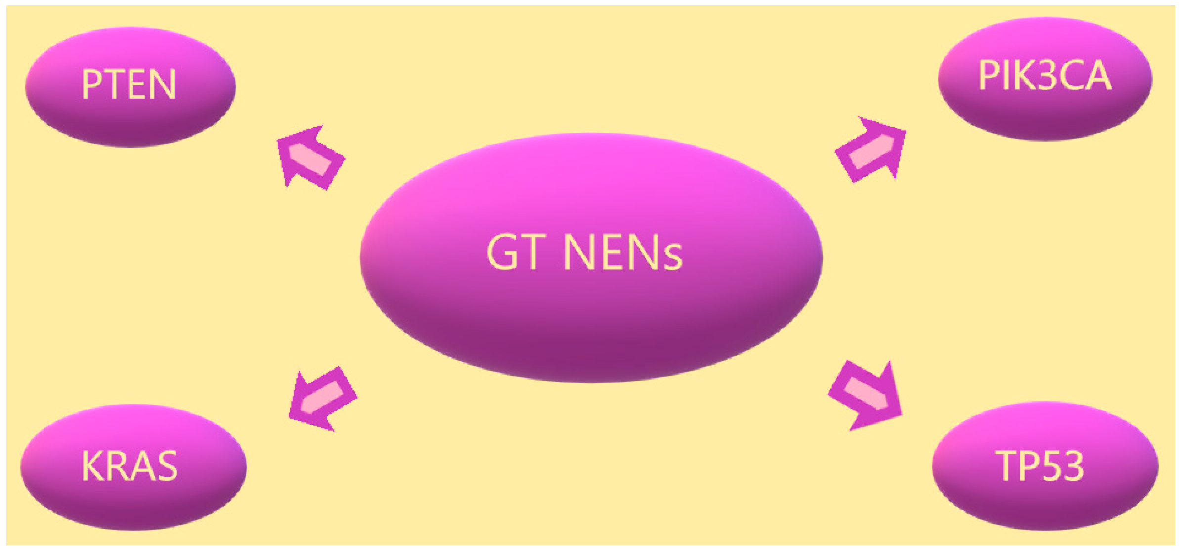
2. Risk Factors
Treatment
3. Peptide Receptor Radionuclide Therapy
4. Radiotherapy
5. Somatostatin Analogues
6. Immunotherapy
7. Chemotherapy
8. Conclusions
Author Contributions
Funding
Institutional Review Board Statement
Informed Consent Statement
Data Availability Statement
Conflicts of Interest
References
- Heller, M.T.; Shah, A.B. Imaging of Neuroendocrine Tumors imaging overview of Neuroendocrine. Radiol. Clin. N. Am. 2011, 49, 529–548. [Google Scholar] [CrossRef]
- Rooper, L.M.; Sharma, R.; Li, Q.K.; Illei, P.B.; Westra, W.H. INSM1 Demonstrates Superior Performance to the Individual and Combined Use of Synaptophysin, Chromogranin and CD56 for Diagnosing Neuroendocrine Tumors of the Thoracic Cavity. Am. J. Surg. Pathol. 2017, 41, 1561–1569. [Google Scholar] [CrossRef]
- Dasari, A.; Shen, C.; Halperin, D.M.; Zhao, B.; Zhou, S.; Xu, Y.; Shih, T.; Yao, J.C. Trends in the incidence, prevalence, and survival outcomes in patients with neuroendocrine tumors in the United States. JAMA Oncol. 2017, 3, 1335–1342. [Google Scholar] [CrossRef]
- Ellis, L.; Shale, M.J.; Coleman, M.P. Carcinoid tumors of the gastrointestinal tract: Trends in incidence in England since 1971. Am. J. Gastroenterol. 2010, 105, 2563–2569. [Google Scholar] [CrossRef]
- Scherübl, H.; Streller, B.; Stabenow, R.; Herbst, H.; Höpfner, M.; Schwertner, C.; Steinberg, J.; Eick, J.; Ring, W.; Tiwari, K.; et al. Clinically detected gastroenteropancreatic neuroendocrine tumors are on the rise: Epidemiological changes in Germany. World J. Gastroenterol. 2013, 19, 9012–9019. [Google Scholar] [CrossRef]
- Hallet, J.; Law, C.H.L.; Cukier, M.; Saskin, R.; Liu, N.; Singh, S. Exploring the rising incidence of neuroendocrine tumors: A population-based analysis of epidemiology, metastatic presentation, and outcomes. Cancer 2015, 121, 589–597. [Google Scholar] [CrossRef]
- Ito, T.; Sasano, H.; Tanaka, M.; Osamura, R.Y.; Sasaki, I.; Kimura, W.; Takano, K.; Obara, T.; Ishibashi, M.; Nakao, K.; et al. Epidemiological study of gastroenteropancreatic neuroendocrine tumors in Japan. J. Gastroenterol. 2010, 45, 234–243. [Google Scholar] [CrossRef]
- Assarzadegan, N.; Montgomery, E. What is new in the 2019 world health organization (WHO) classification of tumors of the digestive system: Review of selected updates on neuroendocrine neoplasms, appendiceal tumors, and molecular testing. Arch. Pathol. Lab. Med. 2021, 145, 664–677. [Google Scholar] [CrossRef] [Green Version]
- Pobłocki, J.; Jasińska, A.; Syrenicz, A.; Andrysiak-Mamos, E.; Szczuko, M. The neuroendocrine neoplasms of the digestive tract: Diagnosis, treatment and nutrition. Nutrients 2020, 12, 1437. [Google Scholar] [CrossRef]
- Sundin, A.; Arnold, R.; Baudin, E.; Cwikla, J.B.; Eriksson, B.; Fanti, S.; Fazio, N.; Giammarile, F.; Hicks, R.J.; Kjaer, A.; et al. ENETS Consensus Guidelines for the Standards of Care in Neuroendocrine Tumors: Radiological, Nuclear Medicine and Hybrid Imaging. Neuroendocrinology 2017, 105, 212–244, 2017. [Google Scholar] [CrossRef] [Green Version]
- Cavalcanti, M.S.; Gönen, M.; Klimstra, D.S. The ENETS/WHO grading system for neuroendocrine neoplasms of the gastroenteropancreatic system: A review of the current state, limitations and proposals for modifications. Int. J. Endocr. Oncol. 2016, 3, 203–219. [Google Scholar] [CrossRef]
- Fahrenkamp, A.G.; Wibbeke, C.; Böcker, W.; Schmid, K.W.; Winde, G.; Öfner, D.; Fischer-Colbrie, R. Immunohistochemical distribution of chromogranins A and B and secretogranin II in neuroendocrine tumours of the gastrointestinal tract. Vichows Archiv A Pathol. Anat. 1995, 426, 361–367. [Google Scholar] [CrossRef]
- Gibbs, J.; Mei, S.; Economos, K.; Lee, Y.C.; Kanis, M.J. Clinicopathologic features, incidence, and survival trends of gynecologic neuroendocrine tumors: A SEER database analysis. Am. J. Obstet. Gynecol. 2019, 221, 53.e1–53.e6. [Google Scholar] [CrossRef]
- Virarkar, M.; Vulasala, S.S.; Morani, A.C.; Waters, R.; Gopireddy, D.R.; Kumar, S.; Bhosale, P.; Lall, C. Neuroendocrine Neoplasms of the Gynecologic Tract. Cancers 2022, 14, 1835. [Google Scholar] [CrossRef]
- Crane, E.K.; Ramos, P.; Farley, J.H.; Naumann, R.W.; Tait, D.L.; Higgins, R.V.; Brown, J. Molecular profiling in a large cohort of gynecologic neuroendocrine tumors. Gynecol. Oncol. 2020, 159, 262. [Google Scholar] [CrossRef]
- Winer, I.; Kim, C.; Gehrig, P. Neuroendocrine tumors of the gynecologic tract update. Gynecol. Oncol. 2021, 162, 210–219. [Google Scholar] [CrossRef]
- Rindi, G.; Klimstra, D.S.; Abedi-Ardekani, B.; Asa, S.L.; Bosman, F.T.; Brambilla, E.; Busam, K.J.; de Krijger, R.R.; Dietel, M.; El-Naggar, A.K.; et al. A common classification framework for neuroendocrine neoplasms: An International Agency for Research on Cancer (IARC) and World Health Organization (WHO) expert consensus proposal. Mod. Pathol. 2018, 31, 1770–1786. [Google Scholar] [CrossRef] [Green Version]
- Ishikawa, M.; Kasamatsu, T.; Tsuda, H.; Fukunaga, M.; Sakamoto, A.; Kaku, T.; Kato, T.; Takahashi, K.; Ariyoshi, K.; Suzuki, K.; et al. Gynecologic Oncology A multi-center retrospective study of neuroendocrine tumors of the uterine cervix: Prognosis according to the new 2018 staging system, comparing outcomes for different chemotherapeutic regimens and histopathological subtypes. Gynecol. Oncol. 2019, 155, 444–451. [Google Scholar] [CrossRef]
- Eichhorn, J.H.; Young, R.H. Neuroendocrine Tumors of the Genital Tract. Pathol. Patterns Rev. 2001, 115 (Suppl. 1), S94–S112. [Google Scholar] [CrossRef]
- Avis, K.E.P.D.; Artmann, L.Y.N.N.K.H.; Eeney, G.A.R.Y.L.K.; Hapiro, H.O.S. Primary Ovarian Carcinoid Tumors. Gynecol. Oncol. 1996, 265, 259–265. [Google Scholar]
- Noh, H.K.; Kwon, B.S.; Kim, Y.H.; Lee, N.K.; Choi, K.U.; Suh, D.S.; Lee, D.H.; Kim, K.H. Peptide YY producing strumal carcinoid tumor of the ovary in a postmenopausal woman: A rare cause of chronic constipation. Obstet. Gynecol. Sci. 2017, 60, 602–607. [Google Scholar] [CrossRef]
- Li, J.D.; Zhuang, Y.; Li, Y.F.; Feng, Y.L.; Hou, J.H.; Chen, L.; Zhu, A.N.; Wu, Q.L.; Yun, J.P. A clinicopathological aspect of primary small-cell carcinoma of the uterine cervix: A single-centre study of 25 cases. J. Clin. Pathol. 2011, 64, 1102–1107. [Google Scholar] [CrossRef]
- McHugh, K.E.; Mukhopadhyay, S.; Doxtader, E.E.; Lanigan, C.; Allende, D.S. INSM1 Is a highly specific marker of neuroendocrine differentiation in primary neoplasms of the gastrointestinal tract, appendix, and pancreas: An immunohistochemical study of 179 cases. Am. J. Clin. Pathol. 2020, 153, 811–820. [Google Scholar] [CrossRef]
- Damian, A.; Lago, G.; Rossi, S.; Alonso, O.; Engler, H. Early Detection of Bone Metastasis in Small Cell Neuroendocrine Carcinoma of the Cervix by 68Ga-DOTATATE PET/CT Imaging. Clin. Nucl. Med. 2017, 42, 216–217. [Google Scholar] [CrossRef]
- Hassan, M.M.; Phan, A.; Li, D.; Dagohoy, C.G.; Leary, C.; Yao, J.C. Risk factors associated with neuroendocrine tumors: A US-based case–control study. Int. J. Cancer 2008, 873, 867–873. [Google Scholar] [CrossRef]
- Paper, O. Risk and Protective Factors for Small Intestine Neuroendocrine Tumors: A Prospective Case-Control Study. Neuroendocrinology 2016, 103, 531–537. [Google Scholar] [CrossRef]
- Haugvik, S.-P.; Ibrahim, I.B.; Hedenström, P.; Valente, R.; Hayes, A.J.; Siuka, D.; Gladhaug, I.P.; Capurso, G. Smoking, alcohol and family history of cancer as risk factors for small intestinal neuroendocrine tumors: A systematic review and meta-analysis. Scand. J. Gastroenterol. 2017, 52, 797–802. [Google Scholar] [CrossRef]
- Halfdanarson, T.R.; Bamlet, W.R.; McWilliams, R.R.; Hobday, T.J.; Burch, P.A.; Rabe, K.G.; Petersen, G.M. Risk factors for pancreatic neuroendocrine tumors (PNETs): A clinic-based case-control study. Pancreas 2014, 43, 1219–1222. [Google Scholar] [CrossRef] [Green Version]
- Ben, Q.; Zhong, J.; Fei, J.; Chen, H.; Yv, L.; Tan, J.; Yuan, Y. Risk Factors for Sporadic Pancreatic Neuroendocrine Tumors: A Case-Control Study. Nat. Publ. Gr. 2016, 6, 36073. [Google Scholar] [CrossRef]
- Jung, Y.S.; Yun, K.E.; Chang, Y.; Ryu, S.; Park, J.H.; Kim, H.J. Risk Factors Associated with Rectal Neuroendocrine Tumors: A Cross-Sectional Study. Cancer Epidemiol. Biomark. Prev. 2014, 2004, 1406–1414. [Google Scholar] [CrossRef] [Green Version]
- Pavel, M.; Öberg, K.; Falconi, M.; Krenning, E.P.; Sundin, A.; Perren, A.; Berruti, A. Gastroenteropancreatic neuroendocrine neoplasms: ESMO Clinical Practice Guidelines for diagnosis, treatment and follow-up. Ann. Oncol. 2020, 31, 844–860. [Google Scholar] [CrossRef]
- Pyo, J.H.; Hong, S.N.; Min, B.-H.; Lee, J.H.; Chang, D.K.; Rhee, P.-L.; Kim, J.J.; Choi, S.K.; Jung, S.-H.; Son, H.J.; et al. Evaluation of the risk factors associated with rectal neuroendocrine tumors: A big data analytic study from a health screening center. J. Gastroenterol. 2016, 51, 1112–1121. [Google Scholar] [CrossRef]
- Kim, C.; Kim, H.; Jin, S.; Park, J.; Lee, K. Association of elevated serum ferritin concentration with insulin resistance and impaired glucose metabolism in Korean men and women. Metabolism 2011, 60, 414–420. [Google Scholar] [CrossRef]
- Mateo-gallego, R.; Calmarza, P.; Jarauta, E.; Burillo, E.; Cenarro, A.; Civeira, F. Serum ferritin is a major determinant of lipid phenotype in familial combined hyperlipidemia and familial hypertriglyceridemia. Metabolism 2010, 59, 154–158. [Google Scholar] [CrossRef]
- Available online: https://bloodcancerdiscov.aacrjournals.org (accessed on 3 September 2020).
- Vijayvergia, N.; Boland, P.M.; Handorf, E.; Gustafson, K.S.; Gong, Y.; Cooper, H.S.; Sheriff, F.; Astsaturov, I.; Cohen, S.J.; Engstrom, P.F. Molecular profiling of neuroendocrine malignancies to identify prognostic and therapeutic markers: A Fox Chase Cancer Center Pilot Study. Br. J. Cancer 2016, 115, 564–570. [Google Scholar] [CrossRef] [Green Version]
- Busico, A.; Maisonneuve, P.; Prinzi, N.; Pusceddu, S.; Centonze, G.; Garzone, G.; Pellegrinelli, A.; Giacomelli, L.; Mangogna, A.; Paolino, C.; et al. Gastroenteropancreatic High-Grade Neuroendocrine Neoplasms: Histology and Molecular Analysis, Two Sides of the Same Coin. Neuroendocrinology 2019, 110, 616–629. [Google Scholar] [CrossRef]
- Jiao, Y.; Shi, C.; Edil, B.H.; De Wilde, R.F.; Klimstra, D.S.; Maitra, A.; Schulick, R.D.; Tang, L.H.; Wolfgang, C.L.; Choti, M.A.; et al. DAXX/ATRX, MEN1, and mTOR Pathway Genes Are Frequently Altered in Pancreatic Neuroendocrine Tumors. Science 2011, 331, 1199–1204. [Google Scholar] [CrossRef] [Green Version]
- Konukiewitz, B.; Jesinghaus, M.; Steiger, K.; Schlitter, A.M.; Kasajima, A.; Sipos, B.; Zamboni, G.; Weichert, W.; Pfarr, N.; Klöppel, G. Pancreatic neuroendocrine carcinomas reveal a closer relationship to ductal adenocarcinomas than to neuroendocrine tumors G3. Hum. Pathol. 2018, 77, 70–79. [Google Scholar] [CrossRef]
- Francis, J.M.; Kiezun, A.; Ramos, A.H.; Serra, S.; Pedamallu, C.S.; Qian, Z.R.; Banck, M.S.; Kanwar, R.; A Kulkarni, A.; Karpathakis, A.; et al. Somatic mutation of CDKN1B in small intestine neuroendocrine tumors. Nat. Publ. Gr. 2013, 45, 1483–1486. [Google Scholar] [CrossRef]
- Alrezk, R.; Hannah-shmouni, F. MEN4 and CDKN1B mutations: The latest of the MEN syndromes. Endocr. Relat. Cancer 2017, 24, T195–T208. [Google Scholar] [CrossRef] [Green Version]
- Alejo, M.; Alemany, L.; Clavero, O.; Quiros, B.; Vighi, S.; Seoud, M.; Cheng-Yang, C.; Garland, S.M.; Juanpere, N.; Lloreta, J.; et al. Contribution of Human papillomavirus in neuroendocrine tumors from a series of 10,575 invasive cervical cancer cases. Papillomavirus Res. 2018, 5, 134–142. [Google Scholar] [CrossRef]
- Kuji, S.; Watanabe, R.; Sato, Y.; Iwata, T.; Hirashima, Y.; Takekuma, M.; Ito, I.; Abe, M.; Nagashio, R.; Omae, K.; et al. A new marker, insulinoma-associated protein 1 (INSM1), for high-grade neuroendocrine carcinoma of the uterine cervix: Analysis of 37 cases. Gynecol. Oncol. 2016, 144, 384–390. [Google Scholar] [CrossRef]
- Wang, K.; Wang, T.; Huang, Y.; Lai, J.C.; Chang, T.; Yen, M. Human Papillomavirus Type and Clinical Manifestation in Seven Cases of Large-cell Neuroendocrine Cervical Carcinoma. J. Formos. Med. Assoc. 2009, 108, 428–432. [Google Scholar] [CrossRef] [Green Version]
- Wang, C.-C.; Lai, C.-H.; Huang, H.-J.; Chao, A.; Chang, C.-J.; Chang, T.-C.; Chou, H.-H.; Hong, J.-H. Clinical Effect of Human Papillomavirus Genotypes in Patients With Cervical Cancer Undergoing Primary Radiotherapy. Int. J. Radiat. Oncol. 2010, 78, 1111–1120. [Google Scholar] [CrossRef]
- Siriaunkgul, S.; Utaipat, U.; Settakorn, J.; Sukpan, K. International Journal of Gynecology and Obstetrics CLINICAL ARTICLE HPV genotyping in neuroendocrine carcinoma of the uterine cervix in northern Thailand. Int. J. Gynecol. Obstet. 2011, 115, 175–179. [Google Scholar] [CrossRef]
- Eskander, R.N.; Elvin, J.; Gay, L.; Ross, J.S.; Miller, V.A. Unique Genomic Landscape of High-Grade Neuroendocrine Cervical Carcinoma: Implications for Rethinking Current Treatment Paradigms. JCO Precis. Oncol. 2020, 4, 972–987. [Google Scholar] [CrossRef]
- Cimic, A.; Vranic, S.; Arguello, D.; Contreras, E.; Gatalica, Z.; Swensen, J. Molecular Profiling Reveals Limited Targetable Biomarkers in Neuroendocrine Carcinoma of the Cervix. Appl. Immunohistochem. Mol. Morphol. 2021, 29, 299–304. [Google Scholar] [CrossRef]
- Frumovitz, M.; Burzawa, J.K.; Byers, L.A.; Lyons, Y.A.; Ramalingam, P.; Coleman, R.L.; Brown, J. Sequencing of mutational hotspots in cancer-related genes in small cell neuroendocrine cervical cancer. Gynecol. Oncol. 2016, 141, 588–591. [Google Scholar] [CrossRef] [Green Version]
- Van Ta, T.; Nguyen, Q.N.; Truong, V.-L.; Tran, T.T.; Nguyen, H.P.; Vuong, L.D. Human Papillomavirus Infection, p16INK4a Expression and Genetic Alterations in Vietnamese Cervical Neuroendocrine Cancer. Malaysian J. Med. Sci. 2019, 26, 151–157. [Google Scholar] [CrossRef]
- Kupryjańczyk, J.; Dansonka-Mieszkowska, A.; Moes-Sosnowska, J.; Plisiecka-Hałasa, J.; Szafron, L.; Podgorska, A.; Rzepecka, I.K.; Konopka, B.; Budziłowska, A.; Rembiszewska, A.; et al. Ovarian small cell carcinoma of hypercalcemic type—Evidence of germline origin and smarca4 gene inactivation. a pilot study. Pol. J. Pathol. 2013, 4, 238–246. [Google Scholar] [CrossRef]
- Witkowski, L.; Carrot-Zhang, J.; Albrecht, S.; Fahiminiya, S.; Hamel, N.; Tomiak, E.; Grynspan, D.; Saloustros, E.; Nadaf, J.; Rivera, B.; et al. Germline and somatic SMARCA4 mutations characterize small cell carcinoma of the ovary, hypercalcemic type. Nat. Genet. 2014, 46, 438–443. [Google Scholar] [CrossRef]
- A Clarke, B.; Witkowski, L.; Nu, T.N.T.; A Shaw, P.; Gilks, C.B.; Huntsman, D.; Karnezis, A.N.; Sebire, N.; Lamovec, J.; Roth, L.M.; et al. Loss of SMARCA4 (BRG1) protein expression as determined by immunohistochemistry in small-cell carcinoma of the ovary, hypercalcaemic type distinguishes these tumours from their mimics. Histopathology 2016, 69, 727–738. [Google Scholar] [CrossRef] [Green Version]
- Vélayoudom-Céphise, F.-L.; Duvillard, P.; Foucan, L.; Hadoux, J.; Chougnet, C.N.; Leboulleux, S.; Malka, D.; Guigay, J.; Goere, D.; Debaere, T.; et al. Are G3 ENETS neuroendocrine neoplasms heterogeneous? Endocr. Relat. Cancer 2013, 20, 649–657. [Google Scholar] [CrossRef] [Green Version]
- Srirajaskanthan, R.; Watkins, J.; Marelli, L.; Khan, K.; Caplin, M. Expression of Somatostatin and Dopamine 2 Receptors in Neuroendocrine Tumours and the Potential Role for New Biotherapies. Neuroendocrinology 2009, 89, 308–314. [Google Scholar] [CrossRef]
- Nicolini, S.; Severi, S.; Ianniello, A.; Sansovini, M.; Ambrosetti, A.; Bongiovanni, A.; Scarpi, E.; Di Mauro, F.; Rossi, A.; Matteucci, F.; et al. Investigation of receptor radionuclide therapy with 177Lu-DOTATATE in patients with GEP-NEN and a high Ki-67 proliferation index. Eur. J. Nucl. Med. 2018, 45, 923–930. [Google Scholar] [CrossRef]
- Strosberg, J.; El-Haddad, G.; Wolin, E.; Hendifar, A.; Yao, J.; Chasen, B.; Mittra, E.; Kunz, P.L.; Kulke, M.H.; Jacene, H.; et al. Phase 3 Trial of 177Lu-Dotatate for Midgut Neuroendocrine Tumors. N. Engl. J. Med. 2017, 376, 125–135. [Google Scholar] [CrossRef]
- Carlsen, E.A.; Fazio, N.; Granberg, D.; Grozinsky-Glasberg, S.; Ahmadzadehfar, H.; Grana, C.M.; Zandee, W.T.; Cwikla, J.; A Walter, M.; Oturai, P.S.; et al. Peptide receptor radionuclide therapy in gastroenteropancreatic NEN G3: A multicenter cohort study. Endocr. Relat. Cancer 2019, 26, 227–239. [Google Scholar] [CrossRef]
- Claringbold, P.G.; Harvey, J.; Street, A. Pancreatic Neuroendocrine Tumor Control: Durable Objective Response to Combination 177 Lu-Octreotate-Capecitabine-Temozolomide Radiopeptide Chemotherapy. Neuroendocrinology 2016, 103, 432–439. [Google Scholar] [CrossRef]
- Tumors, L.N.; Claringbold, P.G.; Price, R.A.; Turner, J.H. Phase I-II Study of Radiopeptide 177Lu-Octreotate in Combination with Capecitabine and Temozolomide in Advanced Low-Grade Neuroendocrine Tumors. Cancer Biother. Radiopharm. 2012, 27, 561–569. [Google Scholar] [CrossRef]
- Ezziddin, S.; Khalaf, F.; Vanezi, M. Outcome of peptide receptor radionuclide therapy with 177 Lu-octreotate in advanced grade 1/2 pancreatic neuroendocrine tumours. Eur. J. Nucl. Med. Mol. Imaging 2014, 41, 925–933. [Google Scholar] [CrossRef]
- Inzani, F.; Santoro, A.; Angelico, G.; Feraco, A.; Spadola, S.; Arciuolo, D.; Valente, M.; Carlino, A.; Piermattei, A.; Scaglione, G.; et al. Neuroendocrine Carcinoma of the Uterine Cervix: A Clinicopathologic and Immunohistochemical Study with Focus on Novel Markers (Sst2–Sst5). Cancers 2020, 12, 1211. [Google Scholar] [CrossRef]
- Iwata, T.; Ueno, H.; Itami, J.; Ito, Y.; Inaba, K.; Morizane, C.; Kondo, S.; Sakamoto, Y.; Shiba, S.; Sasaki, M.; et al. Efficacy of radiotherapy for primary tumor in patients with unresectable pancreatic neuroendocrine tumors. Jpn. J. Clin. Oncol. 2017, 47, 826–831. [Google Scholar] [CrossRef]
- Contessa, J.N.; Griffith, K.A.; Wolff, E.; Ensminger, W.; Zalupski, M.; Lawrence, T.S.; Ben-Josef, E. Radiotherapy for Pancreatic Neuroendocrine Tumors. Int. J. Radiat. Oncol. Biol. Phys. 2009, 75, 1196–1200. [Google Scholar] [CrossRef]
- Abrams, R.A.; King, D.; Wilson, J.F. Objective response of malignant carcinoid to radiation therapy. Orig. Contrib. 1987, 13, 869–873. [Google Scholar] [CrossRef]
- Arvold, N.D.; Willett, C.G.; Castillo, C.F.-D.; Ryan, D.P.; Ferrone, C.R.; Clark, J.W.; Blaszkowsky, L.S.; Deshpande, V.; Niemierko, A.; Allen, J.N.; et al. Pancreatic Neuroendocrine Tumors With Involved Surgical Margins: Prognostic Factors and the Role of Adjuvant Radiotherapy. Int. J. Radiat. Oncol. 2012, 83, e337–e343. [Google Scholar] [CrossRef]
- Dong, M.; Gu, X.; Ma, T.; Mi, Y.; Shi, Y.; Fan, R. The role of radiotherapy in neuroendocrine cervical cancer: SEER-based study. Sci. Prog. 2021, 104. [Google Scholar] [CrossRef]
- Hou, W.-H.; Schultheiss, T.E.; Wong, J.Y.; Wakabayashi, M.T.; Chen, Y.-J. Surgery Versus Radiation Treatment for High-Grade Neuroendocrine Cancer of Uterine Cervix: A Surveillance Epidemiology and End Results Database Analysis. Int. J. Gynecol. Cancer 2018, 28, 188–193. [Google Scholar] [CrossRef]
- Pei, X.; Xiang, L.; Ye, S.; He, T.; Cheng, Y.; Yang, W.; Wu, X.; Yang, H. Cycles of cisplatin and etoposide affect treatment outcomes in patients with FIGO stage I-II small cell neuroendocrine carcinoma of the cervix. Gynecol. Oncol. 2017, 147, 589–596. [Google Scholar] [CrossRef]
- Chan, J.K.; Loizzi, V.; Burger, R.A.; Rutgers, J.; Monk, B.J. Prognostic factors in neuroendocrine small cell cervical carcinoma. Cancer 2003, 97, 568–574. [Google Scholar] [CrossRef]
- Caplin, M.E.; Pavel, M.; Ćwikła, J.B.; Phan, A.T.; Raderer, M.; Sedláčková, E.; Cadiot, G.; Wolin, E.M.; Capdevila, J.; Wall, L.; et al. Lanreotide in Metastatic Enteropancreatic Neuroendocrine Tumors. N. Engl. J. Med. 2014, 371, 224–233. [Google Scholar] [CrossRef]
- Faggiano, A.; Carratù, A.C.; Guadagno, E.; Tafuto, S.; Tatangelo, F.; Riccardi, F.; Mocerino, C.; Palmieri, G.; Damiano, V.; Siciliano, R.; et al. Somatostatin Analogues according to Ki67 index in neuroendocrine tumours: An observational retrospective-prospective analysis from real life. Oncotarget 2015, 7, 5538–5547. [Google Scholar] [CrossRef] [Green Version]
- Merola, E.; Gordoa, T.A.; Zhang, P.; Al-Toubah, T.; Pellè, E.; Kolasińska-Ćwikła, A.; Zandee, W.; Laskaratos, F.; Mestier, L.; Lamarca, A.; et al. Somatostatin Analogs for Pancreatic Neuroendocrine Tumors: Any Benefit When Ki-67 Is ≥10%? Oncologist 2020, 26, 294–301. [Google Scholar] [CrossRef]
- Martín-Richard, M.; Massutí, B.; Pineda, E.; Alonso, V.; Mármol, M.; Castellano, D.; Fonseca, E.; Galán, A.; Llanos, M.; Sala, M.A.; et al. Antiproliferative effects of lanreotide autogel in patients with progressive, well-differentiated neuroendocrine tumours: A Spanish, multicentre, open-label, single arm phase II study. BMC Cancer 2013, 13, 427. [Google Scholar] [CrossRef] [Green Version]
- Pavel, M.; Ćwikła, J.B.; Lombard-Bohas, C.; Borbath, I.; Shah, T.; Pape, U.F.; Capdevila, J.; Panzuto, F.; Thanh, X.-M.T.; Houchard, A.; et al. Efficacy and safety of high-dose lanreotide autogel in patients with progressive pancreatic or midgut neuroendocrine tumours: CLARINET FORTE phase 2 study results. Eur. J. Cancer 2021, 157, 403–414. [Google Scholar] [CrossRef]
- Kang, J.; Yoo, C.; Hwang, H.-S.; Hong, S.-M.; Kim, K.-P.; Kim, S.Y.; Hong, Y.-S.; Kim, T.W.; Ryoo, B.-Y. Efficacy and safety of lanreotide in Korean patients with metastatic, well-differentiated gastroenteropancreatic-neuroendocrine tumors: A retrospective analysis. Investig. New Drugs 2018, 37, 763–770. [Google Scholar] [CrossRef]
- Kajiwara, H.; Hirabayashi, K.; Miyazawa, M.; Nakamura, N.; Hirasawa, T.; Muramatsu, T.; Mikami, M.; Yasuda, M.; Osamura, R.Y. Immunohistochemical expression of somatostatin type 2A receptor in neuroendocrine carcinoma of uterine cervix. Arch. Gynecol. Obstet. 2008, 279, 521–525. [Google Scholar] [CrossRef] [Green Version]
- Kolouch, T.; Linkova, H.; Lang, O.; Ciprova, V.; Brunerova, L. Carcinoid heart disease in a primary ovarian carcinoid. Acta Cardiol. Sin. 2016, 32, 112–115. [Google Scholar] [CrossRef]
- Buda, A.; Giuliani, D.; Montano, N.; Perego, P.; Milani, R. Primary insular carcinoid of the ovary with carcinoid heart disease: Unfavourable outcome of a case. Int. J. Surg. Case Rep. 2012, 3, 59–61. [Google Scholar] [CrossRef] [Green Version]
- Damen, N. Ovarian carcinoid presenting with right heart failure. Case Rep. 2014, 2014, bcr2014204518. [Google Scholar] [CrossRef] [Green Version]
- Shah, V.N.; Orlov, O.I.; Plestis, K.A. Carcinoid heart disease and a primary ovarian carcinoid tumor. J. Vis. Surg. 2019, 5, 48. [Google Scholar] [CrossRef]
- Cavalcanti, E.; Armentano, R.; Valentini, A.M.; Chieppa, M.; Caruso, M.L. Role of PD-L1 expression as a biomarker for GEP neuroendocrine neoplasm grading. Cell Death Dis. 2017, 8, e3004. [Google Scholar] [CrossRef] [Green Version]
- Ali, A.S.; Langer, S.W.; Federspiel, B.; Hjortland, G.O.; Grønbæk, H.; Ladekarl, M.; Welin, S.; Vestermark, L.W.; Arola, J.; Osterlund, P.; et al. PD-L1 expression in gastroenteropancreatic neuroendocrine neoplasms grade 3. PLoS ONE 2020, 15, e0243900. [Google Scholar] [CrossRef]
- Rösner, E.; Kaemmerer, D.; Sänger, J.; Lupp, A. Evaluation of PD-L1 expression in a large set of gastroenteropancreatic neuroendocrine tumours and correlation with clinicopathological data. Transl. Oncol. 2022, 25, 101526. [Google Scholar] [CrossRef]
- Mehnert, J.M.; Bergsland, E.; O’Neil, B.H.; Santoro, A.; Schellens, J.H.M.; Cohen, R.B.; Doi, T.; Ott, P.A.; Pishvaian, M.J.; Puzanov, I.; et al. Pembrolizumab for the treatment of programmed death–ligand 1–positive advanced carcinoid or pancreatic neuroendocrine tumors: Results from the KEYNOTE-028 study. Cancer 2020, 126, 3021–3030. [Google Scholar] [CrossRef]
- Strosberg, J.R.; Mizuno, N.; Doi, T.; Grande, E.; Delord, J.-P.; Shapira-Frommer, R.; Bergsland, E.K.; Shah, M.H.; Fakih, M.; Takahashi, S.; et al. Efficacy and Safety of Pembrolizumab in Previously Treated Advanced Neuroendocrine Tumors: Results From the Phase II KEYNOTE-158 Study. Clin. Cancer Res. 2020, 26, 2124–2130. [Google Scholar] [CrossRef]
- MacFarlane, A.W.; Yeung, H.-M.; Alpaugh, R.K.; Dulaimi, E.; Engstrom, P.F.; Dasari, A.; Campbell, K.S.; Vijayvergia, N. Impacts of pembrolizumab therapy on immune phenotype in patients with high-grade neuroendocrine neoplasms. Cancer Immunol. Immunother. 2021, 70, 1893–1906. [Google Scholar] [CrossRef]
- Vijayvergia, N.; Dasari, A.; Deng, M.; Litwin, S.; Al-Toubah, T.; Alpaugh, R.K.; Dotan, E.; Hall, M.J.; Ross, N.M.; Runyen, M.M.; et al. Pembrolizumab monotherapy in patients with previously treated metastatic high-grade neuroendocrine neoplasms: Joint analysis of two prospective, non-randomised trials. Br. J. Cancer 2020, 122, 1309–1314. [Google Scholar] [CrossRef]
- Jia, R.; Li, Y.; Xu, N.; Jiang, H.-P.; Zhao, C.-H.; Liu, R.-R.; Shi, Y.; Zhang, Y.-Y.; Wang, S.-Y.; Zhou, H.; et al. Sintilimab in Patients with Previously Treated Metastatic Neuroendocrine Neoplasms. Oncol. 2022, 27, e625–e632. [Google Scholar] [CrossRef]
- Patel, S.P.; Othus, M.; Chae, Y.K.; Giles, F.J.; Hansel, D.E.; Singh, P.P.; Fontaine, A.; Shah, M.H.; Kasi, A.; Al Baghdadi, T.; et al. A Phase II Basket Trial of Dual Anti–CTLA-4 and Anti–PD-1 Blockade in Rare Tumors (DART SWOG 1609) in Patients with Nonpancreatic Neuroendocrine Tumors. Clin. Cancer Res. 2020, 26, 2290–2296. [Google Scholar] [CrossRef] [Green Version]
- Klein, O.; Kee, D.; Markman, B.; Michael, M.; Underhill, C.R.; Carlino, M.S.; Jackett, L.; Lum, C.; Scott, C.L.; Nagrial, A.; et al. Immunotherapy of Ipilimumab and Nivolumab in Patients with Advanced Neuroendocrine Tumors: A Subgroup Analysis of the CA209-538 Clinical Trial for Rare Cancers. Clin. Cancer Res. 2020, 26, 4454–4459. [Google Scholar] [CrossRef]
- Lu, M.; Zhang, P.; Zhang, Y.; Li, Z.; Gong, J.-F.; Li, J.; Li, J.; Li, Y.; Zhang, X.; Lu, Z.; et al. Efficacy, safety and biomarkers of toripalimab in patients with recurrent or metastatic neuroendocrine neoplasms: A multiple-center phase Ib trial. Clin. Cancer Res. 2020, 26, 2337–2345. [Google Scholar] [CrossRef] [Green Version]
- Frumovitz, M.; Westin, S.N.; Salvo, G.; Zarifa, A.; Xu, M.; Yap, T.A.; Rodon, A.J.; Karp, D.D.; Abonofal, A.; Jazaeri, A.A.; et al. Phase II study of pembrolizumab efficacy and safety in women with recurrent small cell neuroendocrine carcinoma of the lower genital tract. Gynecol. Oncol. 2020, 158, 570–575. [Google Scholar] [CrossRef]
- Xu, Z.; Chen, L.; Chen, M.; Chen, Z. Hyperprogressive Disease in Cervical Small Cell Carcinoma Treated By Immune Check-point Inhibitor. OncoTargets Ther. 2019, 12, 8873. [Google Scholar] [CrossRef] [Green Version]
- Paraghamian, S.E.; Longoria, T.C.; Eskander, R.N. Metastatic small cell neuroendocrine carcinoma of the cervix treated with the PD-1 inhibitor, nivolumab: A case report. Gynecol. Oncol. Res. Pract. 2017, 4, 3. [Google Scholar] [CrossRef] [Green Version]
- Paterniti, T.A.M.; Dorr, K.R.; Ullah, A.; White, J.D.; Williams, H.; Ghamande, S. Complete Response to Combination Nivolumab and Ipilimumab in Recurrent Neuroendocrine Carcinoma of the Cervix. Obstet. Gynecol 2021, 138, 813–816. [Google Scholar] [CrossRef]
- Towner, M.; Novak, K.; Chae, Y.K.; Matei, D. Ipilimumab and nivolumab for recurrent neuroendocrine cervical carcinoma. Gynecol. Oncol. Rep. 2022, 42. [Google Scholar] [CrossRef]
- Zhu, Y.-X.M.; Gao, X.-P.M.; Xin, L.M.; Jia, Y.-C. A case report of small cell ovarian neuroendocrine carcinoma combined with immunochemotherapy. Medicine 2022, 101, e31445. [Google Scholar] [CrossRef]
- Espinosa-Olarte, P.; La Salvia, A.; Riesco-Martinez, M.C.; Anton-Pascual, B.; Garcia-Carbonero, R. Chemotherapy in NEN: Still has a role? Rev. Endocr. Metab. Disord. 2021, 22, 595–614. [Google Scholar] [CrossRef]
- Ducreux, M.; Dahan, L.; Smith, D.; O’Toole, D.; Lepère, C.; Dromain, C.; Vilgrain, V.; Baudin, E.; Lombard-Bohas, C.; Scoazec, J.-Y.; et al. Bevacizumab combined with 5-FU/streptozocin in patients with progressive metastatic well-differentiated pancreatic endocrine tumours (BETTER trial)—A phase II non-randomised trial. Eur. J. Cancer 2014, 50, 3098–3106. [Google Scholar] [CrossRef]
- Yao, J.C.; Fazio, N.; Singh, S.; Buzzoni, R.; Carnaghi, C.; Wolin, E.; Tomasek, J.; Raderer, M.; Lahner, H.; Voi, M.; et al. Everolimus for the treatment of advanced, non-functional neuroendocrine tumours of the lung or gastrointestinal tract (RADIANT-4): A randomised, placebo-controlled, phase 3 study. Lancet 2015, 387, 968–977. [Google Scholar] [CrossRef]
- Yao, J.C.; Pavel, M.; Lombard-Bohas, C.; Van Cutsem, E.; Voi, M.; Brandt, U.; He, W.; Chen, D.; Capdevila, J.; De Vries, E.G.E.; et al. Everolimus for the Treatment of Advanced Pancreatic Neuroendocrine Tumors: Overall Survival and Circulating Biomarkers From the Randomized, Phase III RADIANT-3 Study. J. Clin. Oncol. 2016, 34, 3906–3913. [Google Scholar] [CrossRef]
- Yao, J.C.; Shah, M.H.; Ito, T.; Bohas, C.L.; Wolin, E.M.; Van Cutsem, E.; Hobday, T.J.; Okusaka, T.; Capdevila, J.; de Vries, E.G.; et al. Everolimus for Advanced Pancreatic Neuroendocrine Tumors. N. Engl. J. Med. 2011, 364, 514–523. [Google Scholar] [CrossRef] [Green Version]
- Lacombe, C.; De Rycke, O.; Couvelard, A.; Turpin, A.; Cazes, A.; Hentic, O.; Gounant, V.; Zalcman, G.; Ruszniewski, P.; Cros, J.; et al. Biomarkers of Response to Etoposide-Platinum Chemotherapy in Patients with Grade 3 Neuroendocrine Neoplasms. Cancers 2021, 13, 643. [Google Scholar] [CrossRef]
- Hadoux, J.; Kanaan, C.; Durand, A.; Hescot, S.; Hautefeuille, V.; Cadiot, G.; Tauveron, I.; Laboureau, S.; Cao, C.D.; Walter, T.; et al. Prognostic factors of metastatic neuroendocrine carcinoma under first-line treatment with platinum etoposide with a focus on NEC score and Rb expression: Results from the multicentre RBNEC study of the Groupe d’Etude des Tumeurs Endocrines (GTE) and the ENDOCAN-RENATEN network. Eur. J. Cancer 2021, 152, 100–115. [Google Scholar] [CrossRef]
- Morizane, C.; Machida, N.; Honma, Y.; Okusaka, T.; Boku, N.; Kato, K.; Nomura, S.; Hiraoka, N.; Sekine, S.; Taniguchi, H.; et al. Effectiveness of Etoposide and Cisplatin vs Irinotecan and Cisplatin Therapy for Patients With Advanced Neuroendocrine Carcinoma of the Digestive System. JAMA Oncol. 2022, 8, 1447. [Google Scholar] [CrossRef]
- Collot, T.; Fumet, J.-D.; Klopfenstein, Q.; Vincent, J.; Bengrine, L.; Ghiringhelli, F. Bevacizumab-based Chemotherapy for Poorly-differentiated Neuroendocrine Tumors. Anticancer. Res. 2018, 38, 5963–5968. [Google Scholar] [CrossRef]
- Mitry, E.; Walter, T.; Baudin, E.; Kurtz, J.-E.; Ruszniewski, P.; Dominguez-Tinajero, S.; Bengrine-Lefevre, L.; Cadiot, G.; Dromain, C.; Farace, F.; et al. Bevacizumab plus capecitabine in patients with progressive advanced well-differentiated neuroendocrine tumors of the gastro-intestinal (GI-NETs) tract (BETTER trial)—A phase II non-randomised trial. Eur. J. Cancer 2014, 50, 3107–3115. [Google Scholar] [CrossRef]
- Zhang, P.; Li, J.; Li, J.; Zhang, X.; Zhou, J.; Wang, X.; Peng, Z.; Shen, L.; Lu, M. Etoposide and cisplatin versus irinotecan and cisplatin as the first-line therapy for patients with advanced, poorly differentiated gastroenteropancreatic neuroendocrine carcinoma: A randomized phase 2 study. Cancer 2020, 126, 2086–2092. [Google Scholar] [CrossRef] [Green Version]
- Hobday, T.J.; Qin, R.; Reidy-Lagunes, D.; Moore, M.J.; Strosberg, J.; Kaubisch, A.; Shah, M.; Kindler, H.L.; Lenz, H.-J.; Chen, H.; et al. Multicenter Phase II Trial of Temsirolimus and Bevacizumab in Pancreatic Neuroendocrine Tumors. J. Clin. Oncol. 2015, 33, 1551–1556. [Google Scholar] [CrossRef]
- Tokunaga, H.; Nagase, S.; Yoshinaga, K.; Tanaka, S.; Nagai, T.; Kurosawa, H.; Kaiho-Sakuma, M.; Toyoshima, M.; Otsuki, T.; Utsunomiya, H.; et al. Small Cell Carcinoma of the Uterine Cervix: Clinical Outcome of Concurrent Chemoradiotherapy with a Multidrug Regimen. Tohoku J. Exp. Med. 2013, 229, 75–81. [Google Scholar] [CrossRef] [Green Version]
- Zivanovic, O.; Leitao, M.; Park, K.; Zhao, H.; Diaz, J.; Konner, J.; Alektiar, K.; Chi, D.; Abu-Rustum, N.; Aghajanian, C. Small cell neuroendocrine carcinoma of the cervix: Analysis of outcome, recurrence pattern and the impact of platinum-based combination chemotherapy. Gynecol. Oncol. 2009, 112, 590–593. [Google Scholar] [CrossRef]
- Embry, J.R.; Kelly, M.G.; Post, M.D.; Spillman, M.A. Large cell neuroendocrine carcinoma of the cervix: Prognostic factors and survival advantage with platinum chemotherapy. Gynecol. Oncol. 2011, 120, 444–448. [Google Scholar] [CrossRef]
- Pang, L.; Guo, Z. Primary neuroendocrine tumors of the ovary: Management and outcomes. Cancer Med. 2021, 10, 8558–8569. [Google Scholar] [CrossRef]
- Tsuji, T.; Togami, S.; Shintomo, N.; Fukamachi, N.; Douchi, T.; Taguchi, S. Ovarian large cell neuroendocrine carcinoma. J. Obstet. Gynaecol. Res. 2008, 34, 726–730. [Google Scholar] [CrossRef]
- Safini, F.; Jouhadi, H.; El Attar, H. Primary large cell neuroendocrine carcinoma of the ovary: A rare entity. Gulf J. Oncol. 2021, 1, 82–85. [Google Scholar]


| Progression-Free Survival [Months] | Response Rate [%] | Overall Survival [Months] | Treatment | Study Group |
|---|---|---|---|---|
| nd | 12 (PD-L1 positive) vs. 6.3(PD-L1 negative) | nd | pembrolizumab | KEYNOTE-028 [85] |
| 4.1 | 3.7 | 24.2 | pembrolizumab | KEYNOTE-158 [86] |
| 1.9 | 19.1 | nd | pembrolizumab | MacFarlane et al. [87] |
| 4.5 | 24.1 | 5.1 | pembrolizumab | Vijayvergia et al. [88] |
| 2.1 | 20.8 | 10.8 | sintilimab | Jia et al. [89] |
| 31% (after 6 months) | 25 | 11 | Ipilimumab + nivolumab | Patel et al. [90] |
| 4.8 | 24 | 14.8 | Ipilimumab + nivolumab | Klein et al. [91] |
| 2.5 | 20 | 7.8 | toripalimab | Lu et al. [92] |
| Progression-Free Survival [Months] | Response Rate [%] | Overall Survival [Months] | Treatment | Study Group |
|---|---|---|---|---|
| 23.7 | 56 | 21.1 | bevacizumab | BETTER [100] |
| 11 | 64 | 23.7 | everolimus | RADIANT-4 [101] |
| nd | Nd | 44 | everolimus | RADIANT-3 [103] |
| 5.9 | 60 | 12 | Etoposide + platinum | Hadoux et al. [105] |
| 14 | 63.6 | 15.3 | bevacizumab | Collot et al. [107] |
| 23.4 | 88 | Not reached | Bevacizumab +capecitabine | BETTER [108] |
| 13.2 | 41 | 34 | Bevacizumab + temsirolimus | Hobday et al. [110] |
Disclaimer/Publisher’s Note: The statements, opinions and data contained in all publications are solely those of the individual author(s) and contributor(s) and not of MDPI and/or the editor(s). MDPI and/or the editor(s) disclaim responsibility for any injury to people or property resulting from any ideas, methods, instructions or products referred to in the content. |
© 2023 by the authors. Licensee MDPI, Basel, Switzerland. This article is an open access article distributed under the terms and conditions of the Creative Commons Attribution (CC BY) license (https://creativecommons.org/licenses/by/4.0/).
Share and Cite
Lorenz, A.; Lenkiewicz, S.; Kozłowski, M.; Kwiatkowski, S.; Cymbaluk-Płoska, A. Neuroendocrine Neoplasms of the Gastrointestinal Tract versus Neuroendocrine Neoplasms of the Gynaecological Tract—Comparison of the Risk Factors and Non-Surgical Treatment Efficacy. Int. J. Mol. Sci. 2023, 24, 6853. https://doi.org/10.3390/ijms24076853
Lorenz A, Lenkiewicz S, Kozłowski M, Kwiatkowski S, Cymbaluk-Płoska A. Neuroendocrine Neoplasms of the Gastrointestinal Tract versus Neuroendocrine Neoplasms of the Gynaecological Tract—Comparison of the Risk Factors and Non-Surgical Treatment Efficacy. International Journal of Molecular Sciences. 2023; 24(7):6853. https://doi.org/10.3390/ijms24076853
Chicago/Turabian StyleLorenz, Anna, Sebastian Lenkiewicz, Mateusz Kozłowski, Sebastian Kwiatkowski, and Aneta Cymbaluk-Płoska. 2023. "Neuroendocrine Neoplasms of the Gastrointestinal Tract versus Neuroendocrine Neoplasms of the Gynaecological Tract—Comparison of the Risk Factors and Non-Surgical Treatment Efficacy" International Journal of Molecular Sciences 24, no. 7: 6853. https://doi.org/10.3390/ijms24076853