Article

Different miRNAs Related to FBXW7 Mutations or High Mitotic Indices Contribute to Rectal Neuroendocrine Tumors: A Pilot Study

1
Department of Internal Medicine, Hallym University Sacred Heart Hospital, Hallym University College of Medicine, Anyang 14068, Republic of Korea
2
Department of Pathology, Busan Paik Hospital, Inje University College of Medicine, Busan 47392, Republic of Korea
3
Department of Surgery, Hallym University Sacred Heart Hospital, Hallym University College of Medicine, Anyang 14068, Republic of Korea
4
Department of Radiology, Hallym University Sacred Heart Hospital, Hallym University College of Medicine, Anyang 14068, Republic of Korea
5
Hallym Institute of Translational Genomics and Bioinformatics, Hallym University Medical Center, Anyang 14068, Republic of Korea
6
Department of Surgery, Kangdong Sacred Heart Hospital, Seoul 05355, Republic of Korea
7
Department of Pathology, Kangdong Sacred Heart Hospital, Seoul 05355, Republic of Korea
8
Department of Pathology, Hallym University Sacred Heart Hospital, Hallym University College of Medicine, Anyang 14068, Republic of Korea
*
Author to whom correspondence should be addressed.
Int. J. Mol. Sci. 2023, 24(7), 6329; https://doi.org/10.3390/ijms24076329
Submission received: 22 February 2023 / Revised: 24 March 2023 / Accepted: 27 March 2023 / Published: 28 March 2023
(This article belongs to the Special Issue RNA Therapeutics for Cancer Diseases)

Abstract

:
Recent studies suggest that miRNA may be involved in the development of rectal neuroendocrine tumors (NETs). We explored the frequency of clinicopathologically relevant mutations and miRNA expression in rectal NETs to examine molecular profiles related to prognosis and behavior. Twenty-four eligible specimens with endoscopically excised rectal NETs were selected. Next-generation sequencing and an miRNA expression assay were used to evaluate the expression profile relevant to common genetic mutations in rectal NETs. Kyoto Encyclopedia of Genes and Genomes analysis predicted that the possible target signaling pathways were correlated with dysregulated miRNAs. Nineteen rectal NETs harbored more than one mutation in the 24 cancer-related genes. Seven miRNAs (hsa-miR-769-5p, hsa-miR-221-3p, hsa-miR-34a-5p, hsa-miR-181c-5p, hsa-miR-1246, hsa-miR-324-5p, and hsa-miR-361-3p) were significantly down-regulated in tumors harboring the FBWX7 mutation. Unsupervised hierarchical clustering analysis showed that up-regulation of these seven miRNAs may result in high mitotic indices, indicating the role of miRNAs in tumor progression. Among the down-regulated miRNAs, hsa-miR-769-5p was strongly correlated with extracellular matrix–receptor interaction and lysine degradation. Among the clinicopathological factors, up-regulated hsa-miR-3934-5p was linked to an increased mitotic count. No change in miRNA expression was associated with a tumor size >1 cm, lymphovascular invasion, or Ki-67 index. In summary, we identified different miRNA signatures involved in FBXW7 mutations or high mitotic indices in rectal NETs, which may play a critical role in tumor behavior.

1. Introduction

Neuroendocrine tumors (NETs) are a heterogeneous group of tumors originating from neuroendocrine cells and harbor malignant potential [1,2]. The incidence of NETs is becoming more common worldwide, and their characteristics and behavior can vary widely [1,2]. The incidence of gastrointestinal NETs depends on the primary tumor site, with the greatest prevalence in the rectum [3,4,5]. Rectal NETs are the most frequent in Asia, including the Republic of Korea, taking up 60–89% of all gastroenteropancreatic NETs [3,4,5]. In particular, a nine-fold increase in incidence has been discovered in the Republic of Korea [5]. Most rectal NETs are small (<10 mm) and have a favorable 5-year overall survival rate of 88% [6]. However, its prognostic implications are exceedingly discussed because the tumor behavior varies according to the grade, size, invasion depth, and lymphovascular invasion (LVI) [7,8,9,10,11,12,13,14]. Moreover, its incidence may reflect ethnic or geographical differences, as rectal NETs develop predominantly in Asians. Midgut NETs, including the small intestine, occur more in white and less in Asian patients [7,8,9,10,11,12,13,14]. Virtually, the genetic mutations observed in NETs diverge depending on their anatomic location [15]. Pancreatic NETs have been illustrated to carry somatic mutations in MEN1, DAXX, ATRX, PTEN, and members of the mTOR signaling pathway [16], whereas gastrointestinal NETs have been reported to harbor CDKN1B mutations [15,17]. The inactivation of RB1 and TP53 has been described in so-called neuroendocrine carcinomas rather than NETs [16]. However, this genetic information is exclusively based on the European population [15,16]. Our previous study involving 69 rectal cases from single institutional samples in Korea demonstrated that one-third of well-differentiated, grade 1 or 2 rectal NETs, frequently carry TP53 (10.1%) or FBXW7 (7.2%) mutations [18]. This high prevalence of TP53 mutations differs from the European population-based molecular results [18]. There has been scarce information on the molecular profiling of rectal NETs in the Korean population, wherein piecemeal molecular characterization may limit the explanation of the spectrum of behavior in rectal NETs [18]. Therefore, there is an inevitable need to identify key regulators that could influence the distinct molecular features of rectal NETs in the Korean population.
Epigenetic regulation of gene expression, particularly through miRNA, is an important research topic linked to cancer development [19]. miRNAs are small noncoding RNA molecules that regulate gene expression at the post-translational level by suppressing target mRNAs [20]. Gene silencing by miRNAs governs diverse physiological and pathological steps, which can be used in various cancer classifications as informative tissue markers [20]. However, studies on miRNA expression profiles in NETs have yielded conflicting results [21,22,23,24], making it challenging to identify key tissue markers [25]. For example, a previous study discovered that miR-885-5p could be an early prognostic indicator for LVI in rectal NETs [21]. However, this study did not investigate the connection between miR-885-5p and tumor grade. A more recent study in Japan examined seven rectal NET tissues and found overexpression of the miR-144/miR-451 cluster in NETs of grade 1 with LVI [22]. In another study, higher expression of miR-19a and miR-96 was found in metastatic tissue than in primary tumors of the small intestine and colon; however, the sample size was limited [23]. A meta-analysis of 22 studies found inconsistent miRNA signatures in gastric, pancreatic, small intestinal, and colorectal NETs [24]. miR-222 was associated with regulated p27KIP1 in gastric NETs, while miR-21 and miR-144 showed variable up- and down-regulation in pancreatic NETs depending on the analysis method [24]. No consistent miRNA signatures were identified in small intestinal or colorectal NETs [24]. Nevertheless, a lack of data on rectal NETs requires further validation of rectal NETs in the Korean population. In this study, the Nanostring nCounter Human v3 miRNA Expression assay screens 800 miRNAs, representing one of the vast miRNA assays as high-throughput techniques, out of date.
Therefore, we screened differentially expressed miRNAs clinically or pathologically relevant to the specific genetic mutations involved in rectal NETs based on the tumor grade to identify miRNA biomarkers related to prognosis and tumor behavior.

2. Results

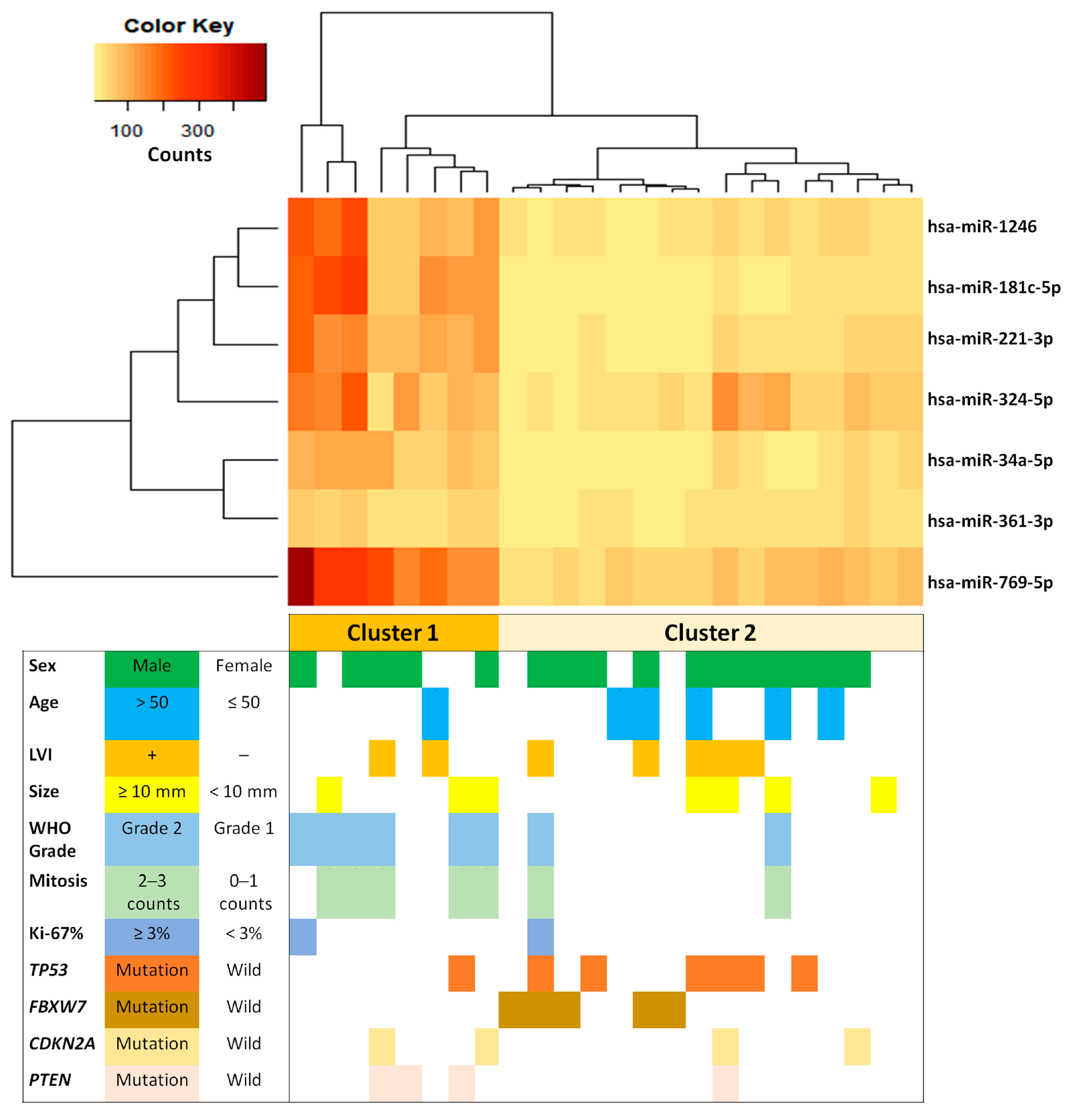
2.1. Baseline Characteristics and Genetic Alterations of Rectal NETs

Overall, 24 patients (17 male and seven female) with a median age of 48.5 years (34–73 years) were included (Table 1). The average tumor size ranged from 7.62 ± 2.81 mm (5 to 15 mm). According to the World Health Organization (WHO) classification, 16 patients showed grade 1 NETs, and eight exhibited grade 2 NETs. Microscopic examination with D2-40 and hematoxylin and eosin (H&E) staining allowed us to observe seven LVIs (29.2%, 7/24). After next-generation sequencing (NGS) and variant analysis, 19 rectal NETs (79.2%, 19/24) revealed more than one mutation in the 24 cancer-related genes: TP53 (7/24, 29.2%), FBXW7 (5/24, 20.8%), CDKN2A (4/24, 16.7%), PTEN (4/24, 16.7%), ATM (3/24, 12.5%), EGFR (3/24, 12.5%), AKT1 (2/24, 8.3%), ALK (2/24, 8.3%), IDH1 (2/24, 8.3%), KIT (2/24, 8.3%), KRAS (2/24, 8.3%), PIK3CA (2/24, 8.3%), RET (2/24, 8.3%), SMARCB1 (2/24, 8.3%), VHL (2/24, 8.3%), BRAF (1/24, 4.1%), CTNNB1 (1/24, 4.1%), ERBB2 (1/24, 4.1%), EZH2 (1/24, 4.1%), FLT3 (1/24, 4.1%), HNF1A (1/24, 4.1%), SMAD4 (1/24, 4.1%), SMO (1/24, 4.1%), and STK11 (1/24, 4.1%). TP53, FBXW7, CDKN2A, and PTEN mutations were the most frequently found in rectal NETs.

2.2. Screening of Candidate miRNAs Associated with Specific Genetic Mutations

We screened the expression of 584 miRNA relevant to the aforementioned 24 genetic mutations in mutated and wild-type tumors. The results revealed that FBXW7 was the only candidate gene relevant to miRNAs differentially expressed between mutated and wild-type NETs. When comparing the miRNA expression levels of 19 tumors harboring FBXW7 wild-type with 5 tumors carrying FBXW7 mutations, we observed seven down-regulated miRNAs (hsa-miR-221-3p, hsa-miR-769-5p, hsa-miR-181c-5p, hsa-miR-34a-5p, hsa-miR-1246, hsa-miR-324-5p, and hsa-miR-361-3p) in FBXW7-mutant tumors compared to FBXW7-wild-type tumors (all p < 0.001, Table 2). Among them, hsa-miR-769-5p was the most correlated with FBXW7 mutation (false discovery rate [FDR] < 0.05).
Then, unsupervised hierarchical clustering analysis was performed to compare the expression profiles of the dysregulated seven miRNAs (Figure 1). The analysis separated the tumors into two main clusters according to miRNAs being up- or down-regulated. The cluster with up-regulated miRNA expression tended to correlate with NETs with higher mitotic indices, grade 2, wild-type FBXW7, and wild-type TP53. In contrast, the cluster with down-regulated miRNAs tended to correlate with NETs with lower mitotic indices, grade 1, FBXW7 mutation, and TP53 mutation. These findings indicated distinct miRNA differential expression (hsa-miR-769-5p, hsa-miR-221-3p, hsa-miR-34a-5p, hsa-miR-181c-5p, hsa-miR-1246, hsa-miR-324-5p, and hsa-miR-361-3p) between the tumor groups with high mitotic indices, grade 2, wild-type FBXW7, and wild-type TP53 and other tumor groups with low mitotic indices, grade 1, FBXW7 mutation, and TP53 mutation.

2.3. miRNAs Related to Clinical and Pathological Characteristics

Evaluation of 584 miRNA expression profiles between clinical and pathological factors, including sex, age (≤50 vs. >50), tumor size (<10 mm vs. ≥10 mm), LVI, Ki-67 index (<3% vs. ≥3%), and WHO grade (grade 1 vs. grade 2), showed no significant differences among miRNA expression levels except for the mitotic index.
When comparing the miRNA expression profiles between tumors with either low (0–1) or high (≥2) mitotic indices, we identified five differentially expressed miRNAs between the two groups: miR-3934-5p was significantly up-regulated, and miR-153-3p, miR-4286, miR-223-3p, and miR-129-5p were down-regulated in the high mitotic index group. After adjustment, only miR-3934-5p was statistically significant (Table 3). However, miR-3934-5p, related to a high mitotic index, was not correlated with the miRNA expression profiles related to FBXW7 mutations (p > 0.99999).

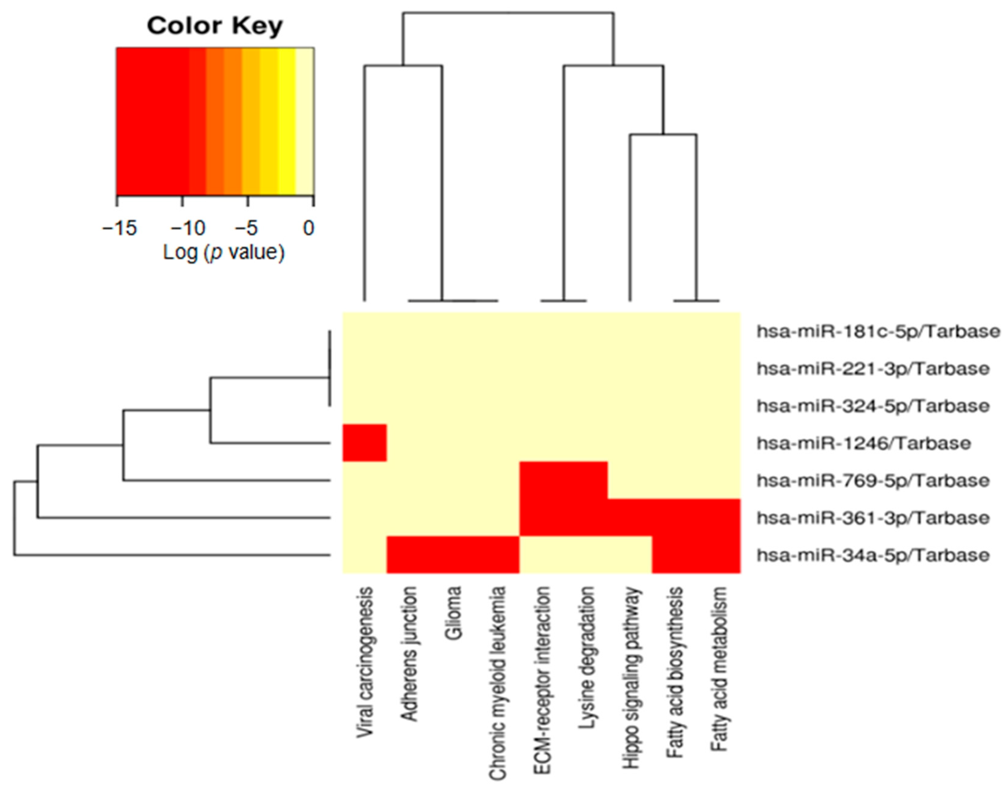
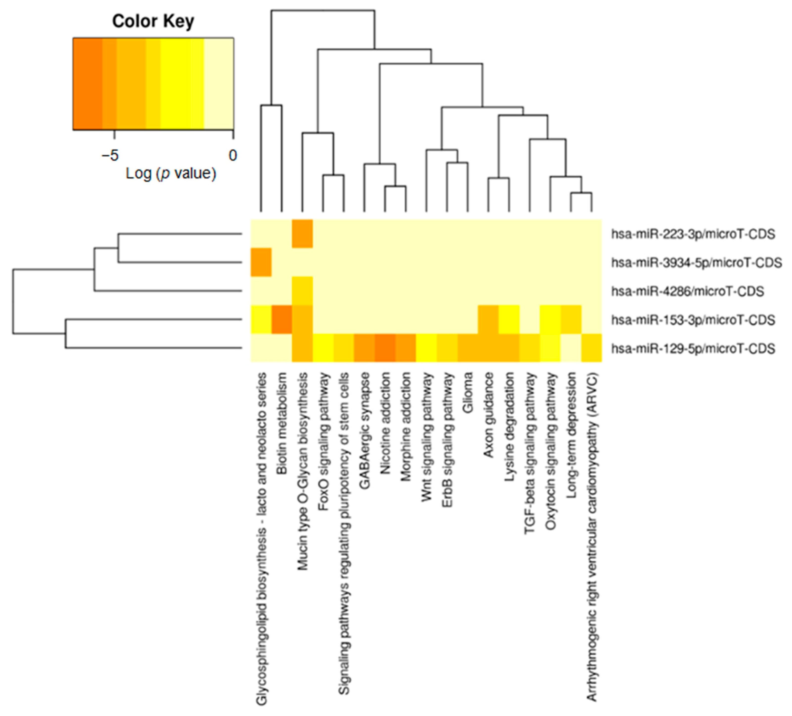
2.4. Identification of Potential Functional Pathways Related to miRNAs

We tested Kyoto Encyclopedia of Genes and Genomes (KEGG) pathway maps with the mentioned miRNAs. Seven significantly differentially expressed miRNAs correlated with FBXW7 mutational status (hsa-miR-769-5p, hsa-miR-34a-5p, hsa-miR-181c-5p, hsa-miR-324-5p, hsa-miR-221-3p, hsa-miR-361-3p, and hsa-miR-1246). This analysis enriched nine possible pathways involved in fatty acid biosynthesis, extracellular matrix (ECM)-receptor interaction, fatty acid metabolism, lysine degradation, viral carcinogenesis, the adherens junction, chronic myeloid leukemia, and glioma and the Hippo signaling pathway to identify potential functionally connected pathways between miRNAs and target genes (Table 4). Specifically, ECM-receptor interactions and lysine degradation were significantly correlated with hsa-miR-769-5p (Figure 2).
Furthermore, we produced additional KEGG pathway maps with five differentially expressed miRNAs that correlated with a high mitotic index (miR-3934-5p, miR-153-3p, miR-4286, miR-223-3p, and miR-129-5p). This analysis enriched 17 possible pathways in which the glycosphingolipid biosynthesis-lacto and neolacto series were significantly correlated with hsa-miR-3934-5p (Figure 3).

3. Discussion

In this study, screening 584 miRNA expressions in rectal NETs revealed that FBXW7 and mitotic index were the sole candidate genes and clinicopathological features specifically relevant to miRNAs, respectively. FBXW7 acts as a potent tumor suppressor that may control the expression levels of multiple oncoproteins (i.e., mTOR, notch, cyclin E, c-Myc, and c-Jun) that engage in cellular signaling pathways by inducing them for ubiquitin-mediated proteasomal degradation, thereby protecting against cancer development [27,28]. Several studies have demonstrated that FBXW7 also significantly influences DNA damage response and repair processes, prime molecular mechanisms for maintaining cellular homeostasis, and genomic integrity [29,30,31,32]. Accordingly, FBXW7 dysregulation in this process may be a critical factor contributing to tumorigenesis [27,28]. A recent study has reported a high frequency of P53 (61%), APC (53%), FBXW7 (25%), and KRAS (25%) mutations in rectal NETs [15]. Likewise, in our previous study, FBXW7 (p.R465H) was found to be likely pathogenic in low-grade rectal NETs [18]. In this context, it may be important to focus on the epigenetic regulation of FBXW7 in rectal NETs, as FBXW7 is a critical regulator of several key signaling pathways involved in cell growth and proliferation [27,28]. Understanding the epigenetic regulation of FBXW7 in rectal NETs may provide insights into the mechanisms underlying tumor development and potentially identify novel therapeutic targets for treating these tumors.
Alterations in FBXW7 expression have been linked to the development and progression of several types of cancer [27,28]. Some FBXW7 regulators include miRNAs, such as miR-27 in lung cancer, miR-32 in breast cancer, miR-92a in cervical cancer, miR-223 in gastric cancer, miR-182 in lung cancer, and miR-223 in T-cell lymphoma [33,34,35]. In the rectal NETs of the present study, seven miRNAs (hsa-miR-769-5p, hsa-miR-221-3p, hsa-miR-34a-5p, hsa-miR-181c-5p, hsa-miR-1246, hsa-miR-324-5p, and hsa-miR-361-3p) were significantly down-regulated in FBXW7-mutant tumors. In gastric cancer, miR-223 acted as an oncogene and negatively modulated FBXW7 expression, governing proliferation, apoptosis, and invasion [33]. Consistent with this, Notch-mediated activation of miR-223 represses FBXW7 in T-cell acute lymphoblastic leukemia [34]. In breast cancer cells, miR-32 induces cell proliferation and migration, evading apoptosis by down-regulating FBXW7 [35]. In cervical cancer cells, an up-regulation of miR-92a causes a down-regulation of FBXW7, thereby stimulating tumor cell proliferation and invasion [36]. miR-182 increases the proliferation of non-small cell lung cancer cells by inhibiting FBXW7 [37].
Here, we noted that hsa-miR-769-5p was most correlated with FBXW7 mutation, further linked to ECM-receptor interaction and lysine degradation via the KEGG analysis of target signaling pathways. The ECM is a central element of both normal and tumor tissues, consisting of a complex texture of cross-linked proteins to play a role in resident cells’ hydration, elasticity, and structural organization [38]. It importantly helps in neoplastic proliferation, angiogenesis, progression, and tumor survival [39]. Specific ECM alterations regulating tissue biochemical and biomechanical properties have been described in NETs [40], which may imply that several ECM glycoproteins may contribute to the tumorigenesis of NETs. In a recent study, the up-regulation of six fibrogenic genes (COL5A2, COL1A2, COL3A1, ITGA5, ITGB1, and ITGB1) in pulmonary neuroendocrine carcinoma and the down-regulation of pulmonary neuroendocrine neoplasms were strongly related to no metastasis and overall survival [41]. Only one study has shown that the activation of the lysine degradation pathway injures tumor cell proliferation [42], which is also related to miR-769-5p tumor suppression, as shown in subsequent studies. miR-769-5p/miR-769-3p-mediated inhibition of proliferation causes apoptosis in gastric cancer cells [43]. Another study has shown that miR-769-5p inhibited non-small cell lung cancer proliferation and invasion, indicating that this type of tumor could benefit from miR-769-5p as a diagnostic and prognostic biomarker [44]. hsa-miR-769-5p has also been described to be expressed at low levels in papillary thyroid carcinomas, and the overexpression of hsa-miR-769-5p highly affected the formation of the tumor microenvironment [45], which appears to be consistent with our findings.
Up-regulated miR-3934-5p and down-regulated miR-4286, miR-223-3p, miR-129-5p, and miR-153-3p were characterized in tumors with high mitotic indices, in which miR-3934-5p was the only one statistically significant after adjustment. Further clustering analysis separated two main groups: (1) high mitotic index, grade 2, wild-type FBXW7, and wild-type TP53 and (2) low mitotic index, grade 1, FBXW7 mutation, and TP53 mutation. This may indicate that these miRNA contributions were mutually exclusive between the high mitotic index and FBXW7 mutation in rectal NETs. The KEGG pathway maps also confirmed the different pathway involvement in different miRNAs between FBXW7-mutated and highly mitotic-active tumors. hsa-miR-3934-5p may be closely implicated in a high mitotic index, eventually contributing to higher grade 2 tumors in rectal NETs.
In contrast, hsa-miR-769-5p related to FBXW7 mutation may be involved in grade 1 tumors and is less likely to be involved in tumor aggressiveness. Indeed, a recent study has discovered that WHO grade 3 gastro-pancreatic NETs tend to harbor NETs with lower FBXW7 mutation frequency than grade 1 and grade 2 NETs [15]. Comparisons of normal pancreatic islets with their matched neoplastic groups disclosed changes in ECM compositions, which also differed depending on histological grade [40,46]. This may explain the finding that the ECM-receptor interaction KEGG pathway (hsa-miR-769-5p) was significantly associated with FBXW7 mutations and was more likely involved in grade 1 tumors. hsa-miR-769-5p appears to be a possible regulator of FBXW7-mutated rectal NETs. These differentially expressed miRNAs could be potential tissue biomarkers for aggressive behavior in rectal NETs.
Our study was limited by a relatively small number of rectal NETs. We did not perform the validation step using quantitative PCR. However, we utilized the advantages of Nanostring technology to identify 24 cancer-related miRNAs out of 584 miRNAs without requiring the amplification of transcripts. We believe that our approach was able to identify low-abundance transcripts that may have been difficult to validate through PCR [47]. Our study focused on identifying candidate miRNAs. In addition, we did not analyze the difference between plasma and tissue miRNAs. miRNAs from tumors may be secreted within membrane vesicles (exosomes) or directly into the blood, indicating that miRNAs play a key role in intercellular communication [48]. Plasma miRNAs communicate the most with the pericardial, adipose, liver, and spleen tissues [49]. As rectal NETs are submucosal tumors, tissue-based analysis of our miRNAs may be more appropriate as they reflect the origin of these tumors. Because some may metastasize, we aim to further investigate the differences in plasma miRNAs between localized rectal NETs and those with metastasis to advance our understanding of these tumors in future studies.
Nevertheless, some conclusions emerged from our results. To our knowledge, this is the first study to identify a correlation between FBXW7 mutations and miRNAs in rectal NETs. hsa-miR-769-5p was most correlated with FBXW7 mutation and further linked to ECM-receptor interaction and lysine degradation via a KEGG analysis of target signaling pathways. hsa-miR-3934-5p may be strongly implicated in a high mitotic index, which may eventually contribute to higher grade 2 tumors in rectal NETs, whereas hsa-miR-769-5p related to FBXW7 mutation may be involved in grade 1 tumors and is likely to play a role in suppressing rectal NET progression.

4. Materials and Methods

4.1. Patients

The institutional review board of Hallym University Sacred Heart Hospital approved this study (HALLYM 2022-11-006-002). All participants provided written informed consent for the procedure, and the study protocol adhered to the guidelines and regulations of the ethics committee of Hallym University. A search in the electronic database of the pathology department of Hallym University Sacred Heart Hospital disclosed 69 consecutive patients with primary rectal NETs who underwent endoscopic resection in the hospital between 1 June 2005, and 31 July 2015. These patients were included in previous studies [6,50]. We excluded 45 patients whose formalin-fixed paraffin-embedded (FFPE) tissue blocks contained too few tumor cells for the assay or whose FFPE tumor blocks were inadequate for molecular analysis.

4.2. Histologic Evaluation

All H&E-stained slides were reviewed by a gastrointestinal and molecular pathologist (M.J.K.) to confirm the diagnosis and evaluate histopathological characteristics. The results of mitotic rates, LVI, and proliferative index established in the previous studies were applied in the present study [6,50].

4.3. DNA Extraction and NGS

A tissue microtome (Leica Biosystems, Wetzlar, Germany) was used to obtain two 10-μm sections from every FFPE tissue block on the glass slides. Tumor areas on the glass slides were manually macrodissected from the unstained tissue sections to enrich tumor cell populations > 50%. DNA was extracted and purified using the Ion AmpliSeq Direct FFPE DNA Kit (Thermo Fisher Scientific, Waltham, MA, USA) and QIAamp DSP DNA FFPE Tissue Kit (Qiagen, Hilden, Germany), respectively. The yield of purified genomic DNA was estimated using a Qubit 2.0 Fluorometer and Qubit dsDNA HS Assay Kit (Thermo Fisher Scientific) prior to library preparation for sequencing.
The NGS platform was an Ion Personal Genome Machine Sequencer (Thermo Fisher Scientific). Library preparation for each sample was conducted using the Ion AmpliSeq Library Kit 2.0 (Thermo Fisher Scientific) and Ion AmpliSeq Cancer HotSpot Panel v2 (Thermo Fisher Scientific), following the manufacturer’s protocols, as previously explained [18]. The Ion Torrent platform-specific pipeline software was employed via the variant calling process. Annotation was performed using the Variant Effect Predictor. To compare our data with findings from other cancer genome studies and check for hotspot mutations, we downloaded all cancer genome studies from cBioPortal using the CGDS-R package (R foundation for Statistical Computing, Vienna, Austria). To identify confident putative somatic variants, annotated raw variants were filtered according to the following criteria: (1) non-synonymous single-nucleotide variant or short insertion or deletion in coding regions; (2) coverage ≥ 50× and variant allele frequency (VAF) ≥ 5% or coverage ≥ 1000× and VAF ≥ 3%; and (3) minor allele frequency < 0.1% in the gnomAD and 1000 Genomes Project. The resulting list of variants was manually reviewed and visually confirmed using the Integrated Genomics Viewer (http://www.broadinstitute.org/igv/, accessed on 1 October 2022).

4.4. miRNA Extraction and Nanostring nCounter miRNA Expression Assay

The miRNAs were isolated from FFPE tissues using the miRNeasy Mini Kit (Qiagen) according to the manufacturer’s instructions. The concentration was assessed using a NanoDrop spectrophotometer (Thermo Fisher Scientific) [51]. nCounter human v3 miRNA expression assays designed and synthesized by NanoString Technologies (NanoString, Seattle, WA, USA) were performed in this study. The miRNA panel included oligonucleotide tags for 798 human miRNAs (from miRBase v21) and 5 housekeeping mRNAs for reference (ACTB, B2M, GAPDH, RPL19, and RPLP0). Twenty-five control probes that recognized either synthetic mRNA or miRNA targets were used to monitor the efficiency and specificity of each reaction step. Hybridization was performed with 150 ng of total RNA (16 h at 65 °C) with probe pairs consisting of reporter probes carrying the signal at their 5′ end and capture probes carrying biotin on their 3′ ends. After hybridization, sample cleanup and digital report counts were performed according to the manufacturer’s instructions. Raw data were analyzed using the NanoString nSolver software (version 4.0). Internal positive controls normalized raw data to remove variability in hybridization, and negative controls to subtract the background. In addition, miRNA input levels were normalized using the spike-in normalization method according to the manufacturer’s instructions. The minimum threshold to consider an miRNA for further analysis was 20 counts in at least 10% of the samples. This threshold allowed us to identify 584 miRNAs suitable for analysis.

4.5. DNA Intelligent Analysis (DIANA)-miRPath for miRNA Pathway Analysis

Pathway analysis using significantly up-regulated and down-regulated miRNAs was performed using DIANA-miRPath v3.0 [52]. This software allowed us to link miRNAs to experimentally validated target genes from Tarbase, v7.0/microT-CDS v5.0 (https://dianalab.e-ce.uth.gr/, accessed on 1 October 2022) and identifies the putative targeted molecular ‘KEGG’ pathways. As our analysis was hypothesis-free, we used the pathway union option of the miRPath software. The pathway analysis was performed using miRPath v3.0 with an FDR < 0.05, using the Benjamini and Hochberg methods.

4.6. Statistical Analyses

The differentially expressed mRNAs and significant pathways were selected based on the p-value and FDR analysis [53]. The miRNA expression levels were compared to each clinicopathological parameter using the Mann–Whitney U test, calculated using GraphPad Prism 9 (GraphPad Software, San Diego, CA, USA). The p-values were obtained through Fisher’s exact test and χ2 test as enrichment analysis methods. The FDR was estimated using the two-stage process of Benjamini, Krieger, and Yekutieli using FDR < 0.05 and p < 0.001 as the significance threshold [26]. Hierarchical clustering was performed using the R package, version 4.13, using Pearson’s correlation.

5. Conclusions

Different miRNAs related to high mitotic indices or FBXW7 mutations may contribute to tumor behaviors in rectal NETs. Specifically, FBXW7 mutations are likely to play an important role in inhibiting rectal NET progression, whereas mitosis may be the only significant histological risk factor related to miRNA expression. Furthermore, differentially expressed miRNAs may be used as potential tissue biomarkers for possible prognostic indicators in rectal NETs.

Author Contributions

H.S.K.: investigation, formal analysis, validation, writing-original draft, writing—review and editing; H.Y.P., I.T.S. and M.-J.K. (Min-Jeong Kim): data curation; H.L.: formal analysis; N.Y.K.: software; E.S.N., S.J.C. and M.J.K. (Min Jeong Kim): project administration; M.J.K. (Mi Jung Kwon): investigation, writing-original draft, writing—review and editing, funding acquisition. All authors have read and agreed to the published version of the manuscript.

Funding

This research was funded by the National Research Foundation of Korea, the Korean Ministry of Science, and I.C.T. (grant no. NRF-2022R1F1A1065335) to M.J.K. (Mi Jung Kwon). The APC was funded by NRF-2022R1F1A1065335.

Institutional Review Board Statement

This study was approved by the institutional review board of Hallym University Sacred Heart Hospital (HALLYM 2022-11-006-002).

Informed Consent Statement

All 24 patients provided informed consent prior to endoscopic resection.

Data Availability Statement

The data used to support the findings of this study are available from the corresponding author upon request.

Conflicts of Interest

The authors declare no conflict of interest.

References

  1. Soga, J. Carcinoids of the rectum: An evaluation of 1271 reported cases. Surg. Today. 1997, 27, 112–119. [Google Scholar] [CrossRef]
  2. Modlin, I.M.; Oberg, K.; Chung, D.C.; Jensen, R.T.; de Herder, W.W.; Thakker, R.V.; Caplin, M.; Delle Fave, G.; Kaltsas, G.A.; Krenning, E.P.; et al. Gastroenteropancreatic neuroendocrine tumours. Lancet Oncol. 2008, 9, 61–72. [Google Scholar] [CrossRef] [PubMed]
  3. Jung, E.S.; Kang, Y.K.; Cho, M.Y.; Kim, J.M.; Lee, W.A.; Lee, H.E.; Park, S.; Sohn, J.H.; Jin, S.Y. Update on the proposal for creating a guideline for cancer registration of the gastrointestinal tumors (I-2). Korean J. Pathol. 2012, 46, 443–453. [Google Scholar] [CrossRef] [PubMed]
  4. Gao, W.; Liu, S.M.; Lu, H.Z.; Liang, J.; Yuan, Y.L.; Liu, X.Y. Analysis of clinicopathological features of intestinal neuroendocrine neoplasms. Zhonghua Zhong Liu Za Zhi 2012, 34, 450–456. [Google Scholar] [CrossRef] [PubMed]
  5. Gastrointestinal Pathology Study Group of Korean Society of Pathologists; Cho, M.Y.; Kim, J.M.; Sohn, J.H.; Kim, M.J.; Kim, K.M.; Kim, W.H.; Kim, H.; Kook, M.C.; Park, D.Y.; et al. Current Trends of the Incidence and Pathological Diagnosis of Gastroenteropancreatic Neuroendocrine Tumors (GEP-NETs) in Korea 2000–2009: Multicenter Study. Cancer Res. Treat. 2012, 44, 157–165. [Google Scholar] [CrossRef]
  6. Kwon, M.J.; Kang, H.S.; Soh, J.S.; Lim, H.; Kim, J.H.; Park, C.K.; Park, H.R.; Nam, E.S. Lymphovascular invasion in more than one-quarter of small rectal neuroendocrine tumors. World J. Gastroenterol. 2016, 22, 9400–9410. [Google Scholar] [CrossRef]
  7. Fahy, B.N.; Tang, L.H.; Klimstra, D.; Wong, W.D.; Guillem, J.G.; Paty, P.B.; Temple, L.K.; Shia, J.; Weiser, M.R. Carcinoid of the rectum risk stratification (CaRRs): A strategy for preoperative outcome assessment. Ann. Surg. Oncol. 2007, 14, 396–404. [Google Scholar] [CrossRef]
  8. Shields, C.J.; Tiret, E.; Winter, D.C.; International Rectal Carcinoid Study Group. Carcinoid tumors of the rectum: A multi-institutional international collaboration. Ann. Surg. 2010, 252, 750–755. [Google Scholar] [CrossRef]
  9. Yoon, S.N.; Yu, C.S.; Shin, U.S.; Kim, C.W.; Lim, S.B.; Kim, J.C. Clinicopathological characteristics of rectal carcinoids. Int. J. Color. Dis. 2010, 25, 1087–1092. [Google Scholar] [CrossRef]
  10. Kasuga, A.; Chino, A.; Uragami, N.; Kishihara, T.; Igarashi, M.; Fujita, R.; Yamamoto, N.; Ueno, M.; Oya, M.; Muto, T. Treatment strategy for rectal carcinoids: A clinicopathological analysis of 229 cases at a single cancer institution. J. Gastroenterol. Hepatol. 2012, 27, 1801–1807. [Google Scholar] [CrossRef]
  11. Yamagishi, D.; Matsubara, N.; Noda, M.; Yamano, T.; Tsukamoto, K.; Kuno, T.; Hamanaka, M.; Kobayashi, M.; Ikeuchi, H.; Matsuda, I.; et al. Clinicopathological characteristics of rectal carcinoid patients undergoing surgical resection. Oncol. Lett. 2012, 4, 910–914. [Google Scholar] [CrossRef] [PubMed]
  12. Caplin, M.; Sundin, A.; Nillson, O.; Baum, R.P.; Klose, K.J.; Kelestimur, F.; Plöckinger, U.; Papotti, M.; Salazar, R.; Pascher, A.; et al. Enets Consensus Guidelines for the management of patients with digestive neuroendocrine neoplasms: Colorectal neuroendocrine neoplasms. Neuroendocrinology 2012, 95, 88–97. [Google Scholar] [CrossRef] [PubMed]
  13. Zhou, X.; Xie, H.; Xie, L.; Li, J.; Fu, W. Factors associated with lymph node metastasis in radically resected rectal carcinoids: A systematic review and meta-analysis. J. Gastrointest. Surg. 2013, 17, 1689–1697. [Google Scholar] [CrossRef] [PubMed]
  14. Han, D.S.; Sohn, J.H.; Byeon, J.S.; Choi, H.; Kim, J.M. Diagnostic coding for intramucosal carcinoma and neuroendocrine tumor in the colorectum: Proposal for avoiding confusing coding in Korea. Clin. Endosc. 2015, 48, 216–220. [Google Scholar] [CrossRef]
  15. Venizelos, A.; Elvebakken, H.; Perren, A.; Nikolaienko, O.; Deng, W.; Lothe, I.M.B.; Couvelard, A.; Hjortland, G.O.; Sundlöv, A.; Svensson, J.; et al. The molecular characteristics of high-grade gastroenteropancreatic neuroendocrine neoplasms. Endocr. Relat. Cancer 2021, 29, 1–14. [Google Scholar] [CrossRef]
  16. Tang, L.H.; Basturk, O.; Sue, J.J.; Klimstra, D.S. A practical approach to the classification of WHO Grade 3 (G3) well-differentiated neuroendocrine tumor (WD-NET) and poorly differentiated neuroendocrine carcinoma (PD-NEC) of the pancreas. Am. J. Surg. Pathol. 2016, 40, 1192–1202. [Google Scholar] [CrossRef] [Green Version]
  17. Capuano, F.; Grami, O.; Pugliese, L.; Paulli, M.; Pietrabissa, A.; Solcia, E.; Vanoli, A. Grade 3 neuroendocrine tumor (G3 NET) in a background of multiple serotonin cell neoplasms of the ileum associated with carcinoid syndrome and aggressive behavior. Endocr. Pathol. 2018, 29, 369–373. [Google Scholar] [CrossRef]
  18. Park, H.Y.; Kwon, M.J.; Kang, H.S.; Kim, Y.J.; Kim, N.Y.; Kim, M.J.; Min, K.W.; Choi, K.C.; Nam, E.S.; Cho, S.J.; et al. Targeted next-generation sequencing of well-differentiated rectal, gastric, and appendiceal neuroendocrine tumors to identify potential targets. Hum. Pathol. 2019, 87, 83–94. [Google Scholar] [CrossRef]
  19. Kagohara, L.T.; Stein-O’Brien, G.L.; Kelley, D.; Flam, E.; Wick, H.C.; Danilova, L.V.; Easwaran, H.; Favorov, A.V.; Qian, J.; Gaykalova, D.A.; et al. Epigenetic regulation of gene expression in cancer: Techniques, resources and analysis. Brief. Funct. Genom. 2018, 17, 49–63. [Google Scholar] [CrossRef] [Green Version]
  20. Ying, S.Y.; Chang, D.C.; Lin, S.L. The microRNA (miRNA): Overview of the RNA genes that modulate gene function. Mol. Biotechnol. 2008, 38, 257–268. [Google Scholar] [CrossRef]
  21. Mitsuhashi, K.; Yamamoto, I.; Kurihara, H.; Kanno, S.; Ito, M.; Igarashi, H.; Ishigami, K.; Sukawa, Y.; Tachibana, M.; Takahashi, H.; et al. Analysis of the molecular features of rectal carcinoid tumors to identify new biomarkers that predict biological malignancy. Oncotarget 2015, 6, 22114–22125. [Google Scholar] [CrossRef] [PubMed] [Green Version]
  22. Murayama, N.; Okamoto, K.; Nakagawa, T.; Miyoshi, J.; Nishida, K.; Kawaguchi, T.; Kagemoto, K.; Kitamura, S.; Ma, B.; Miyamoto, H.; et al. miR-144-3p/miR-451a promotes lymphovascular invasion through repression of PTEN/p19 in rectal neuroendocrine tumors. J. Gastroenterol. Hepatol. 2022, 37, 919–927. [Google Scholar] [CrossRef]
  23. Vega Neira, S.J.; Torres Carvajal, M.M.; Barrera Herrera, L.; Andrade Perez, R.; Lopez Panqueva, R. P-017 Analysis of microRNA expression in gastroenteropancreatic Neuroendocrine Tumor. Ann Oncol. 2016, 27, II5. [Google Scholar] [CrossRef] [Green Version]
  24. Malczewska, A.; Kidd, M.; Matar, S.; Kos-Kudla, B.; Modlin, I.M. A comprehensive assessment of the role of miRNAs as biomarkers in gastroenteropancreatic neuroendocrine tumors. Neuroendocrinology 2018, 107, 73–90. [Google Scholar] [CrossRef] [PubMed]
  25. Precazzini, F.; Detassis, S.; Imperatori, A.S.; Denti, M.A.; Campomenosi, P. Measurements methods for the development of microRNA-based tests for cancer diagnosis. Int. J. Mol. Sci. 2021, 22, 1176. [Google Scholar] [CrossRef]
  26. Benjamini, Y.; Krieger, A.M.; Yekutieli, D. Adaptive linear step-up procedures that control the false discovery rate. Biometrika 2006, 93, 491–507. [Google Scholar] [CrossRef]
  27. Welcker, M.; Clurman, B.E. FBW7 ubiquitin ligase: A tumour suppressor at the crossroads of cell division, growth and differentiation. Nat. Rev. Cancer 2008, 8, 83–93. [Google Scholar] [CrossRef]
  28. Davis, R.J.; Welcker, M.; Clurman, B.E. Tumor suppression by the Fbw7 ubiquitin ligase: Mechanisms and opportunities. Cancer Cell. 2014, 26, 455–464. [Google Scholar] [CrossRef] [Green Version]
  29. Lan, H.; Sun, Y. Tumor suppressor FBXW7 and its regulation of DNA damage response and repair. Front. Cell Dev. Biol. 2021, 9, 751574. [Google Scholar] [CrossRef]
  30. Jiang, Y.; Qi, X.; Liu, X.; Zhang, J.; Ji, J.; Zhu, Z.; Ren, J.; Yu, Y. Fbxw7 haploinsufficiency loses its protection against DNA damage and accelerates MNU-induced gastric carcinogenesis. Oncotarget 2017, 8, 33444–33456. [Google Scholar] [CrossRef] [Green Version]
  31. Galindo-Moreno, M.; Giráldez, S.; Limón-Mortés, M.C.; Belmonte-Fernández, A.; Reed, S.I.; Sáez, C.; Japón, M.Á.; Tortolero, M.; Romero, F. SCF(FBXW7)-mediated degradation of p53 promotes cell recovery after UV-induced DNA damage. FASEB J. 2019, 33, 11420–11430. [Google Scholar] [CrossRef]
  32. Zhang, Q.; Mady, A.S.A.; Ma, Y.; Ryan, C.; Lawrence, T.S.; Nikolovska-Coleska, Z.; Sun, Y.; Morgan, M.A. The WD40 domain of FBXW7 is a poly(ADP-ribose)-binding domain that mediates the early DNA damage response. Nucleic Acids Res. 2019, 47, 4039–4053. [Google Scholar] [CrossRef] [PubMed]
  33. Li, J.; Guo, Y.; Liang, X.; Sun, M.; Wang, G.; De, W.; Wu, W. MicroRNA-223 functions as an oncogene in human gastric cancer by targeting FBXW7/hCdc. J. Cancer Res. Clin. Oncol. 2012, 138, 763–774. [Google Scholar] [CrossRef]
  34. Kumar, V.; Palermo, R.; Talora, C.; Campese, A.F.; Checquolo, S.; Bellavia, D.; Tottone, L.; Testa, G.; Miele, E.; Indraccolo, S.; et al. Notch and NF-kB signaling pathways regulate miR-223/FBXW7 axis in T-cell acute lymphoblastic leukemia. Leukemia 2014, 28, 2324–2335. [Google Scholar] [CrossRef] [PubMed]
  35. Xia, W.; Zhou, J.; Luo, H.; Liu, Y.; Peng, C.; Zheng, W.; Ma, W. MicroRNA-32 promotes cell proliferation, migration and suppresses apoptosis in breast cancer cells by targeting FBXW. Cancer Cell Int. 2017, 17, 14. [Google Scholar] [CrossRef] [Green Version]
  36. Zhou, C.; Shen, L.; Mao, L.; Wang, B.; Li, Y.; Yu, H. miR-92a is upregulated in cervical cancer and promotes cell proliferation and invasion by targeting FBXW. Biochem. Biophys. Res. Commun. 2015, 458, 63–69. [Google Scholar] [CrossRef]
  37. Chang, H.; Liu, Y.H.; Wang, L.L.; Wang, J.; Zhao, Z.H.; Qu, J.F.; Wang, S.F. MiR-182 promotes cell proliferation by suppressing FBXW7 and FBXW11 in non-small cell lung cancer. Am. J. Transl. Res. 2018, 10, 1131–1142. [Google Scholar] [PubMed]
  38. Yue, B. Biology of the extracellular matrix: An overview. J. Glaucoma. 2014, 23 (Suppl. 1), S20–S23. [Google Scholar] [CrossRef] [Green Version]
  39. Saupe, F.; Schwenzer, A.; Jia, Y.; Gasser, I.; Spenlé, C.; Langlois, B.; Kammerer, M.; Lefebvre, O.; Hlushchuk, R.; Rupp, T.; et al. Tenascin-C downregulates wnt inhibitor dickkopf-1, promoting tumorigenesis in a neuroendocrine tumor model. Cell Rep. 2013, 5, 482–492. [Google Scholar] [CrossRef] [Green Version]
  40. Cives, M.; Pelle’, E.; Quaresmini, D.; Rizzo, F.M.; Tucci, M.; Silvestris, F. The Tumor Microenvironment in Neuroendocrine Tumors: Biology and Therapeutic Implications. Neuroendocrinology 2019, 109, 83–99. [Google Scholar] [CrossRef]
  41. Prieto, T.G.; Machado-Rugolo, J.; Baldavira, C.M.; Velosa, A.P.P.; Teodoro, W.R.; Saber, A.M.A.; Capelozzi, V.L. The fibrosis-targeted collagen/integrins gene profile predicts risk of metastasis in pulmonary neuroendocrine neoplasms. Front. Oncol. 2021, 11, 706141. [Google Scholar] [CrossRef] [PubMed]
  42. Dai, Z.; Yang, S.; Xu, L.; Hu, H.; Liao, K.; Wang, J.; Wang, Q.; Gao, S.; Li, B.; Lai, L. Identification of Cancer–associated metabolic vulnerabilities by modeling multi-objective optimality in metabolism. Cell Commun. Signal. 2019, 17, 124. [Google Scholar] [CrossRef] [PubMed] [Green Version]
  43. Dai, W.; Liu, S.; Zhang, J.; Pei, M.; Xiao, Y.; Li, J.; Hong, L.; Lin, J.; Wang, J.; Wu, X.; et al. Vorinostat triggers miR-769-5p/3p-mediated suppression of proliferation and induces apoptosis via the STAT3-IGF1R-HDAC3 complex in human gastric cancer. Cancer Lett. 2021, 521, 196–209. [Google Scholar] [CrossRef]
  44. Sun, Y.; Li, J.; Zheng, S. MiR-769-5p, Which Targets HDGF, Inhibits Cell Proliferation and Invasion in Nonsmall Cell Lung Cancer. Cancer Biotherapy Radiopharm. 2021. [Google Scholar] [CrossRef]
  45. Gao, T.; Zhao, L.; Zhang, F.; Cao, C.; Fan, S.; Shi, X. Evaluate the diagnostic and prognostic value of NUSAP1 in papillary thyroid carcinoma and identify the relationship with genes, proteins, and immune factors. World J. Surg. Oncol. 2022, 20, 207. [Google Scholar] [CrossRef]
  46. Corti, A. Chromogranin A and the tumor microenvironment. Cell Mol. Neurobiol. 2010, 30, 1163–1170. [Google Scholar] [CrossRef]
  47. Foye, C.; Yan, I.K.; David, W.; Shukla, N.; Habboush, Y.; Chase, L.; Ryland, K.; Kesari, V.; Patel, T. Comparison of miRNA quantitation by Nanostring in serum and plasma samples. PLoS ONE 2017, 12, e0189165. [Google Scholar] [CrossRef] [Green Version]
  48. Géczi, D.; Nagy, B.; Szilágyi, M.; Penyige, A.; Klekner, Á.; Jenei, A.; Virga, J.; Birkó, Z. Analysis of circulating miRNA profile in plasma samples of glioblastoma patients. Int. J. Mol. Sci. 2021, 22, 5058. [Google Scholar] [CrossRef] [PubMed]
  49. Cui, C.; Cui, Q. The relationship of human tissue microRNAs with those from body fluids. Sci Rep. 2020, 10, 5644. [Google Scholar] [CrossRef] [Green Version]
  50. Kim, M.J.; Kwon, M.J.; Kang, H.S.; Choi, K.C.; Nam, E.S.; Cho, S.J.; Park, H.R.; Min, S.K.; Seo, J.; Choe, J.Y.; et al. Identification of phosphohistone H3 cutoff values corresponding to original WHO grades but distinguishable in well-differentiated gastrointestinal neuroendocrine tumors. BioMed. Res. Int. 2018, 2018, 1013640. [Google Scholar] [CrossRef] [Green Version]
  51. Denaro, M.; Ugolini, C.; Poma, A.M.; Borrelli, N.; Materazzi, G.; Piaggi, P.; Chiarugi, M.; Miccoli, P.; Vitti, P.; Basolo, F. Differences in miRNA expression profiles between wild-type and mutated NIFTPs. Endocr. Relat. Cancer 2017, 24, 543–553. [Google Scholar] [CrossRef] [PubMed] [Green Version]
  52. Vlachos, I.S.; Zagganas, K.; Paraskevopoulou, M.D.; Georgakilas, G.; Karagkouni, D.; Vergoulis, T.; Dalamagas, T.; Hatzigeorgiou, A.G. Diana-miRPath v3.0: Deciphering microRNA function with experimental support. Nucleic Acids Res. 2015, 43, W460–W466. [Google Scholar] [CrossRef] [PubMed]
  53. Clarke, R.; Ressom, H.W.; Wang, A.; Xuan, J.; Liu, M.C.; Gehan, E.A.; Wang, Y. The properties of high-dimensional data spaces: Implications for exploring gene and protein expression data. Nat. Rev. Cancer. 2008, 8, 37–49. [Google Scholar] [CrossRef] [PubMed] [Green Version]
Figure 1. Hierarchical clustering map of differentially regulated miRNAs between FBXW7 wild-type and mutated rectal neuroendocrine tumors. Each row represents one miRNA, and each column represents one sample. Red and light yellow indicate the high expression level above the mean and the low expression level below the mean.
Ijms 24 06329 g001
Figure 2. KEGG pathway heatmaps reveal that miRNAs are significantly differentially expressed in FBXW7-mutated rectal neuroendocrine tumors.
Ijms 24 06329 g002
Figure 3. KEGG pathway heatmaps of differentially expressed miRNAs with significantly different expression in mitotic counts (p < 0.01).
Ijms 24 06329 g003
Table 1. Demographic and clinical features of patients with rectal neuroendocrine tumors.
CharacteristicN = 24
Sex
    Male17 (70.8%)
    Female7 (29.2%)
Age, mean ± SD (years)48.5 ± 9.51 (range, 34–73)
    ≤5016 (66.7%)
    >508 (33.3%)
Tumor size, mean ± SD (mm)7.62 ± 2.81 (range, 5–15)
    <10 mm17 (70.8%)
    ≥10 mm7 (29.2%)
WHO grade
    Grade 116 (66.7%)
    Grade 28 (33.3%)
Mitotic count/10 HPFs
    0–118 (75.0%)
    2–36 (25.0%)
Ki-67 labeling index
    <3%22 (91.6%)
    ≥3%2 (8.3%)
Lymphovascular invasion
    Negative17 (70.8%)
    Positive7 (29.2%)
Mutated detected tumors
    No detected5 (20.8%)
    Detected 19 (79.2%)
TP53
Wild-type17 (70.8%)
    Mutated7 (29.2%)
FBXW7
Wild-type19 (79.2%)
    Mutated5 (20.8%)
CDKN2A
Wild-type20 (83.3%)
    Mutated4 (16.7%)
PTEN
Wild-type20 (83.3%)
    Mutated4 (16.7%)
SD, standard deviation; HPF, High power field.
Table 2. miRNA expression profiles between FBXW7-wild-type and -mutated rectal neuroendocrine tumors.
Dysregulated miRNAsFBXW7 Wild-Type vs. MutatedExpression in FBXW7-
Mutated Tumors
p-Value *Adjusted p-Value **
hsa-miR-769-5p0.0000470.000047down-regulated
hsa-miR-221-3p0.0005650.000565down-regulated
hsa-miR-34a-5p0.0005650.000565down-regulated
hsa-miR-181c-5p0.0008470.000847down-regulated
hsa-miR-12460.0008940.000894down-regulated
hsa-miR-324-5p0.0008940.000894down-regulated
hsa-miR-361-3p0.0008940.000894down-regulated
* p-values were obtained by using Mann–Whitney U test. (significant, p < 0.001); ** Adjusted p-values using the two-stage method of Benjamini, Krieger, & Yekutieli [26]. (significant, FDR < 0.05).
Table 3. Significantly differentially expressed miRNAs between low (0–1) and high (≥2) mitotic index in rectal neuroendocrine tumors.
Dysregulated miRNAsMitotic Index (Low vs. High)Expression in Tumors with High Mitotic Index
p-Value *Adjusted p-Value **
hsa-miR-3934-5p0.0000400.000047up–regulated
hsa-miR-153-3p0.0032590.366977down-regulated
hsa-miR-42860.0042070.001365down-regulated
hsa-miR-223-3p0.0067960.023951down-regulated
hsa-miR-129-5p0.0085180.235225down-regulated
HPF, High power field. Mitotic index refers to mitotic counts per 10 HPFs. A low mitotic index is considered 0–1 mitosis per 10 HPFs, whereas a high mitotic index is considered ≥2 mitoses per 10 HPFs. * p-values were obtained by using Mann–Whitney U test. (significant, p < 0.001); ** Adjusted p-values using the two-stage method of Benjamini, Krieger, & Yekutieli [26]. (significant, FDR < 0.05).
Table 4. KEGG pathway analysis involved in significantly differentially expressed miRNAs between FBXW7 wild-type and mutated rectal neuroendocrine tumors.
Signaling Pathwayp-ValueTarget GenesmiRNAs
Fatty acid biosynthesis1 × 10−325FASN, ACSL4, ACSL1, ACACAhsa-miR-34a-5p
hsa-miR-361-3p
ECM-receptor interaction1 × 10−325LAMB2, LAMB1, THBS1, COL4A5, COL4A2, COL6A2, COL5A1, COL1A1, DAG1, COL1A2, COL4A6, TNC, SDC4hsa-miR-769-5p
hsa-miR-361-3p
Fatty acid metabolism1.67 × 10−11FASN, SCD5, ACOX1, ACOX3, ACAA2, PPT1, PTPLA, CPT2, ACADVL, PPT2, SCD, ACSL4, ACSL1, HSD17B12, MECR, ACACAhsa-miR-34a-5p
hsa-miR-361-3p
Lysine degradation2.49 × 10−8SETD1B, NSD1, ASH1L, KMT2D, DOT1L, WHSC1, KMT2A, KMT2E, SETD1Ahsa-miR-769-5p
hsa-miR-361-3p
Viral carcinogenesis8.07 × 10−8PIK3CB, BAX, CDK6, TP53, KAT2B, CCNE1hsa-miR-1246
Hippo signaling pathway3.76 × 10−5PPP1CA, YAP1, YWHAG, DLG4, CCND1, PPP2R1A, TEAD2, PARD6Bhsa-miR-361-3p
Glioma4.73 × 10−5BRAF, PDGFRA, E2F1, CDK4, E2F2, MAP2K2, TGFA, PIK3R2, RAF1, EGFR, CDKN2A, CDK6, ARAF, TP53, AKT2, PLCG1, CCND1, E2F3, PRKCB, IGF1, PIK3CA, CDKN1A, MAP2K1, PTEN, MAPK1, PDGFRBhsa-miR-34a-5p
Chronic myeloid
leukemia
3.82 × 10⁻⁴BRAF, E2F1, NFKB1, CDK4, E2F2, CRKL, MAP2K2, CRK, RUNX1, HDAC1, PIK3R2, RAF1, CDKN2A, CDK6, TGFB1, ARAF, TP53, AKT2, CCND1, SMAD4, E2F3, MYC, NFKBIA, PTPN11, PIK3CA, CDKN1A, MAP2K1, MAPK1, TGFBR2, BAD, TGFB3hsa-miR-34a-5p
Adherens junction4.54 × 10−4ACTB, CSNK2A2, MET, RAC2, CTNND1, PVRL2, ACTG1, TCF7L1, IQGAP1, PTPRM, EGFR, VCL, SNAI1, TJP1, SORBS1, FYN, MLLT4, CDH1, SMAD4, CTNNB1, WASF2, CSNK2A1, FARP2, SRC, EP300, LEF1, PARD3, FGFR1, MAPK1, TGFBR2, PVRL1hsa-miR-34a-5p
ECM, extracellular matrix.
Disclaimer/Publisher’s Note: The statements, opinions and data contained in all publications are solely those of the individual author(s) and contributor(s) and not of MDPI and/or the editor(s). MDPI and/or the editor(s) disclaim responsibility for any injury to people or property resulting from any ideas, methods, instructions or products referred to in the content.

