Article

Lanthanides as Calcium Mimetic Species in Calcium-Signaling/Buffering Proteins: The Effect of Lanthanide Type on the Ca2+/Ln3+ Competition

1
Faculty of Chemistry and Pharmacy, Sofia University “St. Kliment Ohridski”, 1164 Sofia, Bulgaria
2
Institute of Optical Materials and Technologies “Acad. J. Malinowski”, Bulgarian Academy of Sciences, 1113 Sofia, Bulgaria
*
Author to whom correspondence should be addressed.
Int. J. Mol. Sci. 2023, 24(7), 6297; https://doi.org/10.3390/ijms24076297
Submission received: 23 February 2023 / Revised: 22 March 2023 / Accepted: 24 March 2023 / Published: 27 March 2023
(This article belongs to the Special Issue Metals in Biology and Medicine)

Abstract

:
Lanthanides, the 14 4f-block elements plus Lanthanum, have been extensively used to study the structure and biochemical properties of metalloproteins. The characteristics of lanthanides within the lanthanide series are similar, but not identical. The present research offers a systematic investigation of the ability of the entire Ln3+ series to substitute for Ca2+ in biological systems. A well-calibrated DFT/PCM protocol is employed in studying the factors that control the metal selectivity in biological systems by modeling typical calcium signaling/buffering binding sites and elucidating the thermodynamic outcome of the competition between the “alien” La3+/Ln3+ and “native” Ca2+, and La3+ − Ln3+ within the lanthanide series. The calculations performed reveal that the major determinant of the Ca2+/Ln3+ selectivity in calcium proteins is the net charge of the calcium binding pocket; the more negative the charge, the higher the competitiveness of the trivalent Ln3+ with respect to its Ca2+ contender. Solvent exposure of the binding site also influences the process; buried active centers with net charge of −4 or −3 are characterized by higher Ln3+ over Ca2+ selectivity, whereas it is the opposite for sites with overall charge of −1. Within the series, the competition between La3+ and its fellow lanthanides is determined by the balance between two competing effects: electronic (favoring heavier lanthanides) and solvation (generally favoring the lighter lanthanides).

Graphical Abstract

1. Introduction

Lanthanum (La3+) and its fellow lanthanides (Ln3+) have been extensively used to study the structure and biochemical properties of vast number of metalloproteins, the calcium-signaling/buffering proteins in particular [1,2,3,4,5,6,7,8,9,10,11,12,13,14,15,16,17,18,19]. The latter (parvalbumin, calmodulin D, calcineurin, recoverin, troponin C, cadherin, and S100P) are involved in a plethora of physiological processes such as muscle contraction, vision, cell cycle regulation, brain cortex and cerebellum modulation, and microtubule organization [20]. These proteins contain Ca2+ binding sites, most of which belong to the so-called EF-hand motif family. The canonical EF hand motif, which is highly selective for Ca2+ over Mg2+ and other physiological metal cations, consists of a 12-residue calcium-binding loop surrounded by two helices creating a signature helix–loop–helix motif. Aspartate/glutamate (Asp/Glu) and asparagine/glutamine (Asn/Gln) side chains as well as backbone carbonyls from the loop ligate the Ca2+ ion, which often retains a bound water molecule [21]. The binding site is characterized by a pentagonal bipyramidal geometry. Notably, the conserved Glu at the last position of the EF-hand binding loop (Glu-12) binds Ca2+ in a bidentate fashion via both carboxylate oxygens, whereas the other Asp/Glu residues bind Ca2+ monodentately, employing one of the carboxylate oxygens (Figure 1).
Employing Ln3+ (this includes La3+ from now on unless specified) in probing Ca2+ binding sites, which have limited number of chemical properties to be examined by experimental techniques, stems from the ability of the lanthanides to closely mimic some characteristics of the native metal. Lanthanides and Ca2+ behave as “hard” acids with high affinity to “hard” bases containing oxygen, rather than “soft” bases comprising nitrogen, phosphorus, and sulfur [25]. Ln3+ also resembles more alkaline earth metal dications than the respective transition metal counterparts, as the bonding with the former is essentially ionic [26]. Directed “covalent” bonding typically observed in the transition metal compounds is not observed in Ln3+ series, mostly because the f-electrons (buried deep in the atom electronic shell) do not play an essential role in the lanthanide–ligand bonding [25,27]. A slight amount of covalency in lanthanide bonds has been attributed to the participation of the lanthanide 6s orbitals rather than the 4f orbitals [28]. Furthermore, lanthanides, exhibiting a range of ionic radii encompassing that of Ca2+, appear to be almost ideal biomimetic agents for Ca2+. For example, La3+ and Ca2+ have similar ionic radii: 1.16/1.1 and 1.12/1.06 Å, respectively, for eight/seven-coordinated ions, whereas the respective ones for the lanthanide series (coordination number of eight) are in the range of 1.14 (for the “early” lanthanides such as Ce3+) and 0.98 Å (for the “late” lanthanides such as Lu3+) [29]. Unlike many other 3+ metal cations (such as Ga3+, Al3+, and Fe3+), Ln3+ do not ionize the bound water molecules (the pKa of the aqua ligands in the La–aqua cluster is 9.0 [30]) which resembles the behavior of the divalent metals (Ca2+ in particular) which also do not deprotonate the coordinated waters (the pKa for Ca2+–aqua complexes is 12.8 [30]).
Triply charged lanthanide cations, however, also possess properties distinct from those of Ca2+. Ln3+ exhibits higher affinity to the partner ligands than Ca2+. Its hydration free energy is also much higher compared to that of Ca2+ (see Methods section). The typical coordination number of the two cations in protein binding sites differs as well: it is eight for lanthanides, but seven for Ca2+ (especially in EF hand motifs) [31,32]. These differences reflect on their binding properties, as demonstrated below.
Most of the physicochemical characteristics (excluding spectroscopic behavior) of lanthanides within the lanthanide series are similar, and therefore the metals are very often referred to as a single chemical element, Ln, with properties reminiscent of that of the namesake La. Notably, although close, the chemical characteristics of the lanthanides are not identical. For example, the atomic/ionic radii decrease as the atomic number in the series increases (lanthanide contraction). Consequently, their hydration free energy and ligand affinity increase in the same direction. However, no systematic study on the binding affinity/selectivity of the members of the entire lanthanide series, pertinent to the metals’ biochemistry, has been performed (to the best of our knowledge). Thus, several questions remain unaddressed. It is not clear which factors control the metal selectivity within the lanthanide series and which lanthanides are the strongest/poorest Ca2+ competitors in biological systems. How do the lanthanides compare with lanthanum itself in substituting for Ca2+ in the respective metal binding centers? Could lanthanum be safely employed as a fully-fledged representative of its fellow lanthanides in biochemical reactions?
In this article, we endeavor to address these questions by modeling typical calcium signaling/buffering binding sites and elucidating the thermodynamic outcome of the competition between “alien” La3+/Ln3+ and “native” Ca2+, and La3+—Ln3+ within the lanthanide series. A well-calibrated DFT/PCM protocol is employed. A combination between density functional theory (DFT) calculations and polarizable continuum method (PCM) computations is employed in the task. Notably, this approach, unlike other theoretical methods, is very well suited for properly treating interactions in highly charged systems, such as the present ones, between polycationic species and polyanionic clusters. The strong electrostatic interactions between metal cations and protein residues are treated by DFT methods to account explicitly for all electronic effects accompanying the process, including charge transfer to/from the dication/trication and polarization of the participating entities. These calculations can also efficiently treat weaker hydrogen bonding and/or van der Waals interactions between protein and/or water ligands. Furthermore, the reduced size of the system enables the use of sufficiently high-level DFT methods and large basis sets in computing the metal exchange’s free energies for the respective metal binding sites, which would be computationally prohibitive, employing other computational protocols. Since the strength of the metal–ligand interactions rapidly attenuates with increasing distance, the effect of the protein matrix on the Ca2+/La3+ selectivity is accounted for by employing PCM evaluations with an effective dielectric constant ε, ranging from 4 to 29 to reflect the increasing solvent accessibility of the metal-binding site. It should be noted that our aim here is to obtain reliable trends in the free energy changes with varying parameters of the system such as the composition, overall charge, rigidity and the solvent exposure of the metal binding site, rather than to reproduce the absolute ion exchange free energies in these metal centers. Notably, trends in the free energies computed using this approach have been found to be consistent with experimental observations in previous works [33,34,35,36,37,38,39,40,41,42,43]. The effect of a number of factors on the process (such as the metal’s affinity for the binding site, its ionic radius and hydration energy, the overall charge of the binding center, and the dielectric properties of the medium) is assessed. Note that the trends of changes in the respective thermodynamic quantities with the varying type and characteristics of the respective influencers are evaluated (which is the power of the current computational protocol), rather than reproducing the absolute value of the binding affinity/selectivity of the metal cation.
The competition between rival cations, such as Ca2+ and Ln3+, in the various model binding sites was evaluated by treating the interactions between the ion and ligands lining the binding pore explicitly using density functional theory (DFT); the region inside the binding site was represented by an effective dielectric constant ε varying from 4 to 29, in order to mimic binding cavities of increasing solvent exposure which are treated by polarizable continuum model (PCM) computations. The binding site selectivity can be expressed in terms of the free energy ΔGx for replacing the Ca2+ bound inside the binding cavity with the “alien” Ln3+:
[Ln3+-aq] + [Ca2+-protein] + H2O → [Ln3+-protein-H2O] + [Ca2+-aq]
In Equation (1), [Ca2+/Ln3+-protein] and [Ca2+/Ln3+-aq] represent the metal cation bound inside the binding pocket and unbound outside the binding cavity (in the bulk solvent), respectively. Note that the metal in the Ca2+ binding site is 7-coordinated, whereas in its La3+ counterpart lanthanum, it is 8-coordinated, at the expense of an additional first-shell water molecule. A positive free energy for Equation (1) implies a Ca2+-selective site, whereas a negative value suggests a Ln3+-selective one.

2. Results and Discussion
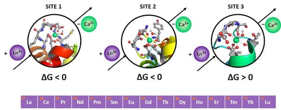
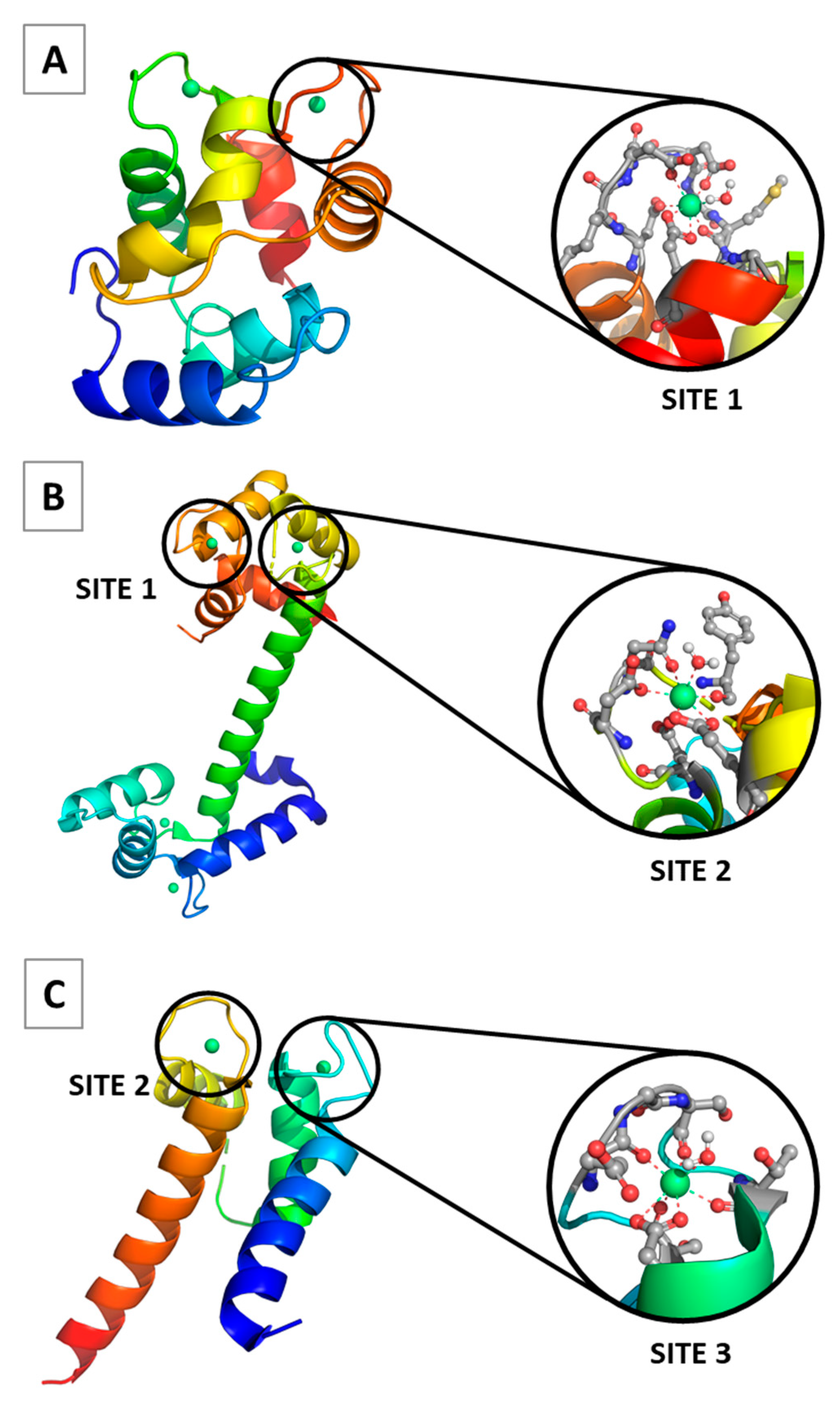
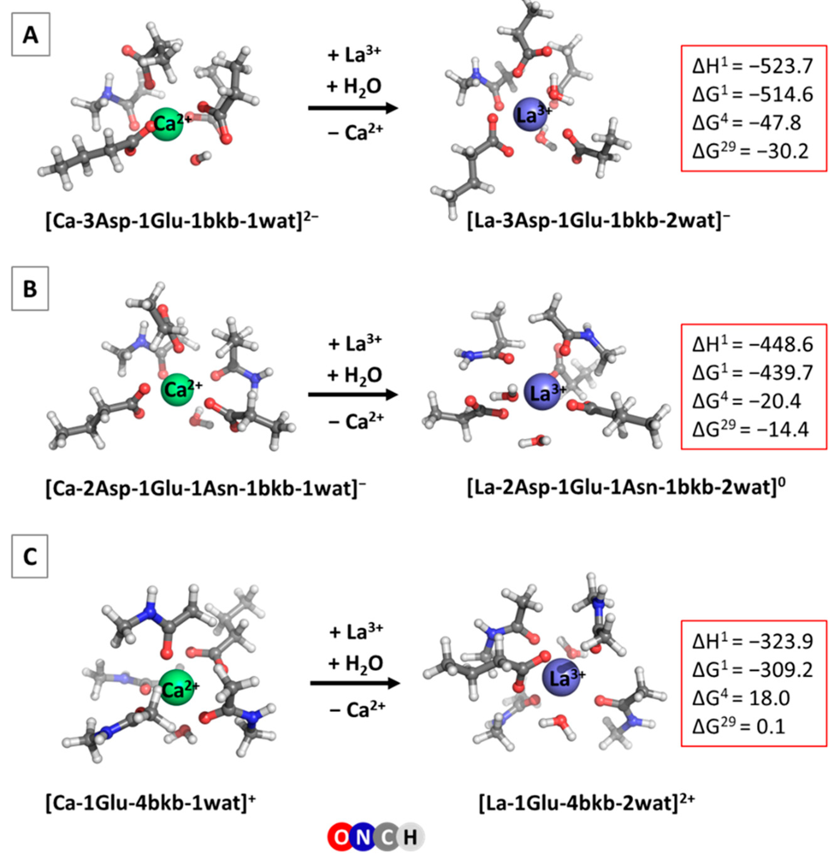
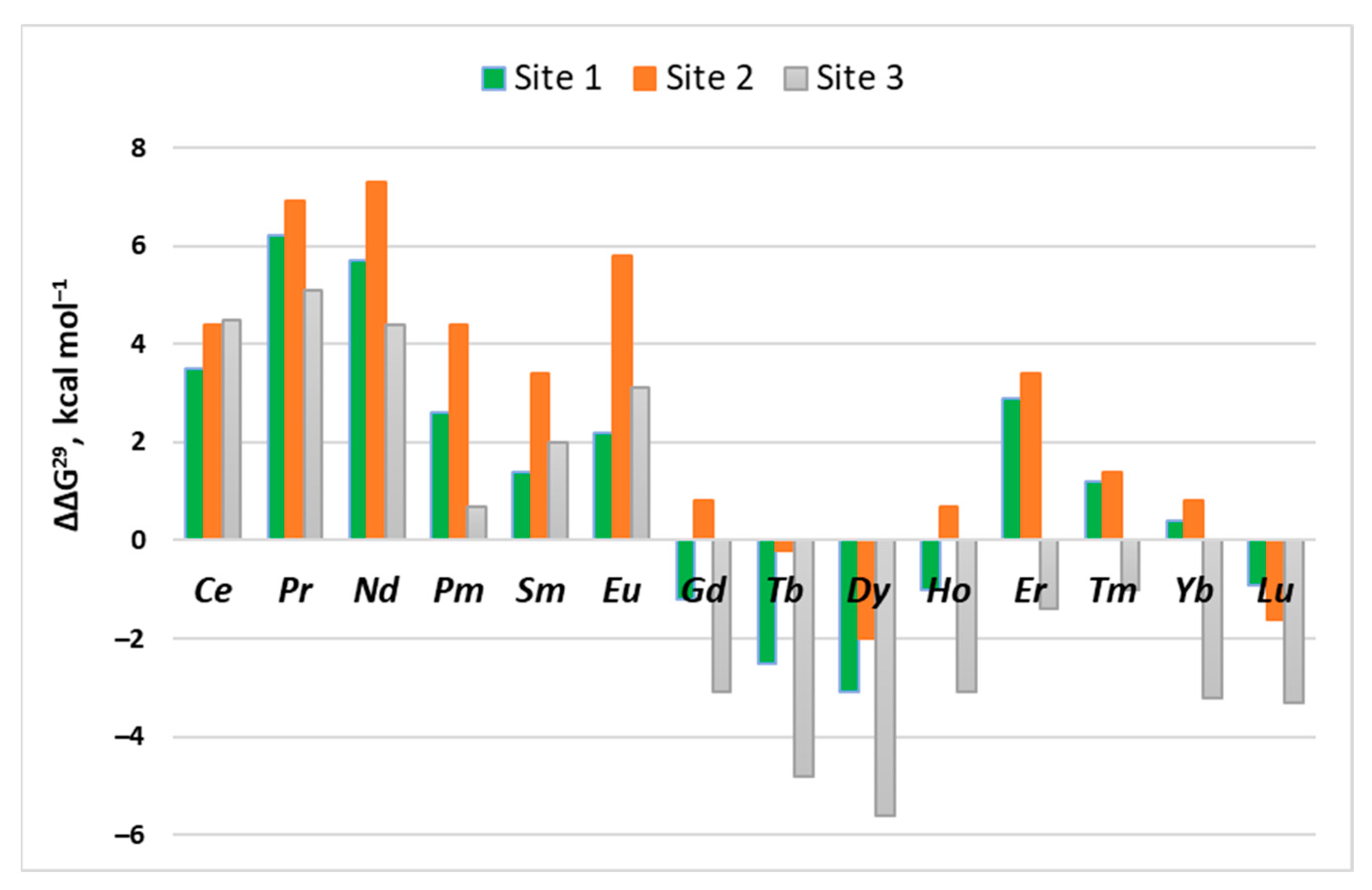
Several calcium-binding sites characteristic of Ca2+-signaling/buffering proteins were modeled, and their metal selectivity was examined. These are designated as Site 1, Site 2 and Site 3, whose optimized Ca2+/La3+-loaded structures, following the experimental observations (Figure 1), are presented in Figure 2.

2.1. Site 1

This is a canonical EF-hand motif binding site comprising three Asp- and one Glu- side chains, and a peptide backbone group donated by the host protein. Such calcium-selective binding sites (with respect to other “native” cytosolic cations) are typical of parvalbimun (EF site) and calmodulin (EF-I and EF-IV sites). One/two water molecules complement the first coordination shell of the metal cation (Figure 2A). Glutamate is bidentately bound to the Ca2+/La3+ cations, whereas the aspartates are coordinated to the metal in a monodentate fashion. The overall charge of the metal-free binding pocket is −4.
Structurally, the two calcium/lanthanum-loaded binding sites differ mostly in their shape and metal coordination number (CN), as Ca2+ cation prefers heptacoordination, whereas lanthanum favors higher coordination numbers in biological systems, CN = 8 in particular [21,22]. The metal–ligand bond distances are not, however, very different; the mean of Ca2+-O (ligand) distance is 2.43 Å, while the respective La3+-O (ligand) distance is 2.55 Å.
The enthalpies and Gibbs free energies for the Ca2+ → La3+ metal exchange (Equation (1)) are also presented in Figure 2A. The gas-phase thermodynamics, as seen, are enthalpy-driven, since ΔH1 is the major contributor to the free energy of the reaction. As expected, ΔG1 is quite negative (−515.6 kcal mol−1), signifying very favorable substitution of the dicationic Ca2+ by the tricationuc La3+ in the gas phase. The solvation, however, strongly attenuates the gas-phase free energy as the desolvation penalty of the incoming La3+ species (−751.7 kcal mol−1), and strongly outweigh the free energy gain by the outgoing Ca2+ (−359.7 kcal mol−1) [44]. Nevertheless, ΔGs in the condensed phase remain negative, implying a calcium-binding site vulnerable to the La3+ substitution. Buried binding sites (ε = 4) are more predisposed to lanthanum attack (ΔG4 = −47.8 kcal mol−1) than their solvent-exposed counterparts (ΔG29 = −30.2 kcal mol−1). The trends outlined above for the Ca2+ → La3+ substitution are followed by those for the Ca2+ → Ln3+ exchange as well, where the gas-phase free energies vary between −524.6 and −613.0 kcal mol−1, and the ΔG4/ΔG29 fluctuate between −41.6 and −50.7/−24.0 and −33.3 kcal mol−1, respectively (Table 1).
Tracking the changes in the free energies of metal exchange within the lanthanide series is of particular interest. The following reaction was considered:
[Ln3+-aq] + [La3+-protein] → [Ln3+-protein] + [La3+-aq]
and the respective La3+/Ln3+ relative free energies, ΔΔGs, were evaluated (Table 1). The gas-phase ΔΔG steadily decreases going down the series (from −10.0 for Ce3+ to −98.4 kcal mol−1 for Lu3+) as the charge density increases in the same direction (due to decreasing ionic radius; see Section 3.2, Table 4), thus securing more favorable interactions with the host binding side for lanthanides rather than lanthanum. The free energies of solvation of lanthanides, however, also decrease in the same direction (incurring higher desolvation penalties) and strongly affect the outcome of the La3+ → Ln3+ competition in condensed media. This apparently depends on the balance between the electronic and solvation effects. As can be seen in the second half of Table 2 and the green columns of Figure 3, ΔΔGs do not follow linear dependency but fluctuate between positive and negative values, with the lowest (most favorable) being for Dy3+ (ΔΔG4/ΔΔG29 = −2.9/−3.1 kcal mol−1) and the highest (least favorable) for Pr3+ (ΔΔG4/ΔΔG29 = 6.2/6.2 kcal mol−1). Interestingly, ΔΔGs distribution seems to follow a semi-sinusoidal pattern (Figure 3; green columns) with a maximum (Pr3+), followed by a minimum (Dy3+) followed by another (less pronounced) maximum (Er3+).

2.2. Site 2

This is a typical EF-hand calcium binding site found in calmodulin (EF-II and EF-III), recoverin (EF-II and EF-III) and S100P (EF-II). The hepta-coordinated Ca2+ complex includes two Asp- (monodentately bound to the metal), one Glu- (bidentately bound), one Asn, a backbone peptide group and a water molecule. The La3+ cation binds to the same set of ligands, with the addition of another water molecule complementing its coordination number to 8. The optimized structures of the Ca2+ and La3+ constructs are presented in Figure 2B. The metal-free binding site bears a negative charge of −3.
The thermodynamic characteristics of the Ca2+ → La3+ metal exchange are also given in Figure 2B. It can be seen that, as in the above case (Site 1), the reaction is enthalpically driven. The metal substitution appears, again, to be highly favorable in both the gas phase and condensed media. However, the Gibbs free energies are higher (less negative) than the respective quantities in Site 1 (Figure 2A), which implies decreased competitiveness of La3+ in Site 2 compared with that in Site 1. This is mainly due to the decreased negative charge density in Site 2 (−3) as compared to that in Site 1 (−4), which affects (unfavorably) the La3+-ligand interactions to a greater extent than the respective Ca2+-ligand interactions.
The Gibbs free energies for the Ca2+ → Ln3+ exchange in the lanthanide series are summarized in the first half of Table 2. These fluctuate in certain limits but remain on a negative ground for the entire series, implying that all the lanthanides are effective Ca2+ competitors in this type of binding site. Furthermore, examining ΔΔG4/29 for the La3+ → Ln3+ substitution, one can conclude that very few of the lanthanides, such as Tb3+, Dy3+ and Lu3+, can successfully compete with La3+ for Site 2 (second half of Table 2 and Figure 3, orange columns), as evidenced by negative values for the three cations but positive values for all the other lanthanides. The positive values for the respective lanthanides are the highest in the series of the three binding sites examined here (Figure 3 and Table 1, Table 2 and Table 3).

2.3. Site 3

Site 3 represents the calcium binding site found in S100P protein (EF-I). It has a low negative charge density, comprising one bidentate anionic Glu-, four neutral backbone peptide groups and one or two water molecules, depending on the metal. The structure of the optimized Ca2+ and La3+ complexes and thermodynamic parameters for the metal competition in this system are shown in Figure 2C.
As expected, in view of the low negative charge density of Site 3 (the overall charge of the metal free binding pocket is −1), the gas-phase free energy gain upon Ca2+ → La3+ substitution decreases further in absolute value as compared with those in Site 1 and Site 2. This weighs heavily on the condensed-phase ΔG4/29, which now are positive, implying calcium binding sites resistant to lanthanum attack.
The free energies of Ca2+ → Ln3+ exchange in the buried binding sites (ε = 4) remain positive throughout the entire lanthanide series as well (Table 3, left-hand side), but change sign in solvent-accessible binding pockets (ε = 29) for metals from the second half of the series (heavier lanthanides). The heavier lanthanides are also better competitors of the namesake La3+ in solvent-exposed binding sites characterized by negative ΔΔG4/29 (Table 3, second half and Figure 3, gray columns). Again, Dy3+ appears to be the lanthanide with the highest affinity for the protein-binding site.

3. Materials and Methods

3.1. Models Used

Model Ca2+ binding sites were built in accordance with the existing X-ray structures of the respective signaling/buffering proteins deposited in the PDB: 1PAL [22] (parvalbumin), 4DJC [23] (calmodulin), 4YI8 [45] (recoverin) and 1J55 [24] (S100P). The side chains of Asp−, Glu− and Asn were represented by CH3CH2COO, CH3CH2CH2COO and CH3CH2CONH2, respectively, whereas the metal-coordinated backbone peptide group was modeled by N-methylacetamide (CH3CONHCH3). The initial model seven-coordinated Ca2+-bound constructs were subjected to geometry optimization (see below). Consequently, Ca2+ from the optimized structures was replaced by a La3+/Ln3+ cation, followed by a water molecule addition (to complement the lanthanide coordination number to 8), and the resulting constructs were fully optimized. The optimized structures of Ca2+ and La3+ complexes are shown in Figure 2. Notably, the overall structure of the respective Ln3+ complexes does not differ significantly from that of the La3+ counterpart, and thus these are not depicted here. An oxidation state of 3+, typical of the lanthanum and lanthanides, was considered throughout the paper.

3.2. DFT/PCM Calculations
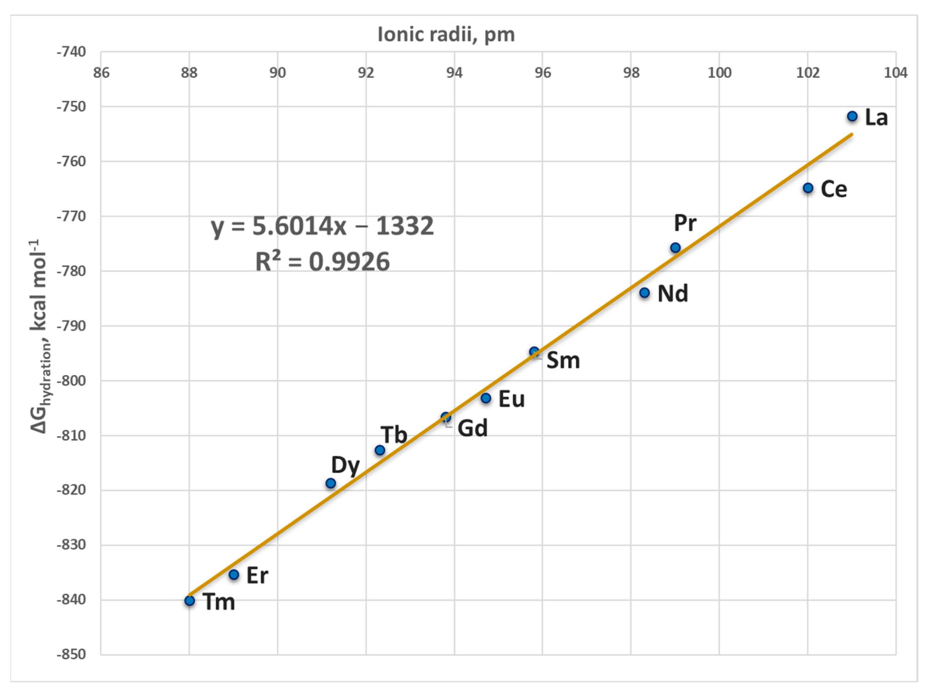
The M062X method [46] in combination with Pople’s triple zeta 6-311++G(d,p) basis set augmented with polarization and diffuse functions for C, H, N, O and Ca atoms, and an SDD basis set/effective core potential for Ln3+ (see Table 4) was employed in the calculations. This combination of theoretical method/basis set has been meticulously calibrated/validated in several of our previous studies with respect to available experimental data, and has proven to be dependable, as it reliably reproduced the structure of a series of representative metal constructs [47] as well as the Gibbs free energies of metal substitution in acetate, imidazole and glycine complexes [48]. The M062X/6-311++G(d,p)//SDD level of theory has been recently used in the study of the competition between Ag+ and Ni2+ in nickel enzymes, and between Ag+ and the key constituents of the bacterial cell wall/membrane [49,50].
Each metal-bound structure was optimized in the gas phase by employing a Gaussian 09 suite of programs [51]. Electronic energies, Eel, were evaluated for each optimized construct. Consequent frequency evaluations were conducted at the same M062X/6-311++G(d,p)//SDD level of theory to confirm a local minimum on the potential energy surface; no imaginary frequency was detected for any of the structures considered. The frequencies were scaled by an empirical factor of 0.983 [46] and used to calculate the thermal energies, Eth, comprising zero-point energy and entropies, S. The differences ΔEel, ΔEth and ΔS between the products and reactants in Equation (1) were used to evaluate the metal substitution Gibbs free energy in the gas phase, ΔG1, at T= 298.15 K, according to:
ΔG1 = ΔEel1 + ΔEth1 − TΔS1
The basis set superposition error for this type of reactions has been found to be insignificant [52], and thus was not taken into account in the present calculations.
The condensed-phase evaluations were conducted in solvents mimicking the dielectric properties of buried and solvent-accessible binding sites, diethyl ether (ε = 4) and propanonitrile (ε = 29), respectively. Solvation effects were simulated by performing PCM computations employing the solvation model based on density (SMD) scheme [31,53] as implemented in the Gaussian 09 program. In so doing, the optimized gas-phase structure of each metal complex was subjected to single point evaluations in the respective solvent at M062X/6-311++G(d,p)//SDD level of theory. The difference between the gas-phase and SMD energies was used to calculate the solvation free energy, ΔGsolvε, of each metal complex. The outgoing Ca2+ and incoming Ln3+ are considered to be in a bulk aqueous environment (ε = 78) outside the binding pocket. Accordingly, their experimentally evaluated/estimated hydration free energies of −359.7 kcal mol−1 (for Ca2+ [44] and Ln3+ (from −751.7 to −849.7 kcal mol−1, see Table 4 below), respectively were used in the computations. The experimental hydration free energy of water of −6.3 kcal mol−1 [54] was employed as well. The cation exchange free energy in a protein-binding pocket characterized by an effective dielectric constant was evaluated as
∆Gε = ∆G1 + ∆Gsolvε ([Ln3+-protein]) − ∆Gsolvε ([Ca2+-protein]) − ∆Gsolv78 ([Ln3+-aq]) + ∆Gsolv78 ([Ca2+-aq])
Note that Equation (4) allows us to separate electronic effects (interactions between the metal cation and ligands lining the binding pocket) from other effects such as the solvent accessibility and the effective dielectric constant of the active center, which are controlled by the protein matrix.
Table 4. Ionic radii, hydration free energies and effective core potentials for lanthanides.
Metal CationIonic Radius a (Å) ΔGhydration (kcal mol−1) bECP
La3+1.03−751.7MWB46
Ce3+1.02−764.8MWB47
Pr3+0.99−775.6MWB48
Nd3+0.983−783.9MWB49
Pm3+0.970−788.7 cMWB50
Sm3+0.958−794.7MWB51
Eu3+0.947−803.1MWB52
Gd3+0.938−806.6MWB53
Tb3+0.923−812.6MWB54
Dy3+0.912−818.6MWB55
Ho3+0.901−827.3 cMWB56
Er3+0.89−835.3MWB57
Tm3+0.88−840.1MWB58
Yb3+0.868−845.8 cMWB59
Lu3+0.861−849.7 cMWB60
a From Greenwood and Earnshaw, 1997 [55]. b Experimentally determined hydration free energies (from Marcus [44]) unless indicated. c Estimated from the ΔGhydration/Ionic radius correlation (R2 = 0.9926); Figure 4.

4. Conclusions

The present research offers the first systematic investigation of the ability of the entire series of lanthanides to substitute for Ca2+ in biological systems (to the best of our knowledge). The calculations performed reveal that the major determinant of the Ca2+/Ln3+ selectivity in calcium proteins is the net charge of the calcium binding pocket; the more negative the charge, the higher the competitiveness of the trivalent Ln3+ with respect to its Ca2+ contender (Figure 2). The solvent exposure of the binding site also influences the process; buried active centers with net charge of −4 or −3 are characterized by higher Ln3+ over Ca2+ selectivity (Figure 2A,B), whereas it is the opposite for sites with overall charge of −1 (Figure 2C). These findings are in line with our earlier results on Ca2+ → La3+ competition [35].
Within the series, the competition between La3+ and its fellow lanthanides is determined by the balance between two competing effects: electronic (favoring heavier lanthanides) and solvation (generally favoring the lighter lanthanides). The dependency is not straightforward, as ΔΔG4/29 fluctuate between positive and negative values, following semi-sinusoidal-like distribution (Figure 3). The calculations demonstrate that lighter lanthanides (especially Ce3+, Pr3+ and Eu3+) are poor competitors to their namesake lanthanum, whereas their counterparts from the middle of the series (Gd3+, Tb3+ and Dy3+) are predicted to have greater affinity to the binding pocket than lanthanum. The dielectric properties of the binding cavity only slightly affect the La3+/Ln3+ competition, since ΔΔG4 and ΔΔG29 alternate in relatively narrow limits (Table 1, Table 2 and Table 3).

Author Contributions

Conceptualization, T.D.; investigation, V.N., N.K., S.D., S.A. and T.D.; writing—original draft preparation, T.D.; writing—review and editing, N.K., S.A. and T.D.; visualization, S.D. and S.A.; supervision, T.D.; All authors have read and agreed to the published version of the manuscript.

Funding

This research received no external funding.

Institutional Review Board Statement

Not applicable.

Informed Consent Statement

Not applicable.

Data Availability Statement

The data presented in this study are available on request from the corresponding author.

Acknowledgments

The authors gratefully acknowledge the provided access to the e-infrastructure of the NCHDC–part of the Bulgarian National Roadmap for RIs, with the financial support by the Grant No D01−168/28.07.2022.

Conflicts of Interest

The authors declare no conflict of interest.

References

  1. Bruno, J.A.; Horrocks, W.D.W.; Zauhar, R.J. Europium(III) Luminescence and Tyrosine to Terbium(III) Energy-Transfer Studies of Invertebrate (Octopus) Calmodulin. Biochemistry 1992, 31, 7016–7026. [Google Scholar] [CrossRef] [PubMed]
  2. Wang, C.L.A.; Leavis, P.C.; Horrocks, W.D.W.; Gergely, J. Binding of Lanthanide Ions to Troponin C. Biochemistry 1981, 20, 2439–2444. [Google Scholar] [CrossRef] [PubMed]
  3. Chaudhuri, D.; Horrocks, W.D.; Amburgey, J.C.; Weber, D.J. Characterization of Lanthanide Ion Binding to the EF-Hand Protein S100 Beta by Luminescence Spectroscopy. Biochemistry 1997, 36, 9674–9680. [Google Scholar] [CrossRef]
  4. Drakenberg, T.; Swärd, M.; Cavé, A.; Parello, J. Metal-Ion Binding to Parvalbumin. A 113Cd-n.m.r. Study of the Binding of Different Lanthanide Ions. Biochem. J. 1985, 227, 711. [Google Scholar] [CrossRef] [Green Version]
  5. Edington, S.C.; Gonzalez, A.; Middendorf, T.R.; Halling, D.B.; Aldrich, R.W.; Baiz, C.R. Coordination to Lanthanide Ions Distorts Binding Site Conformation in Calmodulin. Proc. Natl. Acad. Sci. USA 2018, 115, E3126–E3134. [Google Scholar] [CrossRef] [Green Version]
  6. Bertini, I.; Gelis, I.; Katsaros, N.; Luchinat, C.; Provenzani, A. Tuning the Affinity for Lanthanides of Calcium Binding Proteins. Biochemistry 2003, 42, 8011–8021. [Google Scholar] [CrossRef]
  7. Barbieri, R.; Bertini, I.; Cavallaro, G.; Lee, Y.M.; Luchinat, C.; Rosato, A. Paramagnetically Induced Residual Dipolar Couplings for Solution Structure Determination of Lanthanide Binding Proteins. J. Am. Chem. Soc. 2002, 124, 5581–5587. [Google Scholar] [CrossRef] [PubMed]
  8. Mulqueen, P.; Tingey, J.M.; Horrocks, W.D.W. Characterization of Lanthanide(III) Ion Binding to Calmodulin Using Luminescence Spectroscopy. Biochemistry 1985, 24, 6639–6645. [Google Scholar] [CrossRef]
  9. Buccigross, J.M.; Nelson, D.J. EPR Studies Show That All Lanthanides Do Not Have the Same Order of Binding to Calmodulin. Biochem. Biophys. Res. Commun. 1986, 138, 1243–1249. [Google Scholar] [CrossRef]
  10. Rhee, M.-J.; Sudnick, D.R.; Arkle, V.K.; Horrocks, W.D. Lanthanide Ion Luminescence Probes. Characterization of Metal Ion Binding Sites and Intermetal Energy Transfer Distance Measurements in Calcium-Binding Proteins. 1. Parvalbumin. Biochemistry 1981, 20, 3328–3334. [Google Scholar] [CrossRef]
  11. Lee, L.; Sykes, B.D. Strategies for the Uses of Lanthanide NMR Shift Probes in the Determination of Protein Structure in Solutio. Application to the EF Calcium Binding Site of Carp Parvalbumin. Biophys. J. 1980, 32, 193–210. [Google Scholar] [CrossRef] [Green Version]
  12. Kupke, D.W.; Fox, J.W. Volume Changes in the Binding of Lanthanides to Peptide Analogues of Loop II of Calmodulin. Biochemistry 1989, 28, 4409–4415. [Google Scholar] [CrossRef] [PubMed]
  13. Bertini, I.; Donaire, A.; Jiménez, B.; Luchinat, C.; Parigi, G.; Piccioli, M.; Poggi, L. Paramagnetism-Based versus Classical Constraints: An Analysis of the Solution Structure of Ca Ln Calbindin D9k. J. Biomol. NMR 2001, 21, 85–98. [Google Scholar] [CrossRef]
  14. Cotruvo, J.A.J. The Chemistry of Lanthanides in Biology: Recent Discoveries, Emerging Principles, and Technological Applications. ACS Cent. Sci. 2019, 5, 1496–1506. [Google Scholar] [CrossRef] [Green Version]
  15. Mattocks, J.A.; Cotruvo, J.A.; Deblonde, G.J.-P. Engineering Lanmodulin’s Selectivity for Actinides over Lanthanides by Controlling Solvent Coordination and Second-Sphere Interactions. Chem. Sci. 2022, 13, 6054–6066. [Google Scholar] [CrossRef]
  16. Mattocks, J.A.; Tirsch, J.L.; Cotruvo, J.A. Chapter Two—Determination of Affinities of Lanthanide-Binding Proteins Using Chelator-Buffered Titrations. In Rare-Earth Element Biochemistry: Characterization and Applications of Lanthanide-Binding Biomolecules; Cotruvo, J.A., Jr., Ed.; Academic Press: Cambridge, MA, USA, 2021; Volume 651, pp. 23–61. ISBN 0076-6879. [Google Scholar]
  17. Caldwell, S.J.; Haydon, I.C.; Piperidou, N.; Huang, P.-S.; Bick, M.J.; Sjöström, H.S.; Hilvert, D.; Baker, D.; Zeymer, C. Tight and Specific Lanthanide Binding in a de Novo TIM Barrel with a Large Internal Cavity Designed by Symmetric Domain Fusion. Proc. Natl. Acad. Sci. USA 2020, 117, 30362–30369. [Google Scholar] [CrossRef]
  18. Takacs, M. Adapting Proteins for Clinical and Industrial Use. Ph.D. Thesis, Syracuse University, Syracuse, NY, USA, 2018. [Google Scholar]
  19. Dudev, T.; Chang, L.-Y.; Lim, C. Factors Governing the Substitution of La3+ for Ca2+ and Mg2+ in Metalloproteins:  A DFT/CDM Study. J. Am. Chem. Soc. 2005, 127, 4091–4103. [Google Scholar] [CrossRef] [PubMed]
  20. Cates, M.S.; Berry, M.B.; Ho, E.L.; Li, Q.; Potter, J.D.; Phillips, G.N. Metal-Ion Affinity and Specificity in EF-Hand Proteins: Coordination Geometry and Domain Plasticity in Parvalbumin. Structure 1999, 7, 1269–1278. [Google Scholar] [CrossRef] [Green Version]
  21. Gifford, J.L.; Walsh, M.P.; Vogel, H.J. Structures and Metal-Ion-Binding Properties of the Ca2+-Binding Helix-Loop-Helix EF-Hand Motifs. Biochem. J. 2007, 405, 199–221. [Google Scholar] [CrossRef] [PubMed]
  22. Declercq, J.P.; Tinant, B.; Parello, J.; Rambaud, J. Ionic Interactions with Parvalbumins: Crystal Structure Determination of Pike 4.10 Parvalbumin in Four Different Ionic Environments. J. Mol. Biol. 1991, 220, 1017–1039. [Google Scholar] [CrossRef]
  23. Sarhan, M.F.; Tung, C.C.; Van Petegem, F.; Ahern, C.A. Crystallographic Basis for Calcium Regulation of Sodium Channels. Proc. Natl. Acad. Sci. USA 2012, 109, 3558–3563. [Google Scholar] [CrossRef] [PubMed] [Green Version]
  24. Zhang, H.; Wang, G.; Ding, Y.; Wang, Z.; Barraclough, R.; Rudland, P.S.; Fernig, D.G.; Rao, Z. The Crystal Structure at 2 Å Resolution of the Ca2+-Binding Protein S100P. J. Mol. Biol. 2003, 325, 785–794. [Google Scholar] [CrossRef] [PubMed]
  25. Cotton, F.A.; Wilkinson, G. Advanced Inorganic Chemistry, 5th ed.; John Wiley & Sons: Hoboken, NJ, USA, 1988. [Google Scholar]
  26. Evans, C.H. Biochemistry of the Lanthanides; Springer: Berlin/Heidelberg, Germany, 1990. [Google Scholar]
  27. Gschneidner, K.A.; Eyring, L.; Gschneidner, K.A. Handbook on the Physics and Chemistry of Rare Earths; Gschneidner, K.A., Eyring, L., Eds.; Elsevier: Amsterdam, The Netherlands, 1991; Volume 15. [Google Scholar]
  28. Gschneidner, K.A.; Eyring, L.; Lander, G.H.; Choppin, G.R. Handbook on the Physics and Chemistry of Rare Earths; Elsevier: Amsterdam, The Netherlands, 1994; Volume 18. [Google Scholar]
  29. Shannon, R.D. Revised Effective Ionic Radii and Systematic Studies of Interatomic Distances in Halides and Chalcogenides. Acta Crystallogr. Sect. A 1976, 32, 751–767. [Google Scholar] [CrossRef]
  30. Dean, J.A. Lange’ s Handbook of Chemistry; McGraw-Hill, Inc.: New York, NY, USA, 1985. [Google Scholar]
  31. Abola, E.E.; Sussman, J.L.; Prilusky, J.; Manning, N.O. Protein Data Bank Archives of Three-Dimensional Macromolecular Structures. Methods Enzymol. 1997, 277, 556–571. [Google Scholar] [CrossRef]
  32. Pidcock, E.; Moore, G.R. Structural Characteristics of Protein Binding Sites for Calcium and Lanthanide Ions. J. Biol. Inorg. Chem. 2001, 6, 479–489. [Google Scholar] [CrossRef]
  33. Nikolova, V.; Angelova, S.; Markova, N.; Dudev, T. Gallium as a Therapeutic Agent: A Thermodynamic Evaluation of the Competition between Ga3+ and Fe3+ Ions in Metalloproteins. J. Phys. Chem. B 2016, 120, 2241–2248. [Google Scholar] [CrossRef]
  34. Dudev, T.; Mazmanian, K.; Lim, C. Factors Controlling the Selectivity for Na+ over Mg2+ in Sodium Transporters and Enzymes. Phys. Chem. Chem. Phys. 2016, 18, 16986–16997. [Google Scholar] [CrossRef] [PubMed]
  35. Dudev, T.; Lim, C. Competition between Li+ and Mg2+ in Metalloproteins. Implications for Lithium Therapy. J. Am. Chem. Soc. 2011, 133, 9506–9515. [Google Scholar] [CrossRef]
  36. Dudev, T.; Musset, B.; Morgan, D.; Cherny, V.V.; Smith, S.M.E.; Mazmanian, K.; DeCoursey, T.E.; Lim, C. Selectivity Mechanism of the Voltage-Gated Proton Channel, HV1. Sci. Rep. 2015, 5, 10320. [Google Scholar] [CrossRef] [PubMed] [Green Version]
  37. Dudev, T.; Lim, C. Ion Selectivity in the Selectivity Filters of Acid-Sensing Ion Channels. Sci. Rep. 2015, 5, 7864. [Google Scholar] [CrossRef] [PubMed] [Green Version]
  38. Dudev, T.; Lim, C. Importance of Metal Hydration on the Selectivity of Mg2+ versus Ca2+ in Magnesium Ion Channels. J. Am. Chem. Soc. 2013, 135, 17200–17208. [Google Scholar] [CrossRef] [PubMed]
  39. Dudev, T.; Lim, C. The Effect of Metal Binding on the Characteristic Infrared Band Intensities of Ligands of Biological Interest. J. Mol. Struct. 2012, 1009, 83–88. [Google Scholar] [CrossRef]
  40. Dudev, T.; Lim, C. Ion Selectivity Strategies of Sodium Channel Selectivity Filters. Acc. Chem. Res. 2014, 47, 3580–3587. [Google Scholar] [CrossRef] [PubMed]
  41. Kuppuraj, G.; Dudev, M.; Lim, C. Factors Governing Metal−Ligand Distances and Coordination Geometries of Metal Complexes. J. Phys. Chem. B 2009, 113, 2952–2960. [Google Scholar] [CrossRef] [PubMed]
  42. Dudev, T.; Lim, C. Factors Governing the Na+ vs K+ Selectivity in Sodium Ion Channels. J. Am. Chem. Soc. 2010, 132, 2321–2332. [Google Scholar] [CrossRef]
  43. Dudev, T.; Lim, C. Competition among Ca2+, Mg2+, and Na+ for Model Ion Channel Selectivity Filters: Determinants of Ion Selectivity. J. Phys. Chem. B 2012, 116, 10703–10714. [Google Scholar] [CrossRef]
  44. Marcus, Y. Thermodynamics of Solvation of Ions. Part 5.—Gibbs Free Energy of Hydration at 298.15 K. J. Chem. Soc. Faraday Trans. 1991, 87, 2995–2999. [Google Scholar] [CrossRef]
  45. Kumar, R.P.; Ranaghan, M.J.; Ganjei, A.Y.; Oprian, D.D. Crystal Structure of Recoverin with Calcium Ions Bound to Both Functional EF Hands. Biochemistry 2015, 54, 7222–7228. [Google Scholar] [CrossRef] [Green Version]
  46. Zhao, Y.; Truhlar, D.G. The M06 Suite of Density Functionals for Main Group Thermochemistry, Thermochemical Kinetics, Noncovalent Interactions, Excited States, and Transition Elements: Two New Functionals and Systematic Testing of Four M06-Class Functionals and 12 Other Function. Theor. Chem. Acc. 2008, 120, 215–241. [Google Scholar] [CrossRef] [Green Version]
  47. Dudev, T.; Cheshmedzhieva, D.; Doudeva, L. Competition between Abiogenic Al3+ and Native Mg2+, Fe2+ and Zn2+ Ions in Protein Binding Sites: Implications for Aluminum Toxicity. J. Mol. Model. 2018, 24, 55. [Google Scholar] [CrossRef]
  48. Dudev, T.; Nikolova, V. Determinants of Fe2+ over M2+ (M = Mg, Mn, Zn) Selectivity in Non-Heme Iron Proteins. Inorg. Chem. 2016, 55, 12644–12650. [Google Scholar] [CrossRef] [PubMed] [Green Version]
  49. Dobrev, S.; Kircheva, N.; Nikolova, V.; Angelova, S.; Dudev, T. Competition between Ag+ and Ni2+ in Nickel Enzymes: Implications for the Ag+ Antibacterial Activity. Comput. Biol. Chem. 2022, 101, 107785. [Google Scholar] [CrossRef] [PubMed]
  50. Kircheva, N.; Dobrev, S.; Nikolova, V.; Angelova, S.; Dudev, T. Theoretical Insight into the Phosphate-Targeted Silver’s Antibacterial Action: Differentiation between Gram (+) and Gram (−) Bacteria. Inorg. Chem. 2022, 61, 10089–10100. [Google Scholar] [CrossRef]
  51. Frisch, M.J.; Trucks, G.W.; Schlegel, H.B.; Scuseria, G.E.; Robb, M.A.; Cheeseman, J.R.; Scalmani, G.; Barone, V.; Petersson, G.A.; Nakatsuji, H.; et al. Gaussian 09, Rev. D.01; Gaussian Inc.: Wallingford, CT, USA, 2013. [Google Scholar]
  52. Dudev, T.; Lim, C. Determinants of K+ vs Na+ Selectivity in Potassium Channels. J. Am. Chem. Soc. 2009, 131, 8092–8101. [Google Scholar] [CrossRef] [PubMed]
  53. Marenich, A.V.; Cramer, C.J.; Truhlar, D.G. Universal Solvation Model Based on Solute Electron Density and on a Continuum Model of the Solvent Defined by the Bulk Dielectric Constant and Atomic Surface Tensions. J. Phys. Chem. B 2009, 113, 6378–6396. [Google Scholar] [CrossRef] [PubMed]
  54. Ben-Nairn, A.; Marcus, Y. Solvation Thermodynamics of Nonionic Solutes. J. Chem. Phys. 1998, 81, 2016. [Google Scholar] [CrossRef]
  55. Greenwood, N.N.; Earnshaw, A. Chemistry of the Elements, 2nd ed.; Butterworth-Heinemann: Oxford, UK; Sophia Forster & Andreas Frewer: Erlangen, Germany, 1997. [Google Scholar]
Figure 1. Structures of calcium proteins as given in the protein data bank: (A) parvalbumin—PDB entry 1PAL [22]; (B) calmodulin D—PDB entry 4DJC [23]; (C) S100P—PDB entry 1J55 [24]. Details about Site 1, 2, 3 are given in the text.
Ijms 24 06297 g001
Figure 2. M062X/6-311++G**//SDD fully optimized Ca2+ and La3+-loaded metal binding sites of calcium-signaling/buffering proteins: (A) Site 1, (B) Site 2 and (C) Site 3. Enthalpies and Gibbs energies (in kcal mol−1) of the Ca2+ → La3+ substitution are also given. Superscript 1 stands for the process in the gas phase, whereas superscripts of 4 and 29 signify metal exchange reactions taking place in buried and solvent-accessible binding pockets, respectively.
Ijms 24 06297 g002
Figure 3. Plot of ΔΔG29 (in kcal mol−1) for La3+ → Ln3+ exchange in the series of lanthanides in Site 1, Site 2 and Site 3.
Ijms 24 06297 g003
Figure 4. Plot of the experimental ΔGhydration vs. ionic radii for La3+/Ln3+ cations. There are no experimental data for the hydration free energies of Pm3+, Ho3+, Yb3+ and Lu3+.
Ijms 24 06297 g004
Table 1. Free energies of the Ca2+ → Ln3+ and La3+ → Ln3+ exchange (in kcal mol−1) for Site 1.
MetalCa2+ → Ln3+La3+ → Ln3+
ΔG1ΔG4ΔG29ΔΔG1ΔΔG4ΔΔG29
La3+−514.6−47.8−30.20.00.00.0
Ce3+−524.6−44.4−26.7−10.03.43.5
Pr3+−532.6−41.6−24.0−18.06.26.2
Nd3+−541.7−42.1−24.5−26.75.75.7
Pm3+−549.1−45.2−27.6−34.52.62.6
Sm3+−556.7−46.3−28.8−41.71.51.4
Eu3+−563.8−45.4−28.0−49.22.42.2
Gd3+−570.7−48.8−31.4−56.1−1.0−1.2
Tb3+−577.8−50.1−32.7−63.4−2.3−2.5
Dy3+−584.6−50.7−33.3−70.0−2.9−3.1
Ho3+−591.1−48.5−31.2−76.5−0.7−1.0
Er3+−594.9−44.4−27.3−80.33.42.9
Tm3+−601.3−46.0−29.0−86.71.81.2
Yb3+−607.8−46.8−29.8−93.21.00.4
Lu3+−613.0−48.1−31.1−98.4−0.3−0.9
Table 2. Free energies of the Ca2+ → Ln3+ and La3+ → Ln3+ exchange (in kcal mol−1) for Site 2.
MetalCa2+ → Ln3+La3+ → Ln3+
ΔG1ΔG4ΔG29ΔΔG1ΔΔG4ΔΔG29
La3+−439.7−20.4−14.40.00.00.0
Ce3+−448.7−16.1−10.0−9.04.34.4
Pr3+−457.3−13.7−7.5−17.66.76.9
Nd3+−464.9−13.1−7.1−25.27.37.3
Pm3+−472.7−15.8−10.0−33.04.64.4
Sm3+−480.4−17.1−11.0−40.73.33.4
Eu3+−485.5−14.7−8.6−45.85.75.8
Gd3+−494.9−19.8−13.6−55.20.60.8
Tb3+−501.7−20.8−14.6−62.0−0.4−0.2
Dy3+−509.5−22.5−16.4−69.8−2.1−2.0
Ho3+−515.3−19.6−13.7−75.60.80.7
Er3+−520.9−16.8−11.0−81.23.63.4
Tm3+−527.3−19.1−13.0−87.61.31.4
Yb3+−534.1−19.7−13.6−94.40.70.8
Lu3+−539.9−22.1−16.0−100.2−1.7−1.6
Table 3. Free energies of the Ca2+ → Ln3+ and La3+ → Ln3+ exchange (in kcal mol−1) for Site 3.
MetalCa2+ → Ln3+La3+ → Ln3+
ΔG1ΔG4ΔG29ΔΔG1ΔΔG4ΔΔG29
La3+−309.218.00.10.00.00.0
Ce3+−316.921.24.6−7.73.24.5
Pr3+−327.623.35.2−18.45.35.1
Nd3+−336.422.84.5−27.24.84.4
Pm3+−273.719.20.8−35.51.20.7
Sm3+−349.020.62.1−39.82.62.0
Eu3+−356.121.83.2−46.93.83.1
Gd3+−365.715.7−3.0−56.5−2.3−3.1
Tb3+−373.214.1−4.7−64.0−3.9−4.8
Dy3+−379.613.4−5.5−70.4−4.6−5.6
Ho3+−385.616.0−3.0−76.4−2.0−3.1
Er3+−392.017.6−1.3−82.8−0.4−1.4
Tm3+−396.218.1−0.9−87.00.1−1.0
Yb3+−403.915.9−3.1−94.7−2.1−3.2
Lu3+−407.815.8−3.2−98.6−2.2−3.3
Disclaimer/Publisher’s Note: The statements, opinions and data contained in all publications are solely those of the individual author(s) and contributor(s) and not of MDPI and/or the editor(s). MDPI and/or the editor(s) disclaim responsibility for any injury to people or property resulting from any ideas, methods, instructions or products referred to in the content.

