Article

Exposure to Insecticides Modifies Gene Expression and DNA Methylation in Hematopoietic Tissues In Vitro

by
María del Pilar Navarrete-Meneses
1,
Consuelo Salas-Labadía
1,
María del Rocío Juárez-Velázquez
1,
Dafné Moreno-Lorenzana
1,
Fernando Gómez-Chávez
2,
Alberto Olaya-Vargas
3 and
Patricia Pérez-Vera
1,*
1
Laboratorio de Genética y Cáncer, Instituto Nacional de Pediatría, Mexico City 04530, Mexico
2
Maestría y Doctorado en Ciencia y Tecnología de Vacunas y Bioterapéuticos, Doctorado en Ciencias en Biotecnología, Laboratorio de Enfermedades Osteoarticulares e Inmunológicas, Instituto Politécnico Nacional-ENMyH, Mexico City 07738, Mexico
3
Unidad de Trasplante de Células Hematopoyeticas y Terapia Celular, Instituto Nacional de Pediatría, Mexico City 04530, Mexico
*
Author to whom correspondence should be addressed.
Int. J. Mol. Sci. 2023, 24(7), 6259; https://doi.org/10.3390/ijms24076259
Submission received: 30 January 2023 / Revised: 3 March 2023 / Accepted: 15 March 2023 / Published: 26 March 2023
(This article belongs to the Special Issue Pesticides Exposure and Toxicity)

Abstract

:
The evidence supporting the biological plausibility of the association of permethrin and malathion with hematological cancer is limited and contradictory; thus, further studies are needed. This study aimed to investigate whether in vitro exposure to 0.1 μM permethrin and malathion at 0, 24, 48 and 72 h after cell culture initiation induced changes in the gene expression and DNA methylation in mononuclear cells from bone marrow and peripheral blood (BMMCs, PBMCs). Both pesticides induced several gene expression modifications in both tissues. Through gene ontology analysis, we found that permethrin deregulates ion channels in PBMCs and BMMCs and that malathion alters genes coding proteins with nucleic acid binding capacity, which was also observed in PBMCs exposed to permethrin. Additionally, we found that both insecticides deregulate genes coding proteins with chemotaxis functions, ion channels, and cytokines. Several genes deregulated in this study are potentially associated with cancer onset and development, and some of them have been reported to be deregulated in hematological cancer. We found that permethrin does not induce DNA hypermethylation but can induce hypomethylation, and that malathion generated both types of events. Our results suggest that these pesticides have the potential to modify gene expression through changes in promoter DNA methylation and potentially through other mechanisms that should be investigated.

1. Introduction

In recent decades, insecticide use has increased dramatically, as insecticides are employed in several human activities. Insecticides are widely used in agriculture, homes, aircraft, and hospitals. Thus, humans may potentially be exposed to these chemicals. Although multiple insecticides have been developed, pyrethroids and organophosphates are among the most commonly used [1,2,3,4,5]. The global use and exposure to pyrethroids and organophosphates are of concern because they have been associated with an increased risk of diverse diseases. Thus, the biological effects these pesticides exert on human health have attracted a great deal of interest [1].
The adverse effects insecticides can cause on humans have been associated with neurological alterations, inflammation, and cancer [3,4,6,7,8,9]. Different biological effects at the cellular and molecular levels have been reported as a consequence of pyrethroid and organophosphate exposures, including DNA damage, oxidative stress, epigenetic modifications, altered cell adhesion, endocrine disruption, apoptosis, altered proliferation/differentiation, and altered gene expression [10,11,12,13,14,15]. All these effects may underlie diseases associated with insecticide exposure, such as cancer.
Exposure to pyrethroids and organophosphates can increase the risk of developing different types of cancer, particularly hematopoietic cancer. Epidemiological studies have suggested an association of pyrethroids with multiple myeloma in adults and acute lymphoblastic leukemia in children [16]. Similarly, exposure to organophosphates has been associated with an increased risk of lymphomas, leukemia, and multiple myeloma [17,18]. Although epidemiological studies have suggested an association of pyrethroids and organophosphates with hematological cancer, the experimental evidence showing if this is plausible is controversial [7,10,16,18,19,20]. Thus, it is necessary to further explore if pyrethroids and organophosphates can induce biological events at the cellular and molecular levels that could underlie or promote cancer development.
Limited previous studies have assessed the effect of pesticides on gene expression and DNA methylation deregulation. In farmers exposed to mixtures of pesticides (dicamba, picloram, mesotrione, acetochlor, metolachlor, glyphosate, 2,4- dichlorophenoxyacetic acid (2,4-D), atrazine, aldrin, chlordane, DDT, dieldrin, heptachlor, lindane, toxaphene, and malathion), it was reported that DNA methylation changes occurred in whole blood. In this study, a total of 162 CpGs were differentially methylated in relation to 8 active ingredients (dicamba, picloram, mesotrione, aceto- chlor, metolachlor, glyphosate, atrazine, and malathion) [21]. In an additional study, it was reported that organophosphates induce changes in CpGs methylation in blood samples from controls and Parkinson’s disease patients. The most over-represented pathways were the nicotinic and muscarinic acetylcholine receptor signaling pathways [22]. These studies are particularly relevant and show that pesticides are capable of inducing DNA methylation modifications that could potentially modify gene expression. Unfortunately, these studies did not look for gene expression changes and included a questionary-based assessment of mixtures of pesticides, making it difficult to determine the effects of specific compounds. Regarding gene expression, for other pesticides such as paraquat, glyphosate and phosphonomethyl, changes in gene expression of DNA repair genes in farmers exposed to these chemicals have been reported [23]. However, in this study, permethrin and malathion were not included. An integrated bioinformatic study that included gene expression, microRNA expression, and DNA methylation dataset analysis reported that pesticides can modulate the expression levels of different genes and induce different epigenetic alterations in the expression levels of miRNAs and in the modulation of DNA methylation status. They found that two genes, SLC44A1 and FANCC, were consistently deregulated in the different datasets [24]. Together, these studies show that pesticides can induce genetic and epigenetic changes that may be linked to several health issues. However, studies assessing the effect of individual pesticides are missing. To date, there are no studies evaluating the effect of permethrin and malathion on gene expression in vitro together with changes in DNA methylation.
In this study, we assessed whether in vitro exposure to permethrin, a very common pyrethroid, and malathion, a widely used organophosphate, can induce gene expression and DNA methylation changes in hematopoietic tissues. We analyzed mononuclear cells from bone marrow and peripheral blood (BMMCs and PBMCs) exposed in vitro to 0.1 μM of permethrin and malathion at 0, 24, and 48 h after culture initiation using RNA and DNA microarrays.

2. Results

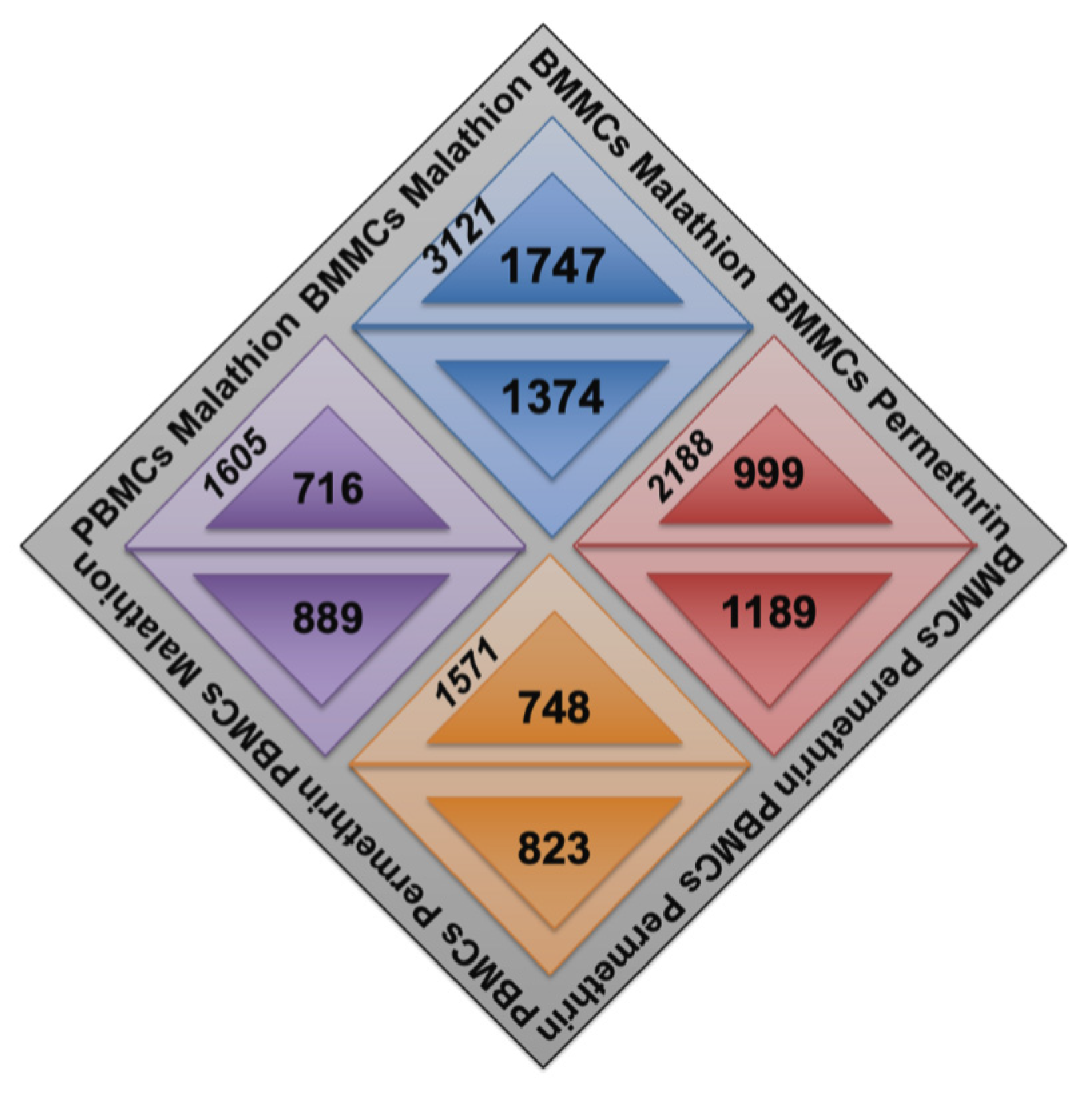
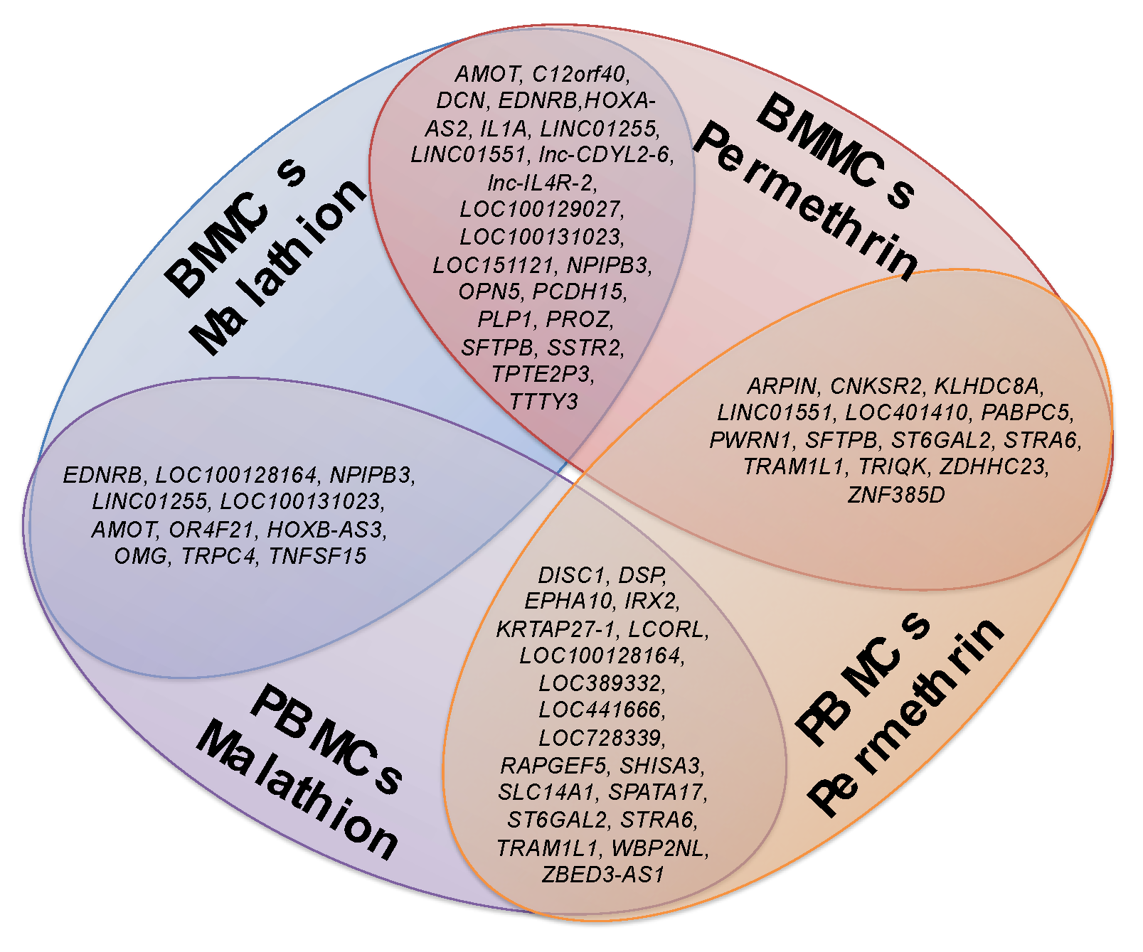
2.1. Changes in Gene Expression of BMMCs and PBMCs Exposed to Insecticides

Permethrin and malathion induced changes in gene expression compared to cells exposed to DMSO (Figure 1). A higher number of genes (3121) with altered expression was detected in BMMCs exposed to malathion, corresponding to 1747 with overexpression and 1374 with low expression. In BMMCs exposed to permethrin, 999 genes were found to be overexpressed and 1189 had low expression. Fewer genes with altered expression were observed in PBMCs compared to BMMCs (Figure 1). In cells exposed to malathion, 716 genes were found to be overexpressed and 889 to be underexpressed. BMMCs treated with permethrin showed 748 genes were overexpressed and 823 had low expression. No significant changes in cell viability were detected, all samples showed cell viabilities above 90%.
Two lists of genes were obtained: a list of the total number of genes with changes in gene expression and a list of the 100 genes with the highest and 100 with the lowest expression. Gene ontology analysis was performed on both lists.

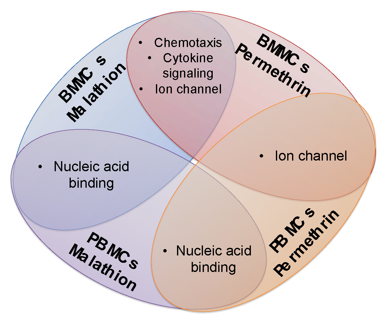
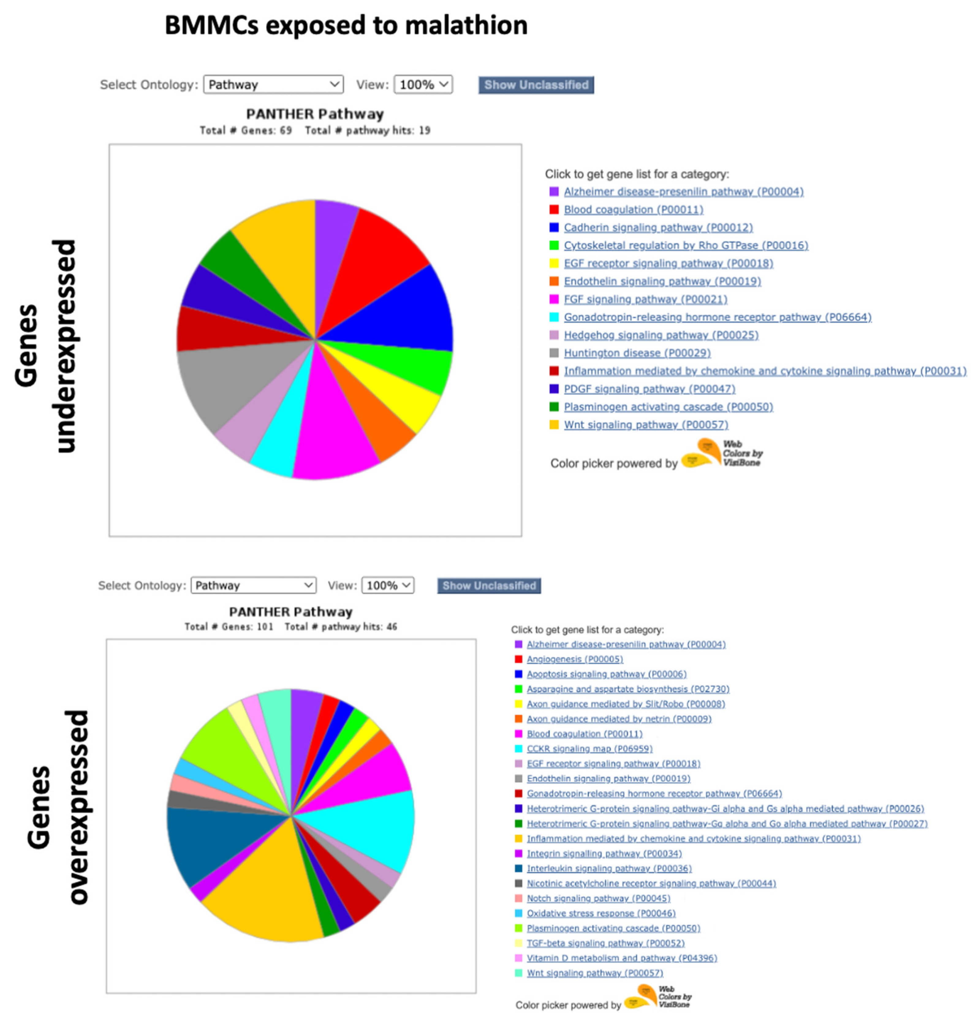
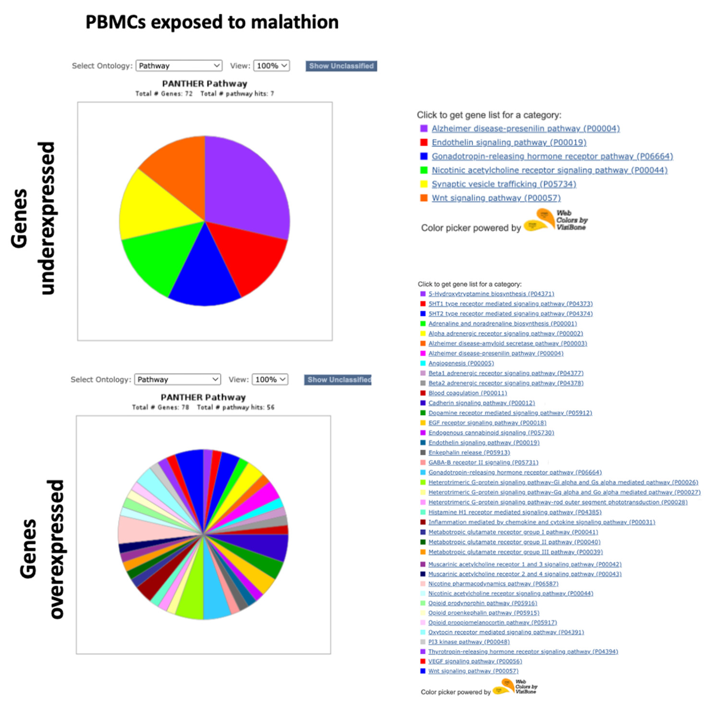
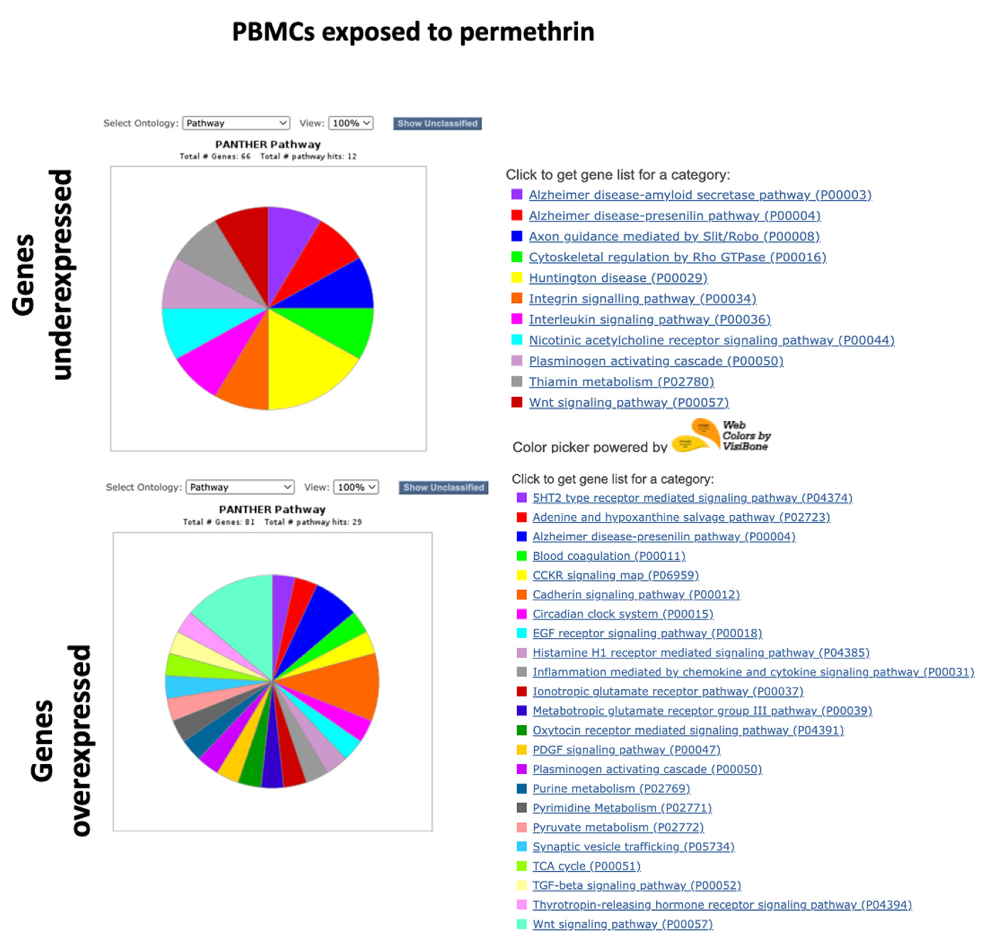
2.2. PANTHER GO Analysis with the Complete Lists of Altered Genes

An analysis with the complete lists of altered genes was performed with PANTHER and included the classifications: “biological process”, “molecular function”, “protein class”, “cell component”, and “signaling pathways”. The over-representation analysis was evaluated by Fisher’s exact test. Supplementary Tables S1–S4 show a summary of the results with statistical significance (p < 0.05) according to the over-representation test using the total of the genes.
More significant results were obtained in cultures of BMMCs exposed to malathion, mainly when analyzing overexpressed genes. We observed an over-representation of cytokine and chemokine-mediated inflammation function, interleukin signaling, and cholecystokinin receptor (CCKR) signaling (Supplementary Table S1). Additionally, an enrichment of diverse biological processes was observed, including leukocyte chemotaxis, kinase activity regulation, tumor necrosis growth factor cell response (TNF), ERK1/ERK2 cascade positive regulation, MAPK positive regulation, cell communication regulation, epidermal growth factor signaling, cytokine signaling, inflammatory response, and cell adhesion (Supplementary Table S1, Figure 2). In addition, in PBMCs exposed to malathion, we observed that underexpressed genes corresponded mostly to proteins with nucleic acid binding activity. We found that the overexpressed genes participated mainly in biological processes such as glutamatergic synaptic transmission, glutamate receptor signaling, synaptic signaling, and cell–cell signaling (Supplementary Table S2, Figure 2).
Analysis of BMMCs exposed to permethrin showed that the underexpressed genes mostly related to cytokines, enzyme binding, and G protein-coupled receptor binding. The most represented biological processes were chemotaxis and cytokine-mediated signaling. In addition, the overexpressed genes corresponded mainly to receptors with transmembrane signaling activity. The most represented pathway was the glutamate ionotropic receptor pathway (Supplementary Table S3, Figure 3). In addition, the analysis of PBMCs exposed to permethrin showed that the underexpressed genes corresponded mainly to cation channels and participated in cadherin signaling, ionotropic glutamate receptor, and Alzheimer–presenilin pathways. Overexpressed genes corresponded mainly to proteins with tyrosine kinase, ion channel, and RNA binding activities (Supplementary Table S4, Figure 3).
Coincidences were observed regarding the most over-represented molecular functions, biological processes, and pathways between BMMCs and PBMCs exposed to the same insecticide. Coincidences between the same type of tissue exposed to a different insecticide were also found (Figure 4). In both types of tissues, malathion deregulated genes that code proteins with nucleic acid binding activity, and permethrin affected the expression of genes that code ion channels. Additionally, we detected that in BMMCs, malathion and permethrin deregulated genes involved in chemotaxis, cytokine signaling, and over-representation of genes coding ion channels were identified. In PBMCs, both insecticides deregulated the expression of genes that code proteins with nucleic acid binding activity (Figure 4).

2.3. PANTHER GO Analysis with the 100 Genes with the Highest Expression and 100 with the Lowest Expression

Functional classification was performed with the PANTHER system, using the lists of the 100 genes with the highest expression and the 100 genes with the lowest expression. The “Pathways” ontology was selected and viewed in pie charts. Unclassified genes were filtered. Figure 5, Figure 6, Figure 7 and Figure 8 show the pie charts of the pathways enriched and Supplementary Tables S1–S4 show the lists of genes and their fold changes. Other ontologies analyzed included “Molecular function” and “Biological Process”, Table 1, Table 2, Table 3 and Table 4 show a summary of genes and their functions.
In the BMMCs exposed to malathion, we found that the genes with the lowest expression coded proteins with DNA binding activity, such as PROX2 and NFIB, and genes that participate in apoptosis, such as ADAM11 and ADAM32 (Table 1). Genes underexpressed in BMMCs exposed to malathion participated in pathways such as the endothelin signaling pathway, Wnt signaling pathway, cadherin signaling pathway, inflammation mediated by chemokine and cytokine, and EGF receptor signaling pathway (Figure 5). Among the genes with the highest expression due to exposure to malathion in BMMCs were the cytokines and chemokines CCL3, CCL7, CCL19, CCL1, CCL3L3, CXCL8, CXCL5, CCL1, CXCL11, and CCL20. Additionally, genes of the TNF pathway, such as TNF and TNFSF15, and the transcription factors HES4, NR4A3, and NR4A1 were overexpressed in BMMCs exposed to malathion (Table 1). Pathways enriched with genes overexpressed in BMMCs exposed to malathion included inflammation mediated by cytokines and chemokines, the Notch pathway, Wnt pathway, Interleukin signaling pathway, oxidative stress pathway, CCKR signaling map, and TGF-beta signaling pathway (Figure 5).
Among the genes with the lowest expression detected in the PBMCs exposed to malathion, transcription factors such as MEIS1, ZFP42, ARNT2, and WBP2NL were detected, as well as genes associated with cell death, such as TNFSF15 and PDIA2, and genes involved in developmental differentiation, such as GATA4 and LEP1 (Table 2). The pathways involved with the genes underexpressed in PBMCs exposed to malathion included the endothelin signaling pathway and Wnt signaling pathway (Figure 6). Among the genes with the highest expression detected in PMBCs exposed to malathion, we found those that code proteins with nucleic acid binding activity, such as ZFPM2 and IRX2; genes that code tyrosine kinase receptors, such as EPHA10, FLT4, and ERBB4, which are involved in the negative regulation of apoptosis; genes associated with epigenetic regulation, such as FAM172BP and MBD2; and genes of the MAPK pathway, such as EPHA10, FLT4, RAPGEF5, and ERBB4 (Table 2). A long list of pathways involved with genes overexpressed in PBMCs exposed to malathion were obtained, which included pathways such as cadherin, endothelin, inflammation, VEGF, and Wnt (Figure 6).
In BMMCs exposed to permethrin, genes with the lowest expression were found to be associated with antioxidant function and stress response, such as GPX8 and PROZ, as well as genes that code nucleic acid binding proteins, such as TBX15 and RBMXL2; genes of the MAPK pathway, such as FGFR and PDGFD; genes involved in cell death, such as ADAM30; genes coding proteins with transcription factor functions, such as TBX15 and GATA4; genes that code sodium channels, such as SCN9A; and genes coding proteins with cadherin activity, such as DCHS2, PCDH25, and PCDH9 (Table 3). Pathways involved with genes underexpressed in BMMCs exposed to permethrin included the cadherin, FGF, endothelin, interleukin, PI3K kinase, Wnt, and p53 signaling pathways (Figure 7). Genes that code proteins with a nucleic acid function, such as MBD2, FAM172BP, and IFIT1, were found among the genes with the highest expression in BMMCs exposed to permethrin. Additionally, genes associated with apoptosis regulation, such as BDNF, genes associated with stress response, such as FIGF, genes coding proteins with cytokine activity, such as IL15R, and genes of the TGFB pathways, such as GDF1, were among the most overexpressed genes in BMMCs exposed to permethrin (Table 3). Pathways enriched in BMMCs exposed to permethrin included the interleukin, PDGF and TGF-beta pathways (Figure 7).
Among the genes with the lowest expression detected in PBMCs exposed to permethrin were those that code nucleic acid binding proteins, such as SAMD7, LHX3, PRX, and IFIT1B; transcription factors, such as ZFP42 and WBPNL; cytokines, such as A2ML1 and IL13RA2; kinases, such as MAPK4, and cell-death-associated genes, such as LHX3 and PRUNE2 (Table 4). Pathways enriched included interleukin, nicotinic acetylcholine, and Wnt (Figure 8). Genes with the highest expression detected in PBMCs exposed to permethrin included those that code nucleic acid binding proteins, such as VGLL3; cytokines, such as TNFSF11; apoptosis-regulation-associated genes, such as EPHA10, EPHA6, TNFSF11, and PPEF1; DNA repair genes, such as HMGA2; stress response genes, such as HOXB5, FOX11, PER3, MYT1, and KDM5S; and inflammation-associated genes, such as PLCH11 (Table 4). Pathways associated with genes overexpressed in PBMCs exposed to permethrin included the CCKR, cadherin, EGF, inflammation, PDGF, ionotropic glutamate receptor, TGF-beta, and Wnt pathways (Figure 8).
Additionally, concordances were found among the altered genes in the different tissues examined, BMMCs and PBMCs, and coincidences among altered genes with the different treatments (malathion and permethrin). Figure 9 shows those genes shared between tissues and treatments.

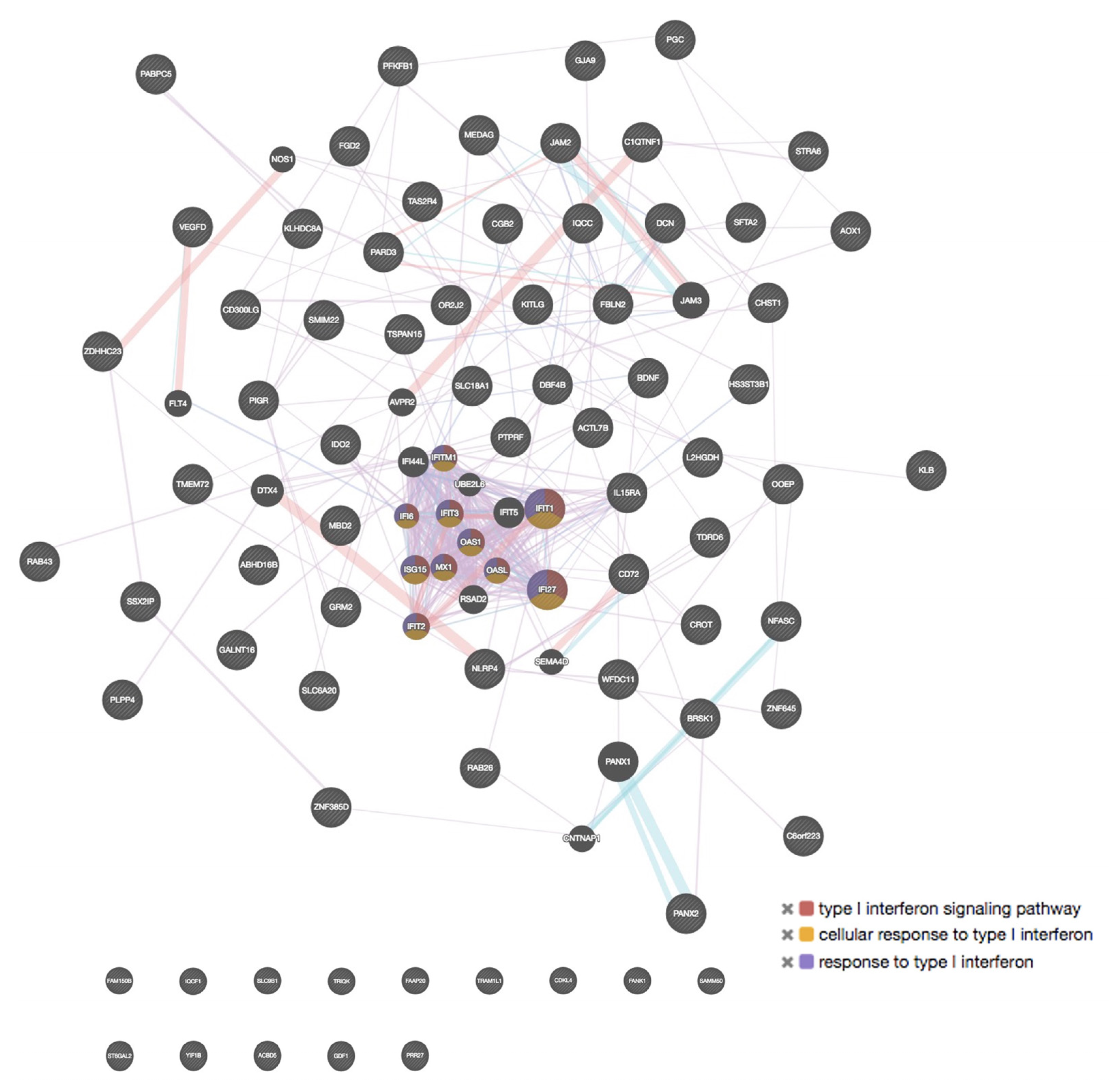
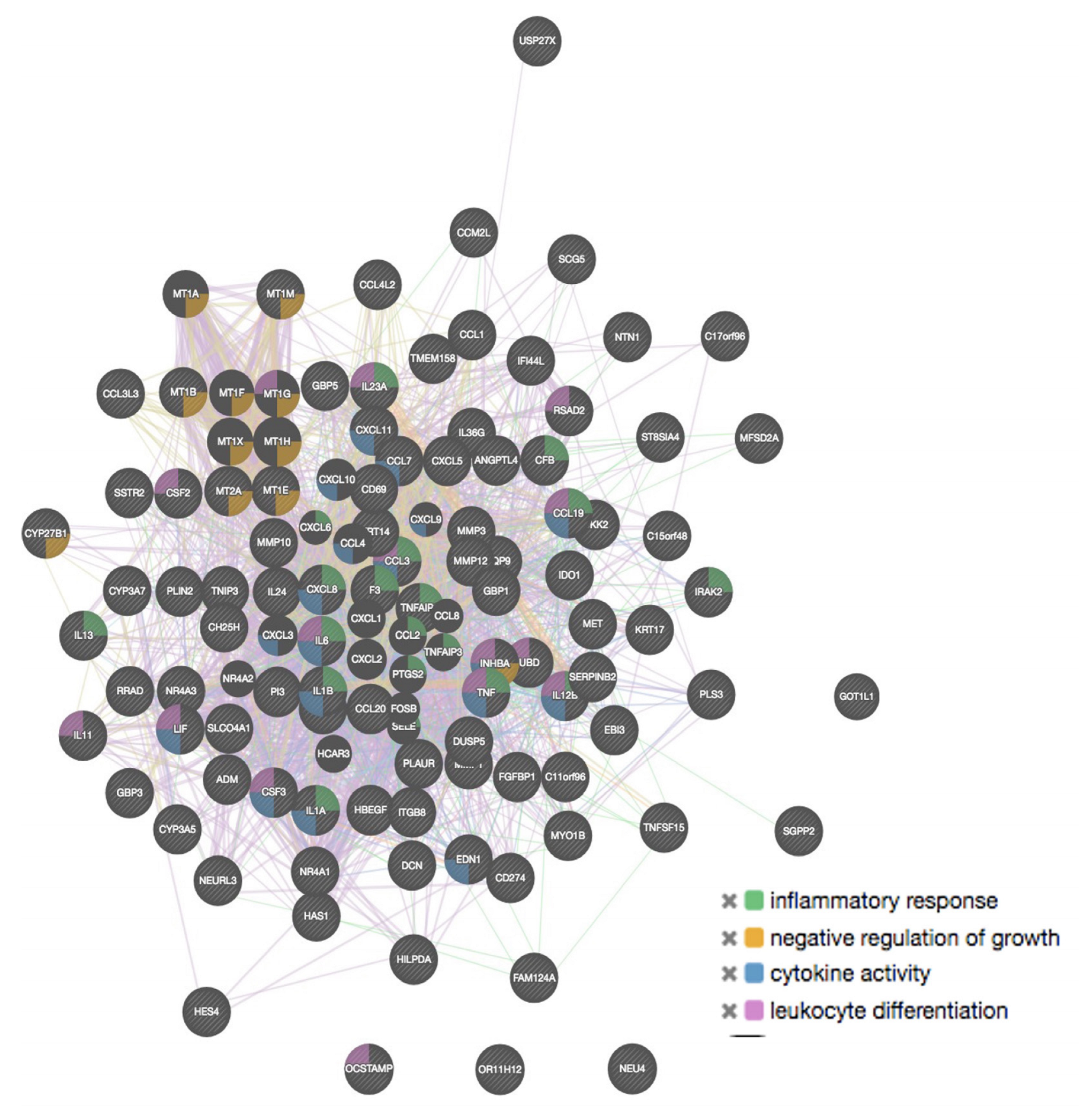
2.4. GeneMANIA Analysis

To determine the interaction among genes, molecular functions, and common pathways, an additional analysis using the GeneMANIA tool was performed, including the 100 genes with the highest and the 100 with the lowest expression. A summary of the most relevant findings is shown in Table 5 and Figure 10 and Figure 11. Regarding molecular functions, the additional results obtained with GeneMANIA were also detected with the PANTHER tool. In BMMCs exposed to malathion, we found that genes with low expression corresponded to genes associated with ion channel activity that negatively regulate differentiation and cell development. In addition, several high-expression genes are co-expressed and associated with cytokine activity, negative growth regulation, leukocyte differentiation, and inflammatory response; most of these functions were also detected with the PANTHER tool.
In PBMCs, we found through GeneMANIA analysis that genes with high expression share specific physical interactions and participate in the regulation of synaptic activity. Regarding BMMCs exposed to permethrin, we observed that several genes with elevated expression are co-expressed and participate in type 1 interferon signaling (IFN1) (Figure 10). Moreover, in PBMCs, genes with high expression showed genetic interactions associated with functions such as ROS metabolism, ephrin receptors and signaling.

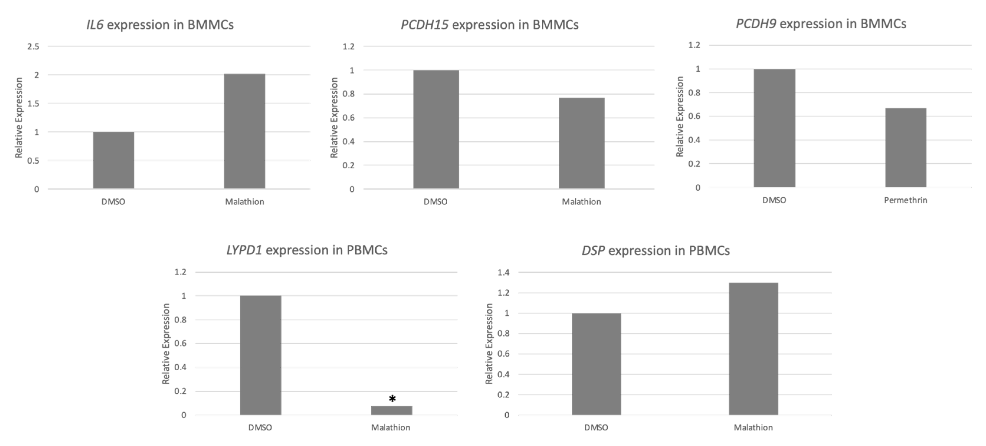
2.5. qPCR Validation

Based on the level of expression and/or relevance in cancer, five genes among the 100 with the highest and 100 with the lowest expression were selected for analysis with qPCR to corroborate microarray expression results. The gene expression of these genes was analyzed on two independent PBMC and BMMC samples from different individuals than those included for the microarrays. The qPCR analysis showed that expression tended to increase or decrease concordantly with the findings of the microarrays. Differences in the degree of increase or decrease were observed between microarray and qPCR analyses, which could be attributed to the differences between both techniques and the use of samples from different individuals. Results were significant for LYPID, in which significant underexpression was detected with malathion treatment in PBMCs (Figure 12).

2.6. Changes in DNA Methylation of BMMCs Exposed to Insecticides

Considering the results of gene expression obtained with BMMCs, we selected 100 genes for methylation analysis: 25 overexpressed genes and 25 underexpressed that were detected in cells exposed to malathion, and 25 overexpressed and 25 underexpressed that were detected in cells exposed to permethrin. These genes were selected because they were among the 100 with the highest or the 100 with the lowest expression and/or showed relevance for cancer as determined by the ontology analysis. Information was successfully obtained for 78 of the 100 selected genes from the DNA methylation microarray. The complete list of genes is shown in Supplementary Table S9. We analyzed 17 genes with underexpression and 21 with overexpression in BMMCs exposed to malathion. Additionally, we detected 24 genes with underexpression and 16 with overexpression in BMMCs exposed to permethrin. We found that 15.3% (12/78) of genes showed some degree of variation in DNA methylation on the promoter region, which was concordant with gene expression since hypermethylation coincided with gene underexpression and hypomethylation with overexpression. In general, we observed more events of hypomethylation induced by the insecticides. We identified 2/17 (11.7%) genes (KCNMA1 and ADAM11) with hypermethylation in the promoter region in BMMCs exposed to malathion concordant with underexpression results. In addition, we identified 4/21 (19%) genes (INHBA, NR4A3, ANGPTL4, and ITGB8) that showed demethylation in the promoter region that matched the overexpression induced by malathion in BMMCS. Regarding permethrin exposure, we found that this insecticide did not induce hypermethylation on the genes analyzed (0/24). On the contrary, demethylation events were frequent, affecting 6/16 (37.5%) of analyzed genes (ST6GAL2, FIGF, BDNF, PARD3, MAPK, and TDRD6), which showed demethylation on the promoter region that was concordant with overexpression in BMMCs exposed to permethrin. Table 6 shows the detected genes in each sample and the corresponding Z values.

3. Discussion

In the present study, we assessed in hematological tissues whether in vitro exposure to permethrin and malathion, two commonly used insecticides, can induce changes in gene expression and DNA methylation, which are events relevant for carcinogenesis [25]. It is important to evaluate whether permethrin and malathion can modify gene expression and if these modifications are due to changes in DNA methylation on promoter regions, since previous studies have demonstrated that environmental agents can alter DNA methylation, which can modify gene expression [13,14,26,27,28]. Moreover, in populations exposed to certain pollutants, such as ultrafine carbon particles and endocrine disruptors, gene expression and methylation have been assessed in PBMCs and characteristic expression profiles have been detected, which are considered to be footprints evidencing the exposure to these specific chemicals [29,30].
Different classes of insecticides can modify DNA methylation and expression of genes coding cytokines, chemokines, transcription factors, and signal transductors [26,27,31,32]. Although studies that have evaluated the effect of pyrethroids are very limited, it has been shown that these insecticides can alter the expression of genes such as WNT10B, THBS1, SPP1, FEZ1, and GPNM. Given the very scarce information regarding the effect of permethrin and malathion on gene expression, we aimed to evaluate in vitro whether these insecticides alter gene expression and methylation in PBMCs and BMMCs, which are hematopoietic tissues that can adequately reflect the scenario where hematological malignancies occur.
Our results showed that both insecticides modify global gene expression in PBMCs and BMMCS exposed in vitro; different results were obtained between these tissues. In general, more alterations in gene expression were observed in BMMCs, which suggests that more undifferentiated and immature cells show more susceptibility to the insecticides. In populations exposed to pesticides, it is more common to study PBMCs because of the feasibility of obtaining the sample and because it is not as invasive as other tissues [26]. However, in this study, we observed that tissues other than PBMCs could show higher susceptibility, such as BMMCs. Thus, studies may underestimate the effects of pesticides at the transcriptional level if only PBMCs are studied. On the other hand, it is important to consider that one limitation of this study is that we included PBMCs and BMMCs from different donors, and differences seen among these tissues could be related to individual biological variability. Unfortunately, in the present study we were not able to include PMBCs and BMMCs from the same donor.
The concentrations of permethrin and malathion used in this study (0.1 µM) are concentrations with biological relevance. We considered that it was of importance to include concentrations reflecting scenarios of human exposure [20]. In addition, the time of exposure of 72 h was selected as it was considered to be an optimal time for the cell culture of PMBCs and BMMCs without having cell viability decrease. We decided to apply the insecticides every 24 h to maintain the 0.1 μM concentrations and to simulate a subchronic exposure.

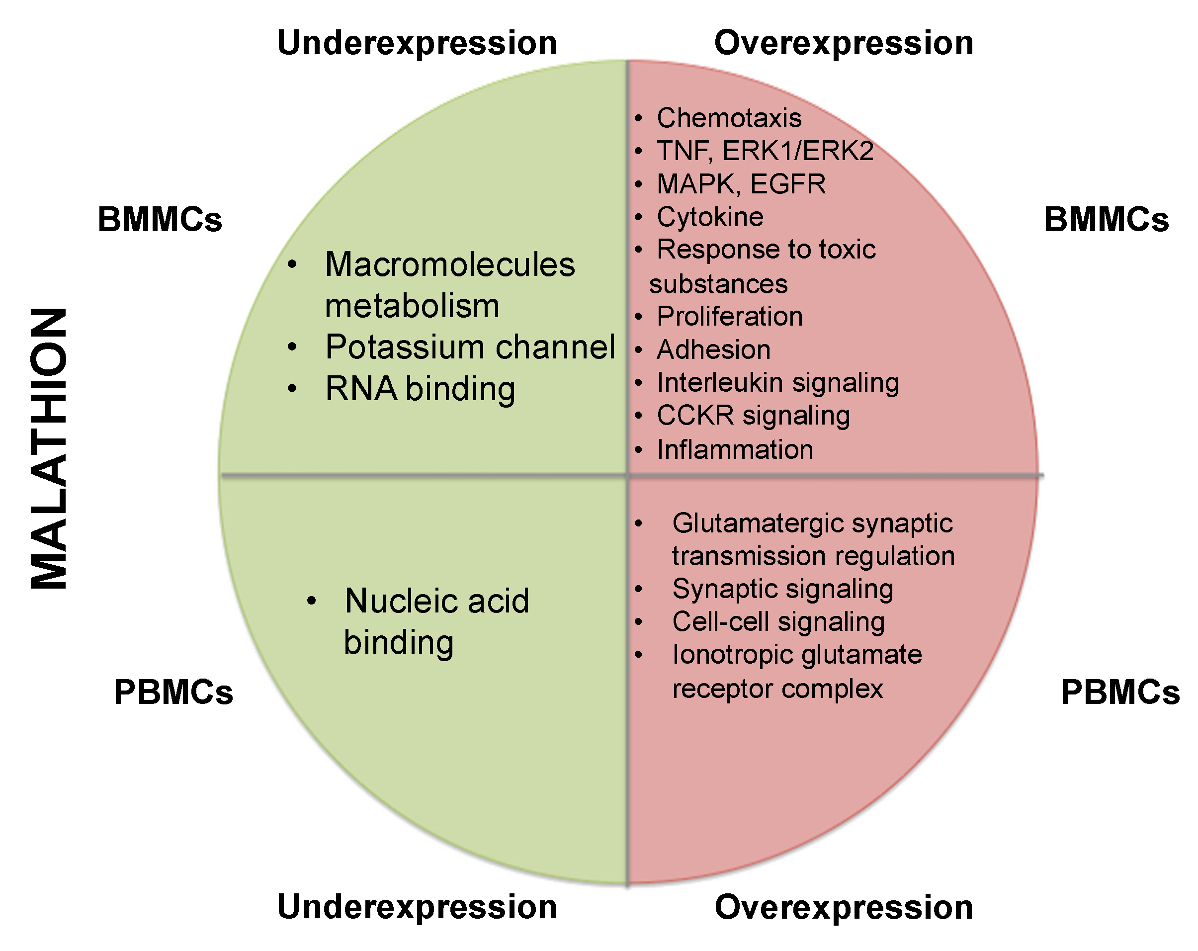
3.1. Underexpression of Genes in BMMCs Exposed to Malathion

Among the genes found to be repressed with malathion exposure in BMMCs, KCNMA1, which is associated with hypokalemia and paralysis, was detected [33,34]. We observed an over-representation of pathways involved in neuronal function, which was expected because this is a common effect of organophosphate pesticide. Several potassium channels were repressed. Besides the effect of malathion on genes associated with neurotoxicity, we detected the repression of genes relevant to cancer onset and development (Supplementary Table S11). We found low expression of the disintegrin and metalloproteinase families of the genes ADAM11 and ADAM32, which have relevance in cancer [35]. An additional family of genes repressed in BMMCs exposed to malathion is the protocadherin family. Hypermethylation of different members of the protocadherin family has been reported in cancer [36,37]. In addition, we detected repression of the EDNRB gene in BMMCs exposed to malathion, which suffers hypermethylation in different types of leukemia [38].

3.2. Overexpression of Genes in BMMCs Exposed to Malathion

Gene ontology analysis revealed that among genes with high expression in BMMCs exposed to malathion were genes coding cytokines and chemokines, proteins associated with processes such as inflammation, oxidative stress response, and response toxic substances, as well as pathways such as MAPK, Wnt, TNF, and Notch were over-represented. Additionally, the Notch and Wnt pathways were enriched when analyzing genes with the highest expression. One important finding was the induction of expression of genes involved in the inflammatory response, such as IL6, IL1B, CXCL8, CCL3, CCL7, CCL3L3, and CCL20. This result was exclusively seen in BMMCs treated with malathion, given that in PBMCs, a lower number of genes associated with inflammation was observed. Previous studies have reported that organophosphate exposure modifies chemokines and cytokines, concordant with our findings [39]. This study adds to earlier reports that show that exposure to low levels of organophosphates induces an inflammatory response [40]. Although the mechanism by which organophosphates trigger the inflammatory response has not been elucidated, it is suggested that these insecticides interact directly with inflammatory cells and trigger the release of inflammation mediators. It has been reported that exposure to malathion stimulates macrophages for generating ROS and cathepsin D, can potentiate phagocytosis and antigen presentation, and induces the release of histamine by mast cells and basophils. Among the overexpressed genes that encode pro-inflammatory cytokines, IL6 was detected, which is a relevant finding because it has a role in leukemogenesis (Supplementary Table S11) [41]. Together with the increase in this cytokine, we found an increase in TNFα and IL1A expression, which are signals that promote the production of IL6 [41,42]. This suggests that in BMMCs, the effect of malathion induces a pro-inflammatory environment by increasing the levels of TNF and IL1A, which lead to an increase in IL-6. The increase in TNF-α expression as an effect of malathion is a relevant finding because this cytokine participates in all the steps of leukemogenesis and is overexpressed in pre-B ALL [43]. It is thus relevant to study the role of malathion exposure on TNF deregulation to a greater extent in other models (Supplementary Table S11).
Additionally, among the pathways with more over-representation, we detected MAPK, ERK1/ERK2, NFkB, Notch, Wnt, and JNK, which are involved in different cell functions such as proliferation, survival, differentiation, maturation, angiogenesis, inflammatory response, and migration, and have a very important role in hematopoiesis; their participation in leukemia and other types of cancer has been reported [44,45,46,47,48].

3.3. Gene Expression Deregulation in PBMCs Exposed to Malathion

We detected that genes with underexpression corresponded to nucleic acid binding proteins, such as the transcription factor ZFP42. ZFP42 (REX1) regulates the proliferation of human mesenchymal cells through MAPK signaling [49]. In PBMCS, the expression of the MAP4K3 gene was increased; however, it remains to be investigated if the MAPK pathway deregulation occurs through the inhibition of REX1 [43], which seems to be repressed by malathion (Supplementary Table S11). Additionally, we observed underexpression of the MEIS1 gene. The repression of this gene is associated with higher levels of ROS. Malathion and other organophosphates induce ROS, and this phenomenon could be potentiated through the inhibition of MEIS1 expression [50,51]. As in BMMCs, the expression of the EDNRB gene was observed to be lowered in PBMCs exposed to malathion, which shows that in both tissues, this gene is repressed by the effect of this insecticide. This gene has shown relevance in cancer (Supplementary Table S11) [38].
In PBMCs exposed to malathion, the overexpressed genes were involved in glutamate receptor signaling, synaptic signaling, and cell–cell signaling; the pathway with the highest over-representation was the cadherin pathway. We detected overexpression of the GRIK2 gene, which is considered a tumor suppressor gene [52,53] (Supplementary Table S11). Like BMMCs, in PBMCs exposed to malathion, we identified overexpression of the Wnt pathway, which is vital in leukemia and lymphoma [54,55]. In addition, we observed the deregulation of genes involved in the cadherin pathway, apoptosis regulation, epigenetic deregulation, and inflammation; these processes have a crucial role in carcinogenesis (Supplementary Table S11) [43].

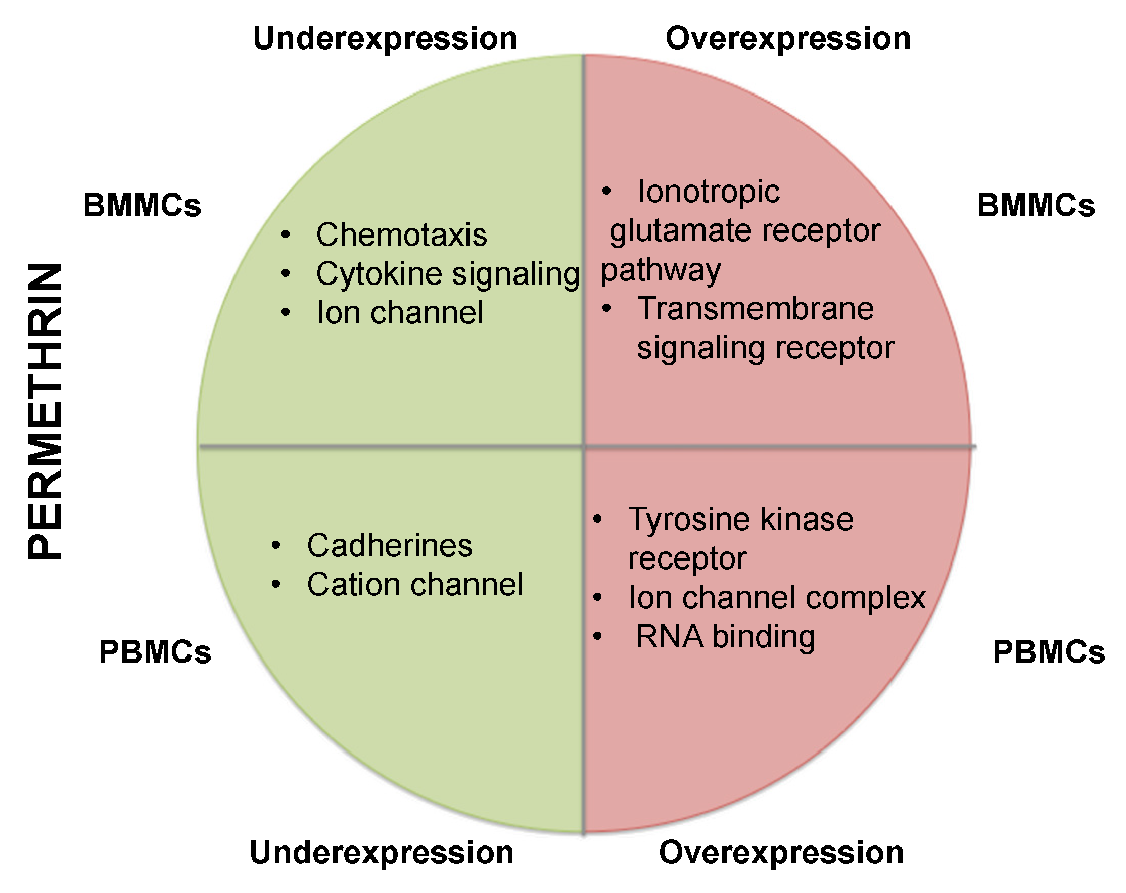
3.4. Gene Repression in BMMCs Exposed to Permethrin

With this tissue and agent, we identified a significant over-representation of repressed genes coding proteins with cytokine and chemokine function. It has been previously suggested that insecticide exposure can have an immunotoxic effect, modifying the activity of specific cell types and altering cytokine secretion [31]. This study agrees with previous reports because we detected expression modification in several genes associated with cytokine and chemokine functions. Moreover, these molecules show repressed expression in certain types of cancer. We found repression of the CXCL8 chemokine with permethrin exposure; this is an important finding because this gene can have an antitumor role [56]. However, this contrasts with the low expression of other cytokines and chemokines, such as CXCL12, that have a pro-tumoral effect. Among cytokines with a low expression, we found the OSM gene, which has the potential to inhibit myeloid leukemic cell proliferation [57]. In addition, we observed that permethrin represses the expression of the TGFB2 gene in BMMCs, which has a tumor suppressor function (Supplementary Table S11) [58,59].
Similarly, we detected low expression of the GPX8 gene in BMMCs exposed to permethrin. GPx proteins regulate oxidative stress. The low expression of GPX8 may enable permethrin toxicity through the accumulation of ROS and lipid peroxidation [60]. Another gene that showed repression was GATA4, which has been recognized as a tumor suppressor gene in different types of cancer [61]. In addition, we detected a high representation of genes repressed encoding ion channels such as SCN2A, SCNN1, SCN9A, and SCN3B, which encode voltage-dependent sodium channels. Our study shows that pyrethroids can alter ion channels at the gene expression level [62]. Additionally, we found the repression of genes coding protocadherins, such as PCDH15 and PCDH9; the deregulation of these genes is a characteristic of leukemic cells (Supplementary Table S11) [43].

3.5. Gene Overexpression in BMMCs Exposed to Permethrin

Among the genes with overexpression, we detected mainly genes of the ionotropic glutamate receptor pathway. This is concordant with previous reports and confirms that certain neuroactive insecticides, such as pyrethroids, act over ionic channels, including the ionotropic glutamate receptor [63]. Overexpression was also detected for the MBD2 nucleic acid binding gene, which can promote DNA demethylation [64]. An important finding was the overexpression of the IFIT1 gene in BMMCs exposed to permethrin; overexpression of this gene has been detected in myelodysplastic syndromes before the development of leukemia [65], and is relevant for pre-B ALL (Supplementary Table S11) [66]. IFIT1 is modulated by the JAK/STAT pathway and is an inflammation related protein that can be aberrantly expressed in cancer. The overexpression of IFIT1 was also highlighted with the Genemania analyis, in which response to IFN1 pathway was observed. IFITs are quickly induced by multiple stimuli, such as IFN dependent or IFN independent pathways and have been linked to cancer [67]. Our data agrees with previous reports that showed that the pyrethroid ivermectin induces expression of IFIT1, IFIT2, IF144, ISG20, OASL and IRF9 [68].

3.6. Gene Repression in PBMCs Exposed to Permethrin

We detected low expression of genes coding cadherins, sodium channels, and genes associated with Alzheimer–presenilin signaling (Supplementary Table S11). Some genes coding cadherins participate in the Wnt pathway, which is relevant in hematopoiesis and leukemogenesis [47]. Previous studies suggest that pyrethroids can alter Wnt pathway genes (WNT10) [69]. In this study, we found deregulation of members of the Wnt pathway family (WNT2, WNT4, WNT7A). Among the repressed genes, we found a high number of genes involved in cadherin signaling, such as CADH19, TCF7L2, and protocadherins PCDH9, PCDH15, PCDH12, PCDH114, and PCDHGA5. Protocadherins are cell–cell adhesion proteins that participate in aging and cancer. [70]. Additionally, repression of PRUNE2, which participates in cancer, was detected in PMBCs exposed to permethrin [71].
In PBMCs exposed to permethrin, we also detected repression of genes that participate in Alzheimer’s development, such as the metalloproteins MMP12, MMP2, and MMP13, among others [72]. Additionally, we found deregulated genes coding cationic channels, which have also been associated with the neurotoxicity of permethrin. This study provides relevant evidence showing that permethrin and malathion have a strong effect on genes involved in the central nervous system, which was evident even in cells of hematopoietic origin. It is possible that the topology of the regions in which the affected genes are located makes them particularly sensitive to the effect of insecticides. It is also possible that genes coding transcription factors associated with neurological processes were deregulated. In fact, with both insecticides, we found an over-representation of genes coding proteins with nucleic acid binding activity, many of which were transcription factors. We also found correlations among genes deregulated by malathion and permethrin, suggesting that although having different chemical structures, these chemicals can share mechanisms of action, which has already been reported [73]. Although metalloproteins are mainly involved in the central nervous system, the results obtained in this study and those described in the literature suggest that they can also participate in other processes, including cancer [74].

3.7. Overexpression of Genes in PMBCs Exposed to Permethrin

The over-representation analysis showed that the genes with increased expression were significantly associated with the vasopressin synthesis pathway, transmembrane receptors functions, ion channels, and nuclear processes. Regarding vasopressin synthesis, overexpression of the NEU2 gene was evidenced. It is reported that overexpression of NEU2 impedes cell growth, blocking different cell cycle checkpoints and affecting all cell cycle phases [75] (Supplementary Table S11). We also detected a significant over-representation of genes coding proteins with transmembrane receptor function, including ROS1, KIT, and ALK, which showed overexpression in PBMCs exposed to permethrin. These genes belong to the tyrosine kinase receptor family and have been identified as oncogenes in diverse types of cancer [76,77,78].

3.8. Methylation Alteration in BMMCs Exposed to Insecticides

We observed that both malathion and permethrin induce changes in gene expression. Organophosphates are reported to cause changes in DNA methylation [13]. Concordant with previous studies, we found that malathion can alter DNA methylation, and those changes matched changes in gene expression. These relevant findings support studies on exposed farmers showing changes in DNA methylation due to exposure to organophosphate mixtures [79]. We found that permethrin does not induce DNA hypermethylation; however, demethylation events were very frequent, matching the overexpression of the affected genes. However, we also found repressed genes, in which no hypermethylation on promoter region was detected, suggesting that permethrin can lead to gene silencing through alternative mechanisms different from DNA methylation. Although the effect of pyrethroids on DNA methylation has not been widely studied in human beings, murine models have reported that permethrin can induce a decrease in DNA methylation, which is concordant with our findings [28,80]. It is proposed that permethrin could induce DNA demethylation through mechanisms dependent on ROS. Previous studies have not shown that changes in DNA methylation induced by permethrin match with changes in the expression of DNMTs (DNA methyltransferases).
The present study found higher expression on DNMTs, and thus the mechanism involved remains to be elucidated. An alternative to partially explain demethylation induced by permethrin is through the MBD2 gene. We detected that permethrin could induce MBD2 gene expression, which is associated with active DNA demethylation. MBD2 can directly remove the methyl group through hydrolysis [81]. Additionally, in PBMCs exposed to permethrin, we found overexpression of the ROS1 gene, which encodes a DNA glycosylase that participates in DNA demethylation through the base excision DNA repair pathway [81]. The induction of ROS1 overexpression represents a possible pathway by which permethrin increases DNA demethylation and another possible mechanism is the activation of RELN, whose activation has been associated with demethylation in rat neurons [81]. In our study, we detected overexpression of the RELN gene in PBMCs exposed to permethrin that could contribute to DNA demethylation events.

4. Materials and Methods

4.1. Cell Culture and Treatment

Informed consent was obtained according to the recommendations of the Helsinki Declaration for all samples. The project (INP 001/2013) was approved and followed the guidelines of Research and Institutional Ethics. A 30 mL peripheral blood sample was acquired from two young, healthy, non-smoker male donors. Two 10 mL bone marrow samples, left over from the samples used for transplants, were obtained. Mononuclear cells were isolated from peripheral blood or bone marrow using a density gradient medium (Lymphoprep, Serumwerk, Bernburg, Germany) and cultured at a final density of 5–7 × 10⁵ cells/mL in triplicate, in RPMI1640 medium (Thermo Fisher Scientific, Waltham, MA, USA) supplemented with 10% fetal bovine serum (Thermo Fisher Scientific, Waltham, MA, USA), 1% non-essential amino acids (Thermo Fisher Scientific, Waltham, MA, USA), 1% sodium pyruvate (Thermo Fisher Scientific, Waltham, MA, USA), and 1% L-glutamine (Thermo Fisher Scientific, Waltham, MA, USA). Cells were cultured for 72 h at 37 °C and 5% CO2 and were treated at 0, 24, and 48 h after culture initiation with permethrin and malathion at a final concentration of 0.1 μM. Treatment with 0.2% DMSO (Merck, Darmstadt, Germany) was a negative control because it dissolved the insecticides. Cell viability was assessed with Trypan blue (Merck, Darmstadt, Germany) exclusion assay at the beginning and the end of cell culture (0 and 72 h).

4.2. Nucleic acid Extraction and Microarrays

After 72 h of culture, cells were washed with PBS 1× (Thermo Fisher Scientific, Waltham, MA, USA) and cell viability was determined by trypan blue exclusion assay. At least 1 × 10⁶ cells were separated in 300 μL of TRIzol (Thermo Fisher Scientific, Waltham, MA, USA) for RNA extraction according to the manufacturer’s guidelines. The remaining cells were used for DNA extraction using Qiamp DNA midi Kit (Qiagen, Hilden, Germany). Pools of equal parts of cells in TRIzol to obtain RNA or equivalent amounts of DNA from each donor were prepared for the microarrays. A microarray containing pools of RNA (gene expression microarray) or pools of DNA (DNA methylation microarray) was performed for each treatment condition and tissue. Agilent “Human GE 4×44K v2 Microarray” (G4845A) was used for gene expression analysis (Agilent Technologies, Santa Clara, CA, USA). In each PBMCs or BMMCs gene expression microarray, cRNA from cells exposed to insecticide (permethrin or malathion) were hybridized together with cRNA from cells exposed to solvent (DMSO), according to Agilent USA protocol. Briefly, after RNA extraction, amplification and labeling with fluorochrome were performed to generate cRNA Cy5 or Cy3 using a Low Input Quick Amp Labeling two-color kit (Agilent Technologies, Santa Clara, CA, USA) in the presence of a two-color spike-in control. After purification, labeled cRNA was hybridized onto “Human GE 4×44K v2 Microarray, “containing RNA sequences of 27,958 genes from the Entrez Gene database. Scanning was performed with an Agilent scanner G2505, and data extraction was performed with Feature Extraction Software Scan Control 8.3 [82]. Data were analyzed and processed with the Gene Spring GX 14.9 program (Agilent Technologies, Santa Clara, CA, USA).
DNA methylation analysis was carried out with 6–12 μg of DNA of each BMMCs sample exposed to insecticides or solvent. Then, sonication and immunoprecipitation of methylated DNA were performed. DNA was washed and purified with a mixture of phenol:chloroform:isoamyl alcohol and was labeled with Cy5. Control genomic DNA was labeled with Cy3. Both immunoprecipitated and control genomic DNA were hybridized on the “Human DNA Methylation Microarray” (G4495A-023795) 1×244K (Agilent Technologies, Santa Clara, CA, USA), which covers 27,672 CpG islands of promoter and non-promoter regions. Finally, washing, scanning, and data extraction were performed according to the manufacturer’s recommendations [83]. Data analysis was performed with the Agilent Workbench 7.0 program.

4.3. Data Analysis

Gene expression was analyzed with Gene Spring GX 14.9 Agilent Software (Agilent Technologies, Santa Clara, CA, USA), which was used to obtain lists of genes that had a >2.0-fold change in gene expression compared to the control. One hundred genes with the highest and one hundred with the lowest expression were selected. Using the total number of genes with >2.0 fold change and with the top and bottom 100 genes, molecular function, biological processes, cellular components, protein classes, and pathways were analyzed with PANTHER PATHWAYS 14.1 (Protein Analysis Through Evolutionary Relationships), which belongs to the Gene Ontology Reference Genome Project (GO) [84]. The PANTHER tool was used to analyze the distribution of each category of ontology. The over-representation test tool was used for statistical analysis, comparing the list of genes of interest with the reference list using the Fisher test and determining if a particular class of genes was over-represented. A p < 0.05 was considered significant [85]. Additionally, interaction among the 100 genes with higher expression and the 100 with the lowest gene expression was achieved with the GeneMANIA web tool [86].
Considering the expression data obtained, 78 genes were analyzed for changes in DNA methylation with Workbench 7.0 Agilent software, comparing samples exposed to insecticides with samples exposed to DMSO (Supplementary Table S5). Z factors of each gene were compared, considering a high or positive Z factor to be methylated DNA and a low or negative Z factor to be non-methylated DNA. The analysis focused on methylation changes in promoter regions, considering DNA methylation status and gene expression concordance. The sum of the Z values of all the probes in the promoter region of the gene of interest exposed to insecticides was compared to the sum of the Z values of all the probes in the promoter region of the gene of interest exposed to DMSO. Differences between the sum of Z values were considered when higher than 20.

4.4. qPCR Validation

From the list of genes with significant fold changes in gene expression, five were selected for qPCR assay in additional independent PBMC and BMMC samples from different volunteers than those included in the microarray. Relative quantification of the gene expression levels of the transcripts of PCDH15, PCDH9, IL6, DSP, and LYPD1 genes was acquired on a LightCycler 2.0 (Roche Applied Science, Penzberg, Germany) (Supplementary Table S10). Cell cultures were conducted as described before. At 72 h after cell culture, the viability with trypan blue exclusion was analyzed, and RNA extraction was carried out with the Trizol (Thermo Fisher Scientific, Waltham, MA, USA) method. RNA integrity was verified on an agarose gel, and RNA quantification was assessed on an Epoch (BioTek, Winooski, VT, USA). cDNA was obtained with the Transcriptor First Strand cDNA kit (Roche, Penzberg, Germany). LightCycler probes from Universal Probe Library System (Roche, Penzberg, Germany) were used for qPCR amplification. Primers and probes used for each gene are described in Supplementary Table S10. Transcript quantification was calculated in duplicates with the ΔΔCT method, using the GUSβ gene as endogen control for data normalization. Statistical analysis was performed with a T-test, with a p < 0.05 considered significant.

5. Conclusions

We observed that malathion and permethrin induce several gene expression modifications in both tissues. Notably, many genes showed changes in expression in BMMCs exposed to malathion. Through gene ontology analysis, we found that permethrin deregulates ion channels in PBMCs and BMMCs, and malathion alters genes coding proteins with nucleic acid binding capacity, which was also observed in PBMCs exposed to permethrin. Additionally, we found that both insecticides deregulated genes with chemotaxis functions, ion channels, and cytokines. Several genes deregulated in this study are potentially associated with cancer onset and development, and some of them have been reported to be deregulated in hematological cancer. It is important to take into consideration that the cellular model used here included cells without malignant transformation and our model was limited to study changes in gene expression and methylation; further studies confirming if these events could drive leukemogenesis are needed. In addition, we detected that both pesticides could induce DNA methylation changes that matched changes in gene expression. We found that permethrin does not induce hypermethylation but can induce hypomethylation, and malathion generated both phenomena. These results suggest that both pesticides have the potential to modify gene expression through changes in promoter DNA methylation and probably through other mechanisms that should be investigated. Our study shows that permethrin and malathion induce biological changes that could be associated with diseases such as cancer. Further studies, including those using animal models, are needed to assess whether these results are also observable in vivo.

Supplementary Materials

The following supporting information can be downloaded at: https://www.mdpi.com/article/10.3390/ijms24076259/s1.

Author Contributions

The authors made the following contributions to this study: Conceptualization, investigation, data curation, formal analysis, writing, and original draft preparation, M.d.P.N.-M.; methodology, investigation, and data analysis, C.S.-L. and M.d.R.J.-V.; data curation, visualization, writing—review and editing, F.G.-C.; data analysis, data curation, and editing, D.M.-L.; methodology and supervision, A.O.-V.; project administration, conceptualization, and funding acquisition, P.P.-V. All authors have read and agreed to the published version of the manuscript.

Funding

This research was funded by CONACYT, grant number CB-2012-01-183467, and by Fondos Federales del Instituto Nacional de Pediatría 2013 INP 001/13.

Institutional Review Board Statement

This study was conducted in accordance with the Declaration of Helsinki, and approved by the Investigation, Ethics and Biosecurity Committees of the National Institute of Pediatrics, protocol ID INP 001/13.

Informed Consent Statement

Informed consent was obtained from all subjects involved in the study in accordance with the Declaration of Helsinki.

Data Availability Statement

Additional datasets supporting the findings of this study can be found in Supplementary Materials. Complete datasets are not publicly available but can be made available upon request, following ethics committee approval and a data transfer agreement. Please contact the author María del Pilar Navarrete-Meneses (peachnavarrete@gmail.com) to request access to the data.

Acknowledgments

We acknowledge the volunteers for their participation in this study.

Conflicts of Interest

The authors declare no conflict of interest. The funders had no role in the design of the study; in the collection, analyses, or interpretation of data; in the writing of the manuscript; or in the decision to publish the results.

References

  1. Matthews, G. Pesticides: Health, Safety and the Environment, 2nd ed.; Wiley: Hoboken, NJ, USA, 2016; Available online: https://www.wiley.com/en-us/Pesticides%3A+Health%2C+Safety+and+the+Environment%2C+2nd+Edition-p-9781118976029 (accessed on 16 December 2022).
  2. Agopian, J.; Navarro, J.-M.; Gac, A.-C.; Lecluse, Y.; Briand, M.; Grenot, P.; Gauduchon, P.; Ruminy, P.; LeBailly, P.; Nadel, B.; et al. Agricultural pesticide exposure and the molecular connection to lymphomagenesis. J. Exp. Med. 2009, 206, 1473–1483. [Google Scholar] [CrossRef] [PubMed] [Green Version]
  3. Ding, G.; Shi, R.; Gao, Y.; Zhang, Y.; Kamijima, M.; Sakai, K.; Wang, G.; Feng, C.; Tian, Y. Pyrethroid pesticide exposure and risk of childhood acute lymphocytic leukemia in Shanghai. Environ. Sci. Technol. 2012, 46, 13480–13487. [Google Scholar] [CrossRef] [PubMed]
  4. Menegaux, F.; Baruchel, A.; Bertrand, Y.; Lescoeur, B.; Leverger, G.; Nelken, B.; Sommelet, D.; Hémon, D.; Clavel, J. Household exposure to pesticides and risk of childhood acute leukaemia. Occup. Environ. Med. 2006, 63, 131–134. [Google Scholar] [CrossRef] [PubMed]
  5. Lu, C.; Liu, X.; Liu, C.; Wang, J.; Li, C.; Liu, Q.; Li, Q.; Li, S.; Sun, S.; Yan, J.; et al. Chlorpyrifos induces MLL translocations through caspase 3-dependent genomic instability and topoisomerase II inhibition in human fetal liver hematopoietic stem cells. Toxicol. Sci. Off. J. Soc. Toxicol. 2015, 147, 588–606. [Google Scholar] [CrossRef] [Green Version]
  6. Alavanja, M.C.R.; Bonner, M.R. Occupational pesticide exposures and cancer risk: A review. J. Toxicol. Environ. Heal. Part B 2012, 15, 238–263. [Google Scholar] [CrossRef]
  7. Hernández, A.F.; Menéndez, P. Linking pesticide exposure with pediatric leukemia: Potential underlying mechanisms. Int. J. Mol. Sci. 2016, 17, 461. [Google Scholar] [CrossRef] [Green Version]
  8. Mohammadi, H.; Ghassemi-Barghi, N.; Malakshah, O.; Ashari, S. Pyrethroid exposure and neurotoxicity: A mechanistic approach. Arch. Ind. Hyg. Toxicol. 2019, 70, 74–89. [Google Scholar] [CrossRef] [Green Version]
  9. Naughton, S.X.; Terry, A.V. Neurotoxicity in acute and repeated organophosphate exposure. Toxicology 2018, 408, 101–112. [Google Scholar] [CrossRef]
  10. Navarrete-Meneses, M.D.P.; Pérez-Vera, P. Pyrethroid pesticide exposure and hematological cancer: Epidemiological, biological and molecular evidence. Rev. Environ. Health 2019, 34, 197–210. [Google Scholar] [CrossRef]
  11. Perry, J.; Cotton, J.; Rahman, M.A.; A Brumby, S. Organophosphate exposure and the chronic effects on farmers: A narrative review. Rural Remote Health 2020, 20, 4508. [Google Scholar] [CrossRef]
  12. Fghihi-Zarandi, A.; Dabaghzadeh, F.; Vaziri, A.; Karami-Mohajeri, S.; Ghorbaninejad, B.; Zamani, A.; Rahimi-Sadegh, K. Occupational risk assessment of organophosphates with an emphasis on psychological and oxidative stress factors. Toxicol. Ind. Health 2022, 38, 342–350. [Google Scholar] [CrossRef] [PubMed]
  13. Zhang, X.; Wallace, A.D.; Du, P.; Kibbe, W.A.; Jafari, N.; Xie, H.; Lin, S.; Baccarelli, A.; Soares, M.B.; Hou, L. DNA methylation alterations in response to pesticide exposure in vitro. Environ. Mol. Mutagen. 2012, 53, 542–549. [Google Scholar] [CrossRef] [PubMed] [Green Version]
  14. Anjitha, R.; Antony, A.; Shilpa, O.; Anupama, K.P.; Mallikarjunaiah, S.; Gurushankara, H.P. Malathion induced cancer-linked gene expression in human lymphocytes. Environ. Res. 2020, 182, 109131. [Google Scholar] [CrossRef] [PubMed]
  15. Romero, A.; Ramos, E.; Ares, I.; Castellano, V.; Martínez, M.; Martínez-Larrañaga, M.-R.; Anadón, A.; Martínez, M.-A. Oxidative stress and gene expression profiling of cell death pathways in alpha-cypermethrin-treated SH-SY5Y cells. Arch. Toxicol. 2016, 91, 2151–2164. [Google Scholar] [CrossRef] [PubMed]
  16. Boffetta, P.; Desai, V. Exposure to permethrin and cancer risk: A systematic review. Crit. Rev. Toxicol. 2018, 48, 433–442. [Google Scholar] [CrossRef] [PubMed]
  17. McDuffie, H.H.; Pahwa, P.; McLaughlin, J.R.; Spinelli, J.J.; Fincham, S.; Dosman, J.A.; Robson, D.; Skinnider, L.F.; Choi, N.W. Non-Hodgkin’s lymphoma and specific pesticide exposures in men: Cross-Canada study of pesticides and health. Cancer Epidemiol. Biomark. Prev. Publ. Am. Assoc. Cancer Res. Cosponsored. Am. Soc. Prev. Oncol. 2001, 10, 1155–1163. [Google Scholar]
  18. de Moura, L.T.R.; Bedor, C.N.G.; Lopez, R.V.M.; Santana, V.S.; de Silviera da Rocha, T.M.B.; Filho, V.W.; Curado, M.P. Occupational exposure to organophosphate pesticides and hematologic neoplasms: A systematic review. Rev. Bras. Epidemiol. Braz. J. Epidemiol. 2020, 23, e200022. [Google Scholar]
  19. Rodriguez-Cortez, V.C.; Navarrete-Meneses, M.P.; Molina, O.; Velasco-Hernandez, T.; Gonzalez, J.; Romecin, P.; Gutierrez-Agüera, F.; Roca-Ho, H.; Vinyoles, M.; Kowarz, E.; et al. The insecticides permethrin and chlorpyrifos show limited genotoxicity and no leukemogenic potential in human and murine hematopoietic stem progenitor cells. Haematologica 2022, 107, 544–559. [Google Scholar] [CrossRef]
  20. Navarrete-Meneses, M.P.; Pedraza-Meléndez, A.I.; Salas-Labadía, C.; Moreno-Lorenzana, D.; Pérez-Vera, P. Low concentrations of permethrin and malathion induce numerical and structural abnormalities in KMT2A and IGH genes in vitro. J. Appl. Toxicol. 2018, 38, 1262–1270. [Google Scholar] [CrossRef]
  21. Hoang, T.T.; Qi, C.; Paul, K.C.; Lee, M.; White, J.D.; Richards, M.; Auerbach, S.S.; Long, S.; Shrestha, S.; Wang, T.; et al. Epigenome-wide DNA methylation and pesticide use in the agricultural lung health study. Environ. Health Perspect. 2021, 129, 097008. [Google Scholar] [CrossRef]
  22. Paul, K.C.; Chuang, Y.-H.; Cockburn, M.; Bronstein, J.M.; Horvath, S.; Ritz, B. Organophosphate pesticide exposure and differential genome-wide DNA methylation. Sci. Total Environ. 2018, 645, 1135–1143. [Google Scholar] [CrossRef]
  23. Costa, M.B.; Farias, I.R.; da Silva, C.M.; Filho, L.I.P.F.; Borges, D.D.P.; de Oliveira, R.T.G.; Ribeiro-Junior, H.L.; Magalhães, S.M.M.; Pinheiro, R.F. Chromosomal abnormalities and dysregulated DNA repair gene expression in farmers exposed to pesticides. Environ. Toxicol. Pharmacol. 2020, 82, 103564. [Google Scholar] [CrossRef]
  24. Giambò, F.; Leone, G.M.; Gattuso, G.; Rizzo, R.; Cosentino, A.; Cinà, D.; Teodoro, M.; Costa, C.; Tsatsakis, A.; Fenga, C.; et al. Genetic and epigenetic alterations induced by pesticide exposure: Integrated analysis of gene expression, microRNA expression, and DNA methylation datasets. Int. J. Environ. Res. Public Health 2021, 18, 8697. [Google Scholar] [CrossRef]
  25. Wajed, S.A.; Laird, P.W.; Demeester, T.R. DNA methylation: An alternative pathway to cancer. Ann. Surg. 2001, 234, 10–20. [Google Scholar] [CrossRef]
  26. Mandarapu, R.; Prakhya, B.M. Exposure to cypermethrin and mancozeb alters the expression profile of THBS1, SPP1, FEZ1 and GPNMB in human peripheral blood mononuclear cells. J. Immunotoxicol. 2016, 13, 463–473. [Google Scholar] [CrossRef] [Green Version]
  27. Collotta, M.; Bertazzi, P.; Bollati, V. Epigenetics and pesticides. Toxicology 2013, 307, 35–41. [Google Scholar] [CrossRef]
  28. Bordoni, L.; Nasuti, C.; Mirto, M.; CaraDonna, F.; Gabbianelli, R. Intergenerational effect of early life exposure to permethrin: Changes in global DNA methylation and in Nurr1 gene expression. Toxics 2015, 3, 451–461. [Google Scholar] [CrossRef] [Green Version]
  29. Huang, Y.-C.T.; Schmitt, M.; Yang, Z.; Que, L.G.; Stewart, J.C.; Frampton, M.W.; Devlin, R.B. Gene expression profile in circulating mononuclear cells after exposure to ultrafine carbon particles. Inhal. Toxicol. 2010, 22, 835–846. [Google Scholar] [CrossRef] [Green Version]
  30. Wens, B.; De Boever, P.; Verbeke, M.; Hollanders, K.; Schoeters, G. Cultured human peripheral blood mononuclear cells alter their gene expression when challenged with endocrine-disrupting chemicals. Toxicology 2013, 303, 17–24. [Google Scholar] [CrossRef]
  31. Gangemi, S.; Gofita, E.; Costa, C.; Teodoro, M.; Briguglio, G.; Nikitovic, D.; Tzanakakis, G.; Tsatsakis, A.M.; Wilks, M.F.; Spandidos, D.A.; et al. Occupational and environmental exposure to pesticides and cytokine pathways in chronic diseases. Int. J. Mol. Med. 2016, 38, 1012–1020. Available online: https://pubmed.ncbi.nlm.nih.gov/27600395/ (accessed on 16 December 2022). [CrossRef] [Green Version]
  32. Yang, J.S.; Symington, S.; Clark, J.M.; Park, Y. Permethrin, a pyrethroid insecticide, regulates ERK1/2 activation through membrane depolarization-mediated pathway in HepG2 hepatocytes. Food Chem. Toxicol. Int. J. Publ. Br. Ind. Biol. Res. Assoc. 2018, 121, 387–395. [Google Scholar] [CrossRef] [PubMed]
  33. Kim, J.-B.; Kim, S.-J.; Kang, S.-Y.; Yi, J.W.; Kim, S.-M. The large-conductance calcium-activated potassium channel holds the key to the conundrum of familial hypokalemic periodic paralysis. Korean J. Pediatr. 2014, 57, 445–450. [Google Scholar] [CrossRef] [PubMed] [Green Version]
  34. Prasad, D.R.M.M. Reduced levels of serum potassium and plasma cholinesterase in acute organophosphate poisoning: Possible predictive markers. Asia Pac. J. Med. Toxicol. 2014, 3, 68–72. [Google Scholar]
  35. Sieuwerts, A.M.; Gelder, M.E.M.-V.; Timmermans, M.; Trapman, A.M.; Garcia, R.R.; Arnold, M.; Goedheer, A.J.; Portengen, H.; Klijn, J.G.; Foekens, J.A. How ADAM-9 and ADAM-11 differentially from estrogen receptor predict response to tamoxifen treatment in patients with recurrent breast cancer: A retrospective study. Clin. Cancer Res. 2005, 11, 7311–7321. [Google Scholar] [CrossRef] [Green Version]
  36. Figueroa, M.E.; Chen, S.-C.; Andersson, A.K.; Phillips, L.A.; Li, Y.; Sotzen, J.; Kundu, M.; Downing, J.R.; Melnick, A.; Mullighan, C.G. Integrated genetic and epigenetic analysis of childhood acute lymphoblastic leukemia. J. Clin. Investig. 2013, 123, 3099–3111. [Google Scholar] [CrossRef] [Green Version]
  37. Slotkin, T.A.; Seidler, F.J.; Fumagalli, F. Exposure to organophosphates reduces the expression of neurotrophic factors in neonatal rat brain regions: Similarities and differences in the effects of chlorpyrifos and diazinon on the fibroblast growth factor superfamily. Environ. Health Perspect. 2007, 115, 909–916. [Google Scholar] [CrossRef] [Green Version]
  38. Hsiao, P.-C.; Liu, M.-C.; Chen, L.-M.; Tsai, C.-Y.; Wang, Y.-T.; Chen, J.; Hsu, L.-S. Promoter methylation of p16 and EDNRB gene in leukemia patients in Taiwan. Chin. J. Physiol. 2008, 51, 27–31. [Google Scholar]
  39. Taghavian, F.; Vaezi, G.; Abdollahi, M.; Malekirad, A.A. Comparative toxicological study between exposed and non-exposed farmers to organophosphorus pesticides. Cell J. 2016, 18, 89–96. [Google Scholar]
  40. Banks, C.N.; Lein, P.J. A review of experimental evidence linking neurotoxic organophosphorus compounds and inflammation. Neurotoxicology 2012, 33, 575–584. [Google Scholar] [CrossRef] [Green Version]
  41. Hemmati, S.; Haque, T.; Gritsman, K. Inflammatory signaling pathways in preleukemic and leukemic stem cells. Front. Oncol. 2017, 7, 265. [Google Scholar] [CrossRef] [Green Version]
  42. Zhou, X.; Li, Z.; Zhou, J. Tumor necrosis factor α in the onset and progression of leukemia. Exp. Hematol. 2017, 45, 17–26. [Google Scholar] [CrossRef]
  43. Navarrete-Meneses, M.D.P.; Pérez-Vera, P. Epigenetic alterations in acute lymphoblastic leukemia. Bol. Med. Hosp. Infant. Mex. 2017, 74, 243–264. [Google Scholar] [CrossRef]
  44. Knight, T.; Irving, J.A.E. Ras/Raf/MEK/ERK pathway activation in childhood acute lymphoblastic leukemia and its therapeutic targeting. Front. Oncol. 2014, 4, 160. [Google Scholar] [CrossRef]
  45. Nwabo Kamdje, A.H.; Krampera, M. Notch signaling in acute lymphoblastic leukemia: Any role for stromal microenvironment? Blood 2011, 118, 6506–6514. [Google Scholar] [CrossRef] [Green Version]
  46. Bubici, C.; Papa, S. JNK signalling in cancer: In need of new, smarter therapeutic targets. Br. J. Pharmacol. 2013, 171, 24–37. [Google Scholar] [CrossRef]
  47. Staal, F.J.T.; Famili, F.; Perez, L.G.; Pike-Overzet, K. Aberrant Wnt signaling in leukemia. Cancers 2016, 8, 78. [Google Scholar] [CrossRef] [Green Version]
  48. Kordes, U.; Krappmann, D.; Heissmeyer, V.; Ludwig, W.D.; Scheidereit, C. Transcription factor NF-kappaB is constitutively activated in acute lymphoblastic leukemia cells. Leukemia 2000, 14, 399–402. [Google Scholar] [CrossRef] [Green Version]
  49. Bhandari, D.R.; Seo, K.-W.; Roh, K.-H.; Jung, J.-W.; Kang, S.-K.; Kang, K.-S. REX-1 expression and p38 MAPK activation status can determine proliferation/differentiation fates in human mesenchymal stem cells. PLoS ONE 2010, 5, e10493. [Google Scholar] [CrossRef] [Green Version]
  50. Kocabas, F.; Zheng, J.; Thet, S.; Copeland, N.G.; Jenkins, N.A.; DeBerardinis, R.J.; Zhang, C.; Sadek, H.A. Meis1 regulates the metabolic phenotype and oxidant defense of hematopoietic stem cells. Blood 2012, 120, 4963–4972. [Google Scholar] [CrossRef] [Green Version]
  51. Soltaninejad, K.; Abdollahi, M. Current opinion on the science of organophosphate pesticides and toxic stress: A systematic review. Med. Sci. Monit. Int. Med. J. Exp. Clin. Res. 2009, 15, RA75–RA90. [Google Scholar]
  52. Sturgill, J.L.; Mathews, J.; Scherle, P.; Conrad, D.H. Glutamate signaling through the kainate receptor enhances human immunoglobulin production. J. Neuroimmunol. 2011, 233, 80–89. [Google Scholar] [CrossRef] [PubMed] [Green Version]
  53. Hu, H.; Takano, N.; Xiang, L.; Gilkes, D.M.; Luo, W.; Semenza, G.L. Hypoxia-inducible factors enhance glutamate signaling in cancer cells. Oncotarget 2014, 5, 8853–8868. [Google Scholar] [CrossRef] [PubMed] [Green Version]
  54. Grainger, S.; Traver, D.; Willert, K. Wnt signaling in hematological malignancies. Prog. Mol. Biol. Transl. Sci. 2018, 153, 321–341. [Google Scholar] [PubMed]
  55. Dwyer, M.A.; Joseph, J.D.; Wade, H.E.; Eaton, M.L.; Kunder, R.S.; Kazmin, D. WNT11 expression is induced by estrogen-related receptor alpha and beta-catenin and acts in an autocrine manner to increase cancer cell migration. Cancer Res. 2010, 70, 9298–9308. [Google Scholar] [CrossRef] [PubMed] [Green Version]
  56. Nagarsheth, N.; Wicha, M.S.; Zou, W. Chemokines in the cancer microenvironment and their relevance in cancer immunotherapy. Nat. Rev. Immunol. 2017, 17, 559–572. [Google Scholar] [CrossRef] [Green Version]
  57. Rose, T.M.; Bruce, A.G. Oncostatin M is a member of a cytokine family that includes leukemia-inhibitory factor, granulocyte colony-stimulating factor, and interleukin 6. Proc. Natl. Acad. Sci. USA 1991, 88, 8641–8645. [Google Scholar] [CrossRef] [Green Version]
  58. Dong, M.; Blobe, G.C. Role of transforming growth factor-beta in hematologic malignancies. Blood 2006, 107, 4589–4596. [Google Scholar] [CrossRef] [Green Version]
  59. Seth, B.; Yadav, A.; Agarwal, S.; Tiwari, S.K.; Chaturvedi, R.K. Inhibition of the transforming growth factor-β/SMAD cascade mitigates the anti-neurogenic effects of the carbamate pesticide carbofuran. J. Biol. Chem. 2017, 292, 19423–19440. [Google Scholar] [CrossRef] [Green Version]
  60. Zhang, Z.; Liu, Q.; Cai, J.; Yang, J.; Shen, Q.; Xu, S. Chlorpyrifos exposure in common carp (Cyprinus carpio L.) leads to oxidative stress and immune responses. Fish Shellfish. Immunol. 2017, 67, 604–611. [Google Scholar] [CrossRef]
  61. Tao, Y.-F.; Fang, F.; Hu, S.-Y.; Lu, J.; Cao, L.; Zhao, W.-L.; Xiao, P.-F.; Li, Z.-H.; Wang, N.-N.; Xu, L.-X.; et al. Hypermethylation of the GATA binding protein 4 (GATA4) promoter in Chinese pediatric acute myeloid leukemia. BMC Cancer 2015, 15, 756. [Google Scholar] [CrossRef] [Green Version]
  62. Soderlund, D.M. Molecular mechanisms of pyrethroid insecticide neurotoxicity: Recent advances. Arch. Toxicol. 2012, 86, 165–181. [Google Scholar] [CrossRef] [Green Version]
  63. Raymond, V.; Matsuda, K.; Sattelle, B.M.; Rauh, J.J.; Sattelle, D.B. Ion channels: Molecular targets of neuroactive insecticides. Invertebr. Neurosci. 2005, 5, 119–133. [Google Scholar] [CrossRef]
  64. Alvarado, S.; Wyglinski, J.; Suderman, M.; Andrews, S.A.; Szyf, M. Methylated DNA binding domain protein 2 (MBD2) coordinately silences gene expression through activation of the MicroRNA hsa-mir-496 promoter in breast cancer cell line. PLoS ONE 2013, 8, e74009. [Google Scholar] [CrossRef] [Green Version]
  65. Sarma, S.N.; Kim, Y.-J.; Ryu, J.-C. Gene expression profiles of human promyelocytic leukemia cell lines exposed to volatile organic compounds. Toxicology 2010, 271, 122–130. [Google Scholar] [CrossRef]
  66. Vainchenker, W.; Constantinescu, S.N. JAK/STAT signaling in hematological malignancies. Oncogene 2012, 32, 2601–2613. [Google Scholar] [CrossRef] [Green Version]
  67. Pidugu, V.K.; Pidugu, H.B.; Wu, M.-M.; Liu, C.-J.; Lee, T.-C. Emerging functions of human IFIT proteins in cancer. Front. Mol. Biosci. 2019, 6, 148. [Google Scholar] [CrossRef] [Green Version]
  68. Seth, C.; Mas, C.; Conod, A.; Mueller, J.; Siems, K.; Kuciak, M.; Borges, I.; Altaba, A.R. Long-lasting WNT-TCF response blocking and epigenetic modifying activities of withanolide F in human cancer cells. PLoS ONE 2016, 11, e0168170. [Google Scholar] [CrossRef] [Green Version]
  69. Kasat, K.; Go, V.; Pogo, B.G. Effects of pyrethroid insecticides and estrogen on WNT10B proto-oncogene expression. Environ. Int. 2002, 28, 429–432. [Google Scholar] [CrossRef]
  70. El Hajj, N.; Dittrich, M.; Haaf, T. Epigenetic dysregulation of protocadherins in human disease. Semin. Cell Dev. Biol. 2017, 69, 172–182. [Google Scholar] [CrossRef]
  71. Salameh, A.; Lee, A.K.; Cardó-Vila, M.; Nunes, D.N.; Efstathiou, E.; Staquicini, F.I.; Dobroff, A.S.; Marchiò, S.; Navone, N.M.; Hosoya, H.; et al. PRUNE2 is a human prostate cancer suppressor regulated by the intronic long noncoding RNA PCA3. Proc. Natl. Acad. Sci. USA 2015, 112, 8403–8408. [Google Scholar] [CrossRef] [Green Version]
  72. Wang, X.X.; Tan, M.S.; Yu, J.T.; Tan, L. Matrix metalloproteinases and their multiple roles in Alzheimer’s Disease. BioMed Res. Int. 2014, 2014, 908636. [Google Scholar] [CrossRef] [PubMed] [Green Version]
  73. Meijer, M.; Dingemans, M.M.; Berg, M.V.D.; Westerink, R.H. Inhibition of voltage-gated calcium channels as common mode of action for (Mixtures of) distinct classes of insecticides. Toxicol. Sci. 2014, 141, 103–111. [Google Scholar] [CrossRef] [PubMed] [Green Version]
  74. Lin, L.I.; Lin, D.T.; Chang, C.J.; Lee, C.Y.; Tang, J.L.; Tien, H.F. Marrow matrix metalloproteinases (MMPs) and tissue inhibitors of MMP in acute leukaemia: Potential role of MMP-9 as a surrogate marker to monitor leukaemic status in patients with acute myelogenous leukaemia. Br. J. Haematol. 2002, 117, 835–841. [Google Scholar] [CrossRef] [PubMed]
  75. Nath, S.; Mandal, C.; Chatterjee, U.; Mandal, C. Association of cytosolic sialidase Neu2 with plasma membrane enhances Fas-mediated apoptosis by impairing PI3K-Akt/mTOR-mediated pathway in pancreatic cancer cells. Cell Death Dis. 2018, 9, 210. [Google Scholar] [CrossRef] [PubMed] [Green Version]
  76. Cilloni, D.; Carturan, S.; Bracco, E.; Campia, V.; Rosso, V.; Torti, D.; Calabrese, C.; Gaidano, V.; Niparuck, P.; Favole, A.; et al. Aberrant activation of ROS1 represents a new molecular defect in chronic myelomonocytic leukemia. Leuk. Res. 2013, 37, 520–530. [Google Scholar] [CrossRef]
  77. Abbaspour Babaei, M.; Kamalidehghan, B.; Saleem, M.; Huri, H.Z.; Ahmadipour, F. Receptor tyrosine kinase (c-Kit) inhibitors: A potential therapeutic target in cancer cells. Drug Des. Dev. Ther. 2016, 10, 2443–2459. [Google Scholar] [CrossRef] [Green Version]
  78. Holla, V.R.; Elamin, Y.Y.; Bailey, A.M.; Johnson, A.M.; Litzenburger, B.C.; Khotskaya, Y.B.; Sanchez, N.S.; Zeng, J.; Shufean, A.; Shaw, K.R.; et al. ALK: A tyrosine kinase target for cancer therapy. Mol. Case Stud. 2017, 3, a001115. [Google Scholar] [CrossRef] [Green Version]
  79. Rusiecki, J.A.; Freeman, L.E.B.; Bonner, M.R.; Alexander, M.; Chen, L.; Andreotti, G.; Barry, K.H.; Moore, L.E.; Byun, H.-M.; Kamel, F.; et al. High pesticide exposure events and DNA methylation among pesticide applicators in the agricultural health study. Environ. Mol. Mutagen. 2016, 58, 19–29. [Google Scholar] [CrossRef] [Green Version]
  80. Singh, A.; Agrahari, A.; Singh, R.; Yadav, S.; Srivastava, V.; Parmar, D. Imprinting of cerebral cytochrome P450s in offsprings prenatally exposed to cypermethrin augments toxicity on rechallenge. Sci. Rep. 2016, 6, 37426. [Google Scholar] [CrossRef] [Green Version]
  81. Zhu, J.-K. Active DNA demethylation mediated by DNA glycosylases. Annu. Rev. Genet. 2009, 43, 143–166. [Google Scholar] [CrossRef] [Green Version]
  82. Two-Color Microarray-Based Gene Expression Analysis Low Input Quick Amp Labeling Protocol. Available online: https://www.agilent.com/cs/library/usermanuals/public/G4140-90050_GeneExpression_TwoColor_6.9.pdf (accessed on 16 December 2022).
  83. Agilent Microarray Analysis of Methylated DNA Immunoprecipitation Protocol. Available online: https://www.agilent.com/cs/library/usermanuals/public/G4170-90012_Methylation_2.3.pdf (accessed on 16 December 2022).
  84. Mi, H.; Muruganujan, A.; Ebert, D.; Huang, X.; Thomas, P.D. PANTHER version 14: More genomes, a new PANTHER GO-slim and improvements in enrichment analysis tools. Nucleic Acids Res. 2018, 47, D419–D426. [Google Scholar] [CrossRef] [PubMed]
  85. Mi, H.; Muruganujan, A.; Casagrande, J.T.; Thomas, P.D. Large-scale gene function analysis with the PANTHER classification system. Nat. Protoc. 2013, 8, 1551–1566. [Google Scholar] [CrossRef] [PubMed]
  86. Warde-Farley, D.; Donaldson, S.L.; Comes, O.; Zuberi, K.; Badrawi, R.; Chao, P.; Franz, M.; Grouios, C.; Kazi, F.; Lopes, C.T.; et al. The GeneMANIA prediction server: Biological network integration for gene prioritization and predicting gene function. Nucleic Acids Res. 2010, 38, W214–W220. [Google Scholar] [CrossRef] [PubMed] [Green Version]
Figure 1. Distribution of genes with low expression (upside down triangles) and with overexpression (triangles looking up) on each microarray. Gene expression was analyzed in PBMCs and BMMCs exposed in vitro to permethrin or malathion. Each colored rhombus belongs to a specific treatment condition.
Ijms 24 06259 g001
Figure 2. Summary of over-representation test results of genes with altered expression in BMMCs and PBMCs exposed to malathion. Green section shows cellular functions associated with genes with underexpression, and red section with genes with overexpression. The upper part displays results of BMMCs and lower part shows results of PBMCs.
Ijms 24 06259 g002
Figure 3. Summary of over-representation test results of genes with altered expression in BMMCs and PBMCs exposed to permethrin. The green section shows cellular functions associated with genes with underexpression and the red section with genes with overexpression. The upper part displays the results of BMMCs and the lower part shows the results of PBMCs.
Ijms 24 06259 g003
Figure 4. Common cellular functions altered by modification in gene expression in BMMCs and PBMCs exposed to insecticides. Results derived from over-representation test analyzing the total of genes with gene expression changes determined by Panther Pathways program.
Ijms 24 06259 g004
Figure 5. Pathway ontology analysis in BMMCs exposed to malathion.
Ijms 24 06259 g005
Figure 6. Pathway ontology analysis in PBMCs exposed to malathion.
Ijms 24 06259 g006
Figure 7. Pathway ontology analysis in BMMCs exposed to permethrin.
Ijms 24 06259 g007
Figure 8. Pathway ontology analyisis in PBMCs exposed to permethrin.
Ijms 24 06259 g008
Figure 9. Genes with altered expression in more than one treatment condition and tissue. Results derived from the analysis of the top and bottom 100 genes over or underexpressed in BMMCs and PBMCs, exposed to malathion or permethrin. Figure shows genes that where concordant between two different assays.
Ijms 24 06259 g009
Figure 10. GeneMANIA tool interaction analysis of overexpressed genes in BMMCs exposed to permethrin.
Ijms 24 06259 g010
Figure 11. GeneMANIA tool interaction analysis of overexpressed genes in BMMCs exposed to malathion.
Ijms 24 06259 g011
Figure 12. qPCR validation of changes in gene expression in cells exposed to insecticides. Gene expression alterations were assessed on independent PBMCs and BMMCs exposed to malathion and permethrin (0.1 μM, 72 h) * p < 0.05, t test.
Ijms 24 06259 g012
Table 1. Highlights of gene ontology analysis of the top and bottom 100 genes with over and underexpression in BMMCs exposed to malathion.
BMMCs Exposed to Malathion
UnderexpressionOverexpression
Molecular FunctionStructural, cytoskeleton: TUBB8Cytokines: CCL3, CCL7, CCL19, CCL1, CCL3L3, CCL20
Cytokine receptor binding: IL23A
Hormonal receptor binding: ADM, SCG5, EDN1
Biological processCell growth: LINGO1, LRIG3Negative regulation of apoptosis: MET
Cell differentiation: PLP1
Cell death: ADAM11, ADAM32
Mesoderm development: PITX1, FGF22
Stimulus response: OPN5, LINGO1, LRIG3, DDR1, OR4D1, CEACAM7, OR2AT4, OR4F29, PROZ, OR4F21, AMOT, OR6C2, OR9G4
Protein classDefense/Immunity: CRISP3Adhesion: ITGB8, ANGPTL4
Growth factor: INHBA, HBEGF
Signaling: PF4V1, FGGChemokines: CXCL8, CCL19, CXCL5, CXCL11
Transcription factor: PROX2, NFIBTumoral necrosis factor family: TNF, TNFSF15
Transcription factor: HES4, NR4A3, NR4A1
Table 2. Highlights of gene ontology analysis of the top and bottom 100 genes with over and underexpression in PBMCs exposed to malathion.
PBMCs Exposed to Malathion
UnderexpressionOverexpression
Molecular FunctionMethyltransferase: HNMTProtein kinase: CDK15, GRK1
Biological ProcessCell cycle: S100A1, CROCCP3Negative regulation of apoptosis: EPHA10, FLT4, ERBB4
Development differentiation: GATA4, LEP1Epigenetic regulation: FAM172BP, MBD2
Immune response: CMA1Signals transduction: RAPGEF5
Stress response: APLF, PDIA2Differentiation during development: GP5
Embryonic development: DLG2
DNA replication: GMNC
Cell componentIntracellular junctions: AMOTNucleus: TCF7L1
Microtubules: KIF6
Protein classCytokines: TNFSF15, SSTAdhesion: RS1
Nuclease: TDRD6, TDRD1
Transcription factor: IRX2, ZFPM2
Transcription Factor: MEIS1, ARNT2, ZFP42, WBP2NL, EN1
PathwayEndothelin: EDNRBInflammation: PLCB4, GNAI1
Wnt: PCDHGB1
Table 3. Highlights of gene ontology analysis of the top and bottom 100 genes with over and underexpression in BMMCs exposed to permethrin.
BMMCs Exposed to Permethrin
UnderexpressionOverexpression
Molecular FunctionAntioxidant: GPX8Nucleic acid binding: IFIT1
Hormonal activity: CGB2
Biological processNegative regulation of apoptosis: FGFR2Cell adhesion: ZNF645, PARD3, SSX21P
Negative regulation of apoptosis: BDNF
Epigenetic regulation: FAM172BP, MBD2
Chromatin organization: MBD2
Cell cycle: MAPRE3, TPTE2Signal transduction: MAPK: GDF1
Cell cycle: CDKL4, ACTL7B
Differentiation during development: FGFR2, HMCN2, BSPH1, PLP1, GATA4, LMOD1Differentiation during development: DCN, GDF1, BDNF, BRSK1
Cell death: ADAM30Cell Death: IFI27
Stress response: PROZStress response: FIGF
Cell componentNucleus: CSN3, RBMXL2Nucleus: CDKL4
Protein classAdhesion: ITGB6Adhesion: FBLN2
Nuclease: TDRD6
Transcription factor: TBX15Growth factors: BDNF
Voltage dependent sodium channel: SCN9A
PathwaysAngiogenesis: PDGFDInterleukins: IL15RA
Cadherins: DCHS2, PCDH15, PCDH9
Endothelin: EDNRB
PI3K pathway: TPTE2
P53: TPTE2
Table 4. Highlights of gene ontology analysis of the top and bottom 100 genes with over and underexpression in PBMCs exposed to permethrin.
PBMCs Exposed to Permethrin
UnderexpressionOverexpression
Molecular FunctionNucleic acid binding: PRX, IFIT1B
Transcription factor: ZFP42, WBP2NL
Cytokines: A2ML1, IL13RA2
Biological ProcessNegative regulation of apoptosis: LHX3Chromatin ensemble: HIST2H3BF
Cytoskeleton organization: TUBB8MAPK cascade: EPHA10, RAPGEF5, EPHA6, GAB1
Cell cycle: SAMD7Differentiation during development: SPRR1A
Cell Growth: MXRA5, SEMA5B
Differentiation during development: PSAPL1Cell death: TNFSF11, PPEF1
DNA repair: HMGA2
Cell death: PRUNE2Splicing: RBMX2
Mesoderm development: COL8A1Cytokine production: BTNL3
Stress response: ATG10
Cell component Nucleus: PUS7L, VGLL3
Protein ClassKinases: MAPK4Transcription factor: HOXB5, FOXI1, PER3, MYT1, KDM5A
PathwaysWnt: ANKYRINInflammation: PLCH11
Wnt: PCDHA8, PCDHB18P, MMTV, WNT11, MMP7
Table 5. GeneMANIA analyses of the 100 genes with the highest overexpression and the 100 with the lowest under expression. (-) No results were obtained.
UnderexpressionOverexpression
MalathionBMMCsChannel activityInflammatory response
Negative regulation of cell developmentGrowth negative regulation
Ion channel activityCytokine activity
Cell differentiation negative regulationLeukocyte differentiation
PBMCs-Synaptic regulation activity
PermethrinBMMCs-Type 1 Interferon signaling pathway
Type 1 Interferon cellular response
PBMCsWide pore cannel activityReactive oxygen species regulation
Ephrins signaling
Protein-protein interaction, bridging
Table 6. Methylation analysis of selected genes.
Repressed Genes in BMMCs Exposed to Malathion
GeneLocusΣZ DMSOΣZ MalathionMethylation StatusΣZ Malathion-
ΣZ DMSO
KCNMA110q22.339.6562.97Hypermethylated23.32
ADAM1117q21.31−44.5855.14Hypermethylated99.72
Overexpressed genes in BMMCs exposed to malathion
GeneLocusΣZ DMSOΣZ MalathionMethylation statusΣZ DMSO-
ΣZ Malathion
INHBA7p14.19.74−11.36Hypomethylated21.10
NR4A39q22167.3065.15Hypomethylated102.15
ANGPTL419p13.231.23−62.18Hypomethylated93.41
ITGB87p21.166.010.25Hypomethylated65.77
Overexpressed genes in BMMCs exposed to permethrin
GeneLocusΣZ DMSOΣZ PermethrinMethylation statusΣZ DMSO-
ΣZ Permethrin
ST6GAL22q12.386.51−3.67Hypomethylated90.17
FIGFXp22.228.400.92Hypomethylated27.48
BDNF11p14.1128.06−1.27Hypomethylated129.32
PARD310p11.22-p11.2127.95−5.93Hypomethylated33.88
MAPK22q11.2218.07−13.69Hypomethylated31.76
TDRD66p12.362.780.03Hypomethylated62.75
Disclaimer/Publisher’s Note: The statements, opinions and data contained in all publications are solely those of the individual author(s) and contributor(s) and not of MDPI and/or the editor(s). MDPI and/or the editor(s) disclaim responsibility for any injury to people or property resulting from any ideas, methods, instructions or products referred to in the content.

