Article

Predicting Key Genes and Therapeutic Molecular Modelling to Explain the Association between Porphyromonas gingivalis (P. gingivalis) and Alzheimer’s Disease (AD)

by
Ahmed Hamarsha
,
Kumarendran Balachandran
,
Ahmad Tarmidi Sailan
and
Nurrul Shaqinah Nasruddin
*
Department of Craniofacial Diagnostics and Biosciences, Faculty of Dentistry, University Kebangsaan Malaysia, Jalan Raja Muda Abdul Aziz, Kuala Lumpur 50300, Malaysia
*
Author to whom correspondence should be addressed.
Int. J. Mol. Sci. 2023, 24(6), 5432; https://doi.org/10.3390/ijms24065432
Submission received: 10 February 2023 / Revised: 1 March 2023 / Accepted: 3 March 2023 / Published: 12 March 2023
(This article belongs to the Special Issue Bioinformatics, Omics Tools and Tutorials)

Abstract

:
The association between Porphyromonas gingivalis (P. gingivalis) and Alzheimer’s disease (AD) remains unclear. The major aim of this study was to elucidate the role of genes and molecular targets in P. gingivalis-associated AD. Two Gene Expression Omnibus (GEO) datasets, GSE5281 for AD (n = 84 Alzheimer’s, n = 74 control) and GSE9723 (n = 4 P. gingivalis, n = 4 control), were downloaded from the GEO database. Differentially expressed genes (DEGs) were obtained, and genes common to both diseases were drawn. Additionally, Kyoto Encyclopedia of Genes and Genomes (KEGG) and Gene Ontology (GO) analysis was performed from the top 100 genes (50 upregulated and 50 downregulated genes). We then proceeded with CMap analysis to screen for possible small drug molecules targeting these genes. Subsequently, we performed molecular dynamics simulations. A total of 10 common genes (CALD1, HES1, ID3, PLK2, PPP2R2D, RASGRF1, SUN1, VPS33B, WTH3DI/RAB6A, and ZFP36L1) were identified with a p-value < 0.05. The PPI network of the top 100 genes showed UCHL1, SST, CHGB, CALY, and INA to be common in the MCC, DMNC, and MNC domains. Out of the 10 common genes identified, only 1 was mapped in CMap. We found three candidate small drug molecules to be a fit for PLK2, namely PubChem ID: 24971422, 11364421, and 49792852. We then performed molecular docking of PLK2 with PubChem ID: 24971422, 11364421, and 49792852. The best target, 11364421, was used to conduct the molecular dynamics simulations. The results of this study unravel novel genes to P. gingivalis-associated AD that warrant further validation.

1. Introduction

Alzheimer’s disease (AD) is characterised as a progressive neurological disorder that impacts thinking, behaviour, and memory [1]. The etiology of AD is poorly understood [2]. Many studies have found that AD is linked with genetics. Increasingly, much evidence has shown that AD can also be caused by environmental risk.
Many bacteria, including Streptococcus mutans and Actinomyces viscosus, have been associated with an increased risk of AD development via modifying gene expression [3]. Studies found that infection with the herpes simplex virus 1 (HSV-1) could lead to changes in gene expression in human brain cells that were similar to those observed in the brains of Alzheimer’s patients [4]. The researchers suggested that these changes could contribute to the development of AD. However, it is not yet clear exactly how microbes might be capable of altering genes in the brain, but one possibility is that they may be able to trigger changes in the epigenome, which is the set of chemical modifications to DNA and associated proteins that regulate gene expression [5]. Some studies have suggested that infection with certain microbes may lead to changes in the epigenome that could contribute to the development of AD [6]. While the exact mechanisms by which microbes might alter genes in the brain are not yet fully understood, there is growing evidence to suggest that this could be one way in which these microbes contribute to the development and progression of AD [7]. Additionally, microbes may induce changes in the epigenome through a process called DNA methylation. DNA methylation is the addition of a methyl group to DNA, which can alter gene expression [8]. Some studies have suggested that infection with certain microbes may lead to changes in DNA methylation patterns in the brain, which could contribute to the development of AD [9].
More recently, studies have found that Porphyromonas gingivalis (P. gingivalis), a Gram-negative bacterium, is localised in the brain of humans with AD [10]. According to recent research, P. gingivalis can lead to systemic inflammation and is associated with other chronic inflammatory disorders such as rheumatoid arthritis and cardiovascular disease [11]. P. gingivalis has also been connected to systemic diseases such as endocarditis and sepsis [12].
Gene expression profiling using microarray and sequencing technologies has become a common tool in research and has the potential to provide a more comprehensive understanding of the molecular mechanisms underlying these diseases [13]. Multiple gene expression studies on AD have been conducted by different researchers, resulting in the availability of a large number of gene expression datasets [14]. By integrating these datasets, it is possible to identify key genes that are involved in the development and prognosis of P. gingivalis-associated AD [15].
Therefore, in this study, gene expressions of AD and P. gingivalis were downloaded from Gene Expression Omnibus (GEO) database. We identified differentially expressed genes (DEGs) by bioinformatic tools and identified DEGs related to AD and P. gingivalis. Additionally, we utilised clustering tools to identify hub genes using Gene Ontology (GO) enrichment analysis, Kyoto Encyclopedia of Genes and Genomes (KEGG), and protein–protein network interaction (PPI) analysis. Hub genes identified from this study provide insights into the molecular mechanism to elucidate the association between AD and P. gingivalis.

2. Results

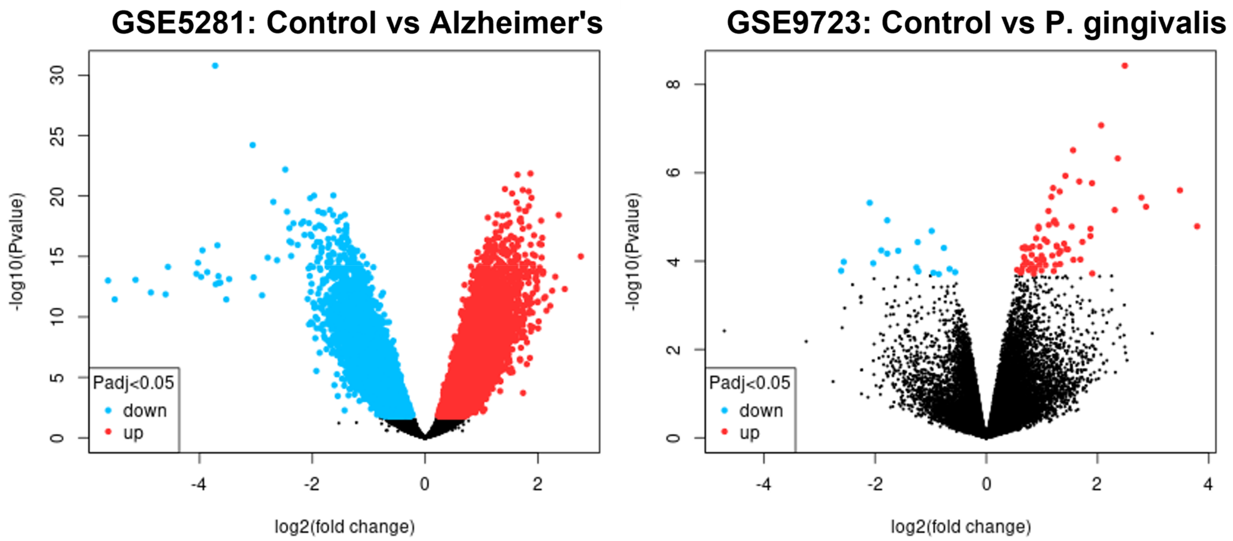
2.1. Data Normalisation

R 4.2.1 software was used to analyse the original data. Firstly, quality was evaluated, data were normalised, and the expression density is shown in Figures S1 and S2 (Supplementary Data). We found 2540 upregulated and 1776 downregulated in GSE 5281. A total of 39 were upregulated and 11 downregulated in GSE9723, as shown in Figure 1. These DEGs are shown by volcano plots in Figure 2. The volcano plots shown identify genes that are p < 0.05 and are used as criteria for significance. The heat maps (Figure 2) show the hierarchical clustering of the DEGs.

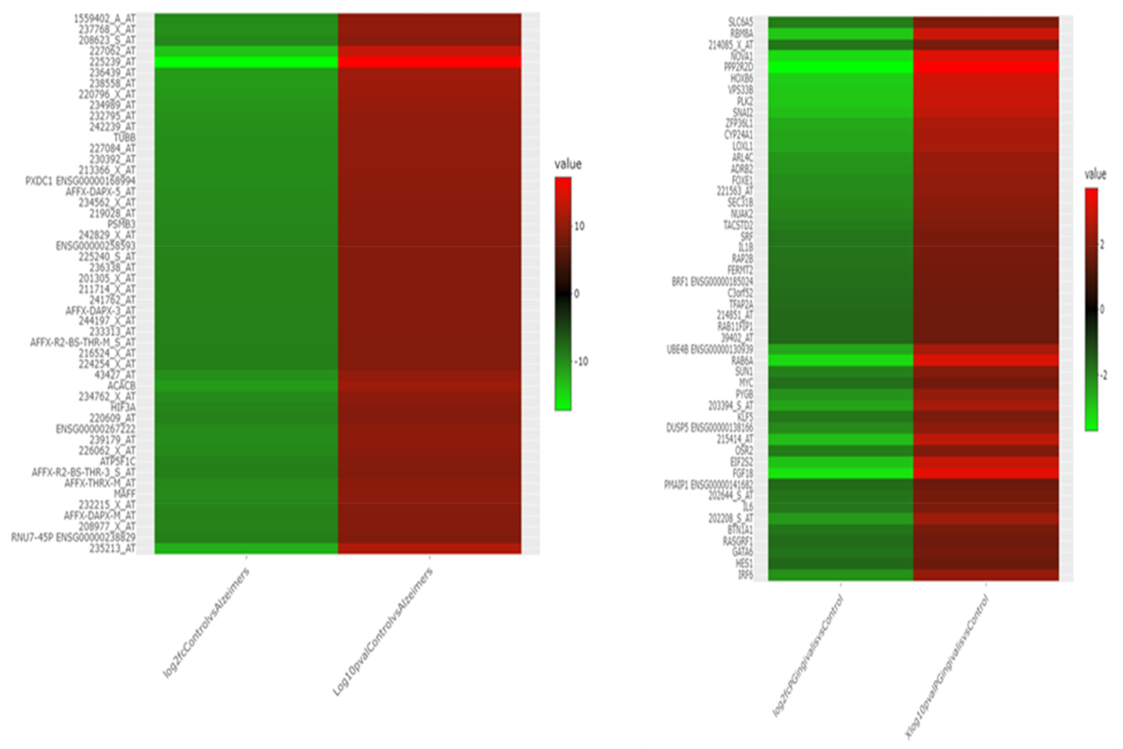
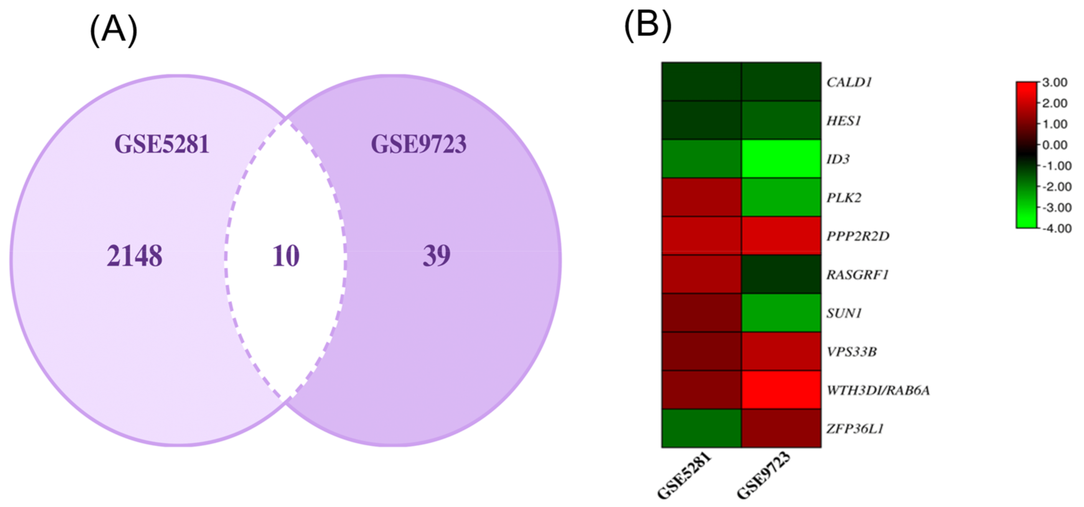
2.2. DEGs Common to AD and P. gingivalis

Using R, we found the common genes associated with datasets GSE5281 and GSE9723. The 10 genes were CALD1, HES1, ID3, PLK2, PPP2R2D, RASGRF1, SUN1, VPS33B, WTH3DI/RAB6A, and ZFP36L1. We then built a heat map to show the relativity of these found genes. Both of these findings are depicted in Figure 3.

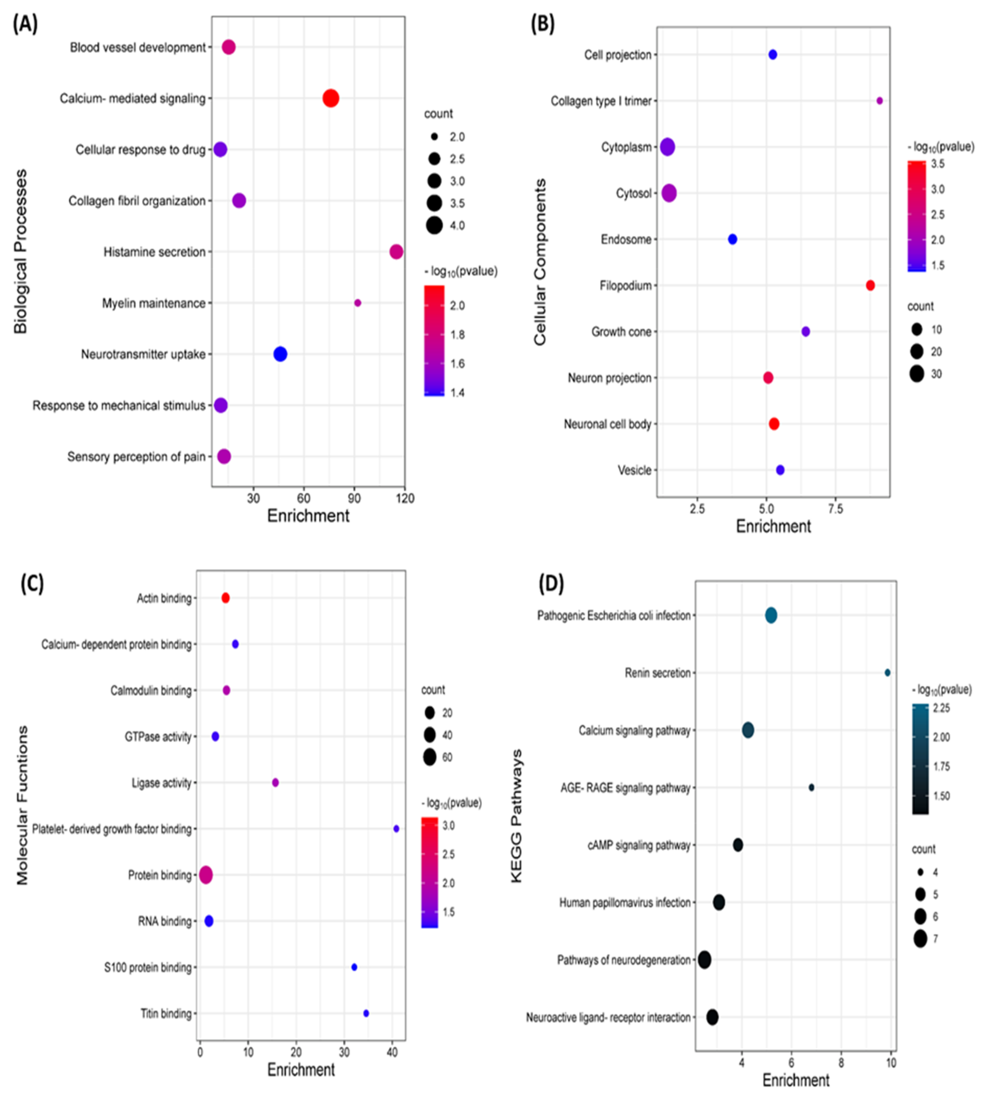
2.3. GO and KEGG Analysis

To elucidate the role of these genes, a Gene Ontology (GO) and Kyoto Encyclopedia of Genes and Genomes (KEGG) pathway was constructed. Most of the functions of the common genes were involved in calcium-mediated signalling, actin binding, filopodium, and some involvement in pathogenic E. coli infection. The summary of this finding is presented in Figure 4.

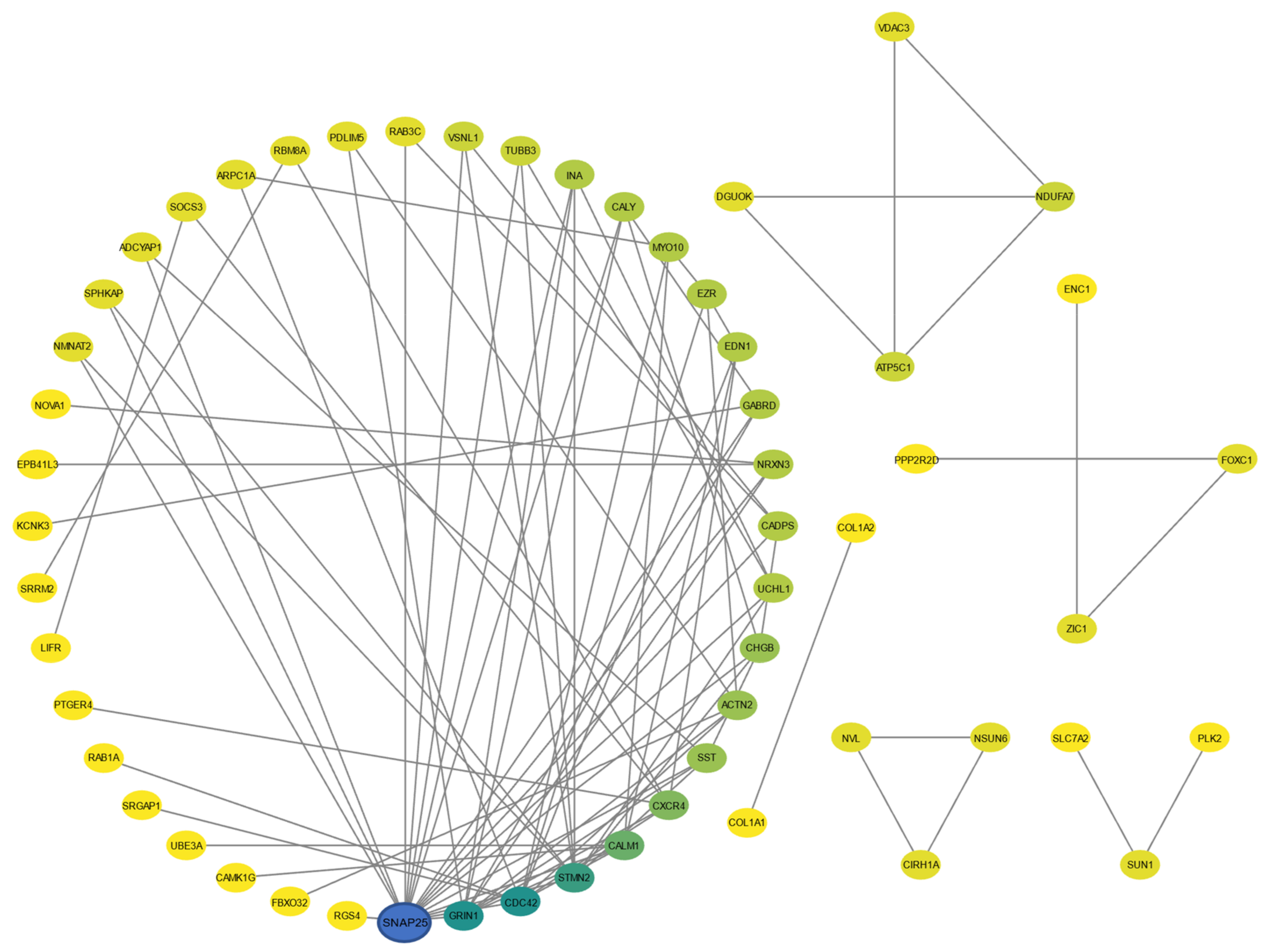
2.4. PPI Network and Hub Gene Selection

The PPI network from 10 identified common genes showed limited interaction. We then selected the top 100 genes (top 50 upregulated and top 50 downregulated) for our PPI network. The nodes indicate proteins, and the edges indicate their interactions, as shown in Figure 5. The specific functions of the genes and their indications are provided in Supplementary File S1.

2.5. CMap and Molecular Docking

Our CMap analysis only yielded one result of the ten common genes screened. PLK2 was then chosen for further analysis. A total of three drug targets were identified to exert biological changes to protein PLK2. They are summarised in Table 1, and the results of molecular docking are shown in Figure 6.

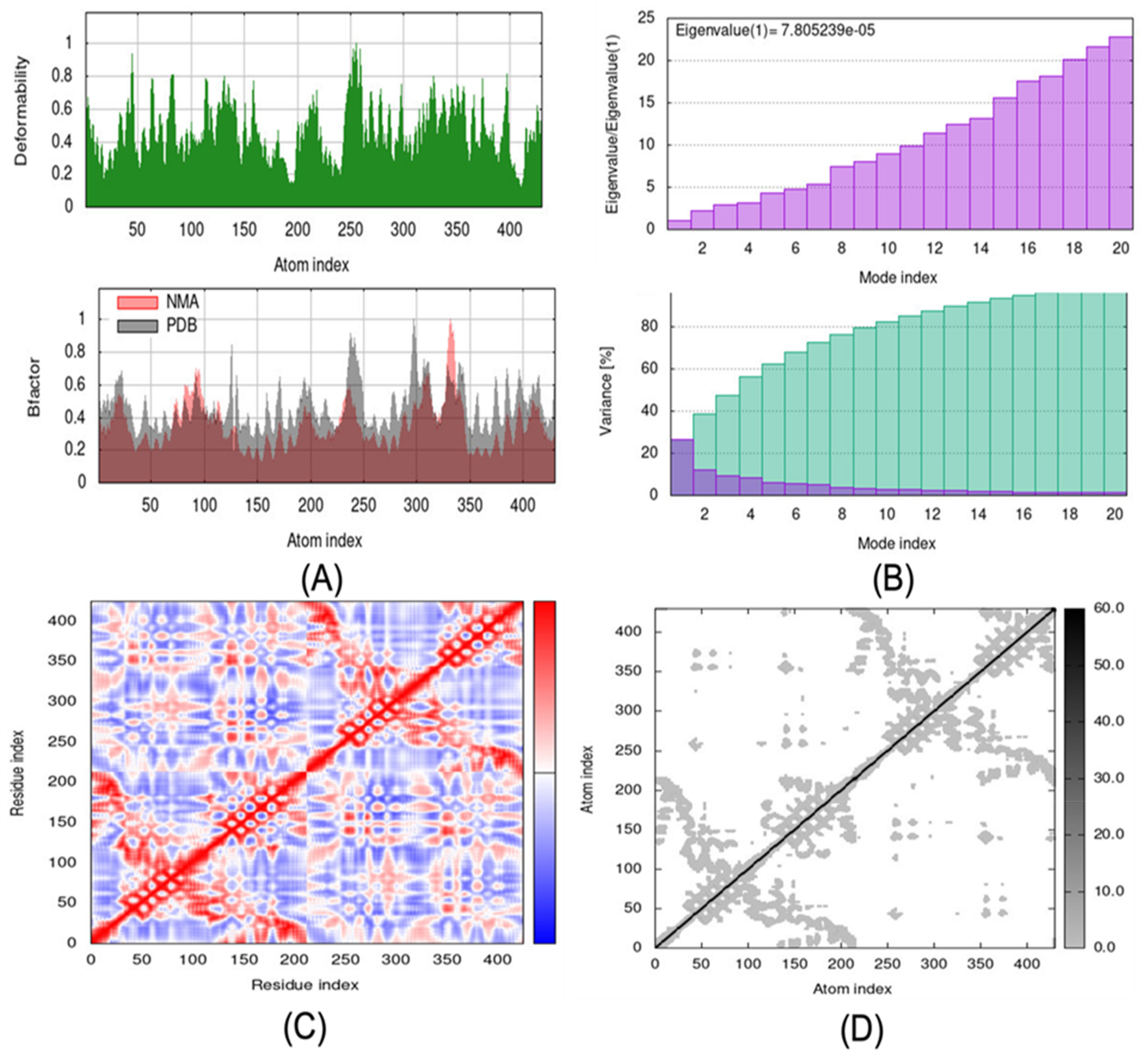
2.6. Molecular Dynamic Simulations

The best docking complex was chosen (PubChem ID: 11364421). The mobility characteristics of docked proteins are determined by deformability and B-factor. The deformability and B-factors of the PLK2 and 11364421 complexes show the peaks corresponding to deformable regions in the proteins, with the greatest peaks representing high deformability regions (Figure 7A). Figure 7B shows the eigenvalue and variance graphs of the PLK2 and 11364421 complexes. The variation graph of 11364421 with the target PLK2 shows individual variance with purple-shaded bars and cumulative variance with green-shaded bars. The complex covariance matrix depicts the correlations between residues in a complex. The red colour in Figure 7C in the matrix represents a good correlation between residues, while the white colour represents uncorrelated motion. Furthermore, the blue tint indicates anticorrelations. The higher the correlation, the more complicated the system. The docked proteins’ elastic maps (Figure 7D) show the atoms’ connections, with darker grey areas indicating stiffer regions.

3. Discussion

3.1. Common Genes Found in P.gingivalis and AD

CALD1 (Calcium-binding protein 1) is a protein that is encoded by the CALD1 gene in humans. It is a member of the calcyphosine family of calcium-binding proteins and is expressed in various tissues, including the brain. Some research has suggested that CALD1 may be involved in the development of AD, although more research is needed to fully understand its role in this condition [16]. In a particular study, it was found that CALD1 expression was significantly increased in the brains of people with AD compared to those without the condition and that CALD1 may be involved in the production of amyloid beta, a protein that is believed to be a key contributor to the development of AD [17,18]. Some research has suggested that CALD1 may be involved in the development of periodontitis, a type of gum disease characterised by inflammation and loss of the tissue and bone that support the teeth [19,20]. It was found that CALD1 expression was significantly increased in the gingiva of people with periodontitis compared to those without the condition and that CALD1 may be involved in the immune response to periodontitis.
The HES1 protein is a member of the family of transcription factors known as basic helix–loop–helix (bHLH) factors, and it functions as a transcriptional repressor for genes whose transcription is dependent on the bHLH protein. The protein attaches to the N-box promoter region rather than the typical enhancer box because it has a particular type of basic domain with a helix-interrupting protein (E-box). According to a study, HSE1 Melatonin’s protective effect on soluble A1-42-induced memory impairment, astrogliosis, and synaptic dysfunction in the rat hippocampus through the Musashi1/Notch1/Hes1 signalling pathway [21]. According to certain studies, HES1 may contribute to the onset of oral cancer cells sustainably infected with P. gingivalis exhibit resistance to Taxol and have a higher metastatic potential [22].
The human ID3 gene produces the DNA-binding protein inhibitor ID-3 protein [23]. Helix–loop–helix (HLH) proteins belonging to the ID family lack a fundamental DNA-binding domain and suppress transcription by forming dimers that are ineffective at binding to DNA. Regarding histone H3K9me3-based epigenome signatures, research has indicated that ID3 is connected with synaptic impairment in AD [24]. In one study conducted, gene expression changes in the various functional categories related to periodontitis in adults and aged animals the ID3 were decreased with periodontitis [25].
Protein kinase serine/threonine, the PLK2 gene in humans, codes for the enzyme PLK2. The ‘polo’ family of serine/threonine protein kinases, which includes serum-inducible kinase, is involved in healthy cell division. They discovered that PLK2 activity inhibition alters APP and tau pathology and enhances synaptic content in a sex-dependent manner in Alzheimer’s dementia, suggesting that it may contribute to the aetiology of the illness [26]. A study has shown that PLK2’s significance in cancer is somewhat debatable; evidence points to both an oncogenic and a tumour suppressor role in a variety of malignancies [27].
The PPP2R2D gene in humans encodes the protein known as PP2A subunit B isoform delta, often referred to as serine/threonine-protein phosphatase 2A 55 kDa regulatory subunit B delta isoform. There is no research on this gene’s relationship to P. gingivalis or AD.
A nuclear envelope protein with an UNC84 (SUN) domain is encoded by the protein-coding gene SUN1 (Sad1 and UNC84 domain containing 1). This gene belongs to the unc-84 homolog family. The protein aids in nuclear migration and anchoring. Spliced transcript variations have also been reported as an alternative (provided by RefSeq, January 2019). A previous study found that the accumulation of the inner nuclear envelope protein Sun1 is pathogenic in progeroid and dystrophic laminopathies, which results in AD [28]. No study has shown SUN1-gene-associated P. gingivalis.
VPS33B (vacuolar protein sorting-associated protein 33B) is a protein that, in humans, is encoded by the VPS33B gene. The translational profile of striatopallidal neurons is preferentially altered by deep brain stimulation of the subthalamic nucleus in an animal model of Parkinson’s disease, and one of the genes they identified was VPS33B [29]. By integrating transcriptome analysis, the gene VPS33B is implicated in the different gene expression traits in the interactions between epithelial cells and P. gingivalis [30].
WTH3DI/RAB6A is a type of protein coding; members of the small GTPase superfamily’s RAB family are encoded by this gene. The targeting and fusing of transport carriers to acceptor compartments are regulated by the binding of GTPases of the RAB family to different effectors. This protein is found at the Golgi apparatus, which controls both retrograde and forward trafficking provided by HUGO Gene Nomenclature. No study was found related to this gene-associated AD and P. gingivalis.
The ZFP36L1 gene belongs to the TIS11 family of early response genes, which are activated by a variety of agonists, including the polypeptide mitogen EGF and the phorbol ester TPA. This gene is highly conserved between species and features motifs found in other early-response genes in its promoter. A distinctive putative zinc finger domain with a recurring cys-his pattern can be found in the encoded protein. Most likely, this putative nuclear transcription factor controls how the body reacts to growth stimuli. This gene has been associated with a variety of alternatively spliced transcript variants that encode distinct isoforms (provided by RefSeq, September 2011). Characterisation of ZFP36L1 in the context of multiple sclerosis and functional immunological effects connected to the susceptibility to the disease, according to a study [31]. APN impairs the ability of macrophages, which play a significant role in periodontitis, to function. Through dependent signalling pathways, APN first stimulates the synthesis of TNF-α, which increases the expression of IL-10 and, as a result, reduces the inflammatory response of macrophages exposed to LPS [32]. Moreover, by promoting macrophage autophagy, APN can also reduce the expression of inflammatory mediators brought on by LPS. APN increases the production of ZFP36L1, which inhibits the interaction between Bcl-2 and Beclin-1 and, in turn, stimulates Beclin-1-activated autophagy in macrophages by destabilizing the mRNA of Bcl-2 [33]. A summary of the functions of the genes and possible roles is provided in Figure 8.

3.2. PPI Network Selected Hub Genes UCHL1, SST, CHGB, CALY, and INA

Ubiquitin carboxy-terminal hydrolase L1 (UCHL1) belongs to a gene family whose members hydrolyze short C-terminal ubiquitin adducts to produce the ubiquitin monomer [34]. Highly specialised neurons, diffuse neuroendocrine system cells, and their tumours express UCHL1 [35]. It is known to play a role in AD [36]. It is thought that UCHL1 may be involved in the clearance of proteins that accumulate in the presence of AD, such as amyloid-beta [37]. UCHL1 has been identified as a potential marker for periodontitis [38]. The presence of UCHL1 in periodontitis may be associated with increased inflammation and tissue destruction [39]. UCHL1 may be a potential target for the treatment of AD and periodontitis [40]. Somatostatin is present throughout the body and binds to high-affinity G-protein-coupled somatostatin receptors to prevent the production of multiple secondary hormones. Through its interactions with thyroid stimulating hormone and pituitary growth hormone, this hormone plays a key role in the regulation of the endocrine system (provided by RefSeq, July 2008). It is also believed to play a role in the progression of AD [41]. Studies have shown that somatostatin levels are significantly lower in individuals with AD and that this decrease is associated with increased levels of the amyloid beta peptide, which is known to be a major cause of AD [42]. Somatostatin has been found to play a role in periodontitis, or inflammation of the gums; it is believed that somatostatin may be involved in the inflammatory process that leads to periodontitis [43]. Secretogranin-1, also known as Chromogranin B, is a protein that the CHGB gene in humans codes for; it is a member of the grain protein family. Chromogranin B is a gene that has been associated with AD. Studies have shown that individuals with AD have higher levels of CHGB in their brains than those without the disease. It is believed that CHGB may be involved in the accumulation of tau proteins, which are associated with AD, and the formation of amyloid plaques, which are also associated with the disease [44]. Currently, there is no evidence to suggest that CHGB, or Chromogranin B, is involved in periodontitis. However, research into this gene and its role in other inflammatory diseases may provide insight into its potential role in periodontitis. Neuron-specific vesicular protein is a type II single transmembrane protein that is expressed by the CALY gene in humans. It is necessary for the maximum accelerated calcium release upon stimulation of purinergic or muscarinic receptors [45]. CALY has been associated with AD. Studies have shown that levels of Calcyon are significantly lower in individuals with AD than in those without the disease [46]. It is believed that Calcyon may be able to regulate the activity of certain proteins that are involved in the progression of AD [47]. Currently, there is no evidence to suggest that neuron-specific vesicular protein Calcyon is involved in periodontitis. Alpha-internexin is a type of class IV intermediate filament with a mass of 66 kDa. The rat spinal cord and optic nerve were originally used to purify the protein [48]. A comparable central rod domain found in alpha-internexin includes about 310 amino acid residues and forms a highly conserved alpha-helical region. An area called the amino-terminal head, and a region called the carboxy-terminal tail, surround the core rod domain, which is in charge of the coiled-coil structure [49]. INA encodes neuronal intermediate filament protein found mostly in the neurons of the nervous system during early development [50]. No study has shown a relationship between the INA gene and periodontitis.

3.3. Molecular Docking of PLK2 with 11364421 (C28H39N703)

We used iMODS to evaluate and define the flexibility of PLK2 with 11364421. We hypothesize that based on the conceivable interactions of the identified proteins with PLK2, they can serve as prospective therapeutic candidates and targets to attenuate the pathological process in P. gingivalis-associated AD.

4. Materials and Methods

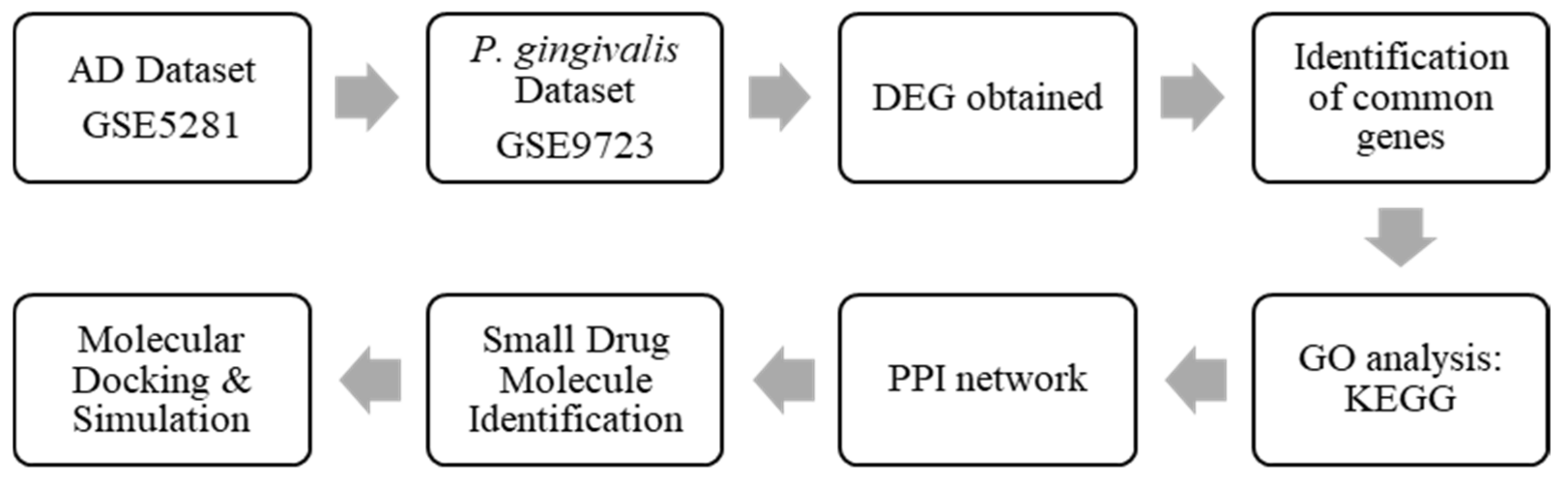
4.1. Pipeline of Research

The flowchart of the conduct of this study is summarised in Figure 9.

4.2. Data Acquisition

The National Centre for Biotechnology Information (NCBI) produced and maintains the gene expression database known as the GEO database. It includes high throughput gene expression data provided by international research institutions [51]. To determine the molecular mechanism of the occurrence and development of P. gingivalis-associated AD, two microarray datasets from the GEO database, namely, GSE5281 and GSE9723, were downloaded. The former was a study conducted on Alzheimer’s patients, and the latter was using cells infected with P. gingivalis. The data’s executive summary is presented in Table 2 below.

4.3. DEGs Screening

Using the limma package in R, we identified the differentially expressed genes (DEGs) between AD and P. gingivalis. The criteria of p < 0.05 and log2FC > 1 were set before entering to VENN tool to visualise the intersecting genes.

4.4. Functional Analysis of Common Genes

The Database for Annotation, Visualization, and Integrated Discovery (DAVID) web tool is used to carry out GO enrichment analysis and KEGG pathway analysis [55]. The three categories of GO analysis, which include biological processes (BPs), cellular components (CCs), and molecular functions (MFs), were performed [56]. A p-value < 0.05 is considered significant.

4.5. Construction of PPI Network

A protein–protein interaction (PPI) analysis was carried out using the STRING database to investigate the relationships between common genes (https://string-db.org/) (accessed on 26 December 2022). All known and anticipated protein–protein interactions, including both physical and details functional relationships, are listed in the STRING database [57]. The minimum interaction score was set to 0.400 as a criterion for statistical significance. The PPI is illustrated by the nodes as proteins and lines as interactions.

4.6. CMap Analysis, Molecular Docking, and Simulation

We will be predicting small drug molecules using CMap in accordance with specific gene expression signatures offered by the database. A small drug molecule with a negative mean score may be able to reverse biological effects and so have potential therapeutic utility (p < 0.05 is considered significant). The effectiveness and binding potential were assessed using molecular docking. Firstly, crystal structures are obtained from RCSB Protein Data Bank (PDB, http://www.rcsb.org) (accessed on 26 December 2022), and the mol2 file formats of the compounds were retrieved from the PubChem database. PyMOL 2.3.1 was used to dehydrate target proteins and remove ligands. AutoDock Tool 1.5.6 software was used to hydrogenate, calculate its charge, and store it in PDBQT format. PyRx and Autodock Vina v1.2.0 software was used to visualize the findings of molecular docking. The iMOD server iMOD server (iMODS) (http://imods.chaconlab.org) (accessed on 26 December 2022) was used to run molecular dynamics simulations to calculate the stability and molecular mobility of the bound protein-113644421 complexes. In addition to calculating molecular mobility, iMODS was employed to analyse the structural dynamics of the docking complexes. The elastic network, deformability, B-factor, eigenvalues, variance, and covariance map were all used to show how stable the protein-11364421 complexes were. All parameters for the docked PDB files used as input were left at default when they were uploaded to the iMODS server.

5. Conclusions

In this study, 10 crossover genes were identified, indicating a potential association between AD and P. gingivalis as the key pathogen in periodontitis. Hence, one potential therapeutic target was identified for P. gingivalis-induced-AD treatment.
However, further in vivo and in vitro studies are needed to confirm these findings.

Supplementary Materials

The following supporting information can be downloaded at: https://www.mdpi.com/article/10.3390/ijms24065432/s1.

Author Contributions

Conceptualization, A.H. and K.B.; methodology, A.H. and K.B.; software, A.H. and K.B.; supervision, N.S.N. and A.T.S.; funding acquisition, N.S.N. All authors have read and agreed to the published version of the manuscript.

Funding

This study was funded by the Fundamental Research Grant Scheme, Ministry of Education; FRGS/1/2020/SKK0/UKM/02/10.

Institutional Review Board Statement

Not Applicable.

Informed Consent Statement

Not Applicable.

Data Availability Statement

Not Applicable.

Conflicts of Interest

The authors declare no conflict of interest.

References

  1. Ball, S.L.; Holland, A.J.; Watson, P.C.; Huppert, F.A. Theoretical Exploration of the Neural Bases of Behavioural Disinhibition, Apathy and Executive Dysfunction in Preclinical Alzheimer’s Disease in People with Down’s Syndrome: Potential Involvement of Multiple Frontal-Subcortical Neuronal Circuits. J. Intellect. Disabil. Res. 2010, 54, 320–336. [Google Scholar] [CrossRef] [PubMed]
  2. Jellinger, K.A. Alzheimer’s Disease. Neurobiol. Dis. 2007, 69–82. [Google Scholar] [CrossRef]
  3. Balolong, M.P.; Mendoza, M.A.F.; Balolong, M.P.; Mendoza, M.A.F. Understanding Oral Diseases: Exploring Opportunities from Filipino Oral Microbiome Research. Dent. Caries 2021. [Google Scholar] [CrossRef]
  4. Viral Role in Alzheimer’s Disease Discovered|University of Oxford. Available online: https://www.ox.ac.uk/news/2022-08-02-viral-role-alzheimers-disease-discovered (accessed on 25 February 2023).
  5. Sharma, V.K.; Mehta, V.; Singh, T.G. Alzheimer’s Disorder: Epigenetic Connection and Associated Risk Factors. Curr. Neuropharmacol. 2020, 18, 740. [Google Scholar] [CrossRef] [PubMed]
  6. Lunnon, K.; Mill, J. Epigenetic Studies in Alzheimer’s Disease: Current Findings, Caveats and Considerations for Future Studies. Am. J. Med. Genet. Part B Neuropsychiatr. Genet. Off. Publ. Int. Soc. Psychiatry Genet. 2013, 162, 789. [Google Scholar] [CrossRef] [PubMed] [Green Version]
  7. Fülöp, T.; Itzhaki, R.F.; Balin, B.J.; Miklossy, J.; Barron, A.E. Role of Microbes in the Development of Alzheimer’s Disease: State of the Art—An International Symposium Presented at the 2017 IAGG Congress in San Francisco. Front. Genet. 2018, 9, 362. [Google Scholar] [CrossRef] [Green Version]
  8. Jackson, J.P.; Lindroth, A.M.; Cao, X.; Jacobsen, S.E. Control of CpNpG DNA Methylation by the KRYPTONITE Histone H3 Methyltransferase. Nature 2002, 416, 556–560. [Google Scholar] [CrossRef] [PubMed]
  9. Balazs, R. Epigenetic Mechanisms in Alzheimer’s Disease. Degener. Neurol. Neuromuscul. Dis. 2014, 4, 85. [Google Scholar] [CrossRef] [Green Version]
  10. Elwishahy, A.; Antia, K.; Bhusari, S.; Ilechukwu, N.C.; Horstick, O.; Winkler, V. Porphyromonas Gingivalis as a Risk Factor to Alzheimer’s Disease: A Systematic Review. J. Alzheimers Dis. Rep. 2021, 5, 721. [Google Scholar] [CrossRef]
  11. Mei, F.; Xie, M.; Huang, X.; Long, Y.; Lu, X.; Wang, X.; Chen, L. Porphyromonas gingivalis and Its Systemic Impact: Current Status. Pathogens 2020, 9, 944. [Google Scholar] [CrossRef]
  12. Li, X.; Kolltveit, K.M.; Tronstad, L.; Olsen, I. Systemic Diseases Caused by Oral Infection. Clin. Microbiol. Rev. 2000, 13, 547. [Google Scholar] [CrossRef]
  13. O’Brien, M.A.; Costin, B.N.; Miles, M.F. Using Genome-Wide Expression Profiling to Define Gene Networks Relevant to the Study of Complex Traits: From RNA Integrity to Network Topology. Int. Rev. Neurobiol. 2012, 104, 91. [Google Scholar] [CrossRef] [Green Version]
  14. Kim, K.; Zakharkin, S.O.; Allison, D.B. Expectations, Validity, and Reality in Gene Expression Profiling. J. Clin. Epidemiol. 2010, 63, 950. [Google Scholar] [CrossRef] [PubMed] [Green Version]
  15. Olsen, I.; Singhrao, S.K. Interaction between Genetic Factors, Porphyromonas Gingivalis and Microglia to Promote Alzheimer’s Disease. J. Oral Microbiol. 2020, 12, 1820834. [Google Scholar] [CrossRef] [PubMed]
  16. What Causes Alzheimer’s Disease?|National Institute on Aging. Available online: https://www.nia.nih.gov/health/what-causes-alzheimers-disease (accessed on 6 January 2023).
  17. Kumari, A.; Rahaman, A.; Zeng, X.-A.; Farooq, M.A.; Huang, Y.; Yao, R.; Ali, M.; Ishrat, R.; Ali, R. Temporal Cortex Microarray Analysis Revealed Impaired Ribosomal Biogenesis and Hyperactivity of the Glutamatergic System: An Early Signature of Asymptomatic Alzheimer’s Disease. Front. Neurosci. 2022, 16, 966877. [Google Scholar] [CrossRef] [PubMed]
  18. Pereira, A.C.; Gray, J.D.; Kogan, J.F.; Davidson, R.L.; Rubin, T.G.; Okamoto, M.; Morrison, J.H.; McEwen, B.S. Age and Alzheimer’s Disease Gene Expression Profiles Reversed by the Glutamate Modulator Riluzole. Mol. Psychiatry 2017, 22, 296. [Google Scholar] [CrossRef] [PubMed]
  19. Zhang, W.; Wu, Q.; Su, J.; Wang, W.; Zhao, X.; Cao, P. Circulating MiRNAs as Epigenetic Mediators of Periodontitis and Preeclampsia Association. Dis. Markers 2022, 2022, 2771492. [Google Scholar] [CrossRef]
  20. Gonzalez, O.A.; Kirakodu, S.; Nguyen, L.M.; Orraca, L.; Novak, M.J.; Gonzalez-Martinez, J.; Ebersole, J.L. Comparative Analysis of Gene Expression Patterns for Oral Epithelial Cell Functions in Periodontitis. Front. Oral Health 2022, 3, 863231. [Google Scholar] [CrossRef]
  21. Zhang, S.; Wang, P.; Ren, L.; Hu, C.; Bi, J. Protective Effect of Melatonin on Soluble Aβ1-42-Induced Memory Impairment, Astrogliosis, and Synaptic Dysfunction via the Musashi1/Notch1/Hes1 Signaling Pathway in the Rat Hippocampus. Alzheimer’s Res. Ther. 2016, 8, 40. [Google Scholar] [CrossRef] [Green Version]
  22. Woo, B.H.; Kim, D.J.; Choi, J.I.; Kim, S.J.; Park, B.S.; Song, J.M.; Lee, J.H.; Park, H.R. Oral Cancer Cells Sustainedly Infected with Porphyromonas Gingivalis Exhibit Resistance to Taxol and Have Higher Metastatic Potential. Oncotarget 2017, 8, 46981. [Google Scholar] [CrossRef] [Green Version]
  23. Ellmeier, W.; Aguzzi, A.; Kleiner, E.; Kurzbauer, R.; Weith, A. Mutually Exclusive Expression of a Helix-Loop-Helix Gene and N-Myc in Human Neuroblastomas and in Normal Development. EMBO J. 1992, 11, 2563–2571. [Google Scholar] [CrossRef] [PubMed]
  24. Lee, J.; Roberts, J.A.S.; Atanasova, K.R.; Chowdhury, N.; Han, K.; Yilmaz, Ö. Human Primary Epithelial Cells Acquire an Epithelial-Mesenchymal-Transition Phenotype during Long-Term Infection by the Oral Opportunistic Pathogen, Porphyromonas Gingivalis. Front. Cell. Infect. Microbiol. 2017, 7, 493. [Google Scholar] [CrossRef] [PubMed] [Green Version]
  25. Ebersole, J.L.; Al-Sabbagh, M.; Dawson, D.R. Heterogeneity of Human Serum Antibody Responses to P. Gingivalis in Periodontitis: Effects of Age, Race/Ethnicity, and Sex. Immunol. Lett. 2020, 218, 11. [Google Scholar] [CrossRef]
  26. Martínez-Drudis, L.; Sheta, R.; Pellegrinato, R.; Loukili, S.; Tremblay, C.; Calon, F.; Rivest, S.; Oueslati, A. Inhibition of PLK2 Activity Affects APP and Tau Pathology and Improves Synaptic Content in a Sex-Dependent Manner in a 3xTg Mouse Model of Alzheimer’s Disease. Neurobiol. Dis. 2022, 172, 105833. [Google Scholar] [CrossRef]
  27. de Cárcer, G. The Mitotic Cancer Target Polo-Like Kinase 1: Oncogene or Tumor Suppressor? Genes 2019, 10, 208. [Google Scholar] [CrossRef] [Green Version]
  28. Chen, C.Y.; Chi, Y.H.; Mutalif, R.A.; Starost, M.F.; Myers, T.G.; Anderson, S.A.; Stewart, C.L.; Jeang, K.T. Accumulation of the Inner Nuclear Envelope Protein Sun1 Is Pathogenic in Progeric and Dystrophic Laminopathies. Cell 2012, 149, 565–577. [Google Scholar] [CrossRef] [PubMed] [Green Version]
  29. Visanji, N.P.; Sarvestani, I.K.; Creed, M.C.; Shoaei, Z.S.; Nobrega, J.N.; Hamani, C.; Hazrati, L.N. Deep Brain Stimulation of the Subthalamic Nucleus Preferentially Alters the Translational Profile of Striatopallidal Neurons in an Animal Model of Parkinson’s Disease. Front. Cell. Neurosci. 2015, 9, 221. [Google Scholar] [CrossRef] [Green Version]
  30. Zhang, D.; Hou, J.; Wu, Y.; Liu, Y.; Li, R.; Xu, T.; Liu, J.; Pan, Y. Distinct Gene Expression Characteristics in Epithelial Cell-Porphyromonas Gingivalis Interactions by Integrating Transcriptome Analyses. Int. J. Med. Sci. 2019, 16, 1320. [Google Scholar] [CrossRef] [Green Version]
  31. Ottoboni, L.; Radassi, K.; De Jager, P.; Comi, G.; Farina, C.; Martino, G. Characterization of ZFP36L1 in the Context of Multiple Sclerosis and Functional Immunological Consequences Associated with the Susceptibility to the Disease. J. Neuroimmunol. 2014, 275, 52. [Google Scholar] [CrossRef]
  32. Park, P.H.; McMullen, M.R.; Huang, H.; Thakur, V.; Nagy, L.E. Short-Term Treatment of RAW264.7 Macrophages with Adiponectin Increases Tumor Necrosis Factor-α (TNF-α) Expression via ERK1/2 Activation and Egr-1 Expression: Role of TNF-α in adiponectin-stimulated interleukin-10 production. J. Biol. Chem. 2007, 282, 21695–21703. [Google Scholar] [CrossRef] [PubMed] [Green Version]
  33. Shrestha, A.; Pun, N.T.; Park, P.H. ZFP36L1 and AUF1 Induction Contribute to the Suppression of Inflammatory Mediators Expression by Globular Adiponectin via Autophagy Induction in Macrophages. Biomol. Ther. 2018, 26, 446. [Google Scholar] [CrossRef] [PubMed]
  34. Doran, J.F.; Jackson, P.; Kynoch, P.A.M.; Thompson, R.J. Isolation of PGP 9.5, a New Human Neurone-Specific Protein Detected by High-Resolution Two-Dimensional Electrophoresis. J. Neurochem. 1983, 40, 1542–1547. [Google Scholar] [CrossRef] [PubMed]
  35. Das, C.; Hoang, Q.Q.; Kreinbring, C.A.; Luchansky, S.J.; Meray, R.K.; Ray, S.S.; Lansbury, P.T.; Ringe, D.; Petsko, G.A. Structural Basis for Conformational Plasticity of the Parkinson’s Disease-Associated Ubiquitin Hydrolase UCH-L1. Proc. Natl. Acad. Sci. USA 2006, 103, 4675–4680. [Google Scholar] [CrossRef] [PubMed] [Green Version]
  36. Zhao, Z.B.; Wu, L.; Xiong, R.; Wang, L.L.; Zhang, B.; Wang, C.; Li, H.; Liang, L.; Chen, S.D. MicroRNA-922 Promotes Tau Phosphorylation by Downregulating Ubiquitin Carboxy-Terminal Hydrolase L1 (UCHL1) Expression in the Pathogenesis of Alzheimer’s Disease. Neuroscience 2014, 275, 232–237. [Google Scholar] [CrossRef]
  37. Oddo, S. The Ubiquitin-Proteasome System in Alzheimer’s Disease. J. Cell. Mol. Med. 2008, 12, 363. [Google Scholar] [CrossRef] [Green Version]
  38. Lin, L.; Li, S.; Hu, S.; Yu, W.; Jiang, B.; Mao, C.; Li, G.; Yang, R.; Miao, X.; Jin, M.; et al. UCHL1 Impairs Periodontal Ligament Stem Cell Osteogenesis in Periodontitis. J. Dent. Res. 2022, 102, 00220345221116031. [Google Scholar] [CrossRef]
  39. Cobb, C.M.; Singla, O.; Feil, P.H.; Theiesen, F.C.; Schultz, R.E. Comparison of NK-Cell (Leu-7+ and Leu-11b+) Populations in Clinically Healthy Gingiva, Chronic Gingivitis and Chronic Adult Periodontitis. J. Periodontal Res. 1989, 24, 1–7. [Google Scholar] [CrossRef]
  40. Jiang, Z.; Shi, Y.; Zhao, W.; Zhou, L.; Zhang, B.; Xie, Y.; Zhang, Y.; Tan, G.; Wang, Z. Association between Chronic Periodontitis and the Risk of Alzheimer’s Disease: Combination of Text Mining and GEO Dataset. BMC Oral Health 2021, 21, 466. [Google Scholar] [CrossRef]
  41. Solarski, M.; Wang, H.; Wille, H.; Schmitt-Ulms, G. Somatostatin in Alzheimer’s Disease: A New Role for an Old Player. Prion 2018, 12, 1–8. [Google Scholar] [CrossRef] [Green Version]
  42. Saido, T.C. Metabolism of Amyloid β Peptide and Pathogenesis of Alzheimer’s Disease. Proc. Jpn. Acad. Ser. B Phys. Biol. Sci. 2013, 89, 321. [Google Scholar] [CrossRef] [PubMed] [Green Version]
  43. Memmert, S.; Damanaki, A.; Nokhbehsaim, M.; Nogueira, A.V.B.; Eick, S.; Cirelli, J.A.; Jäger, A.; Deschner, J. Regulation of Somatostatin Receptor 2 by Proinflammatory, Microbial and Obesity-Related Signals in Periodontal Cells and Tissues. Head Face Med. 2019, 15, 2. [Google Scholar] [CrossRef] [PubMed]
  44. Lechner, T.; Adlassnig, C.; Humpel, C.; Kaufmann, W.A.; Maier, H.; Reinstadler-Kramer, K.; Hinterhölzl, J.; Mahata, S.K.; Jellinger, K.A.; Marksteiner, J. Chromogranin Peptides in Alzheimer’s Disease. Exp. Gerontol. 2004, 39, 101–113. [Google Scholar] [CrossRef]
  45. Heijtz, R.D.; Alexeyenko, A.; Xavier, F.X. Calcyon MRNA Expression in the Frontal-Striatal Circuitry and Its Relationship to Vesicular Processes and ADHD. Behav. Brain Funct. BBF 2007, 3, 33. [Google Scholar] [CrossRef] [Green Version]
  46. Muthusamy, N.; Chen, Y.J.; Yin, D.M.; Mei, L.; Bergson, C. Complementary Roles of the Neuron-Enriched Endosomal Proteins NEEP21 and Calcyon in Neuronal Vesicle Trafficking. J. Neurochem. 2015, 132, 20–31. [Google Scholar] [CrossRef] [PubMed]
  47. Vazdarjanova, A.; Bunting, K.; Muthusamy, N.; Bergson, C. Calcyon Up-Regulation in Adolescence Impairs Response Inhibition and Working Memory in Adulthood. Mol. Psychiatry 2011, 16, 672. [Google Scholar] [CrossRef] [PubMed] [Green Version]
  48. Levavasseur, F.; Zhu, Q.; Julien, J.P. No Requirement of α-Internexin for Nervous System Development and for Radial Growth of Axons. Mol. Brain Res. 1999, 69, 104–112. [Google Scholar] [CrossRef]
  49. Lariviere, R.C.; Julien, J.P. Functions of Intermediate Filaments in Neuronal Development and Disease. J. Neurobiol. 2004, 58, 131–148. [Google Scholar] [CrossRef]
  50. Wang, P.; Wang, S.M.; Hsieh, C.J.; Chien, C.L. Neural Expression of α-Internexin Promoter In Vitro and In Vivo. J. Cell. Biochem. 2006, 97, 275–287. [Google Scholar] [CrossRef] [PubMed]
  51. Edgar, R.; Domrachev, M.; Lash, A.E. Gene Expression Omnibus: NCBI Gene Expression and Hybridization Array Data Repository. Nucleic Acids Res. 2002, 30, 207–210. [Google Scholar] [CrossRef] [Green Version]
  52. Readhead, B.; Haure-Mirande, J.V.; Funk, C.C.; Richards, M.A.; Shannon, P.; Haroutunian, V.; Sano, M.; Liang, W.S.; Beckmann, N.D.; Price, N.D.; et al. Multiscale Analysis of Independent Alzheimer’s Cohorts Finds Disruption of Molecular, Genetic, and Clinical Networks by Human Herpesvirus. Neuron 2018, 99, 64–82.e7. [Google Scholar] [CrossRef] [Green Version]
  53. Liang, W.S.; Dunckley, T.; Beach, T.G.; Grover, A.; Mastroeni, D.; Walker, D.G.; Caselli, R.J.; Kukull, W.A.; McKeel, D.; Morris, J.C.; et al. Gene Expression Profiles in Anatomically and Functionally Distinct Regions of the Normal Aged Human Brain. Physiol. Genom. 2007, 28, 311–322. [Google Scholar] [CrossRef]
  54. Handfield, M.; Mans, J.J.; Zheng, G.; Lopez, M.C.; Mao, S.; Progulske-Fox, A.; Narasimhan, G.; Baker, H.V.; Lamont, R.J. Distinct Transcriptional Profiles Characterize Oral Epithelium-Microbiota Interactions. Cell. Microbiol. 2005, 7, 811–823. [Google Scholar] [CrossRef] [PubMed]
  55. Huang, D.W.; Sherman, B.T.; Lempicki, R.A. Systematic and Integrative Analysis of Large Gene Lists Using DAVID Bioinformatics Resources. Nat. Protoc. 2009, 4, 44–57. [Google Scholar] [CrossRef]
  56. Blake, J.A.; Christie, K.R.; Dolan, M.E.; Drabkin, H.J.; Hill, D.P.; Ni, L.; Sitnikov, D.; Burgess, S.; Buza, T.; Gresham, C.; et al. Gene Ontology Consortium: Going Forward. Nucleic Acids Res. 2015, 43, D1049–D1056. [Google Scholar] [CrossRef]
  57. Szklarczyk, D.; Gable, A.L.; Nastou, K.C.; Lyon, D.; Kirsch, R.; Pyysalo, S.; Doncheva, N.T.; Legeay, M.; Fang, T.; Bork, P.; et al. The STRING Database in 2021: Customizable Protein–Protein Networks, and Functional Characterization of User-Uploaded Gene/Measurement Sets. Nucleic Acids Res. 2021, 49, D605–D612. [Google Scholar] [CrossRef] [PubMed]
Figure 1. Volcano plots showing the upregulated and downregulated genes in GSE5281 and GSE9723.
Ijms 24 05432 g001
Figure 2. Heat maps of the differentially expressed genes (DEGs) according to their genes and expressions. The gene symbols are shown on the left side of the heat maps.
Ijms 24 05432 g002
Figure 3. (A) Common genes and (B) heat map of P. gingivalis-associated AD.
Ijms 24 05432 g003
Figure 4. Gene Ontology and Kyoto Encyclopedia of Genes and Genomes (KEGG) analysis of the differentially expressed genes (DEGs) according to (A) biological processes (BPs), (B) cellular components (CCs), (C) molecular functions (MFs), and (D) KEGG pathways.
Ijms 24 05432 g004
Figure 5. Protein–protein interaction (PPI) network from top 100 genes. Proteins are denoted as nodes and interactions as edges.
Ijms 24 05432 g005
Figure 6. Molecular docking of PLK2 with 3 drug targets.
Ijms 24 05432 g006
Figure 7. Results of PLK2-11364421 molecular dynamics simulations using iMODS: (A) deformability and B-factor plots; (B) eigenvalue and variance plots (Colored bars show the individual (purpule) and cumulative (green) variances.); (C) elastic network model; and (D) covariance map.
Ijms 24 05432 g007
Figure 8. The functional characterisation of identified common genes.
Ijms 24 05432 g008
Figure 9. Flow chart of this study.
Ijms 24 05432 g009
Table 1. Chemical structure and ID according to CMap for PLK2 targets.
IDChemical StructureIUPAC Name
24971422C21H17N3O33-(1,3-benzodioxol-5-yl)-N-[(1S)-1-phenylethyl]-[1,2] oxazolo [5,4-c] pyridin-5-amine
11364421C28H39N7O34-[[(7R)-8-cyclopentyl-7-ethyl-5-methyl-6-oxo-7H-pteridin-2-yl] amino]-3-methoxy-N-(1-methylpiperidin-4-yl) benzamide
49792852C24H27F3N8O31-(2-hydroxyethyl)-8-[5-(4-methylpiperazin-1-yl)-2-(trifluoromethoxy) anilino]-4,5-dihydropyrazolo[4,3-h] quinazoline-3-carboxamide
Table 2. The summary of the datasets collected from the Gene Expression Omnibus (GEO) database.
ReferenceGEO SeriesPlatformControl
(N)
Effected
(N)
Country
Readhead et al., 2018 [52], Liang et al., 2007 [53]GSE5281GPL5707487USA
Handfield et al., 2005 [54]GSE9723GPL9644USA
Disclaimer/Publisher’s Note: The statements, opinions and data contained in all publications are solely those of the individual author(s) and contributor(s) and not of MDPI and/or the editor(s). MDPI and/or the editor(s) disclaim responsibility for any injury to people or property resulting from any ideas, methods, instructions or products referred to in the content.

