
Silver-Based Surface Plasmon Sensors: Fabrication and Applications


Abstract
:1. Introduction
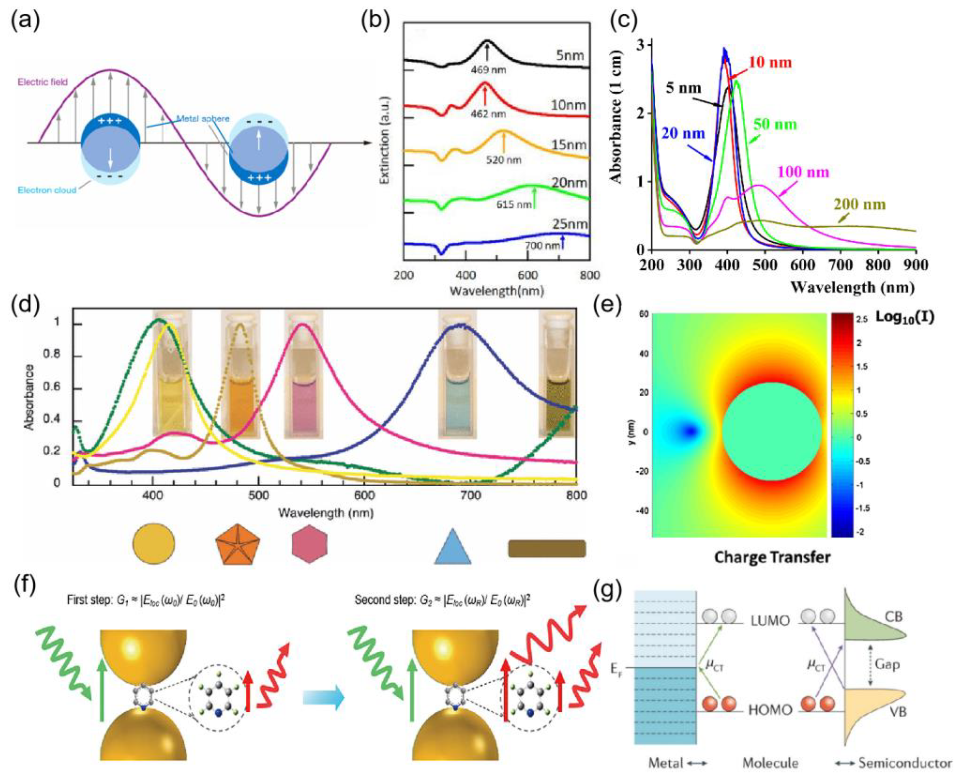
2. Surface Plasmon

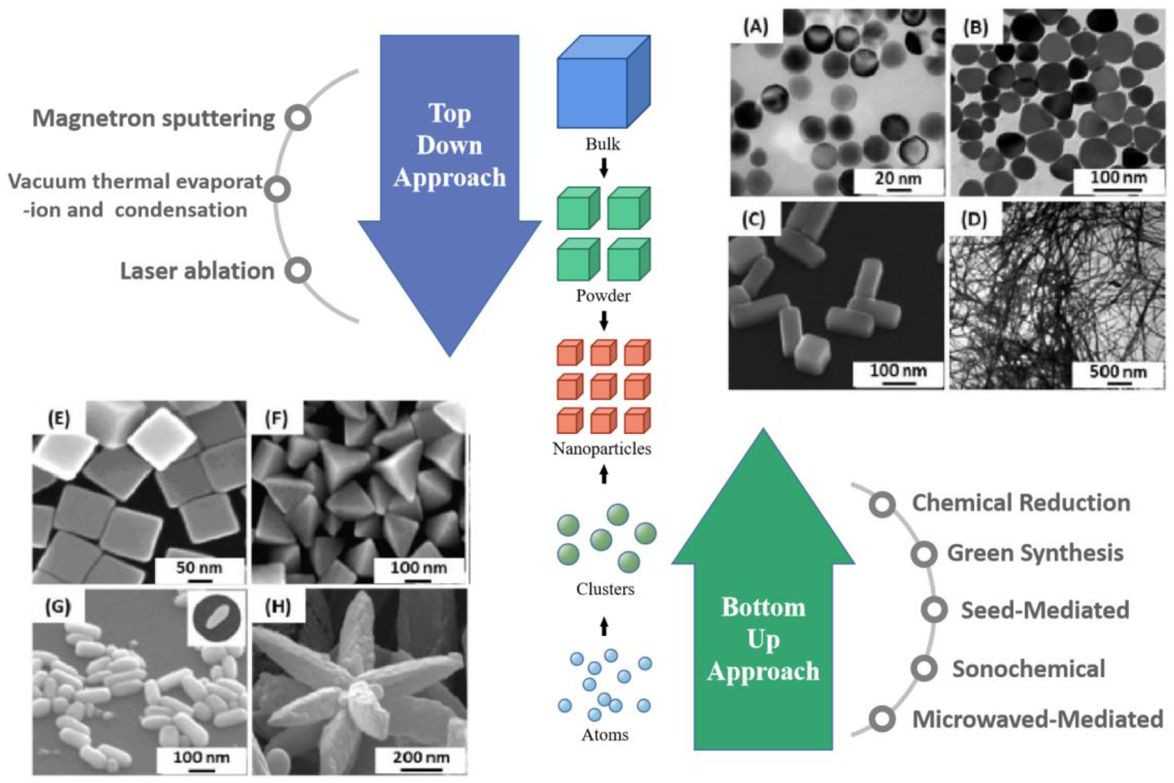
3. Synthesis of Silver Nanostructure
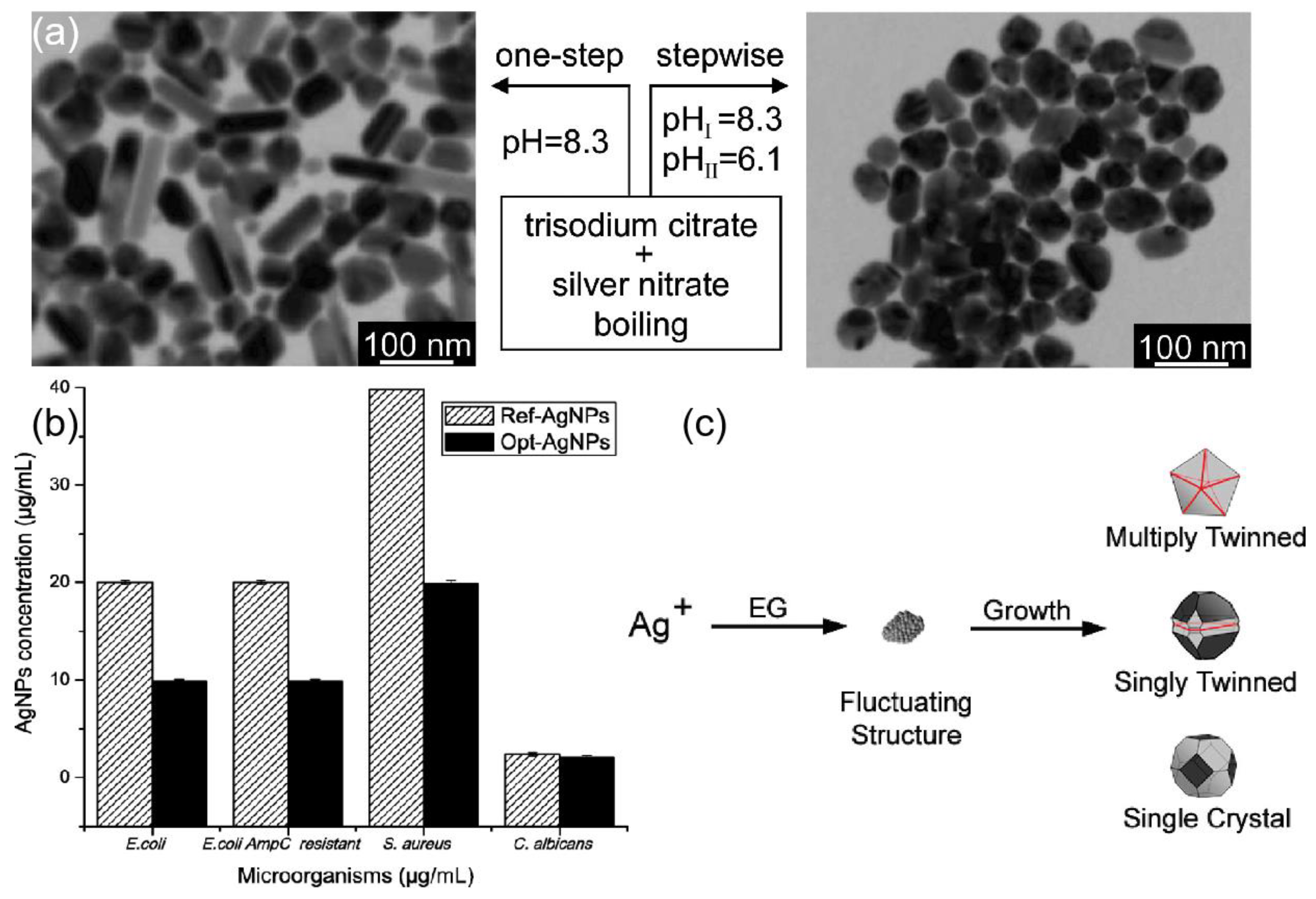
3.1. Chemical Reduction
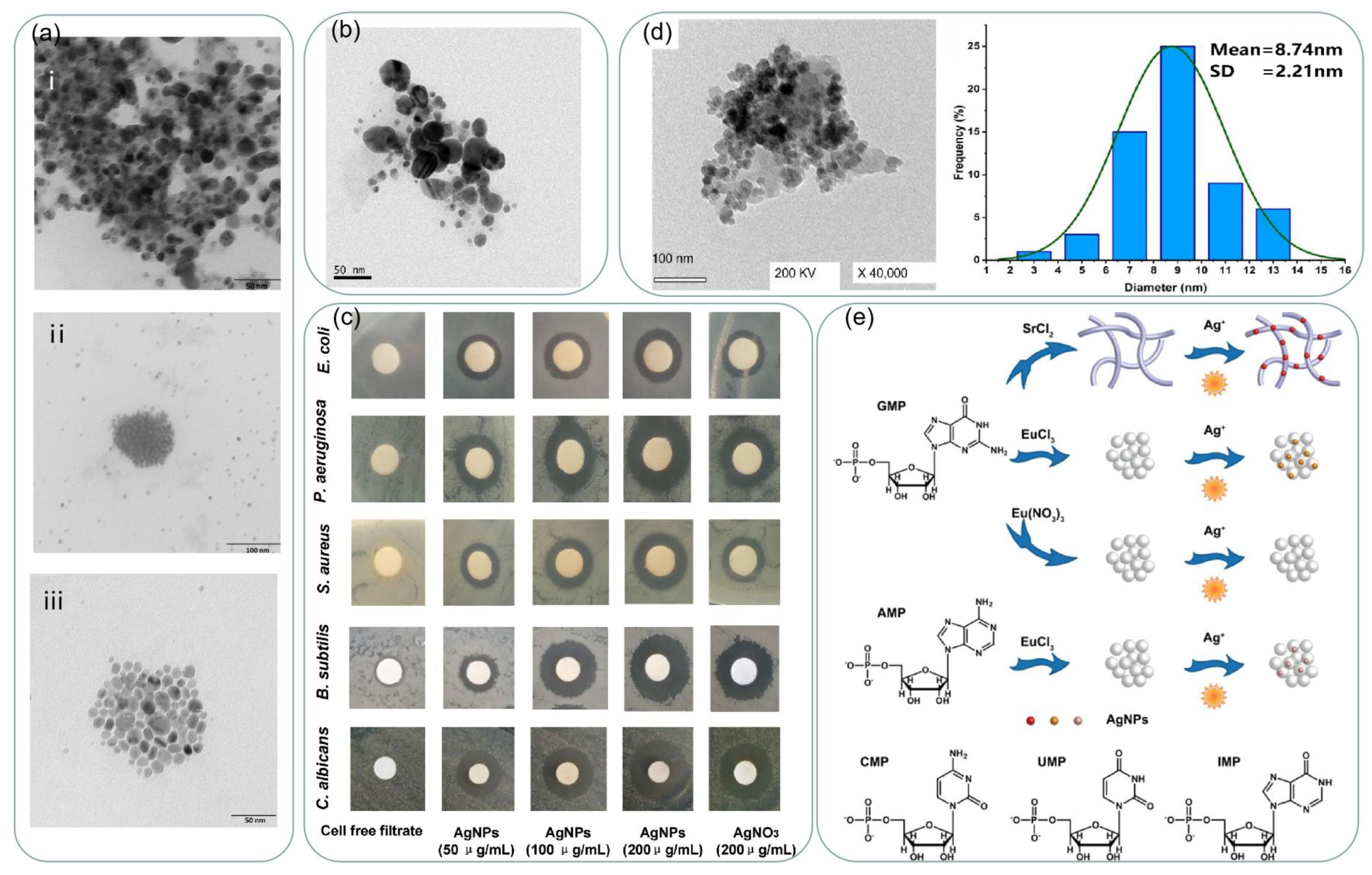
3.2. Green Synthesis

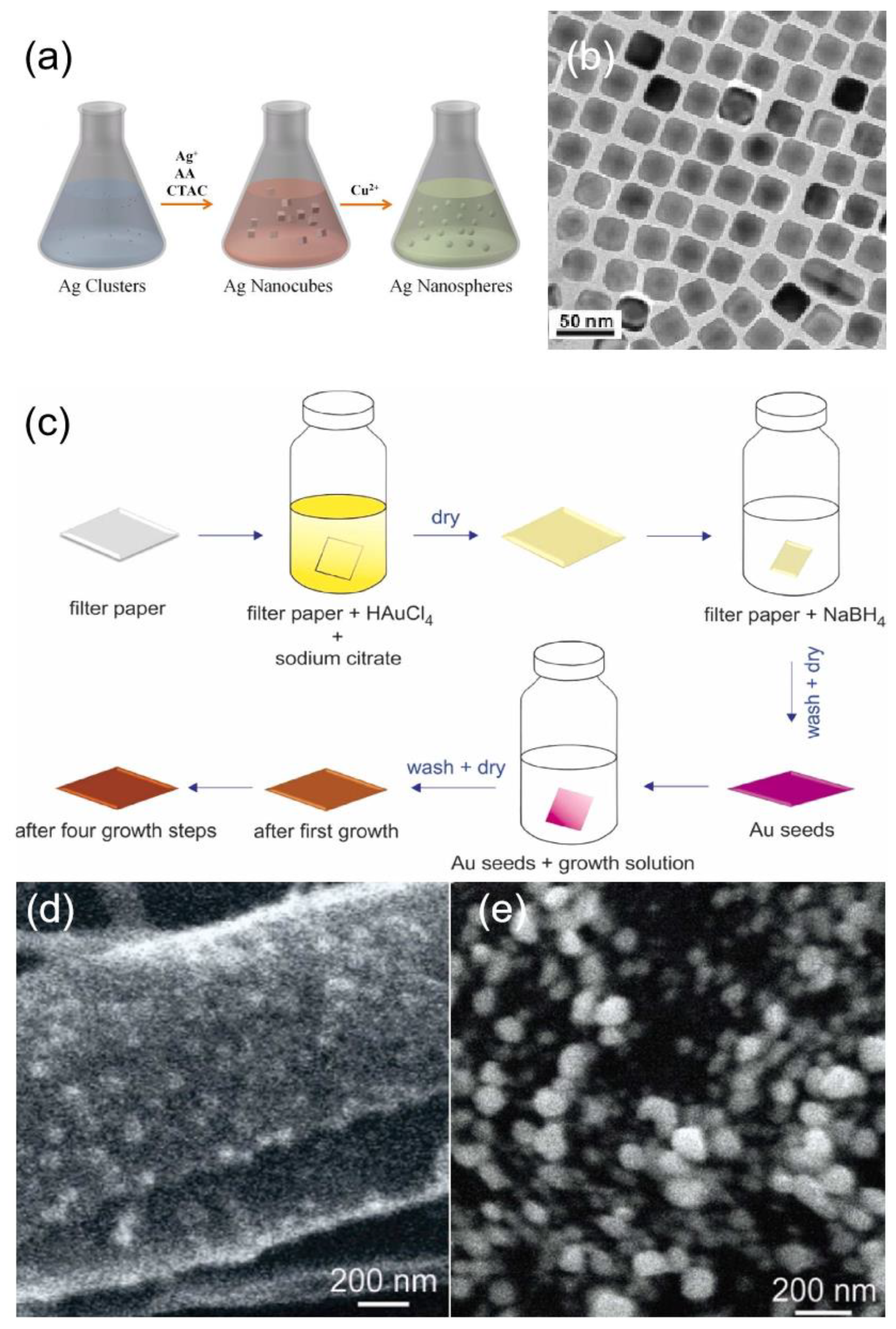
3.3. Seed-Mediated Growth

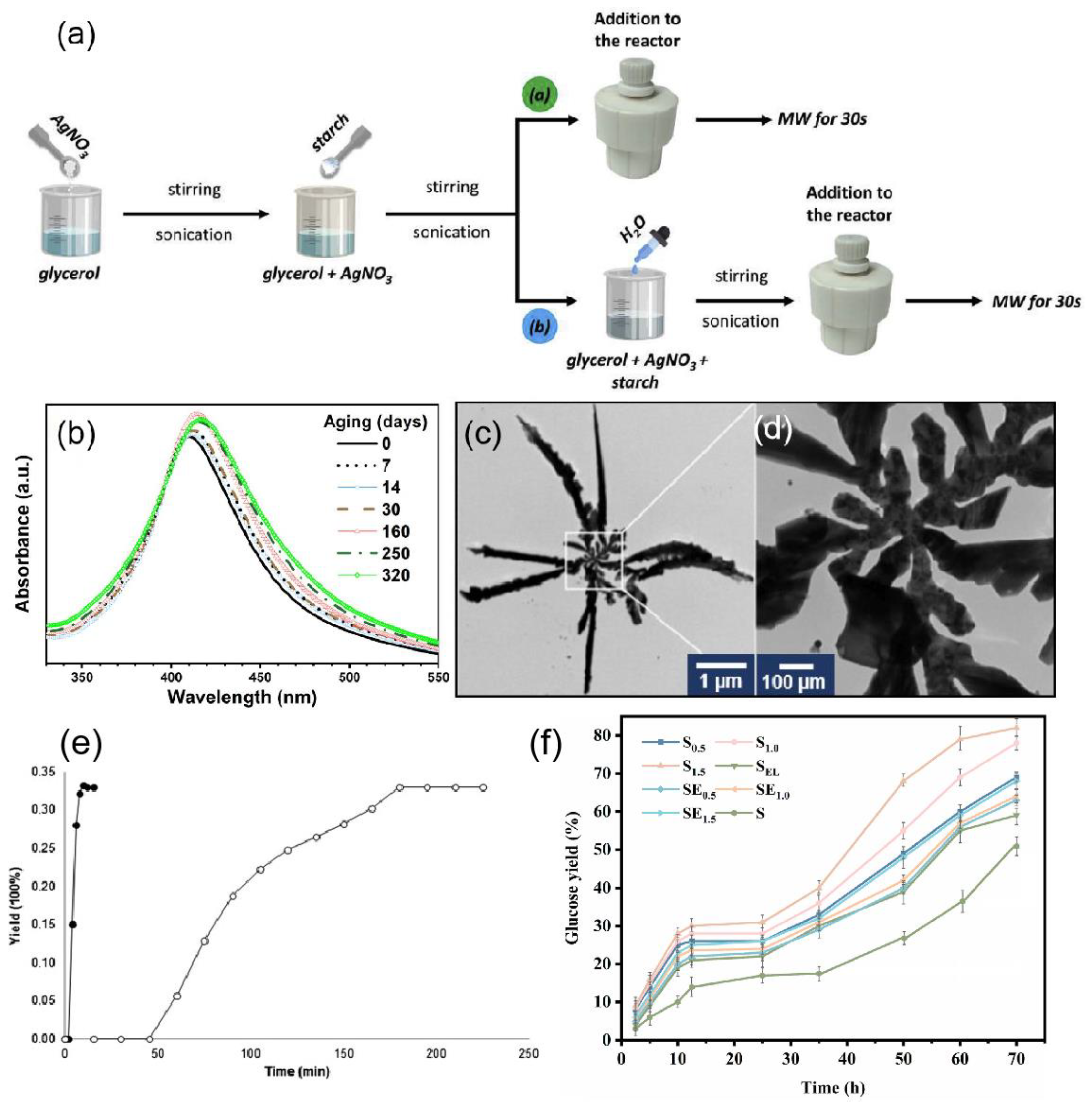
3.4. Microwave-Assisted Growth

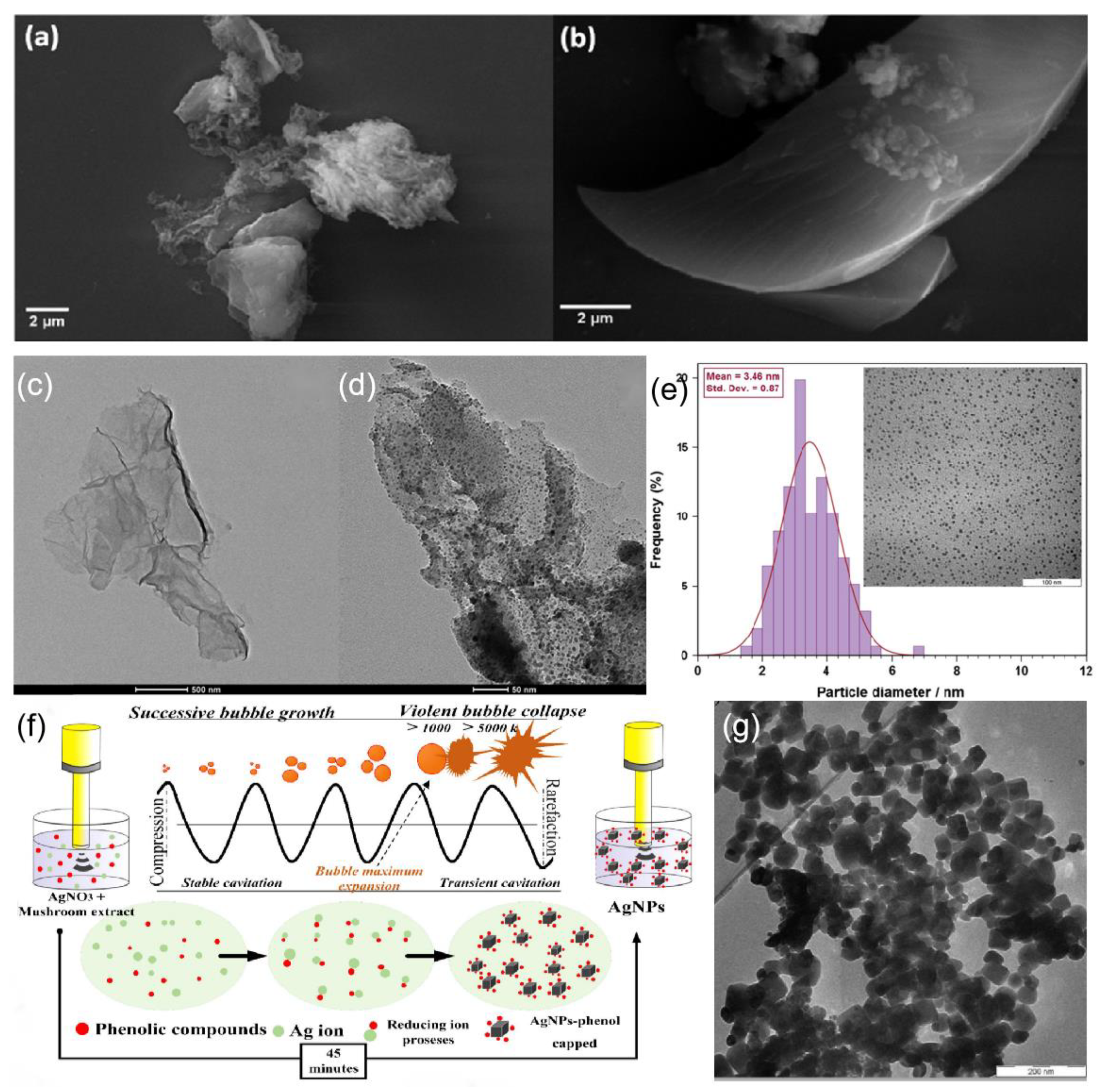
3.5. Sonochemical-Mediated Synthesis
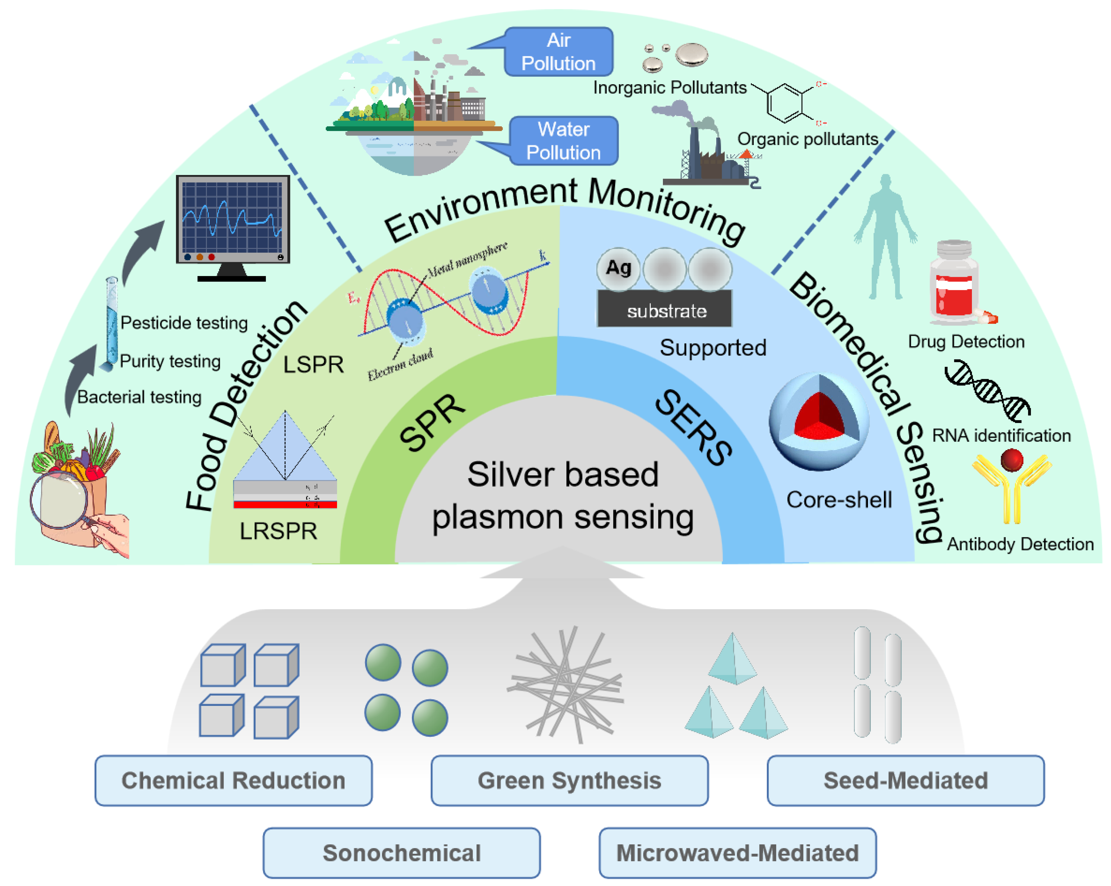
4. Applications of Silver-Based Plasmonic Sensing
4.1. Silver-Based Surface Plasmon Resonance Sensing Applications
4.1.1. Food Detection
4.1.2. Environmental Monitoring
4.1.3. Biomedical Sensing
4.2. Silver-Based Surface Enhanced Raman Scattering Sensing Applications
4.2.1. Supported Structure

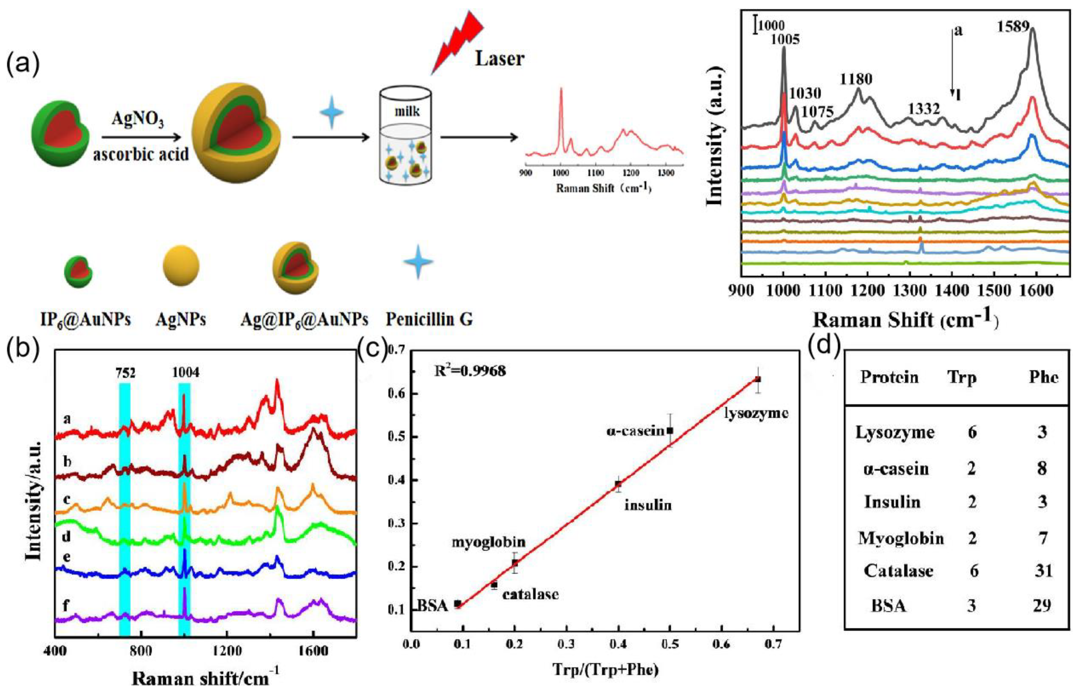
4.2.2. Core-Shell Structure
5. Summary and Future Prospect
Author Contributions
Funding
Institutional Review Board Statement
Informed Consent Statement
Conflicts of Interest
References
- Li, M.; Cushing, S.K.; Wu, N. Plasmon-enhanced optical sensors: A review. Analyst 2015, 140, 386–406. [Google Scholar] [CrossRef] [PubMed] [Green Version]
- Kneipp, J.; Kneipp, H.; Kneipp, K. SERS—A single-molecule and nanoscale tool for bioanalytics. Chem. Soc. Rev. 2008, 37, 1052–1060. [Google Scholar] [CrossRef] [PubMed]
- Zhang, Z.; Lee, Y.; Haque, F.; Leem, J.; Hsieh, E.Y.; Nam, S. Plasmonic sensors based on graphene and graphene hybrid materials. Nano Converg. 2022, 9, 28. [Google Scholar] [CrossRef] [PubMed]
- Tittl, A.; Giessen, H.; Liu, N. Plasmonic Gas and Chemical Sensing. Nanophotonics 2014, 3, 239–272. [Google Scholar] [CrossRef] [Green Version]
- Chung, T.; Lee, S.-Y.; Song, E.Y.; Chun, H.; Lee, B. Plasmonic Nanostructures for Nano-Scale Bio-Sensing. Sensors 2011, 11, 10907–10929. [Google Scholar] [CrossRef] [Green Version]
- Thangamuthu, M.; Raziman, T.; Martin, O.J.F.; Tang, J. Review—Origin and Promotional Effects of Plasmonics in Photocatalysis. J. Electrochem. Soc. 2022, 169, 036512. [Google Scholar] [CrossRef]
- Li, S.; Miao, P.; Zhang, Y.; Wu, J.; Zhang, B.; Du, Y.; Han, X.; Sun, J.; Xu, P. Recent Advances in Plasmonic Nanostructures for Enhanced Photocatalysis and Electrocatalysis. Adv. Mater. 2020, 33, e2000086. [Google Scholar] [CrossRef]
- Dhiman, M. Plasmonic nanocatalysis for solar energy harvesting and sustainable chemistry. J. Mater. Chem. A 2020, 8, 10074–10095. [Google Scholar] [CrossRef]
- Tuniz, A. Nanoscale nonlinear plasmonics in photonic waveguides and circuits. La Riv. Nuovo Cimento 2021, 44, 193–249. [Google Scholar] [CrossRef]
- Wei, H.; Pan, D.; Zhang, S.; Li, Z.; Li, Q.; Liu, N.; Wang, W.; Xu, H. Plasmon Waveguiding in Nanowires. Chem. Rev. 2018, 118, 2882–2926. [Google Scholar] [CrossRef]
- Wang, D.; Wang, W.; Knudson, M.P.; Schatz, G.C.; Odom, T.W. Structural Engineering in Plasmon Nanolasers. Chem. Rev. 2017, 118, 2865–2881. [Google Scholar] [CrossRef] [PubMed]
- Liu, J.; He, H.; Xiao, D.; Yin, S.; Ji, W.; Jiang, S.; Luo, D.; Wang, B.; Liu, Y. Recent Advances of Plasmonic Nanoparticles and their Applications. Materials 2018, 11, 1833. [Google Scholar] [CrossRef] [Green Version]
- Vilela, D.; González, M.C.; Escarpa, A. Sensing colorimetric approaches based on gold and silver nanoparticles aggregation: Chemical creativity behind the assay. A review. Anal. Chim. Acta 2012, 751, 24–43. [Google Scholar] [CrossRef] [PubMed]
- Khurana, K.; Jaggi, N. Localized Surface Plasmonic Properties of Au and Ag Nanoparticles for Sensors: A Review. Plasmonics 2021, 16, 981–999. [Google Scholar] [CrossRef]
- Tuersun, P.; Yusufu, T.; Yimiti, A.; Sidike, A. Refractive index sensitivity analysis of gold nanoparticles. Optik 2017, 149, 384–390. [Google Scholar] [CrossRef]
- Arif, M.; Farooqi, Z.H.; Irfan, A.; Begum, R. Gold nanoparticles and polymer microgels: Last five years of their happy and successful marriage. J. Mol. Liq. 2021, 336, 116270. [Google Scholar] [CrossRef]
- Liang, A.; Liu, Q.; Wen, G.; Jiang, Z. The surface-plasmon-resonance effect of nanogold/silver and its analytical applications. TrAC Trends Anal. Chem. 2012, 37, 32–47. [Google Scholar] [CrossRef]
- Rycenga, M.; Cobley, C.M.; Zeng, J.; Li, W.; Moran, C.H.; Zhang, Q.; Qin, D.; Xia, Y. Controlling the Synthesis and Assembly of Silver Nanostructures for Plasmonic Applications. Chem. Rev. 2011, 111, 3669–3712. [Google Scholar] [CrossRef] [PubMed] [Green Version]
- Pulit-Prociak, J.; Stokłosa, K.; Banach, M. Nanosilver products and toxicity. Environ. Chem. Lett. 2015, 13, 59–68. [Google Scholar] [CrossRef]
- Jiang, N.; Zhuo, X.; Wang, J. Active Plasmonics: Principles, Structures, and Applications. Chem. Rev. 2017, 118, 3054–3099. [Google Scholar] [CrossRef]
- Ordal, M.A.; Long, L.L.; Bell, R.J.; Bell, S.E.; Bell, R.R.; Alexander, R.W., Jr.; Ward, C.A. Optical properties of the metals Al, Co, Cu, Au, Fe, Pb, Ni, Pd, Pt, Ag, Ti, and W in the infrared and far infrared. Appl. Opt. 1983, 22, 1099–1190. [Google Scholar] [CrossRef] [PubMed]
- Kiba, T.; Masui, K.; Inomata, Y.; Furumoto, A.; Kawamura, M.; Abe, Y.; Kim, K.H. Control of localized surface plasmon resonance of Ag nanoparticles by changing its size and morphology. Vacuum 2021, 192, 110432. [Google Scholar] [CrossRef]
- Hlaing, M.; Gebear-Eigzabher, B.; Roa, A.; Marcano, A.; Radu, D.; Lai, C.-Y. Absorption and scattering cross-section extinction values of silver nanoparticles. Opt. Mater. 2016, 58, 439–444. [Google Scholar] [CrossRef] [Green Version]
- Stamplecoskie, K.G.; Scaiano, J.C. Silver as an Example of the Applications of Photochemistry to the Synthesis and Uses of Nanomaterials. Photochem. Photobiol. 2012, 88, 762–768. [Google Scholar] [CrossRef] [PubMed]
- Li, Z.; Xu, H. Nanoantenna effect of surface-enhanced Raman scattering: Managing light with plasmons at the nanometer scale. Adv. Phys. X 2016, 1, 492–521. [Google Scholar] [CrossRef] [Green Version]
- Moskovits, M. Surface-enhanced Raman spectroscopy: A brief retrospective. J. Raman Spectrosc. 2005, 36, 485–496. [Google Scholar] [CrossRef]
- Cao, C.; Zhang, J.; Wen, X.; Dodson, S.L.; Dao, N.T.; Wong, L.M.; Wang, S.; Li, S.; Phan, A.T.; Xiong, Q. Metamaterials-Based Label-Free Nanosensor for Conformation and Affinity Biosensing. ACS Nano 2013, 7, 7583–7591. [Google Scholar] [CrossRef]
- Doughty, A.C.; Hoover, A.R.; Layton, E.; Murray, C.K.; Howard, E.W.; Chen, W.R. Nanomaterial Applications in Photothermal Therapy for Cancer. Materials 2019, 12, 779. [Google Scholar] [CrossRef] [PubMed] [Green Version]
- Chiang, N.; Goubert, G.; Pozzi, E.A.; McAnally, M.O.; Chapman, C.; Jiang, N.; Schatz, G.C.; Van Duyne, R.P. Ultra-High Vacuum Tip-Enhanced Raman Spectroscopy. Chem. Rev. 2017, 117, 231–253. [Google Scholar] [CrossRef]
- Kong, X.-T.; Wang, Z.; Govorov, A.O. Plasmonic Nanostars with Hot Spots for Efficient Generation of Hot Electrons under Solar Illumination. Adv. Opt. Mater. 2017, 5, 15. [Google Scholar] [CrossRef] [Green Version]
- Zong, C.; Xu, M.; Xu, L.-J.; Wei, T.; Ma, X.; Zheng, X.-S.; Hu, R.; Ren, B. Surface-Enhanced Raman Spectroscopy for Bioanalysis: Reliability and Challenges. Chem. Rev. 2018, 118, 4946–4980. [Google Scholar] [CrossRef]
- Langer, J.; Jimenez de Aberasturi, D.; Aizpurua, J.; Alvarez-Puebla, R.A.; Auguié, B.; Baumberg, J.J.; Bazan, G.C.; Bell, S.E.J.; Boisen, A.; Brolo, A.G.; et al. Present and Future of Surface-Enhanced Raman Scattering. ACS Nano 2020, 14, 28–117. [Google Scholar] [CrossRef] [Green Version]
- Kahraman, M.; Mullen, E.R.; Korkmaz, A.; Wachsmann-Hogiu, S. Fundamentals and applications of SERS-based bioanalytical sensing. Nanophotonics 2017, 6, 831–852. [Google Scholar] [CrossRef] [Green Version]
- Szaniawska, A.; Kudelski, A. Applications of Surface-Enhanced Raman Scattering in Biochemical and Medical Analysis. Front. Chem. 2021, 9, 664134. [Google Scholar] [CrossRef] [PubMed]
- Xia, J.; Li, W.; Sun, M.; Wang, H. Application of SERS in the Detection of Fungi, Bacteria and Viruses. Nanomaterials 2022, 12, 3572. [Google Scholar] [CrossRef]
- Le Ru, E.C.; Blackie, E.; Meyer, M.; Etchegoin, P.G. Surface Enhanced Raman Scattering Enhancement Factors: A Comprehensive Study. J. Phys. Chem. C 2007, 111, 13794–13803. [Google Scholar] [CrossRef]
- Slepička, P.; Kasálková, N.S.; Siegel, J.; Kolská, Z.; Švorčík, V. Methods of Gold and Silver Nanoparticles Preparation. Materials 2019, 13, 1. [Google Scholar] [CrossRef] [PubMed] [Green Version]
- OhadiFar, P.; Shahidi, S.; Dorranian, D. Synthesis of Silver Nanoparticles and Exhaustion on Cotton Fabric Simultaneously Using Laser Ablation Method. J. Nat. Fibers 2018, 17, 1295–1306. [Google Scholar] [CrossRef]
- Rezaee, S. The effect of deposition time on the structural properties of silver nanoparticles deposited on anodic alumina templates. Results Phys. 2018, 9, 1521–1524. [Google Scholar] [CrossRef]
- Tuchin, V.V.; Genina, E.A.; Postnov, D.E.; Derbov, V.L.; Baranov, M.S.; Khramov, V.N.; Lotin, A.A.; Khaydukov, E.V. Fabrication, size control and functionalization of silver nanoparticles by pulsed laser ablation synthesis in liquid. In Saratov Fall Meeting 2016: Optical Technologies in Biophysics and Medicine XVIII; SPIE: Cergy-Pontoise, France, 2017. [Google Scholar]
- Wiley, B.; Herricks, T.; Sun, Y.; Xia, Y. Polyol Synthesis of Silver Nanoparticles: Use of Chloride and Oxygen to Promote the Formation of Single-Crystal, Truncated Cubes and Tetrahedrons. Nano Lett. 2004, 4, 1733–1739. [Google Scholar] [CrossRef]
- Darmanin, T.; Nativo, P.; Gilliland, D.; Ceccone, G.; Pascual, C.; De Berardis, B.; Guittard, F.; Rossi, F. Microwave-assisted synthesis of silver nanoprisms/nanoplates using a “modified polyol process”. Colloids Surfaces A Physicochem. Eng. Asp. 2011, 395, 145–151. [Google Scholar] [CrossRef]
- Wiley, B.J.; Chen, Y.; McLellan, J.M.; Xiong, Y.; Li, Z.-Y.; Ginger, D.; Xia, Y. Synthesis and Optical Properties of Silver Nanobars and Nanorice. Nano Lett. 2007, 7, 1032–1036. [Google Scholar] [CrossRef]
- Murphy, C.; Jana, N. Controlling the Aspect Ratio of Inorganic Nanorods and Nanowires. Adv. Mater. 2002, 14, 80–82. [Google Scholar] [CrossRef]
- Wiley, B.J.; Im, S.H.; Li, Z.-Y.; McLellan, J.; Siekkinen, A.; Xia, Y. Maneuvering the Surface Plasmon Resonance of Silver Nanostructures through Shape-Controlled Synthesis. J. Phys. Chem. B 2006, 110, 15666–15675. [Google Scholar] [CrossRef] [PubMed]
- Pourjavadi, A.; Soleyman, R. Novel silver nano-wedges for killing microorganisms. Mater. Res. Bull. 2011, 46, 1860–1865. [Google Scholar] [CrossRef]
- Bin Ahmad, M.; Lim, J.J.; Shameli, K.; Ibrahim, N.A.; Tay, M.Y.; Chieng, B.W. Antibacterial activity of silver bionanocomposites synthesized by chemical reduction route. Chem. Central J. 2012, 6, 101. [Google Scholar] [CrossRef] [PubMed] [Green Version]
- Bahlol, H.S.; Foda, M.F.; Ma, J.; Han, H. Robust Synthesis of Size-Dispersal Triangular Silver Nanoprisms via Chemical Reduction Route and Their Cytotoxicity. Nanomaterials 2019, 9, 674. [Google Scholar] [CrossRef] [PubMed] [Green Version]
- Dzhardimalieva, G.I.; Uflyand, I.E. Preparation of metal-polymer nanocomposites by chemical reduction of metal ions: Functions of polymer matrices. J. Polym. Res. 2018, 25, 255. [Google Scholar] [CrossRef]
- Rafique, M.; Sadaf, I.; Rafique, M.S.; Tahir, M.B. A review on green synthesis of silver nanoparticles and their applications. Artif. Cells Nanomed. Biotechnol. 2017, 45, 1272–1291. [Google Scholar] [CrossRef]
- Bhattarai, B.; Zaker, Y.; Bigioni, T.P. Green synthesis of gold and silver nanoparticles: Challenges and opportunities. Curr. Opin. Green Sustain. Chem. 2018, 12, 91–100. [Google Scholar] [CrossRef]
- Ahmad, S.; Munir, S.; Zeb, N.; Ullah, A.; Khan, B.; Ali, J.; Bilal, M.; Omer, M.; Alamzeb, M.; Salman, S.M.; et al. Green nanotechnology: A review on green synthesis of silver nanoparticles—An ecofriendly approach. Int. J. Nanomed. 2019, 14, 5087–5107. [Google Scholar] [CrossRef] [Green Version]
- Niu, W.; Zhang, L.; Xu, G. Seed-mediated growth method for high-quality noble metal nanocrystals. Sci. China Chem. 2012, 55, 2311–2317. [Google Scholar] [CrossRef]
- Barhoum, A.; Rehan, M.; Rahier, H.; Bechelany, M.; Van Assche, G. Seed-Mediated Hot-Injection Synthesis of Tiny Ag Nanocrystals on Nanoscale Solid Supports and Reaction Mechanism. ACS Appl. Mater. Interfaces 2016, 8, 10551–10561. [Google Scholar] [CrossRef]
- Zhao, T.; Lian, M.-M.; Qin, Y.; Zhu, J.-F.; Kong, X.-G.; Yang, J.-F. Improved performances of lithium disilicate glass-ceramics by seed induced crystallization. J. Adv. Ceram. 2021, 10, 614–626. [Google Scholar] [CrossRef]
- Tsuji, M. Microwave-Assisted Synthesis of Metallic Nanomaterials in Liquid Phase. ChemistrySelect 2017, 2, 805–819. [Google Scholar] [CrossRef]
- Wang, J.; Cao., L.; Zhang, Y.; Liu, Y.; Fang, H.; Chen, J. Effect of mass transfer channels on flexural strength of C/SiC composites fabricated by femtosecond laser assisted CVI method with optimized laser power. Journal of Advanced Ceramics 2021, 10, 227–236. [Google Scholar] [CrossRef]
- Kumar, A.; Kuang, Y.; Liang, Z.; Sun, X. Microwave chemistry, recent advancements, and eco-friendly microwave-assisted synthesis of nanoarchitectures and their applications: A review. Mater. Today Nano 2020, 11, 100076. [Google Scholar] [CrossRef]
- Sáez, V.; Mason, T.J. Sonoelectrochemical Synthesis of Nanoparticles. Molecules 2009, 14, 4284–4299. [Google Scholar] [CrossRef] [PubMed] [Green Version]
- Bayen, S.P.; Mondal, M.K.; Naaz, S.; Mondal, S.K.; Chowdhury, P. Design and sonochemical synthesis of water-soluble fluorescent silver nanoclusters for Hg 2+ sensing. J. Environ. Chem. Eng. 2016, 4, 1110–1116. [Google Scholar] [CrossRef]
- Li, Z.; Zhuang, T.; Dong, J.; Wang, L.; Xia, J.; Wang, H.; Cui, X.; Wang, Z. Sonochemical fabrication of inorganic nanoparticles for applications in catalysis. Ultrason. Sonochemistry 2020, 71, 105384. [Google Scholar] [CrossRef]
- Lee, P.C.; Meisel, D. Adsorption and surface-enhanced Raman of dyes on silver and gold sols. J. Phys. Chem. 1982, 86, 3391–3395. [Google Scholar] [CrossRef]
- Pillai, Z.S.; Kamat, P.V. What Factors Control the Size and Shape of Silver Nanoparticles in the Citrate Ion Reduction Method? J. Phys. Chem. B 2004, 108, 945–951. [Google Scholar] [CrossRef]
- Ranoszek-Soliwoda, K.; Tomaszewska, E.; Socha, E.; Krzyczmonik, P.; Ignaczak, A.; Orlowski, P.; Krzyzowska, M.; Celichowski, G.; Grobelny, J. The role of tannic acid and sodium citrate in the synthesis of silver nanoparticles. J. Nanoparticle Res. 2017, 19, 273. [Google Scholar] [CrossRef] [Green Version]
- Dong, X.; Ji, X.; Wu, H.; Zhao, L.; Li, J.; Yang, W. Shape Control of Silver Nanoparticles by Stepwise Citrate Reduction. J. Phys. Chem. C 2009, 113, 6573–6576. [Google Scholar] [CrossRef]
- Bankura, K.; Maity, D.; Mollick, M.; Mondal, D.; Bhowmick, B.; Bain, M.; Chakraborty, A.; Sarkar, J.; Acharya, K.; Chattopadhyay, D. Synthesis, characterization and antimicrobial activity of dextran stabilized silver nanoparticles in aqueous medium. Carbohydr. Polym. 2012, 89, 1159–1165. [Google Scholar] [CrossRef]
- Quintero-Quiroz, C.; Acevedo, N.; Zapata-Giraldo, J.; Botero, L.E.; Quintero, C.; Triviño, D.G.Z.; Saldarriaga, J.; Pérez, V.Z. Optimization of silver nanoparticle synthesis by chemical reduction and evaluation of its antimicrobial and toxic activity. Biomater. Res. 2019, 23, 27. [Google Scholar] [CrossRef]
- Moradi, N.; Taghizadeh, S.-M.; Hadi, N.; Ghanbariasad, A.; Berenjian, A.; Khoo, K.S.; Varjani, S.; Show, P.L.; Ebrahiminezhad, A. Synthesis of mesoporous antimicrobial herbal nanomaterial-carrier for silver nanoparticles and antimicrobial sensing. Food Chem. Toxicol. 2022, 165, 113077. [Google Scholar] [CrossRef] [PubMed]
- Irfan, M.I.; Amjad, F.; Abbas, A.; Rehman, M.F.U.; Kanwal, F.; Saeed, M.; Ullah, S.; Lu, C. Novel Carboxylic Acid-Capped Silver Nanoparticles as Antimicrobial and Colorimetric Sensing Agents. Molecules 2022, 27, 3363. [Google Scholar] [CrossRef] [PubMed]
- Wiley, B.; Sun, Y.; Xia, Y. Synthesis of silver nanostructures with controlled shapes and properties. Acc. Chem. Res. 2007, 40, 1067–1076. [Google Scholar] [CrossRef] [PubMed]
- Gautam, A.; Singh, G.; Ram, S. A simple polyol synthesis of silver metal nanopowder of uniform particles. Synth. Met. 2007, 157, 5–10. [Google Scholar] [CrossRef]
- Liao, Q.; Hou, W.; Zhang, J.; Qin, L. Controllable Preparation of Silver Nanowires and Its Application in Flexible Stretchable Electrode. Coatings 2022, 12, 1756. [Google Scholar] [CrossRef]
- Wolf, J.B.; Stawski, T.M.; Smales, G.J.; Thünemann, A.F.; Emmerling, F. Towards automation of the polyol process for the synthesis of silver nanoparticles. Sci. Rep. 2022, 12, 5769. [Google Scholar] [CrossRef]
- Huston, M.; DeBella, M.; DiBella, M.; Gupta, A. Green Synthesis of Nanomaterials. Nanomaterials 2021, 11, 2130. [Google Scholar] [CrossRef] [PubMed]
- Slawson, R.M.; Trevors, J.T.; Lee, H. Silver accumulation and resistance in Pseudomonas stutzeri. Arch. Microbiol. 1992, 158, 398–404. [Google Scholar] [CrossRef]
- Pooley, F.D. Bacteria accumulate silver during leaching of sulphide ore minerals. Nature 1982, 296, 642–643. [Google Scholar] [CrossRef]
- Klaus, T.; Joerger, R.; Olsson, E.; Granqvist, C.-G. Silver-based crystalline nanoparticles, microbially fabricated. Proc. Natl. Acad. Sci. USA 1999, 96, 13611–13614. [Google Scholar] [CrossRef] [PubMed] [Green Version]
- Shahverdi, A.R.; Minaeian, S.; Shahverdi, H.R.; Jamalifar, H.; Nohi, A.-A. Rapid synthesis of silver nanoparticles using culture supernatants of Enterobacteria: A novel biological approach. Process. Biochem. 2007, 42, 919–923. [Google Scholar] [CrossRef]
- John, M.S.; Nagoth, J.A.; Ramasamy, K.P.; Mancini, A.; Giuli, G.; Miceli, C.; Pucciarelli, S. Synthesis of Bioactive Silver Nanoparticles Using New Bacterial Strains from an Antarctic Consortium. Mar. Drugs 2022, 20, 558. [Google Scholar] [CrossRef] [PubMed]
- Khan, N.; Khan, M.; Jameel, J.; Jameel, N.; Umer, S. An Overview: Biological Organisms That Serves as Nanofactories for Metallic Nanoparticles Synthesis and Fungi Being the Most Appropriate. Bioceram. Dev. Appl. 2017, 7, 1–4. [Google Scholar] [CrossRef] [Green Version]
- Ma, L.; Su, W.; Liu, J.-X.; Zeng, X.-X.; Huang, Z.; Li, W.; Liu, Z.-C.; Tang, J.-X. Optimization for extracellular biosynthesis of silver nanoparticles by Penicillium aculeatum Su1 and their antimicrobial activity and cytotoxic effect compared with silver ions. Mater. Sci. Eng. C 2017, 77, 963–971. [Google Scholar] [CrossRef]
- Awad, M.A.; Eid, A.M.; Elsheikh, T.M.Y.; Al-Faifi, Z.E.; Saad, N.; Sultan, M.H.; Selim, S.; Al-Khalaf, A.A.; Fouda, A. Mycosynthesis, Characterization, and Mosquitocidal Activity of Silver Nanoparticles Fabricated by Aspergillus niger Strain. J. Fungi 2022, 8, 396. [Google Scholar] [CrossRef]
- Akintelu, S.A.; Bo, Y.; Folorunso, A.S. A Review on Synthesis, Optimization, Mechanism, Characterization, and Antibacterial Application of Silver Nanoparticles Synthesized from Plants. J. Chem. 2020, 2020, 3189043. [Google Scholar] [CrossRef]
- Santos, E.d.B.; Madalossi, N.V.; Sigoli, F.A.; Mazali, I.O. Silver nanoparticles: Green synthesis, self-assembled nanostructures and their application as SERS substrates. New J. Chem. 2015, 39, 2839–2846. [Google Scholar] [CrossRef] [Green Version]
- Sharma, G.; Jasuja, N.D.; Kumar, M.; Ali, M.I. Biological Synthesis of Silver Nanoparticles by Cell-Free Extract of Spirulina platensis. J. Nanotechnol. 2015, 2015, 132675. [Google Scholar]
- Pu, F.; Huang, Y.; Yang, Z.; Qiu, H.; Ren, J. Nucleotide-Based Assemblies for Green Synthesis of Silver Nanoparticles with Controlled Localized Surface Plasmon Resonances and Their Applications. ACS Appl. Mater. Interfaces 2018, 10, 9929–9937. [Google Scholar] [CrossRef] [PubMed]
- Karthik, R.; Govindasamy, M.; Chen, S.-M.; Cheng, Y.-H.; Muthukrishnan, P.; Padmavathy, S.; Elangovan, A. Biosynthesis of silver nanoparticles by using Camellia japonica leaf extract for the electrocatalytic reduction of nitrobenzene and photocatalytic degradation of Eosin-Y. J. Photochem. Photobiol. B Biol. 2017, 170, 164–172. [Google Scholar] [CrossRef]
- Shaikh, W.A.; Chakraborty, S.; Owens, G.; Islam, R.U. A review of the phytochemical mediated synthesis of AgNP (silver nanoparticle): The wonder particle of the past decade. Appl. Nanosci. 2021, 11, 2625–2660. [Google Scholar] [CrossRef]
- Xia, Y.; Gilroy, K.D.; Peng, H.-C.; Xia, X. Seed-Mediated Growth of Colloidal Metal Nanocrystals. Angew. Chem. Int. Ed. 2017, 56, 60–95. [Google Scholar] [CrossRef]
- Lin, X.; Lin, S.; Liu, Y.; Gao, M.; Zhao, H.; Liu, B.; Hasi, W.; Wang, L. Facile Synthesis of Monodisperse Silver Nanospheres in Aqueous Solution via Seed-Mediated Growth Coupled with Oxidative Etching. Langmuir 2018, 34, 6077–6084. [Google Scholar] [CrossRef]
- Hegde, H.; Santhosh, C.; Sinha, R.K. Seed mediated synthesis of highly stable CTAB capped triangular silver nanoplates for LSPR sensing. Mater. Res. Express 2019, 6, 105075. [Google Scholar] [CrossRef]
- Zong, R.; Wang, X.; Shi, S.; Zhu, Y. Kinetically controlled seed-mediated growth of narrow dispersed silver nanoparticles up to 120 nm: Secondary nucleation, size focusing, and Ostwald ripening. Phys. Chem. Chem. Phys. 2014, 16, 4236–4241. [Google Scholar] [CrossRef]
- Fan, F.-R.; Liu, D.-Y.; Wu, Y.-F.; Duan, S.; Xie, Z.-X.; Jiang, Z.-Y.; Tian, Z.-Q. Epitaxial Growth of Heterogeneous Metal Nanocrystals: From Gold Nano-octahedra to Palladium and Silver Nanocubes. J. Am. Chem. Soc. 2008, 130, 6949–6951. [Google Scholar] [CrossRef]
- Tharion, J.; Satija, J.; Mukherji, S. Facile Synthesis of Size-Tunable Silver Nanoparticles by Heteroepitaxial Growth Method for Efficient NIR SERS. Plasmonics 2014, 10, 753–763. [Google Scholar] [CrossRef]
- Nain, A.; Barman, S.R.; Jain, S.; Mukherjee, A.; Satija, J. Dual mechanism-based sensing of mercury using unmodified, heteroepitaxially synthesized silver nanoparticles. Appl. Nanosci. 2017, 7, 299–307. [Google Scholar] [CrossRef] [Green Version]
- Batool, A.; Khan, G.A.; Ahmed, W. Seed-mediated growth of highly concentrated silver nanoparticles on a flexible substrate for applications in SERS-based trace detection. Vib. Spectrosc. 2022, 123, 103438. [Google Scholar] [CrossRef]
- Zeng, J.; Zheng, Y.; Rycenga, M.; Tao, J.; Li, Z.-Y.; Zhang, Q.; Zhu, Y.; Xia, Y. Controlling the Shapes of Silver Nanocrystals with Different Capping Agents. J. Am. Chem. Soc. 2010, 132, 8552–8553. [Google Scholar] [CrossRef] [PubMed]
- Jeon, S.-J.; Yazdi, S.; Thevamaran, R.; Thomas, E.L. Synthesis of Monodisperse Single Crystalline Ag Microcubes via Seed-Mediated Growth. Cryst. Growth Des. 2016, 17, 284–289. [Google Scholar] [CrossRef]
- Kaur, N.; Singh, A.; Ahmad, W. Microwave Assisted Green Synthesis of Silver Nanoparticles and Its Application: A Review. J. Inorg. Organomet. Polym. Mater. 2022, 1–10. [Google Scholar] [CrossRef]
- Anjana, V.N.; Joseph, M.; Francis, S.; Joseph, A.; Koshy, E.P.; Mathew, B. Microwave assisted green synthesis of silver nanoparticles for optical, catalytic, biological and electrochemical applications. Artif. Cells Nanomed. Biotechnol. 2021, 49, 438–449. [Google Scholar] [CrossRef]
- Strapasson, G.B.; Assis, M.; Backes, C.W.; Corrêa, S.A.; Longo, E.; Weibel, D.E. Microwave assisted synthesis of silver nanoparticles and its application in sustainable photocatalytic hydrogen evolution. Int. J. Hydrogen Energy 2021, 46, 34264–34275. [Google Scholar] [CrossRef]
- Revnic, R.N.; Știufiuc, G.F.; Toma, V.; Onaciu, A.; Moldovan, A.; Țigu, A.B.; Fischer-Fodor, E.; Tetean, R.; Burzo, E.; Știufiuc, R.I. Facile Microwave Assisted Synthesis of Silver Nanostars for Ultrasensitive Detection of Biological Analytes by SERS. Int. J. Mol. Sci. 2022, 23, 8830. [Google Scholar] [CrossRef] [PubMed]
- Li, Y.; Fabiano-Tixier, A.S.; Vian, M.A.; Chemat, F. Solvent-free microwave extraction of bioactive compounds provides a tool for green analytical chemistry. TrAC Trends Anal. Chem. 2013, 47, 1–11. [Google Scholar] [CrossRef]
- Gawande, M.B.; Shelke, S.N.; Zboril, R.; Varma, R.S. Microwave-Assisted Chemistry: Synthetic Applications for Rapid Assembly of Nanomaterials and Organics. Acc. Chem. Res. 2014, 47, 1338–1348. [Google Scholar] [CrossRef]
- Cheng, J.; Hu, S.-C.; Geng, Z.-C.; Zhu, M.-Q. Effect of structural changes of lignin during the microwave-assisted alkaline/ethanol pretreatment on cotton stalk for an effective enzymatic hydrolysis. Energy 2022, 254, 124402. [Google Scholar] [CrossRef]
- Devi, M.; Singh, R.; Sindhu, J.; Kumar, A.; Lal, S.; Kumar, R.; Hussain, K.; Sachdeva, M.; Singh, D.; Kumar, P. Sonochemical Protocols for Heterocyclic Synthesis: A Representative Review. Top. Curr. Chem. 2022, 380, 14. [Google Scholar] [CrossRef]
- Zhou, T.; Rong, M.; Cai, Z.; Yang, C.J.; Chen, X. Sonochemical synthesis of highly fluorescent glutathione-stabilized Ag nanoclusters and S2− sensing. Nanoscale 2012, 4, 4103–4106. [Google Scholar] [CrossRef] [Green Version]
- Kumari, S.; Sharma, P.; Yadav, S.; Kumar, J.; Vij, A.; Rawat, P.; Kumar, S.; Sinha, C.; Bhattacharya, J.; Srivastava, C.M.; et al. A Novel Synthesis of the Graphene Oxide-Silver (GO-Ag) Nanocomposite for Unique Physiochemical Applications. ACS Omega 2020, 5, 5041–5047. [Google Scholar] [CrossRef] [Green Version]
- Darroudi, M.; Zak, A.K.; Muhamad, M.; Huang, N.; Hakimi, M. Green synthesis of colloidal silver nanoparticles by sonochemical method. Mater. Lett. 2012, 66, 117–120. [Google Scholar] [CrossRef]
- Murgunde, B.K.; Mulla, R.; Rabinal, M.K. A rapid synthesis of silver nanoparticle foam by ultrasonication. J. Porous Mater. 2020, 27, 1727–1733. [Google Scholar] [CrossRef]
- Jameel, M.S.; Aziz, A.A.; Dheyab, M.A.; Khaniabadi, P.M.; Kareem, A.A.; Alrosan, M.; Ali, A.T.; Rabeea, M.A.; Mehrdel, B. Mycosynthesis of ultrasonically-assisted uniform cubic silver nanoparticles by isolated phenols from Agaricus bisporus and its antibacterial activity. Surf. Interfaces 2022, 29, 101774. [Google Scholar] [CrossRef]
- Okitsu, K.; Ashokkumar, M.; Grieser, F. Sonochemical Synthesis of Gold Nanoparticles: Effects of Ultrasound Frequency. J. Phys. Chem. B 2005, 109, 20673–20675. [Google Scholar] [CrossRef]
- Bang, J.H.; Suslick, K.S. Applications of Ultrasound to the Synthesis of Nanostructured Materials. Adv. Mater. 2010, 22, 1039–1059. [Google Scholar] [CrossRef] [PubMed]
- Wang, W.; You, Y.; Gunasekaran, S. LSPR-based colorimetric biosensing for food quality and safety. Compr. Rev. Food Sci. Food Saf. 2021, 20, 5829–5855. [Google Scholar] [CrossRef] [PubMed]
- Ravindran, N.; Kumar, S.; Yashini, M.; Rajeshwari, S.; Mamathi, C.A.; Nirmal Thirunavookarasu, S.; Sunil, C.K. Recent advances in Surface Plasmon Resonance (SPR) biosensors for food analysis: A review. Crit. Rev. Food Sci. Nutr. 2021, 1–23. [Google Scholar] [CrossRef]
- Vikas; Yadav, M.K.; Kumar, P.; Verma, R.K. Detection of adulteration in pure honey utilizing Ag-graphene oxide coated fiber optic SPR probes. Food Chem. 2020, 332, 127346. [Google Scholar] [CrossRef] [PubMed]
- Hossain, Z. Bacteria: Pseudomonas. In Encyclopedia of Food Safety; Motarjemi, Y., Ed.; Academic Press: Waltham, MA, USA, 2014; pp. 490–500. [Google Scholar]
- Mudgal, N.; Yupapin, P.; Ali, J.; Singh, G. BaTiO3-Graphene-Affinity Layer–Based Surface Plasmon Resonance (SPR) Biosensor for Pseudomonas Bacterial Detection. Plasmonics 2020, 15, 1221–1229. [Google Scholar] [CrossRef]
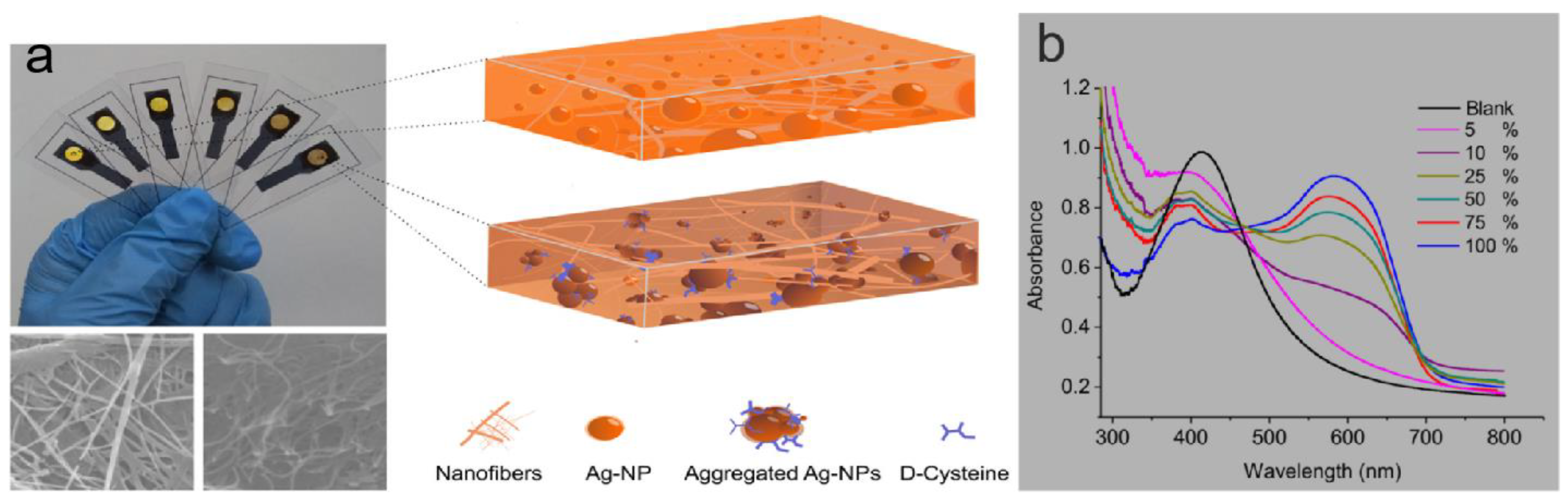
- Zor, E. Silver nanoparticles-embedded nanopaper as a colorimetric chiral sensing platform. Talanta 2018, 184, 149–155. [Google Scholar] [CrossRef]
- Trojanowicz, M.; Kaniewska, M. Chiral Biosensors and Immunosensors. In Biosensors: Emerging Materials and Applications; BoD–Books on Demand: Norderstedt, Germany, 2011. [Google Scholar]
- Baby, R.; Hussein, M.Z.; Abdullah, A.H.; Zainal, Z. Nanomaterials for the Treatment of Heavy Metal Contaminated Water. Polymers 2022, 14, 583. [Google Scholar] [CrossRef] [PubMed]
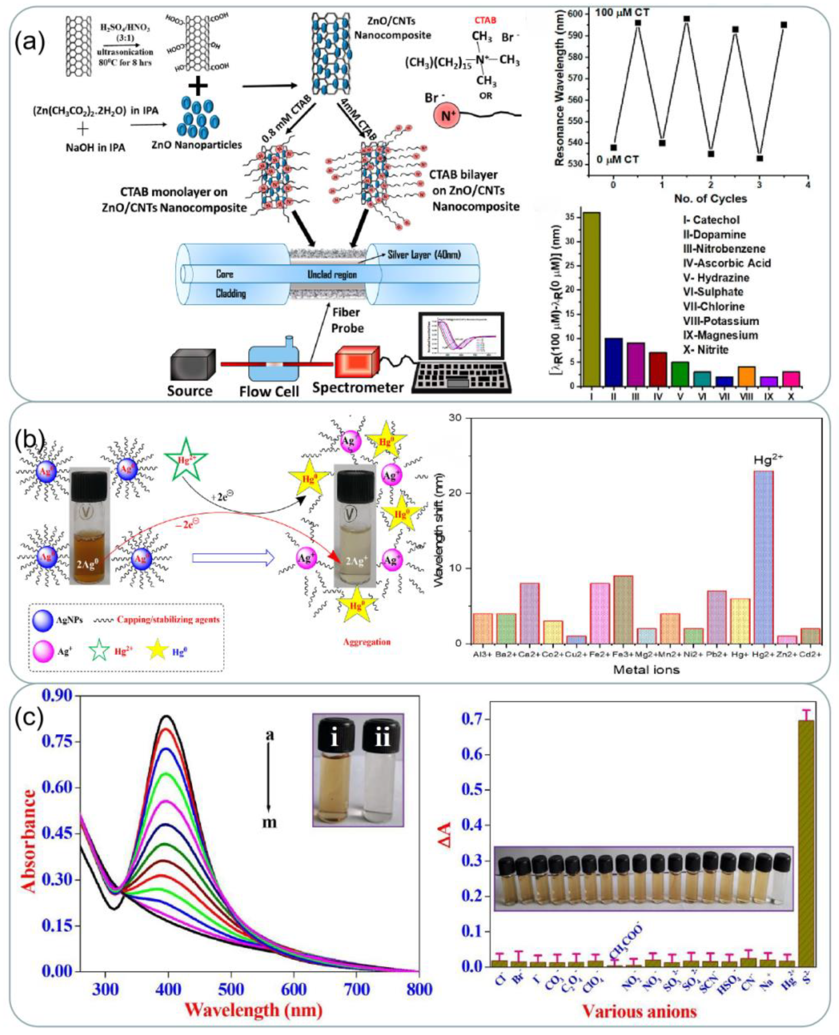
- Pathak, A.; Gupta, B.D. Fiber-Optic Plasmonic Sensor Utilizing CTAB-Functionalized ZnO Nanoparticle-Decorated Carbon Nanotubes on Silver Films for the Detection of Catechol in Wastewater. ACS Appl. Nano Mater. 2020, 3, 2582–2593. [Google Scholar] [CrossRef]
- Rajkumar, G.; Sundar, R. Sonochemical-assisted eco-friendly synthesis of silver nanoparticles (AgNPs) using avocado seed extract: Naked-eye selective colorimetric recognition of Hg2+ ions in aqueous medium. J. Mol. Liq. 2022, 368, 120638. [Google Scholar] [CrossRef]
- Rajamanikandan, R.; Ilanchelian, M. Simple smartphone merged rapid colorimetric platform for the environmental monitoring of toxic sulfide ions by cysteine functionalized silver nanoparticles. Microchem. J. 2022, 174, 107071. [Google Scholar] [CrossRef]
- Pan, N.; Bhattacharyya, N.; Banerjee, A.; Biswas, P.; Roy, L.; Chatterjee, A.; Bhattacharjee, R.; Singh, S.; Ahmed, S.A.; Chattopadhyay, A.; et al. Paper-based plasmonic nanosensor monitors environmental lead pollution in real field. New J. Chem. 2022, 46, 8177–8184. [Google Scholar] [CrossRef]
- Lee, J.-H.; Cho, H.-Y.; Choi, H.K.; Lee, J.-Y.; Choi, J.-W. Application of Gold Nanoparticle to Plasmonic Biosensors. Int. J. Mol. Sci. 2018, 19, 2021. [Google Scholar] [CrossRef] [Green Version]
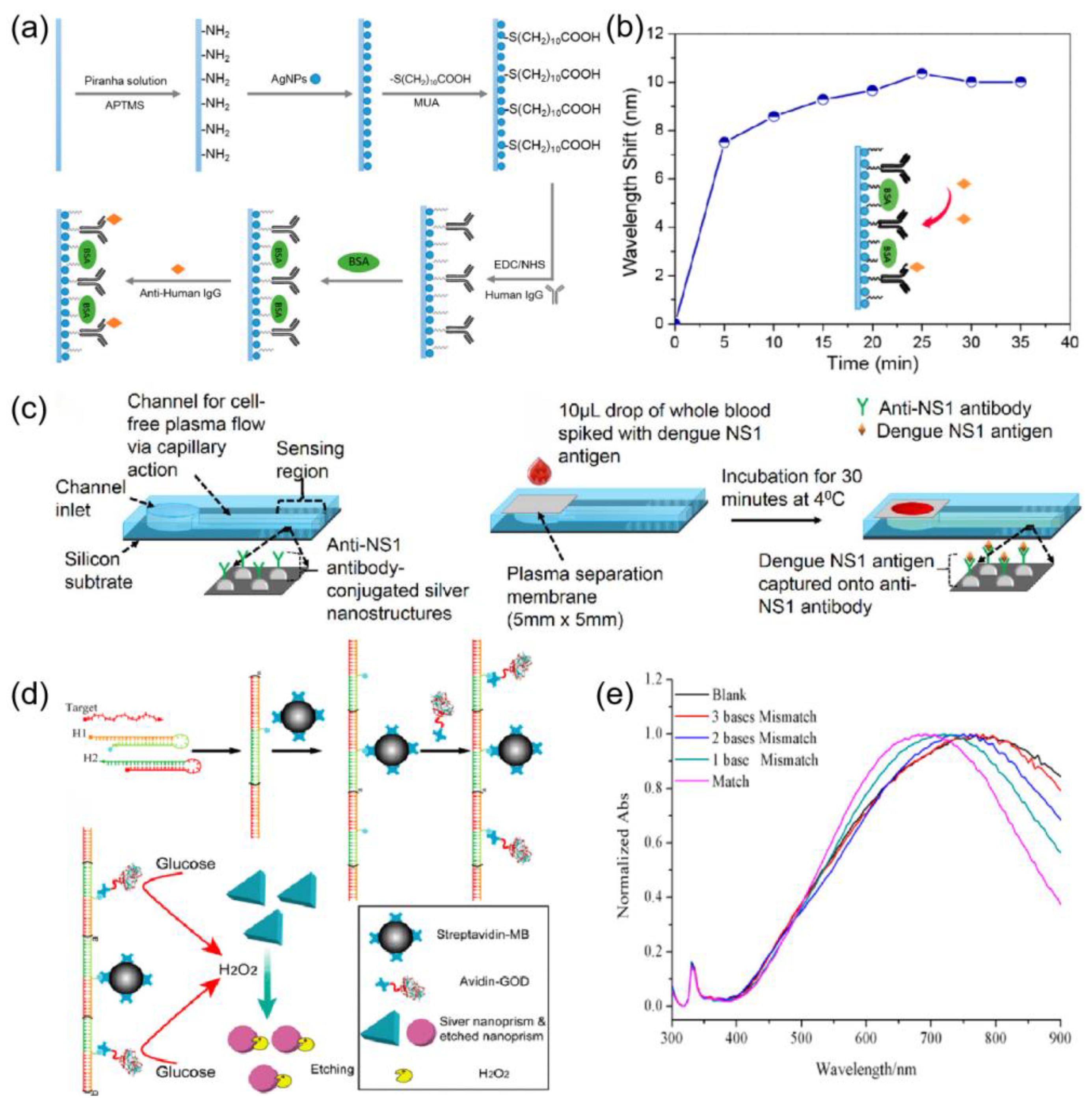
- Chen, J.; Shi, S.; Su, R.; Qi, W.; Huang, R.; Wang, M.; Wang, L.; He, Z. Optimization and Application of Reflective LSPR Optical Fiber Biosensors Based on Silver Nanoparticles. Sensors 2015, 15, 12205–12217. [Google Scholar] [CrossRef]
- Suthanthiraraj, P.P.A.; Sen, A.K. Localized surface plasmon resonance (LSPR) biosensor based on thermally annealed silver nanostructures with on-chip blood-plasma separation for the detection of dengue non-structural protein NS1 antigen. Biosens. Bioelectron. 2019, 132, 38–46. [Google Scholar] [CrossRef]
- Yang, X.; Yu, Y.; Gao, Z. A Highly Sensitive Plasmonic DNA Assay Based on Triangular Silver Nanoprism Etching. ACS Nano 2014, 8, 4902–4907. [Google Scholar] [CrossRef]
- Chen, G.; Wang, D.; Hong, W.; Sun, L.; Zhu, Y.; Chen, X. Fluorescence Enhancement on Large Area Self-Assembled Plasmonic-3D Photonic Crystals. Small 2017, 13, 1602612. [Google Scholar] [CrossRef]
- Liao, Q.; Hou, W.; Liao, K.; Chen, L.; Song, Y.; Gao, G.; Qin, L. Solid-phase sintering and vapor–liquid–solid growth of BP@MgO quantum dot crystals with a high piezoelectric response. J. Adv. Ceram. 2022, 11, 1725–1734. [Google Scholar] [CrossRef]
- Liang, Z.; Nie, Y.; Zhang, X.; Wang, P.; Ma, Q. Multiplex Electrochemiluminescence Polarization Assay Based on the Surface Plasmon Coupling Effect of Au NPs and Ag@Au NPs. Anal. Chem. 2021, 93, 7491–7498. [Google Scholar] [CrossRef]
- Wang, J. Electrochemical biosensing based on noble metal nanoparticles. Microchim. Acta 2012, 177, 245–270. [Google Scholar] [CrossRef]
- Tong, Q.; Wang, W.; Fan, Y.; Dong, L. Recent progressive preparations and applications of silver-based SERS substrates. TrAC Trends Anal. Chem. 2018, 106, 246–258. [Google Scholar] [CrossRef]
- Kassim, S.; Tahrin, R.A.A.; Harun, N.A. Metallic Core-Shell Photonic Crystals for Surface-Enhanced Raman Scattering (SERS). Plasmonics 2020, 15, 1499–1505. [Google Scholar] [CrossRef]
- Chen, C.; Zhou, X.; Ding, T.; Zhang, J.; Wang, S.; Xu, J.; Chen, J.; Dai, J.; Chen, C. Preparation and characterization of ZnO/SiO2/Ag nanoparticles as highly sensitive substrates for surface-enhanced Raman scattering. Mater. Lett. 2016, 165, 55–58. [Google Scholar] [CrossRef]
- Khaywah, M.Y.; Jradi, S.; Louarn, G.; Lacroute, Y.; Toufaily, J.; Hamieh, T.; Adam, P.-M. Ultrastable, Uniform, Reproducible, and Highly Sensitive Bimetallic Nanoparticles as Reliable Large Scale SERS Substrates. J. Phys. Chem. C 2015, 119, 26091–26100. [Google Scholar] [CrossRef]
- Nong, J.; Tang, L.; Lan, G.; Luo, P.; Li, Z.; Huang, D.; Shen, J.; Wei, W. Combined Visible Plasmons of Ag Nanoparticles and Infrared Plasmons of Graphene Nanoribbons for High-Performance Surface-Enhanced Raman and Infrared Spectroscopies. Small 2020, 17, 2004640. [Google Scholar] [CrossRef]
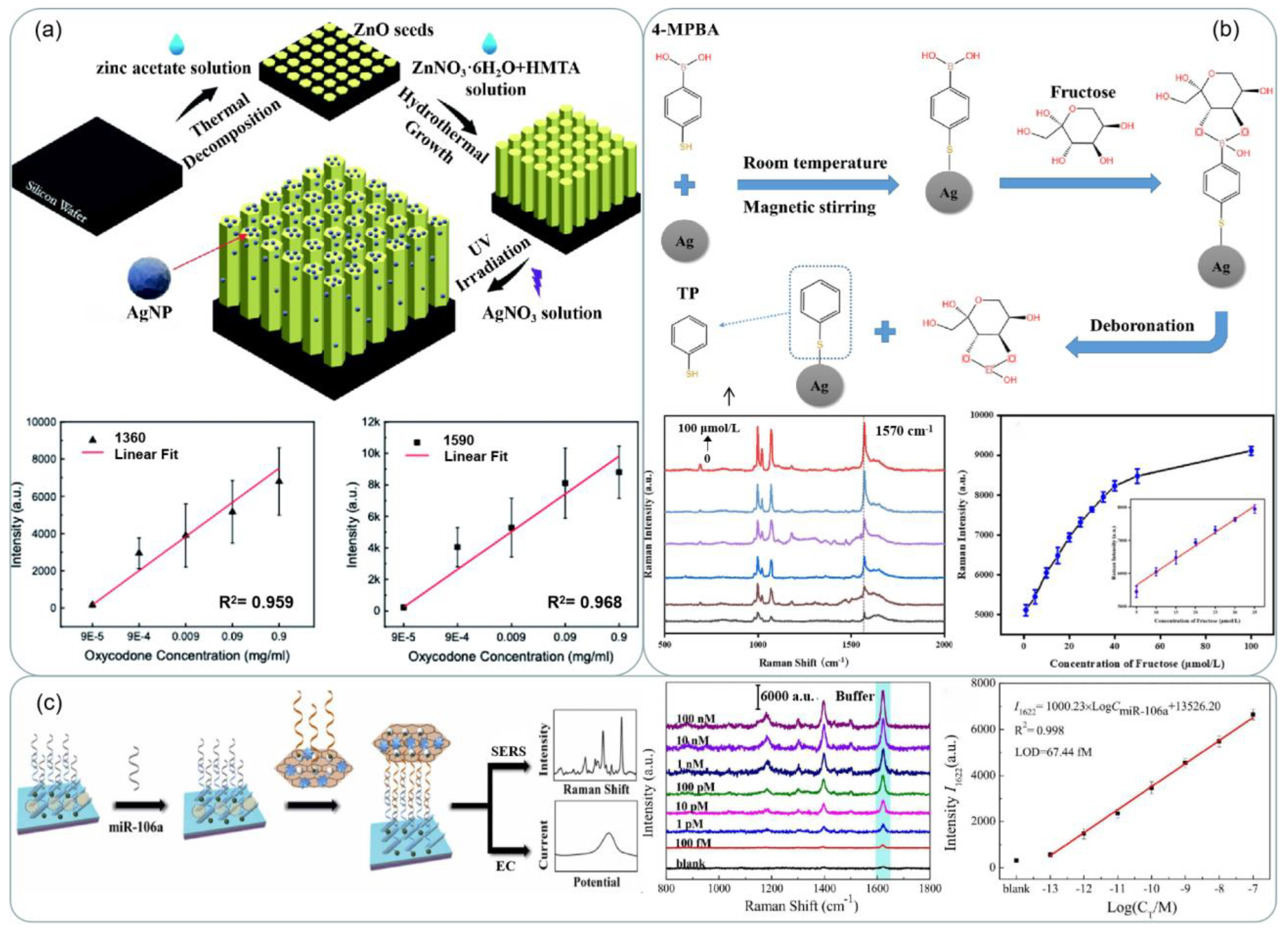
- Zhang, M.; Jin, C.; Nie, Y.; Ren, Y.; Hao, N.; Xu, Z.; Dong, L.; Zhang, J.X.J. Silver nanoparticle on zinc oxide array for label-free detection of opioids through surface-enhanced raman spectroscopy. RSC Adv. 2021, 11, 11329–11337. [Google Scholar] [CrossRef]
- Wang, K.; Qiu, Z.; Qin, Y.; Feng, L.; Huang, L.; Xiao, G. Preparation and SERS performance of silver nanowires arrays on paper by automatic writing method. Spectrochim. Acta Part A Mol. Biomol. Spectrosc. 2022, 281, 121580. [Google Scholar] [CrossRef]
- Chen, P.; Qiu, H.; Xu, S.; Liu, X.; Li, Z.; Hu, L.; Li, C.; Guo, J.; Jiang, S.; Huo, Y. A novel surface-enhanced Raman spectroscopy substrate based on a large area of MoS2 and Ag nanoparticles hybrid system. Appl. Surf. Sci. 2016, 375, 207–214. [Google Scholar] [CrossRef]
- Lv, M.; Sun, D.-W.; Pu, H.; Zhu, H. A core-shell-satellite structured Fe3O4@MIL-100(Fe)@Ag SERS substrate with adsorption, detection, degradation and recovery functionalities for selective detection of cationic dyes. Microchem. J. 2022, 183, 108137. [Google Scholar] [CrossRef]
- Bao, Y.; Li, Y.; Ling, L.; Xiang, X.; Han, X.; Zhao, B.; Guo, X. Label-Free and Highly Sensitive Detection of Native Proteins by Ag IANPs via Surface-Enhanced Raman Spectroscopy. Anal. Chem. 2020, 92, 14325–14329. [Google Scholar] [CrossRef]
- Sun, Y.; Zhai, X.; Xu, Y.; Liu, C.; Zou, X.; Li, Z.; Shi, J.; Huang, X. Facile fabrication of three-dimensional gold nanodendrites decorated by silver nanoparticles as hybrid SERS-active substrate for the detection of food contaminants. Food Control. 2020, 122, 107772. [Google Scholar] [CrossRef]
- Chen, J.; Huang, M.; Kong, L. Flexible Ag/nanocellulose fibers SERS substrate and its applications for in-situ hazardous residues detection on food. Appl. Surf. Sci. 2020, 533, 147454. [Google Scholar] [CrossRef]
- Hou, W.; Liao, Q.; Xie, S.; Song, Y.; Qin, L. Prospects and Challenges of Flexible Stretchable Electrodes for Electronics. Coatings 2022, 12, 558. [Google Scholar] [CrossRef]
- Gao, W.; Xu, J.; Cheng, C.; Qiu, S.; Jiang, S. Rapid and highly sensitive SERS detection of fungicide based on flexible “wash free” metallic textile. Appl. Surf. Sci. 2019, 512, 144693. [Google Scholar] [CrossRef]
- Liao, Q.; Yin, Y.; Zhang, J.; Si, W.; Hou, W.; Qin, L. In Situ Growth of Nanosilver on Fabric for Flexible Stretchable Electrodes. Int. J. Mol. Sci. 2022, 23, 13236. [Google Scholar] [CrossRef]
- Kamal, S.; Yang, T.C.-K. Silver enriched silver phosphate microcubes as an efficient recyclable SERS substrate for the detection of heavy metal ions. J. Colloid Interface Sci. 2022, 605, 173–181. [Google Scholar] [CrossRef]
- Butmee, P.; Samphao, A.; Tumcharern, G. Reduced graphene oxide on silver nanoparticle layers-decorated titanium dioxide nanotube arrays as SERS-based sensor for glyphosate direct detection in environmental water and soil. J. Hazard. Mater. 2022, 437, 129344. [Google Scholar] [CrossRef]
- He, L.; Ding, K.; Luo, J.; Li, Q.; Tan, J.; Hu, J. Hydrophobic plasmonic silver membrane as SERS-active catcher for rapid and ultrasensitive Cu(II) detection. J. Hazard. Mater. 2022, 440, 129731. [Google Scholar] [CrossRef]
- Loiseau, A.; Asila, V.; Boitel-Aullen, G.; Lam, M.; Salmain, M.; Boujday, S. Silver-Based Plasmonic Nanoparticles for and Their Use in Biosensing. Biosensors 2019, 9, 78. [Google Scholar] [CrossRef] [Green Version]
- Shen, J.; Chen, G.; Yang, Z.; Wu, Y.; Ma, C.; Li, L.; Yang, T.; Gu, J.; Gao, H.; Zhu, C. Boric acid-functionalized silver nanoparticles as SERS substrate for sensitive and rapid detection of fructose in artificial urine. Spectrochim. Acta Part A Mol. Biomol. Spectrosc. 2023, 288, 122179. [Google Scholar] [CrossRef]
- Zhai, J.; Li, X.; Zhang, J.; Pan, H.; Peng, Q.; Gan, H.; Su, S.; Yuwen, L.; Song, C. SERS/electrochemical dual-mode biosensor based on multi-functionalized molybdenum disulfide nanosheet probes and SERS-active Ag nanorods array electrodes for reliable detection of cancer-related miRNA. Sensors Actuators B Chem. 2022, 368, 132245. [Google Scholar] [CrossRef]
- Qi, Y.; Xing, T.-Y.; Zhao, J.; Weng, G.-J.; Li, J.-J.; Zhu, J.; Zhao, J.-W. Tuning the surface enhanced Raman scattering performance of anisotropic Au core−Ag shell hetero-nanostructure: The effect of core geometry. J. Alloys Compd. 2019, 776, 934–947. [Google Scholar] [CrossRef]
- Li, Y.; Ma, X.; Ma, J.; Zhang, Z.; Niu, Z.; Chen, F. Facile fabrication and SERS performance of polymer/Ag core-shell microspheres via the reverse breath figure accompanied by in situ reduction. Polymer 2022, 253, 125003. [Google Scholar] [CrossRef]
- Si, W.; Liao, Q.; Hou, W.; Chen, L.; Li, X.; Zhang, Z.; Sun, M.; Song, Y.; Qin, L. Low-Frequency Broadband Absorbing Coatings Based on MOFs: Design, Fabrication, Microstructure and Properties. Coatings 2022, 12, 766. [Google Scholar] [CrossRef]
- Jin, L.; She, G.; Li, J.; Xia, J.; Wang, X.; Mu, L.; Shi, W. A facile fabrication of Ag-Au-Ag nanostructures with nanogaps for intensified surface-enhanced Raman scattering. Appl. Surf. Sci. 2016, 389, 67–72. [Google Scholar] [CrossRef]
- Xin, X.; Li, Y.; Yu, L.; Li, W.; Li, J.; Lu, R. Facile synthesis of Ag@C@Ag hybrid nanoparticles as SERS substrate. Anal. Bioanal. Chem. 2021, 413, 5767–5777. [Google Scholar] [CrossRef] [PubMed]
- Wang, T.; Wang, H.; Zhu, A.; Wu, Y.; Guo, X.; Wen, Y.; Yang, H. Preparation of gold core and silver shell substrate with inositol hexaphosphate inner gap for Raman detection of trace Penicillin G. Sensors Actuators B Chem. 2021, 346, 130591. [Google Scholar] [CrossRef]


| Synthesis Technique | Application | Advantages | Disadvantages | Environment Safety | References |
|---|---|---|---|---|---|
| Chemical reduction | Biosensors Catalysis Antimicrobial | Size and structure are controllable Easier to manipulate growth and morphology by simply monitoring the reaction conditions | High cost of the reducing agent Toxic by-products are present | No | [47,48,49] |
| Green synthesis | Biosensors Catalysis Antimicrobial | Non-toxic chemicals involved Sustainable Cost-effective approach Inherently safer technique | Difficult to control the size and distribution of nanoparticles Mechanism of NP synthesis through bioreduction reaction requires additional experimentation | Yes | [50,51,52] |
| Seed mediated growth | Bactericidal Catalysis LSPR sensing | High degree of control over the size, shape, and structure | High cost The growth mechanism is not fully understood. Time intensive as well as expensive | Yes | [53,54,55] |
| Microwave-assisted synthesis | Catalysis Antimicrobial Water purification | Good reproducibility Rapid High yield and improved selectivity Narrow size distribution | In mass production, microwave penetration is insufficient. The principle of microwave-assisted synthesis needs further investigation: thermal and non-thermal effect | Yes | [56,57,58] |
| Sonochemical mediated synthesis | Catalysis Biomedical | Fast reaction The size of the generated nanostructures is small, and the dispersion of nanoparticles is low | The sonochemical reduction rate depends on the ultrasonic frequency and controlling the shape of the nanoparticles is challenging | Yes | [59,60,61] |
Disclaimer/Publisher’s Note: The statements, opinions and data contained in all publications are solely those of the individual author(s) and contributor(s) and not of MDPI and/or the editor(s). MDPI and/or the editor(s) disclaim responsibility for any injury to people or property resulting from any ideas, methods, instructions or products referred to in the content. |
© 2023 by the authors. Licensee MDPI, Basel, Switzerland. This article is an open access article distributed under the terms and conditions of the Creative Commons Attribution (CC BY) license (https://creativecommons.org/licenses/by/4.0/).
Share and Cite
Li, Y.; Liao, Q.; Hou, W.; Qin, L. Silver-Based Surface Plasmon Sensors: Fabrication and Applications. Int. J. Mol. Sci. 2023, 24, 4142. https://doi.org/10.3390/ijms24044142
Li Y, Liao Q, Hou W, Qin L. Silver-Based Surface Plasmon Sensors: Fabrication and Applications. International Journal of Molecular Sciences. 2023; 24(4):4142. https://doi.org/10.3390/ijms24044142
Chicago/Turabian StyleLi, Yinghao, Qingwei Liao, Wei Hou, and Lei Qin. 2023. "Silver-Based Surface Plasmon Sensors: Fabrication and Applications" International Journal of Molecular Sciences 24, no. 4: 4142. https://doi.org/10.3390/ijms24044142