Article

Pomegranate Seed Oil as a Source of Conjugated Linolenic Acid (CLnA) Has No Effect on Atherosclerosis Development but Improves Lipid Profile and Affects the Expression of Lipid Metabolism Genes in apoE/LDLR−/− Mice

by
Magdalena Franczyk-Żarów
1,*,
Tomasz Tarko
2,*,
Anna Drahun-Misztal
1,
Izabela Czyzynska-Cichon
3,
Edyta Kus
3 and
Renata B. Kostogrys
1
1
Department of Human Nutrition and Dietetics, Faculty of Food Technology, University of Agriculture in Krakow, A. Mickiewicz Av. 21, 31-120 Kraków, Poland
2
Department of Fermentation Technology and Microbiology, Faculty of Food Technology, University of Agriculture in Krakow, A. Mickiewicz Av. 21, 31-120 Kraków, Poland
3
Jagiellonian Centre for Experimental Therapeutics (JCET), Jagiellonian University, Bobrzynskiego St. 14, 30-348 Kraków, Poland
*
Authors to whom correspondence should be addressed.
Int. J. Mol. Sci. 2023, 24(2), 1737; https://doi.org/10.3390/ijms24021737
Submission received: 15 November 2022 / Revised: 5 January 2023 / Accepted: 13 January 2023 / Published: 15 January 2023
(This article belongs to the Special Issue Inflammatory Response in Endocrine Disorders)

Abstract

:
The aim of this study was to evaluate the anti-atherosclerotic effect of pomegranate seed oil as a source of conjugated linolenic acid (CLnA) (cis-9,trans-11,cis-13; punicic acid) compared to linolenic acid (LnA) and conjugated linoleic acid (CLA) (cis-9,trans-11) in apoE/LDLR−/− mice. In the LONG experiment, 10-week old mice were fed for the 18 weeks. In the SHORT experiment, 18-week old mice were fed for the 10 weeks. Diets were supplied with seed oils equivalent to an amount of 0.5% of studied fatty acids. In the SHORT experiment, plasma TCh and LDL+VLDL cholesterol levels were significantly decreased in animals fed CLnA and CLA compared to the Control. The expression of PPARα in liver was four-fold increased in CLnA group in the SHORT experiment, and as a consequence the expression of its target gene ACO was three-fold increased, whereas the liver’s expression of SREBP-1 and FAS were decreased in CLnA mice only in the LONG experiment. Punicic acid and CLA isomers were determined in the adipose tissue and liver in animals receiving pomegranate seed oil. In both experiments, there were no effects on the area of atherosclerotic plaque in aortic roots. However, in the SHORT experiment, the area of atherosclerosis in the entire aorta in the CLA group compared to CLnA and LnA was significantly decreased. In conclusion, CLnA improved the lipid profile and affected the lipid metabolism gene expression, but did not have the impact on the development of atherosclerotic plaque in apoE/LDLR−/− mice.

1. Introduction

Conjugated fatty acids refer to a group of positional and geometric isomers of polyunsaturated fatty acids (PUFA) with conjugated double bonds, where double bonds are separated by a single C-C bond, but not by a methylene group (-CH2-). The most studied are conjugated linoleic acids (CLA) of animal origin, with two conjugated double bonds, and conjugated linolenic acids (CLnA) of vegetable origin, with three conjugated double bonds. Recently, not only for CLA but also for conjugated linolenic acid, several unique biological effects have been found. One of the highly unsaturated forms of conjugated fatty acids of linolenic acid (LnA, cis-9,cis-12,cis-15 C18:3 n-3) is CLnA. Punicic acid (cis-9,trans-11,cis-13) was found at up to 83% in pomegranate seed oil. Seed oils of certain plants include conjugated triene fatty acids such as α-eleostearic acid (α-ESA, cis-9,trans-11,trans-13 C18:3) and calendic acid (trans-8,trans-10,cis-12 C18:3) at levels of 30–80%. There is an increasing amount of specific scientific literature concerning the biological effects and properties of CLnA. In mammals, there have been reported that some isomers of -9,-11,-13 CLnA could improve the immune response [1], markers of obesity [2,3] or have an anti-carcinogenic effect [4,5,6]. In vitro studies showed among others that punicic acid reduces apolipoprotein B100 secretion and triacylglycerol synthesis in HepG2 cells [7] and inhibits breast cancer cell proliferation [8]. These properties are closely related to those of CLA, because there have been found that -9,-11,-13 CLnA are converted into cis-9,trans-11 or trans-9,trans-11 CLA isomers [9,10,11,12].
While some progress has been made in characterizing some of the health-related effects of punicic acid, its underlying mechanisms of action are not fully understood. Punicic acid exerts an anti-inflammatory effect through the inhibition of tumor necrosis factor-α (TNF-α)-induced neutrophil hyperactivation and reactive oxygen species (ROS) production and authors concluded that this natural dietary compound may provide a novel alternative therapeutic strategy in inflammatory bowel disease [13]. Punicic acid acts as an efficient antioxidant in the rat model [14,15] and showed anti-inflammatory activity [16]. In terms of the inhibitory effect on NOS activity, the expression of hepatic NF-κB, plasma TNF-α and IL-6 level, punicic acid has a potent anti-inflammatory role. However, the anti-inflammatory effect of CLnA via suppressing the synthesis of proinflammatory cytokines (i.e., IL-6, TNF-α) seems to be related to PPARγ expression, which is supported by the findings of Changhua [17]. Moreover, naturally occurring, orally active agonists of PPAR, such as CLnA, have demonstrated some promising effects as dual or PAR agonists of PPAR [18,19]. Punicic acid ameliorates glucose tolerance and obesity-related inflammation in animal models of obesity and type 2 diabetes by acting as dual PPARα and γ agonists [7,20] with no adverse side effects detected in toxicological studies [21].
Thus, as an anti-inflammatory agent and efficient antioxidant, we have attempted to evaluate if punicic acid will have influence on the development of atherosclerotic plaque. Effects of dietary supplementation with pomegranate seed oil as a source of punicic acid on the development of atherosclerosis is not known; only hypolipidemic and hypocholesterolemic effects on plasma lipids have been shown in animal and human studies [22,23]. There is no data available so far as to whether CLnA fed to apoE/LDLR−/− mice could have health-related effects. Therefore, the major aim of our studies was to examine potential anti-atherogenic effects of conjugated trienoic fatty acid (C18:3 cis-9,trans-11,cis-13) compared to non-conjugated linolenic acid and cis-9,trans-11 CLA in a mice model of atherogenesis, e.g., apolipoprotein E and low-density lipoprotein receptor double-knockout mice (apoE/LDLR−/−) (Figure 1), as well as to describe the potential mechanisms involved in CLnA effects on metabolism.

2. Results

2.1. Effect of CLnA on Body Weight

The initial and final body weights of animals fed experimental diets (Table 1) are shown in Table 2. Mice fed the control and experimental diets in the SHORT study had similar initial body weight. After 10 weeks of treatment the final body weight of animals did not differ between groups. In the LONG study, due to younger mice (10 weeks old), initial body weight was lower in comparison to the SHORT study, but was similar among groups. After 18 weeks of study the body weight of mice fed the LnA diet was significantly increased compared to the control (23.17 vs. 21.58 g).

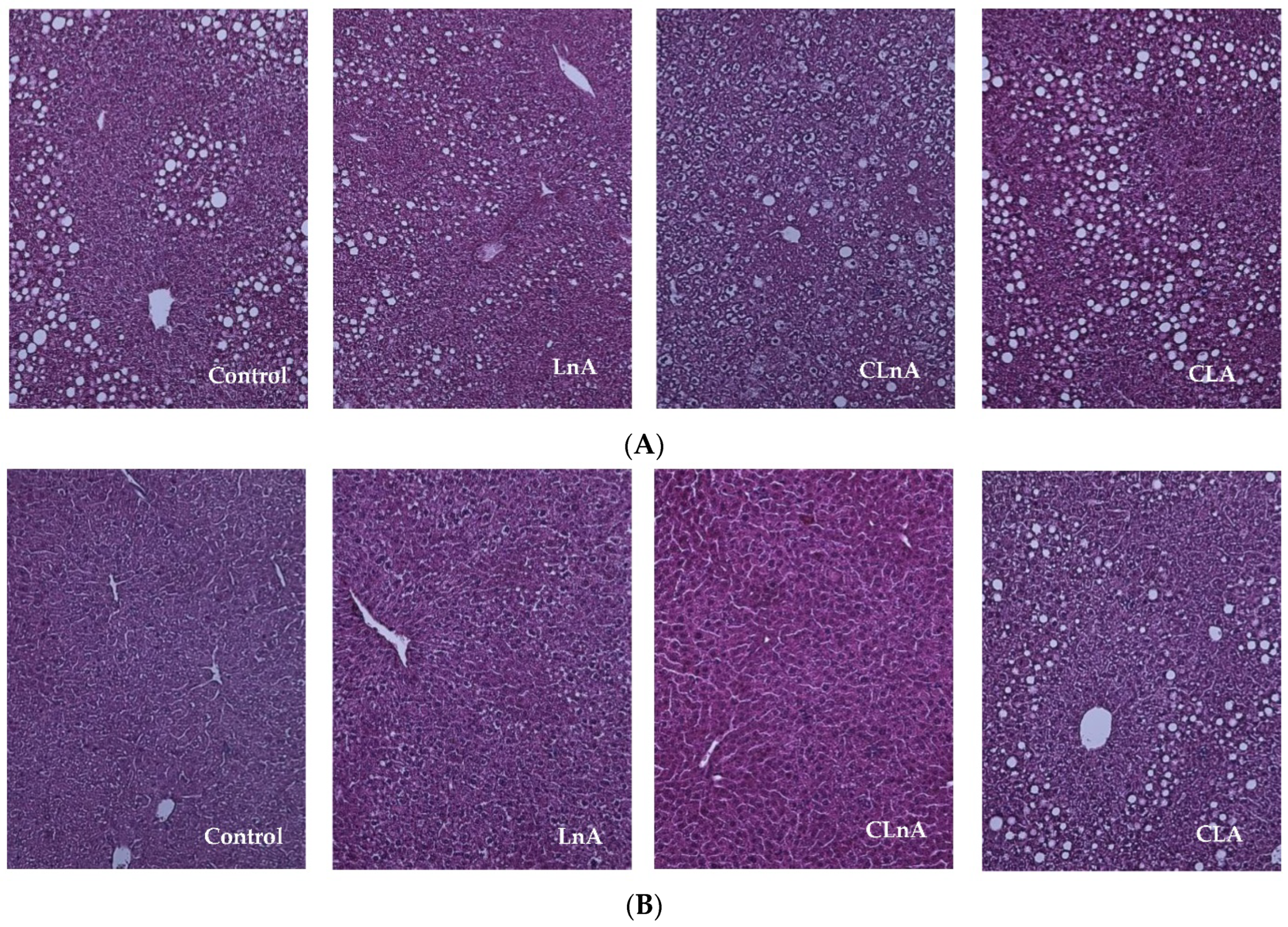
2.2. Effect of CLnA on Liver Weight and Histology

There were no significant differences in liver weight between the control and experimental groups in the SHORT and LONG studies (Table 2). In the same line, the histological examination of the livers showed no signs of the steatosis in mice fed diets with the addition of studied fatty acids. However, representative images of livers stained with H-E of the CLA-fed group seems to present slight fat accumulation compared to the control and LnA as well as CLnA groups (Figure 2A,B).

2.3. Effect of CLnA on Glucose Concentration

No significant differences in final blood glucose were observed between the experimental groups after 10 or 18 weeks of studies (Table 2). However, we observed that only the glucose concentration in the LnA group after 12 weeks of the LONG experiment was significantly (p < 0.05) increased compared to the control and CLA groups (143.2 mg/dL vs. 119.3 mg/dL and 117.0 mg/dL, respectively).

2.4. Effect of CLnA on Plasma Lipid Profile and Expression of PPARα and SREBP-1 and Their Target Genes (ACO and FAS )

The effect of dietary treatments on plasma lipid concentration is shown in Table 3. The only evident effect of inserting different fatty-acid supplementation into the diet was the significantly (p < 0.05) decreased plasma TCh and LDL+VLDL cholesterol levels in animals fed CLnA and CLA as compared to the effect of the control diet in the SHORT study. Moreover, the LDL+VLDL cholesterol concentration was the lowest in the CLnA group (6.94 mmol/L) compared to the control (14.51 mmol/L) and non-conjugated linolenic acid (LnA: 12.14 mmol/L). At the same time, HDL and TAG levels did not differ significantly. However, the concentration of TAG tended to decrease in mice fed the CLnA diet in comparison to the control (1.92 vs. 3.76 mmol/L). Nevertheless, no changes in lipid profile were observed in the LONG experiment.
Expression of transcription factors PPARα (peroxisome proliferator-activated receptor α) and SREBP-1 (sterol regulatory element binding protein-1) and their target genes ACO (acyl-CoA oxidase) and FAS (fatty acid synthase), respectively, were detected in the liver of the animals and is shown in Table 3. The results were obtained by dividing the expression of genes mentioned above by expression β-actin and are presented in arbitrary units. In the SHORT experiment, the PPARα mRNA level differed between the four groups of mice. Mice fed the diet containing pomegranate seed oil had almost four-fold higher mRNA levels of PPARα in the liver than the control mice. Moreover, the same mice showed three-fold higher level of SREBP-1 mRNA compared to the control mice. The expression of their target genes, such as ACO and FAS, were about three-fold increased in mice fed the diet in the SHORT study. In the LONG study, the SREBP-1 mRNA level as well as expression of FAS were decreased in CLnA mice. The same was true for animals fed the diet containing non-conjugated linolenic acid, whereas in CLA mice the increased expressions of SREBP-1 and FAS as well as PPARα and ACO were observed.

2.5. Effect of CLnA on Fatty Acid Profile in Tissues

The fatty acid profile in the adipose tissue and liver were investigated using chromatographic analysis.
When the fatty acid profile of adipose tissue is considered, only SFA concentration was not changed in both experiments (Table 4). Additionally, a significantly decreased level of MUFA and increased PUFA were observed in the CLA group in the SHORT study. In the same line, in the LONG study the level of MUFA was significantly decreased in the CLA group. The amount of stearic acid (C18:0) was significantly decreased in groups fed CLnA and CLA in the SHORT study. The concentration of palmitoleic acid (C16:1) was significantly increased, whereas the amount of stearic acid was significantly decreased in group fed the CLA diet in the LONG study. Moreover, the level of α-linolenic acid (C18:3) was significantly increased in the LnA group in both experiments. In groups fed pomegranate seed oil and CLA supplemented diets, the cis-9,trans-11 and trans-10,cis-12 isomers were present, but the amount of these CLA isomers was significantly increased only in the CLA group in both experiments. Additionally, in mice fed pomegranate seed oil, the supplemented-diet punicic acid was detected in the SHORT as well as in the LONG experiment.
Taking into account the fatty acid profile of liver, no significant differences between the amount of SFA, MUFA and PUFA were shown in the SHORT as well as in the LONG experiment (Table 5). The amount of α-linolenic acid (C18:3) was significantly increased in mice fed flaxseed oil supplemented diets (LnA) in both experiments. Additionally, in the SHORT study, in groups where pomegranate seed oil and CLA were provided, the cis-9,trans-11 CLA isomer was present. A the same time in the LONG experiment, not only the cis-9,trans-11 CLA isomer but also trans-10,cis-12 CLA were present, but the concentration of the cis-9,trans-11 CLA isomer was significantly increased in the group fed with the pomegranate seed oil supplemented diet. Moreover, in this group punicic acid was detected.
To summarize, the fatty acid profile was affected by different fatty-acid supplementation into the animals’ diets. Although individual isomers of CLA were not supplemented into the diet for group fed with pomegranate seed oil, CLA isomers were present in liver as well as in the adipose tissue of mice; what is more, trans-10,cis-12 was incorporated into the tissue lipids in a significantly lower proportion than cis-9,trans-11. These findings confirm that CLnA is converted into CLA isomers in model organisms.

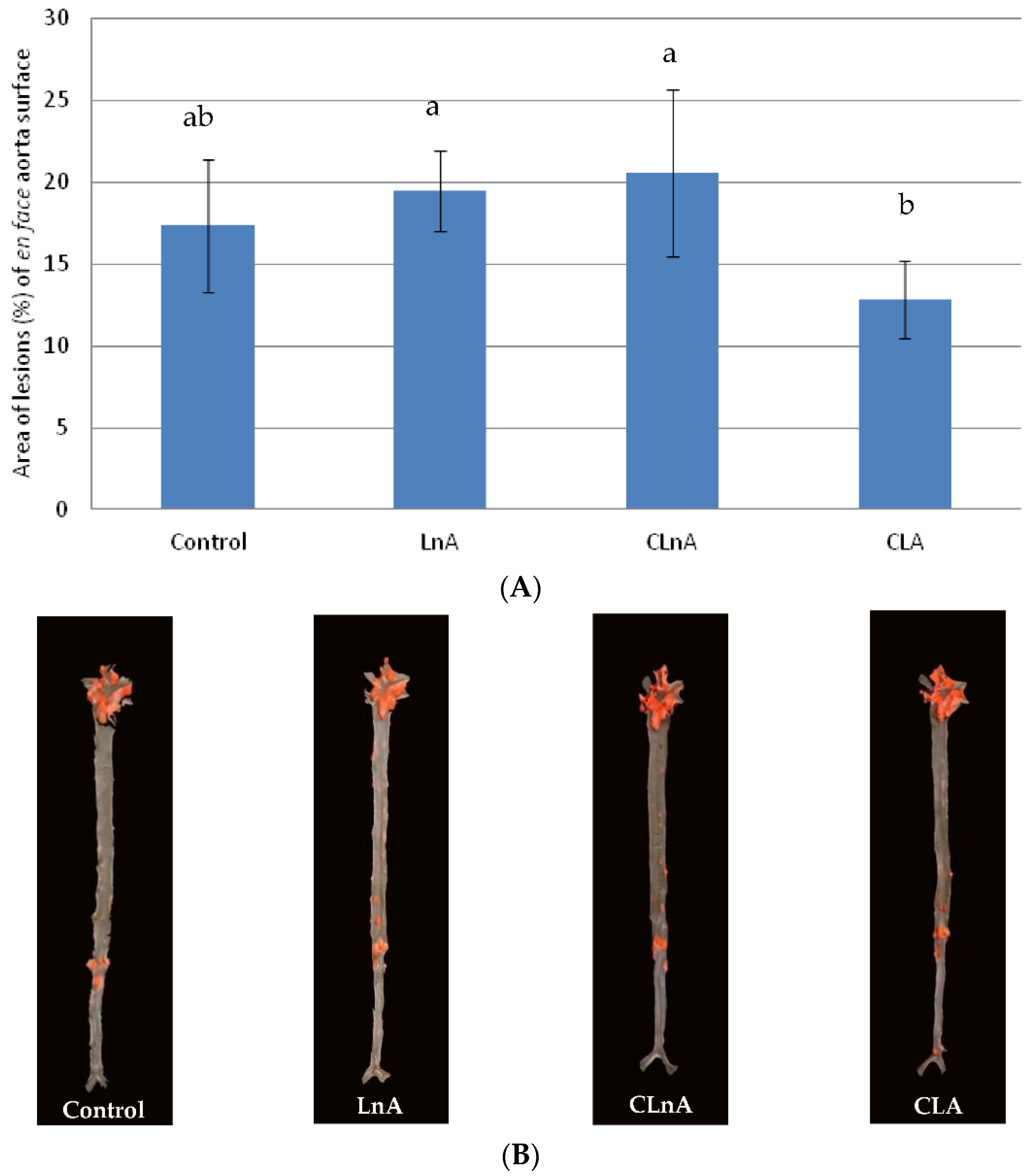
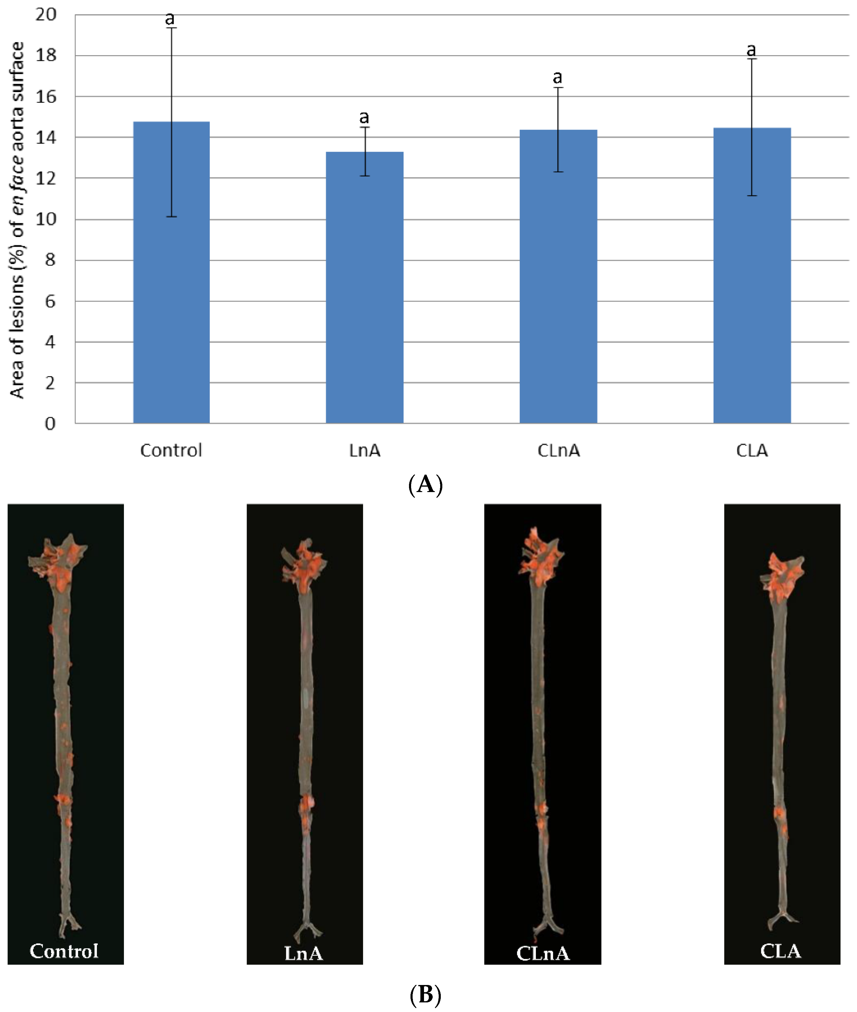
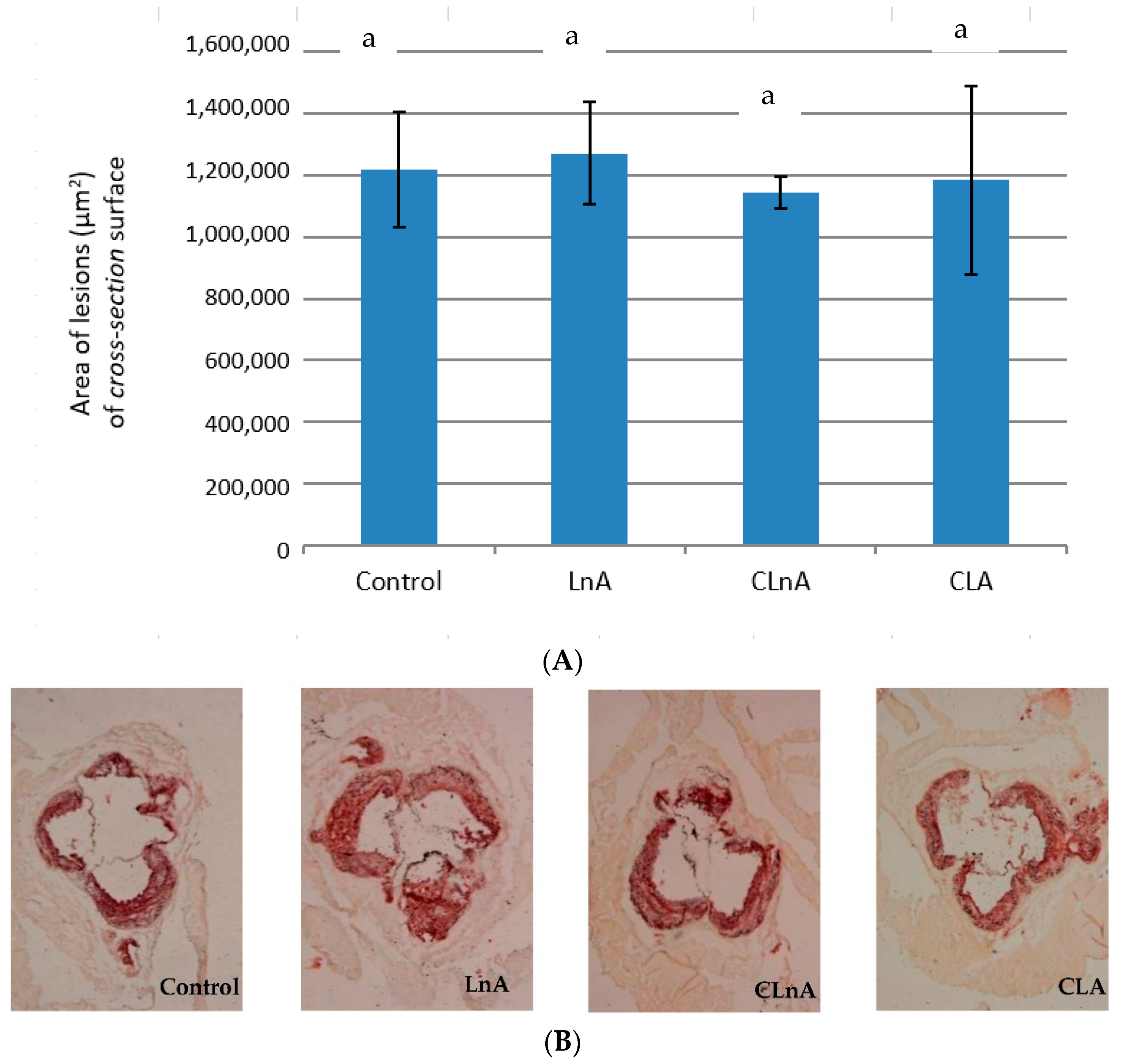
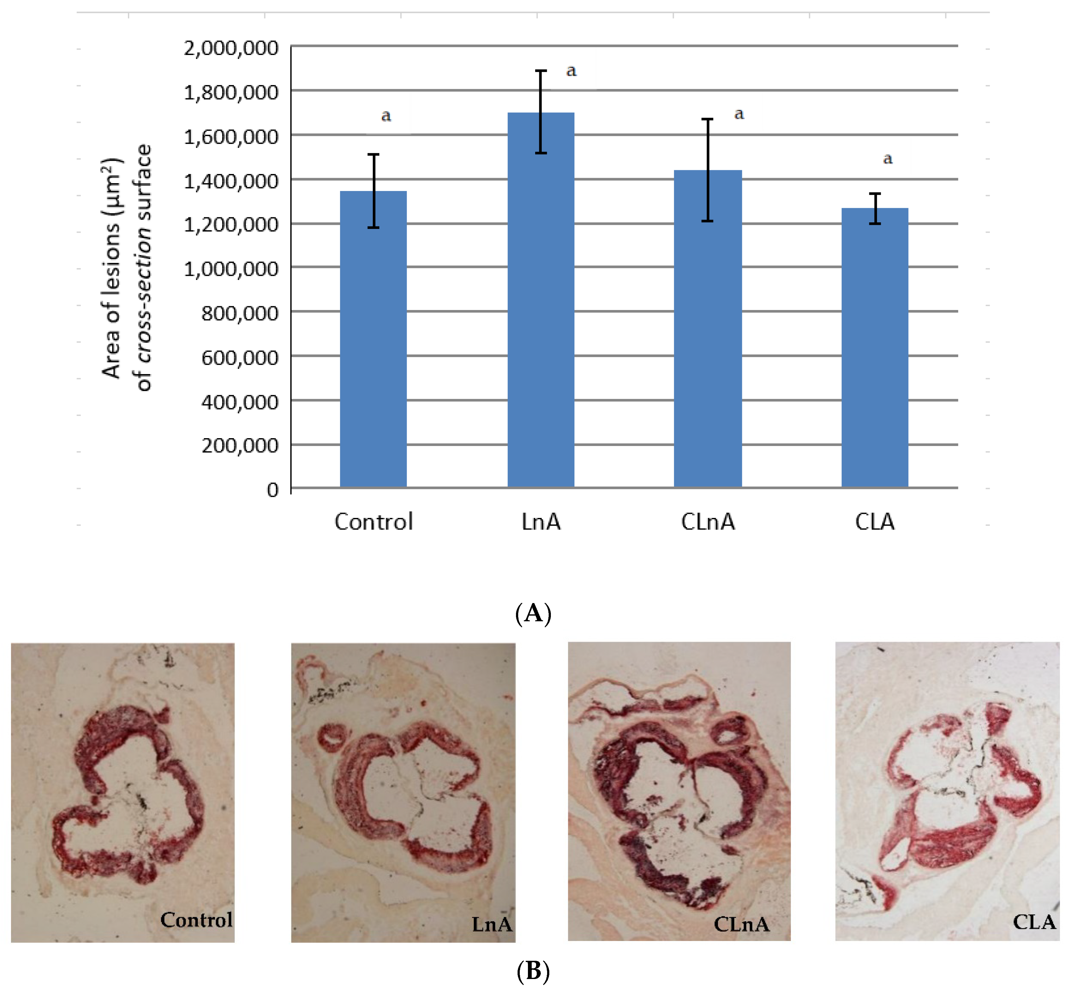
2.6. Effect of CLnA on the Development of Atherosclerosis

Quantification of atherosclerotic plaque as measured in the entire aorta (en face method) stained using Sudan IV, showed that the only significant (p < 0.05) difference is in CLA-fed animals in the SHORT study. In this group, the smallest development of lesion was observed (Figure 3A,B) compared to the LnA and CLnA groups (12.83% vs. 19.47 and 20.57%, respectively). However, in the evaluated groups of the LONG experiment, a significant difference in area of atherosclerotic plaque was not observed (Figure 4A,B). Also area of the plaques as measured in aortic roots (cross-section method) did not differ significantly between experimental groups in the SHORT (Figure 5A,B) as well as in the LONG experiment (Figure 6A,B). However, in the LONG study, the area of atherosclerotic lesions tended to decrease in the CLA group (1,266,174 µm2), whereas in LnA-fed animals it tended to increase (1,702,467 µm2) compared to the control (1,347,660 µm2). Additionally, the diets containing studied fatty acids had no effect on macrophage accumulation in atherosclerotic plaques as determined by immunohistochemical staining for a specific macrophage marker (CD68), as well as on the area covered by SMA-positive cells in aortic plaques.

3. Discussion

Many studies indicated that CLnA isomers have some pro-health effects. In our experiment, the effect of pomegranate seed oil in an animal model of atherosclerosisapoE/LDLR−/− mice was evaluated. Whereas, previous investigations focused mainly on the hypocholesterolemic effects of plasma lipids, we aimed to assess the development of atherosclerotic plaque, as the endpoint.

3.1. Effect of CLnA on Body Weight

In the SHORT study, no effects of experimental treatments on body weight of mice were evident. Only in the LONG study, in animals fed the diet containing non-conjugated linolenic acid, was an increase in final body weight observed. The lack of influence of punicic acid on animals’ body weight was observed in some studies [1,11,24]. Similarly, other authors did not observe the effect of different CLnAs isomers on body weight in model animals [18,25,26]. However, a high-fat diet (HFD) supplemented with 1% pomegranate seed oil for 12 weeks significantly decreased the body weight of mice without affecting the food intake or energy expenditure [27].

3.2. Effect of CLA Isomers on Liver Weight and Histology

In the current study, no effect on the liver weight of animals was observed; these results are consistent with previously reported studies [1,6,7,11]. Nevertheless, some reports indicated that a mixture of CLnA induce hepatic lipid accumulation in rats and that the relative liver weight was significantly heavier in rats fed CLnA [28]. In contrast, the liver weight was significantly decreased in animals after CLnA-isomers feeding [24,29,30]. Hypoplasia induced in the liver due to the antiproliferative effect on hepatocytes, together with the tendency toward increased transaminase levels, should be considered before proposing CLnA isomers as a functional ingredient [24]. Moreover, in some studies increased liver TAG concentration was observed [28].
In our study, the histological examination of the livers showed no signs of steatosis. However, representative images of the liver-stained H-E of CLA fed group seems to present slight fat accumulation compared to the control and LnA as well as CLnA groups. The increased liver mass produced by CLA may be explained by the accumulation of lipids derived from the mobilization of delipidated adipose tissue stores [31]. Raffaele et al. [32] showed that the supplementation of obese mice with pomegranate seed oil decreased hepatic steatosis and fibrosis. Additionally, the lipid droplet diameter was decreased when compared with the liver of mice fed a high fat diet. Authors concluded that pomegranate seed oil has the potential therapeutic effect in the prevention of obesity-derived non-alcoholic fatty liver disease as well as other metabolic disorders [32].

3.3. Effect of CLA Isomers on Glucose Concentration

In the current project, no significant differences in final blood glucose were observed between the experimental groups after 10 or 18 weeks of studies. Our results are consistent with previous reports [24,25,27]. However, pomegranate seed oil has been shown to decrease plasma glucose. Nekooeian et al. [33] showed that diabetic rats treated with pomegranate seed oil had significantly higher levels of serum insulin, but there were no statistically significant differences in terms of blood glucose between them and the diabetic control group. Saha and Ghosh [34] observed that administration of CLnA isomers significantly lowered the blood sugar level of STZ-induced diabetic rats as compared to the diabetic control group.

3.4. Effect of CLA Isomers on Plasma Lipid Profile and Expression of PPARα and SREBP-1 and Their Target Genes (ACO and FAS )

Among the markers of biological response, the serum lipid profile of the experimental mice was evaluated in detail. However, our studies produced equivocal results. The effect was evident only in the SHORT study, whereas in the LONG study all parameters of the lipid profile were unchanged. In the mice fed with CLnA isomers in the SHORT study, total plasma cholesterol as well as LDL+VLDL cholesterol were significantly decreased. Moreover, the TAG concentrations tended to decrease as compared to the effect of the control diet. Thus, in our study we confirmed hypolipidemic and hypocholesterolemic activities of punicic acid in apoE/LDLR−/− mice during 10 weeks of experiment. The cholesterol-lowering effect is probably only short-term and therefore does not affect the long-term reduction of atherosclerotic plaque. It is supposed that punicic acid is more efficient in reducing TCh content than other CLnA isomers. This can be attributed to the fact that punicic acid has 66% cis configuration, so it might have acted like essential fatty acids such as linoleic or n-6 linolenic acid, which normally reduce the cholesterol level in plasma and other tissues [23].
The results of previous research of CLnA isomers are inconclusive. Some authors observed decreased parameters of lipid profile (TCh, LDL, TAG) [18,25,35], whereas others did not report any differences [29,36]. Additionally, hypolipidemic and hypocholesterolemic activities of CLnA isomers present in bitter gourd and snake gourd seed oils was evaluated [23]. The authors reported that CLnA isomers normalized cholesterol, LDL-cholesterol, HDL-cholesterol and triacylogliceride contents in rat-plasma-treated sodium arsenite. The results of some studies evaluating the effect of punicic acid on the lipid profile indicate no changes in the studied parameters [7,24,30,33]. Also, Koba et al. [37] and Yuan et al. [11] did not find any significant effect of punicic acid on lipids but the liver TAG levels were significantly decreased when compared to the control group. On the other hand, Koba et al. [28] showed that feeding 1% of dietary CLnA significantly increased serum and liver TAG in rats. Similarly, Yamasaki et al. [1] reported significant increases in serum TAG and phospholipid levels but not in TCh in mice fed 0.12 and 1.2% punicic acid.
Study conducted on hyperlipidaemic patients showed that concentration of TAG and the TAG:HDL cholesterol ratio were significantly decreased after 4 weeks of 400 mg pomegranate seed oil treatment (one capsule twice daily). This human study confirmed that CLnA had favorable effects on lipid profiles [22].
The attempt to explain the mechanism of CLnA isomers effects on plasma lipids was made. The findings of results seems to indicate that trans or cis configuration of conjugated trienoic fatty acids isomers affects differently. With the advent of nutritional genomics, novel mechanisms by which dietary CLnA might modulate plasma lipids have been suggested. Conjugated fatty acids (CLnA, CLA) are considered to be ligands of nuclear transcription factors which are involved in lipid hepatic metabolism. For instance, activation of PPARα by CLnA may lead to several effects, e.g., the reduction of fatty acid and triacylglycerol synthesis and VLDL production. The SREBP-1 regulates genes encoding lipogenesis and cholesterol biosynthesis enzymes. CLnA may decrease plasma cholesterol and TAG levels via this mechanism. Authors indicated that CLnA affects lipid metabolism through activation of the PPAR.
In vitro studies have reported that conjugated linolenic acid isomers act as agonists for PPARs [19,38,39], a family of nuclear receptors that regulate the transcription of a large number of genes involved in numerous metabolic pathways. Verification of if CLnA isomers serve as ligands in living organisms is unavoidable. Our results showed that expression of PPARα in apoE/LDLR−/− mice in the SHORT study was increased by the CLnA isomer, and the effect was even stronger than for CLA. Surprisingly, there was no such an effect in the LONG study. Moreover, the expression of PPARα was decreased by CLnA. Conjugated linolenic acids, such as punicic acid, catalpic acid and eleostearic acid have demonstrated some promising effects as dual or PAN agonists of PPAR [18,19]. Punicic acid supplementation did not modify ACO gene expression as well as PPARδ [24] in the rats’ liver. Moreover, a significant increase in PPARα expression was shown. These results are in line with those reported by Arao et al. [7]. Bassaganya-Riera et al. [3] shown that punicic acid regulates macrophage and T-cell function through PPARγ- and δ-dependent mechanisms. We hypothesized that a potent anti-atherosclerotic effect of punicic acid in apoE/LDLR−/− mice could occur due to anti-inflammatory efficacy, primarily by targeting PPAR. At the molecular level, PPAR represent important targets for the actions of dietary lipid.
The results of our studies are inconclusive; we showed that the expression of SREBP-1 in apoE/LDLR−/− mice in the SHORT study was increased by the CLnA isomer, and that the effect was even stronger than for CLA. In contrast, the mRNA levels of SREBP-1 and FAS were decreased by punicic acid in the LONG study. The decreased expression of genes related to fatty acid synthesis also suggest that punicic acid can directly inhibit SREBP-1 transcription in a similar manner to PUFA but only in the LONG study. Shinohara et al. [26] showed that jacardic acid fed to ICR mice decreased the expression level of SCD-1 mRNA causing inhibition of SCD activity which may have anti-obesity and anti-diabetes effects. SCD-1 and FAS are regulated via SREBP-1 and PUFA modulates SCD-1 by directly inhibiting SREBP-1c transcription.

3.5. Effect of CLA on Fatty Acid Profile in Tissues

In our experiment carried out on apoE/LDLR−/− mice, we have evaluated the degree of exposure of the mice to dietary punicic acid as well as the cis-9,trans-11 CLA isomer by measuring the CLnA and CLA isomer concentrations in the adipose tissue and liver.
It was observed that experimental treatments had a significant effect on the fatty-acid profile in adipose tissue, where MUFA were decreased at the same time PUFA were increased; SFA were unchanged. These findings are in line with the results of Shinohara et al. [26] who analyzed the fatty-acid profile of different tissue of ICR mice fed jacardic acid. They reported that levels of MUFA in liver and white adipose tissue were also decreased. As we expected, in groups fed flaxseed oil the amount of non-conjugated linolenic acid was significantly increased in the adipose tissue and liver. In groups fed pomegranate seed oil and CLA-supplemented diets, the cis-9,trans-11 and trans-10,cis-12 isomers were present, but the amount of these CLA isomers was significantly increased only in th CLA group in both experiments. Additionally, in mice fed the pomegranate seed oil-supplemented diet, punicic acid was detected in both experiments. Many studies have confirmed that CLnA incorporates into the tissue lipids of model animals following dietary CLnA consumption [40]. The present study confirmed that CLnA isomers can be converted to cis-9,trans-11 CLA. It was shown that CLnAs are not only well absorbed into the body but also metabolized to CLA in vivo [9,10,11] as well as in vitro [12]. Moreover, human studies also show that feeding of punicic acid was clearly reflected by the elevated blood concentration of CLnA, which is incorporated into red blood cell membranes and human plasma [41]. Saha and Ghosh [42] reported that CLnA isomers caused the restoration of renal fatty acid altered by aresnite-induced renal oxidative stress. Moreover, the same authors showed that CLnA isomers are effective in protecting tissue lipid profiles which were altered due to oxidative stress in rats [16]. Additionally, supplementation of CLnA isomers restored erythrocyte membrane lipid peroxidation and lipoprotein oxidation in rats with induced oxidative stress [43]. In contrast, Yuan et al. [44] discovered that supplementation with punicic acid of healthy humans caused a significantly elevated concentration of 8-iso-PGF2α in urine, which indicates that punicic acid increases lipid peroxidation in humans.

3.6. Effect of CLnA on the Development of Atherosclerosis

The development of atherosclerosis in apoE/LDLR−/− mice, which represent a unique and reliable model of atherosclerosis [45,46,47] that allows the quantification of anti-atherogenic activities of food components, was studied for the first time.
The area of atherosclerotic lesions evaluated using the en face method did not vary between CLnA and LnA. The only significant effect was shown for the CLA group compared to LnA and CLnA, where a decreased area of atherosclerosis was observed. The same effect of cis-9,trans-11 CLA was observed by Arbones-Mainar et al. [48]. However, in Nestel et al. [49], supplemental 0.9% CLA (cis-9,trans-11) to the diabetic apoE-deficient mouse failed to reduce the severity of aortic atherosclerosis. In our previous studies on apoE/LDLR−/− mice fed CLA-enriched egg yolks, we observed no significant changes in the amount of atherosclerosis, but we reported the stabilization of the plaque introduced as lower-macrophage accumulation (CD68), and the greater area covered by SMA-positive cells in aortic plaques in CLA-enriched eggs [50]. Moreover, we have demonstrated that CLnA supplemented to margarine did not possess an anti-atherosclerotic effect [51].
Despite metabolic changes, no atherosclerotic plaque size differences between groups have been observed in the current study. In order to assess the atherosclerosis, two methods have been used (en face and cross-section). These quantitative methods evaluate the atherosclerotic plaque area only.
The limitation of the study was the lack of the qualitative assessment, e.g., the composition of the atherosclerotic plaque. Masson trichrome staining could be used to evaluate the collagen accumulation. Immunohistochemistry analysis would show the composition of the atherosclerotic plaque, such as the content of macrophages and SMA. It could be assessed whether the lesions were stable. The most appropriate tissue for such an assessment is the brachiocephalic artery (BCA); unfortunately this vessel was not collected.

4. Materials and Methods

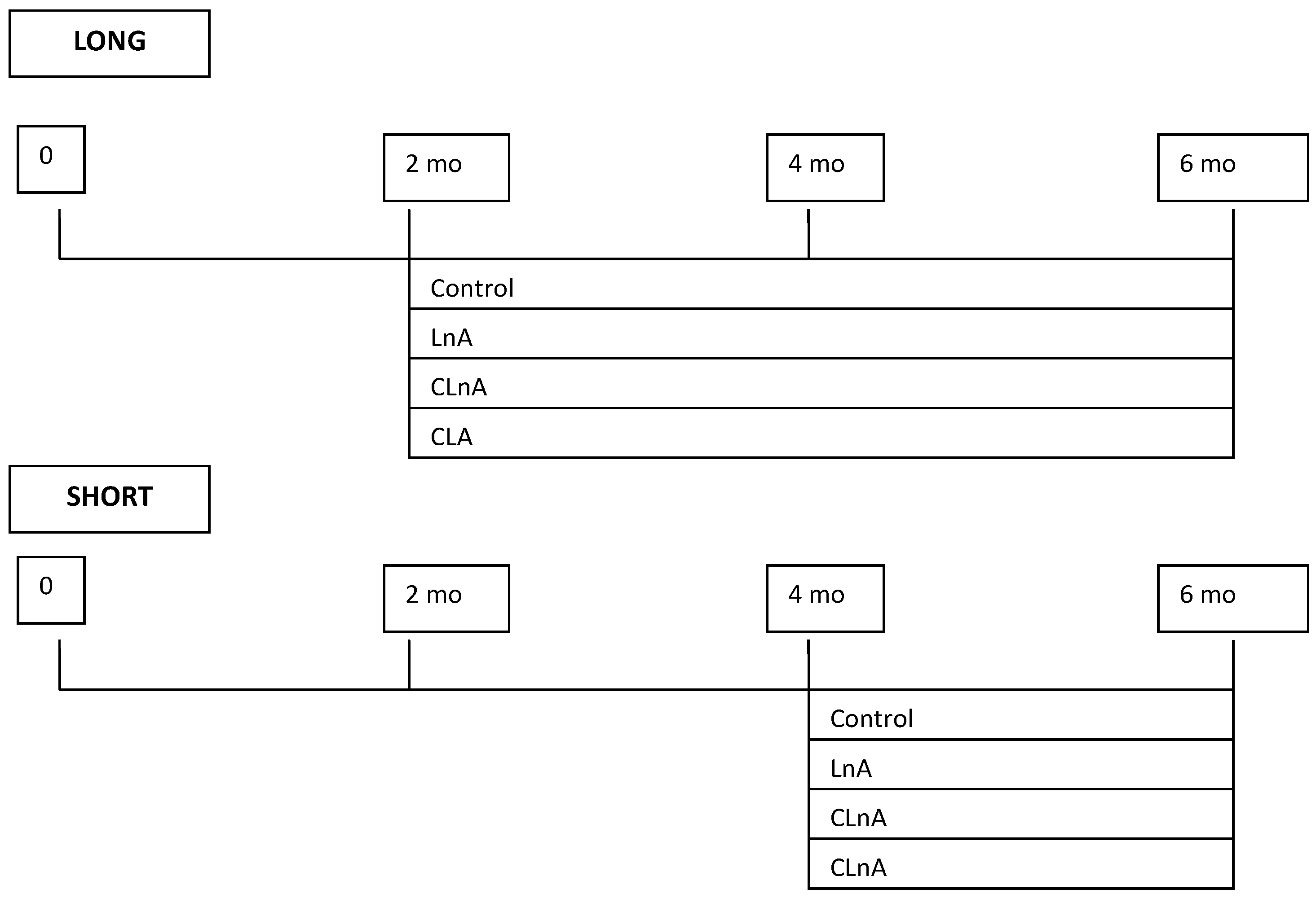
4.1. Animals and Housing

Animals (apolipoprotein E and low-density lipoprotein receptor double-knockout mice apoE/LDLR−/−) were provided by Taconic (Ejby, Denmark). ApoE/LDLR−/− mice spontaneously develop severe atherosclerotic plaques as well as hyperlipidaemia without using cholesterol-rich diets, representing unique and reliable model of atherogenesis. In the studies using the single knockout mice models (apoE or LDLR deficient mice), the high fat diet should be used. Such a factor could interfere the results of the study. Using the double knockout mice model (apoE/LDLR−/−) it is possible to avoid the diet modification (such as high fat diet). Female mice were used in the study. According to Man et al. [52] male animals appear to have more inflamed but smaller plaques compared to female animals.
Animals were housed in colony cages in a 22–25 °C with a 12h light/dark cycle. They were fed ad libitum.
All procedures involving animals were approved by the 1st Local Animal Ethics Commission in Krakow.

4.2. Diets and Feeding

8–10 weeks old (no signs of atherosclerosis) and 16–18 weeks old (pre-established atherosclerosis) female mice were used in this study. Up to the age of 10 or 18 weeks, the mice were fed a commercial, cholesterol-free, pelleted diet (Sniff M-Z Spezialdiäten GmbH; Soest, Germany).
The nutritional experiment was conducted in two steps. Based on our previous studies [53] it was observed that atherosclerotic plaque in apoE/LDLR−/− mice develops much faster comparing to single knockout. Therefore, we decided that the experimental design will be divided into two parts (SHORT—regression and LONG—progression).
Mice were randomly assigned to four experimental groups (n = 10 in each group) at the age of 10 or 18 weeks and fed AIN-93G-based diets [54] for the next 18 weeks (LONG) or 10 weeks (SHORT), respectively.
The experimental design is demonstrated in Figure 1 and the composition of modified diets fed to model animals is shown in Table 1.
The following experimental diets were used: I—control (AIN-93G), II—flaxseed oil, LnA (as a source of linolenic acid), III—pomegranate seed oil, CLnA (as a source of punicic acid), IV—CLA (cis-9,trans-11). The amount of 0.5% of studied fatty acids was supplied with seed oils to the diets.
At the end of the nutritional experiment the mice were injected intraperitoneally (1000 IU of heparin, Sanofi-Synthelabo; Paris, France) and after 10 min anesthetized (40 mg/kg of sodium thiopental, Biochemie; Vienna, Austria) and sacrificed by cervical translocation.

4.3. Blood Sampling, Plasma Lipid Profile and Glucose Analyses

To obtain plasma samples, blood was taken from the left ventricle of the heart and centrifuged (4000× g, 10 min). Until further analysis the samples were stored in −80 °C. Lipid profile was analyzed using commercially available kits for: total cholesterol (TCh; Liquick Cor-Chol 60 No 2-204; Cormay, Lublin, Poland), HDL-cholesterol (HDL; Olympus Diagnostica GmbH No OSR 6287; Hamburg, Germany) and triacylglycerols (TAG; Liquick Cor-TG 30 No 2-262; Cormay, Lublin, Poland). The LDL+VLDL-cholesterol were calculated as the difference between TCh and HDL-cholesterol. Blood glucose concentrations were measured with an Accu-Chek Active strips (Roche Diagnostics GmbH, Mannheim, Germany).

4.4. Fatty Acid Profile of Tissues

The abdominal adipose tissue and part of liver were removed from the mice and snap frozen −80 °C. The lipid profile of adipose tissue and liver were analyzed using GC-MS analyzer (Shimadzu GC-MS, Model QP 5050A) [55].

4.5. Quantification of Atherosclerosis in Descending Aortas (en Face Analysis)

In anesthetized mice, the thorax was longitudinally opened, the right atrium was incised, and the heart was perfused with phosphate-buffered saline (PBS, pH 7.4) through the apex of the left ventricle. The entire aorta was dissected and fixed in 4% paraformaldehyde, opened longitudinally, pinned and stained with Sudan IV (Sigma-Aldrich, St. Louis, MO, USA) [50]. The aortic lesion area was measured using LSM Image Browser 3 software (Zeiss, Jena, Germany).

4.6. Quantitation of Atherosclerosis in Aortic Roots (Cross-Section Analysis)

Heart and the ascending aorta were dissected. The detailed method was described previously [51]. Briefly, the heart and ascending aorta were embedded in OCT compound (CellPath, Oxford, UK), frozen and cut in 10 μm-thick serial cryosections. Eight sections were collected at 100 μm intervals starting at a 100 μm distance from the appearance of the aortic valves. Sections were fixed and stained with oil-red-O (Sigma-Aldrich, St. Louis, MO, USA) and examined under an Olympus BX50 (Olympus, Tokyo, Japan) microscope. Images of the aorta were recorded using an Olympus Camedia 5050 digital camera and stored as TIFF files of resolution 1024 × 768 pixels. The area of the lesion was measured using LSM Image Browser 3 software (Zeiss, Jena, Germany). For each animal, a mean lesion area was calculated from eight sections, reflecting the cross-section area covered by atherosclerosis.

4.7. Liver Histological Examination

Liver was removed, fixed in 4% buffered paraformaldehyde (pH 7.4) and dehydrated in alcohol, embedded in paraffin and sectioned serially (7 µm). Then the sections were stained with hematoxylin and eosin [53].

4.8. Real-Time qRT-PCR

The detailed method was described previously [24]. Briefly, total RNA kit (A&A Biotechnology, Gdańsk, Poland) was used according to the manufacturer’s protocol. RNA quantity was measured using NanoDrop (NanoDrop Technologies, Wilmington, DE, USA) and BioAnalyzer (Agilent, Santa Clara, CA, USA).
Expression of the selected genes was checked using qRT-PCR with the following primers:
PPAR-F 5′-GAGGGTTGAGCTCAGTCAGG-3′
PPAR-R 5′-GGTCACCTACGAGTGGCATT-3′,
SREBP-F 5′-CACTCAGCAGCCACCATCTA-3′
SREBP-R 5′-GCTGTCAGCAGCAGTGAGTC-3′
ACO-F 5′-GCT GGC CGT GTC CAT AGC-3′
ACO-R 5′-TTA TCC GTG GGT CCA AAC TGA-3′,
FAS-F 5′-TTG CTG GCA CTA CAG AAT GC-3′
FAS-R 5′-AAC AGC CTC AGA GCG ACA AT-3′,
β-actin-F: 5′-AGCCATGTACGTAGCCATCCA-3′
β-actin-R: 5′-TCTCAGCTGTGGTGGTGAAG-3′ (primer sequences designed in Primer3 software, based on sequence BC102589 from the GenBank). We used a LightCycler FastStart DNA Master SYBR Green I kit (Roche Diagnostics, Germany) according to the following procedure: Mg2+ added at a final concentration of 3 mM; preincubation step at 95 °C for 10 min; amplification step (40 cycles) including denaturation at 95 °C for 10 s, annealing at 58 °C for 10 s, extension at 72 °C for 10 s; melting curve including denaturation at 95 °C for 0 s, annealing at 65 °C for 15 s, continuous melting at 95 °C for 0 s (slope = 0.1 °C s−1); and cooling step at 40 °C for 30 s. For β-actin, annealing was at 58 °C. Results are presented as the ratio of genes to β-actin expression. The housekeeping gene β-actin was used for normalization.

4.9. Statistical Analysis

The one-way analysis of variance ANOVA calculated using STATISTICA 8.0 package (StatSoft Inc., Tulsa OK Oklahoma, USA) was used. To compare the treatment means the Duncan’s multiple range test was applied. A significant difference between treatment means was considered for p values < 0.05.

5. Conclusions

Since the CLnAs are present in some plant seeds in large amounts and could be purified relatively easily, they could be alternative sources for obtaining the health effects of themselves as well of CLAs. Therefore, CLnAs, which are more available and have a stronger physiological effect than CLA, are expected to be effective as a physiologically active lipid. These natural dietary compounds may provide a novel alternative therapeutic strategy in many diseases such as inflammatory diseases and cancer. Although we failed to prove an anti-atherosclerotic effect assessed directly on the atheroma area, we confirmed hypocholesterolemic properties of serum lipids. Moreover, we have attempted to understand the molecular mechanism of action of CLnA by determining gene expression. More studies are warranted to evaluate if natural resources containing CLnAs could be a potential dietary source of CLA and if they are expected to be useful as a functional food and nutraceuticals.

Author Contributions

Conceptualization, M.F.-Ż. and R.B.K.; methodology, M.F.-Ż.; formal analysis, A.D.-M. and I.C.-C.; investigation, I.C.-C. and E.K.; writing—original draft preparation, M.F.-Ż.; writing—review and editing, M.F.-Ż.; visualization, T.T.; supervision, R.B.K.; project administration, M.F.-Ż.; funding acquisition, M.F.-Ż. and R.B.K. All authors have read and agreed to the published version of the manuscript.

Funding

This research was funded by The National Science Centre in Poland, grant number N312 439437.

Institutional Review Board Statement

The study was conducted according to the guidelines for Animal Care and Treatment of the European Union, and approved by the Ethics Committee of the 1st Local Animal Ethics Committee in Krakow (9 Gronostajowa Street, 30-387 Krakow, No 42/2009).

Informed Consent Statement

Not applicable.

Data Availability Statement

Not applicable.

Conflicts of Interest

The authors declare no conflict of interest.

References

  1. Yamasaki, M.; Kitagawa, T.; Koyanagi; Chujo, N.H.; Maeda, H.; Kohno-Murase, J.; Imamura, J.; Tachibana, H.; Yamada, K. Dietary effect of pomegranate seed oil on immune function and lipid metabolism in mice. Nutrition 2006, 22, 54–59. [Google Scholar] [CrossRef] [PubMed]
  2. Koba, K.; Belury, M.A.; Sugano, M. Potential health benefits of conjugated trienoic acids. Lipid Technol. 2007, 19, 200–220. [Google Scholar] [CrossRef]
  3. Bassaganya-Riera, J.; DiGuardo, M.; Climent, M.; Vives, C.; Carbo, A.; Jouni, Z.E.; Einerhand, A.W.C.; O’Shea, M.; Hontecillas, R. Activation of PPARγ and δ by dietary punicic acid ameliorates intestinal inflammation in mice. Br. J. Nutr. 2011, 106, 878–886. [Google Scholar] [CrossRef] [Green Version]
  4. Suzuki, R.; Noguchi, R.; Ota, T.; Abe, M.; Miyashita, K.; Kawada, T. Cytotoxic Effect of Conjugated Trienoic Fatty Acids on Mouse Tumor and Human Monocytic Leukemia Cells. Lipids 2001, 36, 477–482. [Google Scholar] [CrossRef] [PubMed]
  5. Kim, N.D.; Mehta, R.; Yu, W.; Neeman, I.; Livney, T.; Amichay, A.; Poirier, D.; Nicholls, P.; Kirby, A.; Jiang, W.; et al. Chemopreventive and adjuvant therapeutic potential of pomegranate (Punica granatum) for human breast cancer. Breast Can. Res. Treat. 2002, 71, 203–217. [Google Scholar] [CrossRef]
  6. Kohno, H.; Suzuki, R.; Yasui, Y.; Hosokawa, M.; Miyashita, K.; Tanakaet, T. Pomegranate seed oil rich in conjugated linolenic acid suppresses chemically induced colon carcinogenesis in rats. Cancer Sci. 2004, 95, 481–486. [Google Scholar] [CrossRef] [PubMed]
  7. Arao, K.; Wang, Y.M.; Inoue, N.; Hirata, J.; Cha, J.-Y.; Nagao, K.; Yanagitaet, T. Dietary effect of pomegranate seed oil rich in 9cis, 11trans, 13cis conjugated linolenic acid on lipid metabolism in obese, hyperlipidemic OLETF rats. Lipids Health Dis. 2004, 9, 24–30. [Google Scholar] [CrossRef] [Green Version]
  8. Grossmann, M.E.; Mizuno, N.K.; Schuster, T.; Cleary, M.P. Punicic acid is an ω-5 fatty acid capable of inhibiting breast cancer proliferation. Int. J. Oncol. 2010, 36, 421–426. [Google Scholar]
  9. Tsuzuki, T.; Tokuyama, Y.; Igarashi, M.; Nakagawa, K.; Ohsaki, Y.; Komai, M.; Miyazawat, T. α-Eleostearic Acid (9Z11E13E-18:3) Is Quickly Converted to Conjugated Linoleic Acid (9Z11E-18:2) in Rats. J. Nutr. 2004, 134, 2634–2639. [Google Scholar] [CrossRef] [PubMed] [Green Version]
  10. Tsuzuki, T.; Kawakami, Y.; Abe, R.; Nakagawa, K.; Koba, K.; Imamura, J.; Iwata, T.; Ikeda, I.; Miyazawa, T. Conjugated Linolenic Acid Is Slowly Absorbed in Rat Intestine, but Quickly Converted to Conjugated Linoleic Acid. J. Nutr. 2006, 136, 2153–2159. [Google Scholar] [CrossRef] [Green Version]
  11. Yuan, G.; Sun, H.; Sinclair, A.J.; Li, D. Effects of conjugated linolenic acid and conjugated linoleic acid on lipid metabolism in mice. Eur. J. Lipid Sci. Technol. 2009, 111, 537–545. [Google Scholar] [CrossRef]
  12. Schneider, A.C.; Mignolet, E.; Schneider, Y.J.; Larondelle, Y. Uptake of conjugated linolenic acids and conversion to cis-9, trans-11-or trans-9, trans-11-conjugated linoleic acids in Caco-2 cells. Br. J. Nutr. 2013, 109, 57–64. [Google Scholar] [CrossRef] [Green Version]
  13. Boussetta, T.; Raad, H.; Lettéron, P.; Gougerot-Pocidalo, M.-A.; Marie, J.C.; Driss, F.; El-Benna, J. Punicic Acid a Conjugated Linolenic Acid Inhibits TNFα-Induced Neutrophil Hyperactivation and Protectsfrom Experimental Colon Inflammation in Rats. PLoS ONE 2009, 4, e6458–e6469. [Google Scholar] [CrossRef] [PubMed] [Green Version]
  14. Dhar, P.; Ghosh, S.; Bhattacharyya, D.K. Dietary effects of conjugated octadecatrienoic fatty acid (9 cis, 11 trans, 13 trans) levels on blood lipids and nonenzymatic in vitro lipid peroxidation in rats. Lipids 1999, 34, 109–114. [Google Scholar] [CrossRef]
  15. Mukherjee, C.; Bhattacharyya, S.; Ghosh, S.; Bhattacharyya, D.K. Dietary effects of punicic acid on the composition and peroxidation of rat plasma lipid. J. Oleo Sci. 2002, 51, 513–522. [Google Scholar] [CrossRef] [Green Version]
  16. Saha, S.S.; Ghosh, M. Antioxidant effect of vegetable oils containing conjugated linolenic acid isomers against induced tissue lipid peroxidation and inflammation in rat model. Chem. Biol. Interact. 2011, 190, 109–120. [Google Scholar] [CrossRef]
  17. Changhua, L.; Jindong, Y.; Defa, L.; Lidan, Z.; Shiyan, Q.; Jianjun, X. Conjugated linoleic acid attenuates the production and gene expression of proinflammatory cytokines in weaned pigs challenged with lipopolysaccharide. J. Nutr. 2005, 135, 239–244. [Google Scholar] [CrossRef] [PubMed] [Green Version]
  18. Hontecillas, R.; Diguardo, M.; Duran, E.; Orpi, M.; Bassaganya-Riera, J. Catalpic acid decreases abdominal fat deposition, improves glucose homeostasis and upregulates PPAR alpha expression in adipose tissue. Clin. Nutr. 2008, 27, 764–772. [Google Scholar] [CrossRef]
  19. Hontecillas, R.; O’Shea, M.; Einerhand, A.; Diguardo, M.; Bassaganya-Riera, J. Activation of PPAR gamma and alpha by punicic acid ameliorates glucose tolerance and suppresses obesity-related inflammation. J. Am. Coll. Nutr. 2009, 28, 184–195. [Google Scholar] [CrossRef]
  20. McFarlin, B.K.; Strohacker, K.A.; Kueht, M.L. Pomegranate seed oil consumption during a period of high-fat feeding reduces weight gain and reduces type 2 diabetes risk in CD-1 mice. Br. J. Nutr. 2009, 102, 54–59. [Google Scholar] [CrossRef] [Green Version]
  21. Meerts, I.A.; Verspeek-Rip, C.M.; Buskens, C.A.; Keizer, H.G.; Bassaganya-Riera, J.; Jouni, Z.E.; van Huygevoort, A.H.B.M.; van Otterdijk, F.M.; van de Waartet, E.J. Toxicological evaluation of pomegranate seed oil. Food Chem. Toxicol. 2009, 47, 1085–1092. [Google Scholar] [CrossRef]
  22. Mirmiran, P.; Fazeli, M.R.; Asghari, G.; Shafiee, A.; Azizi, F. Effect of pomegranate seed oil on hyperlipidaemic subjects: A doubleblind placebo-controlled clinical trial. Br. J. Nutr. 2010, 104, 402–406. [Google Scholar] [CrossRef] [Green Version]
  23. Saha, S.S.; Chakraborty, A.; Ghosh, S.; Ghosh, M. Comparative study of hypocholesterolemic and hypolipidemic effects of conjugated linolenic acid isomers against induced biochemical perturbations and aberration in erythrocyte membrane fluidity. Eur. J. Nutr. 2012, 51, 483–495. [Google Scholar] [CrossRef]
  24. Miranda, J.; Aguirre, L.; Fernández-Quintela, A.; Macarulla, M.T.; Guadalupe Martínez-Castaño, M.; Ayo, J.; Bilbao, E.; Portillo, M.P. Effects of pomegranate seed oil on glucose and lipid metabolism-related organs in rats fed an obesogenic diet. J. Agric. Food Chem. 2013, 61, 5089–5096. [Google Scholar] [CrossRef] [PubMed]
  25. Miranda, J.; Fernández-Quintela, A.; Macarulla, M.T.; Churruca, I.; García, C.; Rodríguez, V.M.; Simón, E.; Portillo, M.P. A comparison between CLNA and CLA effects on body fat, serum parameters and liver composition. J. Physiol. Biochem. 2009, 65, 25–32. [Google Scholar] [CrossRef]
  26. Shinohara, N.; Tsuduki, T.; Ito, J.; Honma, T.; Kijima, R.; Sugawara, S.; Arai, T.; Yamasaki, M.; Ikezaki, A.; Yokoyama, M.; et al. Jacaric acid, a linolenic acid isomer with a conjugated triene system, reduces stearoyl-CoA desaturase expression in liver of mice. J. Oleo Sci. 2012, 61, 433–441. [Google Scholar] [CrossRef] [PubMed] [Green Version]
  27. Vroegrijk, I.O.; van Diepen, J.A.; van den Berg, S.; Westbroek, I.; Keizer, H.; Gambelli, L.; Hontecillas, R.; Bassaganya-Riera, J.; Zondag, G.C.M.; Romijn, J.A.; et al. Pomegranate seed oil, a rich source of punicic acid, prevents diet-induced obesity and insulin resistance in mice. Food Chem. Toxicol. 2011, 49, 1426–1430. [Google Scholar] [CrossRef]
  28. Koba, K.; Akahoshi, A.; Yamasaki, M.; Tanaka, K.; Yamada, K.; Iwata, T.; Kamegai, T.; Tsutsumi, K.; Sugano, M. Dietary conjugated linolenic acid in relation to CLA differently modifies body fat mass and serum and liver lipid levels in rats. Lipids 2002, 37, 343–350. [Google Scholar] [CrossRef] [PubMed]
  29. Noguchi, R.; Yasui, Y.; Suzuki, R.; Hosokawa, M.; Fukunaga, K.; Miyashita, K. Dietary Effects of Bitter Gourd Oil on Blood and Liver Lipids of Rats. Arch. Biochem. Biophys. 2001, 396, 207–212. [Google Scholar] [CrossRef]
  30. Yang, L.; Leung, K.A.; Cao, Y.; Huang, Y.; Ratnayake, W.M.N.; Chen, Z.-Y. α-Linolenic acid but not conjugated linolenic acid is hypocholesterolaemic in hamsters. Br. J. Nutr. 2005, 93, 433–438. [Google Scholar] [CrossRef]
  31. Ashwell, M.S.; Ceddia, R.P.; House, R.L.; Cassady, J.P.; Eisen, E.J.; Eling, T.E.; Collins, J.B.; Grissom, S.F.; Odle, J. Trans-10, cis-12-conjugated linoleic acid alters hepatic gene expression in a polygenic obese line of mice displaying hepatic lipidosis. J. Nutr. Biochem. 2010, 21, 848–855. [Google Scholar] [CrossRef] [PubMed]
  32. Raffaele, M.; Licari, M.; Amin, S.; Alex, R.; Shen, H.; Singh, S.P.; Vanella, L.; Rezzani, R.; Bonomini, F.; Peterson, S.J.; et al. Cold Press Pomegranate Seed Oil Attenuates Dietary-Obesity Induced Hepatic Steatosis and Fibrosis through Antioxidant and Mitochondrial Pathways in Obese Mice. Int. J. Mol. Sci. 2020, 21, 5469. [Google Scholar] [CrossRef]
  33. Nekooeian, A.A.; Eftekhari, M.H.; Adibi, S.; Rajaeifard, A. Effects of pomegranate seed oil on insulin release in rats with type 2 diabetes. Iran J. Med. Sci. 2014, 39, 130–135. [Google Scholar] [PubMed]
  34. Saha, S.S.; Ghosh, M. Antioxidant and anti-inflammatory effect of conjugated linolenic acid isomers against streptozotocin-induced diabetes. Br. J. Nutr. 2012, 108, 974–983. [Google Scholar] [CrossRef] [Green Version]
  35. Dhar, P.; Bhattacharyya, D.; Bhattacharyya, D.K.; Ghosh, S. Dietary comparison of conjugated linolenic acid (9cis, 11trans, 13trans) and α-tocopherol effects on blood lipids and lipid peroxidation in alloxan-induced diabetes mellitus rats. Lipids 2006, 41, 49–54. [Google Scholar] [CrossRef]
  36. Lam, C.K.; Chen, J.; Cao, Y.; Yang, L.; Wong, Y.M.; Yeung, S.Y.V.; Yao, X.; Huang, Y.; Chen, Z.Y. Conjugated and non-conjugated octadecaenoic acids affect differently intestinal acyl coenzyme A: Cholesterol acyltransferase activity. Atherosclerosis 2008, 198, 85–93. [Google Scholar] [CrossRef]
  37. Koba, K.; Imamura, J.; Akashoshi, A.; Kohno-Murase, J.; Nishizono, S.; Iwabuchi, M.; Tanaka, K.; Sugano, M. Genetically Modified Rapeseed Oil Containing cis-9, trans-11, cis-13-Octadecatrienoic Acid Affects Body Fat Mass and Lipid Metabolism in Mice. J. Agric. Food Chem. 2007, 55, 3741–3748. [Google Scholar] [CrossRef]
  38. Chuang, C.Y.; Hsu, C.; Chao, C.Y.; Wein, Y.-S.; Kuo, Y.-H.; Huanget, C. Fractionation and identification of 9c, 11t, 13t-conjugated linolenic acid as an activator of PPARalpha in bitter gourd (Momordica charantia L.). J. Biomed. Sci. 2006, 13, 763–772. [Google Scholar] [CrossRef] [Green Version]
  39. Lewis, S.N.; Brannan, L.; Guri, A.J.; Lu, P.; Hontecillas, R.; Bassaganya-Riera, J.; Bevanet, D.R. Dietary α-eleostearic acid ameliorates experimental inflammatory bowel disease in mice by activating peroxisome proliferator-activated receptor-γ. PLoS ONE 2011, 6, e24031. [Google Scholar] [CrossRef] [PubMed]
  40. Lepionka, T.; Białek, M.; Czauderna, M.; Białek, A. Pomegranate seed oil and bitter melon extract supplemented in diet influence the lipid profile and intensity of peroxidation in livers of SPRD rats exposed to a chemical carcinogen. Prostaglandins Other Lipid Mediat. 2021, 152, 106495. [Google Scholar] [CrossRef]
  41. Yuan, G.; Sinclair, A.J.; Xu, C.; Li, D. Incorporation and metabolism of punicic acid in healthy young humans. Mol. Nutr. Food Res. 2009, 53, 1336–1342. [Google Scholar] [CrossRef]
  42. Saha, S.S.; Ghosh, M. Protective effect of conjugated linolenic acid isomers present in vegetable oils against arsenite-induced renal toxicity in rat model. Nutrition 2013, 29, 903–910. [Google Scholar] [CrossRef]
  43. Saha, S.S.; Dasgupta, P.; Sengupta, B.S.; Ghosh, M. Synergistic effect of conjugated linolenic acid isomers against induced oxidative stress, inflammation and erythrocyte membrane disintegrity in rat model. Biochim. Biophys. Acta 2012, 1820, 1951–1970. [Google Scholar] [CrossRef] [PubMed]
  44. Yuan, G.; Wahlqvist, M.L.; Yuan, J.Q.; Wangband, Q.-M.; Li, D. Effect of punicic acid naturally occurring in food on lipid peroxidation in healthy young humans. J. Sci. Food Agric. 2009, 89, 2331–2335. [Google Scholar] [CrossRef]
  45. Olszanecki, R.; Jawien, J.; Gajda, M.; Mateuszuk, L.; Gebska, A.; Korabiowska, M.; Chłopicki, S.; Korbut, R. Effect of curcumin on atherosclerosis in apoE/LDLR-double knockout mice. J. Physiol. Pharmacol. 2005, 56, 627–635. [Google Scholar] [PubMed]
  46. Jawien, J.; Gajda, M.; Rudling, M.; Mateuszuk, L.; Olszanecki, R.; Guzik, T.J.; Cichocki, T.; Chlopicki, S.; Korbut, R. Inhibition of five lipoxygenase activating protein (FLAP) by MK-886 decreases atherosclerosis in apoE/LDLR-double knockout mice. Eur. J. Clin. Investig. 2006, 36, 141–146. [Google Scholar] [CrossRef]
  47. Jawien, J.; Csanyi, G.; Gajda, M.; Mateuszuk, L.; Lomnicka, M.; Korbut, R.; Chlopicki, S. Ticlopidine attenuates progression of atherosclerosis in apolipoprotein E and low density lipoprotein receptor double knockout mice. Eur. J. Pharmacol. 2006, 556, 129–135. [Google Scholar] [CrossRef]
  48. Arbonés-Mainar, J.M.; Navarro, M.A.; Acín, S.; Guzmán, M.A.; Arnal, C.; Surra, J.C.; Carnicer, R.; Roche, H.M.; Osada, J. Trans-10, cis-12- and cis-9, trans-11-Conjugated Linoleic Acid Isomers Selectively Modify HDL-Apolipoprotein Composition in Apolipoprotein E Knockout Mice. J. Nutr. 2006, 136, 353–359. [Google Scholar] [CrossRef] [PubMed] [Green Version]
  49. Nestel, P.; Fujii, A.; Allen, T. The cis-9,trans-11 isomer of conjugated linoleic acid (CLA) lowers plasma triglyceride and raises HDL cholesterol concentrations but does not suppress aortic atherosclerosis in diabetic apoE-deficient mice. Atherosclerosis 2006, 189, 282–287. [Google Scholar] [CrossRef]
  50. Franczyk-Żarów, M.; Kostogrys, R.B.; Szymczyk, B.; Jawień, J.; Gajda, M.; Cichocki, T.; Wojnar, L.; Chlopicki, S.; Pisulewski, P.M. Functional effects of eggs, naturally enriched with conjugated linoleic acid (CLA), on the blood lipid profile, development of atherosclerosis and composition of atherosclerotic plaque in apolipoprotein E and low density lipoprotein receptor double-knockout mice (apoE/LDLR−/−). Brit. J. Nutr. 2008, 99, 49–58. [Google Scholar] [PubMed] [Green Version]
  51. Franczyk-Żarów, M.; Czyżyńska, I.; Drahun, A.; Maślak, E.; Chłopicki, S.; Kostogrys, R.B. Margarine supplemented with conjugated linolenic acid (CLnA) has no effect on atherosclerosis but alleviates the liver steatosis and affects the expression of lipid metabolism genes in apoE/LDLR−/− mice. Eur. J. Lipid Sci. Technol. 2015, 117, 589–600. [Google Scholar] [CrossRef]
  52. Man, J.J.; Beckman, J.A.; Jaffe, I.Z. Sex as a Biological Variable in Atherosclerosis. Circ Res. 2020, 24, 1297–1319. [Google Scholar] [CrossRef] [PubMed]
  53. Csányi, G.; Gajda, M.; Franczyk-Zarow, M.; Kostogrys, R.; Gwoźdź, P.; Mateuszuk, Ł.; Sternak, M.; Wojcik, L.; Zalewska, T.; Walski, M.; et al. Functional alterations in endothelial NO, PGI₂ and EDHF pathways in aorta in ApoE/LDLR−/− mice. Prostaglandins Other Lipid Mediat. 2012, 98, 107–115. [Google Scholar] [CrossRef] [PubMed]
  54. Reeves, P.G.; Nielsen, F.H.; Fahey, G.C. AIN-93 purified diets for laboratory rodents: Final report of the American Institute of Nutrition ad hoc Writing Committee on the reformulation of the AIN-76A rodent diet. J. Nutr. 1993, 123, 1939–1951. [Google Scholar] [CrossRef]
  55. Kiernan, J.A. Histological & Histochemical Methods. Theory & Practice, 2nd ed.; Pergamon Press, Great Britain BPCC, Wheatons Ltd.: Exeter, UK, 1990; pp. 413–421. [Google Scholar]
Figure 1. Experimental design.
Ijms 24 01737 g001
Figure 2. (A) Representative images of liver (stained hematoxylin and eosin) in apoE/LDLR−/− mice (n = 10) fed experimental diets in SHORT. (B) Representative images of liver (stained hematoxylin and eosin) in apoE/LDLR−/− mice (n = 10) fed experimental diets in LONG. Magnification of 200x.
Ijms 24 01737 g002
Figure 3. (A) Quantification (% area) of atherosclerotic plaque in entire aorta (en face) in apoE/LDLR−/− mice (n = 10) fed experimental diets in SHORT. Values are means with their standard errors depicted by vertical bars. The means marked with letters a and b are significantly different (p < 0.05). (B) Representative images of en face aorta showing atherosclerotic plaque in apoE/LDLR−/− mice (n = 10) fed experimental diets in SHORT.
Ijms 24 01737 g003
Figure 4. (A) Quantification (% area) of atherosclerotic plaque in entire aorta (en face) in apoE/LDLR−/− mice (n = 10) fed experimental diets in LONG. Values are means with their standard errors depicted by vertical bars. The means marked with letters a are not significantly different (p < 0.05). (B) Representative images of en face aorta showing atherosclerotic plaque in apoE/LDLR−/− mice (n = 10) fed experimental diets in LONG.
Ijms 24 01737 g004
Figure 5. (A) Area (µm2) of atherosclerosis of aortic roots (cross-section) in apoE/LDLR−/− mice (n = 10) fed experimental diets in SHORT. Values are means with their standard errors depicted by vertical bars. The means marked with letters a are not significantly different (p < 0.05). (B) Representative images of cross-section of aortic roots (stained ORO) showing aortic plaque in apoE/LDLR−/− mice (n = 10) fed experimental diets in SHORT. Magnification of 50×.
Ijms 24 01737 g005
Figure 6. (A) Area (µm2) of atherosclerosis of aortic roots (cross-section) in apoE/LDLR−/− mice (n = 10) fed experimental diets in LONG. Values are means with their standard errors depicted by vertical bars. The means marked with letters a are not significantly different (p < 0.05). (B) Representative images of cross-section of aortic roots (stained ORO) showing aortic plaque in apoE/LDLR−/− mice (n = 10) fed experimental diets in LONG. Magnification of 50×.
Ijms 24 01737 g006
Table 1. Composition of experimental diets (g/kg).
ControlLnACLnACLA
Corn starch532.486532.486532.486532.486
Caseine200200200200
Sucrose100100100100
Soybean oil7059.7561.363.34
Celulose powder50505050
Mineral mixture a35353535
Vitamin mixture b10101010
Choline2.52.52.52.5
tert-butylhydrochinon0.0140.0140.0140.014
Flaxseed oil c-10.25--
Pomegranate seed oil d --8.7-
cis-9,trans-11 CLA e---6.66
a AIN-93G mineral mixture. b AIN-93G vitamin mixture. c Flaxseed oil (Earthoil Plantations Ltd. Bury St Edmunds, UK) contained 48.8 g LnA/100 g. d Pomegranate seed oil (Earthoil Plantations Ltd. Bury St Edmunds UK) contained 57.5 g CLnA/100 g. e CLA isomers (Larodan Fine Chemicals) cis-9,trans-11 CLA methyl ester, purity 75%.
Table 2. Body weight (g), glucose (mg/dL), total cholesterol (TCh) (mmol/L), triacylglycerols (TAG) (mmol/L) level in plasma in apoE/LDLR−/− mice (n = 10) fed experimental diets. The means bearing different letters were significantly different (p < 0.05). Small letters indicate a separate comparison between groups in SHORT experiment. Capital letters indicate a separate comparison between groups in LONG experiment.
SHORTLONG
ControlLnACLnACLAControlLnACLnACLA
Initial body weight (g)23.06 ± 1.0221.77 ± 1.6822.91 ± 2.0422.17 ± 1.6217.82 ± 1.4518.97 ± 1.0618.13 ± 1.8317.46 ± 1.51
Final body weight (g)23.25 ± 0.74 a22.13 ± 2.32 a22.97 ± 2.44 a21.94 ± 0.97 a21.58 ± 1.56 A23.17 ± 1.25 B21.97 ± 1.20 AB21.24 ± 2.09 A
Liver weight (g/100g BW)4.45 ± 0.35 a4.19 ± 0.45 a4.70 ± 0.58 a4.58 ± 0.52 a4.28 ± 0.54 A4.36 ± 0.39 A4.61 ± 0.61 A4.37 ± 0.46 A
Final glucose (mg/dL)135.2 ± 12.5 a144.1 ± 25.2 a140.2 ± 19.3 a122.8 ± 15.4 a120.4 ± 11.6 A123.6 ± 14.8 A133.0 ± 13.7 A123.2 ± 9.4 A
TCh (mmol/L)28.59 ± 5.06 b23.67 ± 2.45 ab19.93 ± 5.29 a21.84 ± 0.58 a20.60 ± 1.24 A20.78 ± 2.49 A21.05 ± 2.45 A21.82 ± 1.69 A
LDL + VLDL (mmol/L)14.51 ± 1.97 c12.14 ± 2.19 bc6.94 ± 0.73 a10.57 ± 1.10 ab8.31 ± 0.76 A8.56 ± 1.51 A8.90 ± 1.50 A9.20 ± 1.27 A
HDL (mmol/L)0.94 ± 0.22 a0.73 ± 0.15 a0.78 ± 0.13 a0.65 ± 0.03 a0.74 ± 0.13 A0.78 ± 0.15 A0.78 ± 0.19 A0.80 ± 0.16 A
TAG (mmol/L)3.76 ± 1.59 a2.27 ± 0.30 a1.92 ± 0.09 a2.46 ± 0.80 a1.85 ± 0.52 A2.04 ± 0.81 A1.96 ± 0.74 A2.12 ± 0.95 A
Table 3. Genes expression (peroxisome proliferator-activated receptor; PPARα sterol regulatory element binding proteins; SREBP-1, acyl-CoA oxidase; ACO and fatty acid synthase; FAS ) in apoE/LDLR−/− mice (n = 10) fed experimental diets.
SHORTLONG
ControlLnACLnACLAControlLnACLnACLA
PPARα (a.u.)1.001.323.771.941.001.970.932.58
SREBP-1 (a.u.)1.001.493.251.231.000.730.783.14
ACO (a.u.)1.000.722.830.661.001.011.111.71
FAS (a.u.)1.000.722.830.661.000.690.561.16
Table 4. Fatty acid composition in adipose tissue of apoE/LDLR−/− mice fed experimental diets. The means bearing different letters were significantly different (p < 0.05). Small letters indicate a separate comparison between groups in SHORT experiment. Capital letters indicate a separate comparison between groups in LONG experiment.
Fatty AcidSHORTLONG
ControlLnACLnACLAControlLnACLnACLA
C 14:01.37 ± 0.24 a1.44 ± 0.20 a1.34 ± 0.27 a1.30 ± 0.30 a1.15 ± 0.09 A1.29 ± 0.15 A1.30 ± 0.11 AB1.47 ± 0.18 B
C 16:016.93 ± 1.32 a17.33 ± 1.29 a18.31 ± 1.26 a17.50 ± 1.17 a17.01 ± 1.04 AB15.71 ± 0.86 A16.74 ± 0.69 A18.74 ± 2.43 B
C 16:14.16 ± 0.61 a4.43 ± 0.80 ab4.63 ± 0.61 ab5.21 ± 0.78 ab4.13 ± 0.84 A3.94 ± 0.54 A4.64 ± 0.90 A5.62 ± 0.27 B
C 18:04.42 ± 0.42 b4.39 ± 0.37 b3.44 ± 0.57 a2.88 ± 0.65 a3.37 ± 0.58 A4.12 ± 0.51 A3.51 ± 0.62 A2.48 ± 0.79 B
C 18:135.45 ± 1.01 b34.73 ± 1.34 ab33.41 ± 1.04 a33.44 ± 1.61 a37.23 ± 0.95 BC38.09 ± 1.03 C35.77 ± 1.01 AB35.57 ± 1.69 A
C 18:234.09 ± 1.92 b31.25 ± 1.56 a32.52 ± 2.17 ab32.78 ± 1.95 ab33.79 ± 1.46 B30.77 ± 0.89 A30.74 ± 1.01 A30.47 ± 2.14 A
C 20:00.30 ± 0.05 a0.28 ± 0.07 a0.24 ± 0.06 a0.22 ± 0.08 a0.23 ± 0.04 A0.30 ± 0.05 C0.23 ± 0.06 A0.17 ± 0.03 B
C 20:11.12 ± 0.19 b0.88 ± 0.17 a0.82 ± 0.09 a0.83 ± 0.11 a0.83 ± 0.15 A1.03 ± 0.10 C0.85 ± 0.18 A0.62 ± 0.08 B
C 18:32.17 ± 0.32 b5.28 ± 0.48 c1.64 ± 0.11 a1.89 ± 0.22 ab1.85 ± 0.19 A4.77 ± 0.74 B1.79 ± 0.33 A1.37 ± 0.17 A
CLA cis-9,trans-11 1.76 ± 0.16 a3.33 ± 0.30 b0.37 ± 0.14 A 2.24 ± 0.24 B2.89 ± 0.26 C
CLA trans-10,cis-12 0.52 ± 0.11 a0.65 ± 0.10 b0.16 ± 0.04 B 0.69 ± 0.16 A0.59 ± 0.09 A
CLnA cis-9,trans-11,cis-13 1.37 ± 0.27 1.51 ± 0.47
SFA23.01 ± 1.70 a23.43 ± 1.50 a23.33 ± 1.73 a21.87 ± 1.85 a21.76 ± 1.39 A21.41 ± 1.25 A21.77 ± 0.89 A22.86 ± 3.50 A
MUFA40.72 ± 2.69 a40.04 ± 2.77 ab38.85 ± 2.77 ab39.48 ± 2.17 b42.19 ± 2.83 A43.06 ± 2.10 AB41.25 ± 1.65 AB41.81 ± 2.03 B
PUFA36.27 ± 1.97 a36.54 ± 1.58 ab37.82 ± 2.13 ab38.65 ± 1.82 b36.16 ± 1.41 A35.53 ± 1.49 A36.97 ± 0.93 A35.33 ± 2.43 A
16:1/16:00.25 ± 0.02 a0.26 ± 0.05 ab0.25 ± 0.03 ab0.30 ± 0.04 b0.24 ± 0.05 A0.25 ± 0.04 AB0.28 ± 0.05 AB0.30 ± 0.04 B
18:1/18:08.03 ± 0.77 a7.91 ± 0.72 ab9.72 ± 1.41 b11.60 ± 2.52 c11.05 ± 1.80 A9.25 ± 1.07 A10.20 ± 1.71 A14.34 ± 4.21 B
16:0/18:20.50 ± 0.06 a0.55 ± 0.06 a0.56 ± 0.06 a0.53 ± 0.06 a0.50 ± 0.04 A0.51 ± 0.04 A0.54 ± 0.03 A0.62 ± 0.13 B
Table 5. Fatty-acid composition in liver of apoE/LDLR−/− mice fed experimental diets. The means bearing different letters were significantly different (p < 0.05). Small letters indicate a separate comparison between groups in SHORT experiment. Capital letters indicate a separate comparison between groups in LONG experiment.
Fatty AcidSHORTLONG
ControlLnACLnACLAControlLnACLnACLA
C 14:00.56 ± 0.11 ab0.48 ± 0.05 ab0.38 ± 0.07 a0.61 ± 0.09 b0.48 ± 0.05 A0.45 ± 0.07 A0.52 ± 0.13 A0.52 ± 0.17 A
C 16:025.80 ± 0.48 ab25.83 ± 1.74 ab24.67 ± 1.11 a28.00 ± 0.41 b24.51 ± 2.74 A24.41 ± 0.88 A22.95 ± 2.13 A24.33 ± 4.32 A
C 16:12.21 ± 0.21 bc1.74 ± 0.18 ab1.25 ± 0.24 a2.39 ± 0.21 c1.61 ± 0.23 A1.63 ± 0.58 A2.13 ± 0.56 A2.54 ± 0.48 A
C 18:04.23 ± 0.51 a4.26 ± 0.74 a3.84 ± 0.52 a3.47 ± 0.27 a4.31 ± 1.06 A3.47 ± 0.87 A5.15 ± 0.49 A3.97 ± 0.88 A
C 18:130.64 ± 0.30 a31.63 ± 3.44 a35.72 ± 2.42 a30.46 ± 0.85 a34.52 ± 3.53 A31.44 ± 2.15 A33.10 ± 1.81 A32.34 ± 1.23 A
C 18:235.13 ± 1.17 b32.97 ± 0.65 ab31.63 ± 1.66 a32.34 ± 0.77 ab32.92 ± 2.23 A34.60 ± 2.26 A31.72 ± 1.96 A33.12 ± 2.68 A
C 18:31.44 ± 0.03 a3.08 ± 0.59 b1.18 ± 0.23 a1.27 ± 0.04 a1.66 ± 0.47 A4.01 ± 0.52 B1.82 ± 0.17 A1.79 ± 0.32 A
CLA cis-9,trans-11 1.34 ± 0.30 a1.31 ± 0.03 a 1.62 ± 0.18 B1.20 ± 0.22 A
CLA trans-10,cis-12 0.20 ± 0.05 A0.20 ± 0.02 A
CLnA cis-9,trans-11,cis-13 0.81 ± 0.11
SFA30.59 ± 0.83 a30.57 ± 2.40 a28.88 ± 0.71 a32.08 ± 0.34 a29.30 ± 2.49 A28.33 ± 1.54 B28.62 ± 2.19 A28.82 ± 4.01 A
MUFA32.84 ± 0.50 a33.37 ± 3.29 a36.97 ± 2.18 a32.85 ± 1.05 a36.13 ± 3.57 A33.06 ± 1.83 A35.22 ± 1.80 A34.88 ± 0.87 A
PUFA36.57 ± 1.14 a36.06 ± 1.24 a34.15 ± 1.73 a34.92 ± 0.79 a34.58 ± 2.47 A38.61 ± 2.34 A36.17 ± 2.24 A36.30 ± 3.20 A
16:1/16:00.09 ± 0.01 b0.07 ± 0.00 a0.05 ± 0.01 a0.09 ± 0.01 b0.07 ± 0.01 A0.07 ± 0.02 A0.09 ± 0.02 AB0.10 ± 0.01 B
18:1/18:07.31 ± 0.97 a7.42 ± 1.93 a9.36 ± 0.70 a8.82 ± 0.90 a8.55 ± 2.94 A9.56 ± 2.95 A6.49 ± 0.88 A8.45 ± 1.85 A
16:0/18:20.74 ± 0.04 a0.78 ± 0.05 ab0.78 ± 0.03 ab0.87 ± 0.03 b0.75 ± 0.10 A0.71 ± 0.07 A0.73 ± 0.11 A0.75 ± 0.18 A
Disclaimer/Publisher’s Note: The statements, opinions and data contained in all publications are solely those of the individual author(s) and contributor(s) and not of MDPI and/or the editor(s). MDPI and/or the editor(s) disclaim responsibility for any injury to people or property resulting from any ideas, methods, instructions or products referred to in the content.

