
Rationale for a Multi-Factorial Approach for the Reversal of Cognitive Decline in Alzheimer’s Disease and MCI: A Review
Abstract
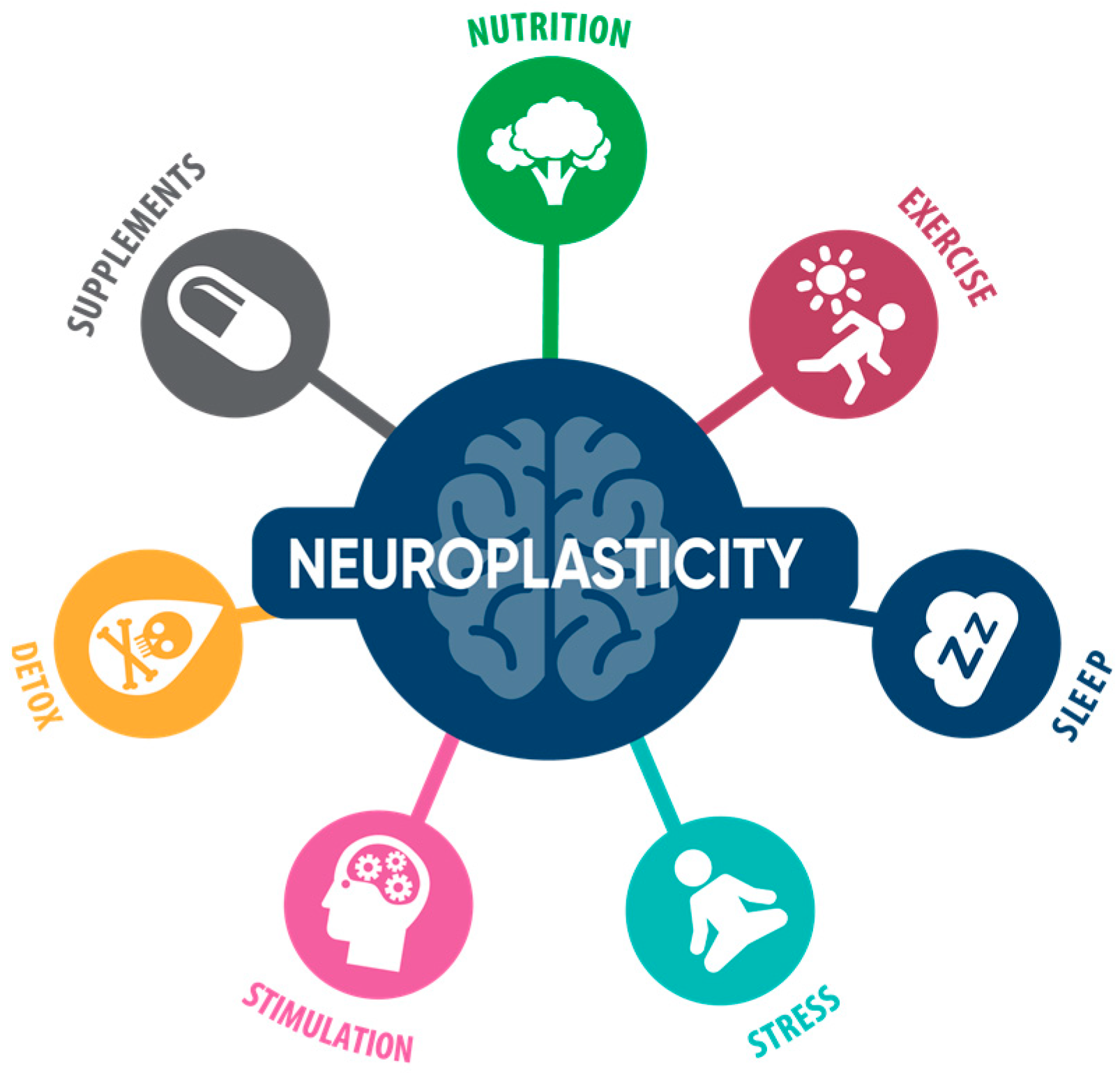
:1. Introduction
2. Multiple Strategies to Optimize Brain Health
2.1. Diet & Nutrition
2.2. Physical Exercise
- (1)
- Aerobic exercise increases heart rate and oxygen uptake, thereby improving cardiovascular health, which in turn benefits the brain. Studies have shown that aerobic exercise improves blood flow to the brain and stimulates the release of brain-derived neurotrophic factor (BDNF), which promotes neuroplasticity, thereby preserving brain volume. Moderate to vigorous aerobic exercise also activates the glymphatic system that promotes the clearance of β-amyloid and other toxins.
- (2)
- Strength training physical exercises improve muscle strength, muscle mass, and endurance, thereby preventing sarcopenia. Furthermore, strength training exercise improves higher-level cognitive processes and memory, stabilizes brain volume, and decreases white matter lesions.
- (3)
- Mind-body exercise combined with body movement improves balance, coordination, gait, and agility; improves neuronal, synaptic, and vascular systems of the brain; promotes the connectivity of brain regions, thereby improving executive function, memory, and emotional status; and curbs neuronal inflammation, all of which improve brain health [26,27,37,61,62,71,72,73,74,75,76,77,78,79,80,81,82,83].
2.3. Sleep
2.4. Mind and Mental Exercise
2.5. Stress Management
2.6. Toxicity and Detoxification
2.6.1. Metal Toxicity
2.6.2. Chemical Toxicity
2.6.3. Infections and Biotoxins
2.7. Supplements & Neuroprotective Herbs
3. Conclusions
Author Contributions
Funding
Institutional Review Board Statement
Informed Consent Statement
Data Availability Statement
Acknowledgments
Conflicts of Interest
References
- 2022 Alzheimer’s disease facts and figures. Alzheimers Dement 2022, 18, 700–789. [CrossRef] [PubMed]
- Toups, K.; Hathaway, A.; Gordon, D.; Chung, H.; Raji, C.; Boyd, A.; Hill, B.D.; Hausman-Cohen, S.; Attarha, M.; Chwa, W.J.; et al. Precision Medicine Approach to Alzheimer’s Disease: Successful Pilot Project. J. Alzheimers Dis. 2022, 88, 1411–1421. [Google Scholar] [CrossRef] [PubMed]
- James, B.D.; Leurgans, S.E.; Hebert, L.E.; Scherr, P.A.; Yaffe, K.; Bennett, D.A. Contribution of Alzheimer disease to mortality in the United States. Neurology 2014, 82, 1045–1050. [Google Scholar] [CrossRef] [PubMed] [Green Version]
- Ngandu, T.; Lehtisalo, J.; Solomon, A.; Levalahti, E.; Ahtiluoto, S.; Antikainen, R.; Backman, L.; Hanninen, T.; Jula, A.; Laatikainen, T.; et al. A 2 year multidomain intervention of diet, exercise, cognitive training, and vascular risk monitoring versus control to prevent cognitive decline in at-risk elderly people (FINGER): A randomised controlled trial. Lancet 2015, 385, 2255–2263. [Google Scholar] [CrossRef] [PubMed]
- Kivipelto, M.; Solomon, A.; Ahtiluoto, S.; Ngandu, T.; Lehtisalo, J.; Antikainen, R.; Backman, L.; Hanninen, T.; Jula, A.; Laatikainen, T.; et al. The Finnish Geriatric Intervention Study to Prevent Cognitive Impairment and Disability (FINGER): Study design and progress. Alzheimers Dement 2013, 9, 657–665. [Google Scholar] [CrossRef] [PubMed]
- Isaacson, R.S.; Hristov, H.; Saif, N.; Hackett, K.; Hendrix, S.; Melendez, J.; Safdieh, J.; Fink, M.; Thambisetty, M.; Sadek, G.; et al. Individualized clinical management of patients at risk for Alzheimer’s dementia. Alzheimers Dement 2019, 15, 1588–1602. [Google Scholar] [CrossRef]
- Raulin, A.C.; Doss, S.V.; Trottier, Z.A.; Ikezu, T.C.; Bu, G.; Liu, C.C. ApoE in Alzheimer’s disease: Pathophysiology and therapeutic strategies. Mol. Neurodegener. 2022, 17, 72. [Google Scholar] [CrossRef]
- Theendakara, V.; Peters-Libeu, C.A.; Spilman, P.; Poksay, K.S.; Bredesen, D.E.; Rao, R.V. Direct Transcriptional Effects of Apolipoprotein E. J. Neurosci. 2016, 36, 685–700. [Google Scholar] [CrossRef] [Green Version]
- Diaz, J.R.; Marta-Ariza, M.; Khodadadi-Jamayran, A.; Heguy, A.; Tsirigos, A.; Pankiewicz, J.E.; Sullivan, P.M.; Sadowski, M.J. Apolipoprotein E4 Effects a Distinct Transcriptomic Profile and Dendritic Arbor Characteristics in Hippocampal Neurons Cultured in vitro. Front. Aging Neurosci. 2022, 14, 845291. [Google Scholar] [CrossRef]
- Huang, Y.A.; Zhou, B.; Nabet, A.M.; Wernig, M.; Sudhof, T.C. Differential Signaling Mediated by ApoE2, ApoE3, and ApoE4 in Human Neurons Parallels Alzheimer’s Disease Risk. J. Neurosci. 2019, 39, 7408–7427. [Google Scholar] [CrossRef]
- Blanchard, J.W.; Akay, L.A.; Davila-Velderrain, J.; von Maydell, D.; Mathys, H.; Davidson, S.M.; Effenberger, A.; Chen, C.Y.; Maner-Smith, K.; Hajjar, I.; et al. APOE4 impairs myelination via cholesterol dysregulation in oligodendrocytes. Nature 2022, 611, 769–779. [Google Scholar] [CrossRef] [PubMed]
- Theendakara, V.; Peters-Libeu, C.A.; Bredesen, D.E.; Rao, R.V. Transcriptional Effects of ApoE4: Relevance to Alzheimer’s Disease. Mol. Neurobiol. 2018, 55, 5243–5254. [Google Scholar] [CrossRef] [PubMed]
- Levros, L.C., Jr.; Labrie, M.; Charfi, C.; Rassart, E. Binding and repressive activities of apolipoprotein E3 and E4 isoforms on the human ApoD promoter. Mol. Neurobiol. 2013, 48, 669–680. [Google Scholar] [CrossRef]
- Urfer-Buchwalder, A.; Urfer, R. Identification of a Nuclear Respiratory Factor 1 Recognition Motif in the Apolipoprotein E Variant APOE4 linked to Alzheimer’s Disease. Sci. Rep. 2017, 7, 40668. [Google Scholar] [CrossRef] [PubMed] [Green Version]
- Yiannopoulou, K.G.; Anastasiou, A.I.; Zachariou, V.; Pelidou, S.H. Reasons for Failed Trials of Disease-Modifying Treatments for Alzheimer Disease and Their Contribution in Recent Research. Biomedicines 2019, 7, 97. [Google Scholar] [CrossRef] [Green Version]
- Mehta, D.; Jackson, R.; Paul, G.; Shi, J.; Sabbagh, M. Why do trials for Alzheimer’s disease drugs keep failing? A discontinued drug perspective for 2010–2015. Expert Opin. Investig. Drugs 2017, 26, 735–739. [Google Scholar] [CrossRef]
- Dubois, B.; Hampel, H.; Feldman, H.H.; Scheltens, P.; Aisen, P.; Andrieu, S.; Bakardjian, H.; Benali, H.; Bertram, L.; Blennow, K.; et al. Proceedings of the Meeting of the International Working, G.; the American Alzheimer’s Association on “The Preclinical State of, A.D.; July; Washington Dc, U.S.A. Preclinical Alzheimer’s disease: Definition, natural history, and diagnostic criteria. Alzheimers Dement 2016, 12, 292–323. [Google Scholar] [CrossRef]
- Bredesen, D.E.; Amos, E.C.; Canick, J.; Ackerley, M.; Raji, C.; Fiala, M.; Ahdidan, J. Reversal of cognitive decline in Alzheimer’s disease. Aging 2016, 8, 1250–1258. [Google Scholar] [CrossRef] [Green Version]
- Rao, R.V.; Kumar, S.; Gregory, J.; Coward, C.; Okada, S.; Lipa, W.; Kelly, L.; Bredesen, D.E. ReCODE: A Personalized, Targeted, Multi-Factorial Therapeutic Program for Reversal of Cognitive Decline. Biomedicines 2021, 9, 1348. [Google Scholar] [CrossRef]
- Seto, M.; Weiner, R.L.; Dumitrescu, L.; Hohman, T.J. Protective genes and pathways in Alzheimer’s disease: Moving towards precision interventions. Mol. Neurodegener. 2021, 16, 29. [Google Scholar] [CrossRef]
- Schneider, N.; Yvon, C. A review of multidomain interventions to support healthy cognitive ageing. J. Nutr. Health Aging 2013, 17, 252–257. [Google Scholar] [CrossRef] [PubMed]
- Bredesen, D.E.; Sharlin, K.; Jenkins, D.; Okuno, M.; Youngberg, W.; Cohen, S.H.; Stefani, A.; Brown, R.L.; Conger, S.; Tanio, C.; et al. Reversal of Cognitive Decline: 100 Patients. J. Alzheimer’s Dis. Park. 2018, 8, 450. [Google Scholar] [CrossRef]
- Schechter, G.; Azad, G.K.; Rao, R.; McKeany, A.; Matulaitis, M.; Kalos, D.M.; Kennedy, B.K. A Comprehensive, Multi-Modal Strategy to Mitigate Alzheimer’s Disease Risk Factors Improves Aspects of Metabolism and Offsets Cognitive Decline in Individuals with Cognitive Impairment. J. Alzheimer’s Dis. Rep. 2020, 4, 223–230. [Google Scholar] [CrossRef] [PubMed]
- Lista, S.; Dubois, B.; Hampel, H. Paths to Alzheimer’s disease prevention: From modifiable risk factors to biomarker enrichment strategies. J. Nutr. Health Aging 2015, 19, 154–163. [Google Scholar] [CrossRef]
- Ross, M.K.; Raji, C.; Lokken, K.L.; Bredesen, D.E.; Roach, J.C.; Funk, C.C.; Price, N.; Rappaport, N.; Hood, L.; Heath, J.R. Case Study: A Precision Medicine Approach to Multifactorial Dementia and Alzheimer’s Disease. J. Alzheimer’s Dis. Parkinsonism 2021, 11 (Suppl. 5), 18. [Google Scholar]
- Fotuhi, M.; Lubinski, B.; Trullinger, M.; Hausterman, N.; Riloff, T.; Hadadi, M.; Raji, C.A. A Personalized 12-week “Brain Fitness Program” for Improving Cognitive Function and Increasing the Volume of Hippocampus in Elderly with Mild Cognitive Impairment. J. Prev. Alzheimer’s Dis. 2016, 3, 133–137. [Google Scholar] [CrossRef]
- Nguyen, S.A.; Oughli, H.A.; Lavretsky, H. Complementary and Integrative Medicine for Neurocognitive Disorders and Caregiver Health. Curr. Psychiatry Rep. 2022, 24, 469–480. [Google Scholar] [CrossRef]
- Roach, J.C.; Hara, J.; Fridman, D.; Lovejoy, J.C.; Jade, K.; Heim, L.; Romansik, R.; Swietlikowski, A.; Phillips, S.; Rapozo, M.K.; et al. The Coaching for Cognition in Alzheimer’s (COCOA) trial: Study design. Alzheimer’s Dement. 2022, 8, e12318. [Google Scholar] [CrossRef]
- Talbot, K.; Wang, H.Y.; Kazi, H.; Han, L.Y.; Bakshi, K.P.; Stucky, A.; Fuino, R.L.; Kawaguchi, K.R.; Samoyedny, A.J.; Wilson, R.S.; et al. Demonstrated brain insulin resistance in Alzheimer’s disease patients is associated with IGF-1 resistance, IRS-1 dysregulation, and cognitive decline. J. Clin. Investig. 2012, 122, 1316–1338. [Google Scholar] [CrossRef] [Green Version]
- Darweesh, S.K.L.; Wolters, F.J.; Ikram, M.A.; de Wolf, F.; Bos, D.; Hofman, A. Inflammatory markers and the risk of dementia and Alzheimer’s disease: A meta-analysis. Alzheimers Dement 2018, 14, 1450–1459. [Google Scholar] [CrossRef]
- Pugazhenthi, S.; Qin, L.; Reddy, P.H. Common neurodegenerative pathways in obesity, diabetes, and Alzheimer’s disease. Biochim. Et Biophys. Acta Mol. Basis Dis. 2017, 1863, 1037–1045. [Google Scholar] [CrossRef] [PubMed]
- Gutierrez, L.; Folch, A.; Rojas, M.; Cantero, J.L.; Atienza, M.; Folch, J.; Camins, A.; Ruiz, A.; Papandreou, C.; Bullo, M. Effects of Nutrition on Cognitive Function in Adults with or without Cognitive Impairment: A Systematic Review of Randomized Controlled Clinical Trials. Nutrients 2021, 13, 3728. [Google Scholar] [CrossRef] [PubMed]
- Feart, C.; Samieri, C.; Rondeau, V.; Amieva, H.; Portet, F.; Dartigues, J.F.; Scarmeas, N.; Barberger-Gateau, P. Adherence to a Mediterranean diet, cognitive decline, and risk of dementia. JAMA 2009, 302, 638–648. [Google Scholar] [CrossRef] [PubMed] [Green Version]
- Morris, M.C.; Tangney, C.C.; Wang, Y.; Sacks, F.M.; Barnes, L.L.; Bennett, D.A.; Aggarwal, N.T. MIND diet slows cognitive decline with aging. Alzheimers Dement 2015, 11, 1015–1022. [Google Scholar] [CrossRef] [Green Version]
- Chen, X.; Maguire, B.; Brodaty, H.; O’Leary, F. Dietary Patterns and Cognitive Health in Older Adults: A Systematic Review. J. Alzheimers Dis. 2019, 67, 583–619. [Google Scholar] [CrossRef]
- Vinciguerra, F.; Graziano, M.; Hagnas, M.; Frittitta, L.; Tumminia, A. Influence of the Mediterranean and Ketogenic Diets on Cognitive Status and Decline: A Narrative Review. Nutrients 2020, 12, 1019. [Google Scholar] [CrossRef] [PubMed] [Green Version]
- Blumenthal, J.A.; Smith, P.J.; Mabe, S.; Hinderliter, A.; Lin, P.H.; Liao, L.; Welsh-Bohmer, K.A.; Browndyke, J.N.; Kraus, W.E.; Doraiswamy, P.M.; et al. Lifestyle and neurocognition in older adults with cognitive impairments: A randomized trial. Neurology 2019, 92, e212–e223. [Google Scholar] [CrossRef] [PubMed] [Green Version]
- Blumenthal, J.A.; Smith, P.J.; Mabe, S.; Hinderliter, A.; Welsh-Bohmer, K.; Browndyke, J.N.; Doraiswamy, P.M.; Lin, P.H.; Kraus, W.E.; Burke, J.R.; et al. Longer Term Effects of Diet and Exercise on Neurocognition: 1-Year Follow-up of the ENLIGHTEN Trial. J. Am. Geriatr. Soc. 2020, 68, 559–568. [Google Scholar] [CrossRef] [Green Version]
- Smith, P.J.; Mabe, S.; Sherwood, A.; Babyak, M.A.; Doraiswamy, P.M.; Welsh-Bohmer, K.A.; Kraus, W.; Burke, J.; Hinderliter, A.; Blumenthal, J.A. Association Between Insulin Resistance, Plasma Leptin, and Neurocognition in Vascular Cognitive Impairment. J. Alzheimers Dis. 2019, 71, 921–929. [Google Scholar] [CrossRef]
- Liu, X.; Morris, M.C.; Dhana, K.; Ventrelle, J.; Johnson, K.; Bishop, L.; Hollings, C.S.; Boulin, A.; Laranjo, N.; Stubbs, B.J.; et al. Mediterranean-DASH Intervention for Neurodegenerative Delay (MIND) study: Rationale, design and baseline characteristics of a randomized control trial of the MIND diet on cognitive decline. Contemp. Clin. Trials 2021, 102, 106270. [Google Scholar] [CrossRef]
- Lourida, I.; Soni, M.; Thompson-Coon, J.; Purandare, N.; Lang, I.A.; Ukoumunne, O.C.; Llewellyn, D.J. Mediterranean diet, cognitive function, and dementia: A systematic review. Epidemiology 2013, 24, 479–489. [Google Scholar] [CrossRef] [PubMed]
- Rosenberg, A.; Ngandu, T.; Rusanen, M.; Antikainen, R.; Backman, L.; Havulinna, S.; Hanninen, T.; Laatikainen, T.; Lehtisalo, J.; Levalahti, E.; et al. Multidomain lifestyle intervention benefits a large elderly population at risk for cognitive decline and dementia regardless of baseline characteristics: The FINGER trial. Alzheimers Dement 2018, 14, 263–270. [Google Scholar] [CrossRef] [PubMed]
- Morris, M.C.; Tangney, C.C.; Wang, Y.; Sacks, F.M.; Bennett, D.A.; Aggarwal, N.T. MIND diet associated with reduced incidence of Alzheimer’s disease. Alzheimers Dement 2015, 11, 1007–1014. [Google Scholar] [CrossRef] [PubMed] [Green Version]
- Feart, C.; Samieri, C.; Barberger-Gateau, P. Mediterranean diet and cognitive function in older adults. Curr. Opin. Clin. Nutr. Metab. Care 2010, 13, 14–18. [Google Scholar] [CrossRef] [Green Version]
- Berendsen, A.A.; Kang, J.H.; van de Rest, O.; Jankovic, N.; Kampman, E.; Kiefte-de Jong, J.C.; Franco, O.H.; Ikram, M.A.; Pikhart, H.; Nilsson, L.M.; et al. Association of Adherence to a Healthy Diet with Cognitive Decline in European and American Older Adults: A Meta-Analysis within the CHANCES Consortium. Dement. Geriatr. Cogn. Disord. 2017, 43, 215–227. [Google Scholar] [CrossRef]
- Berendsen, A.A.M.; Kang, J.H.; van de Rest, O.; Feskens, E.J.M.; de Groot, L.; Grodstein, F. The Dietary Approaches to Stop Hypertension Diet, Cognitive Function, and Cognitive Decline in American Older Women. J. Am. Med. Dir. Assoc. 2017, 18, 427–432. [Google Scholar] [CrossRef]
- Van den Brink, A.C.; Brouwer-Brolsma, E.M.; Berendsen, A.A.M.; van de Rest, O. The Mediterranean, Dietary Approaches to Stop Hypertension (DASH), and Mediterranean-DASH Intervention for Neurodegenerative Delay (MIND) Diets Are Associated with Less Cognitive Decline and a Lower Risk of Alzheimer’s Disease-A Review. Adv. Nutr. 2019, 10, 1040–1065. [Google Scholar] [CrossRef] [Green Version]
- Duplantier, S.C.; Gardner, C.D. A Critical Review of the Study of Neuroprotective Diets to Reduce Cognitive Decline. Nutrients 2021, 13, 2264. [Google Scholar] [CrossRef]
- Mottaghi, T.; Amirabdollahian, F.; Haghighatdoost, F. Fruit and vegetable intake and cognitive impairment: A systematic review and meta-analysis of observational studies. Eur. J. Clin. Nutr. 2018, 72, 1336–1344. [Google Scholar] [CrossRef]
- Theodore, L.E.; Kellow, N.J.; McNeil, E.A.; Close, E.O.; Coad, E.G.; Cardoso, B.R. Nut Consumption for Cognitive Performance: A Systematic Review. Adv. Nutr. 2021, 12, 777–792. [Google Scholar] [CrossRef]
- Roman, G.C.; Jackson, R.E.; Reis, J.; Roman, A.N.; Toledo, J.B.; Toledo, E. Extra-virgin olive oil for potential prevention of Alzheimer disease. Rev. Neurol. 2019, 175, 705–723. [Google Scholar] [CrossRef] [PubMed]
- Omar, S.H. Mediterranean and MIND Diets Containing Olive Biophenols Reduces the Prevalence of Alzheimer’s Disease. Int. J. Mol. Sci. 2019, 20, 2797. [Google Scholar] [CrossRef] [PubMed] [Green Version]
- Solfrizzi, V.; Colacicco, A.M.; D’Introno, A.; Capurso, C.; Torres, F.; Rizzo, C.; Capurso, A.; Panza, F. Dietary intake of unsaturated fatty acids and age-related cognitive decline: A 8.5-year follow-up of the Italian Longitudinal Study on Aging. Neurobiol. Aging 2006, 27, 1694–1704. [Google Scholar] [CrossRef]
- Letenneur, L.; Proust-Lima, C.; Le Gouge, A.; Dartigues, J.F.; Barberger-Gateau, P. Flavonoid intake and cognitive decline over a 10-year period. Am. J. Epidemiol. 2007, 165, 1364–1371. [Google Scholar] [CrossRef]
- Desideri, G.; Kwik-Uribe, C.; Grassi, D.; Necozione, S.; Ghiadoni, L.; Mastroiacovo, D.; Raffaele, A.; Ferri, L.; Bocale, R.; Lechiara, M.C.; et al. Benefits in cognitive function, blood pressure, and insulin resistance through cocoa flavanol consumption in elderly subjects with mild cognitive impairment: The Cocoa, Cognition, and Aging (CoCoA) study. Hypertension 2012, 60, 794–801. [Google Scholar] [CrossRef] [PubMed] [Green Version]
- Grassi, D.; Desideri, G.; Necozione, S.; Ruggieri, F.; Blumberg, J.B.; Stornello, M.; Ferri, C. Protective effects of flavanol-rich dark chocolate on endothelial function and wave reflection during acute hyperglycemia. Hypertension 2012, 60, 827–832. [Google Scholar] [CrossRef] [Green Version]
- Lehtisalo, J.; Levalahti, E.; Lindstrom, J.; Hanninen, T.; Paajanen, T.; Peltonen, M.; Antikainen, R.; Laatikainen, T.; Strandberg, T.; Soininen, H.; et al. Dietary changes and cognition over 2 years within a multidomain intervention trial-The Finnish Geriatric Intervention Study to Prevent Cognitive Impairment and Disability (FINGER). Alzheimers Dement 2019, 15, 410–417. [Google Scholar] [CrossRef] [PubMed]
- Cunnane, S.C.; Swerdlow, R.H.; Inzitari, M.; Olaso-Gonzalez, G.; Vina, J. Multimodal strategy to rescue the brain in mild cognitive impairment: Ketogenic oral nutrition supplementation with B vitamins and aerobic exercise. Eur. J. Clin. Investig. 2022, 52, e13806. [Google Scholar] [CrossRef] [PubMed]
- Cunnane, S.C.; Sieber, C.C.; Swerdlow, R.H.; Cruz-Jentoft, A.J. Mild cognitive impairment: When nutrition helps brain energy rescue-a report from the EuGMS 2020 Congress. Eur. Geriatr. Med. 2021, 12, 1285–1292. [Google Scholar] [CrossRef]
- Barha, C.K.; Falck, R.S.; Best, J.R.; Nagamatsu, L.S.; Hsiung, G.R.; Sheel, A.W.; Hsu, C.L.; Kramer, A.F.; Voss, M.W.; Erickson, K.I.; et al. Reshaping the path of mild cognitive impairment by refining exercise prescription: A study protocol of a randomized controlled trial to understand the “what,” “for whom,” and “how” of exercise to promote cognitive function. Trials 2022, 23, 766. [Google Scholar] [CrossRef]
- Williams, C.L.; Tappen, R.M. Exercise training for depressed older adults with Alzheimer’s disease. Aging Ment. Health 2008, 12, 72–80. [Google Scholar] [CrossRef] [PubMed]
- Teixeira, C.V.L.; Ribeiro de Rezende, T.J.; Weiler, M.; Magalhaes, T.N.C.; Carletti-Cassani, A.; Silva, T.; Joaquim, H.P.G.; Talib, L.L.; Forlenza, O.V.; Franco, M.P.; et al. Cognitive and structural cerebral changes in amnestic mild cognitive impairment due to Alzheimer’s disease after multicomponent training. Alzheimer’s Dement. 2018, 4, 473–480. [Google Scholar] [CrossRef] [PubMed]
- Nayor, M.; Shah, R.V.; Miller, P.E.; Blodgett, J.B.; Tanguay, M.; Pico, A.R.; Murthy, V.L.; Malhotra, R.; Houstis, N.E.; Deik, A.; et al. Metabolic Architecture of Acute Exercise Response in Middle-Aged Adults in the Community. Circulation 2020, 142, 1905–1924. [Google Scholar] [CrossRef]
- Lake, S.L.; Guadagni, V.; Kendall, K.D.; Chadder, M.; Anderson, T.J.; Leigh, R.; Rawling, J.M.; Hogan, D.B.; Hill, M.D.; Poulin, M.J. Aerobic exercise training in older men and women-Cerebrovascular responses to submaximal exercise: Results from the Brain in Motion study. Physiol. Rep. 2022, 10, e15158. [Google Scholar] [CrossRef]
- Portugal, E.M.; Vasconcelos, P.G.; Souza, R.; Lattari, E.; Monteiro-Junior, R.S.; Machado, S.; Deslandes, A.C. Aging process, cognitive decline and Alzheimer`s disease: Can strength training modulate these responses? CNS Neurol. Disord. Drug Targets 2015, 14, 1209–1213. [Google Scholar] [CrossRef]
- Okonkwo, O.C.; Schultz, S.A.; Oh, J.M.; Larson, J.; Edwards, D.; Cook, D.; Koscik, R.; Gallagher, C.L.; Dowling, N.M.; Carlsson, C.M.; et al. Physical activity attenuates age-related biomarker alterations in preclinical AD. Neurology 2014, 83, 1753–1760. [Google Scholar] [CrossRef] [PubMed] [Green Version]
- Coutinho, L.A.; Leao, L.L.; Cassilhas, R.C.; de Paula, A.M.B.; Deslandes, A.C.; Monteiro-Junior, R.S. Alzheimer’s disease genes and proteins associated with resistance and aerobic training: An in silico analysis. Exp. Gerontol. 2022, 168, 111948. [Google Scholar] [CrossRef] [PubMed]
- Nascimento, C.M.; Pereira, J.R.; de Andrade, L.P.; Garuffi, M.; Talib, L.L.; Forlenza, O.V.; Cancela, J.M.; Cominetti, M.R.; Stella, F. Physical exercise in MCI elderly promotes reduction of pro-inflammatory cytokines and improvements on cognition and BDNF peripheral levels. Curr. Alzheimer Res. 2014, 11, 799–805. [Google Scholar] [CrossRef]
- Baker, L.D.; Frank, L.L.; Foster-Schubert, K.; Green, P.S.; Wilkinson, C.W.; McTiernan, A.; Plymate, S.R.; Fishel, M.A.; Watson, G.S.; Cholerton, B.A.; et al. Effects of aerobic exercise on mild cognitive impairment: A controlled trial. Arch. Neurol. 2010, 67, 71–79. [Google Scholar] [CrossRef] [Green Version]
- Maass, A.; Duzel, S.; Goerke, M.; Becke, A.; Sobieray, U.; Neumann, K.; Lovden, M.; Lindenberger, U.; Backman, L.; Braun-Dullaeus, R.; et al. Vascular hippocampal plasticity after aerobic exercise in older adults. Mol. Psychiatry 2015, 20, 585–593. [Google Scholar] [CrossRef] [Green Version]
- Yu, F.; Vock, D.M.; Zhang, L.; Salisbury, D.; Nelson, N.W.; Chow, L.S.; Smith, G.; Barclay, T.R.; Dysken, M.; Wyman, J.F. Cognitive Effects of Aerobic Exercise in Alzheimer’s Disease: A Pilot Randomized Controlled Trial. J. Alzheimers Dis. 2021, 80, 233–244. [Google Scholar] [CrossRef] [PubMed]
- Yoshino, M.; Yoshino, J.; Smith, G.I.; Stein, R.I.; Bittel, A.J.; Bittel, D.C.; Reeds, D.N.; Sinacore, D.R.; Cade, W.T.; Patterson, B.W.; et al. Worksite-based intensive lifestyle therapy has profound cardiometabolic benefits in people with obesity and type 2 diabetes. Cell Metab. 2022, 34, 1431–1441.e5. [Google Scholar] [CrossRef] [PubMed]
- Freberg, E.; Taglialatela, G. Exercise as a Potential Therapeutic Strategy to Target the Clinical Link Between Depression and Alzheimer’s Disease: A Narrative Review. J. Alzheimers Dis. 2022, 89, 759–767. [Google Scholar] [CrossRef]
- Liu-Ambrose, T.; Best, J.R.; Davis, J.C.; Eng, J.J.; Lee, P.E.; Jacova, C.; Boyd, L.A.; Brasher, P.M.; Munkacsy, M.; Cheung, W.; et al. Aerobic exercise and vascular cognitive impairment: A randomized controlled trial. Neurology 2016, 87, 2082–2090. [Google Scholar] [CrossRef] [Green Version]
- Fiatarone, M.A.; O’Neill, E.F.; Ryan, N.D.; Clements, K.M.; Solares, G.R.; Nelson, M.E.; Roberts, S.B.; Kehayias, J.J.; Lipsitz, L.A.; Evans, W.J. Exercise training and nutritional supplementation for physical frailty in very elderly people. N. Engl. J. Med. 1994, 330, 1769–1775. [Google Scholar] [CrossRef] [PubMed] [Green Version]
- Teri, L.; Logsdon, R.G.; Uomoto, J.; McCurry, S.M. Behavioral treatment of depression in dementia patients: A controlled clinical trial. J. Gerontol. B Psychol. Sci. Soc. Sci. 1997, 52, P159–P166. [Google Scholar] [CrossRef] [PubMed] [Green Version]
- Wu, C.; Yang, L.; Feng, S.; Zhu, L.; Yang, L.; Liu, T.C.; Duan, R. Therapeutic non-invasive brain treatments in Alzheimer’s disease: Recent advances and challenges. Inflamm. Regen. 2022, 42, 31. [Google Scholar] [CrossRef] [PubMed]
- Hsu, C.L.; Best, J.R.; Davis, J.C.; Nagamatsu, L.S.; Wang, S.; Boyd, L.A.; Hsiung, G.R.; Voss, M.W.; Eng, J.J.; Liu-Ambrose, T. Aerobic exercise promotes executive functions and impacts functional neural activity among older adults with vascular cognitive impairment. Br. J. Sport. Med. 2018, 52, 184–191. [Google Scholar] [CrossRef]
- Yu, F.; Kolanowski, A.M.; Strumpf, N.E.; Eslinger, P.J. Improving cognition and function through exercise intervention in Alzheimer’s disease. J. Nurs. Scholarsh. Off. Publ. Sigma Theta Tau Int. Honor Soc. Nurs. 2006, 38, 358–365. [Google Scholar] [CrossRef]
- Wilckens, K.A.; Stillman, C.M.; Waiwood, A.M.; Kang, C.; Leckie, R.L.; Peven, J.C.; Foust, J.E.; Fraundorf, S.H.; Erickson, K.I. Exercise interventions preserve hippocampal volume: A meta-analysis. Hippocampus 2021, 31, 335–347. [Google Scholar] [CrossRef]
- Lange-Asschenfeldt, C.; Kojda, G. Alzheimer’s disease, cerebrovascular dysfunction and the benefits of exercise: From vessels to neurons. Exp. Gerontol 2008, 43, 499–504. [Google Scholar] [CrossRef] [PubMed]
- Phillips, C.; Baktir, M.A.; Das, D.; Lin, B.; Salehi, A. The Link Between Physical Activity and Cognitive Dysfunction in Alzheimer Disease. Phys. Ther. 2015, 95, 1046–1060. [Google Scholar] [CrossRef] [PubMed] [Green Version]
- Huttenrauch, M.; Lopez-Noguerola, J.S.; Castro-Obregon, S. Connecting Mind-Body Therapy-Mediated Effects to Pathological Features of Alzheimer’s Disease. J. Alzheimers Dis. 2021, 82, S65–S90. [Google Scholar] [CrossRef] [PubMed]
- Scarmeas, N.; Luchsinger, J.A.; Schupf, N.; Brickman, A.M.; Cosentino, S.; Tang, M.X.; Stern, Y. Physical activity, diet, and risk of Alzheimer disease. JAMA 2009, 302, 627–637. [Google Scholar] [CrossRef] [Green Version]
- Saez de Asteasu, M.L.; Martinez-Velilla, N.; Zambom-Ferraresi, F.; Casas-Herrero, A.; Izquierdo, M. Role of physical exercise on cognitive function in healthy older adults: A systematic review of randomized clinical trials. Ageing Res. Rev. 2017, 37, 117–134. [Google Scholar] [CrossRef]
- Savage, V.M.; West, G.B. A quantitative, theoretical framework for understanding mammalian sleep. Proc. Natl. Acad. Sci. USA 2007, 104, 1051–1056. [Google Scholar] [CrossRef] [Green Version]
- Bishir, M.; Bhat, A.; Essa, M.M.; Ekpo, O.; Ihunwo, A.O.; Veeraraghavan, V.P.; Mohan, S.K.; Mahalakshmi, A.M.; Ray, B.; Tuladhar, S.; et al. Sleep Deprivation and Neurological Disorders. Biomed Res. Int. 2020, 2020, 5764017. [Google Scholar] [CrossRef]
- Reddy, O.C.; van der Werf, Y.D. The Sleeping Brain: Harnessing the Power of the Glymphatic System through Lifestyle Choices. Brain Sci. 2020, 10, 868. [Google Scholar] [CrossRef]
- Rasch, B.; Born, J. About sleep’s role in memory. Physiol. Rev. 2013, 93, 681–766. [Google Scholar] [CrossRef]
- Stickgold, R.; Walker, M.P. Sleep-dependent memory triage: Evolving generalization through selective processing. Nat. Neurosci. 2013, 16, 139–145. [Google Scholar] [CrossRef] [Green Version]
- Borges, C.R.; Poyares, D.; Piovezan, R.; Nitrini, R.; Brucki, S. Alzheimer’s disease and sleep disturbances: A review. Arq. Neuropsiquiatr. 2019, 77, 815–824. [Google Scholar] [CrossRef] [PubMed]
- Shokri-Kojori, E.; Wang, G.J.; Wiers, C.E.; Demiral, S.B.; Guo, M.; Kim, S.W.; Lindgren, E.; Ramirez, V.; Zehra, A.; Freeman, C.; et al. beta-Amyloid accumulation in the human brain after one night of sleep deprivation. Proc. Natl. Acad. Sci. USA 2018, 115, 4483–4488. [Google Scholar] [CrossRef] [PubMed] [Green Version]
- Yulug, B.; Hanoglu, L.; Kilic, E. Does sleep disturbance affect the amyloid clearance mechanisms in Alzheimer’s disease? Psychiatry Clin. Neurosci. 2017, 71, 673–677. [Google Scholar] [CrossRef] [PubMed]
- Cordone, S.; Annarumma, L.; Rossini, P.M.; De Gennaro, L. Sleep and beta-Amyloid Deposition in Alzheimer Disease: Insights on Mechanisms and Possible Innovative Treatments. Front. Pharmacol. 2019, 10, 695. [Google Scholar] [CrossRef] [Green Version]
- Spinedi, E.; Cardinali, D.P. Neuroendocrine-Metabolic Dysfunction and Sleep Disturbances in Neurodegenerative Disorders: Focus on Alzheimer’s Disease and Melatonin. Neuroendocrinology 2019, 108, 354–364. [Google Scholar] [CrossRef] [Green Version]
- Lim, A.S.; Kowgier, M.; Yu, L.; Buchman, A.S.; Bennett, D.A. Sleep Fragmentation and the Risk of Incident Alzheimer’s Disease and Cognitive Decline in Older Persons. Sleep 2013, 36, 1027–1032. [Google Scholar] [CrossRef] [Green Version]
- Jackson, M.L.; Gunzelmann, G.; Whitney, P.; Hinson, J.M.; Belenky, G.; Rabat, A.; Van Dongen, H.P. Deconstructing and reconstructing cognitive performance in sleep deprivation. Sleep Med. Rev. 2013, 17, 215–225. [Google Scholar] [CrossRef] [Green Version]
- Bombois, S.; Derambure, P.; Pasquier, F.; Monaca, C. Sleep disorders in aging and dementia. J. Nutr. Health Aging 2010, 14, 212–217. [Google Scholar] [CrossRef]
- Foley, D.; Ancoli-Israel, S.; Britz, P.; Walsh, J. Sleep disturbances and chronic disease in older adults: Results of the 2003 National Sleep Foundation Sleep in America Survey. J. Psychosom. Res. 2004, 56, 497–502. [Google Scholar] [CrossRef]
- Wennberg, A.M.V.; Wu, M.N.; Rosenberg, P.B.; Spira, A.P. Sleep Disturbance, Cognitive Decline, and Dementia: A Review. Semin. Neurol. 2017, 37, 395–406. [Google Scholar]
- Deschenes, C.L.; McCurry, S.M. Current treatments for sleep disturbances in individuals with dementia. Curr. Psychiatry Rep. 2009, 11, 20–26. [Google Scholar] [CrossRef] [PubMed] [Green Version]
- Tranah, G.J.; Blackwell, T.; Stone, K.L.; Ancoli-Israel, S.; Paudel, M.L.; Ensrud, K.E.; Cauley, J.A.; Redline, S.; Hillier, T.A.; Cummings, S.R.; et al. Circadian activity rhythms and risk of incident dementia and mild cognitive impairment in older women. Ann. Neurol. 2011, 70, 722–732. [Google Scholar] [CrossRef] [PubMed]
- Bubu, O.M.; Brannick, M.; Mortimer, J.; Umasabor-Bubu, O.; Sebastiao, Y.V.; Wen, Y.; Schwartz, S.; Borenstein, A.R.; Wu, Y.; Morgan, D.; et al. Sleep, Cognitive impairment, and Alzheimer’s disease: A Systematic Review and Meta-Analysis. Sleep 2017, 40, zsw032. [Google Scholar] [CrossRef]
- Ettcheto, M.; Olloquequi, J.; Sanchez-Lopez, E.; Busquets, O.; Cano, A.; Manzine, P.R.; Beas-Zarate, C.; Castro-Torres, R.D.; Garcia, M.L.; Bullo, M.; et al. Benzodiazepines and Related Drugs as a Risk Factor in Alzheimer’s Disease Dementia. Front. Aging Neurosci. 2019, 11, 344. [Google Scholar] [CrossRef] [PubMed]
- Rose, K.M.; Lorenz, R. Sleep disturbances in dementia. J. Gerontol. Nurs. 2010, 36, 9–14. [Google Scholar] [CrossRef] [PubMed] [Green Version]
- Bliwise, D.L. Sleep disorders in Alzheimer’s disease and other dementias. Clin. Cornerstone 2004, 6 (Suppl. 1A), S16–S28. [Google Scholar] [CrossRef]
- Tractenberg, R.E.; Singer, C.M.; Kaye, J.A. Characterizing sleep problems in persons with Alzheimer’s disease and normal elderly. J. Sleep Res. 2006, 15, 97–103. [Google Scholar] [CrossRef]
- Cole, C.S.; Richards, K.C. Sleep and cognition in people with Alzheimer’s disease. Issues Ment. Health Nurs. 2005, 26, 687–698. [Google Scholar] [CrossRef]
- Hossain, M.F.; Uddin, M.S.; Uddin, G.M.S.; Sumsuzzman, D.M.; Islam, M.S.; Barreto, G.E.; Mathew, B.; Ashraf, G.M. Melatonin in Alzheimer’s Disease: A Latent Endogenous Regulator of Neurogenesis to Mitigate Alzheimer’s Neuropathology. Mol. Neurobiol. 2019, 56, 8255–8276. [Google Scholar] [CrossRef]
- Shukla, M.; Govitrapong, P.; Boontem, P.; Reiter, R.J.; Satayavivad, J. Mechanisms of Melatonin in Alleviating Alzheimer’s Disease. Curr. Neuropharmacol. 2017, 15, 1010–1031. [Google Scholar] [CrossRef] [Green Version]
- Cardinali, D.P.; Furio, A.M.; Brusco, L.I. Clinical aspects of melatonin intervention in Alzheimer’s disease progression. Curr. Neuropharmacol. 2010, 8, 218–227. [Google Scholar] [CrossRef] [PubMed] [Green Version]
- Wu, Y.H.; Swaab, D.F. The human pineal gland and melatonin in aging and Alzheimer’s disease. J. Pineal Res. 2005, 38, 145–152. [Google Scholar] [CrossRef] [PubMed]
- Nous, A.; Engelborghs, S.; Smolders, I. Melatonin levels in the Alzheimer’s disease continuum: A systematic review. Alzheimers Res Ther. 2021, 13, 52. [Google Scholar] [CrossRef] [PubMed]
- Zhou, J.N.; Liu, R.Y.; Kamphorst, W.; Hofman, M.A.; Swaab, D.F. Early neuropathological Alzheimer’s changes in aged individuals are accompanied by decreased cerebrospinal fluid melatonin levels. J. Pineal Res. 2003, 35, 125–130. [Google Scholar] [CrossRef]
- Reiter, R.J.; Manchester, L.C.; Tan, D.X. Neurotoxins: Free radical mechanisms and melatonin protection. Curr. Neuropharmacol. 2010, 8, 194–210. [Google Scholar] [CrossRef] [Green Version]
- Shen, S.; Liao, Q.; Wong, Y.K.; Chen, X.; Yang, C.; Xu, C.; Sun, J.; Wang, J. The role of melatonin in the treatment of type 2 diabetes mellitus and Alzheimer’s disease. Int. J. Biol. Sci. 2022, 18, 983–994. [Google Scholar] [CrossRef]
- Rosales-Corral, S.A.; Acuna-Castroviejo, D.; Coto-Montes, A.; Boga, J.A.; Manchester, L.C.; Fuentes-Broto, L.; Korkmaz, A.; Ma, S.; Tan, D.X.; Reiter, R.J. Alzheimer’s disease: Pathological mechanisms and the beneficial role of melatonin. J. Pineal Res. 2012, 52, 167–202. [Google Scholar] [CrossRef]
- Rondanelli, M.; Opizzi, A.; Faliva, M.; Mozzoni, M.; Antoniello, N.; Cazzola, R.; Savare, R.; Cerutti, R.; Grossi, E.; Cestaro, B. Effects of a diet integration with an oily emulsion of DHA-phospholipids containing melatonin and tryptophan in elderly patients suffering from mild cognitive impairment. Nutr. Neurosci. 2012, 15, 46–54. [Google Scholar] [CrossRef]
- Cardinali, D.P.; Vigo, D.E.; Olivar, N.; Vidal, M.F.; Furio, A.M.; Brusco, L.I. Therapeutic application of melatonin in mild cognitive impairment. Am. J. Neurodegener. Dis. 2012, 1, 280–291. [Google Scholar]
- Wade, A.G.; Farmer, M.; Harari, G.; Fund, N.; Laudon, M.; Nir, T.; Frydman-Marom, A.; Zisapel, N. Add-on prolonged-release melatonin for cognitive function and sleep in mild to moderate Alzheimer’s disease: A 6-month, randomized, placebo-controlled, multicenter trial. Clin. Interv. Aging 2014, 9, 947–961. [Google Scholar]
- Teter, B.; Ashford, J.W. Neuroplasticity in Alzheimer’s disease. J. Neurosci Res. 2002, 70, 402–437. [Google Scholar] [PubMed]
- Merzenich, M.M.; Van Vleet, T.M.; Nahum, M. Brain plasticity-based therapeutics. Front. Hum. Neurosci. 2014, 8, 385. [Google Scholar] [CrossRef] [PubMed] [Green Version]
- Voss, P.; Thomas, M.E.; Cisneros-Franco, J.M.; de Villers-Sidani, E. Dynamic Brains and the Changing Rules of Neuroplasticity: Implications for Learning and Recovery. Front. Psychol. 2017, 8, 1657. [Google Scholar] [CrossRef] [Green Version]
- Skaper, S.D.; Facci, L.; Zusso, M.; Giusti, P. Synaptic Plasticity, Dementia and Alzheimer Disease. CNS Neurol. Disord. Drug Targets 2017, 16, 220–233. [Google Scholar] [CrossRef] [PubMed]
- Chen, Y.; Fu, A.K.Y.; Ip, N.Y. Synaptic dysfunction in Alzheimer’s disease: Mechanisms and therapeutic strategies. Pharmacol. Ther. 2019, 195, 186–198. [Google Scholar] [CrossRef]
- Selkoe, D.J. Alzheimer’s disease is a synaptic failure. Science 2002, 298, 789–791. [Google Scholar] [CrossRef] [Green Version]
- Vermunt, L.; Sikkes, S.A.M.; van den Hout, A.; Handels, R.; Bos, I.; van der Flier, W.M.; Kern, S.; Ousset, P.J.; Maruff, P.; Skoog, I.; et al. Duration of preclinical, prodromal, and dementia stages of Alzheimer’s disease in relation to age, sex, and APOE genotype. Alzheimers Dement 2019, 15, 888–898. [Google Scholar] [CrossRef]
- Morris, J.C.; Storandt, M.; Miller, J.P.; McKeel, D.W.; Price, J.L.; Rubin, E.H.; Berg, L. Mild cognitive impairment represents early-stage Alzheimer disease. Arch. Neurol. 2001, 58, 397–405. [Google Scholar] [CrossRef] [Green Version]
- Petersen, R.C.; Smith, G.E.; Waring, S.C.; Ivnik, R.J.; Tangalos, E.G.; Kokmen, E. Mild cognitive impairment: Clinical characterization and outcome. Arch. Neurol. 1999, 56, 303–308. [Google Scholar] [CrossRef]
- Davis, M.T.O.C.; Johnson, S.; Cline, S.; Merikle, E.; Martenyi, F.; Simpson, K. Estimating Alzheimer’s Disease Progression Rates from Normal Cognition Through Mild Cognitive Impairment and Stages of Dementia. Curr. Alzheimer Res. 2018, 15, 777–788. [Google Scholar] [CrossRef]
- Bahar-Fuchs, A.; Martyr, A.; Goh, A.M.; Sabates, J.; Clare, L. Cognitive training for people with mild to moderate dementia. Cochrane Database Syst. Rev. 2019, 3, CD013069. [Google Scholar] [CrossRef] [PubMed]
- Backman, L.; Jones, S.; Berger, A.K.; Laukka, E.J.; Small, B.J. Cognitive impairment in preclinical Alzheimer’s disease: A meta-analysis. Neuropsychology 2005, 19, 520–531. [Google Scholar] [CrossRef] [PubMed] [Green Version]
- Forstl, H.; Kurz, A. Clinical features of Alzheimer’s disease. Eur. Arch. Psychiatry Clin. Neurosci. 1999, 249, 288–290. [Google Scholar] [CrossRef]
- Llano, D.A.; Kwok, S.S.; Devanarayan, V.; Alzheimer’s Disease Neuroimaging, I. Reported Hearing Loss in Alzheimer’s Disease Is Associated With Loss of Brainstem and Cerebellar Volume. Front. Hum. Neurosci. 2021, 15, 739754. [Google Scholar] [CrossRef]
- Ralli, M.; Gilardi, A.; Stadio, A.D.; Severini, C.; Salzano, F.A.; Greco, A.; Vincentiis, M. Hearing loss and Alzheimer’s disease: A Review. Int. Tinnitus J. 2019, 23, 79–85. [Google Scholar] [CrossRef] [PubMed]
- Jayakody, D.M.P.; Friedland, P.L.; Martins, R.N.; Sohrabi, H.R. Impact of Aging on the Auditory System and Related Cognitive Functions: A Narrative Review. Front. Neurosci. 2018, 12, 125. [Google Scholar] [CrossRef] [PubMed] [Green Version]
- Mendez, M.F. Degenerative dementias: Alterations of emotions and mood disorders. Handb. Clin. Neurol. 2021, 183, 261–281. [Google Scholar]
- Cortes, N.; Andrade, V.; Maccioni, R.B. Behavioral and Neuropsychiatric Disorders in Alzheimer’s Disease. J. Alzheimers Dis. 2018, 63, 899–910. [Google Scholar] [CrossRef] [Green Version]
- Pfennig, A.; Littmann, E.; Bauer, M. Neurocognitive impairment and dementia in mood disorders. J. Neuropsychiatry Clin. Neurosci. 2007, 19, 373–382. [Google Scholar] [CrossRef]
- Baldwin, S.; Farias, S.T. Neuropsychological assessment in the diagnosis of Alzheimer’s disease. Curr. Protoc. Neurosci. 2009, 49, 10–13. [Google Scholar] [CrossRef] [Green Version]
- Mahncke, H.W.; Connor, B.B.; Appelman, J.; Ahsanuddin, O.N.; Hardy, J.L.; Wood, R.A.; Joyce, N.M.; Boniske, T.; Atkins, S.M.; Merzenich, M.M. Memory enhancement in healthy older adults using a brain plasticity-based training program: A randomized, controlled study. Proc. Natl. Acad. Sci. USA 2006, 103, 12523–12528. [Google Scholar] [CrossRef] [PubMed] [Green Version]
- Vemuri, P.; Fields, J.; Peter, J.; Kloppel, S. Cognitive interventions in Alzheimer’s and Parkinson’s diseases: Emerging mechanisms and role of imaging. Curr. Opin. Neurol. 2016, 29, 405–411. [Google Scholar] [CrossRef] [PubMed]
- Smith, G.E.; Housen, P.; Yaffe, K.; Ruff, R.; Kennison, R.F.; Mahncke, H.W.; Zelinski, E.M. A cognitive training program based on principles of brain plasticity: Results from the Improvement in Memory with Plasticity-based Adaptive Cognitive Training (IMPACT) study. J. Am. Geriatr. Soc. 2009, 57, 594–603. [Google Scholar] [CrossRef] [PubMed] [Green Version]
- Clare, L.; Kudlicka, A.; Oyebode, J.R.; Jones, R.W.; Bayer, A.; Leroi, I.; Kopelman, M.; James, I.A.; Culverwell, A.; Pool, J.; et al. Goal-oriented cognitive rehabilitation for early-stage Alzheimer’s and related dementias: The GREAT RCT. Health Technol. Assess. 2019, 23, 1–242. [Google Scholar] [CrossRef] [PubMed] [Green Version]
- Perez-Gonzalez, D.; Schreiner, T.G.; Llano, D.A.; Malmierca, M.S. Alzheimer’s Disease, Hearing Loss, and Deviance Detection. Front. Neurosci. 2022, 16, 879480. [Google Scholar] [CrossRef]
- Justice, N.J. The relationship between stress and Alzheimer’s disease. Neurobiol. Stress 2018, 8, 127–133. [Google Scholar] [CrossRef]
- Avila-Villanueva, M.; Gomez-Ramirez, J.; Maestu, F.; Venero, C.; Avila, J.; Fernandez-Blazquez, M.A. The Role of Chronic Stress as a Trigger for the Alzheimer Disease Continuum. Front. Aging Neurosci. 2020, 12, 561504. [Google Scholar] [CrossRef]
- Kudielka, B.M.; Buske-Kirschbaum, A.; Hellhammer, D.H.; Kirschbaum, C. HPA axis responses to laboratory psychosocial stress in healthy elderly adults, younger adults, and children: Impact of age and gender. Psychoneuroendocrinology 2004, 29, 83–98. [Google Scholar] [CrossRef]
- Lupien, S.J.; Gaudreau, S.; Tchiteya, B.M.; Maheu, F.; Sharma, S.; Nair, N.P.; Hauger, R.L.; McEwen, B.S.; Meaney, M.J. Stress-induced declarative memory impairment in healthy elderly subjects: Relationship to cortisol reactivity. J. Clin. Endocrinol. Metab. 1997, 82, 2070–2075. [Google Scholar] [CrossRef]
- Lupien, S.J.; McEwen, B.S. The acute effects of corticosteroids on cognition: Integration of animal and human model studies. Brain Res. Brain Res. Rev. 1997, 24, 1–27. [Google Scholar] [CrossRef]
- Sotiropoulos, I.; Catania, C.; Pinto, L.G.; Silva, R.; Pollerberg, G.E.; Takashima, A.; Sousa, N.; Almeida, O.F. Stress acts cumulatively to precipitate Alzheimer’s disease-like tau pathology and cognitive deficits. J. Neurosci. 2011, 31, 7840–7847. [Google Scholar] [CrossRef] [PubMed] [Green Version]
- Du, X.; Pang, T.Y. Is Dysregulation of the HPA-Axis a Core Pathophysiology Mediating Co-Morbid Depression in Neurodegenerative Diseases? Front. Psychiatry 2015, 6, 32. [Google Scholar]
- Canet, G.; Hernandez, C.; Zussy, C.; Chevallier, N.; Desrumaux, C.; Givalois, L. Is AD a Stress-Related Disorder? Focus on the HPA Axis and Its Promising Therapeutic Targets. Front. Aging Neurosci. 2019, 11, 269. [Google Scholar] [CrossRef] [PubMed] [Green Version]
- Ennis, G.E.; An, Y.; Resnick, S.M.; Ferrucci, L.; O’Brien, R.J.; Moffat, S.D. Long-term cortisol measures predict Alzheimer disease risk. Neurology 2017, 88, 371–378. [Google Scholar] [CrossRef] [PubMed] [Green Version]
- Ouanes, S.; Popp, J. High Cortisol and the Risk of Dementia and Alzheimer’s Disease: A Review of the Literature. Front. Aging Neurosci. 2019, 11, 43. [Google Scholar] [CrossRef] [Green Version]
- Orihashi, R.; Imamura, Y.; Yamada, S.; Monji, A.; Mizoguchi, Y. Association between cortisol and aging-related hippocampus volume changes in community-dwelling older adults: A 7-year follow-up study. BMC Geriatr. 2022, 22, 765. [Google Scholar] [CrossRef]
- O’Brien, J.T.; Lloyd, A.; McKeith, I.; Gholkar, A.; Ferrier, N. A longitudinal study of hippocampal volume, cortisol levels, and cognition in older depressed subjects. Am. J. Psychiatry 2004, 161, 2081–2090. [Google Scholar] [CrossRef]
- Green, K.N.; Billings, L.M.; Roozendaal, B.; McGaugh, J.L.; LaFerla, F.M. Glucocorticoids increase amyloid-beta and tau pathology in a mouse model of Alzheimer’s disease. J. Neurosci. 2006, 26, 9047–9056. [Google Scholar] [CrossRef] [Green Version]
- Pietrzak, R.H.; Laws, S.M.; Lim, Y.Y.; Bender, S.J.; Porter, T.; Doecke, J.; Ames, D.; Fowler, C.; Masters, C.L.; Milicic, L.; et al. Plasma Cortisol, Brain Amyloid-beta, and Cognitive Decline in Preclinical Alzheimer’s Disease: A 6-Year Prospective Cohort Study. Biol. Psychiatry Cogn. Neurosci. Neuroimaging 2017, 2, 45–52. [Google Scholar]
- Harrison, N.A.; Cercignani, M.; Voon, V.; Critchley, H.D. Effects of inflammation on hippocampus and substantia nigra responses to novelty in healthy human participants. Neuropsychopharmacology 2015, 40, 831–838. [Google Scholar] [CrossRef] [Green Version]
- Fonken, L.K.; Frank, M.G.; Gaudet, A.D.; Maier, S.F. Stress and aging act through common mechanisms to elicit neuroinflammatory priming. Brain. Behav. Immun. 2018, 73, 133–148. [Google Scholar] [CrossRef] [PubMed]
- Su, F.; Bai, F.; Zhang, Z. Inflammatory Cytokines and Alzheimer’s Disease: A Review from the Perspective of Genetic Polymorphisms. Neurosci. Bull. 2016, 32, 469–480. [Google Scholar] [CrossRef] [PubMed] [Green Version]
- Danucalov, M.A.; Kozasa, E.H.; Ribas, K.T.; Galduroz, J.C.; Garcia, M.C.; Verreschi, I.T.; Oliveira, K.C.; Romani de Oliveira, L.; Leite, J.R. A yoga and compassion meditation program reduces stress in familial caregivers of Alzheimer’s disease patients. Evid Based Complement Altern. Med. 2013, 2013, 513149. [Google Scholar] [CrossRef] [Green Version]
- Sampedro-Piquero, P.; Alvarez-Suarez, P.; Begega, A. Coping with Stress During Aging: The Importance of a Resilient Brain. Curr. Neuropharmacol. 2018, 16, 284–296. [Google Scholar] [CrossRef]
- Sood, A.; Prasad, K.; Schroeder, D.; Varkey, P. Stress management and resilience training among Department of Medicine faculty: A pilot randomized clinical trial. J. Gen. Intern. Med. 2011, 26, 858–861. [Google Scholar] [CrossRef] [PubMed] [Green Version]
- Khalsa, D.S. Stress, Meditation, and Alzheimer’s Disease Prevention: Where The Evidence Stands. J. Alzheimers Dis. 2015, 48, 1–12. [Google Scholar] [CrossRef] [PubMed] [Green Version]
- Newberg, A.B.; Wintering, N.; Khalsa, D.S.; Roggenkamp, H.; Waldman, M.R. Meditation effects on cognitive function and cerebral blood flow in subjects with memory loss: A preliminary study. Ann. Neurosci. 2012, 19, 81. [Google Scholar] [CrossRef]
- Yang, H.; Leaver, A.M.; Siddarth, P.; Paholpak, P.; Ercoli, L.; St Cyr, N.M.; Eyre, H.A.; Narr, K.L.; Khalsa, D.S.; Lavretsky, H. Neurochemical and Neuroanatomical Plasticity Following Memory Training and Yoga Interventions in Older Adults with Mild Cognitive Impairment. Front. Aging Neurosci. 2016, 8, 277. [Google Scholar] [CrossRef] [Green Version]
- Farhang, M.; Miranda-Castillo, C.; Rubio, M.; Furtado, G. Impact of mind-body interventions in older adults with mild cognitive impairment: A systematic review. Int. Psychogeriatr. 2019, 31, 643–666. [Google Scholar] [CrossRef] [Green Version]
- Wei, L.; Chai, Q.; Chen, J.; Wang, Q.; Bao, Y.; Xu, W.; Ma, E. The impact of Tai Chi on cognitive rehabilitation of elder adults with mild cognitive impairment: A systematic review and meta-analysis. Disabil. Rehabil. 2022, 44, 2197–2206. [Google Scholar] [CrossRef]
- Brenes, G.A.; Sohl, S.; Wells, R.E.; Befus, D.; Campos, C.L.; Danhauer, S.C. The Effects of Yoga on Patients with Mild Cognitive Impairment and Dementia: A Scoping Review. Am. J. Geriatr. Psychiatry 2019, 27, 188–197. [Google Scholar] [CrossRef] [PubMed]
- Lavretsky, H. Yoga and Meditation Can Help Improve Cognitive Functioning in Older Adults With Mild Cognitive Impairment and Dementia. Am. J. Geriatr. Psychiatry 2019, 27, 198–199. [Google Scholar] [CrossRef] [PubMed]
- Innes, K.E.; Selfe, T.K.; Khalsa, D.S.; Kandati, S. Effects of Meditation versus Music Listening on Perceived Stress, Mood, Sleep, and Quality of Life in Adults with Early Memory Loss: A Pilot Randomized Controlled Trial. J. Alzheimers Dis. 2016, 52, 1277–1298. [Google Scholar] [CrossRef] [PubMed] [Green Version]
- Rehfeld, K.; Muller, P.; Aye, N.; Schmicker, M.; Dordevic, M.; Kaufmann, J.; Hokelmann, A.; Muller, N.G. Dancing or Fitness Sport? The Effects of Two Training Programs on Hippocampal Plasticity and Balance Abilities in Healthy Seniors. Front. Hum. Neurosci. 2017, 11, 305. [Google Scholar] [CrossRef]
- Lyu, J.; Zhang, J.; Mu, H.; Li, W.; Champ, M.; Xiong, Q.; Gao, T.; Xie, L.; Jin, W.; Yang, W.; et al. The Effects of Music Therapy on Cognition, Psychiatric Symptoms, and Activities of Daily Living in Patients with Alzheimer’s Disease. J. Alzheimers Dis. 2018, 64, 1347–1358. [Google Scholar] [CrossRef] [Green Version]
- Ito, E.; Nouchi, R.; Dinet, J.; Cheng, C.H.; Husebo, B.S. The Effect of Music-Based Intervention on General Cognitive and Executive Functions, and Episodic Memory in People with Mild Cognitive Impairment and Dementia: A Systematic Review and Meta-Analysis of Recent Randomized Controlled Trials. Healthcare 2022, 10, 1462. [Google Scholar] [CrossRef]
- Dorris, J.L.; Neely, S.; Terhorst, L.; VonVille, H.M.; Rodakowski, J. Effects of music participation for mild cognitive impairment and dementia: A systematic review and meta-analysis. J. Am. Geriatr. Soc. 2021, 69, 2659–2667. [Google Scholar] [CrossRef]
- Kattenstroth, J.C.; Kalisch, T.; Holt, S.; Tegenthoff, M.; Dinse, H.R. Six months of dance intervention enhances postural, sensorimotor, and cognitive performance in elderly without affecting cardio-respiratory functions. Front. Aging Neurosci. 2013, 5, 5. [Google Scholar] [CrossRef] [Green Version]
- Kattenstroth, J.C.; Kolankowska, I.; Kalisch, T.; Dinse, H.R. Superior sensory, motor, and cognitive performance in elderly individuals with multi-year dancing activities. Front. Aging Neurosci. 2010, 2, 31. [Google Scholar] [CrossRef] [Green Version]
- Zhu, Y.; Gao, Y.; Guo, C.; Qi, M.; Xiao, M.; Wu, H.; Ma, J.; Zhong, Q.; Ding, H.; Zhou, Q.; et al. Effect of 3-Month Aerobic Dance on Hippocampal Volume and Cognition in Elderly People With Amnestic Mild Cognitive Impairment: A Randomized Controlled Trial. Front. Aging Neurosci. 2022, 14, 771413. [Google Scholar] [CrossRef]
- Zhu, Y.; Zhong, Q.; Ji, J.; Ma, J.; Wu, H.; Gao, Y.; Ali, N.; Wang, T. Effects of Aerobic Dance on Cognition in Older Adults with Mild Cognitive Impairment: A Systematic Review and Meta-Analysis. J. Alzheimers Dis. 2020, 74, 679–690. [Google Scholar] [CrossRef] [PubMed]
- Thumuluri, D.; Lyday, R.; Babcock, P.; Ip, E.H.; Kraft, R.A.; Laurienti, P.J.; Barnstaple, R.; Soriano, C.T.; Hugenschmidt, C.E. Improvisational Movement to Improve Quality of Life in Older Adults With Early-Stage Dementia: A Pilot Study. Front. Sport Act. Living 2021, 3, 796101. [Google Scholar] [CrossRef] [PubMed]
- Karkou, V.; Meekums, B. Dance movement therapy for dementia. Cochrane Database Syst. Rev. 2017, 2, CD011022. [Google Scholar] [CrossRef] [PubMed]
- Wu, C.C.; Xiong, H.Y.; Zheng, J.J.; Wang, X.Q. Dance movement therapy for neurodegenerative diseases: A systematic review. Front. Aging Neurosci. 2022, 14, 975711. [Google Scholar] [CrossRef]
- Kattenstroth, J.C.; Kalisch, T.; Kolankowska, I.; Dinse, H.R. Balance, sensorimotor, and cognitive performance in long-year expert senior ballroom dancers. J. Aging Res. 2011, 2011, 176709. [Google Scholar] [CrossRef] [Green Version]
- Kumar, A.M.; Tims, F.; Cruess, D.G.; Mintzer, M.J.; Ironson, G.; Loewenstein, D.; Cattan, R.; Fernandez, J.B.; Eisdorfer, C.; Kumar, M. Music therapy increases serum melatonin levels in patients with Alzheimer’s disease. Altern. Ther. Health Med. 1999, 5, 49–57. [Google Scholar]
- Flo, B.K.; Matziorinis, A.M.; Skouras, S.; Sudmann, T.T.; Gold, C.; Koelsch, S. Study protocol for the Alzheimer and music therapy study: An RCT to compare the efficacy of music therapy and physical activity on brain plasticity, depressive symptoms, and cognitive decline, in a population with and at risk for Alzheimer’s disease. PLoS ONE 2022, 17, e0270682. [Google Scholar] [CrossRef]
- Guetin, S.; Portet, F.; Picot, M.C.; Pommie, C.; Messaoudi, M.; Djabelkir, L.; Olsen, A.L.; Cano, M.M.; Lecourt, E.; Touchon, J. Effect of music therapy on anxiety and depression in patients with Alzheimer’s type dementia: Randomised, controlled study. Dement Geriatr. Cogn. Disord. 2009, 28, 36–46. [Google Scholar] [CrossRef]
- De Souza, L.B.R.; Gomes, Y.C.; de Moraes, M.G.G. The impacts of visual Art Therapy for elderly with Neurocognitive disorder: A systematic review. Dement. Neuropsychol. 2022, 16, 8–18. [Google Scholar] [CrossRef]
- Jensen, A.; Bonde, L.O. The use of arts interventions for mental health and wellbeing in health settings. Perspect. Public Health 2018, 138, 209–214. [Google Scholar] [CrossRef]
- Windle, G.; Joling, K.J.; Howson-Griffiths, T.; Woods, B.; Jones, C.H.; van de Ven, P.M.; Newman, A.; Parkinson, C. The impact of a visual arts program on quality of life, communication, and well-being of people living with dementia: A mixed-methods longitudinal investigation. Int. Psychogeriatr. 2018, 30, 409–423. [Google Scholar] [CrossRef] [PubMed] [Green Version]
- Jeppson, T.A.; Nudo, C.A.; Mayer, J.F. Painting for a Purpose: A Visual Arts Program as a Method to Promote Engagement, Communication, Cognition, and Quality of Life for Individuals With Dementia. Am. J. Speech-Lang. Pathol. 2022, 31, 1687–1701. [Google Scholar] [CrossRef] [PubMed]
- Schofield, P. Dementia associated with toxic causes and autoimmune disease. Int. Psychogeriatr. 2005, 17 (Suppl. 1), S129–S147. [Google Scholar] [CrossRef] [PubMed]
- Genuis, S.J.; Kelln, K.L. Toxicant exposure and bioaccumulation: A common and potentially reversible cause of cognitive dysfunction and dementia. Behav. Neurol. 2015, 2015, 620143. [Google Scholar] [CrossRef] [Green Version]
- Zaganas, I.; Kapetanaki, S.; Mastorodemos, V.; Kanavouras, K.; Colosio, C.; Wilks, M.F.; Tsatsakis, A.M. Linking pesticide exposure and dementia: What is the evidence? Toxicology 2013, 307, 3–11. [Google Scholar] [CrossRef]
- Jurewicz, J.; Polanska, K.; Hanke, W. Exposure to widespread environmental toxicants and children’s cognitive development and behavioral problems. Int. J. Occup. Med. Environ. Health 2013, 26, 185–204. [Google Scholar]
- Bredesen, D.E. Inhalational Alzheimer’s disease: An unrecognized–and treatable–epidemic. Aging 2016, 8, 304–313. [Google Scholar] [CrossRef]
- Vasefi, M.; Ghaboolian-Zare, E.; Abedelwahab, H.; Osu, A. Environmental toxins and Alzheimer’s disease progression. Neurochem. Int. 2020, 141, 104852. [Google Scholar] [CrossRef]
- Mir, R.H.; Sawhney, G.; Pottoo, F.H.; Mohi-Ud-Din, R.; Madishetti, S.; Jachak, S.M.; Ahmed, Z.; Masoodi, M.H. Role of environmental pollutants in Alzheimer’s disease: A review. Environ. Sci. Pollut. Res. Int. 2020, 27, 44724–44742. [Google Scholar] [CrossRef]
- Shcherbatykh, I.; Carpenter, D.O. The role of metals in the etiology of Alzheimer’s disease. J. Alzheimers Dis. 2007, 11, 191–205. [Google Scholar] [CrossRef]
- Arce-Lopez, B.; Alvarez-Erviti, L.; De Santis, B.; Izco, M.; Lopez-Calvo, S.; Marzo-Sola, M.E.; Debegnach, F.; Lizarraga, E.; Lopez de Cerain, A.; Gonzalez-Penas, E.; et al. Biomonitoring of Mycotoxins in Plasma of Patients with Alzheimer’s and Parkinson’s Disease. Toxins 2021, 13, 477. [Google Scholar] [CrossRef] [PubMed]
- Killin, L.O.; Starr, J.M.; Shiue, I.J.; Russ, T.C. Environmental risk factors for dementia: A systematic review. BMC Geriatr. 2016, 16, 175. [Google Scholar] [CrossRef] [PubMed] [Green Version]
- Zhao, Y.L.; Qu, Y.; Ou, Y.N.; Zhang, Y.R.; Tan, L.; Yu, J.T. Environmental factors and risks of cognitive impairment and dementia: A systematic review and meta-analysis. Ageing Res. Rev. 2021, 72, 101504. [Google Scholar] [CrossRef] [PubMed]
- Pisa, D.; Alonso, R.; Rabano, A.; Rodal, I.; Carrasco, L. Different Brain Regions are Infected with Fungi in Alzheimer’s Disease. Sci. Rep. 2015, 5, 15015. [Google Scholar] [CrossRef] [Green Version]
- Tran, V.T.A.; Lee, L.P.; Cho, H. Neuroinflammation in neurodegeneration via microbial infections. Front. Immunol. 2022, 13, 907804. [Google Scholar] [CrossRef]
- Bakulski, K.M.; Seo, Y.A.; Hickman, R.C.; Brandt, D.; Vadari, H.S.; Hu, H.; Park, S.K. Heavy Metals Exposure and Alzheimer’s Disease and Related Dementias. J. Alzheimers Dis. 2020, 76, 1215–1242. [Google Scholar] [CrossRef]
- Huat, T.J.; Camats-Perna, J.; Newcombe, E.A.; Valmas, N.; Kitazawa, M.; Medeiros, R. Metal Toxicity Links to Alzheimer’s Disease and Neuroinflammation. J. Mol. Biol. 2019, 431, 1843–1868. [Google Scholar] [CrossRef]
- Aloizou, A.M.; Siokas, V.; Vogiatzi, C.; Peristeri, E.; Docea, A.O.; Petrakis, D.; Provatas, A.; Folia, V.; Chalkia, C.; Vinceti, M.; et al. Pesticides, cognitive functions and dementia: A review. Toxicol. Lett. 2020, 326, 31–51. [Google Scholar] [CrossRef]
- Medehouenou, T.C.M.; Ayotte, P.; Carmichael, P.H.; Kroger, E.; Verreault, R.; Lindsay, J.; Dewailly, E.; Tyas, S.L.; Bureau, A.; Laurin, D. Exposure to polychlorinated biphenyls and organochlorine pesticides and risk of dementia, Alzheimer’s disease and cognitive decline in an older population: A prospective analysis from the Canadian Study of Health and Aging. Environ. Health A Glob. Access. Sci. Sour. 2019, 18, 57. [Google Scholar] [CrossRef]
- Manivannan, B.; Yegambaram, M.; Supowit, S.; Beach, T.G.; Halden, R.U. Assessment of Persistent, Bioaccumulative and Toxic Organic Environmental Pollutants in Liver and Adipose Tissue of Alzheimer’s Disease Patients and Age-matched Controls. Curr. Alzheimer Res. 2019, 16, 1039–1049. [Google Scholar] [CrossRef]
- Yegambaram, M.; Manivannan, B.; Beach, T.G.; Halden, R.U. Role of environmental contaminants in the etiology of Alzheimer’s disease: A review. Curr. Alzheimer Res. 2015, 12, 116–146. [Google Scholar] [CrossRef] [PubMed] [Green Version]
- Crous-Bou, M.; Gascon, M.; Gispert, J.D.; Cirach, M.; Sanchez-Benavides, G.; Falcon, C.; Arenaza-Urquijo, E.M.; Gotsens, X.; Fauria, K.; Sunyer, J.; et al. Impact of urban environmental exposures on cognitive performance and brain structure of healthy individuals at risk for Alzheimer’s dementia. Environ. Int. 2020, 138, 105546. [Google Scholar] [CrossRef] [PubMed]
- Winstone, J.K.; Pathak, K.V.; Winslow, W.; Piras, I.S.; White, J.; Sharma, R.; Huentelman, M.J.; Pirrotte, P.; Velazquez, R. Glyphosate infiltrates the brain and increases pro-inflammatory cytokine TNFalpha: Implications for neurodegenerative disorders. J. Neuroinflamm. 2022, 19, 193. [Google Scholar] [CrossRef]
- Schikowski, T.; Altug, H. The role of air pollution in cognitive impairment and decline. Neurochem. Int. 2020, 136, 104708. [Google Scholar] [CrossRef]
- Park, S.Y.; Han, J.; Kim, S.H.; Suk, H.W.; Park, J.E.; Lee, D.Y. Impact of Long-Term Exposure to Air Pollution on Cognitive Decline in Older Adults Without Dementia. J. Alzheimers Dis. 2022, 86, 553–563. [Google Scholar] [CrossRef] [PubMed]
- Mortamais, M.; Gutierrez, L.A.; de Hoogh, K.; Chen, J.; Vienneau, D.; Carriere, I.; Letellier, N.; Helmer, C.; Gabelle, A.; Mura, T.; et al. Long-term exposure to ambient air pollution and risk of dementia: Results of the prospective Three-City Study. Environ. Int. 2021, 148, 106376. [Google Scholar] [CrossRef] [PubMed]
- Delgado-Saborit, J.M.; Guercio, V.; Gowers, A.M.; Shaddick, G.; Fox, N.C.; Love, S. A critical review of the epidemiological evidence of effects of air pollution on dementia, cognitive function and cognitive decline in adult population. Sci. Total Environ. 2021, 757, 143734. [Google Scholar] [CrossRef]
- Singh, N.; Chhillar, N.; Banerjee, B.; Bala, K.; Basu, M.; Mustafa, M. Organochlorine pesticide levels and risk of Alzheimer’s disease in north Indian population. Hum. Exp. Toxicol. 2013, 32, 24–30. [Google Scholar] [CrossRef]
- Singh, N.K.; Chhillar, N.; Banerjee, B.D.; Bala, K.; Mukherjee, A.K.; Mustafa, M.D. Mitrabasu, Gene-environment interaction in Alzheimer’s disease. Am. J. Alzheimer’s Dis. Other Dement. 2012, 27, 496–503. [Google Scholar] [CrossRef]
- Yan, D.; Zhang, Y.; Liu, L.; Yan, H. Pesticide exposure and risk of Alzheimer’s disease: A systematic review and meta-analysis. Sci. Rep. 2016, 6, 32222. [Google Scholar] [CrossRef] [Green Version]
- Tang, B.L. Neuropathological Mechanisms Associated with Pesticides in Alzheimer’s Disease. Toxics 2020, 8, 21. [Google Scholar] [CrossRef] [PubMed] [Green Version]
- Queiroz, S.A.L.; Ton, A.M.M.; Pereira, T.M.C.; Campagnaro, B.P.; Martinelli, L.; Picos, A.; Campos-Toimil, M.; Vasquez, E.C. The Gut Microbiota-Brain Axis: A New Frontier on Neuropsychiatric Disorders. Front. Psychiatry 2022, 13, 872594. [Google Scholar] [CrossRef] [PubMed]
- Piekut, T.; Hurla, M.; Banaszek, N.; Szejn, P.; Dorszewska, J.; Kozubski, W.; Prendecki, M. Infectious agents and Alzheimer’s disease. J. Integr. Neurosci. 2022, 21, 73. [Google Scholar] [CrossRef]
- Chacko, A.; Delbaz, A.; Walkden, H.; Basu, S.; Armitage, C.W.; Eindorf, T.; Trim, L.K.; Miller, E.; West, N.P.; St John, J.A.; et al. Chlamydia pneumoniae can infect the central nervous system via the olfactory and trigeminal nerves and contributes to Alzheimer’s disease risk. Sci. Rep. 2022, 12, 2759. [Google Scholar] [CrossRef]
- Hill, J.M.; Clement, C.; Pogue, A.I.; Bhattacharjee, S.; Zhao, Y.; Lukiw, W.J. Pathogenic microbes, the microbiome, and Alzheimer’s disease (AD). Front. Aging Neurosci. 2014, 6, 127. [Google Scholar]
- Panza, F.; Lozupone, M.; Solfrizzi, V.; Watling, M.; Imbimbo, B.P. Time to test antibacterial therapy in Alzheimer’s disease. Brain 2019, 142, 2905–2929. [Google Scholar] [CrossRef] [PubMed]
- Itzhaki, R.F. Overwhelming Evidence for a Major Role for Herpes Simplex Virus Type 1 (HSV1) in Alzheimer’s Disease (AD); Underwhelming Evidence against. Vaccines 2021, 9, 679. [Google Scholar] [CrossRef] [PubMed]
- Wiatrak, B.; Balon, K.; Jawien, P.; Bednarz, D.; Jeskowiak, I.; Szelag, A. The Role of the Microbiota-Gut-Brain Axis in the Development of Alzheimer’s Disease. Int. J. Mol. Sci. 2022, 23, 4862. [Google Scholar] [CrossRef]
- Alexandrov, P.; Zhai, Y.; Li, W.; Lukiw, W. Lipopolysaccharide-stimulated, NF-kB-, miRNA-146a- and miRNA-155-mediated molecular-genetic communication between the human gastrointestinal tract microbiome and the brain. Folia Neuropathol. 2019, 57, 211–219. [Google Scholar] [CrossRef]
- Kraft, S.; Buchenauer, L.; Polte, T. Mold, Mycotoxins and a Dysregulated Immune System: A Combination of Concern? Int. J. Mol. Sci. 2021, 22, 12269. [Google Scholar] [CrossRef]
- Sadrameli, M.; Bathini, P.; Alberi, L. Linking mechanisms of periodontitis to Alzheimer’s disease. Curr. Opin. Neurol. 2020, 33, 230–238. [Google Scholar] [CrossRef] [PubMed]
- Beydoun, M.A.; Beydoun, H.A.; Hossain, S.; El-Hajj, Z.W.; Weiss, J.; Zonderman, A.B. Clinical and Bacterial Markers of Periodontitis and Their Association with Incident All-Cause and Alzheimer’s Disease Dementia in a Large National Survey. J. Alzheimers Dis. 2020, 75, 157–172. [Google Scholar] [CrossRef] [PubMed]
- Mao, S.; Huang, C.P.; Lan, H.; Lau, H.G.; Chiang, C.P.; Chen, Y.W. Association of periodontitis and oral microbiomes with Alzheimer’s disease: A narrative systematic review. J. Dent. Sci. 2022, 17, 1762–1779. [Google Scholar] [CrossRef] [PubMed]
- Cairns, D.M.; Itzhaki, R.F.; Kaplan, D.L. Potential Involvement of Varicella Zoster Virus in Alzheimer’s Disease via Reactivation of Quiescent Herpes Simplex Virus Type 1. J. Alzheimers Dis. 2022, 88, 1189–1200. [Google Scholar] [CrossRef]
- Lehrer, S.; Rheinstein, P.H. Vaccination Reduces Risk of Alzheimer’s Disease, Parkinson’s Disease and Other Neurodegenerative Disorders. Discov. Med. 2022, 34, 97–101. [Google Scholar]
- Nemergut, M.; Batkova, T.; Vigasova, D.; Bartos, M.; Hlozankova, M.; Schenkmayerova, A.; Liskova, B.; Sheardova, K.; Vyhnalek, M.; Hort, J.; et al. Increased occurrence of Treponema spp. and double-species infections in patients with Alzheimer’s disease. Sci. Total Environ. 2022, 844, 157114. [Google Scholar] [CrossRef]
- Hemmat, N.; Asadzadeh, H.; Asadzadeh, Z.; Shadbad, M.A.; Baradaran, B. The Analysis of Herpes Simplex Virus Type 1 (HSV-1)-Encoded MicroRNAs Targets: A Likely Relationship of Alzheimer’s Disease and HSV-1 Infection. Cell. Mol. Neurobiol. 2022, 42, 2849–2861. [Google Scholar] [CrossRef]
- Wang, T.; Rumbaugh, J.A.; Nath, A. Viruses and the brain: From inflammation to dementia. Clin. Sci. 2006, 110, 393–407. [Google Scholar] [CrossRef] [Green Version]
- Wainberg, M.; Luquez, T.; Koelle, D.M.; Readhead, B.; Johnston, C.; Darvas, M.; Funk, C.C. The viral hypothesis: How herpesviruses may contribute to Alzheimer’s disease. Mol. Psychiatry 2021, 26, 5476–5480. [Google Scholar] [CrossRef]
- Linard, M.; Letenneur, L.; Garrigue, I.; Doize, A.; Dartigues, J.F.; Helmer, C. Interaction between APOE4 and herpes simplex virus type 1 in Alzheimer’s disease. Alzheimers Dement 2020, 16, 200–208. [Google Scholar] [CrossRef]
- Empting, L.D. Neurologic and neuropsychiatric syndrome features of mold and mycotoxin exposure. Toxicol. Ind. Health 2009, 25, 577–581. [Google Scholar] [CrossRef] [PubMed]
- Abbott, A. Are infections seeding some cases of Alzheimer’s disease? Nature 2020, 587, 22–25. [Google Scholar] [CrossRef] [PubMed]
- Ganz, T.; Fainstein, N.; Ben-Hur, T. When the infectious environment meets the AD brain. Mol. Neurodegener. 2022, 17, 53. [Google Scholar] [CrossRef]
- Rosenblum Lichtenstein, J.H.; Hsu, Y.H.; Gavin, I.M.; Donaghey, T.C.; Molina, R.M.; Thompson, K.J.; Chi, C.L.; Gillis, B.S.; Brain, J.D. Environmental mold and mycotoxin exposures elicit specific cytokine and chemokine responses. PLoS ONE 2015, 10, e0126926. [Google Scholar] [CrossRef] [Green Version]
- Ratnaseelan, A.M.; Tsilioni, I.; Theoharides, T.C. Effects of Mycotoxins on Neuropsychiatric Symptoms and Immune Processes. Clin. Ther. 2018, 40, 903–917. [Google Scholar] [CrossRef] [PubMed]
- Howes, M.J.; Houghton, P.J. Ethnobotanical treatment strategies against Alzheimer’s disease. Curr. Alzheimer Res. 2012, 9, 67–85. [Google Scholar] [CrossRef] [PubMed]
- Abascal, K.; Yarnell, E. Alzheimer’s Disease-Part 2—A Botanical Treatment Plan. Altern. Complement. Ther. 2004, 10, 67–72. [Google Scholar] [CrossRef]
- Gregory, J.; Vengalasetti, Y.V.; Bredesen, D.E.; Rao, R.V. Neuroprotective Herbs for the Management of Alzheimer’s Disease. Biomolecules 2021, 11, 543. [Google Scholar] [CrossRef]
- Rao, R.V.; Descamps, O.; John, V.; Bredesen, D.E. Ayurvedic medicinal plants for Alzheimer’s disease: A review. Alzheimers Res. Ther. 2012, 4, 22. [Google Scholar] [CrossRef]
- Rasoanaivo, P.; Wright, C.W.; Willcox, M.L.; Gilbert, B. Whole plant extracts versus single compounds for the treatment of malaria: Synergy and positive interactions. Malar. J. 2011, 10 (Suppl. 1), S4. [Google Scholar] [CrossRef] [Green Version]
- Wagner, H.; Ulrich-Merzenich, G. Synergy research: Approaching a new generation of phytopharmaceuticals. Phytomedicine 2009, 16, 97–110. [Google Scholar] [CrossRef] [PubMed]
- Patwardhan, B.; Bodeker, G. Ayurvedic genomics: Establishing a genetic basis for mind-body typologies. J. Altern. Complement. Med. 2008, 14, 571–576. [Google Scholar] [CrossRef]
- Parasuraman, S.; Thing, G.S.; Dhanaraj, S.A. Polyherbal formulation: Concept of ayurveda. Pharmacogn. Rev. 2014, 8, 73–80. [Google Scholar] [CrossRef] [PubMed] [Green Version]
- Barkat, M.A.; Goyal, A.; Barkat, H.A.; Salauddin, M.; Pottoo, F.H.; Anwer, E.T. Herbal Medicine: Clinical Perspective & Regulatory Status. Comb. Chem. High Throughput Screen. 2020, 24, 1573–1582. [Google Scholar]
- Howes, M.J.; Houghton, P.J. Plants used in Chinese and Indian traditional medicine for improvement of memory and cognitive function. Pharm. Biochem. Behav. 2003, 75, 513–527. [Google Scholar] [CrossRef] [PubMed]
- Eckert, G.P. Traditional used Plants against Cognitive Decline and Alzheimer Disease. Front. Pharmacol. 2010, 1, 138. [Google Scholar] [CrossRef] [Green Version]
- Zieneldien, T.; Kim, J.; Cao, C. The Multifaceted Role of Neuroprotective Plants in Alzheimer’s Disease Treatment. Geriatrics 2022, 7, 24. [Google Scholar] [CrossRef]
- Varteresian, T.; Lavretsky, H. Natural products and supplements for geriatric depression and cognitive disorders: An evaluation of the research. Curr. Psychiatry Rep. 2014, 16, 456. [Google Scholar] [CrossRef]
- Baker, L.D.; Manson, J.E.; Rapp, S.R.; Sesso, H.D.; Gaussoin, S.A.; Shumaker, S.A.; Espeland, M.A. Effects of cocoa extract and a multivitamin on cognitive function: A randomized clinical trial. Alzheimers Dement 2022. [Google Scholar] [CrossRef]
- Wang, Z.; Zhu, W.; Xing, Y.; Jia, J.; Tang, Y. B vitamins and prevention of cognitive decline and incident dementia: A systematic review and meta-analysis. Nutr. Rev. 2022, 80, 931–949. [Google Scholar] [CrossRef]
- Smith, A.D.; Refsum, H. Homocysteine, B Vitamins, and Cognitive Impairment. Annu. Rev. Nutr. 2016, 36, 211–239. [Google Scholar] [CrossRef] [PubMed]
- Tangvik, R.J.; Bruvik, F.K.; Drageset, J.; Kyte, K.; Hunskar, I. Effects of oral nutrition supplements in persons with dementia: A systematic review. Geriatr. Nurs. 2021, 42, 117–123. [Google Scholar] [CrossRef] [PubMed]
- Allen, V.J.; Methven, L.; Gosney, M.A. Use of nutritional complete supplements in older adults with dementia: Systematic review and meta-analysis of clinical outcomes. Clin. Nutr. 2013, 32, 950–957. [Google Scholar] [CrossRef] [PubMed]
- Pagano, G.; Aiello Talamanca, A.; Castello, G.; Cordero, M.D.; d’Ischia, M.; Gadaleta, M.N.; Pallardo, F.V.; Petrovic, S.; Tiano, L.; Zatterale, A. Current experience in testing mitochondrial nutrients in disorders featuring oxidative stress and mitochondrial dysfunction: Rational design of chemoprevention trials. Int. J. Mol. Sci. 2014, 15, 20169–20208. [Google Scholar] [CrossRef] [Green Version]
- Mantle, D.; Hargreaves, I.P. Mitochondrial Dysfunction and Neurodegenerative Disorders: Role of Nutritional Supplementation. Int. J. Mol. Sci. 2022, 23, 12603. [Google Scholar] [CrossRef]
- Saharan, S.; Mandal, P.K. The emerging role of glutathione in Alzheimer’s disease. J. Alzheimers Dis. 2014, 40, 519–529. [Google Scholar] [CrossRef]
- Mandal, P.K.; Shukla, D.; Tripathi, M.; Ersland, L. Cognitive Improvement with Glutathione Supplement in Alzheimer’s Disease: A Way Forward. J. Alzheimers Dis. 2019, 68, 531–535. [Google Scholar] [CrossRef] [Green Version]
- Arellanes, I.C.; Choe, N.; Solomon, V.; He, X.; Kavin, B.; Martinez, A.E.; Kono, N.; Buennagel, D.P.; Hazra, N.; Kim, G.; et al. Brain delivery of supplemental docosahexaenoic acid (DHA): A randomized placebo-controlled clinical trial. EBioMedicine 2020, 59, 102883. [Google Scholar] [CrossRef]
- Cole, G.M.; Frautschy, S.A. DHA may prevent age-related dementia. J. Nutr. 2010, 140, 869–874. [Google Scholar] [CrossRef] [Green Version]
- Velazquez, R.; Winslow, W.; Mifflin, M.A. Choline as a prevention for Alzheimer’s disease. Aging 2020, 12, 2026–2027. [Google Scholar] [CrossRef]
- Rahman, A.; Jackson, H.; Hristov, H.; Isaacson, R.S.; Saif, N.; Shetty, T.; Etingin, O.; Henchcliffe, C.; Brinton, R.D.; Mosconi, L. Sex and Gender Driven Modifiers of Alzheimer’s: The Role for Estrogenic Control Across Age, Race, Medical, and Lifestyle Risks. Front. Aging Neurosci. 2019, 11, 315. [Google Scholar] [CrossRef] [PubMed] [Green Version]
- Jett, S.; Schelbaum, E.; Jang, G.; Boneu Yepez, C.; Dyke, J.P.; Pahlajani, S.; Diaz Brinton, R.; Mosconi, L. Ovarian steroid hormones: A long overlooked but critical contributor to brain aging and Alzheimer’s disease. Front. Aging Neurosci. 2022, 14, 948219. [Google Scholar] [CrossRef] [PubMed]
- Scheyer, O.; Rahman, A.; Hristov, H.; Berkowitz, C.; Isaacson, R.S.; Diaz Brinton, R.; Mosconi, L. Female Sex and Alzheimer’s Risk: The Menopause Connection. J. Prev. Alzheimer’s Dis. 2018, 5, 225–230. [Google Scholar] [CrossRef] [PubMed]
- Pike, C.J. Sex and the development of Alzheimer’s disease. J. Neurosci. Res. 2017, 95, 671–680. [Google Scholar] [CrossRef] [Green Version]
- Kornblith, E.; Bahorik, A.; Boscardin, W.J.; Xia, F.; Barnes, D.E.; Yaffe, K. Association of Race and Ethnicity With Incidence of Dementia Among Older Adults. JAMA 2022, 327, 1488–1495. [Google Scholar] [CrossRef]
- Mehta, K.M.; Yeo, G.W. Systematic review of dementia prevalence and incidence in United States race/ethnic populations. Alzheimers Dement 2017, 13, 72–83. [Google Scholar] [CrossRef]
- Yeo, G. Association of Race and Ethnicity With Dementia. JAMA 2022, 327, 1454–1455. [Google Scholar] [CrossRef]
- Cummings, J.L.; Goldman, D.P.; Simmons-Stern, N.R.; Ponton, E. The costs of developing treatments for Alzheimer’s disease: A retrospective exploration. Alzheimers Dement 2022, 18, 469–477. [Google Scholar] [CrossRef]
- Servick, K. Alzheimer’s drug approved despite murky results. Science 2021, 372, 1141. [Google Scholar] [CrossRef]
- Zucchella, C.; Sinforiani, E.; Tamburin, S.; Federico, A.; Mantovani, E.; Bernini, S.; Casale, R.; Bartolo, M. The Multidisciplinary Approach to Alzheimer’s Disease and Dementia. A Narrative Review of Non-Pharmacological Treatment. Front. Neurol. 2018, 9, 1058. [Google Scholar] [CrossRef] [Green Version]
- Poulos, C.J.; Bayer, A.; Beaupre, L.; Clare, L.; Poulos, R.G.; Wang, R.H.; Zuidema, S.; McGilton, K.S. A comprehensive approach to reablement in dementia. Alzheimer’s Dement. 2017, 3, 450–458. [Google Scholar] [CrossRef] [PubMed]


| Increase/Optimize | Decrease/Prevent/Optimize |
|---|---|
| APP α-cleavage | homocysteine |
| Neprilysin | APP β-cleavage |
| IDE | APP γ-cleavage |
| Aβ clearance | Caspase-6 cleavage |
| Autophagy | Caspase-3 cleavage |
| BDNF | APP b-oligomerization |
| NGF | P-tau and PHF |
| Netrin-1 | Oxidative damage and ROS production |
| ADNP | NFkB |
| SIRT1 | Glial scarring |
| PP2A activity | Inflammation |
| Phagocytosis | Synaptoclastic signaling |
| Insulin sensitivity | Neuronal cell death |
| Axoplasmic transport | |
| Mitochondrial function | |
| Cholinergic neurotransmission | |
| Long-term potentiation | |
| Vit D, B12, and Zinc | |
| Resolvins | |
| Detoxification | |
| Vascularization | |
| cAMP | |
| Glutathione | |
| Estradiol, progesterone, pregnenolone, DHEA, GABA, free T3, free T4, TSH |
| Multi-Therapeutic Strategies | Goals |
|---|---|
| Nutrition | Improves cognition and supports brain health by
|
| Exercise |
|
| Sleep |
|
| Stress Management |
|
| Mental exercise (Brain stimulation) |
|
| Detoxification | Improves cognition and supports brain health by
|
| Herbs & Supplements | Improve cognition and support brain health by
|
Disclaimer/Publisher’s Note: The statements, opinions and data contained in all publications are solely those of the individual author(s) and contributor(s) and not of MDPI and/or the editor(s). MDPI and/or the editor(s) disclaim responsibility for any injury to people or property resulting from any ideas, methods, instructions or products referred to in the content. |
© 2023 by the authors. Licensee MDPI, Basel, Switzerland. This article is an open access article distributed under the terms and conditions of the Creative Commons Attribution (CC BY) license (https://creativecommons.org/licenses/by/4.0/).
Share and Cite
Rao, R.V.; Subramaniam, K.G.; Gregory, J.; Bredesen, A.L.; Coward, C.; Okada, S.; Kelly, L.; Bredesen, D.E. Rationale for a Multi-Factorial Approach for the Reversal of Cognitive Decline in Alzheimer’s Disease and MCI: A Review. Int. J. Mol. Sci. 2023, 24, 1659. https://doi.org/10.3390/ijms24021659
Rao RV, Subramaniam KG, Gregory J, Bredesen AL, Coward C, Okada S, Kelly L, Bredesen DE. Rationale for a Multi-Factorial Approach for the Reversal of Cognitive Decline in Alzheimer’s Disease and MCI: A Review. International Journal of Molecular Sciences. 2023; 24(2):1659. https://doi.org/10.3390/ijms24021659
Chicago/Turabian StyleRao, Rammohan V., Kaavya G. Subramaniam, Julie Gregory, Aida L. Bredesen, Christine Coward, Sho Okada, Lance Kelly, and Dale E. Bredesen. 2023. "Rationale for a Multi-Factorial Approach for the Reversal of Cognitive Decline in Alzheimer’s Disease and MCI: A Review" International Journal of Molecular Sciences 24, no. 2: 1659. https://doi.org/10.3390/ijms24021659