Article

Synthesis, DFT, Biological and Molecular Docking Analysis of Novel Manganese(II), Iron(III), Cobalt(II), Nickel(II), and Copper(II) Chelate Complexes Ligated by 1-(4-Nitrophenylazo)-2-naphthol

1
Department of Veterinary Public Health and Care, College of Veterinary Medicine, King Faisal University, Al-Ahsa 31982, Saudi Arabia
2
Department of Chemistry, College of Science, King Faisal University, Al-Ahsa 31982, Saudi Arabia
3
Department of Chemistry, Faculty of Science, Sohag University, Sohag 82534, Egypt
4
Department of Chemistry, Faculty of Science, Albaha University, Albaha 65528, Saudi Arabia
5
Chemistry Department, Faculty of Science, Mansoura University, Mansoura 35516, Egypt
*
Authors to whom correspondence should be addressed.
Int. J. Mol. Sci. 2022, 23(24), 15614; https://doi.org/10.3390/ijms232415614
Submission received: 30 October 2022 / Revised: 18 November 2022 / Accepted: 29 November 2022 / Published: 9 December 2022
(This article belongs to the Special Issue Novel Metal Coordination Complexes for Biomedical Applications)

Abstract

:
Novelmanganese(II), iron(III), cobalt(II), nickel(II), and copper(II) chelates were synthesized and studied using elemental analysis (EA), infrared spectroscopy, mass spectrometry, ultraviolet-visible spectroscopy, and conductivity, as well as magnetic measurements and thermogravimetric analysis (TG). The azo-ligand 1-[(4-nitrophenyl)diazenyl]-2-naphthol (HL) chelates to the metal ions via the nitrogen and oxygen centers of the azo group and the hydroxyl, respectively. The amounts of H2O present and its precise position were identified by thermal analysis. Density functional theory (DFT) was employed to theoretically elucidate the molecular structures of the ligand and the metal complexes. Furthermore, the quantum chemical parameters were also evaluated. The antimicrobial properties were evaluated against a group of fungal and bacterial microbes. Interestingly, the bioactivity of the complexes is enhanced compared to free ligands. Within this context, the CuL complex manifested the lowest activity, whereas the FeL complex had the greatest. Molecular docking was used to foretell the drugs’ binding affinity for the structure of Escherichia coli (PDB ID: 1hnj). Protein-substrate interactions were resolved, and binding energies were accordingly calculated.

1. Introduction

Several harmful bacterial and fungal microbes have recently spread worldwide, causing diseases affecting all living organisms. These microbes also affect the food, water, and soil components. Therefore, there is an immediate need to disclose and develop new drug candidates to suppress hazardous microbial development [1,2]. Among the most investigated, metal complexes have gained much concern in pharmaceutical chemistry. Bio-metals, such as Mn, Fe, Co, Ni, and Cu, have an essential role in biochemistry arising from their coordination potential with different biological targets. Moreover, metal coordination with different bio-ligands plays a crucial role in understanding biological processes [3,4]. Within this context, coordination chemistry has shown considerable therapeutic successes in the field of controlling severe ailments, and advancements in various medications have captivated chemists and researchers for decades.
Azo-dyes are primarily used as a coloring agent, but they also have many other applications in physiochemistry, analysis, bioinorganic, pharmaceutics, and catalysis [5,6]. For example, the sulphonamide-linked azo-dye, Prontosil, is used to treat bacterial infections [7,8] Furthermore, azo-compounds have been the core of concern in drug development owing to their anti-oxidant, anti-viral (HIV), anti-inflammatory, ant-cancer, fungicidal, anti-diabetic, bacteriostatic, and anti-septic activities, Scheme 1 [9,10,11]. Moreover, there has been a great deal of interest in azo-based compounds in both basic and applied research [12]. Moreover, several biological processes linked to azo-based compounds were recently evolved, such as protein synthesis inhibition, DNA and RNA interchelation, and carcinogenesis [13]. In addition, azo-based compounds have received great attention in organic chemistry because of their efficient catalytic activities and their industrial importance in the production of the polymer [14].
In the field of chemical biology, rational design (RD) is an umbrella term that refers to the strategy of creating new molecules with specific functionality. This strategy is based on the ability to predict how the structure will affect the molecule’s behavior when it is simulated using physical models. This is typically performed in conjunction with directed evolution and can be conducted either from scratch or by making calculated modifications to a known structure. In this framework, a rational design was conducted for the preparation of new metal complexes based on a well-known bioactive azo-dye ligand, 1-[(4-nitrophenyl)diazenyl]-2-naphthol (HL). Thus, the goal of this study is to develop new azo-dye-based Co(II), Cu(II), Fe(III), Ni(II), and Mn(II) chelates and assess their bioactivity. In addition, the three-dimensional (3D) structure of the complexes will also be calculated by the DFT computations. Therefore, the current research is expected to provide novel compounds that might be feasible agents for treating differential antimicrobial illnesses.

2. Results and Discussion

2.1. Characterization of the Structure of the Azo Ligand

The structure of 1-(4-nitrophenylazo)-2-naphthol (HL)was established using IR and NMR spectral data as well as EA. HL showed a broad absorption band of the OH group from 3306-3202 cm−1 and aromatic C-H bands at 3057 cm−1. Furthermore, the characteristic bands of the N=N group were found at stretching vibration absorption 1550 cm−1. The 1H-NMR spectra of HL revealed singlet signals at δ 8.44 for the OH group. Also, the aromatic signals were found at δ 8.42−6.71. On the other hand, the 13CNMR spectrum showed the following signals: δ 144.59,143.96, 133.04, 131.76, 130.32, 129.90, 128.97, 128.11, 126.53, 126.06, 122.75 and 117.83, Supplementary Materials; Figure S1.

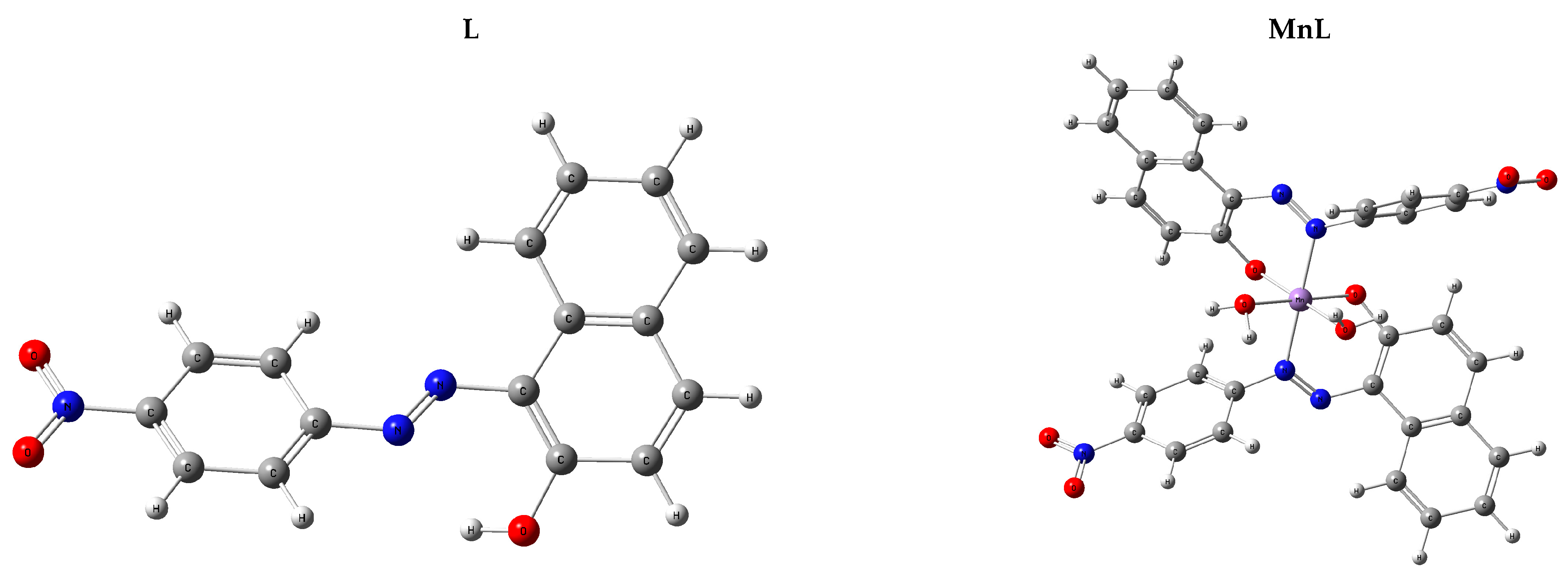
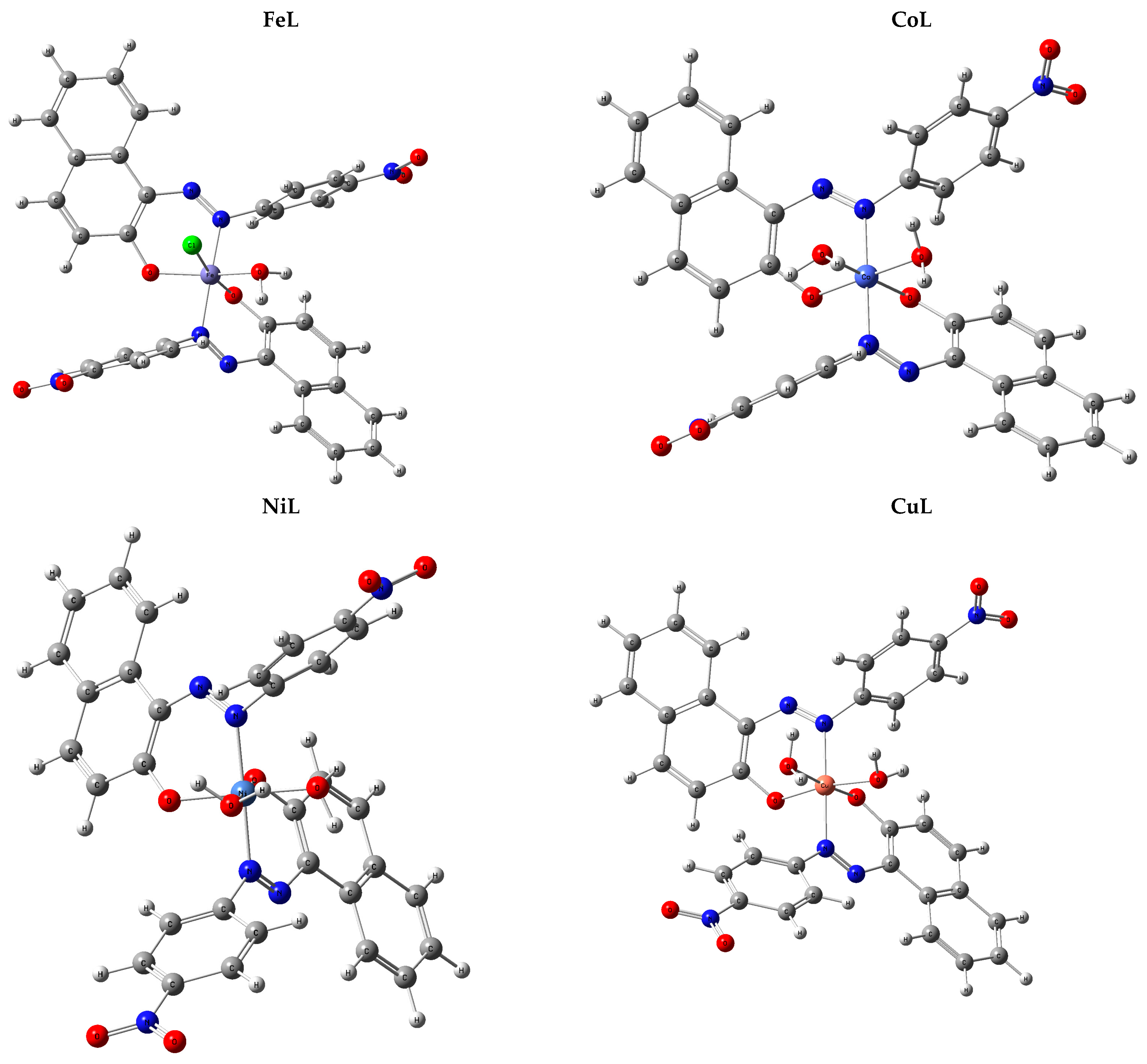
2.2. Complexes Structures’ Elucidation

2.2.1. Conductivity and EA Measurements

The produced complexes are H2O-insoluble but soluble in DMF and CH3CN and stable at 27 °C. Table 1 contains the data on the molar conductivity and EA of the produced compounds. The computed values and the elemental studies of the metal complexes are in accepted agreement with the measured values. The low molar conductivity values showed the non-electrolytic nature of the complexes.

2.2.2. IR Spectra

The IR was obtained to understand the ligand-metal ion interaction better. The most characteristic IR bands for the ligand and chelates are shown in Table 1. The bands seen originated either from the ligand or the bonds between the metal ion and the coordinating sites of the ligand. The azo (–N=N– the) band was observed at 1550 cm−1 in the IR spectrum of the free ligand, whereas the hydroxyl (–OH) band was observed at 3306 cm−1.
Comparing the IR bands of the ligand prior to and after chelation with the metal ion, it was found that the bands of the azo (–N=N–) group are still there. However, they were displaced to a lower wave number, 1515–1520, Table 1. This demonstrates that coordination between the metal ion and azo nitrogen took place. It has also been shown that the phenolic (–OH) vibration disappears in all the metal complexes [15]. These results demonstrate the role played by the ligand’s phenolic oxygen in constructing the C–O–M bond during deprotonation. It was previously believed that the –OH groups on H2O molecules were responsible for the presence of a broad band in the complexes above 3400 cm−1. New spectral bands identified as belonging to the molecules υ (M–O) [16,17] and υ (M–N) [18,19], respectively, Table 1.
These data show that the azo-ligand (HL) forms the complexes via its azo (–N=N–) and phenolic oxygen (–OH) groups, indicating that HL works as a mono-negatively bi-dentate ligand.

2.2.3. Mass Spectra

Mass spectra are a vital tool for deciphering intricate structures. Therefore, mass spectra were collected to compare the compounds’ relative amounts of various elements and presented in Supplementary Materials; Figure S2. Good concurring was found between the calculated values and the molecular ion peak (M+) in the mass spectra of the MnL, FeL, CoL, NiL, and CuL complexes at m/z 712.46, 748.24, 734.58, 716.13, and 720.88, were in good agreement with 711.5, 747.9, 733.5, 715.3, and 720.14, respectively. Some of the other peaks in the mass spectrum might be attributed to different fragments of metal complexes. The mass spectrum results show a high degree of consistency with the known values for carbon, hydrogen, and nitrogen, as well as with the proposed formula.

2.2.4. Magnetic Moment and Electronic Spectra Measurements

The UV-Vis spectra of ligand and metal complexes were processed in acetonitrile and recorded from 200 to 800 nm, Supplementary Materials, Figure S3 and Table 1. Electronic transitions at 280 and 335 nm in the spectrum of the unbound ligand may be ascribed to π → π* and n → π* states, respectively. These bands are shifted to longer wavelengths after coordinating with a metal ion. One of the best ways to figure out the internal structure of metal complexes is to calculate their effective magnetic moment, μeff = 2.83 [(Xg * Mwt) − (dia magnetic correction * T)]0.5], Table 1.
At 395 nm, a band in the electronic spectra of the MnL complex was seen that might be attributed to the 4T2g (G) → 6A1g transition [20]. The 1.89 B.M. value of the magnetic moment may be explained by the presence of a single unpaired electron with low spin (t2g5) in an octahedral geometry around the Mn (II) ion.
An electronic spectrum band at 410 nm was seen for the FeL complex, which has been attributed to the 6A1g → T2g (G) transition in the complex’s octahedral geometry. The 1.91 B.M. magnetic moment reported for the Fe (III) complex can be explained by the presence of d5 low spin (t2g5) atoms in an octahedral geometry around the Fe(III) center.
A band suggested the octahedral shape in the electronic spectra of the CoL complex at 445 nm, which might be attributed to the 4T1g (F) → 4T2g (F) transition. The 1.83 B.M. magnetic moment of the CoL complex at room temperature was assigned to the d7 low spin (t2g6 eg1) electron configuration. Then the CoL complex would have an octahedral shape [21], Table 1.
At 540 nm, bands can be seen in the NiL complex’s electronic spectra that may be attributed to 3T1 (F) → 3T1 (P) transitions, suggesting an octahedral geometry around the Ni(II) center, Table 1. NiL complex has a μeff of 3.14 B.M, which indicates a return to the d8 (t2g6 eg2) electron configuration [22], Table 1.
Bands at 525 nm can be attributed to 2B1g2A1g transitions in the electronic spectra of the CuL complex, suggesting an octahedral geometry centered on Cu(II), as shown in Table 1. In addition, the μeff of the CuL complex was measured to be 1.76 B.M, which is consistent with a return to the d9 (t2g6 eg3) electron configuration [23], Table 1.

2.2.5. Stoichiometry of the Metal Complexes

Job’s method of continuous variation was used to calculate the Stoichiometry of the metal complexes [24]. This method is based on the measurement of absorption of a series of solutions in which molar concentrations of two reactants vary, but their sum remains constant. Job’s Method is also known as the method of continuous variation. The principle of the method is that the mole ratio of the metal ion and the ligand is varied between 0 and 1 at constant total concentration C = Cligand + Cmetal. Then the absorbance of each mixture was obtained after allowing the reaction mixtures (M & L) to equilibrate. As a result, the absorbance of each solution was plotted against the ligand mole fraction ([L]/[L]  +  [M]). So, on drawing the graph between the absorbance of the prepared series of solution and their corresponding mole fraction, the exact ratio of metal and ligand at equilibrium can be observed at the maxima of the graph. Maximum absorbance, as measured by the curve of continuous change, occurred at a ligand mole fraction ([L]/([L] + [M])) of 0.66, suggesting the complex construction at a 1:2 (M:L) molar ratio, Figure S4.

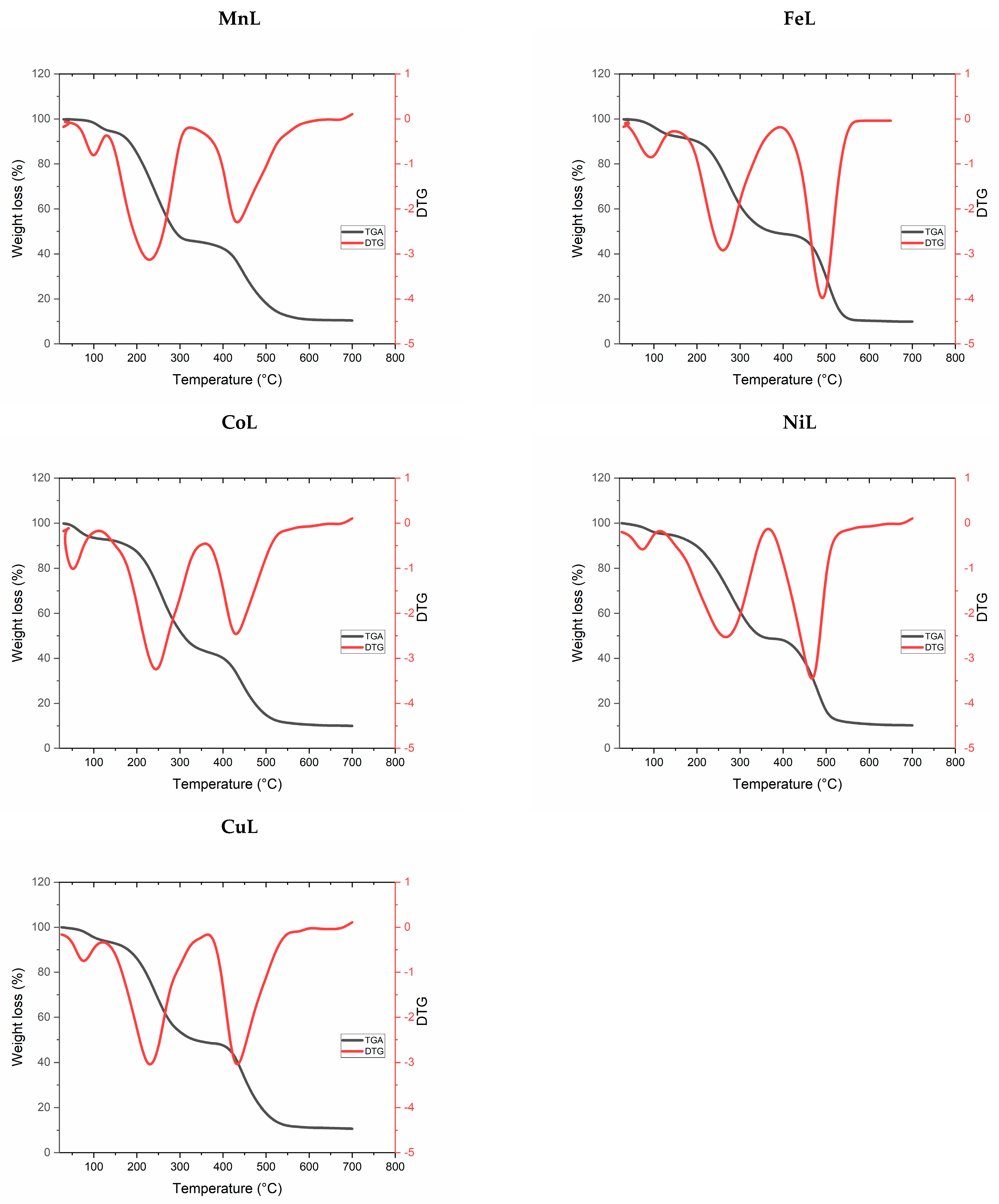
2.2.6. Thermal Decomposition

Determining the relative amounts of coordinated and uncoordinated H2O molecules in the examined metal complexes is a crucial step toward understanding their structural features after being subjected to heat degradation [25,26,27], Figure 1 and Table 2.
The first degradation step was found (calc.) weight loss percentage of 5.31 (5.26), 7.24 (7.35), 7.22 (7.37), 5.18 (5.04), and 5.11 (5.08), which linked to the elimination of two, three, three, two and two H2O of hydration for the MnL, FeL, CoL, NiL and CuL complexes, respectively, Figure 1 and Table 2.
The second degradation step was observed at found (calc.) weight loss percentage of 49.15 (49.37), 44.31 (44.48), 48.87 (48.98), 46.73 (46.85), and 46.08 (46.14) %, which correlated to the elimination of C18H15N4O4, C16H14N3O3Cl, C20H15N4O3, C18H13N3O4, and C19H14N3O3 in the case of MnL, FeL, CoL, NiL and CuL complexes, respectively, Figure 1 and Table 2.
The decomposition was continued for the third degradation step to eliminate the remaining organic moiety leaving the metal oxide as metallic residue, Figure 1 and Table 2.

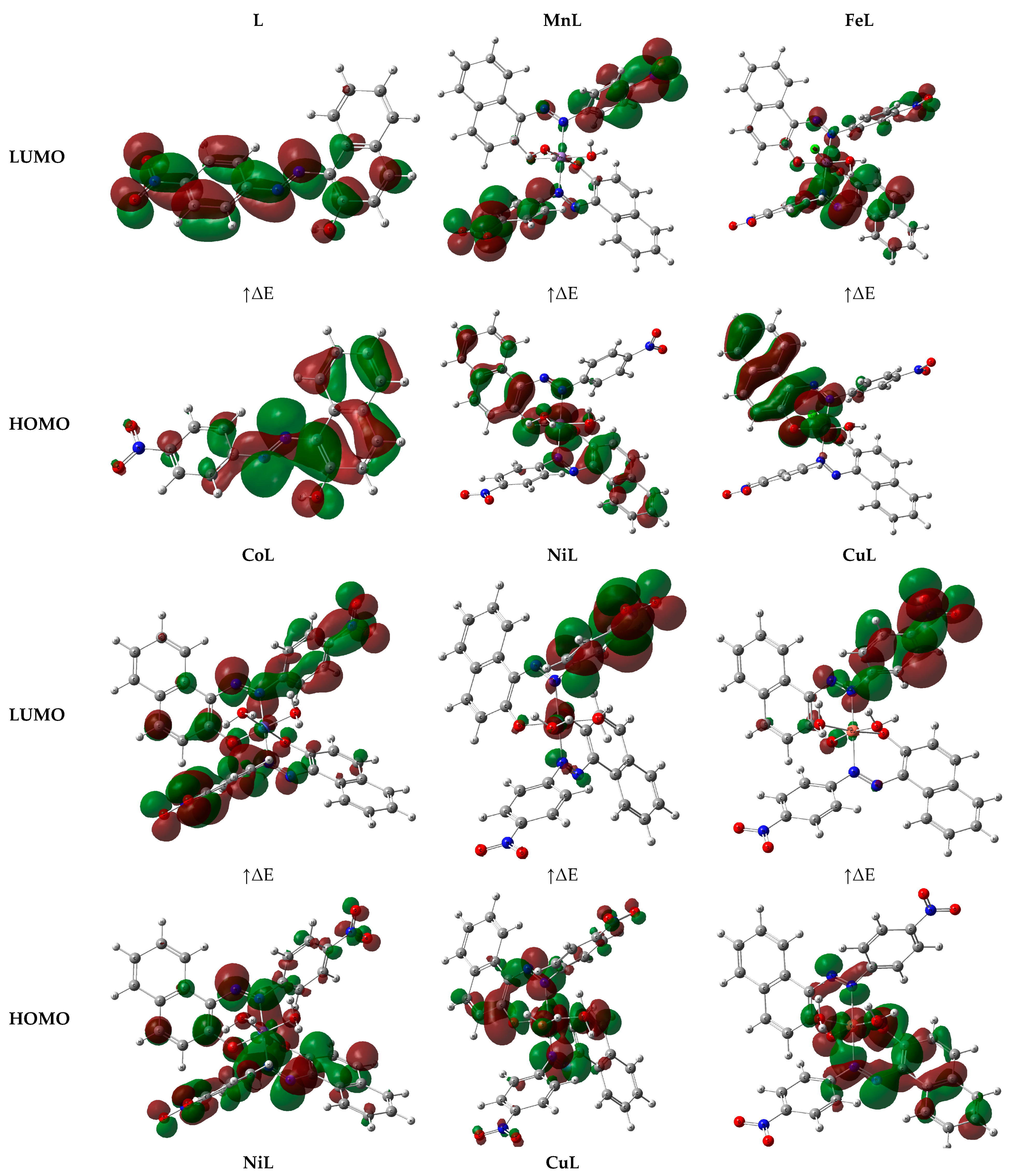
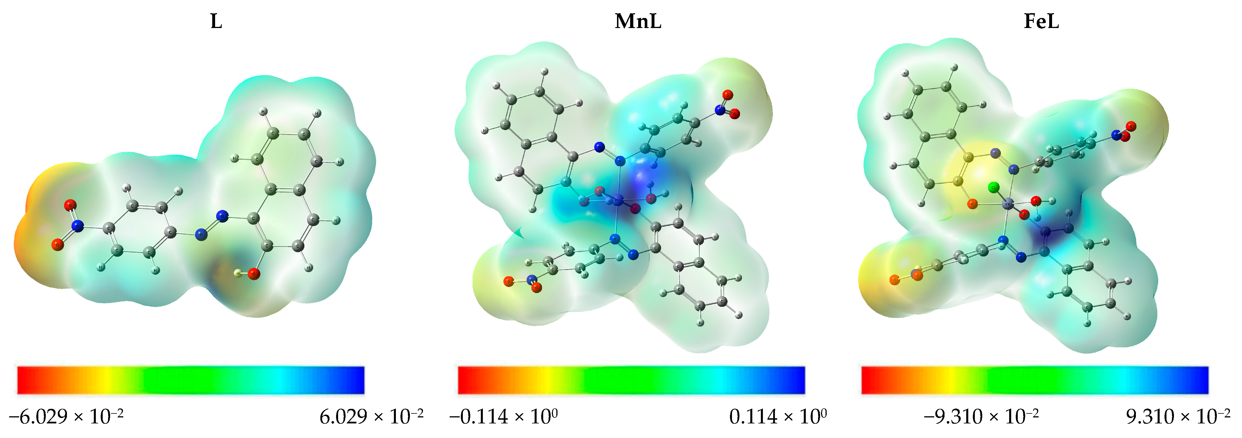
2.3. DFT Calculations

Titled compounds that were optimized had an octahedral geometry around the metal core, as; [Fe (L)2 (H2O) (Cl)] for FeL, and [M (L)2 (H2O)2], for MnL (M=Mn), CoL (M=Co), NiL (M=Ni) and CuL (M=Cu), Figure 2.
The HOMO and LUMO are two examples of frontier molecular orbitals (FMOs), which have a profound impact on reactivity, chemical stability, and electronic properties (LUMO). A diagram depicting the HOMO-LUMO ratio of the compounds mentioned above is presented in Figure 3. This means that the electrons are dispersed throughout the molecule. Therefore, HOMO-LUMO energies are used to determine different chemical properties such as the ΔE, IP, EA, χ, μ, η, σ, ω, and Nu Table 3.
The HOMO-LUMO energies are many-sided because of their potential application to understanding molecules’ chemical reactivity, kinetic stability, hardness-softness, biological properties, and polarizability. The HOMO was the most distant electron-containing orbital and hence the most generous giver of electrons. The lowest unoccupied molecular orbital (LUMO) functioned as an electron acceptor. Since the HOMO and LUMO orbitals define the molecule’s stability, they also define its susceptibility to attack by nucleophiles and electrophiles [28]. The difference between ELUMO and EHOMO energy levels (ΔE gap) indicates how reactive a molecule is [29,30,31,32]. To that end, molecules with smaller ΔE are more receptive to docking (CuL > MnL > NiL > FeL > CoL > HL).
The chemical reactivity scale also considers how hard or soft an element is. To describe how likely a molecule is to connect with another, the hard-soft-acid-base (HSAB) rule can be employed. The Hard Soft Acid Base (HSAB) rule states that hard acids prefer to connect with other hard acids and bases, while soft acids prefer to interact with other soft acids and bases. Biological macromolecules such as proteins and cells are examples of soft molecules [1,2]. When it comes to dealing with biological molecules, it is preferable to use soft molecules rather than rigid ones. As a consequence of this, greater levels of biological activity are linked to surfaces that are both more soft and less hard. Thus, chemical reactivity follows that CuL > MnL > NiL > FeL > CoL > HL. Table 3 shows a rise in biological activity when the values of softness and hardness increase and decrease, respectively.
The negative chemical potential value [29] demonstrates the stability of the cited compounds. It has a high electrophilicity index and low chemical potential value, both of which are conducive to its electrophilic activity [30]. Both the substrate and the protein contain partial charges, and these charges have a considerable influence on how rapidly they connect to one another. The molecular electrostatic potential (MEP) diagram may be used to gain insight into the 3D architecture and topology of substrates. The MEP determines where the nuclei or electrons impact the molecular geometry most.
Each value in an MEP diagram represents a distinct color, from blue to red. The MEP’s positive (blue) and negative (red) regions are linked to nucleophilic and electrophilic reactivity, respectively. Colors in the red spectrum represent portions of the surface that are negatively charged (i.e., the most favorable areas are accepting an electrophile). The attraction of the compound’s suitable sites in interactions with electrophiles is represented by a decrease in the compound’s negative charge [31]. The theoretical map of the MEP diagram for the specified compounds is shown in Figure 4. Negative areas (red) are concentrated around hetero atoms (O and N) in the studied substrates because of the increased electron abundance in these locations. Electrophilic assault is especially effective in these areas. More positive regions (blue) include a coordinated water moiety and metal center, which may function as H-bond donors in protein-substrate intermolecular interactions, Figure 4.

2.4. In Vitro Antimicrobial Activity

The potential antimicrobial applications of the compounds were investigated by testing against P. aeruginosa, E. coli (−ve), S. aureus (+ve), and B. cereus (+ve) bacterial strains and against A. flavus, T. rubrum, and C. albicans fungal strains using the agar diffusion assay Table 4.
The newly prepared complexes showed more significant biological activity with higher IZ and lowered MIC than the free ligand. Within this context, the MnL and CuL complexes showed promising antibacterial activities with high % Activity Index of 94.44%, 94.44% against P. aeruginosa, 90.00%, 90.00% against E. coli, 88.89%, 88.89% against S. aureus, and 88.89%, 94.44% against B. cereus. Similarly, MnL and CuL complexes showed promising antifungal activities with % Activity Index of 89.47%, 89.47% against A. flavus, 81.82%, 81.82% against T. rubrum, and 80.95%, 85.71% against C. albicans.
The minimum inhibitory concentration, abbreviated as MIC, is typically the point of departure for larger preclinical assessments of potentially novel antimicrobial treatments. The minimum inhibitory concentration (MIC) was found by subjecting a sample to a series of dilutions. The lowest compound concentration that inhibits the growth of bacteria/fungi was recorded as the MIC. MICs are normally reported in ppm. In this framework, the MIC was determined for the tested compounds and listed in Table 4. The metal complexes showed lower MIC (25–6.25 ppm) than the free ligand (around 50 ppm), Table 4.
The chelation theory interprets such superior activities [32,33,34]. Within this context, complexation diminishes the metal ions’ polarity via the distribution of their positive charges to the neighbor’s donor atoms. This may lead to electron-delocalization throughout the phenyl ring. Because of this result, the complex is more lipophilic, which increases the likelihood that it will be able to penetrate the lipid bilayer of the cell membrane. Furthermore, the complex may affect microorganisms’ metabolic pathways and respiration by interfering with their binding sites. This stops bacteria from making proteins, stunting their growth and eventually killing them.
It was shown that the activity of the complexes increased with the strength of the bond between the metal and ligand, the size of the cation, the number of receptor sites, the rate of diffusion, and the combined action of the metal and ligand to inactivate the biomolecules [35]. In addition, the IZ values for the specified compounds were compared to those of the antibiotic Chloramphenicol, which indicates a high activity index (%), Table 4.
The biological activity against the E. coli (G−) of the currently studied complexes was compared to that of previously reported metal complexes from the literature survey [1,2,3,4,36,37,38], Supplementary Materials, Table S1. This compression indicated the high biological activity of the current complexes.

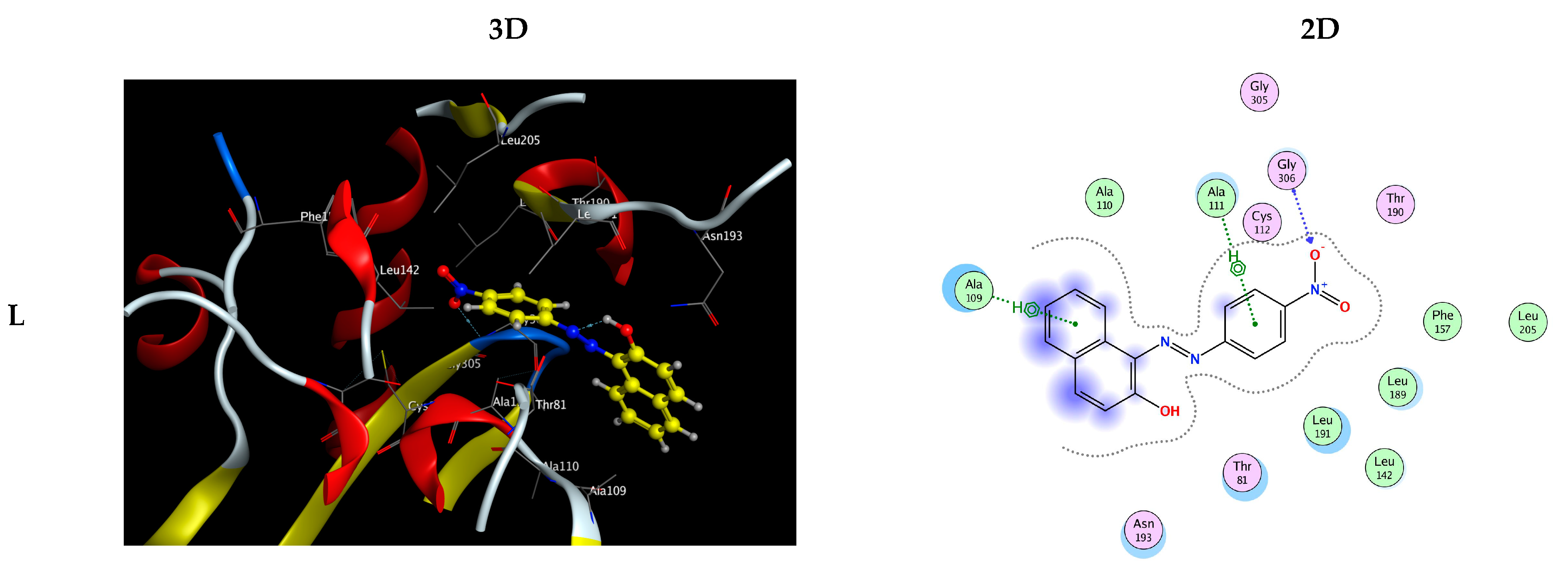
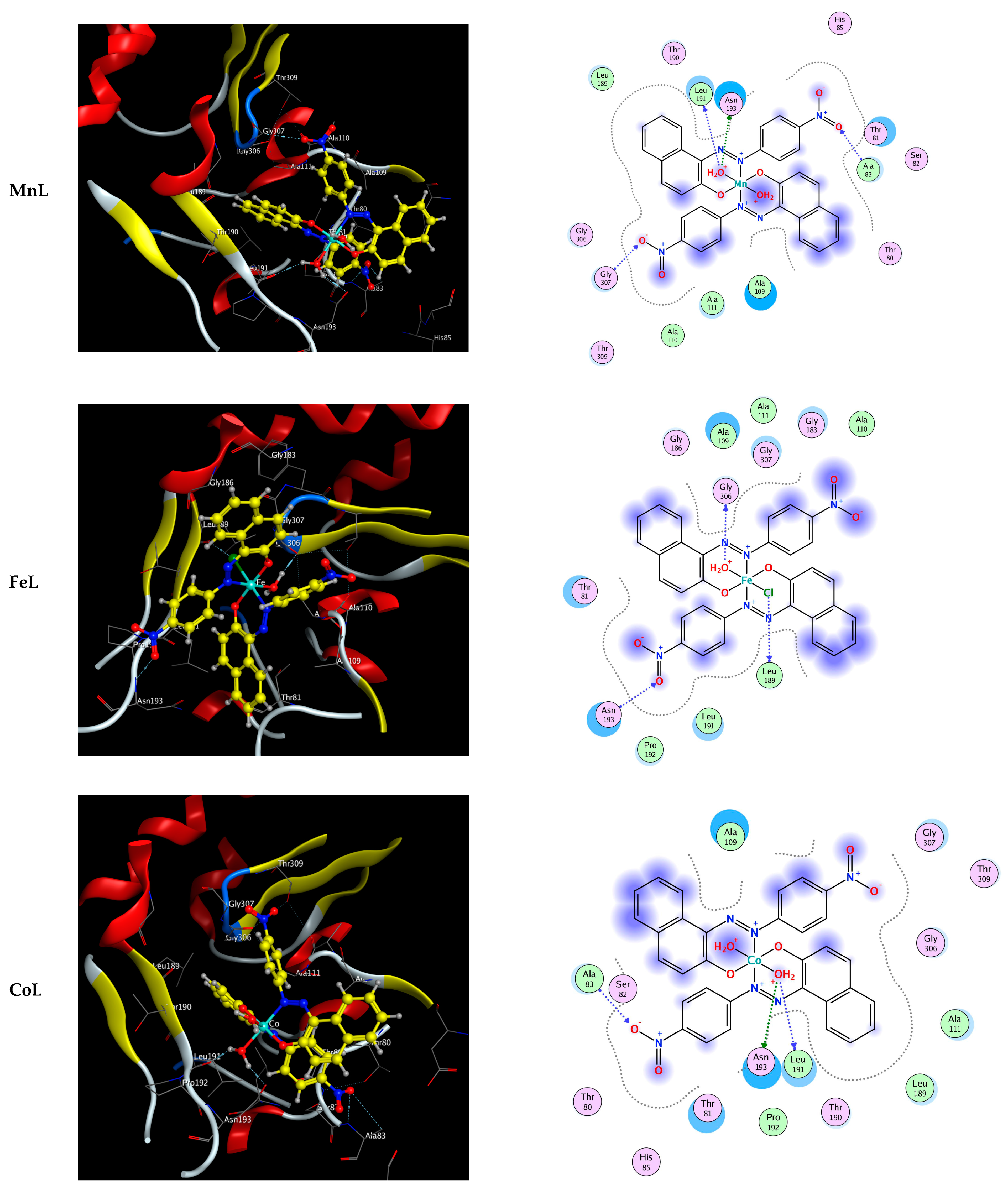
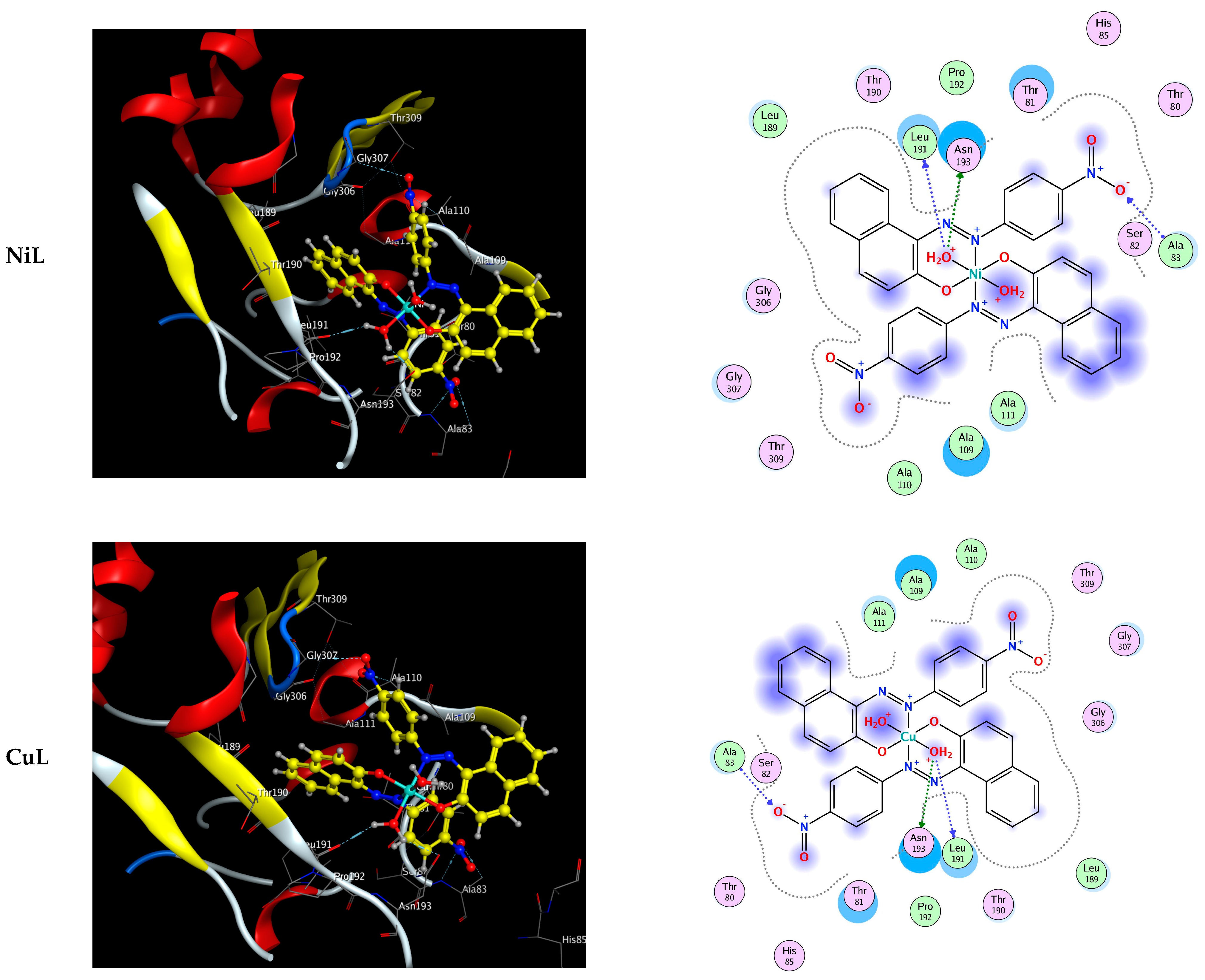
2.5. Molecular Docking

The complexes were docked to the dedicated antimicrobial target protein to verify the relationship between antimicrobial results and the inhibitor’s binding affinities. The highest binding affinities may be predicted by molecular docking studies, which use virtual compound screening and scoring systems to do so. This strategy examines the three-dimensional jigsaw puzzle-like fit between two molecules, such as a substrate and the active site binding of the target receptor.
Molecular docking of the titled compounds was studied to study their potential against 1hnj. First of all, there is a need to understand why this PDB was chosen and downloaded from https://www.rcsb.org/ (accessed on 1 June 2022). 1HNJ is for the E. coli FabH-CoA complex. FabH receptors are considered to be targeted to know the potential of molecules as antimicrobial in nature. FabH is involved in the biosynthesis of fatty acids [35,39]. E. coli (PDB ID: 1hnj) is the target receptor, while the aforementioned chemicals comprise the substrate. Both Table 5 and Figure 5 display the results of the molecular docking analysis. The optimum substrate conformation within the binding pocket is depicted in Figure 5.
Particularly intriguing is the fact that, upon docking to the 1hnj pocket (S), the relevant substrates display both robust hydrophobic contacts and a plethora of hydrogen bonds, Figure 5 and Table 5. This demonstrates that docked substrates make strong contacts with the receptor’s active site. The most inhibitory HL were CuL and MnL, followed by NiL, CoL, and FeL. Figure 5 and Table 5 show that the most active molecules, CuL and MnL, were docked to the substrate binding pocket of 1hnj thanks to a combination of hydrogen bonds and hydrophobic interactions.

3. Materials and Methods

All chemicals used were analytical reagent (AR) grade, the purest form commercially available. One example is 1-(4-nitrophenylazo)-2-naphthol, but others include MnCl2, FeCl3, CoCl2, NiCl2, and CuCl2. In addition, both absolute EtOH and CH3CN were available from BDH as spectroscopically pure organic solvents.

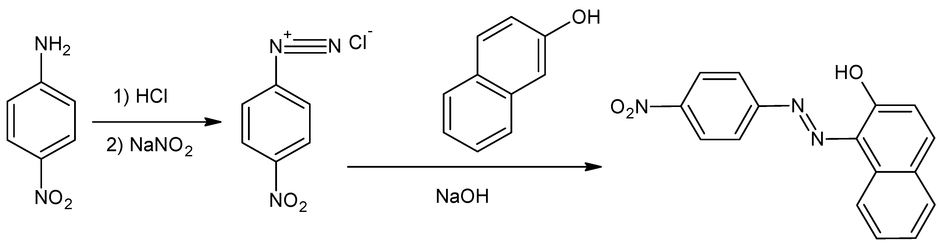
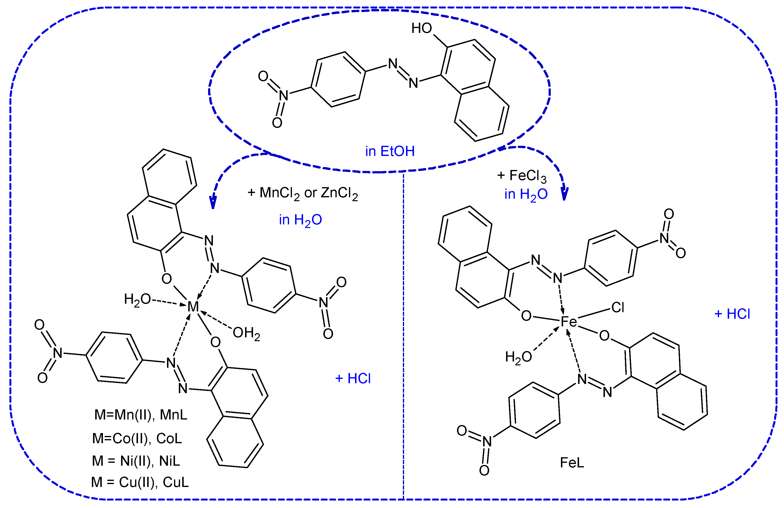
3.1. Synthesis of the 1-(4-Nitrophenylazo)-2-naphthol as Azo-Ligand (HL)

3.1.1. Step I: The Synthesis of Diazonium Salt of 4-Nitroaniline as Azo Compounds

In a round-bottom flask with a capacity of 100 milliliters and a magnetic stirring bar, 1.38 g (10 mmol) of 4-nitroaniline and 36.0 milliliters of concentrated hydrochloric acid were mixed. A progressive addition of 0.69 g (10 millimoles) of sodium nitrite that had been dissolved in 20 milliliters of water was carried out while ensuring that the temperature of the solution remained below 5 degrees Celsius. Keep the diazonium salt solution in an airtight container and store it in the refrigerator. When tested on starch iodide paper, diazonium salt generated a color that may be described as blue-black. The diazonium salt coupling operation was started as soon as it was feasible for us to do so., Scheme 1.

3.1.2. Step II: Coupling Procedure

The 2-naphthol (1.44 g, 10 mmol) was put into a 50 mL round-bottom flask, and then NaOH (10% w/v) was poured into the flask. After that, the flask was placed in an ice bath (0–5 °C). After being agitated at zero degrees Celsius to five degrees Celsius for an hour, the cooled diazonium salt solution was added gradually. Then, after filtering the crude residue, washing it many times in cold water, and recrystallizing it from the appropriate solvent, the final azo product was achieved, Scheme 2.
1-(4-nitrophenylazo)-2-naphthol (HL); red powder; Yield (97%); mp. 248–250 °C; IR (cm−1): 3306-3202 (–OH), 3057 (Ar-H), 1550 (N=N); 1H-NMR (400 MHz, DMSO-d6) δ (ppm): 8.44 (s, 1H, –OH), 8.42−6.71 (m, 10 H, Ar-H); 13C-NMR (100 MHz, DMSO-d6) δ (ppm): 144.59,143.96, 133.04, 131.76, 130.32, 129.90, 128.97, 128.11, 126.53, 126.06, 122.75, 117.83; EA for C16H11N3O3 (Calcd./Found); C, 65.53/65.41; H, 3.78/3.61; N, 14.33/14.21.

3.2. Metal Chelates Preparation

The ligand (2.0 mmol) was dissolved in EtOH and heated, and then a solution of the metal salt (2.0 mmol) in H2O (20 mL) was slowly added. The furnished mixture was stirred continuously while refluxed in an 80 °C H2O bath for 12 h. After being filtered, dried, and washed with the H2O−EtOH (1:2) combination, the final product was recrystallized, Scheme 3. Each product’s yield and melting or decomposition temperature were tabulated in Table 1.

3.3. Characterization

The chelates composition was determined by IR, molar conductance (of 10−3 mol/L ethanol solution), thermal degradation (TGA), electronic spectrum (of 10−3 mol/L acetonitrile solution), magnetic measurements, and EA (C, H, and N content). The latter was run on a [Perkin-Elmer 2408] analyzer in the Central Laboratory at the University of Cairo. The infrared spectra were acquired by observing KBr in a Shimadzu DR-8001 spectrometer. The 1H-NMR was measured using Bruker DRX (400 MHz) (USA) using TMS (standard) and DMSO-d6. UV-Vis spectra were detected using a Jenway spectrophotometer. We used a Shimadzu type 60 H analyzer to conduct thermal decomposition (TGA) on the named compounds. The molar conductance was measured using JENWAY instrument (model 4320) and a 10−3 mol/L EtOH solution. The molar magnetic susceptibility of powdered materials was quantified employing a Bartington Susceptibility apparatus, model 4320. Finally, the compounds’ stoichiometries were determined using the continuous-variation spectrophotometric jobs technique [37].

3.4. DFT Calculations

With the use of the 6-311 (d, p) and LANL2DZ basis sets in conjunction with the hybrid correlation functional (B3LYP) [39,40], for the ligand and its complexes, respectively, geometry optimizations of the subject ligand and its complexes were performed [1,2,3,4]. Quantum chemical properties were determined by using the values of the lowest unoccupied molecular orbital (LUMO) and highest occupied molecular orbital (HOMO) energies of the compounds in question, as follows: chemical potential (µ = −χ), electronegativity (χ = (IP + EA)/2), energy gap (ΔE = ELOMO − EHOMO), chemical hardness (η = (IP − EA)/2), nucleophilicity index (N = 1/ω), softness (σ = ½ η), maximum electronic charge (ΔNmax = −μ/η), and electrophilicity index (ω = µ2/2 η) where EA and IP, are electron affinity (EA = −ELUMO) and ionization potential (IP = −EHOMO) [21,22,28,31].

3.5. Antimicrobial Exploration

The antimicrobial activity of the titled compounds was evaluated using a panel of bacteria (Pseudomonas aeruginosa (P. aeruginosa) (−ve), Escherichia coli (E. coli) (−ve), Staphylococcus aureus (S. aureus) (+ve), and Bacillus cereus (B. cereus) (+ve)) and fungi (Aspergillus flavus (A. flavus), Trichophyton rubrum (T. rubrum), and Candida albicans (C. albicans)). The good agar diffusion technique tested antibacterial and antifungal activities [15,21,23,28]. The findings were monitored using the diameter (in mm) of the inhibitory zone (IZ).
We found the widely used antimicrobial Chloramphenicol effective against bacteria and fungi using the same method. The % Activity Index, defined as (IZ of test compound/IZ of Standard) × 100, was used to compare the anti-microbial efficacy of the identified compounds to that of the reference [1,2,3,4,22,31].
The minimal inhibitory concentration (MIC) values were determined by serial dilution method using the same above protocol. The tested compounds with different concentrations of 100 ppm, 50 ppm, 25 ppm, 12.50 ppm, and 6.25 ppm were used. The lowest compound concentration that inhibits the growth of bacteria/fungi was recorded as the MIC.

3.6. Molecular Docking

Through the MOE software, molecular docking against E. coli (PDB ID: 1hnj) confirmed the therapeutic efficacy of the reported drugs [40,41,42,43].
The 3D structure of the target protein receptor was retrieved from the protein database at http://www.rcsb.org (accessed on 1 June 2022). The compounds of interest were used as a substrate. In this molecular docking evaluation, we use the MOE software, which simulates a molecular smorgasbord.
After establishing a new database for each compound in MDB format, we optimized them for substrate preparation by reducing their energy demands. Hydrogen atom addition, receptor type connections, potential energy fixation, active pocket search, and dummy generating are all steps in the receptor preparation process. Docking patterns and interaction parameters were exported to score inhibitory activity using a scoring function (S, kcal/mol) and analyze interaction features [22,23,28,31].

4. Conclusions

Five novels of CuLQ, CoL, FeL, MnL, and NiL complexes were synthesized and analyzed using physicochemical and spectroscopic techniques. The data showed that the HL ligand binds to the metals in a 1:2 molar ratio, exhibiting the behavior of a monobasic bi-dentate NO ligand. Spectroscopic measurements showed an octahedral shape for the complexes. Furthermore, their antimicrobial activities were evaluated against various bacterial and fungal pathogenic strains. Moreover, the quantum chemical parameters were determined after theoretically optimizing the molecular structures of the metal complexes. The metal complexes showed more promising antibacterial candidates than the free ligand. In addition, molecular docking was used to determine whether the studied chemicals inhibited E. coli growth (PDB ID: 1hnj). The binding of the CuL complex to the target receptor was the most impressive among these drugs.

Supplementary Materials

The following supporting information can be downloaded at: https://www.mdpi.com/article/10.3390/ijms232415614/s1.

Author Contributions

Conceptualization, A.A.A. (Amer A. Amer), A.A. (Aly Abdou), A.A. (Anas Alfarsi), A.A.A. (Antar A. Abdelhamid) and H.M.A.E.-L.; methodology, A.A.A. (Amer A. Amer), A.A. (Aly Abdou), A.A. (Anas Alfarsi), A.A.A. (Antar A. Abdelhamid) and H.M.A.E.-L., S.S.; validation, A.A.A. (Amer A. Amer), A.A. (Aly Abdou), A.A. (Anas Alfarsi), A.A.A. (Antar A. Abdelhamid), S.S., M.G. and M.M.K.; investigation, A.A.A. (Antar A. Abdelhamid), A.A. (Aly Abdou), A.A. (Anas Alfarsi), A.A.A. (Antar A. Abdelhamid), H.M.A.E.-L., M.M.K. and M.M.K.; writing—original draft preparation, A.A.A. (Amer A. Amer), A.A. (Aly Abdou), A.A. (Anas Alfarsi), A.A.A. (Antar A. Abdelhamid), M.G., S.S. and M.M.K.; writing—review and editing, A.A.A. (Amer A. Amer), A.A. (Aly Abdou), A.A. (Anas Alfarsi), A.A.A. (Antar A. Abdelhamid), M.G., S.S. and M.M.K., supervision, A.A.A. (Ahmed A. Alzharani), A.A. (Aly Abdou), A.A. (Anas Alfarsi), and A.A.A. (Antar A. Abdelhamid), project administration, A.A.A. (Ahmed A. Alzharani), A.A. (Aly Abdou), A.A. (Anas Alfarsi), A.A.A. (Antar A. Abdelhamid) and H.M.A.E.-L.; funding acquisition, Y.A.A.A., H.M.A.E.-L., M.G., S.S., A.A.A. (Ahmed A. Alzharani) and M.M.K. All authors have read and agreed to the published version of the manuscript.

Funding

This work was supported by the Deanship of Scientific Research, King Faisal University, Saudi Arabia (GRANT2046).

Institutional Review Board Statement

Not applicable.

Informed Consent Statement

Not applicable.

Data Availability Statement

The raw/processed data generated in this work are available upon request from the corresponding author.

Acknowledgments

This work was supported by the Deanship of Scientific Research, Vice Presidency for Graduate Studies and Scientific Research, King Faisal University, Saudi Arabia [Grant No. 2046], through its KFU Research Summer initiative. Also, the authors extend their appreciation to the faculty of science for funding this work through project No. FC-2200955.

Conflicts of Interest

The authors declare no conflict of interest.

References

  1. Ismael, M.; Abdou, A.; Abdel-Mawgoud, A.-M. Synthesis, Characterization, Modeling, and Antimicrobial Activity of FeIII, CoII, NiII, CuII, and ZnII Complexes Based on Tri-substituted Imidazole Ligand. Z. Anorg. Allg. Chem. 2018, 644, 1203–1214. [Google Scholar] [CrossRef]
  2. Ismael, M.; Abdel-Mawgoud, A.M.; Rabia, M.K.; Abdou, A. Design and synthesis of three Fe(III) mixed-ligand complexes: Exploration of their biological and phenoxazinone synthase-like activities. Inorg. Chim. Acta 2020, 505, 119443. [Google Scholar] [CrossRef]
  3. Ismael, M.; Abdel-Mawgoud, A.M.; Rabia, M.K.; Abdou, A. Synthesis, characterization, molecular modeling and preliminary biochemical evaluation of new copper (II) mixed-ligand complexes. J. Mol. Struct. 2021, 1227, 129695. [Google Scholar] [CrossRef]
  4. Ismael, M.; Abdel-Mawgoud, A.M.; Rabia, M.K.; Abdou, A. Ni(II) mixed-ligand chelates based on 2-hydroxy-1-naphthaldehyde as antimicrobial agents: Synthesis, characterization, and molecular modeling. J. Mol. Liq. 2021, 330, 115611. [Google Scholar] [CrossRef]
  5. Aljamali, N.M. Review in Azo Compounds and its Biological Activity. Biochem. Anal. Biochem. 2015, 4, 1000169. [Google Scholar] [CrossRef] [Green Version]
  6. Eltaboni, F.; Bader, N.; El-Kailany, R.; Elsharif, N.; Ahmida, A. Chemistry and Applications of Azo Dyes: A Comprehensive Review. J. Chem. Rev. 2022, 4, 313–330. [Google Scholar]
  7. Bentley, R. Different roads to discovery; Prontosil (hence sulfa drugs) and penicillin (hence β-lactams). J. Ind. Microbiol. Biotechnol. 2009, 36, 775–786. [Google Scholar] [CrossRef]
  8. Pervaiz, M.; Riaz, A.; Munir, A.; Saeed, Z.; Hussain, S.; Rashid, A.; Younas, U.; Adnan, A. Synthesis and characterization of sulfonamide metal complexes as antimicrobial agents. J. Mol. Struct. 2020, 1202, 127284. [Google Scholar] [CrossRef]
  9. Kyhoiesh, H.A.K.; Al-Adilee, K.J. Synthesis, spectral characterization and biological activities of Ag(I), Pt(IV) and Au(III) complexes with novel azo dye ligand (N, N, O) derived from 2-amino-6-methoxy benzothiazole. Chem. Pap. 2022, 76, 2777–2810. [Google Scholar] [CrossRef]
  10. Husien, N.S.M. Preparation and (Characterization, Chromatography, Antimicrobial Activities) of Some Metal Complexes of 1-(2--hydroxyl-4--nitrophenylazo)-2-naphthol. Biochem. Cell. Arch. 2018, 18, 901–908. [Google Scholar]
  11. Abdallah, S.M. Metal complexes of azo compounds derived from 4-acetamidophenol and substituted aniline. Arab. J. Chem. 2012, 5, 251–256. [Google Scholar] [CrossRef] [Green Version]
  12. Rana, M.; Cho, H.-J.; Roy, T.K.; Mirica, L.M.; Sharma, A.K. Azo-dyes based small bifunctional molecules for metal chelation and controlling amyloid formation. Inorg. Chim. Acta 2018, 471, 419–429. [Google Scholar] [CrossRef]
  13. Badea, M.; Olar, R.; Cristurean, E.; Marinescu, D.; Emandi, A.; Budrugeac, P.; Segal, E. Thermal stability study of some azo-derivatives and their complexes, Part 2. New azo-derivative pigments and their Cu(II) complexes. J. Therm. Anal. Calor. 2004, 77, 815–824. [Google Scholar] [CrossRef]
  14. Mohammed, H. Synthesis, Identification, and Biological Study for Some Complexes of Azo Dye Having Theophylline. Sci. World J. 2021, 2021, 9943763. [Google Scholar] [CrossRef]
  15. Abdou, A.; Mostafa, H.M.; Abdel-Mawgoud, A.M. Seven metal-based bi-dentate NO azocoumarine complexes: Synthesis, characterization, DFT calculations, Drug-Likeness, in vitro antimicrobial screening and molecular docking analysis. Inorg. Chim. Acta 2022, 539, 121043. [Google Scholar] [CrossRef]
  16. Samy, F.; Shebl, M. Synthesis, spectroscopic, biological, and theoretical studies of new complexes from (E)-3-(2-(5, 6- diphenyl-1,2,4- triazin-3- yl)hydrazono)butan-2- one oxime. Appl. Organometal. Chem. 2020, 34, e5502. [Google Scholar] [CrossRef]
  17. Seleem, H.S.; El-Shetary, B.A.; Shebl, M. Synthesis and characterization of a novel series of metallothiocarbohydrazone polymers and their adducts. Heteroatom. Chem. 2007, 18, 100. [Google Scholar] [CrossRef]
  18. Shebl, M.; Khalil, S.M.E. Synthesis, spectral, X-ray diffraction, antimicrobial studies, and DNA binding properties of binary and ternary complexes of pentadentate N2O3 carbohydrazone ligands. Monatsh. Chem. 2015, 146, 15. [Google Scholar] [CrossRef]
  19. Shebl, M.; Adly, O.M.I.; El-Shafiy, H.F.; Khalil, S.; Taha, A.; Mahdi, M.A. Structural variety of mono- and binuclear transition metal complexes of 3-[(2-hydroxy-benzylidene)-hydrazono]-1-(2-hydroxyphenyl)-butan-1-one: Synthesis, spectral, thermal, molecular modeling, antimicrobial and antitumor studies. J. Mol. Struct. 2017, 1134, 649–660. [Google Scholar] [CrossRef]
  20. Morgan, S.M.; El-Sonbati, A.Z.; Eissa, H.R. Geometrical structures, thermal properties and spectroscopic studies of Schiff base complexes: Correlation between ionic radius of metal complexes and DNA binding. J. Mol. Liq. 2017, 240, 752. [Google Scholar] [CrossRef]
  21. Elkanzi, N.A.A.; Ali, A.M.; Hrichi, H.; Abdou, A. New mononuclear Fe(III), Co(II), Ni(II), Cu(II), and Zn(II) complexes incorporating 4-{[(2 hydroxyphenyl)imino]methyl}phenyl-4-methylbenzenesulfonate (HL): Synthesis, characterization, theoretical, anti-inflammatory, and molecular docking investigation. Appl. Organomet Chem. 2022, 36, e6665. [Google Scholar] [CrossRef]
  22. Abdou, A. Synthesis, Structural, Molecular Docking, DFT, Vibrational Spectroscopy, HOMO-LUMO, MEP Exploration, antibacterial and antifungal activity of new Fe(III), Co(II) and Ni(II) hetero-ligand complexes. J. Mol. Struct. 2022, 1262, 132911. [Google Scholar] [CrossRef]
  23. Elkanzi, N.A.A.; Hrichi, H.; Salah, H.; Albqmi, M.; Ali, A.M.; Abdou, A. Synthesis, physicochemical properties, biological, molecular docking and DFT investigation of Fe(III), Co(II), Ni(II), Cu(II) and Zn(II) complexes of the 4-[(5-oxo-4,5-dihydro-1,3-thiazol-2-yl)hydrazono]methyl}phenyl 4-methylbenzenesulfonate Schiff-base ligand. Polyhedron 2023, 230, 116219. [Google Scholar]
  24. Job, P. Formation and Stability of Inorganic Complexes in Solution. Ann. Chem. 1928, 9, 113–203. [Google Scholar]
  25. Abdel-Latif, S.A.; Hassib, H.B.; Issa, Y.M. Studies on some salicylaldehyde Schiff base derivatives and their complexes with Cr(III), Mn(II), Fe(III), Ni(II) and Cu(II). Spectrochim. Acta A Mol. Biomol. Spectrosc. 2007, 67, 950–957. [Google Scholar] [CrossRef] [PubMed]
  26. Prakash, A.; Singh, B.K.; Bhojak, N.; Adhikari, D. Synthesis and characterization of bioactive zinc(II) and cadmium(II) complexes with new Schiff bases derived from 4-nitrobenzaldehyde and acetophenone with ethylenediamine. Spectrochim. Acta A Mol. Biomol. Spectrosc. 2010, 76, 356–362. [Google Scholar] [CrossRef]
  27. Zaky, R.R.; Yousef, T.A. Spectral, magnetic, thermal, molecular modelling, ESR studies and antimicrobial activity of (E)-3-(2-(2-hydroxybenzylidene) hydrazinyl)-3-oxo-n(thiazole-2-yl)propanamide complexes. J. Mol. Struct. 2011, 1002, 76–85. [Google Scholar] [CrossRef]
  28. Alghuwainem, Y.A.A.; Abd El-Lateef, H.M.; Khalaf, M.M.; Abdelhamid, A.A.; Alfarsi, A.; Gouda, M.; Abdelbaset, M.; Abdou, A. Synthesis, Structural, DFT, Antibacterial, Antifungal, Anti-inflammatory, and Molecular Docking Analysis of New VO(II), Fe(III), Mn(II), Zn(II), and Ag(I) complexes based on 4-((2-hydroxy-1-naphthyl)azo) benzenesulfonamide. J. Mol. Liq. 2023, 369, 120936. [Google Scholar] [CrossRef]
  29. Abu-Dief, A.M.; Alotaibi, N.H.; Al-Farraj, E.S.; Qasem, H.A.; Alzahrani, S.; Mahfouz, M.K.; Abdou, A. Fabrication, structural elucidation, theoretical, TD-DFT, vibrational calculation and molecular docking studies of some novel adenine imine chelates for biomedical applications. J. Mol. Liq. 2022, 365, 119961. [Google Scholar] [CrossRef]
  30. Shokr, E.K.; Kamel, M.S.; Abdel-Ghany, H.; Mahmoud El-Remaily, M.A.E.A.A.; Abdou, A. Synthesis, characterization, and DFT study of linear and non-linear optical properties of some novel thieno [2,3-b]thiophene azo dye derivatives. Mater. Chem. Phys. 2022, 290, 126646. [Google Scholar] [CrossRef]
  31. Elkanzi, N.A.A.; Ali, A.M.; Albqmi, M.; Abdou, A. New Benzimidazole-Based Fe (III) and Cr (III) Complexes: Characterization, Bioactivity Screening, and Theoretical Implementations Using DFT and Molecular Docking Analysis. Appl. Organomet. Chem. 2022, 36, e6868. [Google Scholar] [CrossRef]
  32. Abdou, A.; Mostafa, H.M.; Abdel-Mawgoud, A.M. Molecular Modeling, Breast Cancer, and Hepatitis A, B, C Molecular Docking Investigation of (2E)-1-phenyl-butane-1,2,3-trione 2-[(2-oxo-2H-chromene-6-yl)hydrazone]. Sohag. J. Sci. 2022, 6, 167–173. [Google Scholar] [CrossRef]
  33. Al-Wabli, R.I.; Resmi, K.S.; Mary, Y.S.; Panicker, C.Y.; Attia, M.A.; El-Emam, A.A.; Van Alsenoy, C. Vibrational spectroscopic studies, Fukui functions, HOMO-LUMO, NLO, NBO analysis and molecular docking study of (E)-1-(1,3-benzodioxol-5-yl)-4,4-dimethylpent-1-en-3-one, a potential precursor to bioactive agents. J. Mol. Struct. 2016, 1123, 375–383. [Google Scholar] [CrossRef]
  34. Chaudhary, A.P.; Bharti, S.K.; Kumar, S.; Ved, K.; Padam, K. Study of molecular structure, chemical reactivity and first hyperpolarizability of a newly synthesized N-(4-oxo-2-phenylquinazolin-3(4H)-yl)-1H-indole-2-carboxamide using spectral analysis. J. Mol. Struct. 2017, 1148, 356–363. [Google Scholar] [CrossRef]
  35. Abdou, A.; Abdel-Mawgoud, A.M. Synthesis, structural elucidation, and density functional theory investigation of new mononuclear Fe(III), Ni(II), and Cu(II) mixed-ligand complexes: Biological and catalase mimicking activity exploration. Appl. Organomet Chem. 2022, 36, e6600. [Google Scholar] [CrossRef]
  36. Uddin, M.N.; Chowdhury, D.A.; Islam, M.T.; Hoque, F. Evaluation of biological activity of some dioxouranium complexes of some Schiff base and dithiocarbamate ligands. Orbital Electron. J. Chem. 2012, 4, 273–287. [Google Scholar]
  37. Rehman, S.; Ikram, M.; Rehman, S.; Faiz, A. Synthesis, characterization and antimicrobial studies of transition metal complexes of imidazole derivative. Bull. Chem. Soc. Ethiop. 2010, 24, 201–207. [Google Scholar] [CrossRef] [Green Version]
  38. Aiyelabola, T.O.; Ojo, I.A.; Adebajo, A.C.; Ogunlusi, G.O.; Oyetunji, O.; Akinkunmi, E.O.; Adeoye, A.O. Synthesis Characterization and Antimicrobial Activities of Some Metal(II) Amino Acids’ Complexes. Adv. Biol. Chem. 2012, 2, 268–273. [Google Scholar] [CrossRef] [Green Version]
  39. Fekri, A.; Zaky, R. Solvent-free synthesis and computational studies of transition metal complexes of the aceto- and thioaceto-acetanilide derivatives. J. Organomet. Chem. 2016, 818, 15–27. [Google Scholar] [CrossRef]
  40. Kumar, A.; Kumar, D.; Kumari, K.; Mkhize, Z.; Katata Seru, L.M.; Bahadur, I.; Singh, P. Metal-ligand complex formation between ferrous or ferric ion with syringic acid and their anti-oxidant and anti-microbial activities: DFT and molecular docking approach. J. Mol. Liq. 2021, 322, 114872. [Google Scholar] [CrossRef]
  41. Hay, P.J.; Wadt, W.R. Ab initio effective core potentials for molecular calculations. Potentials for main group elements Na to Bi. J. Chem. Phys. 1985, 82, 270–283. [Google Scholar] [CrossRef]
  42. Abdou, A.; Omran, O.A.; Nafady, A.; Antipin, I.S. Structural, spectroscopic, FMOs, and non-linear optical properties exploration of three thiacaix(4)arenes derivatives. Arab. J. Chem. 2022, 15, 103656. [Google Scholar] [CrossRef]
  43. Heba, E. Hashem, Ashutosh Nath, Ajoy Kumer, Synthesis, molecular docking, molecular dynamic, quantum calculation, and antibacterial activity of new Schiff base-metal complexes. J. Mol. Struct. 2022, 1250, 131915. [Google Scholar]
Scheme 1. The structures of azo-derived metal complexes with its featuring biological activity (from Ref. [9]).
Ijms 23 15614 sch001
Figure 1. Thermal decomposition of the synthesized complexes.
Ijms 23 15614 g001
Figure 2. 3D optimized structure of the compounds.
Ijms 23 15614 g002aIjms 23 15614 g002b
Figure 3. HOMO—LUMO of the optimized structures.
Ijms 23 15614 g003
Figure 4. MEP of the compounds.
Ijms 23 15614 g004aIjms 23 15614 g004b
Figure 5. 3D orientation of the substrate-protein complex.
Ijms 23 15614 g005aIjms 23 15614 g005bIjms 23 15614 g005c
Scheme 2. The procedures involved in the production of the free ligand.
Ijms 23 15614 sch002
Scheme 3. Synthesis of the metal complexes.
Ijms 23 15614 sch003
Table 1. EA, conductivity, Uv-vis, and FT-IR results.
LMnLFeLCoLNiLCuL
Physical propertiesColorOrangePale yellowDarck violetDarck orangeOrange redPale pink
Melting point (°C)190280268255295290
Yield (%)808885909590
EA
Found (calc.) %
C65.41 (65.53)54.27
(54.02)
51.64
(51.39)
52.13
(52.40)
53.48
(53.73)
53.82
(53.37)
H3.61
(3.78)
3.55
(3.97)
3.92
(3.77)
4.01
(4.12)
4.15
(3.95)
3.27
(3.92)
N14.21 (14.33)11.43
(11.81)
11.04
(11.24)
11.18
(11.46)
11.98
(11.75)
11.35
(11.67)
M-----7.54
(7.72)
7.81
(7.47)
7.88
(8.03)
8.06
(8.21)
8.44
(8.82)
conductivityµv, Ω−1 cm2 mol−1------8.5710.149.859.1010.08
IR spectraυ (–OH)330634573460345534483466
υ (–N=N)155015151520151815201517
υ (M–O)-------561558559560557
υ (M–N)-------453451450454455
UV-vis.λmax, nm280, 335395410445540525
Magneticµeff (B.M)-------1.891.911.833.141.76
StoichiometryM:L-------1:21:21:21:21:2
Table 2. Thermal decomposition data.
TG (°C)DTG (°C)Mass Loss (%)AssignmentResidue
FoundCalculated
MnL30–140955.315.262 H2OMnO
140–31522549.1549.37C18H15N4O4
315–56043035.2235.39C14H7N2O3
FeL30–140907.247.353 H2O½ Fe2O3
140–36026044.3144.48C16H14N3O3Cl
360–55049036.4436.51C16H6N3O2
CoL30–125607.227.373 H2OCoO
125–35035048.8748.98C20H15N4O3
350–53553033.6533.42C12H9N2O4
NiL30–130755.185.042 H2ONiO
30–1307046.7346.85C18H13N3O4
130–36026037.7637.62C14H11N3O3
CuL360 5204705.115.082 H2OCuO
130–33523046.0846.14C19H14N3O3
335–54044037.8437.78C13H10N3O4
Table 3. Calculated DFT parameters of the subject compounds.
EHOMOELUMO∆EIAχCPησωNuµ
L−6.11−3.242.876.113.244.67−4.671.430.357.610.133.26
MnL−4.69−3.481.214.693.484.08−4.080.600.8313.830.076.77
FeL−6.13−3.602.536.133.604.86−4.861.260.409.350.113.85
CoL−6.16−3.482.686.163.484.82−4.821.340.378.650.123.59
NiL−5.35−3.601.755.353.604.47−4.470.870.5711.440.095.11
CuL−4.09−3.390.704.093.393.74−3.740.351.4320.000.0510.70
Table 4. The antimicrobial properties of the compounds in terms of zone inhibition (IZ, mm), and activity index (%).
CompoundsPseudomonas aeruginosaEscherichia coliStaphylococcus aureusBacillus cereusAspergillus flavusTrichophyton rubrumCandida albicans
IZD (mm) aA%MICIZD (mm) aA%MICIZD (mm) a
844.4450840.00507A%MICIZD (mm) aA%MICIZD (mm) aA%MICIZD (mm) aA%MICIZD (mm) aA%MIC
L1794.446.251890.0012.51638.8950844.4450842.1150940.9150838.1050
MnL1583.3312.51785.0012.51588.8912.51688.8912.51789.47251881.8212.51780.9525
FeL1688.8912.51785.00251583.33251583.33251684.2112.51777.2712.51780.9525
CoL1688.89251890.0012.51583.3312.51688.89251684.21251672.73251885.7112.5
NiL1794.4412.51890.00251683.33251688.8912.51684.21251777.27251780.9525
CuL1794.4412.51890.00251688.89251794.446.251789.4712.51881.8212.51885.7112.5
a Inhibition zone diameters (mm) determined using six mm disks were soaked with 20 μM of the compounds.
Table 5. Molecular docking data.
LigandReceptorInteractionDistanceE (kcal/mol)S (kcal/mol)
LO 20GLY 306H-acceptor3.33−1.30−6.79
6-ringALA 109pi-H3.57−0.70
6-ringALA 111pi-H3.87−0.90
MnLO 66LEU 191H-donor3.35−3.10−8.36
O 66ASN 193H-donor2.99−2.90
O 20GLY 307H-acceptor3.12−1.20
O 53ALA 83H-acceptor3.12−1.10
FeLCL 66LEU 189H-donor3.68−0.80−7.75
O 67GLY 306H-donor3.01−3.60
O 53ASN 193H-acceptor3.03−1.00
CoLO 69LEU 191H-donor3.09−10.10−8.15
O 69ASN 193H-donor3.03−1.80
O 20ALA 83H-acceptor3.19−1.10
NiLO 69LEU 191H-donor3.23−4.30−8.37
O 69ASN 193H-donor2.91−2.70
O 20ALA 83H-acceptor3.15−1.10
CuLO 66LEU 191H-donor3.20−4.20−8.36
O 66ASN 193H-donor2.92−2.50
O 52ALA 83H-acceptor3.15−1.00
Publisher’s Note: MDPI stays neutral with regard to jurisdictional claims in published maps and institutional affiliations.

