Review

Molecular Mechanisms of Ischaemia-Reperfusion Injury and Regeneration in the Liver-Shock and Surgery-Associated Changes

1
Department of General, Visceral, and Transplant Surgery, Ludwig-Maximilians-University Munich, 81377 Munich, Germany
2
Department of Medicine II, University Hospital, LMU Munich, 81377 Munich, Germany
3
Department of General, Visceral and Pediatric Surgery, University Medical Center Goettingen, 37075 Goettingen, Germany
4
Department of Surgery, University of Alabama at Birmingham, Birmingham, AL 35294, USA
*
Author to whom correspondence should be addressed.
Int. J. Mol. Sci. 2022, 23(21), 12942; https://doi.org/10.3390/ijms232112942
Submission received: 17 August 2022 / Revised: 16 October 2022 / Accepted: 20 October 2022 / Published: 26 October 2022
(This article belongs to the Special Issue Molecular Mechanisms of Ischemia/Reperfusion)

Abstract

:
Hepatic ischemia-reperfusion injury (IRI) represents a major challenge during liver surgery, liver preservation for transplantation, and can cause hemorrhagic shock with severe hypoxemia and trauma. The reduction of blood supply with a concomitant deficit in oxygen delivery initiates various molecular mechanisms involving the innate and adaptive immune response, alterations in gene transcription, induction of cell death programs, and changes in metabolic state and vascular function. Hepatic IRI is a major cause of morbidity and mortality, and is associated with an increased risk for tumor growth and recurrence after oncologic surgery for primary and secondary hepatobiliary malignancies. Therapeutic strategies to prevent or treat hepatic IRI have been investigated in animal models but, for the most part, have failed to provide a protective effect in a clinical setting. This review focuses on the molecular mechanisms underlying hepatic IRI and regeneration, as well as its clinical implications. A better understanding of this complex and highly dynamic process may allow for the development of innovative therapeutic approaches and optimize patient outcomes.

1. Introduction

Ischemia reperfusion injury (IRI) is a complex and dynamic process that consists of two phases: a cessation of blood supply to an organ (e.g., due to arterial occlusion) leading to a shortage of oxygen and an imbalance of metabolic supply and demand. As opposed to ischemia occurring with a total lack of blood flow, hypoxia may be caused due to a restriction/reduction of blood flow that occurs with trauma-hemorrhages. The reperfusion following ischemia is characterized by a restoration of blood flow associated with a paradox exacerbation of tissue injury due to the previous lack of oxygen, nutrient delivery, and buildup of metabolic by-products [1,2].
Hepatic IRI can be due to a regional cessation of blood supply during liver surgery (liver resection, transplantation) or, in significantly less cases, due to a systemic reduction of perfusion during shock and trauma. In the latter case, small areas of regional ischemia may occur following trauma and major blood loss in order to preserve blood flow to vital organs. However, if uncorrected, those areas produce similar responses as organ ischemia in the long term, and lead to tissue and organ damage [3]. Hepatic IRI is a major cause of functional liver failure (liver resection, shock) and graft dysfunction (transplantation) and, if severe enough, can result in systemic inflammatory response syndrome (SIRS) or lead to multiple organ failure [4]. In this respect, hepatic IRI represents a major complication in liver surgery and shock, and is associated with increased postoperative morbidity, mortality, and other worse outcomes [4,5]. Furthermore, hepatic IRI is associated with tumor progression and metastasis following liver resections due to an oncological cause [6].
In order to prevent liver dysfunction, attenuate postoperative morbidity, decrease mortality, reduce the likelihood of tumor recurrence due to hepatic IRI, and therapeutically target liver dysfunction, it is pivotal to understand the underlying mechanisms of this complex process.
This review elaborates on the molecular mechanisms of hepatic IRI and regeneration following liver surgery and shock that could be targeted by potential therapeutic strategies to prevent or attenuate hepatic IRI. As there are various mechanisms and signaling pathways that have been investigated in previous studies, this article focuses on mechanisms that have been demonstrated to have the highest relevance in preclinical models, that may be most relevant from a translational standpoint, and might have clinical implications in the near future.

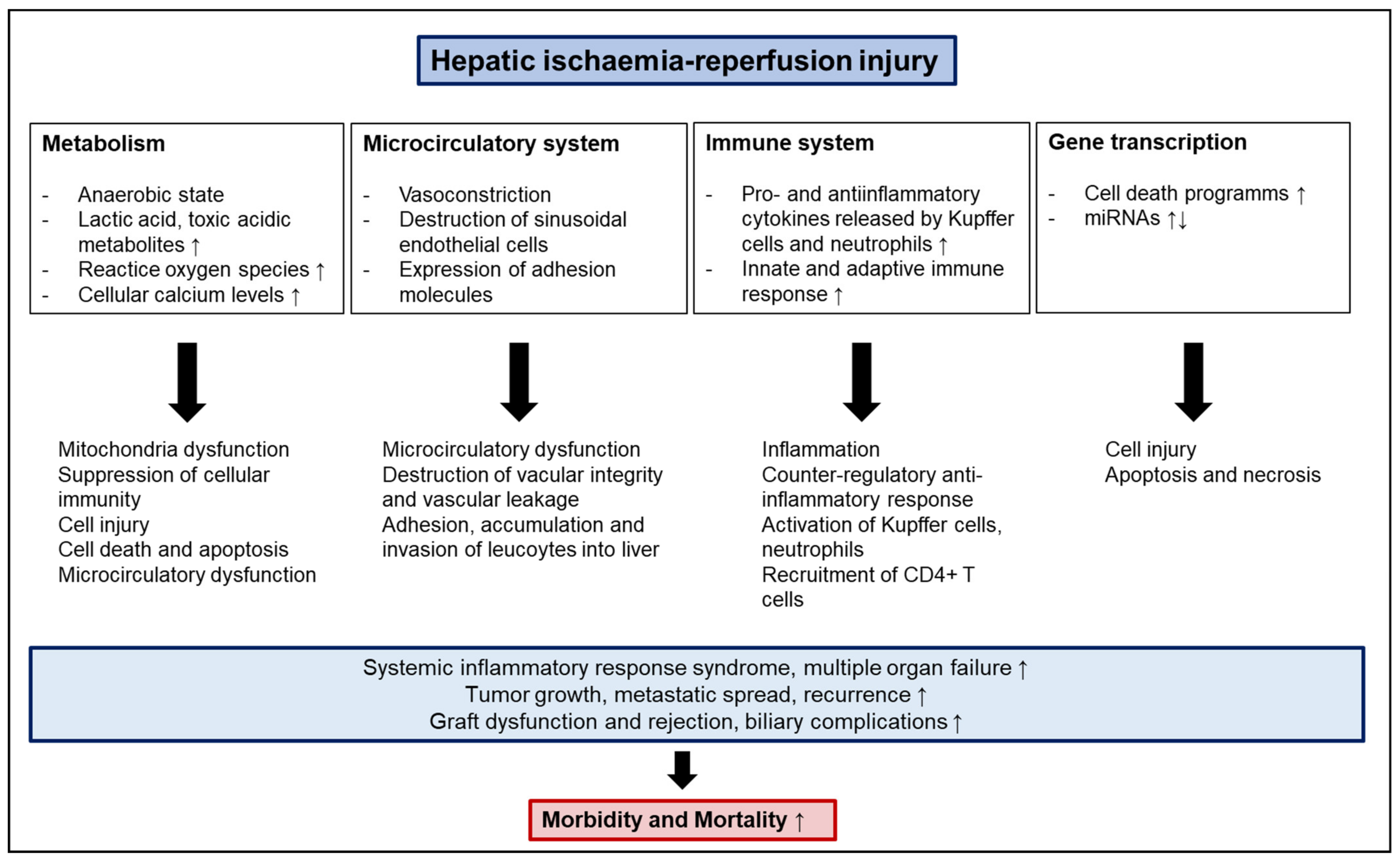
2. Molecular Mechanisms and Pathophysiology of IRI

2.1. Anaerobic Metabolism, Acidosis, and ATP Depletion

Liver ischemia, whether caused by a regional or systemic cessation of blood flow, leads to a variety of alterations in cellular function. The decreased availability of oxygen to the tissue places an increased demand on anaerobic metabolism that results in a shift from an aerobic to an anaerobic metabolic state associated with a depletion in ATP (adenosine triphosphate) levels [7]. Simultaneously, anaerobic glycolysis is accelerated, leading to an increased release of lactic acid, an accumulation of oxygen radicals, and a disequilibrium in H+, Na+, and Ca2+ homeostasis [8,9,10,11]. Lactic acid has been linked to cellular dysfunctions and impaired signaling interactions, including altered macrophage responses and depressed antigen-presentation by Kupffer cells associated with early immunosuppression after shock or extensive surgery [12,13]. Similarly, an overload in cellular calcium levels has been suggested to contribute to the suppression of cellular immunity, potentially by impairing macrophage antigen presentation function [12]. In addition, increased intracellular calcium levels have been shown to activate Ca2+-dependent enzymes, such as protein kinase C and phospholipase C, resulting in apoptosis and cell death [14]. Further consequences of the accumulation of toxic acidic metabolites include mitochondrial hypofunction and damage, microcirculatory dysfunction, and cell injury, and will be further elucidated below [15].

2.2. Mitochondria, Oxidative Stress and Reactive Oxygen Species

Reperfusion of the liver, associated with a concomitant reintroduction of oxygen to ischemic/severely hypoxic tissue, leads to a significant increase in the production of free radicals, such as reactive oxygen (ROS) and reactive nitrogen species. Mainly produced by mitochondria, they exceed the amount produced during the ischemic phase, which is due to the fact that the immediate increase in oxygen supply exceeds the rate of the anaerobic metabolism returning to an aerobic state [10,16]. Insights from transgenic knockout models suggest that the source of ROS production changes during different phases of IRI; whereas, Kupffer cells are the main source of ROS in the early phase, natural killer T cells take over in a later phase, with neutrophils generating the main amount in very late stages [15]. These mitochondria-generated ROS have detrimental effects on enzymes, proteins, nucleic acids, and the cytoskeleton, thereby leading to mitochondrial dysfunction and damage. Furthermore, ROS lead to an upregulation of Tumor Necrosis Factor-α (TNF-α) and the transcription factor Nuclear Factor κB (NF-κB) with an increased release of downstream molecules, such as interleukins (e.g., IL-1), thereby indirectly causing cellular injury [17]. The cellular damage is not limited to hepatocytes, but also includes endothelial cells, which results in microcirculatory dysfunction due to the destruction of the microvascular integrity [16,18,19].

2.3. Microcirculatory Dysfunction

After reperfusion, microvascular dysfunction is among the earliest changes and is characterized by vasoconstriction, the destruction of the sinusoidal endothelial cell (SEC) barrier, expression of adhesion molecules, and an accumulation of leukocytes, including neutrophils in the liver. Within two hours of reperfusion, Kupffer cells release proinflammatory cytokines (e.g., TNF-α, interleukin-1β (IL-1β), interleukin-6 (IL-6)) which leads to an activation of sinusoidal endothelial cells and an imbalance of nitric oxide (NO) production from NO synthase and endothelin-1 (ET-1), which causes vasoconstriction [20,21,22,23]. The activation of SECs is followed by an upregulation of adhesion molecules (e.g., intercellular adhesion molecule 1 (ICAM-1), vascular cell adhesion molecule 1 (VCAM-1)), and integrins (e.g., CD11b/CD18). Attracted by the respective cytokines, neutrophils in the blood stream attach to the adhesion molecules and transmigrate through the impaired sinusoidal endothelial barrier, thereby exiting the vasculature towards the site of the liver damage [24,25]. The infiltration of neutrophils further impairs hepatic perfusion and has deleterious effects on hepatocytes due to the secretion of toxic proteases (e.g., cathepsin G, matrix-metalloproteases) resulting in further hepatic liver injury [26,27].

2.4. Pro- and Anti-Inflammatory Cytokines

As mentioned above, activated Kupffer cells in early stages and neutrophils in late stages secrete IL-1 and TNF-α, which leads to an activation of various downstream pathways inducing an inflammatory response, thereby contributing to hepatic IRI. In this respect, downstream targets of TNF-α include NF-κB, mitogen-activated protein kinase, and c-Jun N-terminal kinase (JNK) [28]. Moreover, TNF-α enhances the upregulation of the previously mentioned adhesion molecules ICAM-1 and VCAM-1, thereby facilitating neutrophil accumulation [29]. Other cytokines involved in IRI include interleukins, such as IL-6, IL-12, IL-23, vascular endothelial growth factor (VEGF), and hepatocyte growth factor (HGF) [30]. IL-12 and IL-23 stimulate CD4 T cells to release IL-17, which serves as another attractant for neutrophils in the liver, thereby aggravating hepatic injury [31,32]. Additionally, they promote TNF-α production via NF-ĸB activation, and IL-23 activates the IFN-γ/IRF-1 pathway [32]. Conversely, TNF-α, IL-1, IL-12, and IL-23 function as proinflammatory cytokines followed by immunosuppression contributing to liver damage; IL-6, VEGF, and HGF seem to act the opposite way contributing to liver regeneration. In this respect, IL-6 reduces liver damage and promotes hepatocyte proliferation by enhancing glutathione expression, activating STAT3 signaling, and eliminating reactive oxygen species [33]. Interestingly, VEGF seems to have a dual role in IRI: while endogenous VEGF is expressed and functional to initiate hepatic IRI, exogenous VEGF has a beneficial effect and contributes to the regeneration of the post-ischemic liver [34]. HGF also seems to have protective functions and has been shown to attenuate IRI by suppressing oxidative stress, promoting hepatocyte proliferation through activation of ERK1/2 signaling, enhancing glutathione expression, and downregulating ICAM-1, thereby impeding neutrophil adhesion and transmigration into the liver [35,36,37].
In summary, IRI due to surgical trauma or shock leads to an inflammatory response characterized by an increased release of pro-inflammatory cytokines. After reperfusion, this state of inflammation is followed by a counter-regulatory anti-inflammatory response with a rise in anti-inflammatory cytokines, while pro-inflammatory cytokine levels may simultaneously remain high. In this respect, IRI leads to a highly vulnerable state with a concurrent expression of pro- and anti-inflammatory cytokines that predominate in an alternating manner [7]. The local inflammatory state of the injured tissue can be followed by a systemic inflammatory response, leading to the systemic inflammatory response syndrome (SIRS) and even multiple organ failure. The anti-inflammatory response has been shown to induce an immunosuppressive state that can even favor tumor growth and metastasis in the liver remnant after hepatic surgery via an activation of cell invasion and migration pathways [4,6,12,38,39]. In this respect, the processes described that start as a local reaction within injured tissue can have “global” consequences involving initially healthy organs.

2.5. NF-κB

NF-ĸB is a transcription factor that plays a key role in mediating inflammatory gene expression during IRI. Activated by hypoxia or cytokines, NF-ĸB translocates to the nucleus of cells, binds to DNA, and induces the transcription of respective target genes. As elucidated above, NF-ĸB regulates gene expression in various cells during IRI. As such, NF-ĸB promotes the expression of TNF-α in hepatocytes, the expression of TNF-α and IL-6 in Kupffer cells, and the expression of adhesion molecules ICAM-1 and VCAM-1 in endothelial cells [40,41,42]. Interestingly, NF-ĸB has a dual role in IRI: while it promotes the pro-inflammatory state during the acute injury phase, it also exhibits hepatoprotective functions and fosters regeneration after injury [43,44].

2.6. Apoptosis and Necrosis

Hepatic reperfusion is not only characterized by an interplay of pro- and anti-inflammatory mediators, but also by massive cell injury mainly affecting hepatocytes and sinusoidal endothelial cells [15]. It remains controversial as to whether this is due to apoptosis via activation of caspase-3 and caspase-8 by TNF-α or necrosis. However, various studies suggest that both pathways might coexist, sharing mechanistic pathways [27,45,46].

2.7. miRNAs

miRNAs are a subset of short single-chain, non-coding RNAs that regulate gene expression, and can thereby modulate IRI on a posttranscriptional and posttranslational level [47]. In this respect, a variety of miRNAs have been identified to impair or ameliorate the state of hepatic IRI, and therefore have been suggested to serve as biomarkers for early diagnosis of IRI as a reflection of the present state of IRI. miRNAs that have been shown to impair the condition of hepatic IRI, when upregulated, include miRNA-223, miRNA-191, miRNA-133a-5p, miRNA-17, miRNA-450-5p, and others. Respective targets are STAT3, NF-κB, IL-1, and TNF-α that have been previously described to be pivotal mediators during IRI [48,49,50,51,52]. Other miRNAs that worsen the condition of IRI, when downregulated, are miRNA-142-3p, miRNA-146a, miRNA-200c, and miRNA-34a targeting factors, such as JNK, ZEB1, and p53 [53,54,55,56,57]. In contrast, there are also miRNAs that ameliorate the condition of IRI, among which are miRNA-370, miRNA-182-5p, miRNA-125b (when upregulated), and miRNA-128-3p (when downregulated) by targeting toll-like receptor 4 (TLR4), TNF-α, IL-1, IL-6, and NF-κB, [58,59,60,61,62]. In this respect, miRNAs worsen or attenuate IRI by modulating inflammatory cell responses, oxidative stress reactions and apoptosis, and cell death, processes that have been elaborated on in previous paragraphs.

2.8. Innate and Adaptive Immune Activation

Both innate and adaptive immune responses play an important part in IRI [63]. The abovementioned cell death, as well as mitochondria dysfunction, induce the production of danger-associated molecular patterns (DAMPs), such as high-mobility group box 1 (HMGB1), that trigger the innate immune response. In this context, toll-like receptors (TLR) such as TLR 4, 7, and 9, as well as nucleotide-binding oligomerization domain (NOD)2, have been shown to promote IRI [64]. TLRs are expressed on various liver cell types, including Kupffer cells, hepatocytes, and dendritic cells. They recognize DAMPs and propagate the inflammatory response. After reperfusion, HMGB1 levels increase and bind to TLR4, leading to an upregulation of downstream pro-inflammatory pathways. However, TLRs have also been shown to alleviate the IRI. In this respect, a lack of intestinal TLR 9 has been demonstrated to increase apoptosis and inflammation, thereby aggravating IRI [65]. CD4+ T-lymphocytes seem to play a major role during adaptive immune responses. Trapped in the constricted sinusoids, they produce granulocyte-macrophage colony-stimulating factor (GM-CSF), interferon gamma (IFN-γ), and TNF-β, which amplify cytokine release and the activation of Kupffer cells [66,67]. Kupffer cells are tissue-fixed macrophages of the liver and are in bidirectional crosstalk with CD4+ T-cells [68,69]. In this respect, Kupffer cells release ROS, TNF-α, and IL-1, thereby triggering the recruitment of CD4+ T-cells, as well as the activation of sinusoidal endothelial cells. In turn, CD4+ T-cells can activate Kupffer cells, thereby promoting further inflammation [68]. However, Kupffer cells might have a dual role in IRI. In this respect, Kupffer cells can release IL-10, an anti-inflammatory cytokine that can suppress NF-κB activation and inhibit the expression of pro-inflammatory factors, such as TNF-α, IL-1β, IFN-γ, IL-2, and adhesion molecules, such as ICAM-1 [70]. Furthermore, CD4+ T-cells help recruit neutrophils to the site of injury through the release of IL-17. Alternatively, depletion of CD4+ T-cells in mice has been shown to be associated with a decrease in neutrophil recruitment [71]. Treg cells have also been demonstrated to be involved in the adaptive immune response, potentially exhibiting a similar protective function. In this context, CD4+CD25+Foxp3+ Treg cells have been shown to attenuate IRI via the PI3K/Akt/mTOR signaling pathway [72,73]. Overall, both innate and adaptive immune responses participate in IRI, crosstalk with each other, and function in partially synergistic or counteractive ways (all components of IRI are summarized in Figure 1).

3. Cold Ischemia Versus Warm Ischemia Reperfusion Injury

Hepatic IRI can be categorized into two types: cold ischemia and warm ischemia. While the previous paragraphs have focused on the molecular mechanisms underlying hepatic IRI in general, the majority of which is present in both types of IRI, this section aims to elucidate the main differences between cold and warm ischemia.
Cold storage ischemia occurs during organ preservation before liver transplantation. Warm ischemia occurs in the setting of transplantation, trauma and shock, and elective liver surgery (e.g., hepatic resections) when hepatic blood supply and oxygen delivery is temporarily interrupted.

3.1. Cold Ischemia

Cold IRI is characterized by massive damage to sinusoidal endothelial cells, a destruction of the endothelial integrity, and microcirculatory dysfunction. Studies have shown that endothelial cells are more sensitive to cold storage than hepatocytes, which results in higher cell death rates through apoptosis and necrosis of endothelial cells compared to liver parenchymal cells [74,75,76]. Endothelial cell damage is followed by Kupffer cell activation, leading to an increased release of cytokines and ROS as previously described. In this respect, Kupffer cell activation has been demonstrated to correlate and increase with increasing cold storage times [75].

3.2. Warm Ischemia

In contrast to cold ischemia, warm IRI is dominated by hepatocellular damage. In this regard, hepatocytes have been shown to be more sensitive to warm IRI (37 °C), while sinusoidal endothelial cells are more susceptible to cold IRI (4 °C) [15]. Warm IRI can be categorized into two phases: the early phase starts within two hours after reperfusion and is characterized by oxidative stress and an activation of immune cells. The late phase starts within 6 to 48 h after reperfusion and is characterized by the accumulation of neutrophils and hepatocellular injury [27,77,78,79]. However, the underlying processes are intertwined and overlap both phases (for more details, please see previous paragraphs).

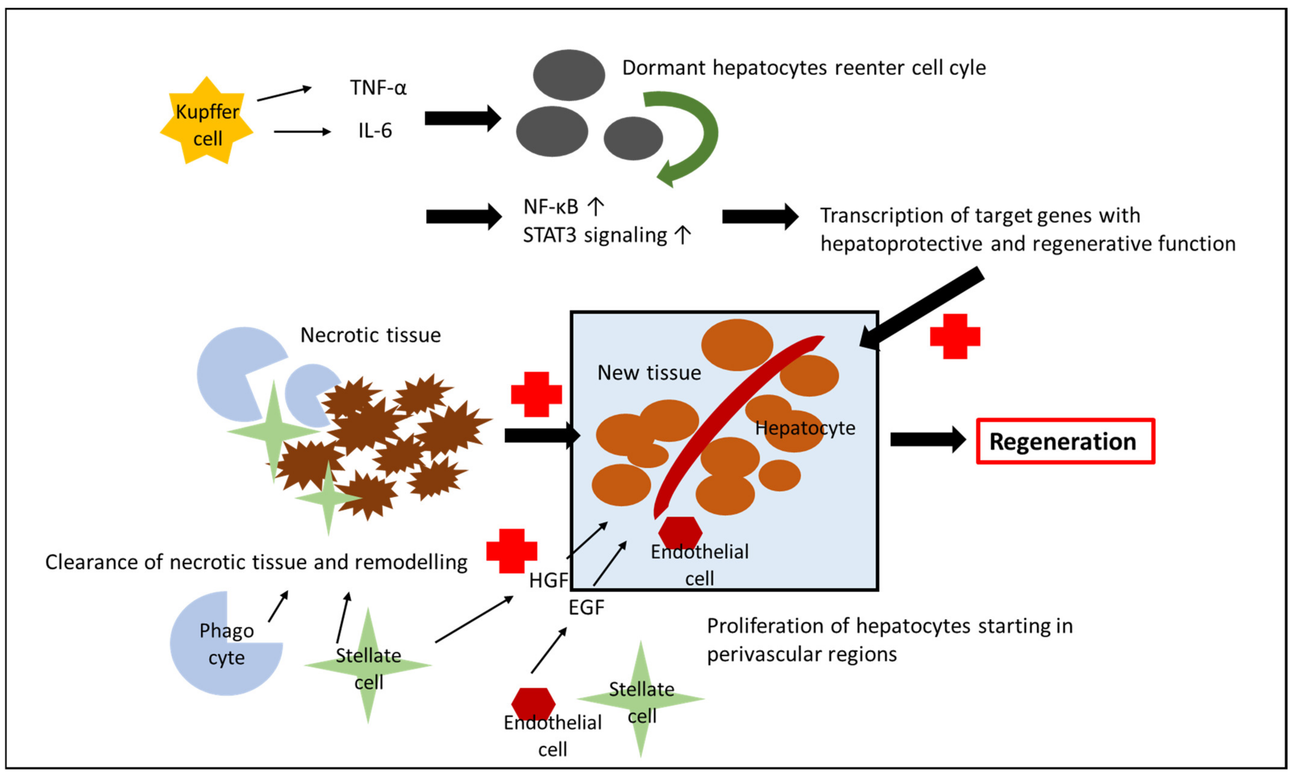
4. Liver Regeneration after IRI

The liver is unique in its capacity to regenerate after injury, a process that is mediated by cytokines, growth factors, and a supportive microenvironment [80]. In general, after uncomplicated hepatic resections, TNF-α and IL-6, which are released from Kupffer cells, prime viable hepatocytes to reenter the cell cycle and initiate NF-κB activation and STAT3 signaling, resulting in the transcription of target genes that are essential for liver regeneration. Subsequently, after reentering the cell-cycle, hepatocytes proliferate, a process that is further promoted by HGF, which is released from the extracellular matrix, hepatic stellate cells and endothelial cells after liver resection, and epidermal growth factor [81,82]. As opposed to uncomplicated liver resections, hepatocytes are stressed after IRI but the stimulus for their proliferation remains unknown [83,84]. During the regeneration process, which begins in perivascular regions, phagocytes, stellate cells, and other nonparenchymal cells clear the necrotic tissue and remodel injured areas so that proliferating hepatocytes can restore the normal architecture, replace the unfunctional liver mass, and generate new functional mass [83,84]. In this regard, NF-ĸB, which has been described in previous paragraphs to not only promote the pro-inflammatory state, but also exert hepatoprotective and regenerative functions, plays an important role in fostering the regenerative potential of hepatocytes [43,44]. Similarly, cytokines (e.g., ELR+ CXC chemokines, such as IL-1) that promote the inflammatory state during the acute injury phase now impact the hepatocytes in an opposite way and promote regeneration, a mechanism that is most likely due to different concentrations of cytokines; while high concentrations seem to induce hepatocyte damage, low concentrations promote hepatocyte regeneration [85] (Figure 2).

5. Clinical Implications of IRI in Different Settings

While the previous paragraphs focused on the molecular mechanisms of IRI during the acute phase and regeneration, this chapter discusses the long-term impact of inflammation induced by IRI on the liver.

5.1. Tumor Growth, Metastasis, and Recurrence

Various studies in both humans and animals have shown that IRI is a strong stimulus for tumor growth, metastasis, and recurrence [6,86,87,88]. During hepatic surgery, hemorrhage and shock are common, leading to IRI due to a systemic reduction in blood perfusion, and have been shown to promote tumor growth and recurrence. In order to control hepatic bleeding, vascular clamping techniques are applied; however, these by themselves can cause hepatic IRI due to a regional obstruction of perfusion and oxygen supply. Overall, the surgical stress due to hemorrhage or vascular clamping and acute phase of IRI with an excess expression of pro-inflammatory cytokines, reactive oxygen species, and adhesion molecules, as well as matrix-metalloproteases (e.g., MMP9), promote tumor cell adhesion, migration, invasion (e.g., via c-MET), and angiogenesis [6,7,86,89,90]. In a colon cancer rat model, hepatic IRI resulted in an increase in liver metastases due to the upregulation of the adhesion molecule E-selectin (and respective E-selectin mRNA). While the amount of liver metastases was time-dependent, with a ten times higher amount of tumor nodules in the 60 (versus 30) minutes ischemia group, metastatic dissemination was present in both clamped and unclamped lobes pointing towards the systemic impact of IRI [86]. Another study investigating colorectal cancer in xenograft models demonstrated that hepatic IRI due to vascular clamping accelerated the outgrowth of pre-existent hepatic micrometastases, resulting in a worse prognosis. However, accelerated tumor growth and tissue necrosis were effectively prevented by intermittently occluding blood flow. As opposed to the previously mentioned study by Doi et al., tumor growth was only stimulated in the clamped lobes [87]. In summary, whether tumor growth after hepatic IRI is due to locally operating mechanisms affecting the clamped lobe and factors potentially disseminating to the unclamped lobe, or whether it is due to the involvement of systemically released factors remains controversial, has not been yet elucidated. Man et al. demonstrated that hepatic IRI of the small liver remnant after major hepatectomy for colorectal liver metastases in mice promoted further metastatic spread to the liver and lungs, due to an upregulation of mRNA levels for Cdc42, ROCK (Rho kinase), vascular endothelial growth factor, and an activation of hepatic stellate cells, all leading to an activation of cell adhesion, invasion, and angiogenesis [6]. Subsequently, ischemia of the liver remnant in patients after resection of colorectal cancer liver metastases was associated with worse cancer-specific and recurrence-free survival [88]. A recent meta-analysis assessing the impact of liver pedicle clamping in patients receiving liver surgery for hepatocellular carcinoma, reported significantly shorter overall survival and higher tumor recurrence rates in patients with pedicle clamping versus the control group [91]. However, prevention of tissue hypoxia through targeting the HIF-1α pathway and improvement of microcirculatory flow might limit recurrence after liver surgery [92].

5.2. Graft Fibrosis

Liver regeneration following hepatic IRI is associated with prolonged fibrosis, especially at the interface between necrotic tissue and regenerating liver due to an increased expression of profibrotic genes in the ischemic liver. In this respect, collagen deposition is enhanced along the injured sites and an increased infiltration of α-smooth muscle actin positive hepatic stellate cells/myofibroblast can be observed [37,93]. However, after full regeneration and restoration of functional liver mass, which takes several weeks, the fibrosis seems to be self-limiting and disappears [94]. In fact, a recent study showed that the fibrotic liver, despite having a higher degree of injury, recovered more quickly than normal liver after hepatic IRI, which was attributed to the increased numbers of hepatic stellate cells, hepatic progenitors cells, and macrophages to the fibrotic sites [93].

5.3. Biliary Complications and Graft Dysfunction

Biliary complications are among the most frequent complications after transplantation, and can lead to graft dysfunction or loss. Additionally, they have been associated with increased morbidity and mortality rates [95]. Hepatic IRI can cause intra- and extrahepatic nonanastomotic biliary strictures after transplantation, a process that is the result of direct injury of cholangiocytes and damage of peribiliary vascular regions leading to apoptosis and necrosis of cholangiocytes [96]. Moreover, direct immunologic cellular injury to the bile duct epithelium is the major underlying cause for tissue damage during liver allograft rejection. The exact mechanisms underlying this process are beyond the theme of this article and are discussed elsewhere [97].

6. Therapeutic Approaches

There are a variety of therapeutic approaches with the aim to attenuate or prevent hepatic IRI, including surgical and pharmacological strategies and the application of perfusion machines [11,98,99,100]. Overall, the therapeutic measures investigated all aimed to target the abovementioned underlying molecular processes of hepatic IRI. These include the reduction of reactive oxygen species by administration of free radical scavengers (e.g., N-acetyl-cysteine), the modulation of the cytokine response and blockage of immune activation (e.g., steroids, gadolinium chloride), and the preservation of organ function through ex vivo organ perfusion and ischemic preconditioning; however, many approaches have only been validated in preclinical studies, or have not been proven successful in the clinical setting underscoring the complexity of IRI [11,98,101,102]. The effect of immunosuppressants (azathioprine, cyclosporine A, tacrolimus, rapamycin, etc.) has been investigated in previous studies, which have been shown to be of a protective nature [103,104,105]. In this respect, immunosuppressants can attenuate IRI of the liver through the reduction of neutrophil and leukocyte accumulation, and modification of the early-local immune response by decreasing the ratio of proinflammatory to anti-inflammatory T lymphocytes and increasing the Treg proportion; however, they cannot fully prevent hepatic IRI. In this respect, additional therapies are required that have the ability to not only eliminate the effector cells and products, but instead harness lymphoid subpopulations (e.g., Treg cells) with regulatory capability to modify the immune response [105]. However, to our knowledge, these strategies have not been tested beyond preclinical studies. From a surgical standpoint, there are a few considerations that can help to minimize hepatic IRI and the respective consequences. As previously described, intraoperative hemorrhage is a common complication during extended liver resections and has been shown to be associated with increased postoperative morbidity, increased tumor recurrence, and worse prognosis [7]. Therefore, various vascular occlusion techniques (e.g., Pringle Maneuver) have been investigated in order to reduce blood loss. However, at the same time, vascular clamping by itself aggravates hepatic IRI; also contributing to postoperative morbidity and worse oncologic outcomes. In this respect, a balance between tolerated blood loss and restriction of hepatic blood flow is required. The ideal clamping time is still under debate; however, intermittent clamping with 30 min of restricted hepatic flow and 5 min of reperfusion seems to be an effective medium [100]. According to a systematic review by Hoekstra et al., there is no evidence supporting further strategies to attenuate hepatic IRI, such as ischemic preconditioning or intraoperative pharmacological interventions [100]. Lastly, it needs to be highlighted that, apart from heavy intraoperative blood loss or vascular clamping, ischemia of the liver remnant after extended liver resections can be due to an additional cause: the presence of insufficiently or nonperfused liver tissue due to imprecise liver resections or due to excessive resections with unintentional damage to a segment’s inflow or out-flow vessels. However, such a factor can be positively influenced by meticulous operative techniques by liver surgeons [88,99].

7. Future Directions

With the aim to attenuate liver damage due to hepatic IRI and decrease morbidity, as well as mortality, future strategies should include studies on IRI prevention not only focusing on the phases of ischemia and reperfusion, but also regeneration after tissue damage. As indicated above, hepatic IRI can be due to regional cessation of blood flow, but could also result from a systemic reduction in blood supply. Thus, recognizing that the liver is only a part of a multi-organ system, future studies may wish to approach hepatic IRI in a more holistic manner. Starting points for further studies could include the following: targeting macrophages and neutrophils to impose reparative phenotypes [106], harnessing lymphoid subpopulations (e.g., Treg cells) with regulatory capability to modify the immune response [105], application of stem cells to restore functional and structural components of the liver [107], and also clinical features, such as pre-operative exercise-based therapy to induce anti-inflammatory trained immunity [108]. Moreover, as the majority of studies are of experimental preclinical nature, translating the results into the clinical arena is required, and might be of immense benefit to improve patient outcomes.

8. Conclusions

Hepatic IRI occurs in a variety of settings, including liver surgery (hepatic resections, liver transplantation), liver preservation for transplantation, trauma, and hemorrhagic shock. Not only does it have adverse effects on morbidity (e.g., graft dysfunction, biliary complications), but it is also associated with increased mortality, as well as metastatic spread and recurrence in cancer patients. The molecular mechanisms of hepatic IRI and regeneration are complex and highly dynamic, and involve a variety of signaling cascades and downstream mediators modulating the cellular metabolic state, the immune response, gene transcription, and microvascular function. While various therapeutic approaches have been investigated in animal models, there is a need for validation in clinical studies to effectively treat and prevent hepatic IRI in the future to optimize patient outcomes.

Author Contributions

Conceptualization: E.P., M.A. and I.H.C.; Methodology: E.P., N.B.K., F.B., M.A. and I.H.C.; Writing original: E.P., H.N., M.A. and I.H.C.; Manuscript editing: F.B., H.N., M.G., J.W., M.A. and I.H.C. All authors have read and agreed to the published version of the manuscript.

Funding

This research received no external funding.

Institutional Review Board Statement

Not applicable.

Informed Consent Statement

Not applicable.

Data Availability Statement

Not applicable.

Conflicts of Interest

The authors declare no conflict of interest.

References

  1. Toledo-Pereyra, L.H.; Simmons, R.L.; Najarian, J.S. Protection of the ischemic liver by donor pretreatment before transplantation. Am. J. Surg. 1975, 129, 513–517. [Google Scholar] [CrossRef]
  2. Singer, M.; Young, P.J.; Laffey, J.G.; Asfar, P.; Taccone, F.S.; Skrifvars, M.B.; Meyhoff, C.S.; Radermacher, P. Dangers of hyperoxia. Crit. Care 2021, 25, 440. [Google Scholar] [CrossRef] [PubMed]
  3. Eltzschig, H.; Eckle, T. Ischemia and reperfusion-from mechanism to translation. Nat. Med. 2011, 17, 1391–1401. [Google Scholar] [CrossRef] [Green Version]
  4. Nastos, C.; Kalimeris, K.; Papoutsidakis, N.; Tasoulis, M.K.; Lykoudis, P.M.; Theodoraki, K.; Nastou, D.; Smyrniotis, V.; Arkadopoulos, N. Global consequences of liver ischemia/reperfusion injury. Oxid. Med. Cell. Longev. 2014, 2014, 906965. [Google Scholar] [CrossRef] [PubMed] [Green Version]
  5. Serracino-Inglott, F.; Habib, N.A.; Mathie, R.T. Hepatic ischemia-reperfusion injury. Am. J. Surg. 2001, 181, 160–166. [Google Scholar] [CrossRef]
  6. Man, K.; Ng, K.; Mau Lo, C.; Ho, J.; Sun, B.; Sun, C.; Lee, T.; Poon, R.; Fan, S. Ischemia-Reperfusion of Small Liver Remnant Promotes Liver Tumor Growth and Metastases—Activation of Cell Invasion and Migration Pathways. Liver Transplant. 2007, 13, 767–768. [Google Scholar] [CrossRef]
  7. Pretzsch, E.; Bösch, F.; Renz, B.; Werner, J.; Angele, M.; Chaudry, I.H. Operative Trauma and Blood Loss—Impact on Tumor Growth and Recurrence. Shock 2021, 55, 455–464. [Google Scholar] [CrossRef]
  8. Kannerup, A.S.; Grønbæk, H.; Funch-Jensen, P.; Karlsen, S.; Mortensen, F.V. Partial liver ischemia is followed by metabolic changes in the normally perfused part of the liver during reperfusion. Eur. Surg. Res. 2010, 45, 61–67. [Google Scholar] [CrossRef] [PubMed]
  9. Nowak, G.; Ungerstedt, J.; Wernerman, J.; Ungerstedt, U.; Ericzon, B.G. Metabolic changes in the liver graft monitored continuously with microdialysis during liver transplantation in a pig model. Liver Transplant. 2002, 8, 424–432. [Google Scholar] [CrossRef] [PubMed]
  10. Zhang, H.; Yan, Q.; Wang, X.; Chen, X.; Chen, Y.; Du, J.; Chen, L. The Role of Mitochondria in Liver Ischemia-Reperfusion Injury: From Aspects of Mitochondrial Oxidative Stress, Mitochondrial Fission, Mitochondrial Membrane Permeable Transport Pore Formation, Mitophagy, and Mitochondria-Related Protective Measures. Oxid. Med. Cell. Longev. 2021, 2021, 6670579. [Google Scholar] [CrossRef]
  11. Cannistrà, M.; Ruggiero, M.; Zullo, A.; Gallelli, G.; Serafini, S.; Maria, M.; Naso, A.; Grande, R.; Serra, R.; Nardo, B. Hepatic ischemia reperfusion injury: A systematic review of literature and the role of current drugs and biomarkers. Int. J. Surg. 2016, 33, S57–S70. [Google Scholar] [CrossRef] [PubMed]
  12. Chaudry, I.H.; Ayala, A. Mechanism of increased susceptibility to infection following hemorrhage. Am. J. Surg. 1993, 165, 59S–67S. [Google Scholar] [CrossRef]
  13. Ayala, A.; Perrin, M.; Ertel, W.; Chaudry, I. Differential effects of hemorrhage on kupffer cells: Decreased antigen presentation despite increased inflammatory cytokine (IL-1, IL-6 and TNF) release. Cytokine 1992, 4, 66–75. [Google Scholar] [CrossRef]
  14. Wang, H.G.; Pathan, N.; Ethell, I.M.; Krajewski, S.; Yamaguchi, Y.; Shibasaki, F.; McKeon, F.; Bobo, T.; Franke, T.F.; Reed, J.C. Ca2+-Induced Apoptosis Through Calcineurin Dephosphorylation of BAD. Science 1999, 284, 339–343. [Google Scholar] [CrossRef]
  15. Datta, G.; Fuller, B.J.; Davidson, B.R. Molecular mechanisms of liver ischemia reperfusion injury: Insights from transgenic knockout models. World J. Gastroenterol. 2013, 19, 1683–1698. [Google Scholar] [CrossRef]
  16. Jaeschke, H. Reactive oxygen and ischemia/reperfusion injury of the liver. Chem. Biol. Interact. 1991, 79, 115–136. [Google Scholar] [CrossRef]
  17. Kis, A.; Yellon, D.M.; Baxter, G.F. Role of nuclear factor-κB activation in acute ischaemia-reperfusion injury in myocardium. Br. J. Pharmacol. 2003, 138, 894–900. [Google Scholar] [CrossRef] [Green Version]
  18. Jaeschke, H. Reactive oxygen and mechanisms of inflammatory liver injury: Present concepts. J. Gastroenterol. Hepatol. 2011, 26, 173–179. [Google Scholar] [CrossRef] [PubMed]
  19. Rauen, U.; Polzar, B.; Stephan, H.; Mannherz, H.; De Groot, H. Cold-induced apoptosis in cultured hepatocytes and liver endothelial cells: Mediation by reactive oxygen species. FASEB J. 1999, 13, 155–168. [Google Scholar] [CrossRef] [PubMed] [Green Version]
  20. Frankenberg, M.V.; Weimann, J.; Fritz, S.; Fiedler, J.; Mehrabi, A.; Büchler, M.W.; Kraus, T.W. Gadolinium chloride-induced improvement of postischemic hepatic perfusion after warm ischemia is associated with reduced hepatic endothelin secretion. Transpl. Int. 2005, 18, 429–436. [Google Scholar] [CrossRef]
  21. Uhlmann, D.; Gaebel, G.; Armann, B.; Ludwig, S.; Hess, J.; Pietsch, U.C.; Fiedler, M.; Tannapfel, A.; Hauss, J.; Witzigmann, H. Attenuation of proinflammatory gene expression and microcirculatory disturbances by endothelinA receptor blockade after orthotopic liver transplantation in pigs. Surgery 2006, 139, 61–72. [Google Scholar] [CrossRef]
  22. Peralta, C.; Fernández, L.; Panés, J.; Prats, N.; Sans, M.; Piqué, J.M.; Gelpí, E.; Roselló-Catafau, J. Preconditioning protects against systemic disorders associated with hepatic ischemia-reperfusion through blockade of tumor necrosis factor-induced P-selectin up-regulation in the rat. Hepatology 2001, 33, 100–113. [Google Scholar] [CrossRef]
  23. Siriussawakul, A.; Zaky, A.; Lang, J.D. Role of nitric oxide in hepatic ischemia-reperfusion injury. World J. Gastroenterol. 2010, 16, 6079–6086. [Google Scholar] [CrossRef]
  24. Burke, J.; Zibari, G.B.; Brown, M.F.; Granger, N.; Kelly, R.; Singh, I.; Mcdonald, J.C. Hepatic Ischemia–Reperfusion Injury Causes E-Selectin Upregulation. Transplant. Proc. 1998, 1345, 2321–2323. [Google Scholar] [CrossRef]
  25. Lentsch, A.B.; Yoshidome, H.; Cheadle, W.G.; Miller, F.N.; Edwards, M.J. Chemokine involvement in hepatic ischemia/reperfusion injury in mice: Roles for macrophage inflammatory protein-2 and Kupffer cells. Hepatology 1998, 27, 507–512. [Google Scholar] [CrossRef]
  26. McKeown, C.M.B.; Edwards, V.; Phillips, M.J.; Harvey, P.R.; Petrunka, C.N.; Strasberg, S.M. Sinusoidal lining cell damage: The critical injury in cold preservation of liver allografts in the rat. Transplantation 1988, 46, 178–191. [Google Scholar] [CrossRef]
  27. Gujral, J.S.; Bucci, T.J.; Farhood, A.; Jaeschke, H. Mechanism of cell death during warm hepatic ischemia-reperfusion in rats: Apoptosis or necrosis? Hepatology 2001, 33, 397–405. [Google Scholar] [CrossRef]
  28. Schwabe, R.F.; Brenner, D.A. Mechanisms of liver injury. I. TNF-α-induced liver injury: Role of IKK, JNK, and ROS pathways. Am. J. Physiol.-Gastrointest. Liver Physiol. 2006, 290, 583–589. [Google Scholar] [CrossRef]
  29. Oe, S.; Hirose, T.; Fujii, H.; Yasuchika, K.; Nishio, T.; Iimuro, Y.; Morimoto, T.; Nagao, M.; Yamaoka, Y. Continuous intravenous infusion of deleted form of hepatocyte growth factor attenuates hepatic ischemia-reperfusion injury in rats. J. Hepatol. 2001, 34, 832–839. [Google Scholar] [CrossRef] [Green Version]
  30. Csak, T.; Ganz, M.; Pespisa, J.; Kodys, K.; Dolganiuc, A.; Szabo, G. Fatty Acid and Endotoxin Activate Inflammasomes in Mouse Hepatocytes that Release Danger Signals to Stimulate Immune Cells. Hepatology 2012, 54, 133–144. [Google Scholar] [CrossRef]
  31. Kato, A.; Graul-Layman, A.; Edwards, M.; Lentsch, A. Promotion Of Hepatic Ischemia/Reperfusion Injury By ILl-12 Is Independent Of STAT4. Transplantation 2002, 73, 1142–1145. [Google Scholar] [CrossRef] [PubMed]
  32. Klune, J.; Bartels, C.; Luo, J.; Yokota, S.; Du, Q.; Geller, D. IL-23 mediated murine liver transplantation reperfusion injury via IFNy/IRF-1 pathway. Am. J. Physiol. Gastrointest. Liver Physiol. 2018, 315, 991–1002. [Google Scholar] [CrossRef] [PubMed]
  33. Teoh, N.; Field, J.; Farrell, G. Interleukin-6 is a key mediator of the hepatoprotective and pro-proliferative effects of ischaemic preconditioning in mice. J. Hepatol. 2006, 45, 20–27. [Google Scholar] [CrossRef] [PubMed]
  34. Tsurui, Y.; Sho, M.; Kuzumoto, Y.; Hamada, K.; Akashi, S.; Kashizuka, H.; Ikeda, N.; Nomi, T.; Mizuno, T.; Kanehiro, H.; et al. Dual role of vascular endothelial growth factor in hepatic ischemia-reperfusion injury. Transplantation 2005, 79, 1110–1115. [Google Scholar] [CrossRef]
  35. Chen, X.H.; Minatoguchi, S.; Kosai, K.; Yuge, K.; Takahashi, T.; Arai, M.; Wang, N.; Misao, Y.; Lu, C.; Onogi, H.; et al. In Vivo Hepatocyte Growth Factor Gene Transfer Reduces Myocardial Ischemia-Reperfusion Injury Through Its Multiple Actions. J. Card. Fail. 2007, 13, 874–883. [Google Scholar] [CrossRef]
  36. Xiao, X.; Liu, D.; Chen, S.; Li, X.; Ge, M.; Huang, W. Sevoflurane preconditioning activates HGF/Met-mediated autophagy to attenuate hepatic ischemia-reperfusion injury in mice. Cell. Signal. 2021, 82, 109966. [Google Scholar] [CrossRef]
  37. Konishi, T.; Lentsch, A.B. Hepatic ischemia/reperfusion: Mechanisms of tissue injury, repair, and regeneration. Gene Expr. 2017, 17, 277–287. [Google Scholar] [CrossRef]
  38. Angele, M.K.; Faist, E. Clinical review: Immunodepression in the surgical patient and increased susceptibility to infection. Crit. Care 2002, 6, 298–305. [Google Scholar] [CrossRef]
  39. Lin, E.; Calvano, S.E.; Lowry, S.F. Inflammatory cytokines and cell response in surgery. Surgery 2000, 127, 117–126. [Google Scholar] [CrossRef]
  40. Li, J.D.; Peng, Y.; Peng, X.Y.; Li, Q.L.; Li, Q. Suppression of Nuclear Factor-κB Activity in Kupffer Cells Protects Rat Liver Graft From Ischemia-Reperfusion Injury. Transplant. Proc. 2010, 42, 1582–1586. [Google Scholar] [CrossRef]
  41. Luedde, T.; Assmus, U.; Wüstefeld, T.; Meyer Zu Vilsendorf, A.; Roskams, T.; Schmidt-Supprian, M.; Rajewsky, K.; Brenner, D.A.; Manns, M.P.; Pasparakis, M.; et al. Deletion of IKK2 in hepatocytes does not sensitize these cells to TNF-induced apoptosis but protects from ischemia/reperfusion injury. J. Clin. Investig. 2005, 115, 849–859. [Google Scholar] [CrossRef] [PubMed] [Green Version]
  42. Suetsugu, H.; Iimuro, Y.; Uehara, T.; Nishio, T.; Harada, N.; Yoshida, M.; Hatano, E.; Son, G.; Fujimoto, J.; Yamaoka, Y. Nuclear factor κB inactivation in the rat liver ameliorates short term total warm ischaemia/reperfusion injury. Gut 2005, 54, 835–842. [Google Scholar] [CrossRef] [Green Version]
  43. Bradham, C.A.; Schemmer, P.; Stachlewitz, R.F.; Thurman, R.G.; Brenner, D.A. Activation of nuclear factor-κB during orthotopic liver transplantation in rats is protective and does not require Kupffer cells. Liver Transplant. Surg. 1999, 5, 282–293. [Google Scholar] [CrossRef] [PubMed]
  44. Íimuro, Y.; Nishiura, T.; Hellerbrand, C.; Behrns, K.E.; Schoonhoven, R.; Grisham, J.W.; Brenner, D.A. NFκB prevents apoptosis and liver dysfunction during liver regeneration. J. Clin. Investig. 1998, 101, 802–811. [Google Scholar] [CrossRef] [PubMed] [Green Version]
  45. Jaeschke, H.; Lemasters, J.J. Apoptosis versus oncotic necrosis in hepatic ischemia/reperfusion injury. Gastroenterology 2003, 125, 1246–1257. [Google Scholar] [CrossRef]
  46. Ben-Ari, Z.; Pappo, O.; Cheporko, Y.; Yasovich, N.; Offen, D.; Shainberg, A.; Leshem, D.; Sulkes, J.; Vidne, B.; Hochhauser, E. Bax Ablation Protects Against Hepatic Ischemia/Reperfusion Injury in Transgenic Mice. Liver Transplant. 2007, 13, 1181–1188. [Google Scholar] [CrossRef]
  47. Sabet Sarvestani, F.; Azarpira, N.; Al-Abdullah, I.H.; Tamaddon, A.M. microRNAs in liver and kidney ischemia reperfusion injury: Insight to improve transplantation outcome. Biomed. Pharmacother. 2021, 133, 110944. [Google Scholar] [CrossRef]
  48. Yu, C.H.; Xu, C.F.; Li, Y.M. Association of microRNA-223 expression with hepatic ischemia/reperfusion injury in mice. Dig. Dis. Sci. 2009, 54, 2362–2366. [Google Scholar] [CrossRef]
  49. Pan, W.; Wang, L.; Zhang, X.F.; Zhang, H.; Zhang, J.; Wang, G.; Xu, P.; Zhang, Y.; Hu, P.; Zhang, X.D.; et al. Hypoxia-induced microRNA-191 contributes to hepatic ischemia/reperfusion injury through the ZONAB/Cyclin D1 axis. Cell Death Differ. 2019, 26, 291–305. [Google Scholar] [CrossRef]
  50. Hao, W.; Zhao, Z.H.; Meng, Q.T.; Tie, M.E.; Lei, S.Q.; Xia, Z.Y. Propofol protects against hepatic ischemia/reperfusion injury via miR-133a-5p regulating the expression of MAPK6. Cell Biol. Int. 2017, 41, 495–504. [Google Scholar] [CrossRef]
  51. Li, S.; Zhang, J.; Wang, Z.; Wang, T.; Yu, Y.; He, J.; Zhang, H.; Yang, T.; Shen, Z. MicroRNA-17 regulates autophagy to promote hepatic ischemia/reperfusion injury via suppression of signal transductions and activation of transcription-3 expression. Liver Transplant. 2016, 22, 1697–1709. [Google Scholar] [CrossRef] [PubMed]
  52. Huang, Z.; Mou, T.; Luo, Y.; Pu, X.; Pu, J.; Wan, L.; Gong, J.; Yang, H.; Liu, Y.; Li, Z.; et al. Inhibition of miR-450b-5p ameliorates hepatic ischemia/reperfusion injury via targeting CRYAB. Cell Death Dis. 2020, 11, 455. [Google Scholar] [CrossRef] [PubMed]
  53. Li, Y.; Gao, M.; Xu, L.N.; Yin, L.H.; Qi, Y.; Peng, J.Y. MicroRNA-142-3p attenuates hepatic ischemia/reperfusion injury via targeting of myristoylated alanine-rich C-kinase substrate. Pharmacol. Res. 2020, 156, 104783. [Google Scholar] [CrossRef] [PubMed]
  54. Chen, Q.; Kong, L.; Xu, X.; Geng, Q.; Tang, W.; Jiang, W. Down-regulation of MicroRNA-146a in the early stage of liver ischemia-reperfusion injury. Transplant. Proc. 2013, 45, 492–496. [Google Scholar] [CrossRef] [PubMed]
  55. Jiang, W.; Kong, L.; Ni, Q.; Lu, Y.; Ding, W.; Liu, G.; Pu, L.; Tang, W.; Kong, L. miR-146a ameliorates liver ischemia/reperfusion injury by suppressing IRAK1 and TRAF6. PLoS ONE 2014, 9, e101530. [Google Scholar] [CrossRef] [PubMed]
  56. Wu, Y.; Gu, C.; Huang, X. Sevoflurane protects against hepatic ischemia/reperfusion injury by modulating microRNA-200c regulation in mice. Biomed. Pharmacother. 2016, 84, 1126–1136. [Google Scholar] [CrossRef] [PubMed]
  57. Huang, X.; Gao, Y.; Qin, J.; Lu, S. The role of miR-34a in the hepatoprotective effect of hydrogen sulfide on ischemia/reperfusion injury in young and old rats. PLoS ONE 2014, 9, e113305. [Google Scholar] [CrossRef]
  58. Li, L.; Li, G.; Yu, C.; Shen, Z.; Xu, C.; Feng, Z.; Zhang, X.; Li, Y. A role of microRNA-370 in hepatic ischaemia-reperfusion injury by targeting transforming growth factor-β receptor II. Liver Int. 2015, 35, 1124–1132. [Google Scholar] [CrossRef]
  59. Zhu, J.; Zhu, F.; Song, W.; Zhang, B.; Zhang, X.; Jin, X.; Li, H. Altered miR-370 expression in hepatic ischemia-reperfusion injury correlates with the level of nuclear kappa B (NF-κB) related factors. Gene 2017, 607, 23–30. [Google Scholar] [CrossRef]
  60. Jiang, W.; Liu, G.; Tang, W. MicroRNA-182-5p Ameliorates Liver Ischemia-Reperfusion Injury by Suppressing Toll-like Receptor 4. Transplant. Proc. 2016, 48, 2809–2814. [Google Scholar] [CrossRef]
  61. Huang, Z.; Zheng, D.; Pu, J.; Dai, J.; Zhang, Y.; Zhang, W.; Wu, Z. MicroRNA-125b protects liver from ischemia/reperfusion injury via inhibiting TRAF6 and NF-κB pathway. Biosci. Biotechnol. Biochem. 2019, 83, 829–835. [Google Scholar] [CrossRef] [PubMed]
  62. Mou, T.; Luo, Y.; Huang, Z.; Zheng, D.; Pu, X.; Shen, A.; Pu, J.; Li, T.; Dai, J.; Chen, W.; et al. Inhibition of microRNA-128-3p alleviates liver ischaemia–reperfusion injury in mice through repressing the Rnd3/NF-κB axis. Innate Immun. 2020, 26, 528–536. [Google Scholar] [CrossRef]
  63. Zhai, Y.; Busuttil, R.; Kupiec-Weglinski, J. Liver Ischemia and Reperfusion Injury: New Insights Into Mechanisms of Innate—Adaptive Immune-Mediated Tissue Inflammation. Am. J. Transpl. 2011, 11, 1563–1569. [Google Scholar] [CrossRef] [PubMed] [Green Version]
  64. Sosa, R.; Rossetti, M.; Naini, B.; Groysberg, V.; Kaldas, F.; Busuttil, R.; Chang, Y.; Gjertson, D.; Kupiec-Weglinski, J.; Reed, E. Pattern Recognition Receptor-Reactivity Screening of Liver Transplant Patients: Potential for Personalized and Precise Organ Matching to Reduce Risks of Ischemia-Reperfusion Injury. Ann. Surg. 2020, 271, 922–931. [Google Scholar] [CrossRef] [PubMed]
  65. Han, S.J.; Kim, M.; Novitsky, E.; D’Agati, V.; Lee, H.T. Intestinal TLR9 deficiency exacerbates hepatic IR injury via altered intestinal inflammation and short-chain fatty acid synthesis. FASEB J. 2020, 34, 12083–12099. [Google Scholar] [CrossRef]
  66. Tsung, A.; Klune, J.R.; Zhang, X.; Jeyabalan, G.; Cao, Z.; Peng, X.; Stolz, D.B.; Geller, D.A.; Rosengart, M.R.; Billiar, T.R. HMGB1 release induced by liver ischemia involves Toll-like receptor 4-dependent reactive oxygen species production and calcium-mediated signaling. J. Exp. Med. 2007, 204, 2913–2923. [Google Scholar] [CrossRef]
  67. Mendes-Braz, M.; Elias-Miró, M.; Jiménez-Castro, M.B.; Casillas-Ramírez, A.; Ramalho, F.S.; Peralta, C. The current state of knowledge of hepatic ischemia-reperfusion injury based on its study in experimental models. J. Biomed. Biotechnol. 2012, 2012, 8657. [Google Scholar] [CrossRef] [Green Version]
  68. Hanschen, M.; Zahler, S.; Krombach, F.; Khandoga, A. Reciprocal activation between CD4+ T cells and Kupffer cells during hepatic ischemia-reperfusion. Transplantation 2008, 86, 710–718. [Google Scholar] [CrossRef]
  69. Nakamitsu, A.; Hiyama, E.; Imamura, Y.; Matsuura, Y.; Yokoyama, T. Kupffer cell function in ischemic and nonischemic livers after hepatic partial ischemia/reperfusion. Surg. Today 2001, 31, 140–148. [Google Scholar] [CrossRef]
  70. Ye, L.; He, S.; Mao, X.; Zhang, Y.; Cai, Y.; Li, S. Effect of Hepatic Macrophage Polarization and Apoptosis on Liver Ischemia and Reperfusion Injury During Liver Transplantation. Front. Immunol. 2020, 11, 1193. [Google Scholar] [CrossRef]
  71. Caldwell, C.C.; Okaya, T.; Martignoni, A.; Husted, T.; Schuster, R.; Lentsch, A.B. Divergent functions of CD4+ T lymphocytes in acute liver inflammation and injury after ischemia-reperfusion. Am. J. Physiol.-Gastrointest. Liver Physiol. 2005, 289, 969–976. [Google Scholar] [CrossRef] [PubMed]
  72. Lu, L.; Li, G.; Rao, J.; Pu, L.; Yu, Y.; Wang, X.; Zhang, F. In vitro induced CD4+CD25+Foxp3+ Tregs attenuate hepatic ischemia-reperfusion injury. Int. Immunopharmacol. 2009, 9, 549–552. [Google Scholar] [CrossRef] [PubMed]
  73. Gan, X.; Zhang, R.; Gu, J.; Ju, Z.; Wu, X.; Wang, Q.; Peng, H.; Qiu, J.; Zhou, J.; Cheng, F.; et al. Acidic Microenvironment Regulates the Severity of Hepatic Ischemia/Reperfusion Injury by Modulating the Generation and Function of Tregs via the PI3K-mTOR Pathway. Front. Immunol. 2020, 10, 2945. [Google Scholar] [CrossRef] [PubMed] [Green Version]
  74. Caldwell-Kenkel, J.C.; Currin, R.T.; Tanaka, Y.; Thurman, R.G.; Lemasters, J.J. Reperfusion injury to endothelial cells following cold ischemic storage of rat livers. Hepatology 1989, 10, 292–299. [Google Scholar] [CrossRef] [PubMed]
  75. Caldwell-Kenkel, J.C.; Currin, R.T.; Tanaka, Y.; Thurman, R.G.; Lemasters, J.J. Kupffer cell activation and endothelial cell damage after storage of rat livers: Effects of reperfusion. Hepatology 1991, 13, 83–95. [Google Scholar] [CrossRef]
  76. Gao, W.; Bentley, R.C.; Madden, J.E.; Clavien, P.A. Apoptosis of sinusoidal endothelial cells is a critical mechanism of preservation injury in rat liver transplantation. Hepatology 1998, 27, 1652–1660. [Google Scholar] [CrossRef]
  77. Jaeschke, H. Mechanisms of reperfusion injury after warm ischemia of the liver. J. Hepatobiliary Pancreat. Surg. 1998, 5, 402–408. [Google Scholar] [CrossRef]
  78. Teoh, N.; Farrell, G. Hepatic ischemia reperfusion injury: Pathogenic mechanisms and basis for hepatoprotection. J. Gastroenterol. Hepatol. 2003, 18, 891–902. [Google Scholar] [CrossRef]
  79. Teoh, N.C. Hepatic ischemia reperfusion injury: Contemporary perspectives on pathogenic mechanisms and basis for hepatoprotection-the good, bad and deadly. J. Gastroenterol. Hepatol. 2011, 26, 180–187. [Google Scholar] [CrossRef]
  80. Fausto, N.; Campbell, J.S.; Riehle, K.J. Liver regeneration. J. Hepatol. 2012, 57, 692–694. [Google Scholar] [CrossRef]
  81. Borowiak, M.; Garratt, A.N.; Wüstefeld, T.; Strehle, M.; Trautwein, C.; Birchmeier, C. Met provides essential signals for liver regeneration. Proc. Natl. Acad. Sci. USA 2004, 101, 10608–10613. [Google Scholar] [CrossRef] [PubMed] [Green Version]
  82. Kang, L.I.; Mars, W.M.; Michalopoulos, G.K. Signals and cells involved in regulating liver regeneration. Cells 2012, 1, 1261–1292. [Google Scholar] [CrossRef] [Green Version]
  83. Kuboki, S.; Shin, T.; Huber, N.; Eismann, T.; Galloway, E.; Schuster, R.; Blanchard, J.; Edwards, M.; Lentsch, A. Hepatocyte Signaling through CXCR2 is Detrimental to Liver Recovery after Ischemia/Reperfusion in Mice. Hepatology 2008, 48, 1213–1223. [Google Scholar] [CrossRef] [PubMed] [Green Version]
  84. Barone, S.; Okaya, T.; Rudich, S.; Petrovic, S.; Tenrani, K.; Wang, Z.; Zahedi, K.; Casero, R.A.; Lentsch, A.B.; Soleimani, M. Distinct and sequential upregulation of genes regulating cell growth and cell cycle progression during hepatic ischemia-reperfusion injury. Am. J. Physiol.-Cell Physiol. 2005, 289, 826–835. [Google Scholar] [CrossRef] [PubMed] [Green Version]
  85. Wilson, G.C.; Kuboki, S.; Freeman, C.M.; Nojima, H.; Schuster, R.M.; Edwards, M.J.; Lentsch, A.B. CXC Chemokines Function as a Rheostat for Hepatocyte Proliferation and Liver Regeneration. PLoS ONE 2015, 10, e0120092. [Google Scholar] [CrossRef] [PubMed] [Green Version]
  86. Doi, K.; Horiuchi, T.; Uchinami, M.; Tabo, T.; Kimura, N.; Yokomachi, J.; Yoshida, M.; Tanaka, K. Hepatic ischemia-reperfusion promotes liver metastasis of colon cancer. J. Surg. Res. 2002, 105, 243–247. [Google Scholar] [CrossRef]
  87. Van Der Bilt, J.D.W.; Kranenburg, O.; Nijkamp, M.W.; Smakman, N.; Veenendaal, L.M.; Te Velde, E.A.; Voest, E.E.; Van Diest, P.J.; Borel Rinkes, I.H.M. Ischemia/reperfusion accelerates the outgrowth of hepatic micrometastases in a highly standardized murine model. Hepatology 2005, 42, 165–175. [Google Scholar] [CrossRef]
  88. Yamashita, S.; Venkatesan, A.M.; Mizuno, T.; Aloia, T.A.; Chun, Y.S.; Lee, J.E.; Vauthey, J.N.; Conrad, C. Remnant liver ischemia as a prognostic factor for cancer-specific Survival after resection of colorectal liver metastases. JAMA Surg. 2017, 152, e172986. [Google Scholar] [CrossRef]
  89. Nagai, S.; Yoshida, A.; Facciuto, M.; Moonka, D.; Abouljoud, M.S.; Schwartz, M.E.; Florman, S.S. Ischemia time impacts recurrence of hepatocellular carcinoma after liver transplantation. Hepatology 2015, 61, 895–904. [Google Scholar] [CrossRef]
  90. Nicoud, I.B.; Jones, C.M.; Pierce, J.M.; Earl, T.M.; Matrisian, L.M.; Chari, R.S.; Gorden, D.L. Warm hepatic ischemia-reperfusion promotes growth of colorectal carcinoma micrometastases in mouse liver via matrix metalloproteinase-9 induction. Cancer Res. 2007, 67, 2720–2728. [Google Scholar] [CrossRef]
  91. Wassmer, C.H.; Moeckli, B.; Berney, T.; Toso, C.; Orci, L.A. Shorter survival after liver pedicle clamping in patients undergoing liver resection for hepatocellular carcinoma revealed by a systematic review and meta-analysis. Cancers 2021, 13, 637. [Google Scholar] [CrossRef]
  92. Van Der Bilt, J.D.W.; Soeters, M.E.; Duyverman, A.M.M.J.; Nijkamp, M.W.; Witteveen, P.O.; Van Diest, P.J.; Kranenburg, O.; Rinkes, I.H.M.B. Perinecrotic hypoxia contributes to ischemia/reperfusion-accelerated outgrowth of colorectal micrometastases. Am. J. Pathol. 2007, 170, 1379–1388. [Google Scholar] [CrossRef] [Green Version]
  93. Konishi, T.; Schuster, R.M.; Goetzman, H.S.; Caldwell, C.C.; Lentsch, A.B. Fibrotic liver has prompt recovery after ischemia-reperfusion injury. Am. J. Physiol.-Gastrointest. Liver Physiol. 2020, 318, G390–G400. [Google Scholar] [CrossRef] [PubMed]
  94. Konishi, T.; Schuster, R.M.; Lentsch, A.B. Liver repair and regeneration after ischemia-reperfusion injury is associated with prolonged fibrosis. Am. J. Physiol.-Gastrointest. Liver Physiol. 2019, 316, G323–G331. [Google Scholar] [CrossRef]
  95. Cursio, R.; Gugenheim, J. Ischemia-Reperfusion Injury and Ischemic-Type Biliary Lesions following Liver Transplantation. J. Transplant. 2012, 2012, 164329. [Google Scholar] [CrossRef] [PubMed] [Green Version]
  96. Feng, L.; Pang, L.; Guo, Y.; Ke, N.; Li, S.; Wei, L.; Li, Q.; Li, Y. Hypoxia/reoxygenation up-regulates death receptor expression and enhances apoptosis in human biliary epithelial cells. Life Sci. 2009, 85, 401–407. [Google Scholar] [CrossRef] [PubMed]
  97. Vierling, J.M.; Fennell, R.H. Histopathology of early and late human hepatic allograft rejection: Evidence of progressive destruction of interlobular bile ducts. Hepatology 1985, 5, 1076–1082. [Google Scholar] [CrossRef]
  98. Gracia-Sancho, J.; Casillas-Ramírez, A.; Peralta, C. Molecular pathways in protecting the liver from ischaemia/reperfusion injury: A 2015 update. Clin. Sci. 2015, 129, 345–362. [Google Scholar] [CrossRef]
  99. Gertsch, P.; Vandoni, R.E.; Pelloni, A.; Krpo, A.; Alerci, M. Localized hepatic ischemia after liver resection: A prospective evaluation. Ann. Surg. 2007, 246, 958–964. [Google Scholar] [CrossRef] [PubMed]
  100. Hoekstra, L.T.; Van Trigt, J.D.; Reiniers, M.J.; Busch, O.R.; Gouma, D.J.; Van Gulik, T.M. Vascular occlusion or not during liver resection: The continuing story. Dig. Surg. 2012, 29, 35–42. [Google Scholar] [CrossRef]
  101. Radermacher, P.; Billiar, T.R.; Ghezzi, P.; Martin, L.; Thiemermann, C. Editorial: Translational Insights Into Mechanisms and Therapy of Organ Dysfunction in Sepsis and Trauma. Front. Immunol. 2020, 11, 1987. [Google Scholar] [CrossRef] [PubMed]
  102. Messerer, D.A.C.; Gaessler, H.; Hoffmann, A.; Gröger, M.; Benz, K.; Huhn, A.; Hezel, F.; Calzia, E.; Radermacher, P.; Datzmann, T. The H2S Donor Sodium Thiosulfate (Na2S2O3) Does Not Improve Inflammation and Organ Damage After Hemorrhagic Shock in Cardiovascular Healthy Swine. Front. Immunol. 2022, 13, 901005. [Google Scholar] [CrossRef] [PubMed]
  103. Matsuda, T.; Yamaguchi, Y.; Matsumura, F.; Akizuki, E.; Okabe, K.; Liang, J.; Ohshiro, H.; Ichiguchi, O.; Yamada, S.; Mori, K.; et al. Immunosuppressants Decrease Neutrophil Chemoattractant and Attenuate Ischemia/Reperfusion Injury of the Liver in Rats. Trauma Acute Care Surg. 1998, 44, 475–484. [Google Scholar] [CrossRef] [PubMed]
  104. Askar, I.; Bozkurt, M. Protective effects of immunosuppressants and steroids against ischemia-reperfusion injury in cremaster muscle flap at microcirculatory level. Microsurgery 2002, 22, 361–366. [Google Scholar] [CrossRef]
  105. Arias-Diaz, J.; Ildefonso, J.A.; Mũoz, J.J.; Zapata, A.; Jiménez, E. Both tacrolimus and sirolimus decrease Th1Th2 ratio, and increase regulatory T lymphocytes in the liver after ischemia/reperfusion. Lab. Investig. 2009, 89, 433–445. [Google Scholar] [CrossRef] [Green Version]
  106. Hirao, H.; Nakamura, K.; Kupiec-Weglinski, J.W. Liver ischaemia–reperfusion injury: A new understanding of the role of innate immunity. Nat. Rev. Gastroenterol. Hepatol. 2022, 19, 239–256. [Google Scholar] [CrossRef]
  107. Barzegar, M.; Kaur, G.; Gavins, F.N.E.; Wang, Y.; Boyer, C.J.; Alexander, J.S. Potential therapeutic roles of stem cells in ischemia-reperfusion injury. Stem Cell Res. 2019, 37, 101421. [Google Scholar] [CrossRef]
  108. Zhang, H.; Chen, T.; Ren, J.; Xia, Y.; Onuma, A.; Wang, Y.; He, J.; Wu, J.; Wang, H.; Hamad, A.; et al. Pre-operative exercise therapy triggers anti-inflammatory trained immunity of Kupffer cells through metabolic reprogramming. Nat. Metab. 2021, 3, 843–858. [Google Scholar] [CrossRef]
Figure 1. Components of hepatic ischemia reperfusion injury.
Ijms 23 12942 g001
Figure 2. Mechanisms of liver regeneration after hepatic IRI. Kupffer cells release TNFα and IL6 stimulating dormant hepatocytes to reenter the cell-cycle. STAT3 signaling is activated, leading to the transcription of hepatoprotective target genes that promote hepatocyte survival and proliferation. The necrotic liver tissue is cleared by, e.g., phagocytes and stellate cells. Extracellular matrix, stellate, and endothelial cells release HGF and EGF which stimulates hepatocyte proliferation starting in perivascular regions resulting in liver regeneration.
Ijms 23 12942 g002
Publisher’s Note: MDPI stays neutral with regard to jurisdictional claims in published maps and institutional affiliations.

