Article

Characterization of the Putative Acylated Cellulose Synthase Operon in Komagataeibacter xylinus E25

by
Izabela Szymczak
,
Agnieszka J. Pietrzyk-Brzezińska
,
Kajetan Duszyński
and
Małgorzata Ryngajłło
*
Institute of Molecular and Industrial Biotechnology, Lodz University of Technology, B. Stefanowskiego 2/22, 90-537 Lodz, Poland
*
Author to whom correspondence should be addressed.
These authors contributed equally to the study.
Int. J. Mol. Sci. 2022, 23(14), 7851; https://doi.org/10.3390/ijms23147851
Submission received: 30 June 2022 / Revised: 12 July 2022 / Accepted: 13 July 2022 / Published: 16 July 2022
(This article belongs to the Collection State-of-the-Art Molecular Microbiology in Poland)

Abstract

:
Bacterial cellulose is a natural polymer with an expanding array of applications. Because of this, the main cellulose producers of the Komagataeibacter genus have been extensively studied with the aim to increase its synthesis or to customize its physicochemical features. Up to now, the genetic studies in Komagataeibacter have focused on the first cellulose synthase operon (bcsI) encoding the main enzyme complex. However, the role of other accessory cellulose operons has been understudied. Here we aimed to fill this gap by performing a detailed analysis of the second cellulose synthase operon (bcsII), which is putatively linked with cellulose acylation. In this study we harnessed the genome sequence, gene expression and protein structure information of K. xylinus E25 and other Komagataeibacter species to discuss the probable features of bcsII and the biochemical function of its main protein products. The results of our study support the previous hypothesis that bcsII is involved in the synthesis of the acylated polymer and expand it by presenting the evidence that it may also function in the regulation of its attachment to the cell surface and to the crystalline cellulose fibers.

1. Introduction

Bacterial nanocellulose (bionanocellulose, BNC) is a natural biopolymer built of β-1-4-linked glucose units, which has found numerous applications due to its outstanding structural, physical and mechanical features [1,2,3,4]. This polymer is naturally produced by many bacterial species as part of their biofilm. Some species produce high amounts of pure cellulose as their main exopolysaccharide. In particular, the strains of the Komagataeibacter (formerly Gluconacetobacter) genus have been widely recognized as the most efficient BNC producers [5,6,7]. Due to the high yield and the production of highly crystalline cellulose ribbons, the strains of this genus have often been selected as models for studying this process at the molecular, physiological and cell culture level [6,8,9,10,11]. Comparisons between the strains of this genus have shown that it is very diverse and that both the yield and properties of cellulose depend strongly on the genotype [7,12,13]. Since genome sequences of this genus accumulate, it became possible to explore the genetic basis of this phenotypic diversity. Answers to these questions are crucial for gaining a better control of the process of cellulose synthesis and its physicochemical features.
The central molecular machinery responsible for BNC biosynthesis consists of the enzyme complex of cellulose synthase (BCS). In the strains of the Komagataeibacter genus, the main enzyme complex is encoded by the operon, which consists of four core and three accessory genes (type I operon, bcsI). The core genes of the cellulose synthase complex are encoding β-glycosyltransferase catalyzing synthesis of β-1,4-glucan from UDP glucose (bcsA) and other subunits, which participate in β-glucan chain synthesis, translocation and packing of the fibrils at the cell surface (bcsB, bcsC, bcsD) [14]. The function of subunit BcsD is connected to the ability of the Bcs enzyme complex to produce highly crystalline cellulose [15]. The accessory genes: cmcAx (endoglucanase), ccpAx (β-glucosidase) and bglAx (unknown function), are likely responsible for the modulation of the cellulose synthesis process [14,16]. The core and the accessory genes of the operon were shown in K. medellinensis ID13488 to be co-transcribed in one polycistronic mRNA [17]. A comparative genomics study has found that this operon is very well conserved in the genus with some variability in the K. hansenii strains (recently proposed to be reclassified to the Novacetimonas gen. nov.) [18], where the bcsA and bcsB subunits are fused [7]. Recently, a very detailed study by M. Bimmer et al. has confirmed that indeed, only the bcsI operon is responsible for the production of crystalline cellulose in K. hansenii ATCC 23760 [19].
Apart from the main bcsI operon, it was discovered that the Komagataeibacter genomes often harbor additional copies of the cellulose synthase operon [13,17,20,21]. The number of additional bcs operons vary between the genomes, but one that is always present encompasses four genes: bcsAB, bcsX, bcsY and bcsCII. This operon is commonly referred to as the second cellulose synthase operon (bcsII, type II); however, its function is much less characterized. Due to the sequence similarity of BcsY to a transacylase and BcsX to a deacylase, it is considered that bcsII is responsible for the production of the acylated, amorphous cellulose [22]. The presence of acylated cellulose has, however, never been confirmed. This operon has remained understudied probably because it is considered to have little influence on the total cellulose yield. Only recently have M. Bimmer et al. shed more light on the likely role of bcsII and shown that it is the product of an amorphous extracellular polymeric substance that covers and connects the bacterial cells [19]. The authors suggested that the role of the polymer produced by the second operon may be important for localization of the biofilm at the air–liquid interphase. The exact role of the proteins encoded by bcsII is yet to be characterized.
In this paper we focused first on a detailed analysis of the second cellulose synthase operon in K. xylinus E25. We further compared the sequence, structure and expression level of bcsII between other Komagataeibacter spp. by taking advantage of the available genomic sequences and the published transcriptomic profiles. Afterwards, we presented and discussed the predicted features and 3D structures of the proteins encoded by the bcsII operon. Finally, we showed the experimental results of solubility tests of bcsII proteins and of BcsX protein crystallization. The obtained results constitute a solid basis for further experimental investigation of the function of the proteins encoded by the bcsII operon.

2. Results and Discussion

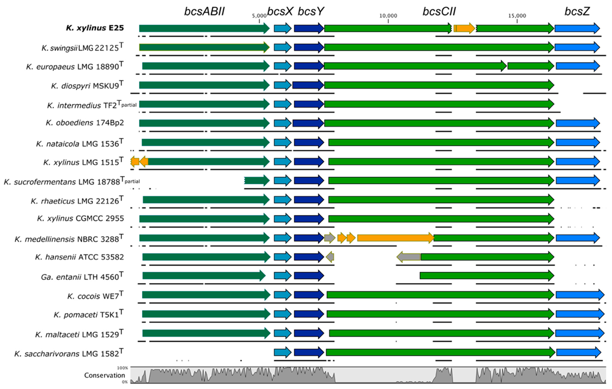
2.1. Diversity of the BcsII Operon in the Genomes of the Komagataeibacter Genus

The genome of K. xylinus E25 harbors three bcs operons: bcsI, bcsII and bcsIII (Figure S1a). The second bcs operon is of a similar size (13,301 bp) as the first operon and is predicted to consist of four, as already mentioned, genes (bcsABII, bcsX, bcsY, bcsCII) and an additional bcsZ gene, which was not described before (Figure 1 and Figure S1a). The bcsA and bcsB subunits are fused in the case of both bcsII and bcsIII (Figure S1a). Based on the annotation, the bcsCII gene is predicted to be disrupted by an insertion sequence of IS1031A-type, which has been described before in K. hansenii ATCC 23769 (former Acetobacter xylinum) and shown to be overrepresented among the Komagataeibacter species [23]. Due to the disruption by IS, the bcsCII is likely not to be functional in K. xylinus E25.
Next, we compared the sequence and the organization of the predicted bcsII operons of the type Komagataeibacter strains to understand how it is conserved in the genus (Figure 1). The second operon was present in all genomes; however, its structure displayed variability among the strains. Some of the differences are due to the incompleteness of the genome sequence (K. intermedius TF2 and K. sucrofermentans LMG 18788). The bcsABII gene is absent in K. saccharivorans LMG 1582; in K. xylinus LMG 1515 it is disrupted by a predicted IS66 family insertion sequence accessory protein TnpB and a transposase, which suggests that this operon is nonfunctional. Gene bcsX is the most highly conserved of all genes in the operon. More nucleotide sequence variability was discovered in the sequence of the bcsY gene, where in such strains as K. diospyri MSKU 9 and K. oboediens 174Bp2 it was slightly longer than in other strains, which translated into differences of 16 or 18 amino acids, respectively. This difference in bcsY size may, however, be due to incorrect prediction of the gene start. In the case of bcsCII, the highest sequence variability among the strains can be observed. This gene is disrupted or incomplete in K. xylinus E25, K. medellinensis NBRC 3288, K. hansenii ATCC 53582 and Gluconacteobacter entanii LTH 4560. Finally, gene bcsZ is much less conserved than bcsX or bcsY; in such strains as K. diospyri MSKU9, K. rhaeticus LMG 22126, K. xylinus CGMCC 2955, K. hansenii ATCC 53582 and Ga. entanii LTH 4560 it is absent. In K. swingsii LMG 22125 and K. xylinus E25 bcsZ was predicted to be slightly longer, which translated into a difference of 16 amino acids of the predicted protein. Similarly, as in the case of bcsY, this may be due to the incorrectly predicted gene start.
We decided to investigate in detail the cases of bcsZ absence by using tblastn. We found that that the genome sequence of K. diospyri MSKU 9 downstream of the bcsII produced significant matches with the translated BcsZ sequence, which suggests that the reading frame of bcsZ accumulated mutations during the strain’s evolution, which probably resulted in gene inactivation. In the case of the K. intermedius TF2 genome, which is highly fragmented, we found a significant match in another contig of the genome. For the genomes of K. rhaeticus LMG 22126 and K. xylinus CGMCC 2955 and the K. hansenii strains, however, no significant matches were produced. Given that the majority of Komagataeibacter spp. harbor bcsZ, it is very exceptional for the K. rhaeticus and K. hansenii strains to lack this gene.
By analyzing a wider genomic context of the bcsII operon, two distinguishable features may be observed for some of the Komagataeibacter species. In the case of K. rhaeticus LMG 22126, K. xylinus CGMCC 2955 and K. medellinensis NBRC 3288, bcsII is constituted of three additional genes (kpsC, kpsS, rfaB), which are located upstream of the bcsABII gene (Figure S1b). This gene cluster may be involved in extracellular matrix formation, as described before for K. medellinensis ID13488 [17]. The presence of these three genes is unusual for the whole genus and is characteristic only to these two species, which have been previously shown to be phylogenetically closely related to each other [6].
Another unusual genomic context of bcsII can be observed for the strains of K. saccharivorans, which distinguishes them from other species of the Komagataeibacter genus (Figure S1c). In these strains, bcsII starts immediately with bcsX, and is located upstream of the bcsI operon, which lacks the bcsCI gene. It can be assumed that the absence of bcsCI, which is crucial for the export of cellulose polymer, is compensated by the presence of bcsCII. What is more, although the type strain K. saccharivorans LMG 1582 does not produce cellulose (there is no phenotypic information for other sequenced strains), other strains of this species have been demonstrated to produce high yields of BNC [24,25]. It would be interesting to experimentally investigate how the structure of this unusual operon influences the properties of synthesized cellulose. The distinctiveness of the K. saccharivorans strains may be expected given the large phylogenetic distance from other Komagataeibacter strains as shown based on the genomic comparisons [6].
Finally, the genomic sequence of K. oboediens 174Bp2 is predicted to harbor four copies of the bcsII operon, which are located in different genomic contexts, as previously described [7]. How this high number of bcsII copies translates into the expression level of its proteins and further on the properties of cellulose polymer is yet to be investigated.
Summarizing, although the second cellulose operon is present in every Komagataeibacter genome, it displays much greater diversity than the first operon. The observed differences in the genomic context of bcsII suggest that it was independently gained during the evolution of the Komagataeibacter species and, therefore, its presence probably provided an adaptational advantage.

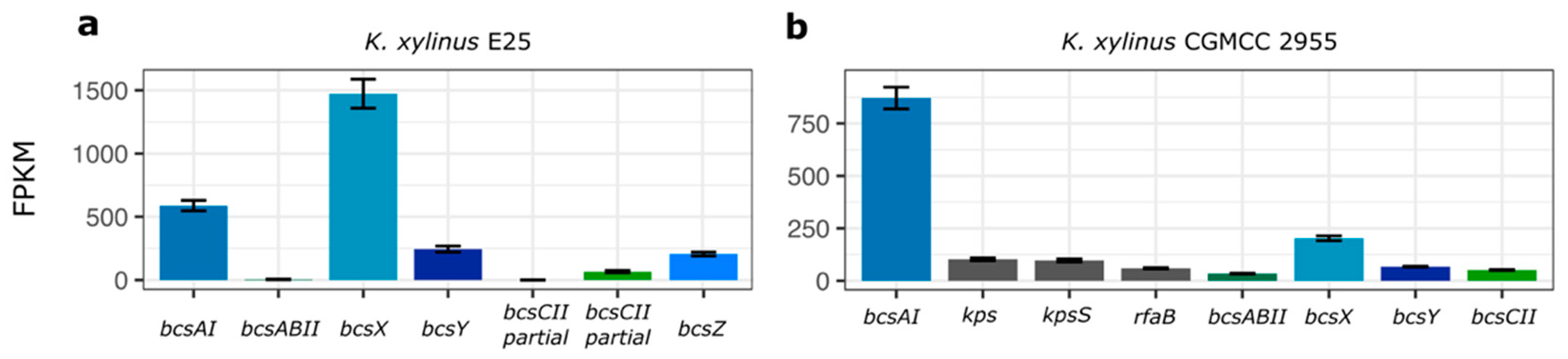
2.2. Expression Profile of the BcsII Operon

Apart from genome sequence data, recently, gene expression profiles have been published for K. xylinus E25 and K. xylinus CGMCC 2955 (probable K. rhaeticus sp.) [6] which opens the possibility to investigate in detail how cellulose operons are expressed [26,27].
The expression of genes of bcsII in K. xylinus E25, which is based on RNA-seq data, is shown in comparison with bcsAI of the first bcs operon (Figure 2a). It can be noticed that bcsX is the most highly expressed gene of bcsII and roughly three times higher than bcsAI. The expression of bcsABII and bcsCII is very low. BcsY and bcsZ are expressed at a much lower level than bcsX. Such variable expression of the bcsII genes suggests that, in the case of K. xylinus E25, they are regulated by a separate promoter or post-transcriptionally tuned. Additionally, we have observed that this operon is differentially regulated by the presence of ethanol in the culture medium (Figure S2). For the majority of bcsII genes this change is positive (for bcsY it is significant), as in the case of bcsAI, whereas bcsZ is significantly negatively regulated.
A slightly different expression profile of bcsII was captured in K. xylinus CGMCC 2955 (Figure 2b). Here, bcsX is the most highly expressed gene of the bcsII operon but at a much lower level, more than four times less than bcsAI of the first operon. The expression of bcsABII and bcsCII is the lowest of all genes of bcsII.
Additional insight into the expression of bcsII has been delivered by a study involving K. medellinensis ID13488, where it was shown that bcsABII was the most weakly expressed gene of all bcs operons present in the genome (the expression of other genes of bcsII was not measured) [17].
Taken together, the presented results suggest that the function of the bcsII operon is the synthesis of a modified cellulose, where bcsX may play the most important role. What is more, the presented profiles suggest that the expression of bcsII may differ in the Komagataeibacter spp. More research, however, is needed to investigate the dependence of genotype and condition on the expression of this operon.

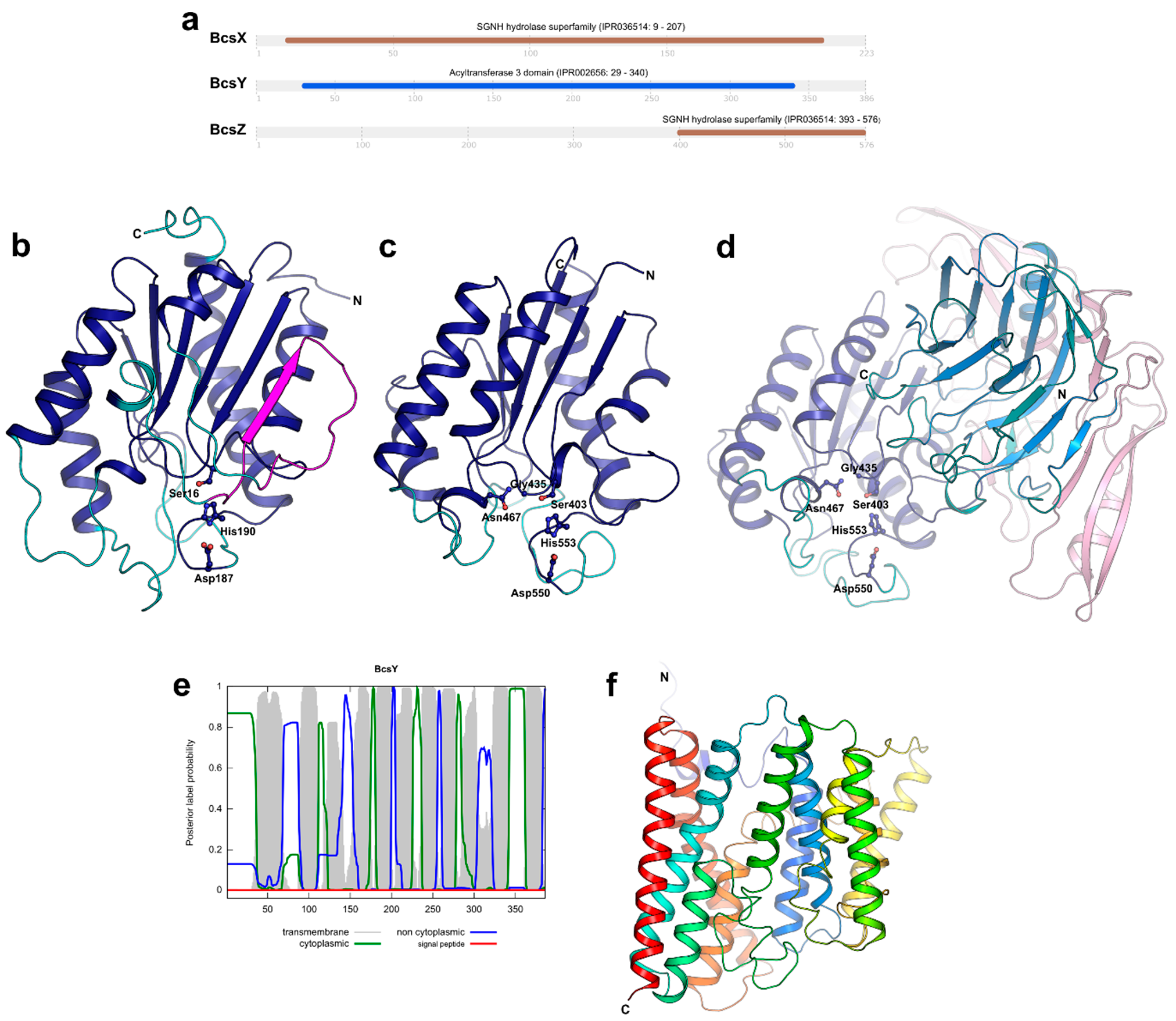
2.3. Function Prediction, Sequence Similarity and Homology Search for the BcsII Proteins

We next focused on the detailed study of the unique proteins encoded by the bcsII operon of K. xylinus E25. First, a prediction of the functional domains within the protein sequences of the investigated proteins was made using the InterProScan database (Figure 3a). Nearly a whole sequence of BcsX is predicted to constitute the SGNH/GDSL hydrolase domain, whereas a sequence of the BcsY protein was predicted to carry the acyltransferase domain, as expected and described before [22]. This suggests that both proteins may play an important role during acylation of a newly-formed bacterial cellulose. Analysis of the BcsZ sequence showed that, similarly to BcsX, it contains the SGNH/GDSL hydrolase domain, but it is located near the C-terminus. Furthermore, these two proteins differ in size (223 vs. 576 amino acids) and share very low sequence similarity (local similarity of 30%). The function of the remaining part of BcsZ seems to be unclear; hypothetically, it may be involved in cooperation with the other protein partners in the BCS complex or may be responsible for allosteric regulation. The prediction of the SGNH/GDSL hydrolase domains in BcsX and BcsZ enhances the hypothesis that the bcsII operon is involved in the cellulose acylation process.
The sequence identity between BcsX, BcsY and BcsZ of K. xylinus E25 and their homologs of the Komagataeibacter species is high (100–75% for BcsX, 98–67% for BcsY and 97–57% for BcsZ; Table S1). These results suggest that BcsX is the most highly conserved protein of all encoded by the bcsII operon. A search for homologues of BcsX and BcsZ in bacterial families other than Acetobacteraceae was also performed. The hydrolases of Polynucleobacter aenigmaticus, Castellaniella defragrans and Pseudomonas alcaligenes (Figure S3) were identical to BcsX of K. xylinus E25 in about 40%. The number of identical residues in a sequence was lower (25–30%) for the comparison of BcsZ with the homologues from non-Acetobacteraceae families (Figure S4). The top-ranked hydrolase homologues of BcsX and BcsZ were different, although both proteins belong to the same hydrolase family. This may be due to the fact that BcsZ contains an extra sequence proceeding the SGNH domain.
Interestingly, BcsX shares high sequence similarity (54%) with the WssF protein from Pseudomonas fluorescens SBW25, which produces cellulosic polymer consisting of partially acetylated cellulose that was found to provide an advantage for colonization of the air–liquid interface as well as rapid spreading across solid surfaces [28]. Studies of wssF and the adjacent downstream genes of wssF (wssGHIJ) mutants have shown that these genes are crucial for the synthesis of acetylated BC. WssF is a predicted GDSL-like lipase/acylhydrolase that was hypothesized to function by presenting acyl groups to the enzymes involved in the acetylation of cellulose. On the other hand, the adjacent downstream genes of wssF (wssGHIJ) of P. fluorescens SBW25, which are associated with the O-acetylation of alginate, are not present in K. xylinus E25. Despite the differences in operon structure, it is possible that BcsX has a similar role as WssF, but more studies are needed to explain its role and interaction with other proteins encoded by bcsII in the process of cellulose acetylation in K. xylinus E25.
The homologues of BcsY are widely distributed not only among the Acetobacteraceae family (Rhodospirillales order), but also among bacteria belonging to different orders, e.g., Enterobacteriales, Rhizobiales and Pseudomonadales (Table S1 and Figure S5). The sequence identity between BcsY and its homologues from different bacteria is variable (usually below 40%); however, the exception is a homologue from Enterobacter lignolyticus, which is identical with BcsY in 52%.

2.4. Prediction of BcsX, BcsY and BcsZ Subcellular Localization

Afterwards, we moved to the prediction of the subcellular localization and analysis of the secondary structure elements of the bcsII operon products.
Initially, we searched for the presence of a signal peptide (SP) in the sequences of the investigated proteins to predict their more probable localization in the cell. A signal peptide was predicted by SignalP only in the sequence of BcsZ (Figure S6). These results suggest that BcsZ functions outside of the cell.
Next, we used the Phobius program to predict the transmembrane (TM) topology of the bcsII proteins. Transmembrane α-helices were predicted only for BcsY (Figure 3e). The putative function of BcsY nicely correlates with the results of this analysis. Many bacterial acyltransferases are localized in the cytoplasmic membranes or lipophilic inclusions present in the cytosol [29]. Acyltransferases belong to the transferase enzyme class and catalyze the transfer of acyl groups from the donor to the acceptor, forming either esters or amides [30]. Sometimes the substrates are long-chain fatty acids; hence, it can be hypothesized that BcsY located in the cytoplasmic membrane might deal with such hydrophobic substrates available there. The secondary structure elements’ prediction of BcsY performed with PRED-TMBB2 (Figure S8) indicated that BcsY can be composed of fourteen transmembrane helices.
Additionally, Phobius predicted the presence of a signal peptide in the sequence of BcsX and BcsZ but not in BcsY. These predictions agree with those of SignalP in the case of BcsY and BcsZ (the same cleavage site predicted in both predictions); however, they differ for BcsX (Figure S7). The presence of a signal peptide in the sequence of BcsX is therefore questionable and so is its subcellular localization.

2.5. Three-Dimensional Structure Prediction for BcsX, BcsY and BcsZ

Recently, the models representing the predicted spatial structures of BcsX and BcsY were generated by AlphaFold and added to the corresponding UniProt entries (BcsX: Q9WX69, BcsY: Q9WX70). Additionally, we modelled the spatial structure of BcsZ using AlphaFold. As a next step, we performed structural homology searches for all proteins with the PDBeFold (SSM) server [31] using the AlphaFold models. BcsX and the C-terminal domain (CTD) of BcsZ (residues 393–576) show homology to many different serine esterases belonging to the SGNH/GDSL hydrolase family, whereas the middle parts of BcsZ (residues 192–382) represent a lectin-like fold. The five top-ranked homologs for each search are listed in Table 1.
The structural homology between BcsX and the listed proteins is relatively high. The superposition of BcsX on its homologs produced the rmsd values (calculated for Cα atoms) in the range from 2.31 to 2.67 Å (for 148–171 out of 262 Cα atoms of BcsX). Based on this structural comparison (Figure 3b), all α-helices and the central β-sheet are conserved among the discussed serine esterases, whereas several loop regions (residues 51–59, 78–84, 106–117, 141–152 and 178–186 in BcsX, residue numbering according to NCBI Gene: WP_025439176.1) have different conformations in the analyzed structures. Moreover, these loops also vary in length. An interesting element of the BcsX structure is an additional β-strand connected with a long loop (residues 51–77, shown in magenta in Figure 3b). All other structural homologues have a central β-sheet formed by five parallel β-strands; BcsX has this additional β-strand, which is, moreover, antiparallel to the adjacent β-strand.
The superposition of BcsZ-CTD on its homologs generated the rmsd values in the range 1.92–2.36 Å (for 147–159 out of 184 Cα atoms of BcsZ-CTD) indicating that the structural homology between BcsZ-CTD and its homologs is even higher than in the case of BcsX. In the model of BcsZ-CTD, all α-helices and the central part of the structure including the β-sheet are highly conserved (Figure 3c) and there are only three flexible loops (residues 470–479, 504–508 and 533–549, residue numbering according to NCBI Gene: WP_025439171.1). Interestingly, the rmsd value for superposition of BcsX and BcsZ-CTD is 2.75 Å (for 152 Cα atoms) and the SGNH/GDSL hydrolase domain of BcsX is much larger (262 residues) compared to BcsZ-CTD (184 residues). Hence, BcsX contains more variable regions.
The most important finding of the structural analysis of BcsX and BcsZ-CTD is the identification of the catalytic triad that involves Ser16, Asp187 and His190 in BcsX and Ser403, Asp550 and His553 in BcsZ-CTD located in conserved loop regions (Figure 3b,c, Figures S3 and S4). Another element important for the catalytic activity of serine esterases is an oxyanion hole. In the analyzed homological SGNH/GDSL hydrolases, it is usually formed by three residues, catalytic serine, glycine/alanine and asparagine. It is of note that in the case of serine and glycine/alanine residues, only their backbone atoms participate in the catalysis. In BcsX the corresponding residues are Ser16 and Ser46; however, if only the atoms of the residue backbone play a role, it does not matter that in BcsX there is serine instead of glycine/alanine. However, the third potential residue of the BcsX oxyanion hole is Thr104 (according to its position in the model). This might suggest that the mechanism of the BcsX catalysis can be slightly different. Another possibility is that the model requires improvement and another residue should be present at this position. For instance, closely located Asp103 could be a potential residue forming the oxyanion hole instead of Thr104. Interestingly, the oxyanion hole of BcsZ-CTD was easily identified, and it is formed by Ser403, Gly435 and Asn467 (Figure 3c and Figure S4). The conservation of the BcsZ-CTD sequence and spatial structure is high, and even the analysis performed by InterProscan indicated putative residues forming the catalytic triad and the oxyanion hole (the residue numbers were in agreement with the results of structural comparison). This finding confirms that BcsZ can act as the SGNH/GDSL hydrolase. Finally, we also compared the substrate-binding site of the analyzed homological enzymes, BcsX and BcsZ-CTD, but no significant similarities were detected. The residues interacting with substrates in other serine esterases were located mainly in the variable loop regions. Moreover, all analyzed enzymes utilize different substrates.
In addition to spatial structure analysis, we tried to pairwise compare the amino acid sequences of the mentioned proteins with the sequence of BcsX or BscZ-CTD using pairwise blastp. Interestingly, the only results were obtained for BcsX/Mycobacterium smegmatis serine esterase and BcsZ-CTD/Escherichia coli multifunctional enzyme. The sequence identity for the first pair was only 26% (residues 144–205 of BcsX, involving mainly conserved structural elements and two residues of the catalytic triad) and 25% for the latter one (residues 395–573 of BcsZ-CTD, covering the almost whole CTD). The comparison of amino acid sequences of BcsX and BcsZ indicates no significant similarity between these two proteins.
The search for structural homologs of the remaining part of BcsZ (residues 41–392 without the signal peptide) indicated that the middle part of the protein (residues 192–382) has a lectin-like fold (Figure 3d). The superposition of the BcsZ middle domain on its homologs produced the rmsd values in the range from 3.15–3.42 Å (for 149–165 out of 190 Cα atoms of the BcsZ middle domain). The top-ranked structural homologs are the L-type lectins. The members of this lectin family are usually present in the seeds of leguminous plants and their carbohydrate recognition domain is composed of two antiparallel β-sheets connected by short loops forming the “jelly-roll fold”. This structural motif can also be found in many other eukaryotic proteins [43] including the human N-terminal domain of thrombospondin that appeared in the results of the structural homologs search. The middle domain of BcsZ contains a lectin-like fold with two central β-sheets connected by a number of flexible loops of a different length. The most similar loop arrangement was observed for the BcsZ lectin-like domain and thrombospondin that mediates cell-to-cell and cell-to-matrix interactions, as it recognizes carbohydrate patterns [38]. However, the conformation of some β-strands forming the central β-sheets is also variable among the compared proteins (Figure 3d). Nevertheless, the obtained results suggest that this domain of BscZ might be involved in interacting with cellulose as it contains a fold that is usually involved in interactions with carbohydrates. The potential function of the N-terminal part of BcsZ (residues 41–191) remains unknown.
The results of the structural analysis of the BcsY model did not provide any information about conserved structural elements or the catalytic residues of this enzyme. The obtained rmsd values were high, except for two proteins, bovine cytochrome c oxidase subunit 3 (PDB ID: 3WG7, chain C) [44] and Saccharomyces cerevisiae translation initiation factor eIF-2B subunit delta (PDB ID: 6QG2, chain H) [45], the values of which were slightly below 4 Å (only over 108–130 out of 386 BcsY Cα atoms). Both proteins play different roles in eukaryotic organisms; hence, it is not very likely that they are structural homologues of BcsY, which is a bacterial protein and a putative acyltransferase. This classification is based only on conserved sequence fragments. To date, there is no information on the spatial structure of this acyltransferase family. The AlphaFold BcsY model contains 12 transmembrane α-helices (Figure 3f), whereas Phobius and PRED-TMBB2 predicted 11 and 14 transmembrane α-helices, respectively. The model contains large loop regions; hence, it is possible that the protein contains more helices than in the current model. The mechanisms of action of this enzyme, its substrates and products also remain unknown.

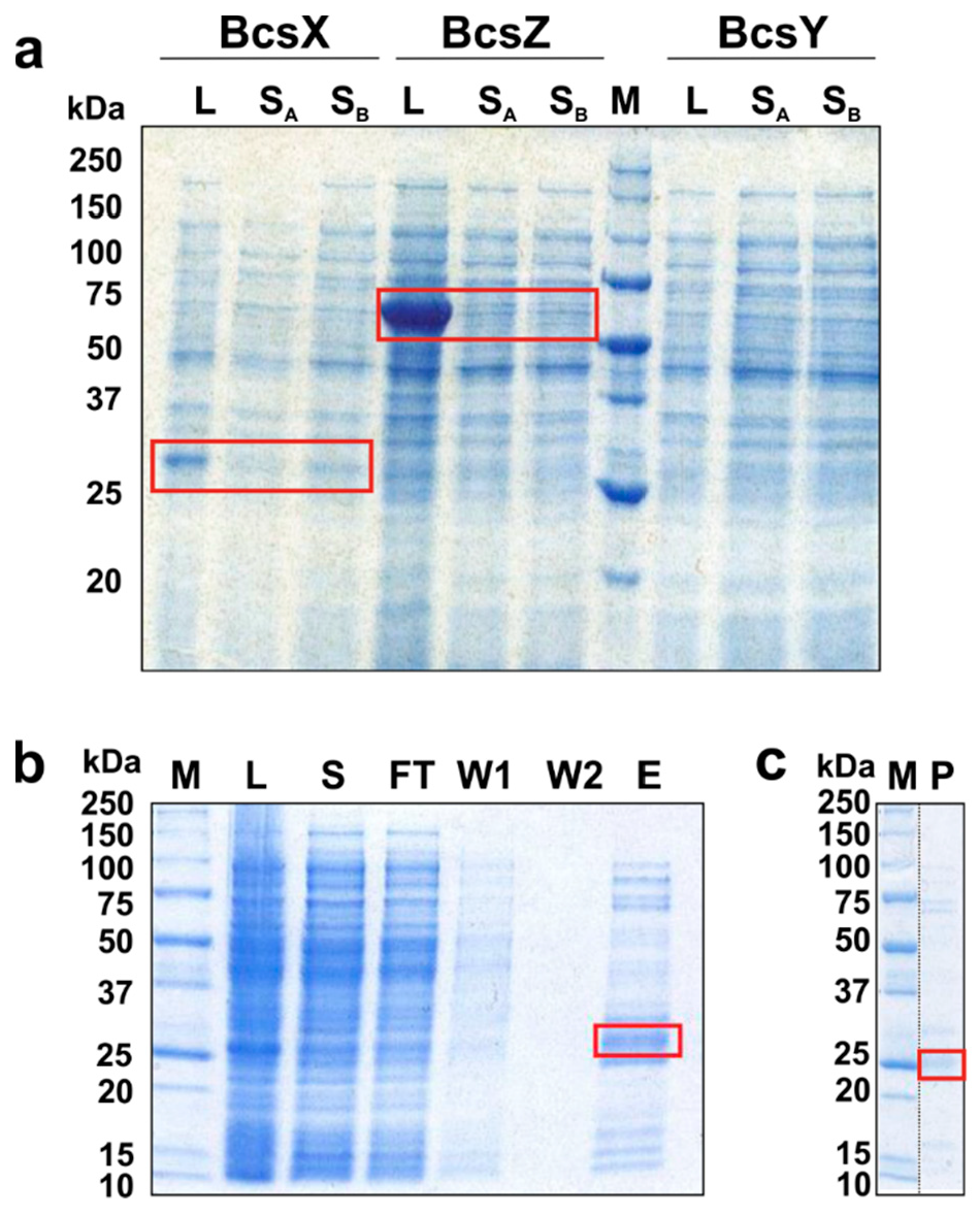
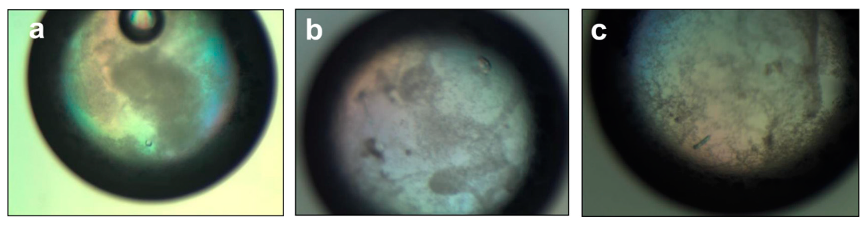
2.6. Overexpression and Solubility Tests of the BcsII Operon Proteins, BcsX Purification and Crystallization

To address the questions about the mechanisms of action of BcsX, BcsY and BcsZ, we tried to produce recombinant proteins that could be then used for structural studies. The expression and solubility of BcsX, BcsY and BcsZ were tested using E. coli BL21 Gold cells as the expression host. The bcsX, bcsY and bcsZ genes were cloned into expression vector pETM-11 and the proteins were bearing the N-terminal cleavable His6-tag. BcsX and BcsZ were detected in lysates obtained after the cultivation of bacteria transformed with the prepared constructs (Figure 4a). BcsY was not produced in E. coli; this might be related to the fact that BcsY is, according to our predictions, a transmembrane protein. Although the amount of BcsZ was higher compared to BcsX, only the latter protein was present in soluble protein fraction. Therefore, BcsX was subjected for further analysis.
Initially, BcsX was overproduced by bacteria growing in the LB medium, and the overexpression was triggered by IPTG. The 500 mL E. coli culture provided about 0.52 mg of purified protein. The use of autoinduction LB medium allowed the amount of protein to increase to 1.2 mg (per 500 mL culture).
BcsX purification was performed using a two-step protocol. After the first step of BcsX purification on Ni-NTA, the elution sample still contained some amount of other proteins (Figure 4b). The second purification step, including removal of the cleaved His6-tag, allowed the protein sample of higher purity to be obtained, although some weak bands corresponding to contaminants were observed on the SDS-PAGE gel (Figure 4c). Nevertheless, the obtained level of purity was sufficient for the initial crystallization trials. Small crystals of BcsX were obtained during the initial screening (Figure 5). The diffraction properties of these crystals were tested; however, the BcsX crystals were diffracted by X-ray only to 5 Å. Further crystallization optimization is necessary to obtain well-diffracting crystals, which can provide good-quality data sufficient for structure determination.

3. Materials and Methods

3.1. Bioinformatical Analysis of Genome and Transcriptome Data

Genome sequences of all Komagataeibacter strains with their annotation were downloaded from NCBI database (access date: December 2021). Alignments of bcsII operons were performed and visualized using CLC Sequence Viewer (v. 8.0).
Gene expression data (RNA sequencing, RNA-seq) for K. xylinus E25 came from [26]. The calculated FPKM (fragments per kilobase per million mapped fragments) values from 3 replicate libraries per condition were used (SH medium or SH medium + ethanol). RNA-seq data for K. xylinus CGMCC 2955 came from the study of [27]. Original NGS (next generation sequencing) libraries were downloaded from SRA (GSM5329218, GSM5329219, GSM5329220). The libraries represented 21% oxygen tension condition (used as reference in the original study), as stated by the authors. The sequencing reads were processed as described in [26]. The K. xylinus CGMCC 2955 (ASM276219v1) genome with annotation was downloaded from NCBI used during reads mapping. Gene expression data from the two studies was visualized using ggplot2 package [46] in R (v. 3.5.1).

3.2. Functional and Structural Prediction

Functional domain search in the protein sequences encoded by bcsII in K. xylinus E25 was performed using InterProScan web server of EBI [47]. Pairwise protein sequence alignment for Komagataeibacter strains was performed with DIAMOND in Blastp mode [48] and by using custom-made scripts written in Python 3.7.9. Search of homologs outside of the Komagataeibacter genus was performed using NCBI Blastp online server [49].
Prediction of signal peptides was conducted using SignalP 6.0 online service [50]. Phobius online service was used to predict transmembrane segments and, additionally, signal peptides [51,52]. The secondary structure elements prediction was performed with PRED-TMBB2 [53].
Three-dimensional structural models for BcsX and BcsY from K. xylinus NBRC 13693 generated by AlphaFold [54] were downloaded from UniProt (BcsX: Q9WX69, BcsY: Q9WX70) and visualized using PyMOL (Schrödinger, LLC, New York, NY, USA). The structure of BcsZ was predicted in this study using AlphaFold v. 2.1.1. Structural homology searches were performed with the PDBeFold (SSM) server [31].

3.3. Overexpression and Solubility Tests of the BcsII Operon Proteins

The bcsX, bcsY and bcsZ genes were PCR-amplified from a genomic DNA of Komagateibacter xylinus E25 (isolated as described before in [7]) and inserted via NcoI and EcoRI (bcsX) or MunI and XhoI (bcsY, bcsZ) restriction sites into the pETM-11 vector (EMBL Heidelberg) under the control of a T7 promotor for production of fusion protein bearing a TEV-cleavable N-terminal His6-tag. The primers used in PCR are listed in Table S2.
E. coli BL21 Gold competent cells were transformed with the prepared vectors. Bacteria were cultivated in lysogeny broth (LB) medium (50 mL culture per construct, supplemented with 50 μg/mL of kanamycin) at 37 °C to an OD600 of ~0.6. Subsequently, isopropyl-β-D-thiogalactopyranoside (IPTG) was added to final concentration of 0.5 mM. After next 3 h of overexpression at 37 °C, the cells were pelleted. The cell pellets were resuspended either in buffer A (20 mM HEPES, pH 7.5, 500 mM NaCl, 10 mM imidazole, 1 mM DTT supplemented with 0.1 mM PMSF) or buffer B (20 mM HEPES, pH 7.5, 500 mM NaCl, 10 mM imidazole, 10% glycerol, 1 mM DTT supplemented with 0.1 mM PMSF) and lysed by sonication using a Sonopuls GM 3200 ultrasonic homogenizer (Bandelin). Lysates were centrifuged for 40 min at 4 °C and 10,000× g. The samples representing lysates (collected prior centrifugation) and soluble protein fraction (collected after centrifugation) were analysed using SDS-PAGE.

3.4. Production and Purification of BcsX Protein

Initially, overproduction of BcsX was carried out as described above. In total culture volume of 500 mL. Subsequently, to increase the amount of produced protein, autoinducing medium, LB Broth Base including trace elements (Formedium), was used and bacteria were cultivated at 37 °C to an OD600 of ~0.6 and subsequently further incubated at 20 °C for 18 h. The cells were harvested at an OD600 of ~10.
BcsX purification protocol was based on the previously described purification of other recombinant proteins [55,56]. The cell extracts were prepared as described in Section 3.3. The target protein was captured from the cleared lysate on 0.5 mL of Ni-NTA agarose beads (Qiagen) and eluted with 400 mM imidazole. The His6-tag was cleaved with TEV protease during overnight dialysis at 4 °C against 20 mM HEPES, pH 7.5, 500 mM NaCl, 10% glycerol, 1 mM DTT. The cleaved protein was loaded onto 0.5 mL of Ni-NTA agarose to remove the His6-tagged TEV protease, uncleaved protein and cleaved His6-tag. The flow-through fractions were concentrated to 11.4 mg/mL.

3.5. Protein Crystallization and Data Collection

Initial screening for crystallization conditions was performed at 18 °C using Index Screen, SaltRx1 Screen and PEG/Ion Screen (Hampton Research) and the hanging-drop vapor diffusion method with drops containing 1 µL of protein solution and 1 µL of reservoir solution. The initial crystals of BcsX were obtained using the following solutions: (1) 2.1 M DL-Malic acid pH 7.0; (2) 0.2 M sodium chloride, 0.1 M Tris pH 8.5, 25% PEG 3350; (3) 0.2 M ammonium acetate, 0.1 M HEPES pH 7.0, 25% PEG 3350. Crystals obtained using solutions 2 and 3 were cryoprotected by transfer into mother liquor containing 22.5 % (v/v) PEG 400 and flash-cooled in liquid nitrogen. X-ray diffraction measurements were carried out on beamline 14.1 of the BESSY II storage ring (Berlin, Germany; [57]).

4. Conclusions

In comparison to the first cellulose synthase operon, the role of bcsII has not been extensively studied and only putatively assigned to the synthesis of amorphous, acylated cellulose. The evidence supporting this hypothesis was, however, lacking.
In this study we aimed to gain a deeper insight into the function of bcsII. First, by taking the advantage of the accumulated genomic sequences of the genus, we compared the conservation of this operon. We found that this operon is present in every Komagataeibacter genome; however, its structure differs depending on the species. We discovered that in K. rhaeticus and K. medellinensis, this operon is associated with kps genes, which are involved in extracellular matrix formation. Association with these genes supports the observation of M. Bimmer et al. that bcsII is responsible for the synthesis of polymer that is attached to the bacterial cell. Further sequence similarity analyses have shown that the proteins encoded by bcsII are quite unique to the Komagataeibacter genus. For BcsX, relatively high sequence similarity was found with the WssF protein of P. fluorescens SBW25, which was confirmed to be important for the synthesis of acetylated cellulose. Our study revealed for the first time the presence of another gene, bcsZ, which directly follows bcsII.
We further found that the expression of bcsX and bcsY, unlike that of bcsZ, was positively regulated in the medium containing ethanol, which are the conditions promoting cellulose biosynthesis in K. xylinus E25. This can suggest that there may be a positive regulatory connection between the cellulose biosynthesis pathway and the expression of bcsX and bcsY. The opposite seems to be the case for bcsZ; however, more expression profiles are needed to confirm the coupling of bcsII genes’ expression with cellulose synthesis.
By focusing on the protein products of the bcsII operon we found that both BcsX and BcsZ contain domains belonging to the SGNH/GDSL family, which is characteristic to serine esterases. Their pairwise sequence similarity was, however, low, which suggests that these enzymes may play different roles. Based on the homology search of BcsZ’s predicted 3D structure, we found that, apart from the SGNH/GDSL domain, the middle part of this protein has a lectin-like fold that is similar to human thrombospondin, which mediates cell-to-cell and cell-to-matrix interactions by recognizing carbohydrate patterns. We therefore postulate that BcsZ may play a similar role in the attachment of Komagataeibacter cells to the acylated and the crystalline cellulose. The prediction of the signal peptide at the N-terminus of BcsZ further suggests that this enzyme functions outside of the cell.
In summary, based on the gathered results, we postulate that the role of bcsII is not only the synthesis of the acylated polymer but also regulation of its attachment to the cell surface and to the crystalline cellulose fibers. As suggested previously for this operon, its function may influence colonization of the air–liquid interphase, and, through analogy to WssF function, spreading across solid surfaces. These findings, together with the experimental results of the bcsII operon proteins’ purification and crystallization, constitute a solid base for further studies on their exact function.

Supplementary Materials

The following supporting information can be downloaded at: https://www.mdpi.com/article/10.3390/ijms23147851/s1.

Author Contributions

I.S., A.J.P.-B. and M.R. conceived and designed the study. M.R., A.J.P.-B. and K.D. performed bioinformatical analyses regarding genomic and protein sequences. A.J.P.-B. performed the analysis of 3D structure models. I.S. and A.J.P.-B. carried out the experiments involving protein production, purification and crystallization. M.R., A.J.P.-B. and I.S. drafted the manuscript. All authors have read and agreed to the published version of the manuscript.

Funding

This research was funded by the MINIATURA2 grant No. 2018/02/X/NZ1/01590 from Narodowe Centrum Nauki (NCN; National Science Centre, Poland).

Institutional Review Board Statement

Not applicable.

Informed Consent Statement

Not applicable.

Data Availability Statement

Not applicable.

Acknowledgments

We are grateful to HZB BESSY for the allocation of synchrotron radiation beamtime at BL 14.1. We thank Stanisław Bielecki for his inspiring discussions. We thank the Computing & Information Services Centre (UCI) of Lodz University of Technology for providing a computing environment, especially to Seweryn Sitarski, Paweł Adamczyk and Przemysław Trzeciak.

Conflicts of Interest

The authors declare no conflict of interest.

References

  1. Szustak, M.; Gendaszewska-Darmach, E. Nanocellulose-Based Scaffolds for Chondrogenic Differentiation and Expansion. Front. Bioeng. Biotechnol. 2021, 9, 733. [Google Scholar] [CrossRef] [PubMed]
  2. Ludwicka, K.; Kaczmarek, M.; Białkowska, A. Bacterial Nanocellulose—A Biobased Polymer for Active and Intelligent Food Packaging Applications: Recent Advances and Developments. Polymers 2020, 12, 2209. [Google Scholar] [CrossRef] [PubMed]
  3. Gorgieva, S.; Trček, J. Bacterial Cellulose: Production, Modification and Perspectives in Biomedical Applications. Nanomaterials 2019, 9, 1352. [Google Scholar] [CrossRef] [Green Version]
  4. Jankau, J.; Błażyńska-Spychalska, A.; Kubiak, K.; Jędrzejczak-Krzepkowska, M.; Pankiewicz, T.; Ludwicka, K.; Dettlaff, A.; Pęksa, R. Bacterial Cellulose Properties Fulfilling Requirements for a Biomaterial of Choice in Reconstructive Surgery and Wound Healing. Front. Bioeng. Biotechnol. 2022, 9, 1492. [Google Scholar] [CrossRef]
  5. Dourado, F.; Ryngajllo, M.; Jedrzejczak-Krzepkowska, M.; Bielecki, S.; Gama, M. Taxonomic Review and Microbial Ecology in Bacterial NanoCellulose Fermentation. Bact. Nanocellul. 2016, 1–17. [Google Scholar]
  6. Ryngajłło, M.; Jędrzejczak-Krzepkowska, M.; Kubiak, K.; Ludwicka, K.; Bielecki, S. Towards Control of Cellulose Biosynthesis by Komagataeibacter Using Systems-Level and Strain Engineering Strategies: Current Progress and Perspectives. Appl. Microbiol. Biotechnol. 2020, 104, 6565–6585. [Google Scholar] [CrossRef]
  7. Ryngajłło, M.; Kubiak, K.; Jędrzejczak-Krzepkowska, M.; Jacek, P.; Bielecki, S. Comparative Genomics of the Komagataeibacter Strains—Efficient Bionanocellulose Producers. Microbiologyopen 2019, 8, e00731. [Google Scholar] [CrossRef] [PubMed] [Green Version]
  8. Nicolas, W.J.; Ghosal, D.; Tocheva, E.I.; Meyerowitz, E.M.; Jensen, G.J. Structure of the Bacterial Cellulose Ribbon and Its Assembly-Guiding Cytoskeleton by Electron Cryotomography. J. Bacteriol. 2021, 203, e00371-20. [Google Scholar] [CrossRef]
  9. Cielecka, I.; Ryngajłło, M.; Maniukiewicz, W.; Bielecki, S. Response Surface Methodology-Based Improvement of the Yield and Differentiation of Properties of Bacterial Cellulose by Metabolic Enhancers. Int. J. Biol. Macromol. 2021, 187, 584–593. [Google Scholar] [CrossRef]
  10. Gullo, M.; La China, S.; Falcone, P.M.; Giudici, P. Biotechnological Production of Cellulose by Acetic Acid Bacteria: Current State and Perspectives. Appl. Microbiol. Biotechnol. 2018, 102, 6885–6898. [Google Scholar] [CrossRef]
  11. Valera, M.J.; Mas, A.; Streit, W.R.; Mateo, E. GqqA, a Novel Protein in Komagataeibacter Europaeus Involved in Bacterial Quorum Quenching and Cellulose Formation. Microb. Cell Fact. 2016, 15, 88. [Google Scholar] [CrossRef] [PubMed] [Green Version]
  12. Matsutani, M.; Ito, K.; Azuma, Y.; Ogino, H.; Shirai, M.; Yakushi, T.; Matsushita, K. Adaptive Mutation Related to Cellulose Producibility in Komagataeibacter Medellinensis (Gluconacetobacter Xylinus) NBRC 3288. Appl. Microbiol. Biotechnol. 2015, 99, 7229–7240. [Google Scholar] [CrossRef] [PubMed]
  13. Ishiya, K.; Kosaka, H.; Inaoka, T.; Kimura, K.; Nakashima, N. Comparative Genome Analysis of Three Komagataeibacter Strains Used for Practical Production of Nata-de-Coco. Front. Microbiol. 2022, 12, 4308. [Google Scholar] [CrossRef] [PubMed]
  14. Jedrzejczak-Krzepkowska, M.; Kubiak, K.; Ludwicka, K.; Bielecki, S. Chapter 2 – Bacterial NanoCellulose Synthesis, Recent Findings. In Bacterial Nanocellulose: From Biotechnology to Bio-Economy; Gama, M., Dourado, F., Bielecki, S., Eds.; Elsevier: Amsterdam, The Netherlands, 2016; pp. 19–46. ISBN 9780444634580. [Google Scholar]
  15. Uto, T.; Ikeda, Y.; Sunagawa, N.; Tajima, K.; Yao, M.; Yui, T. Molecular Dynamics Simulation of Cellulose Synthase Subunit D Octamer with Cellulose Chains from Acetic Acid Bacteria: Insight into Dynamic Behaviors and Thermodynamics on Substrate Recognition. J. Chem. Theory Comput. 2021, 17, 488–496. [Google Scholar] [CrossRef] [PubMed]
  16. Römling, U.; Galperin, M.Y. Bacterial Cellulose Biosynthesis: Diversity of Operons, Subunits, Products, and Functions. Trends Microbiol. 2015, 23, 545–557. [Google Scholar] [CrossRef] [Green Version]
  17. Hernández-Arriaga, A.M.; del Cerro, C.; Urbina, L.; Eceiza, A.; Corcuera, M.A.; Retegi, A.; Auxiliadora Prieto, M. Genome Sequence and Characterization of the Bcs Clusters for the Production of Nanocellulose from the Low PH Resistant Strain Komagataeibacter Medellinensis ID13488. Microb. Biotechnol. 2019, 12, 620–632. [Google Scholar] [CrossRef] [Green Version]
  18. Brandão, P.R.; Crespo, M.T.B.; Nascimento, F.X. Phylogenomic and Comparative Analyses Support the Reclassification of Several Komagataeibacter Species as Novel Members of the Novacetimonas Gen. Nov. and Bring New Insights into the Evolution of Cellulose Synthase Genes. Int. J. Syst. Evol. Microbiol. 2022, 72, 5252. [Google Scholar] [CrossRef]
  19. Bimmer, M.; Mientus, M.; Klingl, A.; Ehrenreich, A.; Liebl, W. The Roles of the Various Cellulose Biosynthesis Operons in Komagataeibacter Hansenii ATCC 23769. Appl. Environ. Microbiol. 2022, 88, e02460-21. [Google Scholar] [CrossRef]
  20. Florea, M.; Reeve, B.; Abbott, J.; Freemont, P.S.; Ellis, T. Genome Sequence and Plasmid Transformation of the Model High-Yield Bacterial Cellulose Producer Gluconacetobacter Hansenii ATCC 53582. Sci. Rep. 2016, 6, 23635. [Google Scholar] [CrossRef] [Green Version]
  21. Zhang, H.; Xu, X.; Chen, X.; Yuan, F.; Sun, B.; Xu, Y.; Yang, J.; Sun, D. Complete Genome Sequence of the Cellulose-Producing Strain Komagataeibacter Nataicola RZS01. Sci. Rep. 2017, 7, 4431. [Google Scholar] [CrossRef] [Green Version]
  22. Umeda, Y.; Hirano, A.; Ishibashi, M.; Akiyama, H.; Onizuka, T.; Ikeuchi, M.; Inoue, Y. Cloning of Cellulose Synthase Genes from Acetobacter Xylinum JCM 7664: Implication of a Novel Set of Cellulose Synthase Genes. DNA Res. 1999, 6, 109–115. [Google Scholar] [CrossRef] [PubMed] [Green Version]
  23. Coucheron, D.H. A Family of IS1031 Elements in the Genome of Acetobacter Xylinum: Nucleotide Sequences and Strain Distribution. Mol. Microbiol. 1993, 9, 211–218. [Google Scholar] [CrossRef] [PubMed]
  24. Gopu, G.; Govindan, S. Production of Bacterial Cellulose from Komagataeibacter Saccharivorans Strain BC1 Isolated from Rotten Green Grapes. Prep. Biochem. Biotechnol. 2018, 48, 842–852. [Google Scholar] [CrossRef] [PubMed]
  25. Abol-Fotouh, D.; Hassan, M.A.; Shokry, H.; Roig, A.; Azab, M.S.; Kashyout, A.E.H.B. Bacterial Nanocellulose from Agro-Industrial Wastes: Low-Cost and Enhanced Production by Komagataeibacter Saccharivorans MD1. Sci. Rep. 2020, 10, 3491. [Google Scholar] [CrossRef] [Green Version]
  26. Ryngajłło, M.; Jacek, P.; Cielecka, I.; Kalinowska, H.; Bielecki, S. Effect of Ethanol Supplementation on the Transcriptional Landscape of Bionanocellulose Producer Komagataeibacter Xylinus E25. Appl. Microbiol. Biotechnol. 2019, 103, 6673–6688. [Google Scholar] [CrossRef] [Green Version]
  27. Huang, L.H.; Liu, Q.J.; Sun, X.W.; Li, X.J.; Liu, M.; Jia, S.R.; Xie, Y.Y.; Zhong, C. Tailoring Bacterial Cellulose Structure through CRISPR Interference-Mediated Downregulation of GalU in Komagataeibacter Xylinus CGMCC 2955. Biotechnol. Bioeng. 2020, 117, 2165–2176. [Google Scholar] [CrossRef]
  28. Spiers, A.J.; Bohannon, J.; Gehrig, S.M.; Rainey, P.B. Biofilm Formation at the Air-Liquid Interface by the Pseudomonas Fluorescens SBW25 Wrinkly Spreader Requires an Acetylated Form of Cellulose. Mol. Microbiol. 2003, 50, 15–27. [Google Scholar] [CrossRef]
  29. Kruis, A.J.; Bohnenkamp, A.C.; Patinios, C.; van Nuland, Y.M.; Levisson, M.; Mars, A.E.; van den Berg, C.; Kengen, S.W.M.; Weusthuis, R.A. Microbial Production of Short and Medium Chain Esters: Enzymes, Pathways, and Applications. Biotechnol. Adv. 2019, 37, 107407. [Google Scholar] [CrossRef]
  30. Park, Y.C.; Shaffer, C.E.H.; Bennett, G.N. Microbial Formation of Esters. Appl. Microbiol. Biotechnol. 2009, 85, 13–25. [Google Scholar] [CrossRef]
  31. PDBeFold (SSM). Available online: http://www.ebi.ac.uk/msd-srv/ssm/ (accessed on 2 June 2022).
  32. Kim, K.; Ryu, B.H.; Kim, S.S.; An, D.R.; Ngo, T.D.; Pandian, R.; Kim, K.K.; Kim, T.D. Structural and Biochemical Characterization of a Carbohydrate Acetylesterase from Sinorhizobium Meliloti 1021. FEBS Lett. 2015, 589, 117–122. [Google Scholar] [CrossRef] [Green Version]
  33. Mathews, I.; Soltis, M.; Saldajeno, M.; Ganshaw, G.; Sala, R.; Weyler, W.; Cervin, M.A.; Whited, G.; Bott, R. Structure of a Novel Enzyme That Catalyzes Acyl Transfer to Alcohols in Aqueous Conditions. Biochemistry 2007, 46, 8969–8979. [Google Scholar] [CrossRef] [PubMed]
  34. Kovačić, F.; Granzin, J.; Wilhelm, S.; Kojić-Prodić, B.; Batra-Safferling, R.; Jaeger, K.E. Structural and Functional Characterisation of TesA - a Novel Lysophospholipase A from Pseudomonas Aeruginosa. PLoS ONE 2013, 8, e69125. [Google Scholar] [CrossRef] [PubMed] [Green Version]
  35. Brzuszkiewicz, A.; Nowak, E.; Dauter, Z.; Dauter, M.; Cieśliski, H.; Dlługolłȩcka, A.; Kur, J. Structure of EstA Esterase from Psychrotrophic Pseudoalteromonas Sp. 643A Covalently Inhibited by Monoethylphosphonate. Acta Crystallogr. Sect. F. Struct. Biol. Cryst. Commun. 2009, 65, 862–865. [Google Scholar] [CrossRef] [PubMed] [Green Version]
  36. Lo, Y.C.; Lin, S.C.; Shaw, J.F.; Liaw, Y.C. Crystal Structure of Escherichia Coli Thioesterase I/Protease I/Lysophospholipase L1: Consensus Sequence Blocks Constitute the Catalytic Center of SGNH-Hydrolases through a Conserved Hydrogen Bond Network. J. Mol. Biol. 2003, 330, 539–551. [Google Scholar] [CrossRef]
  37. Watanabe, M.; Fukada, H.; Inoue, H.; Ishikawa, K. Crystal Structure of an Acetylesterase from Talaromyces Cellulolyticus and the Importance of a Disulfide Bond near the Active Site. FEBS Lett. 2015, 589, 1200–1206. [Google Scholar] [CrossRef] [PubMed] [Green Version]
  38. Tan, K.; Duquette, M.; Liu, J.H.; Zhang, R.; Joachimiak, A.; Wang, J.H.; Lawler, J. The Structures of the Thrombospondin-1 N-Terminal Domain and Its Complex with a Synthetic Pentameric Heparin. Structure 2006, 14, 33. [Google Scholar] [CrossRef] [Green Version]
  39. Geethanandan, K.; Abhilash, J.; Bharath, S.R.; Sadasivan, C.; Haridas, M. X-Ray Structure of a Galactose-Specific Lectin from Spatholobous Parviflorous. Int. J. Biol. Macromol. 2011, 49, 992–998. [Google Scholar] [CrossRef]
  40. Hamelryck, T.W.; Loris, R.; Bouckaert, J.; Dao-Thi, M.H.; Strecker, G.; Imberty, A.; Fernandez, E.; Wyns, L.; Etzler, M.E. Carbohydrate Binding, Quaternary Structure and a Novel Hydrophobic Binding Site in Two Legume Lectin Oligomers from Dolichos Biflorus. J. Mol. Biol. 1999, 286, 1161–1177. [Google Scholar] [CrossRef] [Green Version]
  41. Audette, G.F.; Vandonselaar, M.; Delbaere, L.T.J. The 2.2 A Resolution Structure of the O(H) Blood-Group-Specific Lectin I from Ulex Europaeus. J. Mol. Biol. 2000, 304, 423–433. [Google Scholar] [CrossRef] [Green Version]
  42. Delbaere, L.T.J.; Vandonselaar, M.; Prasad, L.; Quail, J.W.; Wilson, K.S.; Dauter, Z. Structure of the Lectin IV of Griffonia Simplicifolia and Its Complex with the Lewis b Human Blood Group Determinant at 2·0 Å Resolution. J. Mol. Biol. 1993, 230, 950–965. [Google Scholar] [CrossRef]
  43. Lis, H.; Sharon, N. Lectins: Carbohydrate-Specific Proteins That Mediate Cellular Recognition. Chem. Rev. 1998, 98, 637–674. [Google Scholar] [CrossRef] [PubMed]
  44. Hirata, K.; Shinzawa-Itoh, K.; Yano, N.; Takemura, S.; Kato, K.; Hatanaka, M.; Muramoto, K.; Kawahara, T.; Tsukihara, T.; Yamashita, E.; et al. Determination of Damage-Free Crystal Structure of an X-Ray-Sensitive Protein Using an XFEL. Nat. Methods 2014, 11, 734–736. [Google Scholar] [CrossRef] [PubMed]
  45. Gordiyenko, Y.; Llácer, J.L.; Ramakrishnan, V. Structural Basis for the Inhibition of Translation through EIF2α Phosphorylation. Nat. Commun. 2019, 10, 2640. [Google Scholar] [CrossRef] [Green Version]
  46. Wickham, H. Ggplot2: Elegant Graphics for Data Analysis; Springer: New York, NY, USA, 2016; ISBN 978-3-319-24277-4. [Google Scholar]
  47. InterProScan. Available online: https://www.ebi.ac.uk/interpro/search/sequence/ (accessed on 9 December 2021).
  48. Buchfink, B.; Xie, C.; Huson, D.H. Fast and Sensitive Protein Alignment Using DIAMOND. Nat. Methods 2014 121 2014, 12, 59–60. [Google Scholar] [CrossRef] [PubMed]
  49. NCBI Blastp. Available online: https://blast.ncbi.nlm.nih.gov/blast.Cgi?PAGE=Proteins (accessed on 12 June 2022).
  50. Teufel, F.; Almagro Armenteros, J.J.; Johansen, A.R.; Gíslason, M.H.; Pihl, S.I.; Tsirigos, K.D.; Winther, O.; Brunak, S.; von Heijne, G.; Nielsen, H. SignalP 6.0 Predicts All Five Types of Signal Peptides Using Protein Language Models. Nat. Biotechnol. 2022, 40, 1023–1025. [Google Scholar] [CrossRef] [PubMed]
  51. Käll, L.; Krogh, A.; Sonnhammer, E.L.L. A Combined Transmembrane Topology and Signal Peptide Prediction Method. J. Mol. Biol. 2004, 338, 1027–1036. [Google Scholar] [CrossRef] [PubMed]
  52. Käll, L.; Krogh, A.; Sonnhammer, E.L.L. Advantages of Combined Transmembrane Topology and Signal Peptide Prediction-the Phobius Web Server. Nucleic Acids Res. 2007, 35, W429–W432. [Google Scholar] [CrossRef] [Green Version]
  53. Tsirigos, K.D.; Elofsson, A.; Bagos, P.G. PRED-TMBB2: Improved Topology Prediction and Detection of Beta-Barrel Outer Membrane Proteins. Bioinformatics 2016, 32, i665–i671. [Google Scholar] [CrossRef] [Green Version]
  54. Jumper, J.; Evans, R.; Pritzel, A.; Green, T.; Figurnov, M.; Ronneberger, O.; Tunyasuvunakool, K.; Bates, R.; Žídek, A.; Potapenko, A.; et al. Highly Accurate Protein Structure Prediction with AlphaFold. Nature 2021, 596, 583–589. [Google Scholar] [CrossRef]
  55. Pietrzyk-Brzezinska, A.J.; Absmeier, E.; Klauck, E.; Wen, Y.; Antelmann, H.; Wahl, M.C. Crystal Structure of the Escherichia Coli DExH-Box NTPase HrpB. Structure 2018, 26, 1462–1473. [Google Scholar] [CrossRef] [Green Version]
  56. Pietrzyk-Brzezinska, A.J.; Cociurovscaia, A. Structures of the TetR-like Transcription Regulator RcdA Alone and in Complexes with Ligands. Proteins Struct. Funct. Bioinforma. 2022, 90, 33–44. [Google Scholar] [CrossRef] [PubMed]
  57. Mueller, U.; Förster, R.; Hellmig, M.; Huschmann, F.U.; Kastner, A.; Malecki, P.; Pühringer, S.; Röwer, M.; Sparta, K.; Steffien, M.; et al. The Macromolecular Crystallography Beamlines at BESSY II of the Helmholtz-Zentrum Berlin: Current Status and Perspectives. Eur. Phys. J. Plus 2015, 130. [Google Scholar] [CrossRef]
Figure 1. Comparison of bcsII operons in the type Komagataeibacter strains. For K. intermedius TF2, K. sucrofermentans LMG 18788, only a partial operon sequence was enclosed within a draft assembly. Additional (not type) strains of the same species were included where the type strain’s genome assembly was fragmented, or a strain was important for the study. Genes colored in orange represent predicted IS elements or transposases. T stands for the type strain.
Ijms 23 07851 g001
Figure 2. Comparison of gene expression profile of the bcsII operon and bcsAI based on published RNA-seq data. (a) K. xylinus E25. (b) K. xylinus CGMCC 2955. Presented values are the mean calculated from 3 replicates. Transcripts mean FPKM values of 3 replicates are shown. Thin black bars denote standard error.
Ijms 23 07851 g002
Figure 3. Predicted features of bcsII proteins. (a) Prediction of functional domains for the amino acid sequences of BcsX, BcsY and BcsZ by InterProScan. (b) The predicted spatial structure of BcsX. The conserved structural elements and flexible loops of BcsX are shown in blue and cyan, respectively. The structural element unique for BcsX is presented in magenta. The side chains of the catalytic triad residues are shown as sticks and spheres. (c) The predicted spatial structure of BcsZ-CTD. The conserved structural elements and flexible loops of BcsX are shown in blue and cyan, respectively. The side chains of the catalytic triad residues are shown as sticks and spheres. (d) The predicted spatial structure of BcsZ. The domains were presented in different colors: NTD—pink, lectin-like domain—marine/teal (conserved structural elements/variable conformations), CTD—blue/cyan (conserved structural elements/flexible loops). (e) Predicted transmembrane topology of BcsY. The analysis was performed using Phobius. (f) The model of BcsY is rainbow-colored to visualize the twelve transmembrane helices.
Ijms 23 07851 g003
Figure 4. Overexpression and purification of bcsII proteins. (a) SDS-PAGE electrophoregram of the lysate and supernatant samples obtained after protein production in E. coli expression system (M—protein molecular weight marker, L—lysate, SA and SB—supernatant samples obtained with either buffer A or B, respectively). (b). SDS-PAGE results of the first step of BcsX purification on Ni-NTA (M—protein molecular weight marker, L—lysate, S—supernatant, FT—flow-through fraction, W1 and W2—wash fractions, E—elution fraction). (c). SDS-PAGE results of the BcsX purification, P represents the final sample of BcsX obtained after purification and used for crystallization. The bands corresponding to the studied proteins are marked with red boxes.
Ijms 23 07851 g004
Figure 5. BcsX crystals. Initial crystals of BcsX were obtained using the following crystallization solutions: (a) 2.1 M DL-Malic acid pH 7.0; (b) 0.2 M sodium chloride, 0.1 M Tris pH 8.5, 25% PEG 3350; (c) 0.2 M ammonium acetate, 0.1 M HEPES pH 7.0, 25% PEG 3350. Microscope magnification: 50×.
Ijms 23 07851 g005
Table 1. Structural homologs of BcsX and BcsZ proteins.
PDB IDEnzymeRmsd (Å)Cα Atoms AlignedReference (Homolog Structure)
Structural homologs of BcsX
4TX1Sinorhizobium meliloti carbohydrate acetylesterase2.63171[32]
3DCIAgrobacterium tumefaciens putative arylesterase2.67168n/a
2Q0QMycobacterium smegmatis serine esterase2.47160[33]
4JGGPseudomonas aeruginosa serine esterase2.57154[34]
3HP4Pseudoalteromonas sp. 643A serine esterase2.31148[35]
Structural homologs of BcsZ-CTD
1IVNEscherichia coli multifunctional enzyme (activities of thioesterase, esterase, arylesterase, protease and lysophospholipase)1.92147[36]
3P94Parabacteroides distasonis GDSL-like lipase1.94151n/a
5B5STalaromyces cellulolyticus carbohydrate esterase2.01152[37]
4TX1Sinorhizobium meliloti carbohydrate acetylesterase2.36159[32]
2Q0QMycobacterium smegmatis serine esterase2.10151[33]
Structural homologs of BcsZ middle part (residues 192–382)
1Z78Homo sapiens thrombospondin-NTD3.21163[38]
3IPVSpatholobus parviflorus seed lectin3.39165[39]
1LU1Vigna unguiculata seed lectin3.42161[40]
1FX5Ulex europaeus lectin I3.15149[41]
1GSLGriffonia simplicifolia lectin3.24150[42]
Publisher’s Note: MDPI stays neutral with regard to jurisdictional claims in published maps and institutional affiliations.

