Review

Could MicroRNAs Be Useful Tools to Improve the Diagnosis and Treatment of Rare Gynecological Cancers? A Brief Overview

1
Department of Anatomy, Faculty of Medicine and Surgery, University of Malta, MSD 2080 Msida, Malta
2
Center for Biotechnology, Sbarro Institute for Cancer Research and Molecular Medicine, College of Science and Technology, Temple University, Philadelphia, PA 19122, USA
3
Cell Biology and Biotherapy Unit, Istituto Nazionale Tumori-IRCCS-Fondazione G. Pascale, I-80131 Napoli, Italy
4
Departments of Obstetrics and Gynaecology and Histopathology, Trinity St James’s Cancer Institute, Trinity College Dublin, 8 Dublin, Ireland
5
Department of Histopathology, Trinity St James’s Cancer Institute, Trinity College Dublin, 8 Dublin, Ireland
6
National Institute for Cellular Biotechnology, Dublin City University, Glasnevin, 9 Dublin, Ireland
7
Laboratory for Hereditary Cancer, Division of Molecular Medicine, Ruđer Bošković Institute, 10000 Zagreb, Croatia
8
Department of Biological Sciences, Middle East Technical University, Ankara 06810, Turkey
9
Beatson Oncology Centre, Gartnavel General Hospital, 1053 Great Western Road, Glasgow G12 0YN, UK
10
Department of Medical Biotechnologies, University of Siena, 53100 Siena, Italy
11
Cancer Research UK Edinburgh Centre, Western General Hospital, University of Edinburgh, Crewe Road South, Edinburgh EH4 2XR, UK
*
Authors to whom correspondence should be addressed.
Int. J. Mol. Sci. 2021, 22(8), 3822; https://doi.org/10.3390/ijms22083822
Submission received: 17 March 2021 / Revised: 1 April 2021 / Accepted: 5 April 2021 / Published: 7 April 2021
(This article belongs to the Special Issue ncRNAS in Therapeutics)

Abstract

:
Gynecological cancers pose an important public health issue, with a high incidence among women of all ages. Gynecological cancers such as malignant germ-cell tumors, sex-cord-stromal tumors, uterine sarcomas and carcinosarcomas, gestational trophoblastic neoplasia, vulvar carcinoma and melanoma of the female genital tract, are defined as rare with an annual incidence of <6 per 100,000 women. Rare gynecological cancers (RGCs) are associated with poor prognosis, and given the low incidence of each entity, there is the risk of delayed diagnosis due to clinical inexperience and limited therapeutic options. There has been a growing interest in the field of microRNAs (miRNAs), a class of small non-coding RNAs of ∼22 nucleotides in length, because of their potential to regulate diverse biological processes. miRNAs usually induce mRNA degradation and translational repression by interacting with the 3′ untranslated region (3′-UTR) of target mRNAs, as well as other regions and gene promoters, as well as activating translation or regulating transcription under certain conditions. Recent research has revealed the enormous promise of miRNAs for improving the diagnosis, therapy and prognosis of all major gynecological cancers. However, to date, only a few studies have been performed on RGCs. In this review, we summarize the data currently available regarding RGCs.

1. Introduction

Gynecological cancers are cancers that arise in the female reproductive organs, encompassing ovarian, fallopian tubal, uterine/endometrial, cervical, vaginal and vulval cancers, and gestational trophoblastic disease [1]. Each gynecological cancer has its own signs, symptoms and risk factors. Gynecological cancers pose an important public health issue, with a high incidence among women of all ages [2]. Patients are often diagnosed at a late stage. This could be due to several reasons including lack of awareness of specific differential symptoms, improper screening and even misdiagnosis [3]. Late diagnosis, combined with limited treatment options for advanced gynecological cancers are major contributing factors to the high mortality, thus emphasizing the need for further advancement in the area. These issues are further exacerbated in the case of rare gynecological cancers (RGCs) [4].
Many gynecological cancers, for example malignant germ-cell tumors, sex cord-stromal tumors, gestational trophoblastic neoplasia, vaginal/vulvar carcinoma, and melanoma of the female genital tract, are uncommon and have different clinicopathological characteristics, thus implicating diverse molecular biological pathogeneses. These tumors are defined as “rare”, with an annual incidence of <6 per 100,000 women and cumulatively account for over 50% of gynecological cancers [5,6,7,8]. RGCs are generally associated with poor prognosis. Since these cancers are rare, patient management becomes difficult in terms of correct diagnosis and limited therapy options, and given the low incidence of each disease, this poses a major hurdle in the management of patients.
The field of miRNAs has been increasingly investigated because of their potential role in the regulation of different biological processes [9]. miRNAs are a class of non-coding RNAs that are approximately 20–22 nucleotides in length, and are involved in the regulation of gene expression. Usually, miRNAs induce mRNA degradation and/or translational repression by interacting with the 3′ untranslated region (3′-UTR) of target mRNAs. There are few cases of miRNAs interacting with different regions on genes including promoters. They have also been reported to be involved in the or activation of and regulation of gene transcription [10]. According to the latest miRbase [11], 38,589 hairpin precursors and 48,860 mature miRNAs have been reported for nearly 300 organisms. For the human genome, the current numbers are 1917 annotated hairpin precursors, and 2654 mature sequences [11]. Given the rate of discovery of new miRNAs, it is predicted that, in fact, miRNAs may regulate the expression of almost one-third of all human genes [12].
Recent research has revealed the enormous promise for miRNAs to improve the diagnosis, and management of all major gynecological cancers (cervical, endometrial and ovarian cancers) [1]. This is backed up by research on miRNAs in other cancers such as thyroid, breast and gastric cancer [13,14,15]. Numerous miRNAs are believed to influence multiple biological functions, leading to modulation of the tumor microenvironment, including stemness, growth, proliferation, invasion and metastasis [1]. In addition, miRNA signatures have been proposed as potential biomarkers that can be used for early detection of gynecological cancers, as well as predictors of response to ongoing therapies [1]. Based on the available and emerging data, miRNAs could impact future therapeutic strategies for ovarian, cervical, and endometrial carcinomas.
Almost 15 years have passed since the first publication on the aberrant expression of miRNAs in human epithelial ovarian cancer [16]. However, to date, only a few studies have been performed on RGCs. In this review, we summarize the data currently available, in order to assess the progress made to date.

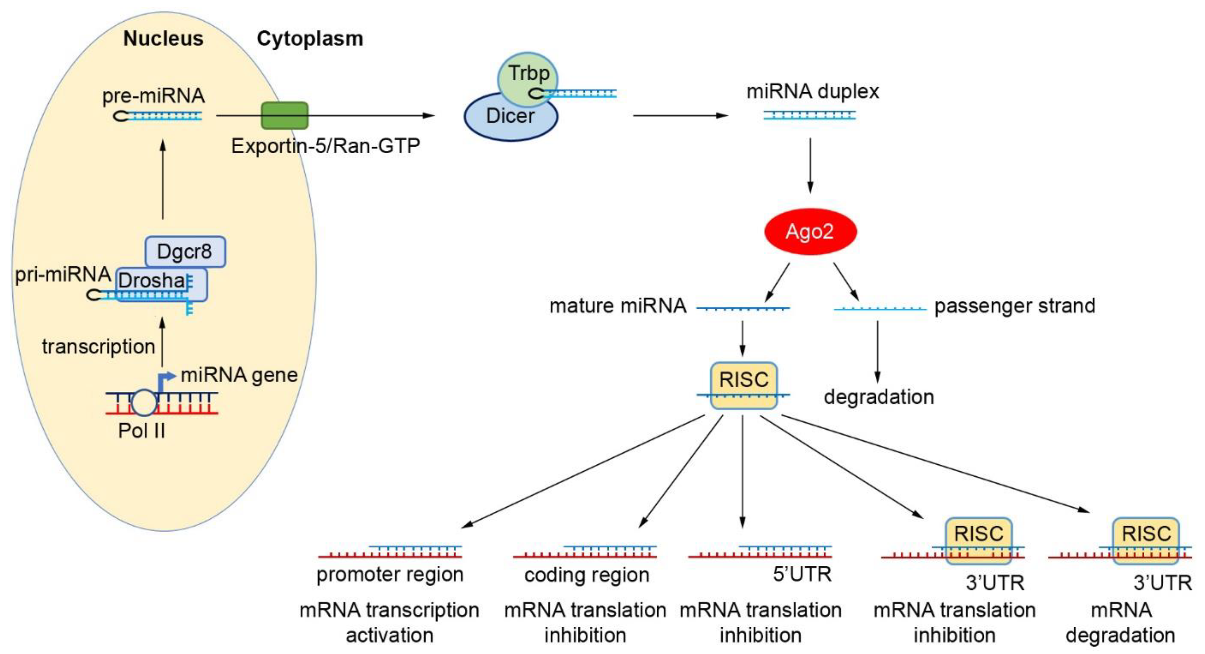
2. Biogenesis and Function of miRNAs

In the search for novel diagnostic and therapeutic targets for cancer, miRNAs have become of significant interest, particularly because of their abundance and potential ease of detection in both tissue and plasma, and therefore they represent potential non-invasive molecular markers for cancer diagnosis and therapeutic response. miRNA biogenesis is a multi-step process that starts in the nucleus (Figure 1) [17,18,19,20]. Here, miRNAs are generally transcribed by RNA Polymerase II into long primary transcripts (pri-miRNA) that are further processed in the nucleus by Drosha/DGCR8 to form an intermediate structure called the pre-miRNA (precursor), made up of 60-70 nucleotides [17,21,22]. The nuclear export factor Exportin-5/Ran-GTP carries the pre-miRNA to the cytoplasm where, following a series of excisions by RNase III endonuclease, Dicer/TRBP and Ago2, a mature 17–25 bp miRNA duplex is generated. A helicase unwinds this miRNA duplex to form a mature single-stranded miRNA, which enters into the RNA-induced silencing complex (RISC). At this point, the complex is directed to target mRNA. It is thought that mature miRNAs regulate gene expression through the binding to the 3′ UTR of target mRNA, thus degrading mRNA or inhibiting translation. In the presence of distinct cofactors and conditions, miRNAs may be capable of activating gene expression directly or indirectly in response to different cell types [10,23,24]. Thus, this process allows the cell to respond rapidly to different cellular conditions due to the reversibility in their post-translational gene regulation. In addition, cells can produce miRNAs via several non-canonical processes. Some miRNA-like species, such as a subclass termed agotrons, are capable of bypassing particular steps of the canonical miRNA biogenesis pathway, escaping both Drosha and Dicer processing [25].

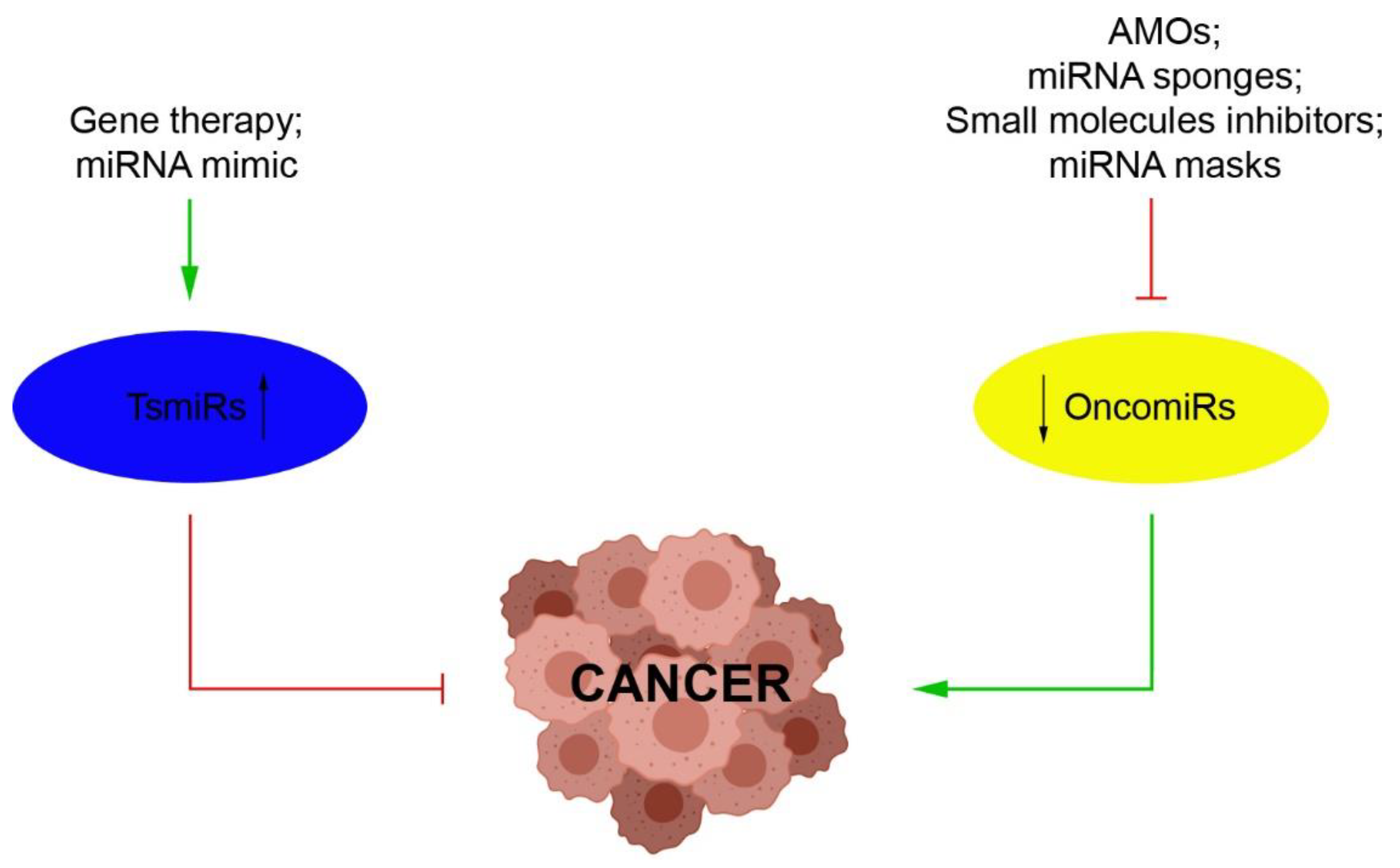
3. miRNAs as Novel Therapeutic Strategies

The link between cancer and miRNAs was first reported almost two decades ago [26,27]. Studies in numerous human cancers have since confirmed that miRNAs are often associated with sites of chromosomal amplification or instability [28]. The aberrant expression of miRNAs has been linked to the stage, progression and metastasis of a wide variety of tumors [28,29]. In fact, miRNAs can function as tumor suppressors (TsmiRs) or tumor promoters (OncomiRs) [30,31]. In addition, miRNAs have also been shown to be involved in cancer stem cells (CSCs) and epithelial–mesenchymal transition (EMT), which are largely responsible for drug resistance and cancer metastasis [1,29]. Hence, targeting miRNAs holds the promise of being an effective option in the management of cancer, with possible therapeutic approaches including achieving “gain” or “loss” of miRNA functions in the tumor cells (Figure 2) [32]. For example, restoring the expression of tumor-suppressive miRNAs may be therapeutically beneficial [32]. In fact, synthetic miRNA mimics have already been used to restore the function of miRNAs in cancer cells [33]. In contrast, several approaches have been applied to achieve the downregulation of oncogenic miRNAs, including the use of miRNA sponges, small molecule inhibitors, anti-miRNA oligonucleotides (AMOs) and miRNA masking [34,35,36]. Overall, restoration or silencing of miRNA function is a promising strategy for cancer treatment. Furthermore, since miRNAs are also involved in radio- and chemo-sensitization of cancer cells, a combination of approaches may be developed for improved therapeutic outcome [37].
However, to date, there are several challenges that need to be overcome before miRNAs can actually be used as therapeutic agents. In fact, RNA oligonucleotides have certain features that complicate drug design and efficacy. Initial hurdles are RNA degradation by nucleases upon addition into biological systems and poor cell membrane penetration. These can potentially be dealt with by undertaking chemical modifications to the oligonucleotides, and using different delivery systems (e.g., liposomes, polymers, exosomes) to make up for their hydrophilic characteristics, negative charge and high molecular weight, which usually block nucleic acids from penetrating the cell membrane [38]. Other challenges include entrapment in the endosome, poor binding affinity for complementary sequences, poor delivery to desired target tissues; activation of innate immune responses and, last but not least, off-target and unwanted toxicities.

4. Dysregulation of miRNAs in Rare Gynecological Cancers

Emerging evidence demonstrates dysregulation of various miRNAs in gynecological cancers, suggesting pathobiological relevance [1]. Here, we highlight the ongoing challenges associated with the early diagnosis and effective treatment of specific RGCs and discuss how miRNAs may further improve the diagnostic, prognostic and therapeutic strategies for these tumor types (Table 1).

4.1. Tubo-Ovarian Cancer

Ovarian cancer is the leading cause of death in women who are diagnosed with gynecological cancer, and overall, it is the fifth most frequent cause of death in women. Ovarian cancer is divided into epithelial and non-epithelial subgroups. Epithelial ovarian cancer is classified according to histopathological appearance and clinical behavior and includes serous (low and high grade), clear cell, mucinous, endometrioid, carcinosarcoma and also fallopian tube and primary peritoneal cancer [39]. Non-epithelial ovarian tumors are relatively rare, but in total, they still account for approximately 10% of all ovarian malignancies. These are challenging from a diagnosis and management point of view, due to their rarity. Non-epithelial tumors include a large variety of different pathological types, including mesenchymal tumors (low- and high-grade endometrioid stromal tumors), mixed epithelial and stromal tumors (adenosarcomas and carcinosarcomas), pure stromal tumors (e.g., fibromas and thecomas), pure sex cord-stromal tumors (e.g., adult granulosa cell tumors and juvenile granulosa cell tumors), mixed-sex cord-stromal tumors (e.g., Sertoli–Leydig cell tumors) and germ cell tumors [40].

4.1.1. miRNAs in the Rare Types of Epithelial Ovarian Cancers

Epithelial ovarian cancer is the most common group of ovarian cancer, however, it also includes the rare clear-cell, mucinous and low-grade serous carcinoma types [41]. High-grade serous carcinomas (which are not rare) almost always arise from the tube, and there is some evidence that low-grade serous carcinomas also do. Most of these types present at a low stage (by contrast with high-grade serous carcinoma) but they do on occasion present at a high stage. Especially when the diagnosis is at a late stage, although the initial treatment might possibly lead to a complete response, relapse is common with minimal response to chemotherapy [42].
Ovarian clear cell cancer (OCCC) diagnosed at an earlier stage tends to have a good prognosis as surgery is often curative. In advanced stages, however, it is more likely to be resistant to chemotherapy and has a worse prognosis. Due to the relative rarity of OCCC, there have been limited efforts in improving outcomes [4]. Similarities in molecular pathways exist among OCCC and clear-cell carcinoma of the kidney [43], where inhibition of angiogenesis, growth-factor signaling and mTOR pathways, might improve survival. Treatments that are used in the case of clear-cell carcinoma of the kidney, such as multikinase inhibitors (sunitinib, axitinib, sorafenib and pazopanib), temsirolimus, bevacizumab, and everolimus might have anti-tumor activity in OCCC, although preliminary clinical data from studies focusing only on OCCC are limited [44].
Loss of ARID1A and activation of PIK3CA are the most common somatic genetic alterations in OCCC (66.7% and 50% respectively), followed by mutations in PPP2R1A (18.8%) and KRAS (16.7%) [45]. Inhibition of the methyltransferase EZH2 and the administration of dasatinib and/or the HDAC6 inhibitor ACY1215 may represent novel treatment strategies for ARID1A mutated OCCC [46,47,48]. It has been shown that ARID1A-mutated OCCC cells have a specific sensitivity to small molecule inhibitors of the bromodomain and extra terminal domain (BET) family of proteins, to which BRD2 belongs, and which in turn causes a reduction in the expression of multiple SWI/SNF members including ARID1B [49].
EGFR inhibitors might be effective therapeutic agents, given that EGFR expression is detected in up to 60% of OCCCs [50]. In addition, high expression of mTOR has been reported in both early- and advanced-stage OCCC, with mTOR inhibitors being promising agents for treatment, especially in recurrent OCCC with cisplatin resistance [51].
Primary mucinous epithelial ovarian cancer (mEOC) accounts for less than 5% of epithelial ovarian cancers, with a decreasing incidence due to the fact that many cases that were previously diagnosed as primary mEOCs were actually metastases from other organs, mostly from the gastrointestinal tract. This highlights the importance of clinico-pathological review, because the pathology does not necessarily distinguish between primary and metastatic mucinous carcinomas, particularly if they have an upper GI phenotype. While it is recommended to treat mEOC with adjuvant carboplatin and paclitaxel, in-depth molecular characterization of mEOC suggests that trastuzumab (Herceptin) and HER2-targeted therapies might be an effective treatment as HER2 is amplified or expressed in 19% [52] or 18.2% of these tumors [4], respectively.
Therefore, in order to improve prognosis through the development of a more specific treatment, thereby improving prognosis improved insight into the molecular characteristics of the different epithelial ovarian cancer subgroups. Several miRNAs, such as miR-509-3-5p, miR-509-3p, miR-509-5p, and miR-510, are differentially expressed in OCCC, high-grade serous ovarian carcinoma (HGSC) and ovarian surface epithelium (OSE), suggesting a carcinogenic role [53]. Furthermore, miR-424 has the capacity to suppress cell invasion and EMT in OCCC through downregulation of DCLK1, thus suggesting potential therapeutic targets [54]. Yanaihara et al. found higher levels of miR-9, miR-34a and miR-126 in OCCC, compared to HGSC [55]. Moreover, miR-9 overexpression may affect pathogenesis in OCCC by targeting E-cadherin and inducing EMT. In addition, it has been shown that miR-449 is under-expressed in OCCC [56] and that miR-29b signaling is involved in the sensitivity to chemotherapy in these cases [57]. Finally, Agostini et al. found that miR-192, miR-194, and miR-215 are upregulated in mEOC, suggesting that the miR192/215 family miRNAs may exert oncogenic functions in this tumor type [58].

4.1.2. miRNAs in Nonepithelial Ovarian Cancers

Nonepithelial ovarian cancers including malignant germ-cell tumors and sex-cord stromal tumors are very rare and account for only 6% of all ovarian malignancies [59,60,61,62].
Malignant germ-cell tumors occur more commonly among women below 20 years of age and are often treated as their testicular counterparts. These tumors can be histologically classified as immature teratoma, dysgerminoma, yolk sac tumor, embryonal carcinoma, choriocarcinoma, mixed germ-cell tumor, malignant struma ovarii, teratoma with malignant transformation and gonadoblastoma [63]. Nowadays, with the use of platinum-based regimens, the five-year overall survival (OS) is estimated to be over 90% for early-stage tumors and above 75% for advanced disease [60]. The role of adjuvant chemotherapy is well established, with regimens such as BEP (bleomycin, etoposide, and cisplatin) being in routine use. However, in the case of relapsed ovarian germ-cell tumors, to date, there are no trials to suggest the benefits of a second-line therapy or the utility of high-dose chemotherapy to be followed by autologous stem cell transplant, as in testicular germ-cell tumors. Current practices include the use of TIP (paclitaxel, ifosfamide, and cisplatin), with extrapolation from treatment used for testicular germ-cell tumors, and even using more complex regimens containing combinations of cisplatin, methotrexate, bleomycin and vincristine, alternating with cyclophosphamide, actinomycin D, and etoposide [59]. Furthermore, targeted therapies that have been investigated consist of tyrosine kinase inhibitors (TKIs) (i.e., imatinib and sunitinib), trastuzumab (anti-HER2 monoclonal antibody) and antiangiogenic agents such as thalidomide and bevacizumab [64].
Granulosa cell tumors are a common type of malignant sex-cord stromal tumor and constitute about 5% of malignant ovarian tumors. There are two main distinct types: adult-type and juvenile granulosa cell tumors, which are different tumor types, but most data refer to adult-type tumors. Sertoli-Leydig cell tumors, steroid cell tumors, gynandroblastomas, and sex cord tumors with annular tubules are infrequently detected. Histologically, granulosa cell tumors are composed of granulosa cells, which secrete progesterone and estrogen [64]. Testing for the C134W FOXL2 mutation is helpful in the diagnosis of adult-type tumors where the morphological appearances are not characteristic [65]. Granulosa cell tumors tend to have a slow progression and late recurrence.
In the case of women with advanced-stage or recurrent granulosa cell tumor, there is limited effectiveness of traditional chemotherapy [66]. The ongoing GOG264 trial (NCT01042522) is currently comparing the efficacy of carboplatin and paclitaxel versus (BEP) in advanced or recurrent sex cord-ovarian stromal cell tumors. Furthermore, targeted therapies such as vascular endothelial growth factor (VEGF) inhibitors, TKIs, and hormonal treatment have been investigated as therapeutic options for granulosa cell tumors [4].
Molecular pathogenesis of these tumor types is starting to be unraveled, especially in relation to the role of FOXL2 [67]. Chang et al. characterized miRNA expression profiles of some germ-cell tumors and sex cord-stromal tumors using small RNA sequencing [68]. Higher expression of miR-302c-3p, miR-372-3p and miR-373-3p, and lower expression of miR-199a-5p, miR-202-3p and miR-214-5p have been observed in malignant germ-cell tumors when compared to benign germ-cell tumors or sex-cord stromal tumors. In sex-cord stromal tumors, miR-513c-5p and miR-202c-3p were more abundant than in benign germ-cell tumors. Additionally, expression of Beclin 1 (BECN1), which is a target of miR-199a-5p, was shown to be higher in malignant germ-cell tumors than benign germ-cell tumors, which corresponds with their lower expression of miR-199a-5p.
Poynter et al. analyzed molecular signatures in dysgerminoma and yolk sac tumor, compared to adjacent tissue samples [69]. Differences in miRNA expression were observed, with miR-122, miR-302a, miR-302d, miR-371-5p and miR-373 showing elevated expression in one or more histologic subtypes. Correlations were also identified across six major hubs with higher expression in yolk sac tumor (miR-302b, miR-302a, miR-122 and miR 126; LEFTY1 and LEFTY2) compared with other germ-cell tumors. Cheng et al. validated six miRNAs (miR-29c-3p, miR-138-5p, miR-184, miR-204-5p, miR-328-3p and miR-501-3p) as novel markers for subtype classification in ovarian granulosa cell tumors with low levels of miR-138-5p correlating with early tumor stage, while low levels of miR-184 were linked with tumor recurrence in early-stage adult-type granulosa cell tumor patients [70].
A comparative study of miRNA regulation on FOXL2 between adult-type and juvenile-type granulosa cell tumors showed that reduction of the miR-17 family indirectly increased FOXL2 mRNA expression [71]. Through miRNA profiling, juvenile- and adult-derived cell-lines have been shown to be biologically distinct, but this still needs to be addressed in vivo. Different studies by Tu et al. showed that miR-10a promotes granulosa cell tumor development via the PTEN-AKT/Wnt regulatory axis, while miR-126 is a tumor suppressor of granulosa cell tumor development via the regulation of EGFL7 [72,73].
Mutations in the RNase IIIb domain of DICER1 are a common feature of nonepithelial ovarian tumors. These mutations lead to impaired miRNA biogenesis and thus disrupt miRNA levels. Mutations in both copies of DICER1 result in the so- called DICER1 syndrome, and ovarian Sertoli-Leydig tumors are highly characteristic of this syndrome. Sertoli–Leydig tumors contain DICER1 mutations in a high proportion of cases [74]. Hence, targeted therapies based on unique molecular pathways may be promising for better cure rates while reducing serious side effects.

4.2. Uterine Cancer

Uterine cancer can arise from both the endometrium and the myometrium. Uterine sarcomas, which arise from the middle muscular layer, are rare but are often aggressive and therefore need prompt diagnosis and treatment. Endometrial carcinoma is the most common type of gynecological cancer in women in developed countries, and it has been traditionally classified into two histological types. Type I tumors make-up 80–90% of endometrial cancers and are typically characterized by a low-grade endometrioid histology, on a background of atypical hyperplasia. These are characterized by estrogen and progesterone receptor positivity and, in most cases, have a favorable prognosis. Type II cancer occurs in 10–20% of endometrial cancers and is associated with typically high-grade non-endometrioid histology (serous endometrial cancer; clear cell endometrial cancer; uterine carcinosarcoma, UCS), arising in atrophic endometria. This is usually estrogen-independent and has a higher risk for metastases and less favorable prognosis [75].

4.2.1. miRNAs in Uterine Sarcomas

Uterine sarcomas are aggressive mesenchymal tumors, with an incidence of 2–3% of all uterine malignancies [76]. There is a lack of consensus on risk factors and optimal treatment due to their rarity and diversity in their histopathology, thus generally leading to poor outcomes. Leiomyosarcoma (LMS), undifferentiated sarcoma and endometrial stromal sarcoma (ESS) are the predominant uterine sarcomas, with even rarer types such as rhabdomyosarcoma (including embryonal type in the cervix), and adenosarcomas [4,77].
ESS represents the second most common category of mesenchymal uterine tumors, in spite of accounting for less than 1% of all uterine tumors [76]. Endometrial sarcomas are further classified into low-grade ESS and high-grade ESS. Low- and high-grade ESS have been found to differ on a molecular level. Low-grade ESS is also a much more hormone-responsive and indolent tumor, whereas high-grade ESS is a more aggressive tumor [76]. This category was re-introduced into the WHO classification in 2014, and it is recognized that there is more than one molecular subtype of this tumor based on translocations [78,79]. Low-grade ESS are treated mainly by hysterectomy and bilateral salpingo-oophorectomy, but may include adjuvant radiation, hormonal treatment or aromatase inhibitors [80].
Patients with high-grade ESS have a higher mortality due to earlier and more frequent recurrences (often <1 year). Advanced or recurrent tumors must be treated aggressively with a combination of chemotherapyand radiation [81]. The role of maintenance therapy with treatments such as cabozantinib in high-grade uterine sarcoma are being investigated [82].
LMS is the most common uterine sarcoma. It occurs in women over 40 years of age and has a 50% 5-year survival rate when confined to the uterus. It appears that adjuvant chemotherapy or radiotherapy does not incur any added benefit [83]. Signs and symptoms of LMS resemble those of leiomyoma, which is more common, and hence the preoperative distinction between the two tumors may be difficult. In postmenopausal women who are not using hormonal replacement therapy, malignancy can be suspected by the tumor growth, although it is rare for a leiomyosarcoma to present as a rapidly growing tumor [76].
Treatment of leiomyosarcomas involves total abdominal hysterectomy with debulking of the tumor in case of local metastasis. Doxorubicin, docetaxel/gemcitabine, and ifosfamide are all possible treatment options for advanced or recurrent disease. Some tumors may respond to hormonal treatment [76]. Targeted therapies such as trabectedin and pazopanib have been investigated as treatment in advanced stage or metastatic leiomyosarcoma, with some degree of efficacy in disease control [76,84]. When compared to usual type leiomyomas, mutations in the cell cycle genes are more common in leiomyosarcoma samples. Hence, cell cycle-related mechanisms could be attractive targets for treatment for these rare tumors [85].
Undifferentiated sarcoma has no identifiable molecular marker and is essentially a diagnosis of exclusion. This is an aggressive cancer, and treatment options are deduced from experience with other high-grade soft tissue sarcomas. Clinical trials for the targeted therapies in soft tissue sarcomas are enrolling patients with uterine sarcoma [4]. While this enables the involvement of more patients into the clinical trials, it may hinder the specific analysis of these subtypes [4].
There is a lack of knowledge about the roles and molecular mechanisms of miRNAs in the physiological and pathological processes and about any correlation with prognosis and their potential to predict treatment outcome in patients with uterine sarcomas. Gonzalez Dos Anjos et al. analyzed miRNA expression profiles linked with the cancer-specific survival (CSS) of patients with uterine sarcomas [86]. In particular, in leiomyosarcoma, an association of lower CSS was found with the downregulation of miR-10a-5p and miR-125a-5p, and the upregulation of miR-34c-5p and miR-196a-5p. In endometrial stromal sarcomas, the down-regulation of miR-23-3p, let-7b-5p and let-7f-5p and the upregulation of miR-372-3p and miR-373-3p were associated with lower CSS. Higher survival rates were linked only to miR-138-5p upregulation. Patients with tumor metastasis and relapse had higher expression of miR-210-3p, miR-301a-3p and miR-335-5p. Finally, expression of miR-138-5p, miR-146b-5p, and miR-218-5p was linked with higher disease-free survival in treated patients. This suggests that these miRNAs represent potential prediction biomarkers for treatment response and prognosis in patients with such tumors.
Evaluation of the expression of 88 miRNAs known to be involved in LMS and ESS showed downregulation of miR-1, miR-23b, let-7c and let-7f in ESS in relation to the benign tissue. However, there were no statistically significant changes in miRNA expression levels between LMS tumors and controls [87]. In a molecular study that was conducted to compare the miRNA profiles of LMS and ESS and to compare the miRNA signatures of primary LMS, primary ESS and metastatic uterine LMS, 94 miRNAs were significantly differentially expressed in LMS and ESS [88]. Out of these miRNAs, 18 were overexpressed in LMS and 76 were overexpressed in ESS. In primary and metastatic LMS, 49 miRNAs were differentially expressed, with 45 being overexpressed in primary LMS and 4 overexpressed in metastases. These differing miRNA profiles in primary and metastatic LMS might help to improve the understanding of the progression of this malignancy. In LMS cells, five miRNAs exhibited an overexpression (miR-129-5p, miR-141-3p, miR-148a-3p, miR-202-3p and miR-203a-3p), and eight were downregulated (miR-1-3p, miR-21-5p, miR-27b-3p, miR-125b-1-3p, miR-140-5p, miR-152-3p, miR-485-5p and miR-495-3p). Of these, only three miRNAs showed significant expression in LMS (miR-1-3p, miR-202-3p and miR-7-5p). In addition, let-7 was also shown to be a potential prognostic biomarker in LMS [89,90].
In 2014, Guled et al. analyzed miRNA profiling on a series of LMS and undifferentiated pleomorphic sarcoma (UPS) samples, and in total, 38 and 46 miRNAs classified UPS and LMS samples, respectively, were compared to control samples. There was differential expression of miR-22, miR-126, miR-199a-3p, miR-199b-5p and miR-320a. In particular, miR-320a and miR-199-5p were highly expressed in LMS and UPS, respectively [91].
Finally, Stope et al. demonstrated that miR-1 is suppressed in LMS, compared to adjacent healthy tissue [92]. Moreover, in vitro studies suggested that miR-1 may be a pivotal tumor suppressor and represent a promising biomarker of diagnosis in LMS therapy. Overall, changes in miRNA levels are potentially important in terms of genomic copy number changes at miRNA gene loci and mRNA targets of these dysregulated miRNAs, which can have further implications in disease mechanisms.

4.2.2. miRNAs in Uterine Carcinosarcomas

Uterine carcinosarcoma (UCS) is another rare gynecological cancer which accounts for less than 5% of uterine cancers [93]. It is a metaplastic carcinoma that is highly lethal with a 5-year survival rate of 33–39% [94]. Adjuvant treatment in case of metastasis largely includes the use of paclitaxel and carboplatin. To date, there is no trial that has shown an OS benefit from adjuvant radiotherapy or chemotherapy, even though most of these trials included a variety of gynecological sarcomas [95,96,97].
UCS is a biphasic tumor consisting of both mesenchymal (sarcomatous) and epithelial (carcinomatous) components. The mesenchymal component can resemble homologous histologic components commonly found in the uterus, or harbor heterologous components that are not normally native to the uterus, such as chondrosarcomatous or rhabdomyosarcomatous differentiation, and is by definition high-grade. The epithelial component is also high-grade and usually shows serous or endometrioid differentiation [98]. UCS shares mutational features similar to serous uterine carcinoma more frequently than endometrioid histologies, with extensive copy number alterations, and the majority harbor somatic TP53 mutations. However, TP53 mutation was found to be less common in “endometrioid” tumors [99]. UCSs are believed to have a monoclonal origin where, according to the conversion theory, carcinomatous subclones can undergo metaplastic differentiation to transform into sarcomatous cells late in tumorigenesis [100]. This conversion theory is backed by the fact that there is the co-expression of epithelial membrane antigens and cytokeratins in sarcomatous and carcinomatous cells, as well as identical patterns of X chromosome inactivation, concordance of TP53 and KRAS mutations, and similar losses of heterozygosity between sarcomatous and carcinomatous components. Other frequent mutations have been found in PIK3CA, FBXW7, TP53, KRAS, PPP2R1A, and PTEN, similar to serous and endometrioid uterine carcinomas [100].
It has still not been determined how carcinomatous cells specifically undergo metaplastic differentiation. According to the conversion theory, it is believed that EMT allows the sarcomatous component to be derived from the carcinomatous component. EMT is a known process that causes cancer progression, metastasis and therapeutic resistance. The mechanism of EMT is also reversible, where mesenchymal–epithelial transition (MET) can decrease the progression of the tumor. Studies demonstrate that the over-expression of miR-200 in UCS cells induces a robust MET, leading to a decreased growth and aggressiveness of cells both in vitro and in vivo [100,101]. This suggests that advanced miRNA therapeutics using ectopic miR-200 expression may be a promising treatment for patients with UCS. A strong negative association has also been shown between expression of the miRNA-200 family and the levels of their promoter methylation. Therefore, epigenetic regulation of these miRNAs indicates a possible a mechanism for EMT in UCS [100].
Brunetti et al. performed molecular investigations on the expression status and mutations of the genes FHIT, HMGA1/2, MTA1 and LIN28A; the pseudogenes HMGA1P6 and HMGA1P7; and the miRNAs known to influence expression of these same genes in ovarian carcinosarcomas and UCS [98]. Mutations in KRAS, PIK3CA, and TP53 were identified in UCS with a frequency of 6%, 31%, and 75%, respectively. In addition, an inverse correlation between downregulation of miRNAs such as miR16, miR26a, miR30c, miR214, let-7a and let-7d, and overexpression of HMGA1/2, and MTA1, were observed [98].

4.3. miRNAs in Vulvar Tumors

Vulvar carcinoma is also considered rare, accounting for 5% of female genital tract cancers, with the highest incidence in women aged 65 to 75 years. Patients with advanced or recurrent disease have a poor outcomes and increased morbidity [64,102]. Over 85% of cases are squamous cell carcinoma, and risk factors include human papillomavirus (HPV) infection, lichen sclerosus, and, especially in young women, vulvar intraepithelial neoplasia. Other histologic types also occur, including basal cell carcinoma, Bartholin gland carcinoma, extramammary Paget disease, sweat gland adenocarcinoma, adenocarcinoma of intestinal type, germ cell tumors, melanoma (see below) and mesenchymal tumors [63,102].
Treatment often consists of surgical management, which is then followed by radiotherapy with or without chemotherapy. Management of these patients is largely based on experience from the treatment of advanced cervical cancers [4].
Targeted therapies with potential benefit in vulvar carcinoma include TKIs such as erlotinib and cetuximab as EGFR genomic amplification and overexpression have been associated with poor survival in these patients. Erlotinib and combination cetuximab with chemotherapy in patients with recurrent vulvar carcinoma showed substantial response [103,104].
In addition, HER2 expression was also detected in extramammary Paget disease of the vulva. Although Paget disease is believed to have a good prognosis, recurrence is frequent. Hence, treatments targeted to HER2 may also benefit recurrent Paget disease of the vulva with HER2 overexpression [105].
High-risk HPV infection is related to vulvar intraepithelial neoplasia, basaloid, and warty carcinomas. In a retrospective study, p16 immunohistochemistry was positive in 166 of 550 tumors (30.2%) and p53 staining in 187 of 597 tumors (31.3%) [106]. Dysregulated cell cycle markers including increased expression of cyclin D1, and cyclin A has also been shown in vulvar carcinomas. These markers are related to poor clinical outcomes. Therefore, targeted agents for these molecular pathways, including a therapeutic HPV vaccine, are potential treatments for vulvar carcinoma [64].
Currently, there is limited information regarding the expression of miRNAs in vulvar carcinoma. de Melo Maia et al. characterized microRNA profile in vulvar tumors, correlating it with clinical and histopathologic data, and the occurrence of HPV infection [107]. There were 25 differentially expressed miRNAs between HPV-negative and HPV-positive groups, and 79 differentially expressed in tumors when compared to normal samples. Moreover, downregulation of both miR-19-b1-5p and miR-223-5p correlated with the presence of lymph node metastasis. Furthermore, downregulation of miR-19-b1-5p and miR-100-3p were associated with vascular invasion. In addition, overexpression of miR-133a and miR-519b were linked with advanced FIGO stage.
Yang and Wu investigated the mechanism of action of miRNAs in vulvar squamous cell carcinoma (VSCC) [108]. Altered expression of 157 miRNAs was detected in this type of carcinoma, with upregulation of miR-182-5p, miR-183-5p and miR-590-5p, and downregulation of miR-103a-3p, miR-107 and miR-603. There was a positive relationship between lymph node metastasis and miR-590-5p expression. Finally, upregulation of miR-590-5p may promote cellular malignant behavior via the target gene TGFβR II. In another study, there was an increased level of expression of miRNA-4712-5p in VSCC, promoting proliferation and invasion, by affecting PTEN and its downstream p-GSK3β, p-AKT, and cyclin D1 signaling pathways [109]. It has also been shown that miR-3147 serves as an oncomiR in VSCC via suppression of Smad4 [110]. These findings suggest future clinical applications related to miRNA deregulation in vulvar carcinoma.

4.4. miRNAs in Melanoma of the Female Genital Tract

Malignant melanoma, which overall accounts for around 1% of all cancers, is a malignant neoplasm of the skin and mucous membranes. The mucosal malignant melanomas, which are rarer and account for 0.03% of all cancers, may occur in various sites including the conjunctiva, oral cavity, esophagus, anus, and even the gynecological tract [111]. In fact, in women, 3% to 7% of all cases of mucosal malignant melanoma develop within the genital tract, mainly in the vulva and vagina. However, primary malignant melanoma of the uterine cervix is even rarer, with a five times lower incidence than primary vaginal or vulva cases of malignant melanoma [112]. Radical hysterectomy with regional lymphadenectomy and/or concurrent chemoradiation therapies are generally recommended, but the prognosis is usually poor and unpredictable. This is because there has been no absolute agreement on comprehensive treatment to date, due to its rarity and difficulty in diagnosis.
Recently, DiVincenzo et al. investigated miRNA expression profiles in melanomas originating from gynecological sites, such as cervix, vulva and vagina [113]. When comparing miRNA expression in vaginal melanoma to normal adjacent vaginal mucosal tissue, 25 differentially expressed miRNAs, were found. Moreover, 45 differentially expressed miRNAs were identified between vulvar melanoma and primary cutaneous melanoma, among which three demonstrated a decrease in expression in vulvar melanoma (miR-200a-3p, miR-200b-3p and miR-494-3p), and 44 demonstrated an increase in expression (including miR-17-5p, miR-146a-5p, and miR19b-3p). Among these differentially expressed miRNAs, both miR-17-5p and miR-146a-5p have been experimentally validated as direct or indirect regulators of PD-L1 expression in melanoma [114,115]. Furthermore, pathway analysis for differentially expressed miRNAs in vulval and vaginal melanoma has shown significant enrichment of 30 and 35 pathways, respectively, each including TGF-β signaling. In these cases, 57 genes in the pathway are validated targets of 13 differentially expressed miRNAs in vaginal melanoma, and 59 genes in the pathway are validated targets of 17 differentially expressed miRNAs in vulvar melanoma. These results indicate that miRNAs have an important role as potential regulators of gene expression in vaginal and vulvar melanomas, thus contributing to tumor progression.

4.5. miRNAs in Gestational Trophoblastic Disease

Gestational trophoblastic disease (GTD), which has an incidence of 2.0 per 1000 pregnancies refers to abnormal trophoblastic proliferation leading to a broad spectrum of lesions ranging from the benign, to premalignant, hydatidiform mole, through to gestational trophoblastic neoplasia (GTN), which encompasses the aggressive invasive mole, choriocarcinoma, placental site trophoblastic tumor and epithelioid trophoblastic tumor [78,116,117]. GTN is also referred to as persistent trophoblastic neoplasia (PTN) because it may arise after a normal term or preterm pregnancy, a molar pregnancy, abortion, or even an ectopic pregnancy. Although patients with GTN generally show a good response with more than 90% cure rate following chemotherapy, around 4% of cases would succumb to the disease [118].
The exact molecular mechanisms of the etiopathogenesis of GTD are still unclear. Human chorionic gonadotropin (HCG) is associated with excessive trophoblastic proliferation and can act as an angiogenic factor during implantation of molar pregnancy [119]. Thus, high HCG concentrations may increase the risk of persistent GTN. Hence, the efficacy of antiangiogenic agents should be explored for GTN.
Although more is now known regarding placenta-associated miRNAs, there is a lack of information regarding their role in the pathogenesis and progression of GTD. In complete hydatidiform mole, there is dysregulation of miR-517a, miR-517b, miR-518b, and miR-519a [120]. In cells derived from choriocarcinoma, miR-371a-5p and miR-518a-3p regulated different pathways related to tumorigenesis and metastasis [121]. These results may offer new clues to the proliferation and metastasis of GTD and may even provide possible diagnostic biomarkers for GTN.
It has been shown that miR-181b-5p, miR-181d-5p and miR-371a-5p are the most significantly altered miRNAs which are associated with progression to GTN [122]. Finally, other studies showed that miR-21 is involved in proliferation, migration, and invasion of choriocarcinoma cells, while miR-34, miR-196b and miR-199b may be tumor suppressors in choriocarcinoma [123,124,125,126].

5. Circulating miRNAs as Potential Biomarkers

To date, miRNAs have been detected in body fluids such as plasma/serum, saliva, cerebrospinal fluid, breast milk, pleural effusion, ascites, urine and vaginal discharge. This presents an opportunity as a non-invasive liquid biopsy approach for the diagnosis of a wide range of cancers [127,128,129]. Thus, miRNAs present in these fluids may serve as biomarkers offering easy and rapid non-invasive tests [130,131,132,133]. Additionally, extracellular miRNAs can be delivered to target cells by binding to proteins, such as argonautes [134] or via vesicles, such as exosomes, acting as endocrine, autocrine, and/or paracrine regulators and modulators of cellular activities [135]. This suggests that miRNAs may have hormone-like activities. Therefore, extracellular and circulating miRNAs can serve as biomarkers for diseases, as well as a means of intercellular communication.
Zhang et al. demonstrated through high-throughput sequencing, that plasma exosomes from women with ovarian cancer and healthy controls differently expressed miRNAs [136]: 31 were found to be downregulated and 34 upregulated. miR-99b-5p, miR-122-5p and miR-185-5p were significantly decreased, and miR-93-5p, miR-106a-5p and let-7d-5p expression levels were significantly increased, in patients with ovarian cancer compared with healthy women. Another study where circulating miRNA profiling was carried out in plasma samples of ovarian cancer patients, a variety of differentially expressed miRNAs were identified as possible biomarkers for the diagnosis, e.g., miR-19b-3p, miR-26b-5p, miR-125a-3p, miR-144-3p, miR-337-5p and miR-500a-5p [137]. However, to date, there are few studies on potential miRNAs as biomarkers for the diagnosis and prognosis of patients with RGCs (Table 2).
In 2014, Chao et al. analyzed the sera of patients with clear cell carcinoma and found that, in a set of 11 pairs of pre- and postoperative sera, the levels of four miRNAs (miR-130a, miR-138, miR-187 and miR-202) were higher in the sera of preoperative patients [138]. In addition, miR-130a remained consistent during the different time points in seven of the 10 patients during clinical follow-up. This suggests that miR-130a may be a useful serum biomarker for detecting the recurrence of OCCC.
Murray et al. showed that there were elevated levels of all eight main members of the miR-371∼373 and miR-302 clusters in the serum of a four-year-old child at diagnosis of yolk sac tumor [139]. Moreover, miRNA levels returned to normal during the clinical follow-up, with kinetics similar to a conventional marker α-fetoprotein. This study indicates that miR-371∼373 and miR-302 clusters could be promising candidate biomarkers for disease monitoring in malignant germ-cell tumors. A serum panel of choriocarcinoma-specific “chromosome-19-microRNA-cluster” (C19MC) microRNAs have been identified and were highly elevated at diagnosis but dropped rapidly upon starting treatment and normalized prior to the start of the second full chemotherapy course [140]. At diagnosis, the same authors also reconfirmed serum elevation of the previously identified marker of malignant germ-cell tumors, miR-371a-3p. Thus, these circulating microRNA markers seem to reflect choriocarcinoma disease activity more accurately than serum hCG, thus having potential in assisting clinical decision-making.
Circulating miRNAs were also investigated as potential biomarkers for leiomyosarcoma (LMS) [141]. The optimal model consisted of two miRNAs (miR-191-5p and miR-1246), with an area under the receiver operating characteristic curve for identifying LMS of 0.97 (95% confidence interval, 0.91–1.00). Seven serum miRNAs, namely miR-191-5p, miR-451a, miR-1246, miR-4430, miR-4485-5p, miR-4635 and miR-6511b-5p, were identified as a promising diagnostic model for LMS.
Finally, there is a significant decrease in plasma levels of miR-520b, -520c-3p and -520f in patients with complete hydatiform mole after evacuation [142]. Interestingly, in GTN patients, these three miRNAs tended to have a similar variation to serum hCG concentration [143].
The combination of multiple circulating miRNAs may be promising biomarkers for the diagnosis of gynecological cancers, including RGCs. However, inconsistent results of different study designs hamper the applicability of these findings as robust biomarkers. Therefore, further studies are required to validate these results.
In addition, 3′-UTR length isoform diversity is another issue in miRNA based gene regulation in cancers. 3′-UTR cis-elements recognized by miRNAs and/or RNA-binding proteins have a significant impact on the fate of mRNAs [144]. Alternative polyadenylation and/or splicing alters the 3′-UTR lengths in normal tissues and in cancer cells [145,146,147,148,149]. These 3′UTR isoforms are generally tissue and cancer-type-specific and hence have been suggested as potential biomarkers with prognostic potential [150,151,152]. Functionally relevant 3′UTR shortening or lengthening events may alter the miRNA binding landscape in cancer transcriptomes. Isoforms with different 3′UTR lengths are likely to be targeted differently by miRNAs, adding an extra level of complexity with implications at the translation step. Currently, there are few studies on ovarian cancers; hence our understanding of the 3′UTR diversity in gynecological cancers is very limited [152,153]. As transcriptome-level complexities are beginning to be investigated in gynecological cancers, a more comprehensive view of miRNA-based regulation is likely in the near future.

6. A Brief Overview on miRNAs and Their Regulated Targets in RGCs

The little evidence to date for miRNA functional targets is largely derived from reporter assays in combination with the cellular effects of modulation of miRNA expression in cell culture. We will proceed with describing miRNAs that may represent novel potential therapeutic targets for RGCs.

6.1. Up-/Down-Regulated miRNAs and Their Roles in RGCs

Several miRNAs that are overexpressed in RGCs have been shown to have oncogenic roles in vitro, as well as defined molecular targets that they regulate (Table 3). miR-9 [55], miR-10a [73], miR-21 [126], miR-590-5p [108], miR-3147 [110] and miR-4712 [109] have been described as “oncomiRs” across multiple mammalian cell types, which is consistent with their role in RGCs. In addition, underexpressed miRNAs in RGCs such as miR-34 [124], miR-126 [72] and miR-196b [125] act as tumor suppressors.

6.1.1. miR-9 Induces EMT by CDH1 Targeting

Depending on the tissue type, miR-9 can act as a tumor suppressor or as an oncomiR. Similar to previous studies describing many other types of cancer [154], Yanaihara et al. [55] observed that there is an increased miR-9 expression in OCCC. In addition, luciferase-based assays have demonstrated direct binding between miR-9 and E-cadherin, which is a tumor suppressor protein encoded by the CDH1 gene. The loss of its expression in association with EMT occurs frequently during tumor metastasis [155]. Moreover, miR-9 knockdown also limits invasion and migration while upregulating E-cadherin expression. This suggests that aberrant miR-9 expression might play an important role in EMT activation in OCCC cells via direct binding to and downregulation of E-cadherin. Therefore, miR-9 upregulation may be involved in OCCC pathogenesis by inducing EMT through E-cadherin modulation. Accordingly, miR-9 may be a promising therapeutic target strategy for OCCC [55].

6.1.2. miR-10a Promotes Tumorigenesis by Regulating PTEN, Akt and Wnt Pathways

In patients with acute myeloid leukemia, miR-10a acts as an oncomiR via its repression of the p53/Rb network [156]. The microRNA-10 family could disturb the normal development of granulosa cells during follicle formation, and there is a strong miR-10a signal in tissues from malignant granulosa cell tumor patients [157]. Moreover, in vitro, forced expression of miR-10a promotes cell proliferation, invasion, migration, ovarian hormone production, and repressed anticancer drug-induced apoptosis. The oncogenic role of miR-10a was also validated in vivo. Interestingly, PTEN, a well-known tumor suppressor, was identified as a direct functional target of miR-10a, and AKT/Wnt as an associated oncogenic pathway of miR-10a in cancerous granulosa cells. These results demonstrate that miR-10a can promote granulosa cell tumor development via regulating PTEN, Akt, and Wnt pathways [73].

6.1.3. miR-21 Targeted PDCD4 and PTEN Genes

miR-21 is one of the most commonly upregulated miRNAs in different types of malignant tumors [158]. Moreover, miR-21 is implicated in various processes involved in carcinogenesis such as proliferation, invasion, and metastasis. Meanwhile, miR-21 also participates in the regulation of multiple signaling pathways such as Nanog/STAT3, PI3K/Akt and PDGF pathways [158]. The regulatory function of miR-21 depends on its target genes such as PTEN and PDCD4 (programmed cell death 4), which are both tumor suppressors [158].
Wang et al. [126] demonstrated the miR-21 could promote proliferation, invasion and migration of choriocarcinoma cells. Furthermore, miR-21 can activate the Akt pathway, negatively regulates PDCD4 and PTEN and targets PDCD4 in choriocarcinoma cells. This suggests that miR-21 is responsible for the aggressive phenotype of gestational trophoblastic disease and may have a potential diagnostic and therapeutic role to play in this condition.

6.1.4. miR-590-5p Promotes Cellular Malignant Behaviors via the Target Gene TGFβRII

It has been reported that miR-590-5p promotes proliferation and invasion in human hepatocellular carcinoma cells via the direct targeting of TGF-βRII, which in turn plays an important role in cell growth and cancer development [159]. Yang and Wu found a positive relationship between miR-590-5p expression and lymph node metastasis [108]. Furthermore, they showed that miR-590-5p plays an oncogenic role in VSCC by promoting cell proliferation and migration through the manipulation of TGFβRII expression. This suggests that miR-590-5p may be a critical therapeutic target in VSCC.

6.1.5. miR-3147 Regulates SMAD4

It has been shown that there is upregulation of miR-3147 in cervical squamous cancer and melanoma [160,161]. In VSCC, the expression of miR-3147 is markedly upregulated and the increased expression of miR-3147 is positively associated with the depth of invasion (4). In addition, miR-3147 regulates SMAD4 by directly binding to its 3′ untranslated region. These results indicate that miR-3147 may have an oncogenic role in VSCC by targeting SMAD4, and miR-3147 may represent a novel potential therapeutic target for VSCC [110].

6.1.6. miR-4712-5p Regulates PTEN and Affects Its Downstream p-AKT, p-GSK3β and Cyclin D1 Signaling Pathways

Yang et al. [109] investigated the role of miR-4712-5p and its regulatory mechanism in VSCC, and found increased levels of miR-4712-5p both in VSCC tissues and the A431 cell line. Moreover, miR-4712-5p overexpression promotes proliferation and invasion of VSCC cells. Luciferase-based assays have also demonstrated direct binding between miR-4712-5p and PTEN. In addition, miR-4712-5p overexpression increased phospho-AKT (p-AKT) and cyclin D1 expression, whilst there was a decrease in PTEN and phospho-GSK3β (p-GSK3β). Therefore, miR-4712-5p can reduce the expression of PTEN, further affecting its downstream p-AKT, p-GSK3β and cyclin D1 signaling pathways, promoting the proliferation and invasion of VSCC.

6.1.7. miR-126 Regulates EGFL7

Epidermal growth factor-like domain-containing protein 7 (EGFL7) has been shown to be a critical oncogene in various types of cancer [162,163,164,165]. Notably, EGFL7 is highly expressed in patients with EOC, and its expression has been correlated with a poor prognosis [166]. In addition, EGFL7 also serves as a potential predictive marker of chemotherapy for cervical cancer [167].
Methylation-associated silencing of miR-126 and its host gene EGFL7 has been demonstrated in pleural mesothelioma [168], suggesting an association between EGFL7 and miR-126. Due to the similarities between granulosa cell tumors and those of mesothelial lineage, Tu et al. [72] showed that miR-126 constrains the tumorigenesis of granulosa cell tumors via directly targeting EGFL7 and suppressing the phosphatidylinositol 3-kinase/ATK (PI3K/AKT) pathway. This suggests that miR-126 may be utilized as a prognostic marker or a therapeutic target for granulosa cell tumor treatment.

6.1.8. miR-34a and miR-196b Are Tumor Suppressors in Choriocarcinoma

These miRNAs include miR-34a and miR-196b, which have been demonstrated to have tumor-suppressor activity in human choriocarcinoma cells [124,125]. The members of the miR-34 family share high sequence homology [169]. Among these, miR-34a is one of the best-known miRNA tumor suppressors and is directly activated by p53 [170,171]. Pang et al. [124] demonstrated that miR-34a suppresses cell proliferation and invasion in choriocarcinoma cells through regulation of the Notch ligand Delta-like one (DLL1). Thus, it is possible that, in the future, miR-34a can be used as a therapeutic target for treating choriocarcinoma.
miR-196b has been shown to function as a tumor suppressor in many different cancer types [172,173,174]. A study conducted by Guo et al. demonstrated that miR-196b suppressed proliferation, migration and invasion of human choriocarcinoma cells by inhibiting its transcriptional target MAP3K1. miR-196b and MAP3K1 may be considered potential targets for the clinical treatment of hydatidiform mole and possibly human choriocarcinoma [125].

7. Conclusions and Future Directions

In summary, multiple lines of evidence suggest that miRNAs are dysregulated in gynecological cancers. The dysregulation patterns, which need to be further confirmed in larger cohorts, could prove to be useful in much-needed applications for diagnosis and prognosis, as well as for therapy prediction. Based on these available and emerging data, miRNAs could have impact in future therapeutic strategies for carcinomas of cervical, endometrial, vaginal, vulval and ovarian origin.
To date, despite improvements in understanding the mechanisms and efficiency of miRNA in therapeutics, there are still particular obstacles to be overcome in order to achieve maximum efficiency. These challenges include: targeted delivery, specificity, stability, immune activation and toxicity in vivo. Once these issues are solved, miRNA therapy will have a major role to play in personalized medicine for various cancers, including RGCs.
GYNOCARE (a European Network for Gynaecological Rare Cancer research: from Concept to Cure) aims to make connections between research (e.g., international, basic, and clinical trials) on RGCs, and the pharmaceutical sector (e.g., focused on innovative, targeted therapies). To bridge the gap between the unmet needs of women afflicted by RGCs and the recent medical and technological advances, both clinicians and their patients need to have good access to current information on possible participation in clinical trials, as well as relevant education and support. For instance, providing a centralized website and medical/coordination assistance may serve as simple steps to accomplish these goals in the clinical setting (e.g., recruitment to research trials, or reinforced adherence to diagnostic and therapeutic management) [5].

Author Contributions

Conceptualization, R.D.F. and J.C.-A.; writing—original draft preparation, R.D.F. and J.C.-A.; writing–review and editing, R.D.F., S.S., F.P., S.A.O., J.J.O., M.P.W., N.T.C., M.S., P.O., A.E.E.-B., N.R., A.G., C.S.H. and J.C.-A.; funding acquisition, J.C.-A. All authors have read and agreed to the published version of the manuscript.

Funding

The publication of this manuscript was funded by the European Cooperation in Science & Technology program (EU COST)–COST Action CA18117: GYNOCARE-“European network for Gynaecological Rare Cancer research: From Concept to Cure”. R.D.F., S.S., S.A.O., J.J.O., M.P.W., N.T.C., M.S., P.O., A.E.E.-B., N.R., A.G., C.S.H. and J.C.-A. are Action management committee members and/or form part of Working Groups 1-3, and J.C.-A. is the Chairperson of this Action.

Institutional Review Board Statement

Not applicable, as no new data on human or animal studies were collected.

Informed Consent Statement

Not applicable, as no new data on human or animal studies were collected.

Data Availability Statement

This review paper does not report any new data.

Conflicts of Interest

The authors declare no conflict of interest.

Abbreviations

BEPbleomycin, etoposide, and cisplatin
CSCCancer Stem Cell
EMTepithelial–mesenchymal transition
ESSendometrial stromal sarcoma
GTDgestational trophoblastic disease
GTNgestational trophoblastic neoplasia
hCGhuman chorionic gonadotropin
HGSChigh-grade serous ovarian carcinoma
HPVhuman papillomavirus
LMSleiomyosarcoma
mEOCmucinous ovarian cancer
miRNA/miRmicro-RNA
OCCCovarian clear cell cancer
PTENphosphatase and tensin homolog deleted on chromosome ten
PTNpersistent trophoblastic neoplasia
RGCrare gynecological cancer
RISCRNA-induced silencing complex
TIPpaclitaxel, ifosfamide, and cisplatin
TKIstyrosine kinase inhibitors
TRBP transactivation response RNA binding protein
TsmiRstumor suppressors miRs
UCSuterine carcinosarcoma
UPSundifferentiated pleomorphic sarcoma
VSCCvulvar squamous cell carcinoma
3′-UTR3′ untranslated region

References

  1. Srivastava, S.K.; Ahmad, A.; Zubair, H.; Miree, O.; Singh, S.; Rocconi, R.P.; Scalici, J.; Singh, A.P. MicroRNAs in gynecological cancers: Small molecules with big implications. Cancer Lett. 2017, 407, 123–138. [Google Scholar] [CrossRef]
  2. Maheshwari, A.; Kumar, N.; Mahantshetty, U. Gynecological cancers: A summary of published Indian data. South Asian J. Cancer 2016, 5, 112–120. [Google Scholar] [CrossRef]
  3. Esserman, L.J.; Thompson, I.M.; Reid, B.; Nelson, P.; Ransohoff, D.F.; Welch, H.G.; Hwang, S.; Berry, D.A.; Kinzler, K.W.; Black, W.C.; et al. Addressing overdiagnosis and overtreatment in cancer: A prescription for change. Lancet Oncol. 2014, 15, e234–e242. [Google Scholar] [CrossRef] [Green Version]
  4. Mandilaras, V.; Karakasis, K.; Clarke, B.; Amit Oza, A.; Lheureux, S. Rare tumors in gynaecological cancers and the lack of therapeutic options and clinical trials. Expert Opin. Orphan Drugs 2017, 5, 71–83. [Google Scholar] [CrossRef]
  5. Di Fiore, R.; Suleiman, S.; Ellul, B.; O’Toole, S.A.; Savona-Ventura, C.; Felix, A.; Napolioni, V.; Conlon, N.T.; Kahramanoglu, I.; Azzopardi, M.J.; et al. GYNOCARE Update: Modern Strategies to Improve Diagnosis and Treatment of Rare Gynecologic Tumors-Current Challenges and Future Directions. Cancers 2021, 13, 493. [Google Scholar] [CrossRef] [PubMed]
  6. Ray-Coquard, I.; Trama, A.; Seckl, M.J.; Fotopoulou, C.; Pautier, P.; Pignata, S.; Kristensen, G.; Mangili, G.; Falconer, H.; Massuger, L.; et al. Rare ovarian tumours: Epidemiology, treatment challenges in and outside a network setting. Eur. J. Surg. Oncol. 2017, 45, 67–74. [Google Scholar] [CrossRef] [PubMed]
  7. Cancer Research UK. Available online: https://www.cancerresearchuk.org (accessed on 20 February 2021).
  8. Rare Cancers Europe. Available online: https://www.rarecancerseurope.org (accessed on 17 February 2021).
  9. Jonas, S.; Izaurralde, E. Towards a molecular understanding of microRNA mediated gene silencing. Nat. Rev. Genet. 2015, 16, 421–433. [Google Scholar] [CrossRef] [PubMed]
  10. O’Brien, J.; Hayder, H.; Zayed, Y.; Peng, C. Overview of MicroRNA Biogenesis, Mechanisms of Actions, and Circulation. Front. Endocrinol. 2018, 9, 402. [Google Scholar] [CrossRef] [Green Version]
  11. Kozomara, A.; Birgaoanu, M.; Griffiths-Jones, S. miRBase: From microRNA sequences to function. Nucleic Acids Res. 2019, 47, D155–D162. [Google Scholar] [CrossRef]
  12. Zhong, X.; Coukos, G.; Zhang, L. miRNAs in human cancer. Methods Mol. Biol. 2012, 822, 295–306. [Google Scholar]
  13. Rajagopal, T.; Talluri, S.; Venkatabalasubramanian, S.; Dunna, N.R. Multifaceted roles of long non-coding RNAs in triple-negative breast cancer: Biology and clinical applications. Biochem. Soc. Trans. 2020, 48, 2791–2810. [Google Scholar] [CrossRef] [PubMed]
  14. Tabatabaeian, H.; Peiling Yang, S.; Tay, Y. Non-Coding RNAs: Uncharted Mediators of Thyroid Cancer Pathogenesis. Cancers 2020, 12, 3264. [Google Scholar] [CrossRef]
  15. Lu, X.; Zhang, Y.; Xie, G.; Ding, Y.; Cong, H.; Xuan, S. Exosomal non coding RNAs: Novel biomarkers with emerging clinical applications in gastric cancer (Review). Mol. Med. Rep. 2020, 22, 4091–4100. [Google Scholar] [CrossRef]
  16. Iorio, M.V.; Visone, R.; Di Leva, G.; Donati, V.; Petrocca, F.; Casalini, P.; Taccioli, C.; Volinia, S.; Liu, C.G.; Alder, H.; et al. MicroRNA signatures in human ovarian cancer. Cancer Res. 2007, 67, 8699–8707. [Google Scholar] [CrossRef] [PubMed] [Green Version]
  17. Miyoshi, K.; Miyoshi, T.; Siomi, H. Many ways to generate microRNA-like small RNAs: Non-canonical pathways for microRNA production. Mol. Genet. Genom. 2010, 284, 95–103. [Google Scholar] [CrossRef] [PubMed]
  18. Kim, V.N. MicroRNA biogenesis: Coordinated cropping and dicing. Nat. Rev. Mol. Cell Biol. 2005, 6, 376–385. [Google Scholar] [CrossRef]
  19. Bartel, D.P. MicroRNAs: Genomics, biogenesis, mechanisms and function. Cell 2004, 116, 281–297. [Google Scholar] [CrossRef] [Green Version]
  20. Abdelfattah, A.M.; Park, C.; Choi, M.Y. Update on non-canonical microRNAs. Biomol. Concepts 2014, 5, 275–287. [Google Scholar] [CrossRef] [PubMed] [Green Version]
  21. Han, J.; Lee, Y.; Yeom, K.H.; Nam, J.W.; Heo, I.; Rhee, J.K.; Sohn, S.Y.; Cho, Y.; Zhang, B.T.; Kim, V.N. Molecular Basis for the Recognition of Primary microRNAs by the Drosha-DGCR8 Complex. Cell 2006, 125, 887–901. [Google Scholar] [CrossRef] [Green Version]
  22. Valinezhad Orang, A.; Safaralizadeh, R.; Kazemzadeh-Bavili, M. Mechanisms of miRNA-Mediated Gene Regulation from Common Downregulation to mRNA-Specific Upregulation. Int. J. Genom. 2014, 2014, 970607. [Google Scholar] [CrossRef] [Green Version]
  23. Yuan, X.; Berg, N.; Lee, J.W.; Le, T.T.; Neudecker, V.; Jing, N.; Eltzschig, H. MicroRNA miR-223 as regulator of innate immunity. J. Leukoc. Biol. 2018, 104, 515–524. [Google Scholar] [CrossRef] [PubMed]
  24. Zhang, Y.; Fan, M.; Zhang, X.; Huang, F.; Wu, K.; Zhang, J.; Liu, J.; Huang, Z.; Luo, H.; Tao, L.; et al. Cellular microRNAs up-regulate transcription via interaction with promoter TATA-box motifs. RNA 2014, 20, 1878–1889. [Google Scholar] [CrossRef] [Green Version]
  25. Daugaard, I.; Hansen, T.B. Biogenesis and Function of Ago- Associated RNAs. Trends Genet. 2017, 33, 208–219. [Google Scholar] [CrossRef] [PubMed]
  26. Calin, G.A.; Dumitru, C.D.; Shimizu, M.; Bichi, R.; Zupo, S.; Noch, E.; Aldler, H.; Rattan, S.; Keating, M.; Rai, K.; et al. Frequent deletions and down-regulation of micro- RNA genes miR15 and miR16 at 13q14 in chronic lymphocytic leukemia. Proc. Natl. Acad. Sci. USA 2002, 99, 15524–15529. [Google Scholar] [CrossRef] [Green Version]
  27. Acunzo, M.; Romano, G.; Wernicke, D.; Croce, C.M. MicroRNA and cancer--a brief overview. Adv. Biol. Regul. 2015, 57, 1–9. [Google Scholar] [CrossRef] [PubMed]
  28. Lu, J.; Getz, G.; Miska, E.A.; Alvarez-Saavedra, E.; Lamb, J.; Peck, D.; Sweet-Cordero, A.; Ebert, B.L.; Mak, R.H.; Ferrando, A.A.; et al. MicroRNA expression profiles classify human cancers. Nature 2005, 435, 834–838. [Google Scholar] [CrossRef] [PubMed]
  29. Shimono, Y.; Zabala, M.; Cho, R.W.; Lobo, N.; Dalerba, P.; Qian, D.; Diehn, M.; Liu, H.; Panula, S.P.; Chiao, E.; et al. Downregulation of miRNA-200c links breast cancer stem cells with normal stem cells. Cell 2009, 138, 592–603. [Google Scholar] [CrossRef] [Green Version]
  30. Calin, G.A.; Croce, C.M. MicroRNA signatures in human cancers. Nat. Rev. Cancer 2006, 6, 857–866. [Google Scholar] [CrossRef]
  31. Zhang, B.; Pan, X.; Cobb, G.P.; Anderson, T.A. microRNAs as oncogenes and tumor suppressors. Dev. Biol. 2007, 302, 1–12. [Google Scholar] [CrossRef] [Green Version]
  32. Bhardwaj, A.; Singh, S.; Singh, A.P. MicroRNA-based Cancer Therapeutics: Big Hope from Small RNAs. Mol. Cell Pharmacol. 2010, 2, 213–219. [Google Scholar]
  33. Tsuda, N.; Ishiyama, S.; Li, Y.; Ioannides, C.G.; Abbruzzese, J.L.; Chang, D.Z. Synthetic microRNA designed to target glioma-associated antigen 1 transcription factor inhibits division and induces late apoptosis in pancreatic tumor cells. Clin. Cancer Res. 2006, 12, 6557–6564. [Google Scholar] [CrossRef] [PubMed] [Green Version]
  34. Lima, J.F.; Cerqueira, L.; Figueiredo, C.; Oliveira, C.; Azevedo, N.F. Anti-miRNA oligonucleotides: A comprehensive guide for design. RNA Biol. 2018, 15, 338–352. [Google Scholar] [CrossRef] [PubMed] [Green Version]
  35. Wen, D.; Danquah, M.; Chaudhary, A.K.; Mahato, R.I. Small molecules targeting microRNA for cancer therapy: Promises and obstacles. J. Control. Release 2015, 219, 237–247. [Google Scholar] [CrossRef] [Green Version]
  36. Gambari, R.; Brognara, E.; Spandidos, D.A.; Fabbri, E. Targeting oncomiRNAs and mimicking tumor suppressor miRNAs: Νew trends in the development of miRNA therapeutic strategies in oncology (Review). Int. J. Oncol. 2016, 49, 5–32. [Google Scholar] [CrossRef] [Green Version]
  37. Mognato, M.; Celotti, L. MicroRNAs Used in Combination with Anti-Cancer Treatments Can Enhance Therapy Efficacy. Mini Rev. Med. Chem. 2015, 15, 1052–1062. [Google Scholar] [CrossRef] [PubMed] [Green Version]
  38. Segal, M.; Slack, F.J. Challenges identifying efficacious miRNA therapeutics for cancer. Expert Opin. Drug Discov. 2020, 15, 987–992. [Google Scholar] [CrossRef] [PubMed]
  39. Yanai, H. Pathology of Epithelial Ovarian Tumors. In Frontiers in Ovarian Cancer Science; Katabuchi, H., Ed.; Springer: Singapore, 2017; pp. 83–113. [Google Scholar]
  40. Fukunaga, M. Pathology of Non-epithelial Ovarian Tumors. In Frontiers in Ovarian Cancer Science; Katabuchi, H., Ed.; Springer: Singapore, 2017; pp. 115–141. [Google Scholar]
  41. Kurman, R.J.; Shih, I. Molecular pathogenesis and extraovarian origin of epithelial ovarian cancer–shifting the paradigm. Hum. Pathol. 2011, 42, 918–931. [Google Scholar] [CrossRef] [PubMed] [Green Version]
  42. Fung-Kee-Fung, M.; Oliver, T.; Elit, L.; Oza, A.; Hirte, H.W.; Bryson, P. Optimal chemotherapy treatment for women with recurrent ovarian cancer. Curr. Oncol. 2007, 14, 195–208. [Google Scholar] [CrossRef] [PubMed] [Green Version]
  43. Zorn, K.K.; Bonome, T.; Gangi, L.; Chandramouli, G.V.; Awtrey, C.S.; Gardner, G.J.; Barrett, J.C.; Boyd, J.; Birrer, M.J. Gene expression profiles of serous, endometrioid, and clear cell subtypes of ovarian and endometrial cancer. Clin. Cancer Res. 2005, 11, 6422–6430. [Google Scholar] [CrossRef] [Green Version]
  44. Yoshida, S.; Furukawa, N.; Haruta, S.; Tanase, Y.; Kanayama, S.; Noguchi, T.; Sakata, M.; Yamada, Y.; Oi, H.; Kobayashi, H. Theoretical model of treatment strategies for clear cell carcinoma of the ovary: Focus on perspectives. Cancer Treat. Rev. 2009, 35, 608–615. [Google Scholar] [CrossRef]
  45. Shibuya, Y.; Tokunaga, H.; Saito, S.; Shimokawa, K.; Katsuoka, F.; Bin, L.; Kojima, K.; Nagasaki, M.; Yamamoto, M.; Yaegashi, N.; et al. Identification of somatic genetic alterations in ovarian clear cell carcinoma with next generation sequencing. Genes Chromosomes Cancer 2018, 57, 51–60. [Google Scholar] [CrossRef]
  46. Bitler, B.G.; Aird, K.M.; Garipov, A.; Li, H.; Amatangelo, M.; Kossenkov, A.V.; Schultz, D.C.; Liu, Q.; Shih, I.; Conejo-Garcia, J.R.; et al. Synthetic lethality by targeting EZH2 methyltransferase activity in ARID1A-mutated cancers. Nat. Med. 2015, 21, 231–238. [Google Scholar] [CrossRef] [PubMed] [Green Version]
  47. Miller, R.E.; Brough, R.; Bajrami, I.; Williamson, C.T.; McDade, S.; Campbell, J.; Kigozi, A.; Rafiq, R.; Pemberton, H.; Natrajan, R.; et al. Synthetic lethal targeting of ARID1A-mutant ovarian clear cell tumors with dasatinib. Mol. Cancer Ther. 2016, 15, 1472–8144. [Google Scholar] [CrossRef] [Green Version]
  48. Bitler, B.G.; Wu, S.; Park, P.H.; Hai, Y.; Aird, K.M.; Wang, Y.; Zhai, Y.; Kossenkov, A.V.; Vara-Ailor, A.; Rauscher, F.J., III; et al. ARID1A-mutated ovarian cancers depend on HDAC6 activity. Nat. Cell Biol. 2017, 19, 962–973. [Google Scholar] [CrossRef] [Green Version]
  49. Berns, K.; Caumanns, J.J.; Hijmans, E.M.; Gennissen, A.M.C.; Severson, T.M.; Evers, B.; Wisman, G.B.A.; Jan Meersma, G.; Lieftink, C.; Beijersbergen, R.L.; et al. ARID1A mutation sensitizes most ovarian clear cell carcinomas to BET inhibitors. Oncogene 2018, 37, 4611–4625. [Google Scholar] [CrossRef] [PubMed]
  50. Fujimura, M.; Hidaka, T.; Saito, S. Selective inhibition of the epidermal growth factor receptor by ZD1839 decreases the growth and invasion of ovarian clear cell adenocarcinoma cells. Clin. Cancer Res. 2002, 8, 2448–2454. [Google Scholar] [PubMed]
  51. Mabuchi, S.; Kawase, C.; Altomare, D.A.; Morishige, K.; Sawada, K.; Hayashi, M.; Tsujimoto, M.; Yamoto, M.; Klein-Szanto, A.J.; Schilder, R.J.; et al. mTOR is a promising therapeutic target both in cisplatin-sensitive and cisplatin-resistant clear cell carcinoma of the ovary. Clin. Cancer Res. 2009, 15, 5404–5413. [Google Scholar] [CrossRef] [Green Version]
  52. Anglesio, M.S.; Kommoss, S.; Tolcher, M.C.; Clarke, B.; Galletta, L.; Porter, H.; Damaraju, S.; Fereday, S.; Winterhoff, B.J.; Kalloger, S.E.; et al. Molecular characterization of mucinous ovarian tumours supports a stratified treatment approach with HER2 targeting in 19% of carcinomas. J. Pathol. 2013, 229, 111–120. [Google Scholar] [CrossRef] [PubMed]
  53. Vilming Elgaaen, B.; Olstad, O.K.; Haug, K.B.F.; Brusletto, B.; Sandvik, L.; Staff, A.C.; Gautvik, K.M.; Davidson, B. Global miRNA expression analysis of serous and clear cell ovarian carcinomas identifies differentially expressed miRNAs including miR-200c-3p as a prognostic marker. BMC Cancer 2014, 14, 80. [Google Scholar] [CrossRef] [Green Version]
  54. Wu, X.; Ruan, Y.; Jiang, H.; Xu, C. MicroRNA-424 inhibits cell migration, invasion, and epithelial mesenchymal transition by downregulating doublecortin-like kinase 1 in ovarian clear cell carcinoma. Int. J. Biochem. Cell Biol. 2017, 85, 66–74. [Google Scholar] [CrossRef] [Green Version]
  55. Yanaihara, N.; Noguchi, Y.; Saito, M.; Takenaka, M.; Takakura, S.; Yamada, K.; Okamoto, A. MicroRNA Gene Expression Signature Driven by miR-9 Overexpression in Ovarian Clear Cell Carcinoma. PLoS ONE 2016, 11, e0162584. [Google Scholar] [CrossRef] [PubMed]
  56. Jang, S.G.; Yoo, C.W.; Park, S.Y.; Kang, S.; Kim, H.K. Low expression of miR-449 in gynecologic clear cell carcinoma. Int. J. Gynecol. Cancer 2014, 24, 1558–1563. [Google Scholar] [CrossRef] [PubMed]
  57. Dai, F.; Zhang, Y.; Chen, Y. Involvement of miR-29b signaling in the sensitivity to chemotherapy in patients with ovarian carcinoma. Hum. Pathol. 2014, 45, 1285–1293. [Google Scholar] [CrossRef] [PubMed]
  58. Agostini, A.; Brunetti, M.; Davidson, B.; Tropé, C.G.; Eriksson, A.G.Z.; Heim, S.; Panagopoulos, I.; Micci, F. The microRNA miR-192/215 family is upregulated in mucinous ovarian carcinomas. Sci. Rep. 2018, 23, 11069. [Google Scholar] [CrossRef] [PubMed]
  59. Bower, M.; Fife, K.; Holden, L.; Paradinas, F.J.; Rustin, G.J.; Newlands, E.S. Chemotherapy for ovarian germ cell tumours. Eur. J. Cancer 1996, 32A, 593–597. [Google Scholar] [CrossRef]
  60. Simone, C.G.; Markham, M.J.; Dizon, D.S. Chemotherapy in ovarian germ cell tumors: A systematic review. Gynecol. Oncol. 2016, 141, 602–607. [Google Scholar] [CrossRef]
  61. Goulvent, T.; Ray-Coquard, I.; Borel, S.; Haddad, V.; Devouassoux-Shisheboran, M.; Vacher-Lavenu, M.C.; Pujade-Laurraine, E.; Savina, A.; Maillet, D.; Gillet, G.; et al. DICER1 and FOXL2 mutations in ovarian sex cord-stromal tumours: A GINECO Group study. Histopathology 2016, 68, 279–285. [Google Scholar] [CrossRef]
  62. Ray-Coquard, I.; Weber, B.; Lotz, J.P.; Tournigand, C.; Provencal, J.; Mayeur, D.; Treilleux, I.; Paraiso, D.; Duvillard, P.; Pujade-Lauraine, E.; et al. Management of rare ovarian cancers: The experience of the French website “Observatory for rare malignant tumours of the ovaries” by the GINECO group: Interim analysis of the first 100 patients. Gynecol. Oncol. 2010, 119, 53–59. [Google Scholar] [CrossRef]
  63. WHO Classification of Tumors Editoral Board. Female Genital Tumors. In WHO Classification of Tumors Series, 5th ed.; IARC: Lyon, France, 2020; Volume 4, Available online: https://publications.iarc.fr/592 (accessed on 20 December 2020).
  64. Manchana, T.; Ittiwut, C.; Mutirangura, A.; Kavanagh, J.J. Targeted therapies for rare gynaecological cancers. Lancet Oncol. 2010, 11, 685–693. [Google Scholar] [CrossRef]
  65. Kommoss, S.; Anglesio, M.S.; Mackenzie, R.; Yang, W.; Senz, J.; Ho, J.; Bell, L.; Lee, S.; Lorette, J.; Huntsman, D.G.; et al. FOXL2 molecular testing in ovarian neoplasms: Diagnostic approach and procedural guidelines. Modern Pathol. 2013, 26, 860–867. [Google Scholar] [CrossRef] [Green Version]
  66. Jamieson, S.; Fuller, P.J. Molecular pathogenesis of granulosa cell tumors of the ovary. Endocr. Rev. 2012, 33, 109–144. [Google Scholar] [CrossRef]
  67. Weis-Banke, S.E.; Lerdrup, M.; Kleine-Kohlbrecher, D.; Mohammad, F.; Sidoli, S.; Jensen, O.N.; Yanase, T.; Nakamura, T.; Iwase, A.; Stylianou, A.; et al. Mutant FOXL2C134W Hijacks SMAD4 and SMAD2/3 to Drive Adult Granulosa Cell Tumors. Cancer Res. 2020, 80, 3466–3479. [Google Scholar] [CrossRef]
  68. Chang, R.K.; Li, X.; Mu, N.; Hrydziuszko, O.; Garcia-Majano, B.; Larsson, C.; Lui, W.O. MicroRNA expression profiles in non-epithelial ovarian tumors. Int. J. Oncol. 2018, 52, 55–66. [Google Scholar] [CrossRef] [PubMed]
  69. Poynter, J.N.; Bestrashniy, J.R.; Silverstein, K.A.; Hooten, A.J.; Lees, C.; Ross, J.A.; Tolar, J. Cross platform analysis of methylation, miRNA and stem cell gene expression data in germ cell tumors highlights characteristic differences by tumor histology. BMC Cancer 2015, 15, 769. [Google Scholar] [CrossRef] [Green Version]
  70. Cheng, W.T.; Rosario, R.; Muthukaruppan, A.; Wilson, M.K.; Payne, K.; Fong, P.C.; Shelling, A.N.; Blenkiron, C. MicroRNA profiling of ovarian granulosa cell tumours reveals novel diagnostic and prognostic markers. Clin. Epigenetics 2017, 9, 72. [Google Scholar] [CrossRef] [PubMed] [Green Version]
  71. Rosario, R.; Blenkiron, C.; Shelling, A.N. Comparative study of microRNA regulation on FOXL2 between adult-type and juvenile-type granulosa cell tumours in vitro. Gynecol. Oncol. 2013, 129, 209–215. [Google Scholar] [CrossRef] [PubMed]
  72. Tu, J.; Cheung, H.H.; Lu, G.; Chan, C.L.K.; Chen, Z.; Chan, W.Y. microRNA-126 Is a Tumor Suppressor of Granulosa Cell Tumor Mediated by Its Host Gene EGFL7. Front. Oncol. 2019, 9, 486. [Google Scholar] [CrossRef]
  73. Tu, J.; Cheung, H.H.; Lu, G.; Chen, Z.; Chan, W.Y. MicroRNA-10a promotes granulosa cells tumor development via PTEN-AKT/Wnt regulatory axis. Cell Death Dis. 2018, 9, 1076. [Google Scholar] [CrossRef] [PubMed]
  74. De Kock, L.; Terzic, T.; McCluggage, W.G.; Stewart, C.J.R.; Shaw, P.; Foulkes, W.D.; Clarke, B.A. DICER1 Mutations Are Consistently Present in Moderately and Poorly Differentiated Sertoli-Leydig Cell Tumors. Am. J. Surg. Pathol. 2017, 41, 1178–1187. [Google Scholar] [CrossRef]
  75. Huvila, J.; Pors, J.; Thompson, E.F.; Gilks, C.B. Endometrial carcinoma: Molecular subtypes, precursors and the role of pathology in early diagnosis. J. Pathol. 2021, 253, 355–365. [Google Scholar] [CrossRef]
  76. Mbatani, N.; Olawaiye, A.; Prat, J. Uterine sarcomas. Int. J. Gynecol. Obstet. 2018, 143, 51–58. [Google Scholar] [CrossRef]
  77. Ferrandina, G.; Aristei, C.; Biondetti, P.R.; Cananzi, F.C.M.; Casali, P.; Ciccarone, F.; Colombo, N.; Comandone, A.; Corvo’, R.; De Iaco, P.; et al. Italian consensus conference on management of uterine sarcomas on behalf of S.I.G.O. (Societa’ italiana di Ginecologia E Ostetricia). Eur. J. Cancer 2020, 139, 149–168. [Google Scholar] [CrossRef] [PubMed]
  78. Kurman, R.J.; Carcangiu, M.L.; Herrington, C.S.; Young, R.H. WHO Classification of Tumors of Female Reproductive Organs; IARC: Lyon, France, 2014. [Google Scholar]
  79. Micci, F.; Heim, S.; Panagopoulos, I. Molecular pathogenesis and prognostication of “low-grade’’ and “high-grade” endometrial stromal sarcoma. Genes Chromosomes Cancer 2020, 60, 160–167. [Google Scholar] [CrossRef] [PubMed]
  80. Spano, J.P.; Soria, J.C.; Kambouchner, M.; Piperno-Neuman, S.; Morin, F.; Morere, J.F.; Martin, A.; Breau, J.L. Long-term survival of patients given hormonal therapy for metastatic endometrial stromal sarcoma. Med. Oncol. 2003, 20, 87–93. [Google Scholar] [CrossRef]
  81. Tanner, E.J.; Garg, K.; Leitao, M.M., Jr.; Soslow, R.A.; Hensley, M.L. High grade undifferentiated uterine sarcoma: Surgery, treatment, and survival outcomes. Gynecol. Oncol. 2012, 127, 27–31. [Google Scholar] [CrossRef]
  82. Ray-Coquard, I.; Hatcher, H.; Bompas, E.; Casado, A.; Westermann, A.; Isambert, N.; Casali, P.G.; Pratap, S.; Stark, D.; Valverde, C.; et al. A randomized double-blind phase II study evaluating the role of maintenance therapy with cabozantinib in high-grade uterine sarcoma after stabilization or response to doxorubicin ± ifosfamide following surgery or in metastatic first line treatment (EORTC62113). Int. J. Gynecol. Cancer 2020, 30, 1633–1637. [Google Scholar]
  83. Hensley, M.L.; Barrette, B.A.; Baumann, K.; Gaffney, D.; Hamilton, A.L.; Kim, J.W.; Maenpaa, J.U.; Pautier, P.; Siddiqui, N.A.; Westermann, A.M.; et al. Gynecologic Cancer InterGroup (GCIG) consensus review: Uterine and ovarian leiomyosarcomas. Int. J. Gynecol. Cancer 2014, 24, S61–S66. [Google Scholar] [CrossRef] [Green Version]
  84. Van der Graaf, W.T.; Blay, J.Y.; Chawla, S.P.; Kim, D.W.; Bui-Nguyen, B.; Casali, P.G.; Schöffski, P.; Aglietta, M.; Staddon, A.P.; Beppu, Y.; et al. Pazopanib for metastatic soft-tissue sarcoma (PALETTE): A randomised, double-blind, placebo-controlled phase 3 trial. Lancet 2012, 379, 1879–1886. [Google Scholar] [CrossRef]
  85. Kobayashi, H.; Uekuri, C.; Akasaka, J.; Ito, F.; Shigemitsu, A.; Koike, N.; Shigetomi, H. The biology of uterine sarcomas: A review and update. Mol. Clin. Oncol. 2013, 1, 599–609. [Google Scholar] [CrossRef] [PubMed] [Green Version]
  86. Gonzalez Dos Anjos, L.; de Almeida, B.C.; Gomes de Almeida, T.; Mourão Lavorato Rocha, A.; De Nardo Maffazioli, G.; Soares, F.A.; Werneck da Cunha, I.; Baracat, E.C.; Carvalho, K.C. Could miRNA Signatures be Useful for Predicting Uterine Sarcoma and Carcinosarcoma Prognosis and Treatment? Cancers 2018, 10, 315. [Google Scholar] [CrossRef] [PubMed] [Green Version]
  87. Kowalewska, M.; Bakula-Zalewska, E.; Chechlinska, M.; Goryca, K.; Nasierowska-Guttmejer, A.; Danska-Bidzinska, A.; Bidzinski, M. microRNAs in uterine sarcomas and mixed epithelial-mesenchymal uterine tumors: A preliminary report. Tumour Biol. 2013, 34, 2153–2160. [Google Scholar] [CrossRef] [Green Version]
  88. Ravid, Y.; Formanski, M.; Smith, Y.; Reich, R.; Davidson, B. Uterine leiomyosarcoma and endometrial stromal sarcoma have unique miRNA signatures. Gynecol. Oncol. 2016, 140, 512–517. [Google Scholar] [CrossRef] [PubMed]
  89. De Almeida, B.C.; Dos Anjos, L.G.; Uno, M.; Cunha, I.W.D.; Soares, F.A.; Baiocchi, G.; Baracat, E.C.; Carvalho, K.C. Let-7 miRNA’s Expression Profile and Its Potential Prognostic Role in Uterine Leiomyosarcoma. Cells 2019, 8, 1452. [Google Scholar] [CrossRef] [Green Version]
  90. De Almeida, B.C.; Garcia, N.; Maffazioli, G.; dos Anjos, L.G.; Baracat, E.C.; Carvalho, K.C. Oncomirs Expression Profiling in Uterine Leiomyosarcoma Cells. Int. J. Mol. Sci. 2017, 19, 52. [Google Scholar] [CrossRef] [PubMed] [Green Version]
  91. Guled, M.; Pazzaglia, L.; Borze, I.; Mosakhani, N.; Novello, C.; Benassi, M.S.; Knuutila, S. Differentiating soft tissue leiomyosarcoma and undifferentiated pleomorphic sarcoma: A miRNA analysis. Genes Chromosomes Cancer 2014, 53, 693–702. [Google Scholar] [CrossRef] [PubMed]
  92. Stope, M.B.; Cernat, V.; Kaul, A.; Diesing, K.; Koensgen, D.; Burchardt, M.; Mustea, A. Functionality of the Tumor Suppressor microRNA-1 in Malignant Tissue and Cell Line Cells of Uterine Leiomyosarcoma. Anticancer Res. 2018, 38, 1547–1550. [Google Scholar] [PubMed]
  93. Berton-Rigaud, D.; Devouassoux-Shisheboran, M.; Ledermann, J.A.; Leitao, M.M.; Powell, M.A.; Poveda, A.; Beale, P.; Glasspool, R.M.; Creutzberg, C.L.; Harter, P.; et al. Gynecologic Cancer InterGroup (GCIG) consensus review for uterine and ovarian carcinosarcoma. Int. J. Gynecol. Cancer 2014, 24, S55–S60. [Google Scholar] [CrossRef] [PubMed] [Green Version]
  94. Cantrell, L.A.; Blank, S.V.; Duska, L.R. Uterine carcinosarcoma: A review of the literature. Gynecol. Oncol. 2015, 137, 581–588. [Google Scholar] [CrossRef]
  95. Pautier, P.; Floquet, A.; Gladieff, L.; Bompas, E.; Ray-Coquard, I.; Piperno-Neumann, S.; Selle, F.; Guillemet, C.; Weber, B.; Largillier, R.; et al. A randomized clinical trial of adjuvant chemotherapy with doxorubicin, ifosfamide, and cisplatin followed by radiotherapy versus radiotherapy alone in patients with localized uterine sarcomas (SARCGYN study). A study of the French Sarcoma group. Ann. Oncol. 2013, 24, 1099–1104. [Google Scholar] [CrossRef]
  96. Reed, N.S.; Mangioni, C.; Malmström, H.; Scarfone, G.; Poveda, A.; Pecorelli, S.; Tateo, S.; Franchi, M.; Jobsen, J.J.; Coens, C.; et al. Phase III randomised study to evaluate the role of adjuvant pelvic radiotherapy in the treatment of uterine sarcomas stages I and II: An European Organisation for Research and Treatment of Cancer Gynaecological Cancer Group Study (protocol 55874). Eur. J. Cancer 2008, 44, 808–818. [Google Scholar] [CrossRef] [PubMed]
  97. Omura, G.A.; Major, F.J.; Blessing, J.A.; Sedlacek, T.V.; Thigpen, J.T.; Creasman, W.T.; Zaino, R.J. A randomized study of adriamycin with and without dimethyl triazenoimidazole carboxamide in advanced uterine sarcomas. Cancer 1983, 52, 626–632. [Google Scholar] [CrossRef]
  98. Brunetti, M.; Agostini, A.; Staurseth, J.; Davidson, B.; Heim, S.; Micci, F. Molecular characterization of carcinosarcomas arising in the uterus and ovaries. Oncotarget 2019, 10, 3614–3624. [Google Scholar] [CrossRef]
  99. McConechy, M.K.; Hoang, L.N.; Chui, M.H.; Senz, J.; Yang, W.; Rozenberg, N.; Mackenzie, R.; McAlpine, J.N.; Huntsman, D.G.; Clarke, B.A.; et al. In-depth molecular profiling of the biphasic components of uterine carcinosarcomas. J. Pathol. Clin. Res. 2015, 1, 173–185. [Google Scholar] [CrossRef] [PubMed]
  100. Cherniack, A.D.; Shen, H.; Walter, V.; Stewart, C.; Murray, B.A.; Bowlby, R.; Hu, X.; Ling, S.; Soslow, R.A.; Broaddus, R.R.; et al. Integrated Molecular Characterization of Uterine Carcinosarcoma. Cancer Cell 2017, 31, 411–423. [Google Scholar] [CrossRef] [PubMed] [Green Version]
  101. Tseng, J.H.; Bisogna, M.; Hoang, L.N.; Olvera, N.; Rodriguez-Aguayo, C.; Lopez-Berestein, G.; Sood, A.K.; Levine, D.A.; Jelinic, P. miR-200c-driven Mesenchymal-To-Epithelial Transition is a Therapeutic Target in Uterine Carcinosarcomas. Sci. Rep. 2017, 7, 3614. [Google Scholar] [CrossRef] [PubMed] [Green Version]
  102. Miccò, M.; Sala, E.; Lakhman, Y.; Hricak, H.; Vargas, H.A. Imaging Features of Uncommon Gynecologic Cancers. AJR Am. J. Roentgenol. 2015, 205, 1346–1359. [Google Scholar] [CrossRef] [Green Version]
  103. Olawaiye, A.; Lee, L.M.; Krasner, C.; Horowitz, N. Treatment of squamous cell vulvar cancer with the anti-EGFR tyrosine kinase inhibitor Tarceva. Gynecol. Oncol. 2007, 106, 628–630. [Google Scholar] [CrossRef]
  104. Richard, S.D.; Krivak, T.C.; Beriwal, S.; Zorn, K.K. Recurrent metastatic vulvar carcinoma treated with cisplatin plus cetuximab. Int. J. Gynecol. Cancer 2008, 18, 1132–1135. [Google Scholar] [CrossRef]
  105. Plaza, J.A.; Torres-Cabala, C.; Ivan, D.; Prieto, V.G. HER-2/neu expression in extramammary Paget disease: A clinicopathologic and immunohistochemistry study of 47 cases with and without underlying malignancy. J. Cutan. Pathol. 2009, 36, 729–733. [Google Scholar] [CrossRef]
  106. Woelber, L.; Prieske, K.; Eulenburg, C.; Oliveira-Ferrer, L.; de Gregorio, N.; Klapdor, R.; Kalder, M.; Braicu, I.; Fuerst, S.; Klar, M.; et al. p53 and p16 expression profiles in vulvar cancer: A translational analysis by the Arbeitsgemeinschaft Gynäkologische Onkologie Chemo and Radiotherapy in Epithelial Vulvar Cancer study group. Am. J. Obstet. Gynecol. 2021. [Google Scholar] [CrossRef]
  107. De Melo Maia, B.; Lavorato-Rocha, A.M.; Rodrigues, L.S.; Coutinho-Camillo, C.M.; Baiocchi, G.; Stiepcich, M.M.; Puga, R.; de A. Lima, L.; Soares, F.A.; Rocha, R.M. microRNA portraits in human vulvar carcinoma. Cancer Prev. Res. 2013, 6, 1231–1241. [Google Scholar] [CrossRef] [Green Version]
  108. Yang, X.; Wu, X. miRNA expression profile of vulvar squamous cell carcinoma and identification of the oncogenic role of miR-590-5p. Oncol. Rep. 2016, 35, 398–408. [Google Scholar] [CrossRef] [Green Version]
  109. Yang, S.; Zhao, Y.; Wang, L.; Liu, C.; Lu, Y.; Fang, Z.; Shi, H.; Zhang, W.; Wu, X. MicroRNA-4712-5p promotes proliferation of the vulvar squamous cell carcinoma cell line A431 by targeting PTEN through the AKT/cyclin D1 signaling pathways. Oncol. Rep. 2019, 42, 1689–1698. [Google Scholar] [CrossRef] [Green Version]
  110. Yang, X.H.; Guo, F. miR-3147 serves as an oncomiR in vulvar squamous cell cancer via Smad4 suppression. Mol. Med. Rep. 2018, 17, 6397–6404. [Google Scholar] [CrossRef] [PubMed]
  111. Yuan, G.; Wu, L.; Li, B.; An, J. Primary malignant melanoma of the cervix: Report of 14 cases and review of literature. Oncotarget 2017, 8, 73162–73167. [Google Scholar] [CrossRef] [PubMed]
  112. Min, K.J.; Kim, Y.S.; Hong, J.H.; Lee, J.K.; Yang, D.S. Primary malignant melanoma of uterine cervix: A suggestion of new scheme of treatment combination. Chin. J. Cancer Res. 2014, 26, 351–354. [Google Scholar] [PubMed]
  113. DiVincenzo, M.; Suarez-Kelly, L.; Moufawad, M.; Ren, C.; Barricklow, Z.; Fadda, P.; Yu, L.; Peters, S.; Gru, A.; Carson, W. 736 MicroRNA expression patterns in melanomas originating from gynecologic sites. J. Immunother. Cancer 2020, 8. [Google Scholar] [CrossRef]
  114. Vergani, E.; Dugo, M.; Cossa, M.; Frigerio, S.; Di Guardo, L.; Gallino, G.; Mattavelli, I.; Vergani, B.; Lalli, L.; Tamborini, E.; et al. miR-146a-5p impairs melanoma resistance to kinase inhibitors by targeting COX2 and regulating NFkB-mediated inflammatory mediators. Cell Commun. Signal. 2020, 18, 156. [Google Scholar] [CrossRef] [PubMed]
  115. Audrito, V.; Serra, S.; Stingi, A.; Orso, F.; Gaudino, F.; Bologna, C.; Neri, F.; Garaffo, G.; Nassini, R.; Baroni, G.; et al. PD-L1 up-regulation in melanoma increases disease aggressiveness and is mediated through miR-17-5p. Oncotarget 2017, 28, 15894–15911. [Google Scholar] [CrossRef] [PubMed] [Green Version]
  116. Ngan, H.Y. The practicability of FIGO 2000 staging for gestational trophoblastic neoplasia. Int. J. Gynecol. Cancer 2004, 14, 202–205. [Google Scholar] [CrossRef]
  117. Steigrad, S.J. Epidemiology of gestational trophoblastic diseases. Best Pract. Res. Clin. Obstet. Gynaecol. 2003, 17, 837–847. [Google Scholar] [CrossRef]
  118. Dhanda, S.; Ramani, S.; Thakur, M. Gestational trophoblastic disease: A multimodality imaging approach with impact on diagnosis and management. Radiol. Res. Pract. 2014, 2014, 842751. [Google Scholar] [CrossRef] [PubMed] [Green Version]
  119. Nagymanyoki, Z.; Growdon, W.B.; Sarno, J.; Callahan, M.J.; Parast, M.M.; Fulop, V.; Mok, S.C.; Horowitz, N.; Berkowitz, R.S. Vascularization and expression of angiogenic factors in partial and complete molar pregnancies. J. Reprod. Med. 2008, 53, 589–594. [Google Scholar] [PubMed]
  120. Na, Q.; Wang, D.; Song, W. Underexpression of 4 placenta-associated microRNAs in complete hydatidiform moles. Int. J. Gynecol. Cancer 2012, 22, 1075–1080. [Google Scholar] [CrossRef] [PubMed]
  121. Zhao, J.R.; Cheng, W.W.; Wang, Y.X.; Cai, M.; Wu, W.B.; Zhang, H.J. Identification of microRNA signature in the progression of gestational trophoblastic disease. Cell Death Dis. 2018, 9, 94. [Google Scholar] [CrossRef] [PubMed]
  122. Lin, L.H.; Maestá, I.; St Laurent, J.D.; Hasselblatt, K.T.; Horowitz, N.S.; Goldstein, D.P.; Quade, B.J.; Sun, S.Y.; Braga, A.; Fisher, R.A.; et al. Distinct microRNA profiles for complete hydatidiform moles at risk of malignant progression. Am. J. Obstet. Gynecol. 2021, 224, 372.e1–372.e30. [Google Scholar] [CrossRef]
  123. Chao, A.; Tsai, C.L.; Wei, P.C.; Hsueh, S.; Chao, A.S.; Wang, C.J.; Tsai, C.N.; Lee, Y.S.; Wang, T.H.; Lai, C.H. Decreased expression of microRNA-199b increases protein levels of SET (protein phosphatase 2A inhibitor) in human choriocarcinoma. Cancer Lett. 2010, 291, 99–107. [Google Scholar] [CrossRef]
  124. Pang, R.T.; Leung, C.O.; Lee, C.L.; Lam, K.K.; Ye, T.M.; Chiu, P.C.; Yeung, W.S. MicroRNA-34a is a tumor suppressor in choriocarcinoma via regulation of Delta-like1. BMC Cancer 2013, 13, 1–10. [Google Scholar] [CrossRef] [Green Version]
  125. Guo, Z.; Sui, L.; Qi, J.; Sun, Q.; Xu, Y.; Zou, N.; Xie, Y.; Kong, Y. miR-196b inhibits cell migration and invasion through targeting MAP3K1 in hydatidiform mole. Biomed. Pharmacother. 2019, 113, 108760. [Google Scholar] [CrossRef] [PubMed]
  126. Wang, Y.-X.; Zhao, J.-R.; Xu, Y.-Y.; Wu, W.-B.; Zhang, H.-J. miR-21 Is Overexpressed in Hydatidiform Mole Tissues and Promotes Proliferation, Migration, and Invasion in Choriocarcinoma Cells. Int. J. Gynecol. Cancer 2017, 27, 364–374. [Google Scholar] [CrossRef]
  127. Palma Flores, C.; García-Vázquez, R.; Gallardo Rincón, D.; Ruiz-García, E.; Astudillo de la Vega, H.; Marchat, L.A.; Salinas Vera, Y.M.; López-Camarillo, C. MicroRNAs driving invasion and metastasis in ovarian cancer: Opportunities for translational medicine (Review). Int. J. Oncol. 2017, 50, 1461–1476. [Google Scholar] [CrossRef] [PubMed] [Green Version]
  128. Prahm, K.P.; Novotny, G.W.; Hogdall, C.; Hogdall, E. Current status on microRNAs as biomarkers for ovarian cancer. APMIS 2016, 124, 337–355. [Google Scholar] [CrossRef]
  129. Resnick, K.E.; Alder, H.; Hagan, J.P.; Richardson, D.L.; Croce, C.M.; Cohn, D.E. The detection of differentially expressed microRNAs from the serum of ovarian cancer patients using a novel real-time PCR platform. Gynecol. Oncol. 2009, 112, 55–59. [Google Scholar] [CrossRef]
  130. Cortez, M.A.; Bueso-Ramos, C.; Ferdin, J.; Lopez-Berestein, G.; Sood, A.K.; Calin, G.A. MicroRNAs in body fluids—the mix of hormones and biomarkers. Nat. Rev. Clin. Oncol. 2011, 8, 467–477. [Google Scholar] [CrossRef] [Green Version]
  131. Kim, Y.K. Extracellular microRNAs as Biomarkers in Human Disease. Chonnam Med. J. 2015, 51, 51–57. [Google Scholar] [CrossRef] [PubMed] [Green Version]
  132. Mitchell, P.S.; Parkin, R.K.; Kroh, E.M.; Fritz, B.R.; Wyman, S.K.; Pogosova-Agadjanyan, E.L.; Peterson, A.; Noteboom, J.; O’Briant, K.C.; Allen, A.; et al. Circulating microRNAs as stable blood-based markers for cancer detection. Proc. Natl. Acad. Sci. USA 2008, 105, 10513–10518. [Google Scholar] [CrossRef] [Green Version]
  133. Reid, G.; Kirschner, M.B.; van Zandwijk, N. Circulating microRNAs: Association with disease and potential use as biomarkers. Crit. Rev. Oncol. Hematol. 2011, 80, 193–208. [Google Scholar] [CrossRef]
  134. Ender, C.; Meister, G. Argonaute proteins at a glance. J. Cell Sci. 2010, 123, 1819–1823. [Google Scholar] [CrossRef] [Green Version]
  135. Iftikhar, H.; Carney, G.E. Evidence and potential in vivo functions for biofluid miRNAs: From expression profiling to functional testing: Potential roles of extracellular miRNAs as indicators of physiological change and as agents of intercellular information exchange. Bioessays 2016, 38, 367–378. [Google Scholar] [CrossRef] [PubMed]
  136. Zhang, H.; Xu, S.; Liu, X. MicroRNA profiling of plasma exosomes from patients with ovarian cancer using high-throughput sequencing. Oncol. Lett. 2019, 17, 5601–5607. [Google Scholar] [CrossRef] [PubMed] [Green Version]
  137. Penyige, A.; Marton, E.; Soltesz, B.; Szilagyi-Bonizs, M.; Poka, R.; Lukacs, J.; Szeles, L.; Nagy, B. Circulating miRNA Profiling in Plasma Samples of Ovarian Cancer Patients. Int. J. Mol. Sci. 2019, 20, 4533. [Google Scholar] [CrossRef] [Green Version]
  138. Chao, A.; Lai, C.H.; Chen, H.C.; Lin, C.Y.; Tsai, C.L.; Tang, Y.H.; Huang, H.J.; Lin, C.T.; Chen, M.Y.; Huang, K.G.; et al. Serum microRNAs in clear cell carcinoma of the ovary. Taiwan J. Obstet. Gynecol. 2014, 53, 536–541. [Google Scholar] [CrossRef] [PubMed] [Green Version]
  139. Murray, M.J.; Halsall, D.J.; Hook, C.E.; Williams, D.M.; Nicholson, J.C.; Coleman, N. Identification of microRNAs from the miR-371~373 and miR-302 clusters as potential serum biomarkers of malignant germ cell tumors. Am. J. Clin. Pathol. 2011, 135, 119–125. [Google Scholar] [CrossRef] [Green Version]
  140. Murray, M.J.; Smith, S.; Ward, D.; Verduci, L.; Nicholson, J.C.; Scarpini, C.G.; Coleman, N. Circulating microRNAs as biomarkers to assist the management of the malignant germ-cell-tumour subtype choriocarcinoma. Transl. Oncol. 2021, 14, 100904. [Google Scholar] [CrossRef]
  141. Yokoi, A.; Matsuzaki, J.; Yamamoto, Y.; Tate, K.; Yoneoka, Y.; Shimizu, H.; Uehara, T.; Ishikawa, M.; Takizawa, S.; Aoki, Y.; et al. Serum microRNA profile enables preoperative diagnosis of uterine leiomyosarcoma. Cancer Sci. 2019, 110, 3718–3726. [Google Scholar] [CrossRef] [PubMed] [Green Version]
  142. Hasegawa, Y.; Miura, K.; Furuya, K.; Yoshiura, K.; Masuzaki, H. Identification of complete hydatidiform mole pregnancy-associated microRNAs in plasma. Clin. Chem. 2013, 59, 1410–1412. [Google Scholar] [CrossRef] [PubMed] [Green Version]
  143. Miura, K.; Miura, S.; Yamasaki, K.; Higashijima, A.; Kinoshita, A.; Yoshiura, K.; Masuzaki, H. Identification of pregnancy-associated microRNAs in maternal plasma. Clin. Chem. 2010, 56, 1767–1771. [Google Scholar] [CrossRef] [PubMed] [Green Version]
  144. Erson-Bensan, A.E. Alternative polyadenylation and RNA-binding proteins. J. Mol. Endocrinol. 2016, 57, F29–F34. [Google Scholar] [CrossRef]
  145. Sandberg, R.; Neilson, J.R.; Sarma, A.; Sharp, P.A.; Burge, C.B. Proliferating cells express mRNAs with shortened 3′ untranslated regions and fewer microRNA target sites. Science 2008, 320, 1643–1647. [Google Scholar] [CrossRef] [Green Version]
  146. Mayr, C.; Bartel, D.P. Widespread shortening of 3’UTRs by alternative cleavage and polyadenylation activates oncogenes in cancer cells. Cell 2009, 138, 673–684. [Google Scholar] [CrossRef] [PubMed] [Green Version]
  147. Akman, B.H.; Can, T.; Erson-Bensan, A.E. Estrogen-induced upregulation and 3’-UTR shortening of CDC6. Nucleic Acids Res. 2012, 40, 10679–10688. [Google Scholar] [CrossRef] [Green Version]
  148. Akman, H.B.; Oyken, M.; Tuncer, T.; Can, T.; Erson-Bensan, A.E. 3′UTR shortening and EGF signaling: Implications for breast cancer. Hum. Mol. Genet. 2015, 24, 6910–6920. [Google Scholar] [CrossRef]
  149. Gruber, A.J.; Zavolan, M. Alternative cleavage and polyadenylation in health and disease. Nat. Rev. Genet. 2019, 20, 599–614. [Google Scholar] [CrossRef]
  150. Singh, P.; Alley, T.L.; Wright, S.M.; Kamdar, S.; Schott, W.; Wilpan, R.Y.; Mills, K.D.; Graber, J.H. Global changes in processing of mRNA 3’ untranslated regions characterize clinically distinct cancer subtypes. Cancer Res. 2009, 69, 9422–9430. [Google Scholar] [CrossRef] [PubMed] [Green Version]
  151. Stricker, T.P.; Brown, C.D.; Bandlamudi, C.; McNerney, M.; Kittler, R.; Montoya, V.; Peterson, A.; Grossman, R.; White, K.P. Robust stratification of breast cancer subtypes using differential patterns of transcript isoform expression. PLoS Genet. 2017, 13, e1006589. [Google Scholar] [CrossRef]
  152. Begik, O.; Oyken, M.; Cinkilli Alican, T.; Can, T.; Erson-Bensan, A.E. Alternative Polyadenylation Patterns for Novel Gene Discovery and Classification in Cancer. Neoplasia 2017, 19, 574–582. [Google Scholar] [CrossRef] [PubMed]
  153. Xue, Z.; Warren, R.L.; Gibb, E.A.; MacMillan, D.; Wong, J.; Chiu, R.; Hammond, S.A.; Yang, C.; Nip, K.M.; Ennis, C.A.; et al. Recurrent tumor-specific regulation of alternative polyadenylation of cancer-related genes. BMC Genom. 2018, 19, 536. [Google Scholar] [CrossRef] [Green Version]
  154. Chehade, M.; Bullock, M.; Glover, A.; Hutvagner, G.; Sidhu, S. Key MicroRNA’s and Their Targetome in Adrenocortical Cancer. Cancers 2020, 12, 2198. [Google Scholar] [CrossRef]
  155. Mendonsa, A.M.; Na, T.Y.; Gumbiner, B.M. E-cadherin in contact inhibition and cancer. Oncogene 2018, 37, 4769–4780. [Google Scholar] [CrossRef] [PubMed]
  156. Vu, T.T.; Stölzel, F.; Wang, K.W.; Röllig, C.; Tursky, M.L.; Molloy, T.J.; Ma, D.D. miR-10a as a therapeutic target and predictive biomarker for MDM2 inhibition in acute myeloid leukemia. Leukemia 2020. [Google Scholar] [CrossRef] [PubMed]
  157. Jiajie, T.; Yanzhou, Y.; Hoi-Hung, A.C.; Zi-Jiang, C.; Wai-Yee, C. Conserved miR-10 family represses proliferation and induces apoptosis in ovarian granulosa cells. Sci. Rep. 2017, 7, 41304. [Google Scholar] [CrossRef] [PubMed] [Green Version]
  158. Feng, Y.H.; Tsao, C.J. Emerging role of microRNA-21 in cancer. Biomed. Rep. 2016, 5, 395–402. [Google Scholar] [CrossRef] [PubMed] [Green Version]
  159. Jiang, X.M.; Xiang, G.; Wang, Y.; Zhang, L.; Yang, X.; Cao, L.; Peng, H.; Xue, P.; Chen, D. MicroRNA-590-5p regulates proliferation and invasion in human hepatocellular carcinoma cells by targeting TGF-β RII. Mol. Cells 2012, 33, 545–551. [Google Scholar] [CrossRef] [PubMed]
  160. Chen, J.; Yao, D.; Li, Y.; Chen, H.; He, C.; Ding, N.; Lu, Y.; Ou, T.; Zhao, S.; Li, L.; et al. Serum microRNA expression levels can predict lymph node metastasis in patients with early-stage cervical squamous cell carcinoma. Int. J. Mol. Med. 2013, 32, 557–567. [Google Scholar] [CrossRef] [Green Version]
  161. Stark, M.S.; Tyagi, S.; Nancarrow, D.J.; Boyle, G.M.; Cook, A.L.; Whiteman, D.C.; Parsons, P.G.; Schmidt, C.; Sturm, R.A.; Hayward, N.K. Characterization of the melanoma miRNAome by deep sequencing. PLoS ONE 2010, 5, e9685. [Google Scholar] [CrossRef]
  162. Wang, F.Y.; Kang, C.S.; Wang-Gou, S.Y.; Huang, C.H.; Feng, C.Y.; Li, X.J. EGFL7 is an intercellular EGFR signal messenger that plays an oncogenic role in glioma. Cancer Lett. 2017, 384, 9–18. [Google Scholar] [CrossRef]
  163. Huang, C.; Yuan, X.; Wan, Y.; Liu, F.; Chen, X.; Zhan, X.; Li, X. VE-statin/Egfl7 expression in malignant glioma and its relevant molecular network. Int. J. Clin. Exp. Pathol. 2014, 7, 1022–1031. [Google Scholar]
  164. Hansen, T.F.; Andersen, R.F.; Olsen, D.A.; Sørensen, F.B.; Jakobsen, A. Prognostic importance of circulating epidermal growth factor-like domain 7 in patients with metastatic colorectal cancer treated with chemotherapy and bevacizumab. Sci. Rep. 2017, 7, 1–9. [Google Scholar] [CrossRef] [Green Version]
  165. Papaioannou, D.; Shen, C.; Nicolet, D.; McNeil, B.; Bill, M.; Karunasiri, M.; Burke, M.H.; Ozer, H.G.; Yilmaz, S.A.; Zitzer, N.; et al. Prognostic and biological significance of the proangiogenic factor EGFL7 in acute myeloid leukemia. Proc. Natl. Acad. Sci. USA 2017, 114, E4641–E4647. [Google Scholar] [CrossRef] [Green Version]
  166. Oh, J.; Park, S.H.; Lee, T.S.; Oh, H.K.; Choi, J.H.; Choi, Y.S. High expression of epidermal growth factor-like domain 7 is correlated with poor differentiation and poor prognosis in patients with epithelial ovarian cancer. J. Gynecol. Oncol. 2014, 25, 334–341. [Google Scholar] [CrossRef] [Green Version]
  167. Yamauchi, M.; Fukuda, T.; Wada, T.; Kawanishi, M.; Imai, K.; Tasaka, R.; Yasui, T.; Sumi, T. Expression of epidermal growth factor-like domain 7 may be a predictive marker of the effect of neoadjuvant chemotherapy for locally advanced uterine cervical cancer. Oncol. Lett. 2016, 12, 5183–5189. [Google Scholar] [CrossRef] [PubMed] [Green Version]
  168. Andersen, M.; Trapani, D.; Ravn, J.; Sørensen, J.B.; Andersen, C.B.; Grauslund, M.; Santoni-Rugiu, E. Methylation-associated Silencing of microRNA-126 and its Host Gene EGFL7 in Malignant Pleural Mesothelioma. Anticancer Res. 2015, 35, 6223–6229. [Google Scholar]
  169. Bommer, G.T.; Gerin, I.; Feng, Y.; Kaczorowski, A.J.; Kuick, R.; Love, R.E.; Zhai, Y.; Giordano, T.J.; Qin, Z.S.; Moore, B.B.; et al. p53-mediated activation of miRNA34 candidate tumor-suppressor genes. Curr. Biol. 2007, 17, 1298–1307. [Google Scholar] [CrossRef] [PubMed] [Green Version]
  170. He, L.; He, X.; Lim, L.P.; de Stanchina, E.; Xuan, Z.; Liang, Y.; Xue, W.; Zender, L.; Magnus, J.; Ridzon, D.; et al. A microRNA component of the p53 tumour suppressor network. Nature 2007, 447, 1130–1134. [Google Scholar] [CrossRef] [Green Version]
  171. Chang, T.C.; Wentzel, E.A.; Kent, O.A.; Ramachandran, K.; Mullendore, M.; Lee, K.H.; Feldmann, G.; Yamakuchi, M.; Ferlito, M.; Lowenstein, C.J.; et al. Transactivation of miR-34a by p53 broadly influences gene expression and promotes apoptosis. Mol. Cell. 2007, 26, 745–752. [Google Scholar] [CrossRef] [PubMed] [Green Version]
  172. Bhatia, S.; Kaul, D.; Varma, N. Functional genomics of tumor suppressor miR-196b in T-cell acute lymphoblastic leukemia. Mol. Cell. Biochem. 2011, 346, 103–116. [Google Scholar] [CrossRef] [PubMed]
  173. Coskun, E.; von der Heide, E.K.; Schlee, C.; Kühnl, A.; Gökbuget, N.; Hoelzer, D.; Hofmann, W.K.; Thiel, E.; Baldus, C.D. The role of microRNA-196a and microRNA-196b as ERG regulators in acute myeloid leukemia and acute T-lymphoblastic leukemia. Leuk. Res. 2011, 35, 208–213. [Google Scholar] [CrossRef]
  174. How, C.; Hui, A.B.; Alajez, N.M.; Shi, W.; Boutros, P.C.; Clarke, B.A.; Yan, R.; Pintilie, M.; Fyles, A.; Hedley, D.W.; et al. MicroRNA-196b regulates the homeobox B7-vascular endothelial growth factor axis in cervical cancer. PLoS ONE 2013, 8, e67846. [Google Scholar] [CrossRef]
Figure 1. miRNA biogenesis and role in gene regulation. In the nucleus, primary miRNA (Pri-miRNA) is transcribed by RNA polymerase II (pol II). Pri-miRNA is cleaved by Drosha complexed with the protein ‘DiGeorge syndrome critical region 8′ (DGCR 8). Precursor miRNA (pre-miRNA) is released and transported to the cytoplasm by Exportin-5/Ran-GTP, a nuclear export factor. Here, a mature 17–25 bp miRNA duplex is generated through a series of cuts by RNase III endonuclease, Dicer/TRBP and Ago2. This miRNA duplex associates with the RNA silencing complex (RISC) to suppress target mRNAs by binding to the 3′ untranslated region (3′UTR) region. Binding to 5′UTR, 3′UTR and coding region could either inhibit the translation or degrade the mRNAs. Binding to the promoter region could activate the transcription.
Ijms 22 03822 g001
Figure 2. miRNA-based therapeutic intervention in cancer. Through inhibition of oncogenic miRNAs (Oncomirs), miRNA expression is modulated using small molecule inhibitors, anti-miR oligonucleotides (AMOs), miRNA masks/target protectors or miRNA sponges, or by reconstituting tumor suppressor miRNAs (TsmiRs), through delivery of miRNA mimics or gene therapy. BioRender has been used to create parts of this figure. (https://biorender.com, accessed on 26 March 2021).
Ijms 22 03822 g002
Table 1. List of deregulated miRNAs in rare gynecological cancers.
MalignancySample TypeReferences
TissueCell Line
UpregulatedDownregulatedUpregulatedDownregulated
Clear cell
ovarian cancer
miR-9
miR-34a
miR-126
miR-509-3-5p
miR-509-3p
miR-509-5p
miR-510
miR-29b
miR-449
miR-9miR-424[53–57]
Mucinous ovarian cancermiR-192
miR-194
miR-215
[58]
Ovarian germ
cell tumor
miR-122
miR-126
miR-302a
miR-302b
miR-302c-3p miR-302d
miR-371-5p
miR-372-3p
miR-373
miR-373-3p
miR-199a-5p
miR-202-3p miR-214-5p
[68,69]
Granulosa cell tumors of the ovarymiR-10amiR-29c-3p
miR-126
miR-138-5p
miR-184
miR-204-5p miR-328-3p
miR-501-3p
miR-17 family
miR-10a
miR-126[70–73]
Sex cord stromal ovary tumormiR-202c-3p
miR-513c-5p
[68]
Uterine sarcomas (Leiomyosarcoma, Endometrial stromal sarcoma, Mixed epithelial–mesenchymal tumors) miR-7-5p
miR-34c-5p
miR-138-5p
miR-196a-5p
miR-202-3p
miR-210-3p
miR-301a-3p
miR-335-5p
miR-372-3p miR-373-3p
miR-1
miR-1-3p
miR-10a-5p
miR-23-3p
miR-23b
miR-125a-5p
let-7 family
miR-129-5p
miR-141-3p
miR-148a-3p
miR-202-3p
miR-203a-3p
miR-1
miR-1-3p
miR-125b-1-3p
miR-140-5p
miR-152-3p
miR-21-5p
miR-27b-3p
miR-485-5p
miR-495-3p
[86–92]
Uterine carcinosarcomamiR-184let-7a
let-7b-5p
let-7d
miR-16
miR-26a
miR-30c
miR-124-3p
miR-200 family miR-214
miR-200c[98,100,101]
Vulvar carcinomamiR-133a
miR-519b
miR-19-b1-5p
miR-100-3p
miR-223-5p
[107]
Vulvar squamous cell carcinomamiR-590-5p
miR-182-5p
miR-183-5p
miR-3147
miR-4712-5p
miR-603
miR-103a-3p
miR-107
miR-182-5p
miR-183-5p
miR-223-5p miR-590-5p
miR-4712-5p
miR-3147
miR-103a-3p
miR-107
miR-603
[108–110]
Melanoma of the female genital tractmiR-17-5p
miR-19b-3p
miR-20a-5p
miR-20b-5p miR-146a-5p
miR-15
miR-99a-5p
miR-145-5p
miR-200a-3p miR-200b-3p
miR-494-p miR-1972
[113]
Gestational trophoblastic diseasemiR-21
miR-181b-5p
miR-181d-5p
miR-371a-5p
miR-518a-3p
miR-519d-3p
miR-520a-3p
miR-934
miR-199b
miR-370-3p
miR-517a
miR-517b
miR-518b
miR-519a
miR-21
miR-371a-5p
miR-518a-3p
miR-34a
miR-196b
miR-199b
[120–126]
Table 2. miRNAs in serum/plasma of rare gynecological cancer patients.
MalignancySerum/Plasma miRNAReferences
UpregulatedDownregulated
Clear cell
ovarian cancer
miR-130a
miR-138
miR-187
miR-202
[138]
Ovarian germ
cell tumor
miR-302
miR-371~373
Chromosome 19 microRNA cluster (C19MC)
miR-371a-3p
[139,140]
Uterine sarcomas (Leiomyosarcoma, Endometrial stromal sarcoma, Mixed epithelial–mesenchymal tumors) miR-191-5p
miR-1246
[141]
Gestational trophoblastic disease miR-520b
miR-520f
miR-520c-3p
[142,143]
Table 3. Validated mRNA targets and affected pathways of miRNAs relevant in RGCs.
miRNAUp- or Down-RegulatedValidated TargetsPathway/Process
Affected
Cell LineReferences
miR-9UpregulatedCDH1EMTOCCC[55]
miR-10aUpregulatedPTENAkt and Wnt pathwaysCancerous granulosa[73]
miR-21UpregulatedPDCD4Akt pathwayChoriocarcinoma [126]
miR-590-5pUpregulatedTGFβRIITGFβ pathwayVSCC[108]
miR-3147UpregulatedSMAD4TGFβ pathwayVSCC[110]
miR-4712-5pUpregulatedPTENAKT, GSK3β and cyclin D1 signaling pathwaysVSCC[109]
miR-34aDownregulatedDLL1Notch pathwayChoriocarcinoma[124]
miR-126DownregulatedEGFL7PI3K/AKT pathwayCancerous granulosa[72]
miR-196bDownregulatedMAP3K1Cell migration and invasionChoriocarcinoma[125]
Publisher’s Note: MDPI stays neutral with regard to jurisdictional claims in published maps and institutional affiliations.

