Next Issue
Volume 22, December-1
Previous Issue
Volume 22, November-1
 
 
ijms-logo

Journal Browser

Journal Browser

Int. J. Mol. Sci., Volume 22, Issue 22 (November-2 2021) – 521 articles

Cover Story (view full-size image): 
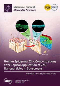
Sunscreens have a pivotal role in photo-protection to prevent skin cancers and reduce photodamage. ZnO is a US FDA approved UV filter used in sunscreens. Agglomerated ZnO nanoparticles are commonly used as they are transparent and therefore more aesthetic. However, ZnO nanoparticles are hydrolysed to zinc ions at the normal pH of the skin and can penetrate into the skin’s viable layers. Zinc ions have been shown to kill human skin cells in vitro. We quantified both exogenous zinc concentrations entering the viable epidermis from the ZnO nanoparticles and natural (endogenous) zinc ion concentrations. We show that the zinc ion concentrations attained after topical application of ZnO nanoparticles to excised human skin are well below those associated with their toxicity. View this paper.
  • Issues are regarded as officially published after their release is announced to the table of contents alert mailing list.
  • You may sign up for e-mail alerts to receive table of contents of newly released issues.
  • PDF is the official format for papers published in both, html and pdf forms. To view the papers in pdf format, click on the "PDF Full-text" link, and use the free Adobe Reader to open them.
Order results
Result details
Section
Select all
Export citation of selected articles as:
11 pages, 1454 KiB  
Article
Identification of the Capsicum baccatum NLR Protein CbAR9 Conferring Disease Resistance to Anthracnose
by Seungmin Son, Soohong Kim, Kyong Sil Lee, Jun Oh, Inchan Choi, Jae Wahng Do, Jae Bok Yoon, Jungheon Han, Doil Choi and Sang Ryeol Park
Int. J. Mol. Sci. 2021, 22(22), 12612; https://doi.org/10.3390/ijms222212612 - 22 Nov 2021
Cited by 5 | Viewed by 2366
Abstract
Anthracnose is caused by Colletotrichum species and is one of the most virulent fungal diseases affecting chili pepper (Capsicum) yield globally. However, the noble genes conferring resistance to Colletotrichum species remain largely elusive. In this study, we identified CbAR9 as the [...] Read more.
Anthracnose is caused by Colletotrichum species and is one of the most virulent fungal diseases affecting chili pepper (Capsicum) yield globally. However, the noble genes conferring resistance to Colletotrichum species remain largely elusive. In this study, we identified CbAR9 as the causal locus underlying the large effect quantitative trait locus CcR9 from the anthracnose-resistant chili pepper variety PBC80. CbAR9 encodes a nucleotide-binding and leucine-rich repeat (NLR) protein related to defense-associated NLRs in several other plant species. CbAR9 transcript levels were induced dramatically after Colletotrichum capsici infection. To explore the biological function, we generated transgenic Nicotiana benthamiana lines overexpressing CbAR9, which showed enhanced resistance to C. capsici relative to wild-type plants. Transcript levels of pathogenesis-related (PR) genes increased markedly in CbAR9-overexpressing N. benthamiana plants. Moreover, resistance to anthracnose and transcript levels of PR1 and PR2 were markedly reduced in CbAR9-silenced chili pepper fruits after C. capsici infection. Our results revealed that CbAR9 contributes to innate immunity against C. capsici. Full article
(This article belongs to the Special Issue Plant Disease Resistance 2.0)
Show Figures

Figure 1

17 pages, 2907 KiB  
Article
Tracing CRISPR/Cas12a Mediated Genome Editing Events in Apple Using High-Throughput Genotyping by PCR Capillary Gel Electrophoresis
by Susan Schröpfer and Henryk Flachowsky
Int. J. Mol. Sci. 2021, 22(22), 12611; https://doi.org/10.3390/ijms222212611 - 22 Nov 2021
Cited by 7 | Viewed by 2853
Abstract
The use of the novel CRISPR/Cas12a system is advantageous, as it expands the possibilities for genome editing (GE) applications due to its different features compared to the commonly used CRISPR/Cas9 system. In this work, the CRISPR/Cas12a system was applied for the first time [...] Read more.
The use of the novel CRISPR/Cas12a system is advantageous, as it expands the possibilities for genome editing (GE) applications due to its different features compared to the commonly used CRISPR/Cas9 system. In this work, the CRISPR/Cas12a system was applied for the first time to apple to investigate its general usability for GE applications. Efficient guide RNAs targeting different exons of the endogenous reporter gene MdPDS, whose disruption leads to the albino phenotype, were pre-selected by in vitro cleavage assays. A construct was transferred to apple encoding for a CRISPR/Cas12a system that simultaneously targets two loci in MdPDS. Using fluorescent PCR capillary electrophoresis and amplicon deep sequencing, all identified GE events of regenerated albino shoots were characterized as deletions. Large deletions between the two neighboring target sites were not observed. Furthermore, a chimeric composition of regenerates and shoots that exhibited multiple GE events was observed frequently. By comparing both analytical methods, it was shown that fluorescent PCR capillary gel electrophoresis is a sensitive high-throughput genotyping method that allows accurate predictions of the size and proportion of indel mutations for multiple loci simultaneously. Especially for species exhibiting high frequencies of chimerism, it can be recommended as a cost-effective method for efficient selection of homohistont GE lines. Full article
(This article belongs to the Special Issue Tree Genetic Engineering, Genome Editing and Genomics)
Show Figures

Figure 1

31 pages, 1280 KiB  
Review
Effects of the Escherichia coli Bacterial Toxin Cytotoxic Necrotizing Factor 1 on Different Human and Animal Cells: A Systematic Review
by Francesca Carlini, Zaira Maroccia, Carla Fiorentini, Sara Travaglione and Alessia Fabbri
Int. J. Mol. Sci. 2021, 22(22), 12610; https://doi.org/10.3390/ijms222212610 - 22 Nov 2021
Cited by 12 | Viewed by 2328
Abstract
Cytotoxic necrotizing factor 1 (CNF1) is a bacterial virulence factor, the target of which is represented by Rho GTPases, small proteins involved in a huge number of crucial cellular processes. CNF1, due to its ability to modulate the activity of Rho GTPases, represents [...] Read more.
Cytotoxic necrotizing factor 1 (CNF1) is a bacterial virulence factor, the target of which is represented by Rho GTPases, small proteins involved in a huge number of crucial cellular processes. CNF1, due to its ability to modulate the activity of Rho GTPases, represents a widely used tool to unravel the role played by these regulatory proteins in different biological processes. In this review, we summarized the data available in the scientific literature concerning the observed in vitro effects induced by CNF1. An article search was performed on electronic bibliographic resources. Screenings were performed of titles, abstracts, and full-texts according to PRISMA guidelines, whereas eligibility criteria were defined for in vitro studies. We identified a total of 299 records by electronic article search and included 76 original peer-reviewed scientific articles reporting morphological or biochemical modifications induced in vitro by soluble CNF1, either recombinant or from pathogenic Escherichia coli extracts highly purified with chromatographic methods. Most of the described CNF1-induced effects on cultured cells are ascribable to the modulating activity of the toxin on Rho GTPases and the consequent effects on actin cytoskeleton organization. All in all, the present review could be a prospectus about the CNF1-induced effects on cultured cells reported so far. Full article
(This article belongs to the Section Molecular Toxicology)
Show Figures

Figure 1

25 pages, 5046 KiB  
Article
Identification of Impacted Pathways and Transcriptomic Markers as Potential Mediators of Pulmonary Fibrosis in Transgenic Mice Expressing Human IGFBP5
by Xinh-Xinh Nguyen, Ludivine Renaud and Carol Feghali-Bostwick
Int. J. Mol. Sci. 2021, 22(22), 12609; https://doi.org/10.3390/ijms222212609 - 22 Nov 2021
Cited by 4 | Viewed by 2928
Abstract
Pulmonary fibrosis is a serious disease characterized by extracellular matrix (ECM) component overproduction and remodeling. Insulin-like growth factor-binding protein 5 (IGFBP5) is a conserved member of the IGFBP family of proteins that is overexpressed in fibrotic tissues and promotes fibrosis. We used RNA [...] Read more.
Pulmonary fibrosis is a serious disease characterized by extracellular matrix (ECM) component overproduction and remodeling. Insulin-like growth factor-binding protein 5 (IGFBP5) is a conserved member of the IGFBP family of proteins that is overexpressed in fibrotic tissues and promotes fibrosis. We used RNA sequencing (RNAseq) to identify differentially expressed genes (DEGs) between primary lung fibroblasts (pFBs) of homozygous (HOMO) transgenic mice expressing human IGFBP5 (hIGFBP5) and wild type mice (WT). The results of the differential expression analysis showed 2819 DEGs in hIGFBP5 pFBs. Functional enrichment analysis confirmed the pro-fibrotic character of IGFBP5 and revealed its impact on fundamental signaling pathways, including cytokine–cytokine receptor interaction, focal adhesion, AGE-RAGE signaling, calcium signaling, and neuroactive ligand-receptor interactions, to name a few. Noticeably, 7% of the DEGs in hIGFBP5-expressing pFBs are receptors and integrins. Furthermore, hub gene analysis revealed 12 hub genes including Fpr1, Bdkrb2, Mchr1, Nmur1, Cnr2, P2ry14, and Ptger3. Validation assays were performed to complement the RNAseq data. They confirmed significant differences in the levels of the corresponding proteins in cultured pFBs. Our study provides new insights into the molecular mechanism(s) of IGFBP5-associated pulmonary fibrosis through possible receptor interactions that drive fibrosis and tissue remodeling. Full article
Show Figures

Figure 1

17 pages, 937 KiB  
Review
Hypoxia: The Cornerstone of Glioblastoma
by Marta Domènech, Ainhoa Hernández, Andrea Plaja, Eva Martínez-Balibrea and Carmen Balañà
Int. J. Mol. Sci. 2021, 22(22), 12608; https://doi.org/10.3390/ijms222212608 - 22 Nov 2021
Cited by 61 | Viewed by 6020
Abstract
Glioblastoma is the most aggressive form of brain tumor in adults and is characterized by the presence of hypervascularization and necrosis, both caused by a hypoxic microenvironment. In this review, we highlight that hypoxia-induced factor 1 (HIF-1), the main factor activated by hypoxia, [...] Read more.
Glioblastoma is the most aggressive form of brain tumor in adults and is characterized by the presence of hypervascularization and necrosis, both caused by a hypoxic microenvironment. In this review, we highlight that hypoxia-induced factor 1 (HIF-1), the main factor activated by hypoxia, is an important driver of tumor progression in GB patients. HIF-1α is a transcription factor regulated by the presence or absence of O2. The expression of HIF-1 has been related to high-grade gliomas and aggressive tumor behavior. HIF-1 promotes tumor progression via the activation of angiogenesis, immunosuppression, and metabolic reprogramming, promoting cell invasion and survival. Moreover, in GB, HIF-1 is not solely modulated by oxygen but also by oncogenic signaling pathways, such as MAPK/ERK, p53, and PI3K/PTEN. Therefore, the inhibition of the hypoxia pathway could represent an important treatment alternative in a disease with very few therapy options. Here, we review the roles of HIF-1 in GB progression and the inhibitors that have been studied thus far, with the aim of shedding light on this devastating disease. Full article
(This article belongs to the Special Issue Glioblastoma 2.0)
Show Figures

Figure 1

15 pages, 5536 KiB  
Article
Single-Cell Atlas of Adult Testis in Protogynous Hermaphroditic Orange-Spotted Grouper, Epinephelus coioides
by Xi Wu, Yang Yang, Chaoyue Zhong, Tong Wang, Yanhong Deng, Hengjin Huang, Haoran Lin, Zining Meng and Xiaochun Liu
Int. J. Mol. Sci. 2021, 22(22), 12607; https://doi.org/10.3390/ijms222212607 - 22 Nov 2021
Cited by 6 | Viewed by 2415
Abstract
Spermatogenesis is a process of self-renewal and differentiation in spermatogonial stem cells. During this process, germ cells and somatic cells interact intricately to ensure long-term fertility and accurate genome propagation. Spermatogenesis has been intensely investigated in mammals but remains poorly understood with regard [...] Read more.
Spermatogenesis is a process of self-renewal and differentiation in spermatogonial stem cells. During this process, germ cells and somatic cells interact intricately to ensure long-term fertility and accurate genome propagation. Spermatogenesis has been intensely investigated in mammals but remains poorly understood with regard to teleosts. Here, we performed single-cell RNA sequencing of ~9500 testicular cells from the male, orange-spotted grouper. In the adult testis, we divided the cells into nine clusters and defined ten cell types, as compared with human testis data, including cell populations with characteristics of male germ cells and somatic cells, each of which expressed specific marker genes. We also identified and profiled the expression patterns of four marker genes (calr, eef1a, s100a1, vasa) in both the ovary and adult testis. Our data provide a blueprint of male germ cells and supporting somatic cells. Moreover, the cell markers are candidates that could be used for further cell identification. Full article
(This article belongs to the Section Molecular Biology)
Show Figures

Figure 1

24 pages, 2149 KiB  
Review
Pleiotropic Outcomes of Glyphosate Exposure: From Organ Damage to Effects on Inflammation, Cancer, Reproduction and Development
by Marianna Marino, Elena Mele, Andrea Viggiano, Stefania Lucia Nori, Rosaria Meccariello and Antonietta Santoro
Int. J. Mol. Sci. 2021, 22(22), 12606; https://doi.org/10.3390/ijms222212606 - 22 Nov 2021
Cited by 22 | Viewed by 6894
Abstract
Glyphosate is widely used worldwide as a potent herbicide. Due to its ubiquitous use, it is detectable in air, water and foodstuffs and can accumulate in human biological fluids and tissues representing a severe human health risk. In plants, glyphosate acts as an [...] Read more.
Glyphosate is widely used worldwide as a potent herbicide. Due to its ubiquitous use, it is detectable in air, water and foodstuffs and can accumulate in human biological fluids and tissues representing a severe human health risk. In plants, glyphosate acts as an inhibitor of the shikimate pathway, which is absent in vertebrates. Due to this, international scientific authorities have long-considered glyphosate as a compound that has no or weak toxicity in humans. However, increasing evidence has highlighted the toxicity of glyphosate and its formulations in animals and human cells and tissues. Thus, despite the extension of the authorization of the use of glyphosate in Europe until 2022, several countries have begun to take precautionary measures to reduce its diffusion. Glyphosate has been detected in urine, blood and maternal milk and has been found to induce the generation of reactive oxygen species (ROS) and several cytotoxic and genotoxic effects in vitro and in animal models directly or indirectly through its metabolite, aminomethylphosphonic acid (AMPA). This review aims to summarize the more relevant findings on the biological effects and underlying molecular mechanisms of glyphosate, with a particular focus on glyphosate's potential to induce inflammation, DNA damage and alterations in gene expression profiles as well as adverse effects on reproduction and development. Full article
(This article belongs to the Special Issue Epigenetic Effects and Toxicity of Environmental Pollutants)
Show Figures

Figure 1

20 pages, 2384 KiB  
Article
Long-Term Effects of Ionizing Radiation on the Hippocampus: Linking Effects of the Sonic Hedgehog Pathway Activation with Radiation Response
by Francesca Antonelli, Arianna Casciati, Montserrat Belles, Noemi Serra, Maria Victoria Linares-Vidal, Carmela Marino, Mariateresa Mancuso and Simonetta Pazzaglia
Int. J. Mol. Sci. 2021, 22(22), 12605; https://doi.org/10.3390/ijms222212605 - 22 Nov 2021
Cited by 4 | Viewed by 2461
Abstract
Radiation therapy represents one of the primary treatment modalities for primary and metastatic brain tumors. Although recent advances in radiation techniques, that allow the delivery of higher radiation doses to the target volume, reduce the toxicity to normal tissues, long-term neurocognitive decline is [...] Read more.
Radiation therapy represents one of the primary treatment modalities for primary and metastatic brain tumors. Although recent advances in radiation techniques, that allow the delivery of higher radiation doses to the target volume, reduce the toxicity to normal tissues, long-term neurocognitive decline is still a detrimental factor significantly affecting quality of life, particularly in pediatric patients. This imposes the need for the development of prevention strategies. Based on recent evidence, showing that manipulation of the Shh pathway carries therapeutic potential for brain repair and functional recovery after injury, here we evaluate how radiation-induced hippocampal alterations are modulated by the constitutive activation of the Shh signaling pathway in Patched 1 heterozygous mice (Ptch1+/−). Our results show, for the first time, an overall protective effect of constitutive Shh pathway activation on hippocampal radiation injury. This activation, through modulation of the proneural gene network, leads to a long-term reduction of hippocampal deficits in the stem cell and new neuron compartments and to the mitigation of radio-induced astrogliosis, despite some behavioral alterations still being detected in Ptch1+/− mice. A better understanding of the pathogenic mechanisms responsible for the neural decline following irradiation is essential for identifying prevention measures to contain the harmful consequences of irradiation. Our data have important translational implications as they suggest a role for Shh pathway manipulation to provide the therapeutic possibility of improving brain repair and functional recovery after radio-induced injury. Full article
(This article belongs to the Special Issue Molecular Research in Radiobiology)
Show Figures

Figure 1

14 pages, 2187 KiB  
Article
Connexin46 Expression Enhances Cancer Stem Cell and Epithelial-to-Mesenchymal Transition Characteristics of Human Breast Cancer MCF-7 Cells
by Rodrigo A. Acuña, Manuel Varas-Godoy, Diego Herrera-Sepulveda and Mauricio A. Retamal
Int. J. Mol. Sci. 2021, 22(22), 12604; https://doi.org/10.3390/ijms222212604 - 22 Nov 2021
Cited by 14 | Viewed by 2852
Abstract
Connexins (Cxs) are a family of proteins that form two different types of ion channels: hemichannels and gap junction channels. These channels participate in cellular communication, enabling them to share information and act as a synchronized syncytium. This cellular communication has been considered [...] Read more.
Connexins (Cxs) are a family of proteins that form two different types of ion channels: hemichannels and gap junction channels. These channels participate in cellular communication, enabling them to share information and act as a synchronized syncytium. This cellular communication has been considered a strong tumor suppressor, but it is now recognized that some type of Cxs can be pro-tumorigenic. For example, Cx46 expression is increased in human breast cancer samples and correlates with cancer stem cell (CSC) characteristics in human glioma. Thus, we explored whether Cx46 and glioma cells, can set up CSC and epithelial-to-mesenchymal transition (EMT) properties in a breast cancer cell line. To this end, we transfected MCF-7 cells with Cx46 attached to a green fluorescent protein (Cx46GFP), and we determined how its expression orchestrates both the gene-expression and functional changes associated with CSC and EMT. We observed that Cx46GFP increased Sox2, Nanog, and OCT4 mRNA levels associated with a high capacity to form monoclonal colonies and tumorspheres. Similarly, Cx46GFP increased the mRNA levels of n-cadherin, Vimentin, Snail and Zeb1 to a higher migratory and invasive capacity. Furthermore, Cx46GFP transfected in MCF-7 cells induced the release of higher amounts of VEGF, which promoted angiogenesis in HUVEC cells. We demonstrated for the first time that Cx46 modulates CSC and EMT properties in breast cancer cells and thus could be relevant in the design of future cancer therapies. Full article
(This article belongs to the Special Issue Cancer Cell Invasion and Metastases 2.0)
Show Figures

Figure 1

9 pages, 522 KiB  
Review
Contribution of Lipid Oxidation and Ferroptosis to Radiotherapy Efficacy
by Ashley N. Pearson, Joseph Carmicheal, Long Jiang, Yu Leo Lei and Michael D. Green
Int. J. Mol. Sci. 2021, 22(22), 12603; https://doi.org/10.3390/ijms222212603 - 22 Nov 2021
Cited by 14 | Viewed by 4534
Abstract
Radiotherapy promotes tumor cell death and senescence through the induction of oxidative damage. Recent work has highlighted the importance of lipid peroxidation for radiotherapy efficacy. Excessive lipid peroxidation can promote ferroptosis, a regulated form of cell death. In this review, we address the [...] Read more.
Radiotherapy promotes tumor cell death and senescence through the induction of oxidative damage. Recent work has highlighted the importance of lipid peroxidation for radiotherapy efficacy. Excessive lipid peroxidation can promote ferroptosis, a regulated form of cell death. In this review, we address the evidence supporting a role of ferroptosis in response to radiotherapy and discuss the molecular regulators that underlie this interaction. Finally, we postulate on the clinical implications for the intersection of ferroptosis and radiotherapy. Full article
(This article belongs to the Special Issue Radiation Biology and Molecular Radiation Oncology)
Show Figures

Figure 1

28 pages, 6061 KiB  
Article
Disulfide Dimerization of Neuronal Calcium Sensor-1: Implications for Zinc and Redox Signaling
by Viktoriia E. Baksheeva, Alexey V. Baldin, Arthur O. Zalevsky, Aliya A. Nazipova, Alexey S. Kazakov, Vasiliy I. Vladimirov, Neonila V. Gorokhovets, François Devred, Pavel P. Philippov, Alexandr V. Bazhin, Andrey V. Golovin, Andrey A. Zamyatnin, Jr., Dmitry V. Zinchenko, Philipp O. Tsvetkov, Sergei E. Permyakov and Evgeni Yu. Zernii
Int. J. Mol. Sci. 2021, 22(22), 12602; https://doi.org/10.3390/ijms222212602 - 22 Nov 2021
Cited by 8 | Viewed by 2820
Abstract
Neuronal calcium sensor-1 (NCS-1) is a four-EF-hand ubiquitous signaling protein modulating neuronal function and survival, which participates in neurodegeneration and carcinogenesis. NCS-1 recognizes specific sites on cellular membranes and regulates numerous targets, including G-protein coupled receptors and their kinases (GRKs). Here, with the [...] Read more.
Neuronal calcium sensor-1 (NCS-1) is a four-EF-hand ubiquitous signaling protein modulating neuronal function and survival, which participates in neurodegeneration and carcinogenesis. NCS-1 recognizes specific sites on cellular membranes and regulates numerous targets, including G-protein coupled receptors and their kinases (GRKs). Here, with the use of cellular models and various biophysical and computational techniques, we demonstrate that NCS-1 is a redox-sensitive protein, which responds to oxidizing conditions by the formation of disulfide dimer (dNCS-1), involving its single, highly conservative cysteine C38. The dimer content is unaffected by the elevation of intracellular calcium levels but increases to 10–30% at high free zinc concentrations (characteristic of oxidative stress), which is accompanied by accumulation of the protein in punctual clusters in the perinuclear area. The formation of dNCS-1 represents a specific Zn2+-promoted process, requiring proper folding of the protein and occurring at redox potential values approaching apoptotic levels. The dimer binds Ca2+ only in one EF-hand per monomer, thereby representing a unique state, with decreased α-helicity and thermal stability, increased surface hydrophobicity, and markedly improved inhibitory activity against GRK1 due to 20-fold higher affinity towards the enzyme. Furthermore, dNCS-1 can coordinate zinc and, according to molecular modeling, has an asymmetrical structure and increased conformational flexibility of the subunits, which may underlie their enhanced target-binding properties. In HEK293 cells, dNCS-1 can be reduced by the thioredoxin system, otherwise accumulating as protein aggregates, which are degraded by the proteasome. Interestingly, NCS-1 silencing diminishes the susceptibility of Y79 cancer cells to oxidative stress-induced apoptosis, suggesting that NCS-1 may mediate redox-regulated pathways governing cell death/survival in response to oxidative conditions. Full article
(This article belongs to the Special Issue Molecular Basis of Sensory Transduction in Health and Disease)
Show Figures

Figure 1

12 pages, 2396 KiB  
Article
Inhibitory Effects of Sulfonamide Derivatives on the β-Carbonic Anhydrase (MpaCA) from Malassezia pachydermatis, a Commensal, Pathogenic Fungus Present in Domestic Animals
by Viviana De Luca, Andrea Angeli, Valeria Mazzone, Claudia Adelfio, Fabrizio Carta, Silvia Selleri, Vincenzo Carginale, Andrea Scaloni, Claudiu T. Supuran and Clemente Capasso
Int. J. Mol. Sci. 2021, 22(22), 12601; https://doi.org/10.3390/ijms222212601 - 22 Nov 2021
Cited by 3 | Viewed by 1929
Abstract
Fungi are exposed to various environmental variables during their life cycle, including changes in CO2 concentration. CO2 has the potential to act as an activator of several cell signaling pathways. In fungi, the sensing of CO2 triggers cell differentiation and [...] Read more.
Fungi are exposed to various environmental variables during their life cycle, including changes in CO2 concentration. CO2 has the potential to act as an activator of several cell signaling pathways. In fungi, the sensing of CO2 triggers cell differentiation and the biosynthesis of proteins involved in the metabolism and pathogenicity of these microorganisms. The molecular machineries involved in CO2 sensing constitute a promising target for the development of antifungals. Carbonic anhydrases (CAs, EC 4.2.1.1) are crucial enzymes in the CO2 sensing systems of fungi, because they catalyze the reversible hydration of CO2 to proton and HCO3-. Bicarbonate in turn boots a cascade of reactions triggering fungal pathogenicity and metabolism. Accordingly, CAs affect microorganism proliferation and may represent a potential therapeutic target against fungal infection. Here, the inhibition of the unique β-CA (MpaCA) encoded in the genome of Malassezia pachydermatis, a fungus with substantial relevance in veterinary and medical sciences, was investigated using a series of conventional CA inhibitors (CAIs), namely aromatic and heterocyclic sulfonamides. This study aimed to describe novel candidates that can kill this harmful fungus by inhibiting their CA, and thus lead to effective anti-dandruff and anti-seborrheic dermatitis agents. In this context, current antifungal compounds, such as the azoles and their derivatives, have been demonstrated to induce the selection of resistant fungal strains and lose therapeutic efficacy, which might be restored by the concomitant use of alternative compounds, such as the fungal CA inhibitors. Full article
Show Figures

Figure 1

14 pages, 3365 KiB  
Article
Analgesic Mechanisms of Steroid Ointment against Oral Ulcerative Mucositis in a Rat Model
by Mako Naniwa, Chihiro Nakatomi, Suzuro Hitomi, Kazunari Matsuda, Takuya Tabuchi, Daijiro Sugiyama, Sayaka Kubo, Yuichi Miyamura, Kenichi Yoshino, Sumio Akifusa and Kentaro Ono
Int. J. Mol. Sci. 2021, 22(22), 12600; https://doi.org/10.3390/ijms222212600 - 22 Nov 2021
Cited by 7 | Viewed by 2192
Abstract
Despite the long history of use of steroid ointments for oral mucositis, the analgesic mechanism has not been fully elucidated. In this study, we examined the effects of triamcinolone acetonide (Tmc) on oral ulcerative mucositis-induced pain in conscious rats by our proprietary assay [...] Read more.
Despite the long history of use of steroid ointments for oral mucositis, the analgesic mechanism has not been fully elucidated. In this study, we examined the effects of triamcinolone acetonide (Tmc) on oral ulcerative mucositis-induced pain in conscious rats by our proprietary assay system. Based on evaluations of the physical properties and retention periods in the oral mucosa of human volunteers and rats, we selected TRAFUL® ointment as a long-lasting base. In oral ulcerative mucositis model rats, TRAFUL® with Tmc suppressed cyclooxygenase-dependent inflammatory responses with upregulations of glucocorticoid receptor-induced anti-inflammatory genes and inhibited spontaneous nociceptive behavior. When an ointment with a shorter residual period was used, the effects of Tmc were not elicited or were induced to a lesser extent. Importantly, TRAFUL® with Tmc also improved oral ulcerative mucositis-induced mechanical allodynia, which has been reported to be independent of cyclooxygenase. Ca2+ imaging in dissociated trigeminal ganglion neurons showed that long-term preincubation with Tmc inhibited the hypertonic stimulation-induced Ca2+ response. These results suggest that the representative steroid Tmc suppresses oral ulcerative mucositis-induced pain by general anti-inflammatory actions and inhibits mechanical sensitivity in peripheral nerves. For drug delivery, long-lasting ointments such as TRAFUL® are needed to sufficiently induce the therapeutic effects. Full article
(This article belongs to the Special Issue Orofacial Pain: Molecular Mechanisms, Diagnosis and Treatment 2021)
Show Figures

Figure 1

21 pages, 3470 KiB  
Review
The Relevance of G-Quadruplexes for DNA Repair
by Rebecca Linke, Michaela Limmer, Stefan A. Juranek, Annkristin Heine and Katrin Paeschke
Int. J. Mol. Sci. 2021, 22(22), 12599; https://doi.org/10.3390/ijms222212599 - 22 Nov 2021
Cited by 27 | Viewed by 3368
Abstract
DNA molecules can adopt a variety of alternative structures. Among these structures are G-quadruplex DNA structures (G4s), which support cellular function by affecting transcription, translation, and telomere maintenance. These structures can also induce genome instability by stalling replication, increasing DNA damage, and recombination [...] Read more.
DNA molecules can adopt a variety of alternative structures. Among these structures are G-quadruplex DNA structures (G4s), which support cellular function by affecting transcription, translation, and telomere maintenance. These structures can also induce genome instability by stalling replication, increasing DNA damage, and recombination events. G-quadruplex-driven genome instability is connected to tumorigenesis and other genetic disorders. In recent years, the connection between genome stability, DNA repair and G4 formation was further underlined by the identification of multiple DNA repair proteins and ligands which bind and stabilize said G4 structures to block specific DNA repair pathways. The relevance of G4s for different DNA repair pathways is complex and depends on the repair pathway itself. G4 structures can induce DNA damage and block efficient DNA repair, but they can also support the activity and function of certain repair pathways. In this review, we highlight the roles and consequences of G4 DNA structures for DNA repair initiation, processing, and the efficiency of various DNA repair pathways. Full article
Show Figures

Figure 1

23 pages, 8266 KiB  
Article
RNA-Seq Analysis Reveals an Essential Role of the Tyrosine Metabolic Pathway and Inflammation in Myopia-Induced Retinal Degeneration in Guinea Pigs
by Ling Zeng, Xiaoning Li, Jian Liu, Hong Liu, Heping Xu and Zhikuan Yang
Int. J. Mol. Sci. 2021, 22(22), 12598; https://doi.org/10.3390/ijms222212598 - 22 Nov 2021
Cited by 20 | Viewed by 3320
Abstract
Myopia is the second leading cause of visual impairment globally. Myopia can induce sight-threatening retinal degeneration and the underlying mechanism remains poorly defined. We generated a model of myopia-induced early-stage retinal degeneration in guinea pigs and investigated the mechanism of action. Methods: The [...] Read more.
Myopia is the second leading cause of visual impairment globally. Myopia can induce sight-threatening retinal degeneration and the underlying mechanism remains poorly defined. We generated a model of myopia-induced early-stage retinal degeneration in guinea pigs and investigated the mechanism of action. Methods: The form-deprivation-induced myopia (FDM) was induced in the right eyes of 2~3-week-old guinea pigs using a translucent balloon for 15 weeks. The left eye remained untreated and served as a self-control. Another group of untreated age-matched animals was used as naïve controls. The refractive error and ocular biometrics were measured at 3, 7, 9, 12 and 15 weeks post-FDM induction. Visual function was evaluated by electroretinography. Retinal neurons and synaptic structures were examined by confocal microscopy of immunolabelled retinal sections. The total RNAs were extracted from the retinas and processed for RNA sequencing analysis. Results: The FDM eyes presented a progressive axial length elongation and refractive error development. After 15 weeks of intervention, the average refractive power was −3.40 ± 1.85 D in the FDM eyes, +2.94 ± 0.59 D and +2.69 ± 0.56 D in the self-control and naïve control eyes, respectively. The a-wave amplitude was significantly lower in FDM eyes and these eyes had a significantly lower number of rods, secretagogin+ bipolar cells, and GABAergic amacrine cells in selected retinal areas. RNA-seq analysis showed that 288 genes were upregulated and 119 genes were downregulated in FDM retinas compared to naïve control retinas. In addition, 152 genes were upregulated and 12 were downregulated in FDM retinas compared to self-control retinas. The KEGG enrichment analysis showed that tyrosine metabolism, ABC transporters and inflammatory pathways were upregulated, whereas tight junction, lipid and glycosaminoglycan biosynthesis were downregulated in FDM eyes. Conclusions: The long-term (15-week) FDM in the guinea pig models induced an early-stage retinal degeneration. The dysregulation of the tyrosine metabolism and inflammatory pathways may contribute to the pathogenesis of myopia-induced retinal degeneration. Full article
Show Figures

Figure 1

17 pages, 26859 KiB  
Article
Singapore Grouper Iridovirus Disturbed Glycerophospholipids Homeostasis: Cytosolic Phospholipase A2 Was Essential for Virus Replication
by Na Ni, Jiaying Zheng, Wenji Wang, Linyong Zhi, Qiwei Qin, Youhua Huang and Xiaohong Huang
Int. J. Mol. Sci. 2021, 22(22), 12597; https://doi.org/10.3390/ijms222212597 - 22 Nov 2021
Cited by 5 | Viewed by 1804
Abstract
Singapore grouper iridovirus (SGIV), belonging to genus Ranavirus, family Iridoviridae, causes great economic losses in the aquaculture industry. Previous studies demonstrated the lipid composition of intracellular unenveloped viruses, but the changes in host-cell glyceophospholipids components and the roles of key enzymes [...] Read more.
Singapore grouper iridovirus (SGIV), belonging to genus Ranavirus, family Iridoviridae, causes great economic losses in the aquaculture industry. Previous studies demonstrated the lipid composition of intracellular unenveloped viruses, but the changes in host-cell glyceophospholipids components and the roles of key enzymes during SGIV infection still remain largely unknown. Here, the whole cell lipidomic profiling during SGIV infection was analyzed using UPLC-Q-TOF-MS/MS. The lipidomic data showed that glycerophospholipids (GPs), including phosphatidylcholine (PC), phosphatidylserine (PS), glycerophosphoinositols (PI) and fatty acids (FAs) were significantly elevated in SGIV-infected cells, indicating that SGIV infection disturbed GPs homeostasis, and then affected the metabolism of FAs, especially arachidonic acid (AA). The roles of key enzymes, such as cytosolic phospholipase A2 (cPLA2), 5-Lipoxygenase (5-LOX), and cyclooxygenase (COX) in SGIV infection were further investigated using the corresponding specific inhibitors. The inhibition of cPLA2 by AACOCF3 decreased SGIV replication, suggesting that cPLA2 might play important roles in the process of SGIV infection. Consistent with this result, the ectopic expression of EccPLA2α or knockdown significantly enhanced or suppressed viral replication in vitro, respectively. In addition, the inhibition of both 5-LOX and COX significantly suppressed SGIV replication, indicating that AA metabolism was essential for SGIV infection. Taken together, our results demonstrated for the first time that SGIV infection in vitro disturbed GPs homeostasis and cPLA2 exerted crucial roles in SGIV replication. Full article
(This article belongs to the Section Molecular Toxicology)
Show Figures

Figure 1

17 pages, 4376 KiB  
Article
Selectivity Determinants of RHO GTPase Binding to IQGAPs
by Niloufar Mosaddeghzadeh, Kazem Nouri, Oliver H. F. Krumbach, Ehsan Amin, Radovan Dvorsky and Mohammad R. Ahmadian
Int. J. Mol. Sci. 2021, 22(22), 12596; https://doi.org/10.3390/ijms222212596 - 22 Nov 2021
Cited by 6 | Viewed by 1916
Abstract
IQ motif-containing GTPase-activating proteins (IQGAPs) modulate a wide range of cellular processes by acting as scaffolds and driving protein components into distinct signaling networks. Their functional states have been proposed to be controlled by members of the RHO family of GTPases, among other [...] Read more.
IQ motif-containing GTPase-activating proteins (IQGAPs) modulate a wide range of cellular processes by acting as scaffolds and driving protein components into distinct signaling networks. Their functional states have been proposed to be controlled by members of the RHO family of GTPases, among other regulators. In this study, we show that IQGAP1 and IQGAP2 can associate with CDC42 and RAC1-like proteins but not with RIF, RHOD, or RHO-like proteins, including RHOA. This seems to be based on the distribution of charged surface residues, which varies significantly among RHO GTPases despite their high sequence homology. Although effector proteins bind first to the highly flexible switch regions of RHO GTPases, additional contacts outside are required for effector activation. Sequence alignment and structural, mutational, and competitive biochemical analyses revealed that RHO GTPases possess paralog-specific residues outside the two highly conserved switch regions that essentially determine the selectivity of RHO GTPase binding to IQGAPs. Amino acid substitution of these specific residues in RHOA to the corresponding residues in RAC1 resulted in RHOA association with IQGAP1. Thus, electrostatics most likely plays a decisive role in these interactions. Full article
(This article belongs to the Section Macromolecules)
Show Figures

Figure 1

14 pages, 2082 KiB  
Review
Pseudophosphatases as Regulators of MAPK Signaling
by Emma Marie Wilber Hepworth and Shantá D. Hinton
Int. J. Mol. Sci. 2021, 22(22), 12595; https://doi.org/10.3390/ijms222212595 - 22 Nov 2021
Cited by 29 | Viewed by 5083
Abstract
Mitogen-activated protein kinase (MAPK) signaling pathways are highly conserved regulators of eukaryotic cell function. These enzymes regulate many biological processes, including the cell cycle, apoptosis, differentiation, protein biosynthesis, and oncogenesis; therefore, tight control of the activity of MAPK is critical. Kinases and phosphatases [...] Read more.
Mitogen-activated protein kinase (MAPK) signaling pathways are highly conserved regulators of eukaryotic cell function. These enzymes regulate many biological processes, including the cell cycle, apoptosis, differentiation, protein biosynthesis, and oncogenesis; therefore, tight control of the activity of MAPK is critical. Kinases and phosphatases are well established as MAPK activators and inhibitors, respectively. Kinases phosphorylate MAPKs, initiating and controlling the amplitude of the activation. In contrast, MAPK phosphatases (MKPs) dephosphorylate MAPKs, downregulating and controlling the duration of the signal. In addition, within the past decade, pseudoenzymes of these two families, pseudokinases and pseudophosphatases, have emerged as bona fide signaling regulators. This review discusses the role of pseudophosphatases in MAPK signaling, highlighting the function of phosphoserine/threonine/tyrosine-interacting protein (STYX) and TAK1-binding protein (TAB 1) in regulating MAPKs. Finally, a new paradigm is considered for this well-studied cellular pathway, and signal transduction pathways in general. Full article
Show Figures

Graphical abstract

18 pages, 1298 KiB  
Review
Epigenetics of Myotonic Dystrophies: A Minireview
by Virginia Veronica Visconti, Federica Centofanti, Simona Fittipaldi, Elisa Macrì, Giuseppe Novelli and Annalisa Botta
Int. J. Mol. Sci. 2021, 22(22), 12594; https://doi.org/10.3390/ijms222212594 - 22 Nov 2021
Cited by 8 | Viewed by 4096
Abstract
Myotonic dystrophy type 1 and 2 (DM1 and DM2) are two multisystemic autosomal dominant disorders with clinical and genetic similarities. The prevailing paradigm for DMs is that they are mediated by an in trans toxic RNA mechanism, triggered by untranslated CTG and CCTG [...] Read more.
Myotonic dystrophy type 1 and 2 (DM1 and DM2) are two multisystemic autosomal dominant disorders with clinical and genetic similarities. The prevailing paradigm for DMs is that they are mediated by an in trans toxic RNA mechanism, triggered by untranslated CTG and CCTG repeat expansions in the DMPK and CNBP genes for DM1 and DM2, respectively. Nevertheless, increasing evidences suggest that epigenetics can also play a role in the pathogenesis of both diseases. In this review, we discuss the available information on epigenetic mechanisms that could contribute to the DMs outcome and progression. Changes in DNA cytosine methylation, chromatin remodeling and expression of regulatory noncoding RNAs are described, with the intent of depicting an epigenetic signature of DMs. Epigenetic biomarkers have a strong potential for clinical application since they could be used as targets for therapeutic interventions avoiding changes in DNA sequences. Moreover, understanding their clinical significance may serve as a diagnostic indicator in genetic counselling in order to improve genotype–phenotype correlations in DM patients. Full article
(This article belongs to the Special Issue Epigenetic Mechanisms and Human Pathology 2.0)
Show Figures

Figure 1

16 pages, 5339 KiB  
Article
The Preliminary Study on the Proapoptotic Effect of Reduced Graphene Oxide in Breast Cancer Cell Lines
by Rafał Krętowski, Agata Jabłońska-Trypuć and Marzanna Cechowska-Pasko
Int. J. Mol. Sci. 2021, 22(22), 12593; https://doi.org/10.3390/ijms222212593 - 22 Nov 2021
Cited by 11 | Viewed by 2007
Abstract
Breast cancer is the most common cancer diagnosed in women, however traditional therapies have several side effects. This has led to an urgent need to explore novel drug approaches to treatment strategies such as graphene-based nanomaterials such as reduced graphene oxide (rGO). It [...] Read more.
Breast cancer is the most common cancer diagnosed in women, however traditional therapies have several side effects. This has led to an urgent need to explore novel drug approaches to treatment strategies such as graphene-based nanomaterials such as reduced graphene oxide (rGO). It was noticed as a potential drug due to its target selectivity, easy functionalisation, chemisensitisation, and high drug-loading capacity. rGO is widely used in many fields, including biological and biomedical, due to its unique physicochemical properties. However, the possible mechanisms of rGO toxicity remain unclear. In this paper, we present findings on the cytotoxic and antiproliferative effects of rGO and its ability to induce oxidative stress and apoptosis of breast cancer cell lines. We indicate that rGO induced time- and dose-dependent cytotoxicity in MDA-MB-231 and ZR-75-1 cell lines, but not in T-47D, MCF-7, Hs 578T cell lines. In rGO-treated MDA-MB-231 and ZR-75-1 cell lines, we noticed increased induction of apoptosis and necrosis. In addition, rGO has been found to cause oxidative stress, reduce proliferation, and induce structural changes in breast cancer cells. Taken together, these studies provide new insight into the mechanism of oxidative stress and apoptosis in breast cancer cells. Full article
(This article belongs to the Special Issue Cell Apoptosis)
Show Figures

Figure 1

19 pages, 5216 KiB  
Article
In Leishmania major, the Homolog of the Oncogene PES1 May Play a Critical Role in Parasite Infectivity
by Miriam Algarabel, Celia Fernández-Rubio, Katerina Musilova, José Peña-Guerrero, Andrés Vacas, Esther Larrea and Paul A. Nguewa
Int. J. Mol. Sci. 2021, 22(22), 12592; https://doi.org/10.3390/ijms222212592 - 22 Nov 2021
Cited by 5 | Viewed by 1851
Abstract
Leishmaniasis is a neglected tropical disease caused by Leishmania spp. The improvement of existing treatments and the discovery of new drugs remain ones of the major goals in control and eradication of this disease. From the parasite genome, we have identified the homologue [...] Read more.
Leishmaniasis is a neglected tropical disease caused by Leishmania spp. The improvement of existing treatments and the discovery of new drugs remain ones of the major goals in control and eradication of this disease. From the parasite genome, we have identified the homologue of the human oncogene PES1 in Leishmania major (LmjPES). It has been demonstrated that PES1 is involved in several processes such as ribosome biogenesis, cell proliferation and genetic transcription. Our phylogenetic studies showed that LmjPES encodes a highly conserved protein containing three main domains: PES N-terminus (shared with proteins involved in ribosomal biogenesis), BRCT (found in proteins related to DNA repair processes) and MAEBL-type domain (C-terminus, related to erythrocyte invasion in apicomplexan). This gene showed its highest expression level in metacyclic promastigotes, the infective forms; by fluorescence microscopy assay, we demonstrated the nuclear localization of LmjPES protein. After generating mutant parasites overexpressing LmjPES, we observed that these clones displayed a dramatic increase in the ratio of cell infection within macrophages. Furthermore, BALB/c mice infected with these transgenic parasites exhibited higher footpad inflammation compared to those inoculated with non-overexpressing parasites. Full article
Show Figures

Figure 1

15 pages, 3970 KiB  
Article
Multiparametric Evaluation of Post-MI Small Animal Models Using Metabolic ([18F]FDG) and Perfusion-Based (SYN1) Heart Viability Tracers
by Tomasz Jan Kolanowski, Weronika Wargocka-Matuszewska, Agnieszka Zimna, Lukasz Cheda, Joanna Zyprych-Walczak, Anna Rugowska, Monika Drabik, Michał Fiedorowicz, Seweryn Krajewski, Łukasz Steczek, Cezary Kozanecki, Zbigniew Rogulski, Natalia Rozwadowska and Maciej Kurpisz
Int. J. Mol. Sci. 2021, 22(22), 12591; https://doi.org/10.3390/ijms222212591 - 22 Nov 2021
Cited by 5 | Viewed by 1959
Abstract
Cardiovascular diseases (CVD), with myocardial infarction (MI) being one of the crucial components, wreak havoc in developed countries. Advanced imaging technologies are required to obtain quick and widely available diagnostic data. This paper describes a multimodal approach to in vivo perfusion imaging using [...] Read more.
Cardiovascular diseases (CVD), with myocardial infarction (MI) being one of the crucial components, wreak havoc in developed countries. Advanced imaging technologies are required to obtain quick and widely available diagnostic data. This paper describes a multimodal approach to in vivo perfusion imaging using the novel SYN1 tracer based on the fluorine-18 isotope. The NOD-SCID mice were injected intravenously with SYN1 or [18F] fluorodeoxyglucose ([18F]-FDG) radiotracers after induction of the MI. In all studies, the positron emission tomography–computed tomography (PET/CT) technique was used. To obtain hemodynamic data, mice were subjected to magnetic resonance imaging (MRI). Finally, the biodistribution of the SYN1 compound was performed using Wistar rat model. SYN1 showed normal accumulation in mouse and rat hearts, and MI hearts correctly indicated impaired cardiac segments when compared to [18F]-FDG uptake. In vivo PET/CT and MRI studies showed statistical convergence in terms of the size of the necrotic zone and cardiac function. This was further supported with RNAseq molecular analyses to correlate the candidate function genes’ expression, with Serpinb1c, Tnc and Nupr1, with Trem2 and Aldolase B functional correlations showing statistical significance in both SYN1 and [18F]-FDG. Our manuscript presents a new fluorine-18-based perfusion radiotracer for PET/CT imaging that may have importance in clinical applications. Future research should focus on confirmation of the data elucidated here to prepare SYN1 for first-in-human trials. Full article
(This article belongs to the Section Molecular Endocrinology and Metabolism)
Show Figures

Figure 1

14 pages, 592 KiB  
Review
Hypoxia-Inducible Factor Stabilizers in End Stage Kidney Disease: “Can the Promise Be Kept?”
by Giuseppina Crugliano, Raffaele Serra, Nicola Ielapi, Yuri Battaglia, Giuseppe Coppolino, Davide Bolignano, Umberto Marcello Bracale, Antonio Pisani, Teresa Faga, Ashour Michael, Michele Provenzano and Michele Andreucci
Int. J. Mol. Sci. 2021, 22(22), 12590; https://doi.org/10.3390/ijms222212590 - 22 Nov 2021
Cited by 8 | Viewed by 4391
Abstract
Anemia is a common complication of chronic kidney disease (CKD). The prevalence of anemia in CKD strongly increases as the estimated Glomerular Filtration Rate (eGFR) decreases. The pathophysiology of anemia in CKD is complex. The main causes are erythropoietin (EPO) deficiency and functional [...] Read more.
Anemia is a common complication of chronic kidney disease (CKD). The prevalence of anemia in CKD strongly increases as the estimated Glomerular Filtration Rate (eGFR) decreases. The pathophysiology of anemia in CKD is complex. The main causes are erythropoietin (EPO) deficiency and functional iron deficiency (FID). The administration of injectable preparations of recombinant erythropoiesis-stimulating agents (ESAs), especially epoetin and darbepoetin, coupled with oral or intravenous(iv) iron supplementation, is the current treatment for anemia in CKD for both dialysis and non-dialysis patients. This approach reduces patients’ dependence on transfusion, ensuring the achievement of optimal hemoglobin target levels. However, there is still no evidence that treating anemia with ESAs can significantly reduce the risk of cardiovascular events. Meanwhile, iv iron supplementation causes an increased risk of allergic reactions, gastrointestinal side effects, infection, and cardiovascular events. Currently, there are no studies defining the best strategy for using ESAs to minimize possible risks. One class of agents under evaluation, known as prolyl hydroxylase inhibitors (PHIs), acts to stabilize hypoxia-inducible factor (HIF) by inhibiting prolyl hydroxylase (PH) enzymes. Several randomized controlled trials showed that HIF-PHIs are almost comparable to ESAs. In the era of personalized medicine, it is possible to envisage and investigate specific contexts of the application of HIF stabilizers based on the individual risk profile and mechanism of action. Full article
(This article belongs to the Special Issue Application of Bio-Molecular Sciences in Peritoneal Dialysis)
Show Figures

Figure 1

18 pages, 4038 KiB  
Article
Leptin Induces Apoptotic and Pyroptotic Cell Death via NLRP3 Inflammasome Activation in Rat Hepatocytes
by Ananda Baral and Pil-Hoon Park
Int. J. Mol. Sci. 2021, 22(22), 12589; https://doi.org/10.3390/ijms222212589 - 22 Nov 2021
Cited by 4 | Viewed by 2382
Abstract
Leptin, a hormone that is predominantly produced by adipose tissue, is closely associated with various liver diseases. However, there is a lack of understanding as to whether leptin directly induces cytotoxic effects in hepatocytes as well as the mechanisms that are involved. Inflammasomes, [...] Read more.
Leptin, a hormone that is predominantly produced by adipose tissue, is closely associated with various liver diseases. However, there is a lack of understanding as to whether leptin directly induces cytotoxic effects in hepatocytes as well as the mechanisms that are involved. Inflammasomes, which are critical components in the innate immune system, have been recently shown to modulate cell death. In this study, we examined the effect of leptin on the viability of rat hepatocytes and the underlying mechanisms, with a particular focus on the role of inflammasomes activation. Leptin treatment induced cytotoxicity in rat hepatocytes, as determined by decreased cell viability, increased caspase-3 activity, and the enhanced release of lactate dehydrogenase. NLRP3 inflammasomes were activated by leptin both in vitro and in vivo, as determined by the maturation of interleukin-1β and caspase-1, and the increased expression of inflammasome components, including NLRP3 and ASC. Mechanistically, leptin-induced inflammasome activation is mediated via the axis of ROS production, ER stress, and autophagy. Notably, the inhibition of inflammasomes by treatment with the NLRP3 inhibitor or the IL-1 receptor antagonist protected the hepatocytes from leptin-induced cell death. Together, these results indicate that leptin exerts cytotoxic effects in hepatocytes, at least in part, via the activation of NLRP3 inflammasomes. Full article
(This article belongs to the Section Molecular Endocrinology and Metabolism)
Show Figures

Figure 1

17 pages, 19244 KiB  
Article
Calcite Nanotuned Chitinous Skeletons of Giant Ianthella basta Marine Demosponge
by Ahmet Kertmen, Iaroslav Petrenko, Christian Schimpf, David Rafaja, Olga Petrova, Viktor Sivkov, Sergey Nekipelov, Andriy Fursov, Allison L. Stelling, Korbinian Heimler, Anika Rogoll, Carla Vogt and Hermann Ehrlich
Int. J. Mol. Sci. 2021, 22(22), 12588; https://doi.org/10.3390/ijms222212588 - 22 Nov 2021
Cited by 15 | Viewed by 2200
Abstract
Marine sponges were among the first multicellular organisms on our planet and have survived to this day thanks to their unique mechanisms of chemical defense and the specific design of their skeletons, which have been optimized over millions of years of evolution to [...] Read more.
Marine sponges were among the first multicellular organisms on our planet and have survived to this day thanks to their unique mechanisms of chemical defense and the specific design of their skeletons, which have been optimized over millions of years of evolution to effectively inhabit the aquatic environment. In this work, we carried out studies to elucidate the nature and nanostructural organization of three-dimensional skeletal microfibers of the giant marine demosponge Ianthella basta, the body of which is a micro-reticular, durable structure that determines the ideal filtration function of this organism. For the first time, using the battery of analytical tools including three-dimensional micro—X-ray Fluorescence (3D-µXRF), X-ray diffraction (XRD), infra-red (FTIR), Raman and Near Edge X-ray Fine Structure (NEXAFS) spectroscopy, we have shown that biomineral calcite is responsible for nano-tuning the skeletal fibers of this sponge species. This is the first report on the presence of a calcitic mineral phase in representatives of verongiid sponges which belong to the class Demospongiae. Our experimental data suggest a possible role for structural amino polysaccharide chitin as a template for calcification. Our study suggests further experiments to elucidate both the origin of calcium carbonate inside the skeleton of this sponge and the mechanisms of biomineralization in the surface layers of chitin microfibers saturated with bromotyrosines, which have effective antimicrobial properties and are responsible for the chemical defense of this organism. The discovery of the calcified phase in the chitinous template of I. basta skeleton is expected to broaden the knowledge in biomineralization science where the calcium carbonate is regarded as a valuable material for applications in biomedicine, environmental science, and even in civil engineering. Full article
Show Figures

Figure 1

15 pages, 2670 KiB  
Article
Genome-Wide Analysis of MYB10 Transcription Factor in Fragaria and Identification of QTLs Associated with Fruit Color in Octoploid Strawberry
by Abinaya Manivannan, Koeun Han, Sun Yi Lee, Hye-Eun Lee, Jong Pil Hong, Jinhee Kim, Ye-Rin Lee, Eun Su Lee and Do-Sun Kim
Int. J. Mol. Sci. 2021, 22(22), 12587; https://doi.org/10.3390/ijms222212587 - 22 Nov 2021
Cited by 4 | Viewed by 2092
Abstract
The genus Fragaria encompass fruits with diverse colors influenced by the distribution and accumulation of anthocyanin. Particularly, the fruit colors of strawberries with different ploidy levels are determined by expression and natural variations in the vital structural and regulatory genes involved in the [...] Read more.
The genus Fragaria encompass fruits with diverse colors influenced by the distribution and accumulation of anthocyanin. Particularly, the fruit colors of strawberries with different ploidy levels are determined by expression and natural variations in the vital structural and regulatory genes involved in the anthocyanin pathway. Among the regulatory genes, MYB10 transcription factor is crucial for the expression of structural genes in the anthocyanin pathway. In the present study, we performed a genome wide investigation of MYB10 in the diploid and octoploid Fragaria species. Further, we identified seven quantitative trait loci (QTLs) associated with fruit color in octoploid strawberry. In addition, we predicted 20 candidate genes primarily influencing the fruit color based on the QTL results and transcriptome analysis of fruit skin and flesh tissues of light pink, red, and dark red strawberries. Moreover, the computational and transcriptome analysis of MYB10 in octoploid strawberry suggests that the difference in fruit colors could be predominantly influenced by the expression of MYB10 from the F. iinumae subgenome. The outcomes of the present endeavor will provide a platform for the understanding and tailoring of anthocyanin pathway in strawberry for the production of fruits with aesthetic colors. Full article
Show Figures

Figure 1

16 pages, 1318 KiB  
Review
Extracellular Vesicles in Osteosarcoma: Antagonists or Therapeutic Agents?
by Viviana De Martino, Michela Rossi, Giulia Battafarano, Jessica Pepe, Salvatore Minisola and Andrea Del Fattore
Int. J. Mol. Sci. 2021, 22(22), 12586; https://doi.org/10.3390/ijms222212586 - 22 Nov 2021
Cited by 12 | Viewed by 2413
Abstract
Osteosarcoma (OS) is a skeletal tumor affecting mainly children and adolescents. The presence of distance metastasis is frequent and it is localized preferentially to the lung, representing the main reason for death among patients. The therapeutic approaches are based on surgery and chemotherapeutics. [...] Read more.
Osteosarcoma (OS) is a skeletal tumor affecting mainly children and adolescents. The presence of distance metastasis is frequent and it is localized preferentially to the lung, representing the main reason for death among patients. The therapeutic approaches are based on surgery and chemotherapeutics. However, the drug resistance and the side effects associated with the chemotherapy require the identification of new therapeutic approaches. The understanding of the complex biological scenario of the osteosarcoma will open the way for the identification of new targets for its treatment. Recently, a great interest of scientific community is for extracellular vesicles (EVs), that are released in the tumor microenvironment and are important regulators of tumor proliferation and the metastatic process. At the same time, circulating extracellular vesicles can be exploited as diagnostic and prognostic biomarkers, and they can be loaded with drugs as a new therapeutic approach for osteosarcoma patients. Thus, the characterization of OS-related EVs could represent a way to convert these vesicles from antagonists for human health into therapeutic and/or diagnostic agents. Full article
(This article belongs to the Special Issue Molecular and Translational Research on Bone Tumors)
Show Figures

Figure 1

15 pages, 8228 KiB  
Review
PSD-95: An Effective Target for Stroke Therapy Using Neuroprotective Peptides
by Lola Ugalde-Triviño and Margarita Díaz-Guerra
Int. J. Mol. Sci. 2021, 22(22), 12585; https://doi.org/10.3390/ijms222212585 - 22 Nov 2021
Cited by 28 | Viewed by 4168
Abstract
Therapies for stroke have remained elusive in the past despite the great relevance of this pathology. However, recent results have provided strong evidence that postsynaptic density protein-95 (PSD-95) can be exploited as an efficient target for stroke neuroprotection by strategies able to counteract [...] Read more.
Therapies for stroke have remained elusive in the past despite the great relevance of this pathology. However, recent results have provided strong evidence that postsynaptic density protein-95 (PSD-95) can be exploited as an efficient target for stroke neuroprotection by strategies able to counteract excitotoxicity, a major mechanism of neuronal death after ischemic stroke. This scaffold protein is key to the maintenance of a complex framework of protein interactions established at the postsynaptic density (PSD) of excitatory neurons, relevant to neuronal function and survival. Using cell penetrating peptides (CPPs) as therapeutic tools, two different approaches have been devised and advanced to different levels of clinical development. First, nerinetide (Phase 3) and AVLX-144 (Phase 1) were designed to interfere with the coupling of the ternary complex formed by PSD-95 with GluN2B subunits of the N-methyl-D-aspartate type of glutamate receptors (NMDARs) and neuronal nitric oxide synthase (nNOS). These peptides reduced neurotoxicity derived from NMDAR overactivation, decreased infarct volume and improved neurobehavioral results in different models of ischemic stroke. However, an important caveat to this approach was PSD-95 processing by calpain, a pathological mechanism specifically induced by excitotoxicity that results in a profound alteration of survival signaling. Thus, a third peptide (TP95414) has been recently developed to interfere with PSD-95 cleavage and reduce neuronal death, which also improves neurological outcome in a preclinical mouse model of permanent ischemia. Here, we review recent advancements in the development and characterization of PSD-95-targeted CPPs and propose the combination of these two approaches to improve treatment of stroke and other excitotoxicity-associated disorders. Full article
(This article belongs to the Special Issue Molecular Mechanisms and Pharmacological Targeting of Neuroprotection)
Show Figures

Figure 1

16 pages, 3435 KiB  
Article
Quantitative Analysis of the Cardiac Phosphoproteome in Response to Acute β-Adrenergic Receptor Stimulation In Vivo
by Alican Güran, Yanlong Ji, Pan Fang, Kuan-Ting Pan, Henning Urlaub, Metin Avkiran and Christof Lenz
Int. J. Mol. Sci. 2021, 22(22), 12584; https://doi.org/10.3390/ijms222212584 - 22 Nov 2021
Cited by 4 | Viewed by 2180
Abstract
β-adrenergic receptor (β-AR) stimulation represents a major mechanism of modulating cardiac output. In spite of its fundamental importance, its molecular basis on the level of cell signalling has not been characterised in detail yet. We employed mass spectrometry-based proteome and phosphoproteome analysis using [...] Read more.
β-adrenergic receptor (β-AR) stimulation represents a major mechanism of modulating cardiac output. In spite of its fundamental importance, its molecular basis on the level of cell signalling has not been characterised in detail yet. We employed mass spectrometry-based proteome and phosphoproteome analysis using SuperSILAC (spike-in stable isotope labelling by amino acids in cell culture) standardization to generate a comprehensive map of acute phosphoproteome changes in mice upon administration of isoprenaline (ISO), a synthetic β-AR agonist that targets both β1-AR and β2-AR subtypes. Our data describe 8597 quantitated phosphopeptides corresponding to 10,164 known and novel phospho-events from 2975 proteins. In total, 197 of these phospho-events showed significantly altered phosphorylation, indicating an intricate signalling network activated in response to β-AR stimulation. In addition, we unexpectedly detected significant cardiac expression and ISO-induced fragmentation of junctophilin-1, a junctophilin isoform hitherto only thought to be expressed in skeletal muscle. Data are available via ProteomeXchange with identifier PXD025569. Full article
(This article belongs to the Special Issue Molecular Mechanisms and Pathophysiology of Cardiovascular Disease)
Show Figures

Figure 1

15 pages, 10014 KiB  
Article
Mutation in OsFWL7 Affects Cadmium and Micronutrient Metal Accumulation in Rice
by Qingsong Gao, Lei Liu, Haiying Zhou, Xi Liu, Wei Li, Yu Min, Yurong Yan, Jianhui Ji, Hao Zhang and Xiangxiang Zhao
Int. J. Mol. Sci. 2021, 22(22), 12583; https://doi.org/10.3390/ijms222212583 - 22 Nov 2021
Viewed by 1947
Abstract
Micronutrient metals, such as Mn, Cu, Fe, and Zn, are essential heavy metals for plant growth and development, while Cd is a nonessential heavy metal that is highly toxic to both plants and humans. Our understanding of the molecular mechanisms underlying Cd and [...] Read more.
Micronutrient metals, such as Mn, Cu, Fe, and Zn, are essential heavy metals for plant growth and development, while Cd is a nonessential heavy metal that is highly toxic to both plants and humans. Our understanding of the molecular mechanisms underlying Cd and micronutrient metal accumulation in plants remains incomplete. Here, we show that OsFWL7, an FW2.2-like (FWL) family gene in Oryza sativa, is preferentially expressed in the root and encodes a protein localized to the cell membrane. The osfwl7 mutation reduces both the uptake and the root-to-shoot translocation of Cd in rice plants. Additionally, the accumulation of micronutrient metals, including Mn, Cu, and Fe, was lower in osfwl7 mutants than in the wildtype plants under normal growth conditions. Moreover, the osfwl7 mutation affects the expression of several heavy metal transporter genes. Protein interaction analyses reveal that rice FWL proteins interact with themselves and one another, and with several membrane microdomain marker proteins. Our results suggest that OsFWL7 is involved in Cd and micronutrient metal accumulation in rice. Additionally, rice FWL proteins may form oligomers and some of them may be located in membrane microdomains. Full article
(This article belongs to the Special Issue Genetics of Plant Metabolism)
Show Figures

Figure 1

Previous Issue
Back to TopTop