Next Issue
Volume 22, August-1
Previous Issue
Volume 22, July-1
 
 
ijms-logo

Journal Browser

Journal Browser

Int. J. Mol. Sci., Volume 22, Issue 14 (July-2 2021) – 522 articles

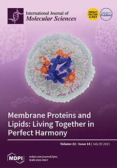
Cover Story (view full-size image): Membrane proteins and lipids, which are integral to the biological membrane, work hand in hand to ensure the smooth running of a myriad of physiological processes. Understanding and observing the molecular interplay between lipids and proteins is an experimental challenge. This review presents the recent biophysical approaches aimed at understanding how membrane proteins and lipids interact. The role of lipids in modulating oligomerization, conformational dynamics and ligand binding is summarized, and different techniques to observe such modulation are introduced along the way. A specific emphasis is put on single-molecule FRET, cryoelectron microscopy and native mass spectrometry techniques, given their important contribution to the study of lipid–protein interactions in the last five years. View this paper
  • Issues are regarded as officially published after their release is announced to the table of contents alert mailing list.
  • You may sign up for e-mail alerts to receive table of contents of newly released issues.
  • PDF is the official format for papers published in both, html and pdf forms. To view the papers in pdf format, click on the "PDF Full-text" link, and use the free Adobe Reader to open them.
Order results
Result details
Section
Select all
Export citation of selected articles as:
22 pages, 1239 KiB  
Review
MYC Ran Up the Clock: The Complex Interplay between MYC and the Molecular Circadian Clock in Cancer
by Jamison B. Burchett, Amelia M. Knudsen-Clark and Brian J. Altman
Int. J. Mol. Sci. 2021, 22(14), 7761; https://doi.org/10.3390/ijms22147761 - 20 Jul 2021
Cited by 13 | Viewed by 3902
Abstract
The MYC oncoprotein and its family members N-MYC and L-MYC are known to drive a wide variety of human cancers. Emerging evidence suggests that MYC has a bi-directional relationship with the molecular clock in cancer. The molecular clock is responsible for circadian (~24 [...] Read more.
The MYC oncoprotein and its family members N-MYC and L-MYC are known to drive a wide variety of human cancers. Emerging evidence suggests that MYC has a bi-directional relationship with the molecular clock in cancer. The molecular clock is responsible for circadian (~24 h) rhythms in most eukaryotic cells and organisms, as a mechanism to adapt to light/dark cycles. Disruption of human circadian rhythms, such as through shift work, may serve as a risk factor for cancer, but connections with oncogenic drivers such as MYC were previously not well understood. In this review, we examine recent evidence that MYC in cancer cells can disrupt the molecular clock; and conversely, that molecular clock disruption in cancer can deregulate and elevate MYC. Since MYC and the molecular clock control many of the same processes, we then consider competition between MYC and the molecular clock in several select aspects of tumor biology, including chromatin state, global transcriptional profile, metabolic rewiring, and immune infiltrate in the tumor. Finally, we discuss how the molecular clock can be monitored or diagnosed in human tumors, and how MYC inhibition could potentially restore molecular clock function. Further study of the relationship between the molecular clock and MYC in cancer may reveal previously unsuspected vulnerabilities which could lead to new treatment strategies. Full article
(This article belongs to the Special Issue Myc Function and Regulation)
Show Figures

Figure 1

16 pages, 2103 KiB  
Review
Regulation of VEGFR Signalling in Lymphatic Vascular Development and Disease: An Update
by Genevieve A. Secker and Natasha L. Harvey
Int. J. Mol. Sci. 2021, 22(14), 7760; https://doi.org/10.3390/ijms22147760 - 20 Jul 2021
Cited by 34 | Viewed by 5003
Abstract
The importance of lymphatic vessels in a myriad of human diseases is rapidly gaining recognition; lymphatic vessel dysfunction is a feature of disorders including congenital lymphatic anomalies, primary lymphoedema and obesity, while improved lymphatic vessel function increases the efficacy of immunotherapy for cancer [...] Read more.
The importance of lymphatic vessels in a myriad of human diseases is rapidly gaining recognition; lymphatic vessel dysfunction is a feature of disorders including congenital lymphatic anomalies, primary lymphoedema and obesity, while improved lymphatic vessel function increases the efficacy of immunotherapy for cancer and neurological disease and promotes cardiac repair following myocardial infarction. Understanding how the growth and function of lymphatic vessels is precisely regulated therefore stands to inform the development of novel therapeutics applicable to a wide range of human diseases. Lymphatic vascular development is initiated during embryogenesis following establishment of the major blood vessels and the onset of blood flow. Lymphatic endothelial progenitor cells arise from a combination of venous and non-venous sources to generate the initial lymphatic vascular structures in the vertebrate embryo, which are then further ramified and remodelled to elaborate an extensive lymphatic vascular network. Signalling mediated via vascular endothelial growth factor (VEGF) family members and vascular endothelial growth factor receptor (VEGFR) tyrosine kinases is crucial for development of both the blood and lymphatic vascular networks, though distinct components are utilised to different degrees in each vascular compartment. Although much is known about the regulation of VEGFA/VEGFR2 signalling in the blood vasculature, less is understood regarding the mechanisms by which VEGFC/VEGFD/VEGFR3 signalling is regulated during lymphatic vascular development. This review will focus on recent advances in our understanding of the cellular and molecular mechanisms regulating VEGFA-, VEGFC- and VEGFD-mediated signalling via VEGFRs which are important for driving the construction of lymphatic vessels during development and disease. Full article
Show Figures

Figure 1

19 pages, 4312 KiB  
Article
Effect of Dimer Structure and Inhomogeneous Broadening of Energy Levels on the Action of Flavomononucleotide in Rigid Polyvinyl Alcohol Films
by Hanna Grajek, Jacek Kubicki, Ignacy Gryczyński, Jerzy Karolczak, Grażyna Żurkowska, Agnieszka I. Piotrowicz-Cieślak and Piotr Bojarski
Int. J. Mol. Sci. 2021, 22(14), 7759; https://doi.org/10.3390/ijms22147759 - 20 Jul 2021
Cited by 1 | Viewed by 2275
Abstract
The results of time-resolved fluorescence measurements of flavin mononucleotide (FMN) in rigid polyvinyl alcohol films (PVA) demonstrate that fluorescence intensity decays are strongly accelerated in the presence of fluorescent dimers and nonradiative energy transfer processes. The fluorescence decay originating both from H and [...] Read more.
The results of time-resolved fluorescence measurements of flavin mononucleotide (FMN) in rigid polyvinyl alcohol films (PVA) demonstrate that fluorescence intensity decays are strongly accelerated in the presence of fluorescent dimers and nonradiative energy transfer processes. The fluorescence decay originating both from H and J dimer states of FMN was experimentally observed for the first time. The mean fluorescence lifetimes for FMN dimers were obtained: τfl = 2.66 ns (at λexc = 445 nm) and τfl = 2.02 (at λexc = 487 nm) at λobs = 600 nm and T = 253 K from H and J state of dimers, respectively. We show that inhomogeneous orientational broadening of energy levels (IOBEL) affects the shape of the fluorescence decay and leads to the dependence of the average monomer fluorescence lifetime on excitation wavelength. IOBEL affected the nonradiative energy transfer and indicated that different flavin positioning in the protein pocket could (1) change the spectroscopic properties of flavins due to the existence of “blue” and “red” fluorescence centers, and (2) diminish the effectiveness of energy transfer between FMN molecules. Full article
(This article belongs to the Collection Feature Papers in Molecular Biophysics)
Show Figures

Figure 1

17 pages, 12369 KiB  
Article
Mutational Analysis of Redβ Single Strand Annealing Protein: Roles of the 14 Lysine Residues in DNA Binding and Recombination In Vivo
by Katerina Zakharova, Brian J. Caldwell, Shalya Ta, Carter T. Wheat and Charles E. Bell
Int. J. Mol. Sci. 2021, 22(14), 7758; https://doi.org/10.3390/ijms22147758 - 20 Jul 2021
Cited by 3 | Viewed by 2523
Abstract
Redβ is a 261 amino acid protein from bacteriophage λ that promotes a single-strand annealing (SSA) reaction for repair of double-stranded DNA (dsDNA) breaks. While there is currently no high-resolution structure available for Redβ, models of its DNA binding domain (residues 1–188) have [...] Read more.
Redβ is a 261 amino acid protein from bacteriophage λ that promotes a single-strand annealing (SSA) reaction for repair of double-stranded DNA (dsDNA) breaks. While there is currently no high-resolution structure available for Redβ, models of its DNA binding domain (residues 1–188) have been proposed based on homology with human Rad52, and a crystal structure of its C-terminal domain (CTD, residues 193-261), which binds to λ exonuclease and E. coli single-stranded DNA binding protein (SSB), has been determined. To evaluate these models, the 14 lysine residues of Redβ were mutated to alanine, and the variants tested for recombination in vivo and DNA binding and annealing in vitro. Most of the lysines within the DNA binding domain, including K36, K61, K111, K132, K148, K154, and K172, were found to be critical for DNA binding in vitro and recombination in vivo. By contrast, none of the lysines within the CTD, including K214, K245, K251, K253, and K258 were required for DNA binding in vitro, but two, K214 and K253, were critical for recombination in vivo, likely due to their involvement in binding to SSB. K61 was identified as a residue that is critical for DNA annealing, but not for initial ssDNA binding, suggesting a role in binding to the second strand of DNA incorporated into the complex. The K148A variant, which has previously been shown to be defective in oligomer formation, had the lowest affinity for ssDNA, and was the only variant that was completely non-cooperative, suggesting that ssDNA binding is coupled to oligomerization. Full article
(This article belongs to the Special Issue Small Prokaryotic Proteins Interacting with Nucleic Acids)
Show Figures

Figure 1

9 pages, 1593 KiB  
Article
Optimal Treatment of 6-Dimethylaminopurine Enhances the In Vivo Development of Canine Embryos by Rapid Initiation of DNA Synthesis
by Hyun Ju Oh, Byeong Chun Lee and Min Kyu Kim
Int. J. Mol. Sci. 2021, 22(14), 7757; https://doi.org/10.3390/ijms22147757 - 20 Jul 2021
Cited by 3 | Viewed by 1943
Abstract
Artificial activation of oocytes is an important step for successful parthenogenesis and somatic cell nuclear transfer (SCNT). Here, we investigated the initiation of DNA synthesis and in vivo development of canine PA embryos and cloned embryos produced by treatment with 1.9 mM 6-dimethylaminopurine [...] Read more.
Artificial activation of oocytes is an important step for successful parthenogenesis and somatic cell nuclear transfer (SCNT). Here, we investigated the initiation of DNA synthesis and in vivo development of canine PA embryos and cloned embryos produced by treatment with 1.9 mM 6-dimethylaminopurine (6-DMAP) for different lengths of time. For experiments, oocytes for parthenogenesis and SCNT oocytes were cultured for 4 min in 10 μM calcium ionophore, and then divided into 2 groups: (1) culture for 2 h in 6-DMAP (DMAP-2h group); (2) culture for 4 h in DMAP (DMAP-4h group). DNA synthesis was clearly detected in all parthenogenetic (PA) embryos and cloned embryos incorporated BrdU 4 h after activation in DMAP-2h and DMAP-4h groups. In vivo development of canine parthenogenetic fetuses was observed after embryo transfer and the implantation rates of PA embryos in DMAP-2h were 34%, which was significantly higher than those in DMAP-4h (6.5%, p < 0.05). However, in SCNT, there was no significant difference in pregnancy rate (DMAP-2h: 41.6% vs. DMAP-4h: 33.3%) and implantation rates (DMAP-2h: 4.94% vs. DMAP-4h: 3.19%) between DMAP-2h and DMAP-4h. In conclusion, the use of DMAP-2h for canine oocyte activation may be ideal for the in vivo development of PA zygotes, but it was not more effective in in vivo development of canine reconstructed SCNT oocytes. The present study demonstrated that DMAP-2h treatment on activation of canine parthenogenesis and SCNT could effectively induce the onset of DNA synthesis during the first cell cycle. Full article
Show Figures

Figure 1

15 pages, 4255 KiB  
Article
Effect of ZnSO4, MnSO4 and FeSO4 on the Partial Hydrogenation of Benzene over Nano Ru-Based Catalysts
by Haijie Sun, Yiru Fan, Xiangrong Sun, Zhihao Chen, Huiji Li, Zhikun Peng and Zhongyi Liu
Int. J. Mol. Sci. 2021, 22(14), 7756; https://doi.org/10.3390/ijms22147756 - 20 Jul 2021
Cited by 4 | Viewed by 2629
Abstract
Nano Ru-based catalysts, including monometallic Ru and Ru-Zn nanoparticles, were synthesized via a precipitation method. The prepared catalysts were evaluated on partial hydrogenation of benzene towards cyclohexene generation, during which the effect of reaction modifiers, i.e., ZnSO4, MnSO4, and [...] Read more.
Nano Ru-based catalysts, including monometallic Ru and Ru-Zn nanoparticles, were synthesized via a precipitation method. The prepared catalysts were evaluated on partial hydrogenation of benzene towards cyclohexene generation, during which the effect of reaction modifiers, i.e., ZnSO4, MnSO4, and FeSO4, was investigated. The fresh and the spent catalysts were thoroughly characterized by XRD, TEM, SEM, XPS, XRF, and DFT studies. It was found that Zn2+ or Fe2+ could be adsorbed on the surface of a monometallic Ru catalyst, where a stabilized complex could be formed between the cations and the cyclohexene. This led to an enhancement of catalytic selectivity towards cyclohexene. Furthermore, electron transfer was observed from Zn2+ or Fe2+ to Ru, hindering the catalytic activity towards benzene hydrogenation. In comparison, very few Mn2+ cations were adsorbed on the Ru surface, for which no cyclohexene could be detected. On the other hand, for Ru-Zn catalyst, Zn existed as rodlike ZnO. The added ZnSO4 and FeSO4 could react with ZnO to generate (Zn(OH)2)5(ZnSO4)(H2O) and basic Fe sulfate, respectively. This further benefited the adsorption of Zn2+ or Fe2+, leading to the decrease of catalytic activity towards benzene conversion and the increase of selectivity towards cyclohexene synthesis. When 0.57 mol·L−1 of ZnSO4 was applied, the highest cyclohexene yield of 62.6% was achieved. When MnSO4 was used as a reaction modifier, H2SO4 could be generated in the slurry via its hydrolysis, which reacted with ZnO to form ZnSO4. The selectivity towards cyclohexene formation was then improved by the adsorbed Zn2+. Full article
(This article belongs to the Special Issue Multifunctional Nanomaterials: Synthesis, Properties and Applications)
Show Figures

Figure 1

19 pages, 1158 KiB  
Review
Microbeam Radiotherapy—A Novel Therapeutic Approach to Overcome Radioresistance and Enhance Anti-Tumour Response in Melanoma
by Verdiana Trappetti, Jennifer M. Fazzari, Cristian Fernandez-Palomo, Maximilian Scheidegger, Vladislav Volarevic, Olga A. Martin and Valentin G. Djonov
Int. J. Mol. Sci. 2021, 22(14), 7755; https://doi.org/10.3390/ijms22147755 - 20 Jul 2021
Cited by 18 | Viewed by 3173
Abstract
Melanoma is the deadliest type of skin cancer, due to its invasiveness and limited treatment efficacy. The main therapy for primary melanoma and solitary organ metastases is wide excision. Adjuvant therapy, such as chemotherapy and targeted therapies are mainly used for disseminated disease. [...] Read more.
Melanoma is the deadliest type of skin cancer, due to its invasiveness and limited treatment efficacy. The main therapy for primary melanoma and solitary organ metastases is wide excision. Adjuvant therapy, such as chemotherapy and targeted therapies are mainly used for disseminated disease. Radiotherapy (RT) is a powerful treatment option used in more than 50% of cancer patients, however, conventional RT alone is unable to eradicate melanoma. Its general radioresistance is attributed to overexpression of repair genes in combination with cascades of biochemical repair mechanisms. A novel sophisticated technique based on synchrotron-generated, spatially fractionated RT, called Microbeam Radiation Therapy (MRT), has been shown to overcome these treatment limitations by allowing increased dose delivery. With MRT, a collimator subdivides the homogeneous radiation field into an array of co-planar, high-dose microbeams that are tens of micrometres wide and spaced a few hundred micrometres apart. Different preclinical models demonstrated that MRT has the potential to completely ablate tumours, or significantly improve tumour control while dramatically reducing normal tissue toxicity. Here, we discuss the role of conventional RT-induced immunity and the potential for MRT to enhance local and systemic anti-tumour immune responses. Comparative gene expression analysis from preclinical tumour models indicated a specific gene signature for an ‘MRT-induced immune effect’. This focused review highlights the potential of MRT to overcome the inherent radioresistance of melanoma which could be further enhanced for future clinical use with combined treatment strategies, in particular, immunotherapy. Full article
(This article belongs to the Special Issue Immunotherapy of Melanoma: Challenges and Solutions)
Show Figures

Figure 1

21 pages, 10012 KiB  
Review
Yeast Interactions and Molecular Mechanisms in Wine Fermentation: A Comprehensive Review
by Francesca Comitini, Alice Agarbati, Laura Canonico and Maurizio Ciani
Int. J. Mol. Sci. 2021, 22(14), 7754; https://doi.org/10.3390/ijms22147754 - 20 Jul 2021
Cited by 34 | Viewed by 5245
Abstract
Wine can be defined as a complex microbial ecosystem, where different microorganisms interact in the function of different biotic and abiotic factors. During natural fermentation, the effect of unpredictable interactions between microorganisms and environmental factors leads to the establishment of a complex and [...] Read more.
Wine can be defined as a complex microbial ecosystem, where different microorganisms interact in the function of different biotic and abiotic factors. During natural fermentation, the effect of unpredictable interactions between microorganisms and environmental factors leads to the establishment of a complex and stable microbiota that will define the kinetics of the process and the final product. Controlled multistarter fermentation represents a microbial approach to achieve the dual purpose of having a less risky process and a distinctive final product. Indeed, the interactions evolved between microbial consortium members strongly modulate the final sensorial properties of the wine. Therefore, in well-managed mixed fermentations, the knowledge of molecular mechanisms on the basis of yeast interactions, in a well-defined ecological niche, becomes fundamental to control the winemaking process, representing a tool to achieve such objectives. In the present work, the recent development on the molecular and metabolic interactions between non-Saccharomyces and Saccharomyces yeasts in wine fermentation was reviewed. A particular focus will be reserved on molecular studies regarding the role of nutrients, the production of the main byproducts and volatile compounds, ethanol reduction, and antagonistic actions for biological control in mixed fermentations. Full article
(This article belongs to the Special Issue Biotechnology of Non-conventional Yeasts)
Show Figures

Figure 1

16 pages, 22214 KiB  
Article
Phosphorylation of H3-Thr3 by Haspin Is Required for Primary Cilia Regulation
by Roberto Quadri, Sarah Sertic, Anna Ghilardi, Diego Rondelli, Guido Roberto Gallo, Luca Del Giacco and Marco Muzi-Falconi
Int. J. Mol. Sci. 2021, 22(14), 7753; https://doi.org/10.3390/ijms22147753 - 20 Jul 2021
Cited by 6 | Viewed by 2480
Abstract
Primary cilia are commonly found on most quiescent, terminally differentiated cells and play a major role in the regulation of the cell cycle, cell motility, sensing, and cell–cell communication. Alterations in ciliogenesis and cilia maintenance are causative of several human diseases, collectively known [...] Read more.
Primary cilia are commonly found on most quiescent, terminally differentiated cells and play a major role in the regulation of the cell cycle, cell motility, sensing, and cell–cell communication. Alterations in ciliogenesis and cilia maintenance are causative of several human diseases, collectively known as ciliopathies. A key determinant of primary cilia is the histone deacetylase HDAC6, which regulates their length and resorption and whose distribution is regulated by the death inducer-obliterator 3 (Dido3). Here, we report that the atypical protein kinase Haspin is a key regulator of cilia dynamics. Cells defective in Haspin activity exhibit longer primary cilia and a strong delay in cilia resorption upon cell cycle reentry. We show that Haspin is active in quiescent cells, where it phosphorylates threonine 3 of histone H3, a known mitotic Haspin substrate. Forcing Dido3 detachment from the chromatin prevents Haspin inhibition from impacting cilia dynamics, suggesting that Haspin activity is required for the relocalization of Dido3–HDAC6 to the basal body. Exploiting the zebrafish model, we confirmed the physiological relevance of this mechanism. Our observations shed light on a novel player, Haspin, in the mechanisms that govern the determination of cilia length and the homeostasis of mature cilia. Full article
(This article belongs to the Section Molecular Biology)
Show Figures

Figure 1

12 pages, 4833 KiB  
Article
Evaluation of MC3T3-E1 Cell Osteogenesis in Different Cell Culture Media
by Makoto Izumiya, Miyu Haniu, Katsuya Ueda, Haruka Ishida, Chuang Ma, Hirokazu Ideta, Atsushi Sobajima, Koki Ueshiba, Takeshi Uemura, Naoto Saito and Hisao Haniu
Int. J. Mol. Sci. 2021, 22(14), 7752; https://doi.org/10.3390/ijms22147752 - 20 Jul 2021
Cited by 24 | Viewed by 8004
Abstract
Many biomaterials have been evaluated using cultured cells. In particular, osteoblast-like cells are often used to evaluate the osteocompatibility, hard-tissue-regeneration, osteoconductive, and osteoinductive characteristics of biomaterials. However, the evaluation of biomaterial osteogenesis-inducing capacity using osteoblast-like cells is not standardized; instead, it is performed [...] Read more.
Many biomaterials have been evaluated using cultured cells. In particular, osteoblast-like cells are often used to evaluate the osteocompatibility, hard-tissue-regeneration, osteoconductive, and osteoinductive characteristics of biomaterials. However, the evaluation of biomaterial osteogenesis-inducing capacity using osteoblast-like cells is not standardized; instead, it is performed under laboratory-specific culture conditions with different culture media. However, the effect of different media conditions on bone formation has not been investigated. Here, we aimed to evaluate the osteogenesis of MC3T3-E1 cells, one of the most commonly used osteoblast-like cell lines for osteogenesis evaluation, and assayed cell proliferation, alkaline phosphatase activity, expression of osteoblast markers, and calcification under varying culture media conditions. Furthermore, the various media conditions were tested in uncoated plates and plates coated with collagen type I and poly-L-lysine, highly biocompatible molecules commonly used as pseudobiomaterials. We found that the type of base medium, the presence or absence of vitamin C, and the freshness of the medium may affect biomaterial regeneration. We posit that an in vitro model that recapitulates in vivo bone formation should be established before evaluating biomaterials. Full article
(This article belongs to the Special Issue Biomaterials for Bone Tissue Engineering 2.0)
Show Figures

Figure 1

15 pages, 5111 KiB  
Article
In Silico Prediction of the Mechanism of Action of Pyriproxyfen and 4′-OH-Pyriproxyfen against A. mellifera and H. sapiens Receptors
by Giulia Spaggiari, Nadia Iovine and Pietro Cozzini
Int. J. Mol. Sci. 2021, 22(14), 7751; https://doi.org/10.3390/ijms22147751 - 20 Jul 2021
Cited by 3 | Viewed by 2301
Abstract
Background. Poisoning from pesticides can be extremely hazardous for non-invasive species, such as bees, and humans causing nearly 300,000 deaths worldwide every year. Several pesticides are recognized as endocrine disruptors compounds that alter the production of the normal hormones mainly by acting through [...] Read more.
Background. Poisoning from pesticides can be extremely hazardous for non-invasive species, such as bees, and humans causing nearly 300,000 deaths worldwide every year. Several pesticides are recognized as endocrine disruptors compounds that alter the production of the normal hormones mainly by acting through their interaction with nuclear receptors (NRs). Among the insecticides, one of the most used is pyriproxyfen. As analogous to the juvenile hormone, the pyriproxyfen acts in the bee’s larval growth and creates malformations at the adult organism level. Methods. This work aims to investigate the possible negative effects of pyriproxyfen and its metabolite, the 4′-OH-pyriproxyfen, on human and bee health. We particularly investigated the mechanism of binding of pyriproxyfen and its metabolite with ultraspiracle protein/ecdysone receptor (USP-EcR) dimer of A. mellifera and the relative heterodimer farnesoid X receptor/retinoid X receptor alpha (FXR-RXRα) of H. sapiens using molecular dynamic simulations. Results. The results revealed that pyriproxyfen and its metabolite, the 4′-OH- pyriproxyfen, stabilize each dimer and resulted in stronger binders than the natural ligands. Conclusion. We demonstrated the endocrine interference of two pesticides and explained their possible mechanism of action. Furthermore, in vitro studies should be carried out to evaluate the biological effects of pyriproxyfen and its metabolite. Full article
(This article belongs to the Section Biochemistry)
Show Figures

Graphical abstract

15 pages, 5154 KiB  
Article
Toxicity of Carbon Nanomaterials—Towards Reliable Viability Assessment via New Approach in Flow Cytometry
by Tomáš Malina, Kateřina Poláková, Cordula Hirsch, Ladislav Svoboda and Radek Zbořil
Int. J. Mol. Sci. 2021, 22(14), 7750; https://doi.org/10.3390/ijms22147750 - 20 Jul 2021
Cited by 6 | Viewed by 2130
Abstract
The scope of application of carbon nanomaterials in biomedical, environmental and industrial fields is recently substantially increasing. Since in vitro toxicity testing is the first essential step for any commercial usage, it is crucial to have a reliable method to analyze the potentially [...] Read more.
The scope of application of carbon nanomaterials in biomedical, environmental and industrial fields is recently substantially increasing. Since in vitro toxicity testing is the first essential step for any commercial usage, it is crucial to have a reliable method to analyze the potentially harmful effects of carbon nanomaterials. Even though researchers already reported the interference of carbon nanomaterials with common toxicity assays, there is still, unfortunately, a large number of studies that neglect this fact. In this study, we investigated interference of four bio-promising carbon nanomaterials (graphene acid (GA), cyanographene (GCN), graphitic carbon nitride (g-C3N4) and carbon dots (QCDs)) in commonly used LIVE/DEAD assay. When a standard procedure was applied, materials caused various types of interference. While positively charged g-C3N4 and QCDs induced false results through the creation of free agglomerates and intrinsic fluorescence properties, negatively charged GA and GCN led to false signals due to the complex quenching effect of the fluorescent dye of a LIVE/DEAD kit. Thus, we developed a new approach using a specific gating strategy based on additional controls that successfully overcame all types of interference and lead to reliable results in LIVE/DEAD assay. We suggest that the newly developed procedure should be a mandatory tool for all in vitro flow cytometry assays of any class of carbon nanomaterials. Full article
(This article belongs to the Special Issue Bio-Nano Interactions)
Show Figures

Graphical abstract

16 pages, 30825 KiB  
Article
Actin Isovariant ACT7 Modulates Root Thermomorphogenesis by Altering Intracellular Auxin Homeostasis
by Sumaya Parveen and Abidur Rahman
Int. J. Mol. Sci. 2021, 22(14), 7749; https://doi.org/10.3390/ijms22147749 - 20 Jul 2021
Cited by 12 | Viewed by 3004
Abstract
High temperature stress is one of the most threatening abiotic stresses for plants limiting the crop productivity world-wide. Altered developmental responses of plants to moderate-high temperature has been shown to be linked to the intracellular auxin homeostasis regulated by both auxin biosynthesis and [...] Read more.
High temperature stress is one of the most threatening abiotic stresses for plants limiting the crop productivity world-wide. Altered developmental responses of plants to moderate-high temperature has been shown to be linked to the intracellular auxin homeostasis regulated by both auxin biosynthesis and transport. Trafficking of the auxin carrier proteins plays a major role in maintaining the cellular auxin homeostasis. The intracellular trafficking largely relies on the cytoskeletal component, actin, which provides track for vesicle movement. Different classes of actin and the isovariants function in regulating various stages of plant development. Although high temperature alters the intracellular trafficking, the role of actin in this process remains obscure. Using isovariant specific vegetative class actin mutants, here we demonstrate that ACTIN 7 (ACT7) isovariant plays an important role in regulating the moderate-high temperature response in Arabidopsis root. Loss of ACT7, but not ACT8 resulted in increased inhibition of root elongation under prolonged moderate-high temperature. Consistently, kinematic analysis revealed a drastic reduction in cell production rate and cell elongation in act7-4 mutant under high temperature. Quantification of actin dynamicity reveals that prolonged moderate-high temperature modulates bundling along with orientation and parallelness of filamentous actin in act7-4 mutant. The hypersensitive response of act7-4 mutant was found to be linked to the altered intracellular auxin distribution, resulted from the reduced abundance of PIN-FORMED PIN1 and PIN2 efflux carriers. Collectively, these results suggest that vegetative class actin isovariant, ACT7 modulates the long-term moderate-high temperature response in Arabidopsis root. Full article
Show Figures

Figure 1

13 pages, 867 KiB  
Review
Androgen/Androgen Receptor Signaling in Ovarian Cancer: Molecular Regulation and Therapeutic Potentials
by Wei-Min Chung, Lumin Chen, Wei-Chun Chang, Sheng-Yuan Su, Yao-Ching Hung and Wen-Lung Ma
Int. J. Mol. Sci. 2021, 22(14), 7748; https://doi.org/10.3390/ijms22147748 - 20 Jul 2021
Cited by 18 | Viewed by 2857
Abstract
Ovarian cancer (OVCA) arises from three cellular origins, namely surface epithelial cells, germ cells, and stromal cells. More than 85% of OVCAs are EOCs (epithelial ovarian carcinomas), which are the most lethal gynecological malignancies. Cancer stem/progenitor cells (CSPCs) are considered to be cancer [...] Read more.
Ovarian cancer (OVCA) arises from three cellular origins, namely surface epithelial cells, germ cells, and stromal cells. More than 85% of OVCAs are EOCs (epithelial ovarian carcinomas), which are the most lethal gynecological malignancies. Cancer stem/progenitor cells (CSPCs) are considered to be cancer promoters due to their capacity for unlimited self-renewal and drug resistance. Androgen receptor (AR) belongs to the nuclear receptor superfamily and can be activated through binding to its ligand androgens. Studies have reported an association between AR expression and EOC carcinogenesis, and AR is suggested to be involved in proliferation, migration/invasion, and stemness. In addition, alternative AR activating signals, including both ligand-dependent and ligand-independent, are involved in OVCA progression. Although some clinical trials have previously been conducted to evaluate the effects of anti-androgens in EOC, no significant results have been reported. In contrast, experimental studies evaluating the effects of anti-androgen or anti-AR reagents in AR-expressing EOC models have demonstrated positive results for suppressing disease progression. Since AR is involved in complex signaling pathways and may be expressed at various levels in OVCA, the aim of this article was to provide an overview of current studies and perspectives regarding the relevance of androgen/AR roles in OVCA. Full article
(This article belongs to the Special Issue Steroid Hormones and Sex Difference in Diseases)
Show Figures

Figure 1

21 pages, 4036 KiB  
Article
Extracellular Hsp90α Promotes Tumor Lymphangiogenesis and Lymph Node Metastasis in Breast Cancer
by Qiaoyun Hou, Shuohua Chen, Qi An, Boya Li, Yan Fu and Yongzhang Luo
Int. J. Mol. Sci. 2021, 22(14), 7747; https://doi.org/10.3390/ijms22147747 - 20 Jul 2021
Cited by 21 | Viewed by 2720
Abstract
Early detection and discovery of new therapeutic targets are urgently needed to improve the breast cancer treatment outcome. Here we conducted an official clinical trial with cross-validation to corroborate human plasma Hsp90α as a novel breast cancer biomarker. Importantly, similar results were noticed [...] Read more.
Early detection and discovery of new therapeutic targets are urgently needed to improve the breast cancer treatment outcome. Here we conducted an official clinical trial with cross-validation to corroborate human plasma Hsp90α as a novel breast cancer biomarker. Importantly, similar results were noticed in detecting early-stage breast cancer patients. Additionally, levels of plasma Hsp90α in breast cancer patients were gradually elevated as their clinical stages of regional lymph nodes advanced. In orthotopic breast cancer mouse models, administrating with recombinant Hsp90α protein increased both the primary tumor lymphatic vessel density and sentinel lymph node metastasis by 2 and 10 times, respectively. What is more, Hsp90α neutralizing antibody treatment approximately reduced 70% of lymphatic vessel density and 90% of sentinel lymph node metastasis. In the in vitro study, we demonstrated the role of extracellular Hsp90α (eHsp90α) as a pro-lymphangiogenic factor, which significantly enhanced migration and tube formation abilities of lymphatic endothelial cells (LECs). Mechanistically, eHsp90α signaled to the AKT pathway through low-density lipoprotein receptor-related protein 1 (LRP1) to upregulate the expression and secretion of CXCL8 in the lymphangiogenic process. Collectively, this study proves that plasma Hsp90α serves as an auxiliary diagnosis biomarker and eHsp90α as a molecular mediator promoting lymphangiogenesis in breast cancer. Full article
Show Figures

Figure 1

12 pages, 932 KiB  
Review
Expression and Role of Ubiquitin-Specific Peptidases in Osteoblasts
by Hadla Hariri and René St-Arnaud
Int. J. Mol. Sci. 2021, 22(14), 7746; https://doi.org/10.3390/ijms22147746 - 20 Jul 2021
Cited by 9 | Viewed by 2291
Abstract
The ubiquitin-proteasome system regulates biological processes in normal and diseased states. Recent investigations have focused on ubiquitin-dependent modifications and their impacts on cellular function, commitment, and differentiation. Ubiquitination is reversed by deubiquitinases, including ubiquitin-specific peptidases (USPs), whose roles have been widely investigated. In [...] Read more.
The ubiquitin-proteasome system regulates biological processes in normal and diseased states. Recent investigations have focused on ubiquitin-dependent modifications and their impacts on cellular function, commitment, and differentiation. Ubiquitination is reversed by deubiquitinases, including ubiquitin-specific peptidases (USPs), whose roles have been widely investigated. In this review, we explore recent findings highlighting the regulatory functions of USPs in osteoblasts and providing insight into the molecular mechanisms governing their actions during bone formation. We also give a brief overview of our work on USP53, a target of PTH in osteoblasts and a regulator of mesenchymal cell lineage fate decisions. Emerging evidence addresses questions pertaining to the complex layers of regulation exerted by USPs on osteoblast signaling. We provide a short overview of our and others’ understanding of how USPs modulate osteoblastogenesis. However, further studies using knockout mouse models are needed to fully understand the mechanisms underpinning USPs actions. Full article
(This article belongs to the Special Issue Osteoblast Differentiation and Activity in Skeletal Diseases)
Show Figures

Graphical abstract

22 pages, 5762 KiB  
Article
6-Gingerol, a Major Constituent of Zingiber officinale Rhizoma, Exerts Anticonvulsant Activity in the Pentylenetetrazole-Induced Seizure Model in Larval Zebrafish
by Kinga Gawel, Wirginia Kukula-Koch, Nancy Saana Banono, Dorota Nieoczym, Katarzyna M. Targowska-Duda, Lidia Czernicka, Jolanta Parada-Turska and Camila V. Esguerra
Int. J. Mol. Sci. 2021, 22(14), 7745; https://doi.org/10.3390/ijms22147745 - 20 Jul 2021
Cited by 25 | Viewed by 2962
Abstract
Zingiber officinale is one of the most frequently used medicinal herbs in Asia. Using rodent seizure models, it was previously shown that Zingiber officinale hydroethanolic extract exerts antiseizure activity, but the active constituents responsible for this effect have not been determined. In this [...] Read more.
Zingiber officinale is one of the most frequently used medicinal herbs in Asia. Using rodent seizure models, it was previously shown that Zingiber officinale hydroethanolic extract exerts antiseizure activity, but the active constituents responsible for this effect have not been determined. In this paper, we demonstrated that Zingiber officinale methanolic extract exerts anticonvulsant activity in the pentylenetetrazole (PTZ)-induced hyperlocomotion assay in larval zebrafish. Next, we isolated 6-gingerol (6-GIN)—a major constituent of Zingiber officinale rhizoma. We observed that 6-GIN exerted potent dose-dependent anticonvulsant activity in the PTZ-induced hyperlocomotion seizure assay in zebrafish, which was confirmed electroencephalographically. To obtain further insight into the molecular mechanisms of 6-GIN antiseizure activity, we assessed the concentration of two neurotransmitters in zebrafish, i.e., inhibitory γ-aminobutyric acid (GABA) and excitatory glutamic acid (GLU), and their ratio after exposure to acute PTZ dose. Here, 6-GIN decreased GLU level and reduced the GLU/GABA ratio in PTZ-treated fish compared with only PTZ-bathed fish. This activity was associated with the decrease in grin2b, but not gabra1a, grin1a, gria1a, gria2a, and gria3b expression in PTZ-treated fish. Molecular docking to the human NR2B-containing N-methyl-D-aspartate (NMDA) receptor suggests that 6-GIN might act as an inhibitor and interact with the amino terminal domain, the glutamate-binding site, as well as within the ion channel of the NR2B-containing NMDA receptor. In summary, our study reveals, for the first time, the anticonvulsant activity of 6-GIN. We suggest that this effect might at least be partially mediated by restoring the balance between GABA and GLU in the epileptic brain; however, more studies are needed to prove our hypothesis. Full article
(This article belongs to the Special Issue Biological Properties of Medicinal Plants)
Show Figures

Figure 1

25 pages, 9516 KiB  
Article
The Process of Binding and Releasing of Genetic Material from Lipoplexes Based on Trimeric Surfactants and Phospholipids
by Żaneta Polańska, Zuzanna Pietralik-Molińska, Daria Wojciechowska, Augustyn Moliński, Marek Weiss, Andrzej Skrzypczak and Maciej Kozak
Int. J. Mol. Sci. 2021, 22(14), 7744; https://doi.org/10.3390/ijms22147744 - 20 Jul 2021
Cited by 1 | Viewed by 1971
Abstract
Nonviral vectors for gene therapy such as lipoplexes are characterized by low toxicity, high biocompatibility, and good transfection efficiency. Specifically, lipoplexes based on polymeric surfactants and phospholipids have great potential as gene carriers due to the increased ability to bind genetic material (multiplied [...] Read more.
Nonviral vectors for gene therapy such as lipoplexes are characterized by low toxicity, high biocompatibility, and good transfection efficiency. Specifically, lipoplexes based on polymeric surfactants and phospholipids have great potential as gene carriers due to the increased ability to bind genetic material (multiplied positive electric charge) while lowering undesirable effects (the presence of lipids makes the system more like natural membranes). This study aimed to test the ability to bind and release genetic material by lipoplexes based on trimeric surfactants and lipid formulations of different compositions and to characterize formed complexes by circular dichroism (CD) spectroscopy and atomic force microscopy (AFM). The cytotoxicity of studied lipoplexes was tested on HeLa cells by the MTT cell viability assay and the dye exclusion test (trypan blue). The presence of lipids in the system lowered the surfactant concentration required for complexation (higher efficiency) and reduced the cytotoxicity of lipoplexes. Surfactant/lipids/DNA complexes were more stable than surfactant/DNA complexes. Surfactant molecules induced the genetic material condensation, but the presence of lipids significantly intensified this process. Systems based on trimeric surfactants and lipid formulations, particularly TRI_N and TRI_IMI systems, could be used as delivery carrier, and have proven to be highly effective, nontoxic, and universal for DNA of various lengths. Full article
(This article belongs to the Special Issue Lipid-Based Drug Carriers and Nanoparticles)
Show Figures

Figure 1

17 pages, 3484 KiB  
Article
Liposomes Loaded with Everolimus and Coated with Hyaluronic Acid: A Promising Approach for Lung Fibrosis
by Laura Pandolfi, Alessandro Marengo, Kamila Bohne Japiassu, Vanessa Frangipane, Nicolas Tsapis, Valeria Bincoletto, Veronica Codullo, Sara Bozzini, Monica Morosini, Sara Lettieri, Valentina Vertui, Davide Piloni, Silvia Arpicco, Elias Fattal and Federica Meloni
Int. J. Mol. Sci. 2021, 22(14), 7743; https://doi.org/10.3390/ijms22147743 - 20 Jul 2021
Cited by 8 | Viewed by 3299
Abstract
Chronic lung allograft dysfunction (CLAD) and interstitial lung disease associated with collagen tissue diseases (CTD-ILD) are two end-stage lung disorders in which different chronic triggers induce activation of myo-/fibroblasts (LFs). Everolimus, an mTOR inhibitor, can be adopted as a potential strategy for CLAD [...] Read more.
Chronic lung allograft dysfunction (CLAD) and interstitial lung disease associated with collagen tissue diseases (CTD-ILD) are two end-stage lung disorders in which different chronic triggers induce activation of myo-/fibroblasts (LFs). Everolimus, an mTOR inhibitor, can be adopted as a potential strategy for CLAD and CTD-ILD, however it exerts important side effects. This study aims to exploit nanomedicine to reduce everolimus side effects encapsulating it inside liposomes targeted against LFs, expressing a high rate of CD44. PEGylated liposomes were modified with high molecular weight hyaluronic acid and loaded with everolimus (PEG-LIP(ev)-HA400kDa). Liposomes were tested by in vitro experiments using LFs derived from broncholveolar lavage (BAL) of patients affected by CLAD and CTD-ILD, and on alveolar macrophages (AM) and lymphocytes isolated, respectively, from BAL and peripheral blood. PEG-LIP-HA400kDa demonstrated to be specific for LFs, but not for CD44-negative cells, and after loading everolimus, PEG-LIP(ev)-HA400kDa were able to arrest cell cycle arrest and to decrease phospho-mTOR level. PEG-LIP(ev)-HA400kDa showed anti-inflammatory effect on immune cells. This study opens the possibility to use everolimus in lung fibrotic diseases, demonstrating that our lipids-based vehicles can vehicle everolimus inside cells exerting the same drug molecular effect, not only in LFs, but also in immune cells. Full article
(This article belongs to the Special Issue Challenges, Opportunities, and Innovation in Local Drug Delivery)
Show Figures

Figure 1

17 pages, 3712 KiB  
Article
Combined Exposure to Diazinon and Nicotine Exerts a Synergistic Adverse Effect In Vitro and Disrupts Brain Development and Behaviors In Vivo
by Bonn Lee, Seon Mi Park, SunHwa Jeong, KangMin Kim and Eui-Bae Jeung
Int. J. Mol. Sci. 2021, 22(14), 7742; https://doi.org/10.3390/ijms22147742 - 20 Jul 2021
Cited by 4 | Viewed by 1985
Abstract
A real-life environment during pregnancy involves multiple and simultaneous exposures to toxic chemicals. Perinatal exposures to toxic chemicals have been reported to exert an inhibitory effect on mouse neural development and behaviors. However, the effect of combined exposures of organophosphate and nicotine has [...] Read more.
A real-life environment during pregnancy involves multiple and simultaneous exposures to toxic chemicals. Perinatal exposures to toxic chemicals have been reported to exert an inhibitory effect on mouse neural development and behaviors. However, the effect of combined exposures of organophosphate and nicotine has not been previously reported. In this study, we investigated whether a combined exposure of diazinon and nicotine can have a synergistic effect. The effects of the combined chemical exposure on cell viability and neuronal differentiation were examined using mouse Sox1-GFP cells. Additionally, mice were maternally administered 0.18 mg/kg diazinon, a no adverse effect level (NOAEL) dose, combined with 0.4, 1, and 2 mg/kg nicotine. Mice offspring underwent behavior tests to assess locomotor, depressive, cognitive, and social behaviors. Morphological change in the brain was investigated with immunolocalization. We revealed that the combined exposure to diazinon and nicotine can have a synergistic adverse effect in vitro. In addition, the chemical-treated mouse offspring showed abnormalities in motor learning, compulsive-like behaviors, spatial learning, and social interaction patterns. Moreover, 0.18 mg/kg diazinon and 2 mg/kg nicotine co-exposure resulted in an increase in tyrosine hydroxylase (TH)-positive dopaminergic neurons. Thus, the findings suggest that perinatal co-exposure to nicotine and diazinon can result in abnormal neurodevelopment and behavior, even at low-level administration. Full article
(This article belongs to the Special Issue Endocrine Disruptors Leading to Obesity and Related Disease)
Show Figures

Figure 1

13 pages, 24962 KiB  
Article
Biological In Vitro Evaluation of PIL Graft Conjugates: Cytotoxicity Characteristics
by Katarzyna Niesyto, Wiktoria Łyżniak, Magdalena Skonieczna and Dorota Neugebauer
Int. J. Mol. Sci. 2021, 22(14), 7741; https://doi.org/10.3390/ijms22147741 - 20 Jul 2021
Cited by 10 | Viewed by 2094
Abstract
In vitro cytotoxicity of polymer-carriers, which in the side chains contain the cholinum ionic liquid units with chloride (Cl) or pharmaceutical anions dedicated for antituberculosis therapy, i.e., p-aminosalicylate (PAS) and clavulanate (CLV), was investigated. The carriers and drug conjugates were examined, in [...] Read more.
In vitro cytotoxicity of polymer-carriers, which in the side chains contain the cholinum ionic liquid units with chloride (Cl) or pharmaceutical anions dedicated for antituberculosis therapy, i.e., p-aminosalicylate (PAS) and clavulanate (CLV), was investigated. The carriers and drug conjugates were examined, in the concentration range of 3.125–100 μg/mL, against human bronchial epithelial cells (BEAS-2B) and adenocarcinomic human alveolar basal epithelial cells (A549) as an experimental model cancer cell line possibly coexisting in tuberculosis. The cytotoxicity was evaluated by MTT test and confluency index, as well as by the cytometric analyses, including Annexin-V FITC apoptosis assay. The polymer systems showed supporting activity towards the normal cells and no tumor progress, especially at the highest concentration (100 μg/mL). The analysis of cell death did not show meaningful changes in the case of the BEAS-2B, whereas in the A549 cell line, the cytostatic activity was observed, especially for the drug-free carriers, causing death in up to 80% of cells. This can be regulated by the polymer structure, including the content of cationic units, side-chain length and density, as well as the type and content of pharmaceutical anions. The results of MTT tests, confluency, as well as cytometric analyses, distinguished the polymer systems with Cl/PAS/CLV containing 26% of grafting degree and 43% of ionic units or 46% of grafting degree and 18% of ionic units as the optimal systems. Full article
(This article belongs to the Collection Feature Papers in Molecular Microbiology)
Show Figures

Figure 1

18 pages, 46059 KiB  
Article
Impact of Dysfunctional Feed-Forward Inhibition on Glutamate Decarboxylase Isoforms and γ-Aminobutyric Acid Transporters
by Sandesh Panthi, Nikita M. A. Lyons and Beulah Leitch
Int. J. Mol. Sci. 2021, 22(14), 7740; https://doi.org/10.3390/ijms22147740 - 20 Jul 2021
Cited by 4 | Viewed by 3228
Abstract
Absence seizures are associated with generalised synchronous 2.5–4 Hz spike-wave discharges causing brief and sudden alteration of awareness during childhood, which is known as childhood absence epilepsy (CAE). CAE is also associated with impaired learning, psychosocial challenges, and physical danger. Absence seizures arise [...] Read more.
Absence seizures are associated with generalised synchronous 2.5–4 Hz spike-wave discharges causing brief and sudden alteration of awareness during childhood, which is known as childhood absence epilepsy (CAE). CAE is also associated with impaired learning, psychosocial challenges, and physical danger. Absence seizures arise from disturbances within the cortico-thalamocortical (CTC) network, including dysfunctional feed-forward inhibition (FFI); however, the precise mechanisms remain unclear. In epileptic stargazers, a genetic mouse model of CAE with chronic seizures, levels of γ-aminobutyric acid (GABA), and expression of GABA receptors are altered within the CTC network, implicating altered GABAergic transmission in absence seizures. However, the expression of GABA synthesising enzymes (GAD65 and GAD67) and GABA transporters (GAT-1 and 3) have not yet been characterised within absence seizure models. We found a specific upregulation of GAD65 in the somatosensory cortex but not the thalamus of epileptic stargazer mice. No differences were detected in GAD67 and GAT-3 levels in the thalamus or somatosensory cortex. Then, we assessed if GAD65 upregulation also occurred in Gi-DREADD mice exhibiting acute absence seizures, but we found no change in the expression profiles of GAD65/67 or GAT-3. Thus, the upregulation of GAD65 in stargazers may be a compensatory mechanism in response to long-term dysfunctional FFI and chronic absence seizures. Full article
(This article belongs to the Section Molecular Neurobiology)
Show Figures

Figure 1

23 pages, 4014 KiB  
Article
The bZIP Transcription Factor HapX Is Post-Translationally Regulated to Control Iron Homeostasis in Aspergillus fumigatus
by Manuel Sánchez López-Berges, Mareike Thea Scheven, Peter Hortschansky, Matthias Misslinger, Clara Baldin, Fabio Gsaller, Ernst R. Werner, Thomas Krüger, Olaf Kniemeyer, Jakob Weber, Axel A. Brakhage and Hubertus Haas
Int. J. Mol. Sci. 2021, 22(14), 7739; https://doi.org/10.3390/ijms22147739 - 20 Jul 2021
Cited by 10 | Viewed by 2322
Abstract
The airborne fungus Aspergillus fumigatus causes opportunistic infections in humans with high mortality rates in immunocompromised patients. Previous work established that the bZIP transcription factor HapX is essential for virulence via adaptation to iron limitation by repressing iron-consuming pathways and activating iron acquisition [...] Read more.
The airborne fungus Aspergillus fumigatus causes opportunistic infections in humans with high mortality rates in immunocompromised patients. Previous work established that the bZIP transcription factor HapX is essential for virulence via adaptation to iron limitation by repressing iron-consuming pathways and activating iron acquisition mechanisms. Moreover, HapX was shown to be essential for transcriptional activation of vacuolar iron storage and iron-dependent pathways in response to iron availability. Here, we demonstrate that HapX has a very short half-life during iron starvation, which is further decreased in response to iron, while siderophore biosynthetic enzymes are very stable. We identified Fbx22 and SumO as HapX interactors and, in agreement, HapX post-translational modifications including ubiquitination of lysine161, sumoylation of lysine242 and phosphorylation of threonine319. All three modifications were enriched in the immediate adaptation from iron-limiting to iron-replete conditions. Interfering with these post-translational modifications, either by point mutations or by inactivation, of Fbx22 or SumO, altered HapX degradation, heme biosynthesis and iron resistance to different extents. Consistent with the need to precisely regulate HapX protein levels, overexpression of hapX caused significant growth defects under iron sufficiency. Taken together, our results indicate that post-translational regulation of HapX is important to control iron homeostasis in A. fumigatus. Full article
(This article belongs to the Special Issue Transition Metals in the Host-Pathogen Interaction)
Show Figures

Figure 1

13 pages, 1724 KiB  
Review
Mechanical and Thermodynamic Properties of Non-Muscle Contractile Tissues: The Myofibroblast and the Molecular Motor Non-Muscle Myosin Type IIA
by Yves Lecarpentier, Victor Claes, Jean-Louis Hébert, Olivier Schussler and Alexandre Vallée
Int. J. Mol. Sci. 2021, 22(14), 7738; https://doi.org/10.3390/ijms22147738 - 20 Jul 2021
Cited by 1 | Viewed by 2051
Abstract
Myofibroblasts are contractile cells found in multiple tissues. They are physiological cells as in the human placenta and can be obtained from bone marrow mesenchymal stem cells after differentiation by transforming growth factor-β (TGF-β). They are also found in the stroma of cancerous [...] Read more.
Myofibroblasts are contractile cells found in multiple tissues. They are physiological cells as in the human placenta and can be obtained from bone marrow mesenchymal stem cells after differentiation by transforming growth factor-β (TGF-β). They are also found in the stroma of cancerous tissues and can be located in non-muscle contractile tissues. When stimulated by an electric current or after exposure to KCl, these tissues contract. They relax either by lowering the intracellular Ca2+ concentration (by means of isosorbide dinitrate or sildenafil) or by inhibiting actin-myosin interactions (by means of 2,3-butanedione monoxime or blebbistatin). Their shortening velocity and their developed tension are dramatically low compared to those of muscles. Like sarcomeric and smooth muscles, they obey Frank-Starling’s law and exhibit the Hill hyperbolic tension-velocity relationship. The molecular motor of the myofibroblast is the non-muscle myosin type IIA (NMIIA). Its essential characteristic is the extreme slowness of its molecular kinetics. In contrast, NMIIA develops a unitary force similar to that of muscle myosins. From a thermodynamic point of view, non-muscle contractile tissues containing NMIIA operate extremely close to equilibrium in a linear stationary mode. Full article
(This article belongs to the Special Issue The Role of Myosins in Cells)
Show Figures

Figure 1

32 pages, 5064 KiB  
Review
The Effects of Oxytocin on Appetite Regulation, Food Intake and Metabolism in Humans
by Liya Kerem and Elizabeth A. Lawson
Int. J. Mol. Sci. 2021, 22(14), 7737; https://doi.org/10.3390/ijms22147737 - 20 Jul 2021
Cited by 33 | Viewed by 8990
Abstract
The hypothalamic peptide oxytocin and its receptor are involved in a range of physiological processes, including parturition, lactation, cell growth, wound healing, and social behavior. More recently, increasing evidence has established the effects of oxytocin on food intake, energy expenditure, and peripheral metabolism. [...] Read more.
The hypothalamic peptide oxytocin and its receptor are involved in a range of physiological processes, including parturition, lactation, cell growth, wound healing, and social behavior. More recently, increasing evidence has established the effects of oxytocin on food intake, energy expenditure, and peripheral metabolism. In this review, we provide a comprehensive description of the central oxytocinergic system in which oxytocin acts to shape eating behavior and metabolism. Next, we discuss the peripheral beneficial effects oxytocin exerts on key metabolic organs, including suppression of visceral adipose tissue inflammation, skeletal muscle regeneration, and bone tissue mineralization. A brief summary of oxytocin actions learned from animal models is presented, showing that weight loss induced by chronic oxytocin treatment is related not only to its anorexigenic effects, but also to the resulting increase in energy expenditure and lipolysis. Following an in-depth discussion on the technical challenges related to endogenous oxytocin measurements in humans, we synthesize data related to the association between endogenous oxytocin levels, weight status, metabolic syndrome, and bone health. We then review clinical trials showing that in humans, acute oxytocin administration reduces food intake, attenuates fMRI activation of food motivation brain areas, and increases activation of self-control brain regions. Further strengthening the role of oxytocin in appetite regulation, we review conditions of hypothalamic insult and certain genetic pathologies associated with oxytocin depletion that present with hyperphagia, extreme weight gain, and poor metabolic profile. Intranasal oxytocin is currently being evaluated in human clinical trials to learn whether oxytocin-based therapeutics can be used to treat obesity and its associated sequela. At the end of this review, we address the fundamental challenges that remain in translating this line of research to clinical care. Full article
(This article belongs to the Special Issue Oxytocin: Involvement in Metabolic Disorders)
Show Figures

Figure 1

19 pages, 8539 KiB  
Article
A Bioglass-Based Antibiotic (Vancomycin) Releasing Bone Void Filling Putty to Treat Osteomyelitis and Aid Bone Healing
by Raquib Hasan, Kambri Schaner, Pranothi Mulinti and Amanda Brooks
Int. J. Mol. Sci. 2021, 22(14), 7736; https://doi.org/10.3390/ijms22147736 - 20 Jul 2021
Cited by 15 | Viewed by 2255
Abstract
While the infection rate after primary total joint replacements (TJR) sits at 1–2%, for trauma-related surgery, it can be as high as 3.6 to 21.2% based on the type of trauma; the risk of reinfection after revision surgery is even higher. Current treatments [...] Read more.
While the infection rate after primary total joint replacements (TJR) sits at 1–2%, for trauma-related surgery, it can be as high as 3.6 to 21.2% based on the type of trauma; the risk of reinfection after revision surgery is even higher. Current treatments with antibiotic-releasing PMMA-based bone cement/ beads and/or systemic antibiotic after surgical debridement do not provide effective treatment due to fluctuating antibiotic levels at the site of infection, leading to insufficient local antibiotic concentration. In addition, non-biodegradable PMMA does not support bone regrowth in the debrided void spaces and often must be removed in an additional surgery. Here, we report a bioactive glass or bioglass (BG) substrate-based biodegradable, easy to fabricate “press fitting” antibiotic-releasing bone void filling (ABVF-BG) putty to provide effective local antibiotic release at the site of infection along with support for bone regeneration. The ABVF-BG putty formulation had homogenously distributed BG particles, a porous structure, and showed putty-like ease of handling. Furthermore, the ABVF-BG putty demonstrated in vitro antibacterial activity for up to 6 weeks. Finally, the ABVF-BG putty was biodegradable in vivo and showed 100% bacterial eradication (as shown by bacterial cell counts) in the treatment group, which received ABVF-BG putty, compared to the infection control group, where all the rats had a high bacterial load (4.63 × 10⁶ ± 7.9 × 10⁵ CFU/gram bone) and sustained osteomyelitis. The ABVF-BG putty also supported bone growth in the void space as indicated by a combination of histology, µCT, and X-ray imaging. The potential for simultaneous infection treatment and bone healing using the developed BG-based ABVF-BG putty is promising as an alternative treatment option for osteomyelitis. Full article
(This article belongs to the Section Materials Science)
Show Figures

Figure 1

28 pages, 14278 KiB  
Article
Synthesis and In Vitro Evaluation of C-7 and C-8 Luteolin Derivatives as Influenza Endonuclease Inhibitors
by Robert Reiberger, Kateřina Radilová, Michal Kráľ, Václav Zima, Pavel Majer, Jiří Brynda, Martin Dračínský, Jan Konvalinka, Milan Kožíšek and Aleš Machara
Int. J. Mol. Sci. 2021, 22(14), 7735; https://doi.org/10.3390/ijms22147735 - 20 Jul 2021
Cited by 9 | Viewed by 4670
Abstract
The part of the influenza polymerase PA subunit featuring endonuclease activity is a target for anti-influenza therapies, including the FDA-approved drug Xofluza. A general feature of endonuclease inhibitors is their ability to chelate Mg2+ or Mn2+ ions located in the enzyme’s [...] Read more.
The part of the influenza polymerase PA subunit featuring endonuclease activity is a target for anti-influenza therapies, including the FDA-approved drug Xofluza. A general feature of endonuclease inhibitors is their ability to chelate Mg2+ or Mn2+ ions located in the enzyme’s catalytic site. Previously, we screened a panel of flavonoids for PA inhibition and found luteolin and its C-glucoside orientin to be potent inhibitors. Through structural analysis, we identified the presence of a 3′,4′-dihydroxyphenyl moiety as a crucial feature for sub-micromolar inhibitory activity. Here, we report results from a subsequent investigation exploring structural changes at the C-7 and C-8 positions of luteolin. Experimental IC50 values were determined by AlphaScreen technology. The most potent inhibitors were C-8 derivatives with inhibitory potencies comparable to that of luteolin. Bio-isosteric replacement of the C-7 hydroxyl moiety of luteolin led to a series of compounds with one-order-of-magnitude-lower inhibitory potencies. Using X-ray crystallography, we solved structures of the wild-type PA-N-terminal domain and its I38T mutant in complex with orientin at 1.9 Å and 2.2 Å resolution, respectively. Full article
(This article belongs to the Special Issue Antiviral Drug Targets: Structure, Function, and Drug Design)
Show Figures

Figure 1

23 pages, 3106 KiB  
Article
Methylprednisolone Induces Extracellular Trap Formation and Enhances Bactericidal Effect of Canine Neutrophils
by Nicole Steffensen, Rabea Imker, Simon Lassnig, Marcus Fulde, Johanna C. Rieder and Nicole de Buhr
Int. J. Mol. Sci. 2021, 22(14), 7734; https://doi.org/10.3390/ijms22147734 - 20 Jul 2021
Cited by 7 | Viewed by 2293
Abstract
Methylprednisolone is a glucocorticoid and can negatively influence immune defense mechanisms. During bacterial infections in the dog, neutrophils infiltrate infected tissue and mediate antimicrobial effects with different mechanisms such as phagocytosis and neutrophil extracellular trap (NET) formation. Here, we investigated the influence of [...] Read more.
Methylprednisolone is a glucocorticoid and can negatively influence immune defense mechanisms. During bacterial infections in the dog, neutrophils infiltrate infected tissue and mediate antimicrobial effects with different mechanisms such as phagocytosis and neutrophil extracellular trap (NET) formation. Here, we investigated the influence of methylprednisolone on canine NET formation and neutrophil killing efficiency of Gram positive and Gram negative bacteria. Therefore, canine blood derived neutrophils were treated with different concentrations of methylprednisolone over time. The survival factor of Staphylococcus pseudintermedius, Streptococcus canis or Escherichia coli was determined in presence of stimulated neutrophils. Additionally, free DNA and nucleosomes as NET marker were analyzed in supernatants and neutrophils were assessed for NET formation by immunofluorescence microscopy. Methylprednisolone concentrations of 62.5 and 625 µg/mL enhanced the neutrophil killing of Gram positive bacteria, whereas no significant influence was detected for the Gram negative Escherichia coli. Interestingly, higher amounts of free DNA were detected under methylprednisolone stimulation in a concentration dependency and in the presence of Streptococcus canis and Escherichia coli. The nucleosome release by neutrophils is induced by bacterial infection and differs depending on the concentration of methylprednisolone. Furthermore, immunofluorescence microscopy analysis identified methylprednisolone at a concentration of 62.5 µg/mL as a NET inducer. In summary, methylprednisolone enhances NET-formation and time-dependent and concentration-dependent the bactericidal effect of canine neutrophils on Gram positive bacteria. Full article
(This article belongs to the Special Issue Molecular Research on Neutrophil Extracellular Trap (NET))
Show Figures

Figure 1

19 pages, 3699 KiB  
Article
Cuticular Hydrocarbon Plasticity in Three Rice Planthopper Species
by Dan-Ting Li, Xiao-Jin Pei, Yu-Xuan Ye, Xin-Qiu Wang, Zhe-Chao Wang, Nan Chen, Tong-Xian Liu, Yong-Liang Fan and Chuan-Xi Zhang
Int. J. Mol. Sci. 2021, 22(14), 7733; https://doi.org/10.3390/ijms22147733 - 20 Jul 2021
Cited by 10 | Viewed by 2076
Abstract
Insect cuticular hydrocarbons (CHCs) are organic compounds of the surface lipid layer, which function as a barrier against water loss and xenobiotic penetration, while also serving as chemical signals. Plasticity of CHC profiles can vary depending upon numerous biological and environmental factors. Here, [...] Read more.
Insect cuticular hydrocarbons (CHCs) are organic compounds of the surface lipid layer, which function as a barrier against water loss and xenobiotic penetration, while also serving as chemical signals. Plasticity of CHC profiles can vary depending upon numerous biological and environmental factors. Here, we investigated potential sources of variation in CHC profiles of Nilaparvata lugens, Laodelphax striatellus and Sogatella furcifera, which are considered to be the most important rice pests in Asia. CHC profiles were quantified by GC/MS, and factors associated with variations were explored by conducting principal component analysis (PCA). Transcriptomes were further compared under different environmental conditions. The results demonstrated that CHC profiles differ among three species and change with different developmental stages, sexes, temperature, humidity and host plants. Genes involved in cuticular lipid biosynthesis pathways are modulated, which might explain why CHC profiles vary among species under different environments. Our study illustrates some biological and ecological variations in modifying CHC profiles, and the underlying molecular regulation mechanisms of the planthoppers in coping with changes of environmental conditions, which is of great importance for identifying potential vulnerabilities relating to pest ecology and developing novel pest management strategies. Full article
(This article belongs to the Section Molecular Plant Sciences)
Show Figures

Figure 1

16 pages, 3153 KiB  
Article
Characterization of a Leptin Receptor Paralog and Its Response to Fasting in Rainbow Trout (Oncorhynchus mykiss)
by Jamie L. Mankiewicz and Beth M. Cleveland
Int. J. Mol. Sci. 2021, 22(14), 7732; https://doi.org/10.3390/ijms22147732 - 20 Jul 2021
Cited by 4 | Viewed by 2220
Abstract
Leptin is a cytokine that regulates appetite and energy expenditure, where in fishes it is primarily produced in the liver and acts to mobilize carbohydrates. Most fishes have only one leptin receptor (LepR/LepRA1), however, paralogs have recently been documented in a few species. [...] Read more.
Leptin is a cytokine that regulates appetite and energy expenditure, where in fishes it is primarily produced in the liver and acts to mobilize carbohydrates. Most fishes have only one leptin receptor (LepR/LepRA1), however, paralogs have recently been documented in a few species. Here we reveal a second leptin receptor (LepRA2) in rainbow trout that is 77% similar to trout LepRA1. Phylogenetic analyses show a salmonid specific genome duplication event as the probable origin of the second LepR in trout. Tissues distributions showed tissue specific expression of these receptors, with lepra1 highest in the ovaries, nearly 50-fold higher than lepra2. Interestingly, lepra2 was most highly expressed in the liver while hepatic lepra1 levels were low. Feed deprivation elicited a decline in plasma leptin, an increase in hepatic lepra2 by one week and remained elevated at two weeks, while liver expression of lepra1 remained low. By contrast, muscle lepra1 mRNA increased at one and two weeks of fasting, while adipose lepra1 was concordantly lower in fasted fish. lepra2 transcript levels were not affected in muscle and fat. These data show lepra1 and lepra2 are differentially expressed across tissues and during feed deprivation, suggesting paralog- and tissue-specific functions for these leptin receptors. Full article
(This article belongs to the Special Issue Endocrine Control of Fish Metabolism)
Show Figures

Figure 1

Previous Issue
Next Issue
Back to TopTop