Next Issue
Volume 22, July-2
Previous Issue
Volume 22, June-2
 
 
ijms-logo

Journal Browser

Journal Browser

Int. J. Mol. Sci., Volume 22, Issue 13 (July-1 2021) – 596 articles

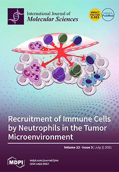
Cover Story (view full-size image): Neutrophils are recruited to the tumor microenvironment where they turn into tumor-associated neutrophils (TANs), and are able to initiate and promote tumor progression and metastasis. Conversely, anti-tumorigenic properties of neutrophils have been documented, highlighting the versatile nature and high pleiotropic plasticity of these polymorphonuclear leukocytes (PMN-L). Here, we dissect the ambivalent roles of TANs in cancer and focus on selected functional aspects that could be therapeutic targets. Indeed, the critical point of targeting TAN functions lies in the fact that an immunosuppressive state could be induced, resulting in unwanted side effects. A deeper knowledge of the mechanisms linked to diverse TAN functions in different cancer types is necessary to define appropriate therapeutic strategies that are able to induce and maintain an anti-tumor microenvironment. View this paper
  • Issues are regarded as officially published after their release is announced to the table of contents alert mailing list.
  • You may sign up for e-mail alerts to receive table of contents of newly released issues.
  • PDF is the official format for papers published in both, html and pdf forms. To view the papers in pdf format, click on the "PDF Full-text" link, and use the free Adobe Reader to open them.
Order results
Result details
Section
Select all
Export citation of selected articles as:
20 pages, 1048 KiB  
Review
Tumor-Associated Macrophages: Combination of Therapies, the Approach to Improve Cancer Treatment
by Pedram Moeini and Paulina Niedźwiedzka-Rystwej
Int. J. Mol. Sci. 2021, 22(13), 7239; https://doi.org/10.3390/ijms22137239 - 05 Jul 2021
Cited by 21 | Viewed by 5117
Abstract
Macrophages are one of the most important cells of the innate immune system and are known for their ability to engulf and digest foreign substances, including cellular debris and tumor cells. They can convert into tumor-associated macrophages (TAMs) when mature macrophages are recruited [...] Read more.
Macrophages are one of the most important cells of the innate immune system and are known for their ability to engulf and digest foreign substances, including cellular debris and tumor cells. They can convert into tumor-associated macrophages (TAMs) when mature macrophages are recruited into the tumor microenvironment. Their role in cancer progression, metastasis, and therapy failure is of special note. The aim of this review is to understand how the presence of TAMs are both advantageous and disadvantageous in the immune system. Full article
Show Figures

Figure 1

19 pages, 4294 KiB  
Article
In Search of Effective Anticancer Agents—Novel Sugar Esters Based on Polyhydroxyalkanoate Monomers
by Wojciech Snoch, Dawid Wnuk, Tomasz Witko, Jakub Staroń, Andrzej J. Bojarski, Ewelina Jarek, Francisco J. Plou and Maciej Guzik
Int. J. Mol. Sci. 2021, 22(13), 7238; https://doi.org/10.3390/ijms22137238 - 05 Jul 2021
Cited by 6 | Viewed by 2877
Abstract
Cancer is one of the deadliest illness globally. Searching for new solutions in cancer treatments is essential because commonly used mixed, targeted and personalized therapies are sometimes not sufficient or are too expensive for common patients. Sugar fatty acid esters (SFAEs) are already [...] Read more.
Cancer is one of the deadliest illness globally. Searching for new solutions in cancer treatments is essential because commonly used mixed, targeted and personalized therapies are sometimes not sufficient or are too expensive for common patients. Sugar fatty acid esters (SFAEs) are already well-known as promising candidates for an alternative medical tool. The manuscript brings the reader closer to methods of obtaining various SFAEs using combined biological, chemical and enzymatic methods. It presents how modification of SFAE’s hydrophobic chains can influence their cytotoxicity against human skin melanoma and prostate cancer cell lines. The compound’s cytotoxicity was determined by an MTT assay, which followed an assessment of SFAEs’ potential metastatic properties in concentrations below IC50 values. Despite relatively high IC50 values (63.3–1737.6 μM) of the newly synthesized SFAE, they can compete with other sugar esters already described in the literature. The chosen bioactives caused low polymerization of microtubules and the depolymerization of actin filaments in nontoxic levels, which suggest an apoptotic rather than metastatic process. Altogether, cancer cells showed no propensity for metastasis after treating them with SFAE. They confirmed that lactose-based compounds seem the most promising surfactants among tested sugar esters. This manuscript creates a benchmark for creation of novel anticancer agents based on 3-hydroxylated fatty acids of bacterial origin. Full article
(This article belongs to the Collection Anticancer Drug Discovery and Development)
Show Figures

Figure 1

15 pages, 3165 KiB  
Article
Comparative Mitogenomic Analysis Reveals Gene and Intron Dynamics in Rubiaceae and Intra-Specific Diversification in Damnacanthus indicus
by Eun-Kyeong Han, Won-Bum Cho, Ichiro Tamaki, In-Su Choi and Jung-Hyun Lee
Int. J. Mol. Sci. 2021, 22(13), 7237; https://doi.org/10.3390/ijms22137237 - 05 Jul 2021
Cited by 6 | Viewed by 2566
Abstract
The dynamic evolution of mitochondrial gene and intron content has been reported across the angiosperms. However, a reference mitochondrial genome (mitogenome) is not available in Rubiaceae. The phylogenetic utility of mitogenome data at a species level is rarely assessed. Here, we assembled mitogenomes [...] Read more.
The dynamic evolution of mitochondrial gene and intron content has been reported across the angiosperms. However, a reference mitochondrial genome (mitogenome) is not available in Rubiaceae. The phylogenetic utility of mitogenome data at a species level is rarely assessed. Here, we assembled mitogenomes of six Damnacanthus indicus (Rubiaceae, Rubioideae) representing two varieties (var. indicus and var. microphyllus). The gene and intron content of D. indicus was compared with mitogenomes from representative angiosperm species and mitochondrial contigs from the other Rubiaceae species. Mitogenome structural rearrangement and sequence divergence in D. indicus were analyzed in six individuals. The size of the mitogenome in D. indicus varied from 417,661 to 419,435 bp. Comparing the number of intact mitochondrial protein-coding genes in other Gentianales taxa (38), D. indicus included 32 genes representing several losses. The intron analysis revealed a shift from cis to trans splicing of a nad1 intron (nad1i728) in D. indicus and it is a shared character with the other four Rubioideae taxa. Two distinct mitogenome structures (type A and B) were identified. Two-step direct repeat-mediated recombination was proposed to explain structural changes between type A and B mitogenomes. The five individuals from two varieties in D. indicus diverged well in the whole mitogenome-level comparison with one exception. Collectively, our study elucidated the mitogenome evolution in Rubiaceae along with D. indicus and showed the reliable phylogenetic utility of the whole mitogenome data at a species-level evolution. Full article
(This article belongs to the Special Issue Organelle Genetics in Plants 2.0)
Show Figures

Figure 1

15 pages, 1537 KiB  
Article
Identification of 3,4-Dihydro-2H,6H-pyrimido[1,2-c][1,3]benzothiazin-6-imine Derivatives as Novel Selective Inhibitors of Plasmodium falciparum Dihydroorotate Dehydrogenase
by Endah Dwi Hartuti, Takaya Sakura, Mohammed S. O. Tagod, Eri Yoshida, Xinying Wang, Kota Mochizuki, Rajib Acharjee, Yuichi Matsuo, Fuyuki Tokumasu, Mihoko Mori, Danang Waluyo, Kazuro Shiomi, Tomoyoshi Nozaki, Shinjiro Hamano, Tomoo Shiba, Kiyoshi Kita and Daniel Ken Inaoka
Int. J. Mol. Sci. 2021, 22(13), 7236; https://doi.org/10.3390/ijms22137236 - 05 Jul 2021
Cited by 8 | Viewed by 3477
Abstract
Plasmodium falciparum’s resistance to available antimalarial drugs highlights the need for the development of novel drugs. Pyrimidine de novo biosynthesis is a validated drug target for the prevention and treatment of malaria infection. P. falciparum dihydroorotate dehydrogenase (PfDHODH) catalyzes the oxidation of [...] Read more.
Plasmodium falciparum’s resistance to available antimalarial drugs highlights the need for the development of novel drugs. Pyrimidine de novo biosynthesis is a validated drug target for the prevention and treatment of malaria infection. P. falciparum dihydroorotate dehydrogenase (PfDHODH) catalyzes the oxidation of dihydroorotate to orotate and utilize ubiquinone as an electron acceptor in the fourth step of pyrimidine de novo biosynthesis. PfDHODH is targeted by the inhibitor DSM265, which binds to a hydrophobic pocket located at the N-terminus where ubiquinone binds, which is known to be structurally divergent from the mammalian orthologue. In this study, we screened 40,400 compounds from the Kyoto University chemical library against recombinant PfDHODH. These studies led to the identification of 3,4-dihydro-2H,6H-pyrimido[1,2-c][1,3]benzothiazin-6-imine and its derivatives as a new class of PfDHODH inhibitor. Moreover, the hit compounds identified in this study are selective for PfDHODH without inhibition of the human enzymes. Finally, this new scaffold of PfDHODH inhibitors showed growth inhibition activity against P. falciparum 3D7 with low toxicity to three human cell lines, providing a new starting point for antimalarial drug development. Full article
(This article belongs to the Special Issue Mitochondrial Coenzyme Q)
Show Figures

Figure 1

26 pages, 3172 KiB  
Review
Citric Acid-Mediated Abiotic Stress Tolerance in Plants
by Md. Tahjib-Ul-Arif, Mst. Ishrat Zahan, Md. Masudul Karim, Shahin Imran, Charles T. Hunter, Md. Saiful Islam, Md. Ashik Mia, Md. Abdul Hannan, Mohammad Saidur Rhaman, Md. Afzal Hossain, Marian Brestic, Milan Skalicky and Yoshiyuki Murata
Int. J. Mol. Sci. 2021, 22(13), 7235; https://doi.org/10.3390/ijms22137235 - 05 Jul 2021
Cited by 78 | Viewed by 9815
Abstract
Several recent studies have shown that citric acid/citrate (CA) can confer abiotic stress tolerance to plants. Exogenous CA application leads to improved growth and yield in crop plants under various abiotic stress conditions. Improved physiological outcomes are associated with higher photosynthetic rates, reduced [...] Read more.
Several recent studies have shown that citric acid/citrate (CA) can confer abiotic stress tolerance to plants. Exogenous CA application leads to improved growth and yield in crop plants under various abiotic stress conditions. Improved physiological outcomes are associated with higher photosynthetic rates, reduced reactive oxygen species, and better osmoregulation. Application of CA also induces antioxidant defense systems, promotes increased chlorophyll content, and affects secondary metabolism to limit plant growth restrictions under stress. In particular, CA has a major impact on relieving heavy metal stress by promoting precipitation, chelation, and sequestration of metal ions. This review summarizes the mechanisms that mediate CA-regulated changes in plants, primarily CA’s involvement in the control of physiological and molecular processes in plants under abiotic stress conditions. We also review genetic engineering strategies for CA-mediated abiotic stress tolerance. Finally, we propose a model to explain how CA’s position in complex metabolic networks involving the biosynthesis of phytohormones, amino acids, signaling molecules, and other secondary metabolites could explain some of its abiotic stress-ameliorating properties. This review summarizes our current understanding of CA-mediated abiotic stress tolerance and highlights areas where additional research is needed. Full article
(This article belongs to the Section Molecular Plant Sciences)
Show Figures

Figure 1

22 pages, 4510 KiB  
Article
The Lrat−/− Rat: CRISPR/Cas9 Construction and Phenotyping of a New Animal Model for Retinitis Pigmentosa
by Céline Koster, Koen T. van den Hurk, Colby F. Lewallen, Mays Talib, Jacoline B. ten Brink, Camiel J. F. Boon and Arthur A. Bergen
Int. J. Mol. Sci. 2021, 22(13), 7234; https://doi.org/10.3390/ijms22137234 - 05 Jul 2021
Cited by 7 | Viewed by 3028
Abstract
Purpose: We developed and phenotyped a pigmented knockout rat model for lecithin retinol acyltransferase (LRAT) using CRISPR/Cas9. The introduced mutation (c.12delA) is based on a patient group harboring a homologous homozygous frameshift mutation in the LRAT gene (c.12delC), causing a dysfunctional visual (retinoid) [...] Read more.
Purpose: We developed and phenotyped a pigmented knockout rat model for lecithin retinol acyltransferase (LRAT) using CRISPR/Cas9. The introduced mutation (c.12delA) is based on a patient group harboring a homologous homozygous frameshift mutation in the LRAT gene (c.12delC), causing a dysfunctional visual (retinoid) cycle. Methods: The introduced mutation was confirmed by DNA and RNA sequencing. The expression of Lrat was determined on both the RNA and protein level in wildtype and knockout animals using RT-PCR and immunohistochemistry. The retinal structure and function, as well as the visual behavior of the Lrat−/− and control rats, were characterized using scanning laser ophthalmoscopy (SLO), optical coherence tomography (OCT), electroretinography (ERG) and vision-based behavioral assays. Results: Wildtype animals had high Lrat mRNA expression in multiple tissues, including the eye and liver. In contrast, hardly any expression was detected in Lrat−/− animals. LRAT protein was abundantly present in wildtype animals and absent in Lrat−/− animals. Lrat−/− animals showed progressively reduced ERG potentials compared to wildtype controls from two weeks of age onwards. Vison-based behavioral assays confirmed reduced vision. Structural abnormalities, such as overall retinal thinning, were observed in Lrat−/− animals. The retinal thickness in knockout rats was decreased to roughly 80% by four months of age. No functional or structural differences were observed between wildtype and heterozygote animals. Conclusions: Our Lrat−/− rat is a new animal model for retinal dystrophy, especially for the LRAT-subtype of early-onset retinal dystrophies. This model has advantages over the existing mouse models and the RCS rat strain and can be used for translational studies of retinal dystrophies. Full article
(This article belongs to the Special Issue Towards an Understanding of Retinal Diseases and Novel Treatment)
Show Figures

Figure 1

20 pages, 7963 KiB  
Article
Fibroblast Growth Factor Type 1 Ameliorates High-Glucose-Induced Oxidative Stress and Neuroinflammation in Retinal Pigment Epithelial Cells and a Streptozotocin-Induced Diabetic Rat Model
by Hsin-Wei Huang, Chung-May Yang and Chang-Hao Yang
Int. J. Mol. Sci. 2021, 22(13), 7233; https://doi.org/10.3390/ijms22137233 - 05 Jul 2021
Cited by 12 | Viewed by 3017
Abstract
Diabetic retinopathy (DR) is a common complication of diabetes that causes severe visual impairment globally. The pathogenesis of DR is related to oxidative stress and chronic inflammation. The fibroblast growth factor type 1 (FGF-1) mitogen plays crucial roles in cell function, development, and [...] Read more.
Diabetic retinopathy (DR) is a common complication of diabetes that causes severe visual impairment globally. The pathogenesis of DR is related to oxidative stress and chronic inflammation. The fibroblast growth factor type 1 (FGF-1) mitogen plays crucial roles in cell function, development, and metabolism. FGF-1 is involved in blood sugar regulation and exerts beneficial antioxidative and anti-inflammatory effects on various organ systems. This study investigated the antioxidative and anti-inflammatory neuroprotective effects of FGF-1 on high-glucose-induced retinal damage. The results revealed that FGF-1 treatment significantly reversed the harmful effects of oxidative stress and inflammatory mediators in retinal tissue in a streptozotocin-induced diabetic rat model. These protective effects were also observed in the in vitro model of retinal ARPE-19 cells exposed to a high-glucose condition. We demonstrated that FGF-1 attenuated p38 mitogen-activated protein kinase and nuclear factor-κB pathway activation under the high-glucose condition. Our results indicated that FGF-1 could effectively prevent retinal injury in diabetes. The findings of this study could be used to develop novel treatments for DR that aim to reduce the cascade of oxidative stress and inflammatory signals in neuroretinal tissue. Full article
Show Figures

Figure 1

34 pages, 16575 KiB  
Article
Norepinephrine Protects against Methamphetamine Toxicity through β2-Adrenergic Receptors Promoting LC3 Compartmentalization
by Gloria Lazzeri, Carla L. Busceti, Francesca Biagioni, Cinzia Fabrizi, Gabriele Morucci, Filippo S. Giorgi, Michela Ferrucci, Paola Lenzi, Stefano Puglisi-Allegra and Francesco Fornai
Int. J. Mol. Sci. 2021, 22(13), 7232; https://doi.org/10.3390/ijms22137232 - 05 Jul 2021
Cited by 9 | Viewed by 2863
Abstract
Norepinephrine (NE) neurons and extracellular NE exert some protective effects against a variety of insults, including methamphetamine (Meth)-induced cell damage. The intimate mechanism of protection remains difficult to be analyzed in vivo. In fact, this may occur directly on target neurons or as [...] Read more.
Norepinephrine (NE) neurons and extracellular NE exert some protective effects against a variety of insults, including methamphetamine (Meth)-induced cell damage. The intimate mechanism of protection remains difficult to be analyzed in vivo. In fact, this may occur directly on target neurons or as the indirect consequence of NE-induced alterations in the activity of trans-synaptic loops. Therefore, to elude neuronal networks, which may contribute to these effects in vivo, the present study investigates whether NE still protects when directly applied to Meth-treated PC12 cells. Meth was selected based on its detrimental effects along various specific brain areas. The study shows that NE directly protects in vitro against Meth-induced cell damage. The present study indicates that such an effect fully depends on the activation of plasma membrane β2-adrenergic receptors (ARs). Evidence indicates that β2-ARs activation restores autophagy, which is impaired by Meth administration. This occurs via restoration of the autophagy flux and, as assessed by ultrastructural morphometry, by preventing the dissipation of microtubule-associated protein 1 light chain 3 (LC3) from autophagy vacuoles to the cytosol, which is produced instead during Meth toxicity. These findings may have an impact in a variety of degenerative conditions characterized by NE deficiency along with autophagy impairment. Full article
(This article belongs to the Special Issue The Role of Norepinephrine in Neuroprotection)
Show Figures

Figure 1

10 pages, 556 KiB  
Review
Metformin as a Treatment Strategy for Sjögren’s Syndrome
by Joa Kim, Yun-Sung Kim and Sung-Hwan Park
Int. J. Mol. Sci. 2021, 22(13), 7231; https://doi.org/10.3390/ijms22137231 - 05 Jul 2021
Cited by 10 | Viewed by 5508
Abstract
Sjögren’s syndrome (SS), a chronic inflammatory disease involving the salivary and lacrimal glands, presents symptoms of sicca as well as systemic manifestations such as fatigue and musculoskeletal pain. Only a few treatments have been successful in management of SS; thus treatment of the [...] Read more.
Sjögren’s syndrome (SS), a chronic inflammatory disease involving the salivary and lacrimal glands, presents symptoms of sicca as well as systemic manifestations such as fatigue and musculoskeletal pain. Only a few treatments have been successful in management of SS; thus treatment of the disease is challenging. Metformin is the first-line agent for type 2 diabetes and has anti-inflammatory potential. Its immunomodulatory capacity is exerted via activation of 5’ adenosine monophosphate-activated protein kinase (AMPK). Metformin inhibits mitochondrial respiratory chain complex I which leads to change in adenosine mono-phosphate (AMP) to adenosine tri-phosphate (ATP) ratio. This results in AMPK activation and causes inhibition of mammalian target of rapamycin (mTOR). mTOR plays an important role in T cell differentiation and mTOR deficient T cells differentiate into regulatory T cells. In this manner, metformin enhances immunoregulatory response in an individual. mTOR is responsible for B cell proliferation and germinal center (GC) differentiation. Thus, reduction of B cell differentiation into antibody-producing plasma cells occurs via downregulation of mTOR. Due to the lack of suggested treatment for SS, metformin has been considered as a treatment strategy and is expected to ameliorate salivary gland function. Full article
(This article belongs to the Section Molecular Pathology, Diagnostics, and Therapeutics)
Show Figures

Figure 1

16 pages, 3680 KiB  
Article
Protective Effects of Ethanolic Extract from Rhizome of Polygoni avicularis against Renal Fibrosis and Inflammation in a Diabetic Nephropathy Model
by Jung-Joo Yoon, Ji-Hun Park, Yun-Jung Lee, Hye-Yoom Kim, Byung-Hyuk Han, Hong-Guang Jin, Dae-Gill Kang and Ho-Sub Lee
Int. J. Mol. Sci. 2021, 22(13), 7230; https://doi.org/10.3390/ijms22137230 - 05 Jul 2021
Cited by 11 | Viewed by 2601
Abstract
Progressive diabetic nephropathy (DN) in diabetes leads to major morbidity and mortality. The major pathological alterations of DN include mesangial expansion, extracellular matrix alterations, tubulointerstitial fibrosis, and glomerular sclerosis. Polygoni avicularis is widely used in traditional oriental medicine and has long been used [...] Read more.
Progressive diabetic nephropathy (DN) in diabetes leads to major morbidity and mortality. The major pathological alterations of DN include mesangial expansion, extracellular matrix alterations, tubulointerstitial fibrosis, and glomerular sclerosis. Polygoni avicularis is widely used in traditional oriental medicine and has long been used as a diuretic, astringent, insecticide and antihypertensive. However, to the best of the authors’ knowledge, the effects of the ethanolic extract from rhizome of Polygoni avicularis (ER-PA) on DN have not yet been assessed. The present study aimed to identify the effect of ER-PA on renal dysfunction, which has been implicated in DN in human renal mesangial cells and db/db mice and investigate its mechanism of action. The in vivo experiment was performed using Polygoni avicularis-ethanol soluble fraction (ER-PA) and was administrated to db/db mice at 10 and 50 mg/kg dose. For the in vitro experiments, the human renal mesangial cells were induced by high glucose (HG, 25 mM). The ER-PA group showed significant amelioration in oral glucose tolerance, and insulin resistance index. ER-PA significantly improved the albumin excretion and markedly reduced plasma creatinine, kidney injury molecule-1 and C-reactive protein. In addition, ER-PA significantly suppressed inflammatory cytokines. Histopathologically, ER-PA attenuated glomerular expansion and tubular fibrosis in db/db mice. Furthermore, ER-PA suppressed the expression of renal fibrosis biomarkers (TGF and Collagen IV). ER-PA also reduced the NLR family pyrin domain containing 3 inflammatory factor level. These results suggest that ER-PA has a protective effect against renal dysfunction through improved insulin resistance as well as the inhibition of nephritis and fibrosis in DN. Full article
(This article belongs to the Special Issue Molecular Mechanisms of Diabetes-Associated Kidney Disease)
Show Figures

Figure 1

16 pages, 997 KiB  
Review
Ineffective Erythropoiesis in β-Thalassaemia: Key Steps and Therapeutic Options by Drugs
by Filomena Longo, Andrea Piolatto, Giovanni Battista Ferrero and Antonio Piga
Int. J. Mol. Sci. 2021, 22(13), 7229; https://doi.org/10.3390/ijms22137229 - 05 Jul 2021
Cited by 20 | Viewed by 5758
Abstract
β-thalassaemia is a rare genetic condition caused by mutations in the β-globin gene that result in severe iron-loading anaemia, maintained by a detrimental state of ineffective erythropoiesis (IE). The role of multiple mechanisms involved in the pathophysiology of the disease has been recently [...] Read more.
β-thalassaemia is a rare genetic condition caused by mutations in the β-globin gene that result in severe iron-loading anaemia, maintained by a detrimental state of ineffective erythropoiesis (IE). The role of multiple mechanisms involved in the pathophysiology of the disease has been recently unravelled. The unbalanced production of α-globin is a major source of oxidative stress and membrane damage in red blood cells (RBC). In addition, IE is tightly linked to iron metabolism dysregulation, and the relevance of new players of this pathway, i.e., hepcidin, erythroferrone, matriptase-2, among others, has emerged. Advances have been made in understanding the balance between proliferation and maturation of erythroid precursors and the role of specific factors in this process, such as members of the TGF-β superfamily, and their downstream effectors, or the transcription factor GATA1. The increasing understanding of IE allowed for the development of a broad set of potential therapeutic options beyond the current standard of care. Many candidates of disease-modifying drugs are currently under clinical investigation, targeting the regulation of iron metabolism, the production of foetal haemoglobin, the maturation process, or the energetic balance and membrane stability of RBC. Overall, they provide tools and evidence for multiple and synergistic approaches that are effectively moving clinical research in β-thalassaemia from bench to bedside. Full article
(This article belongs to the Special Issue Regulation of Erythropoiesis)
Show Figures

Graphical abstract

12 pages, 2609 KiB  
Article
Low-Dose Acrolein, an Endogenous and Exogenous Toxic Molecule, Inhibits Glucose Transport via an Inhibition of Akt-Regulated GLUT4 Signaling in Skeletal Muscle Cells
by Ching-Chia Wang, Huang-Jen Chen, Ding-Cheng Chan, Chen-Yuan Chiu, Shing-Hwa Liu and Kuo-Cheng Lan
Int. J. Mol. Sci. 2021, 22(13), 7228; https://doi.org/10.3390/ijms22137228 - 05 Jul 2021
Cited by 6 | Viewed by 2344
Abstract
Urinary acrolein adduct levels have been reported to be increased in both habitual smokers and type-2 diabetic patients. The impairment of glucose transport in skeletal muscles is a major factor responsible for glucose uptake reduction in type-2 diabetic patients. The effect of acrolein [...] Read more.
Urinary acrolein adduct levels have been reported to be increased in both habitual smokers and type-2 diabetic patients. The impairment of glucose transport in skeletal muscles is a major factor responsible for glucose uptake reduction in type-2 diabetic patients. The effect of acrolein on glucose metabolism in skeletal muscle remains unclear. Here, we investigated whether acrolein affects muscular glucose metabolism in vitro and glucose tolerance in vivo. Exposure of mice to acrolein (2.5 and 5 mg/kg/day) for 4 weeks substantially increased fasting blood glucose and impaired glucose tolerance. The glucose transporter-4 (GLUT4) protein expression was significantly decreased in soleus muscles of acrolein-treated mice. The glucose uptake was significantly decreased in differentiated C2C12 myotubes treated with a non-cytotoxic dose of acrolein (1 μM) for 24 and 72 h. Acrolein (0.5–2 μM) also significantly decreased the GLUT4 expression in myotubes. Acrolein suppressed the phosphorylation of glucose metabolic signals IRS1, Akt, mTOR, p70S6K, and GSK3α/β. Over-expression of constitutive activation of Akt reversed the inhibitory effects of acrolein on GLUT4 protein expression and glucose uptake in myotubes. These results suggest that acrolein at doses relevant to human exposure dysregulates glucose metabolism in skeletal muscle cells and impairs glucose tolerance in mice. Full article
(This article belongs to the Section Biochemistry)
Show Figures

Figure 1

22 pages, 1108 KiB  
Review
The Implications of Pruritogens in the Pathogenesis of Atopic Dermatitis
by Lai-San Wong, Yu-Ta Yen and Chih-Hung Lee
Int. J. Mol. Sci. 2021, 22(13), 7227; https://doi.org/10.3390/ijms22137227 - 05 Jul 2021
Cited by 19 | Viewed by 5662
Abstract
Atopic dermatitis (AD) is a prototypic inflammatory disease that presents with intense itching. The pathophysiology of AD is multifactorial, involving environmental factors, genetic susceptibility, skin barrier function, and immune responses. A recent understanding of pruritus transmission provides more information about the role of [...] Read more.
Atopic dermatitis (AD) is a prototypic inflammatory disease that presents with intense itching. The pathophysiology of AD is multifactorial, involving environmental factors, genetic susceptibility, skin barrier function, and immune responses. A recent understanding of pruritus transmission provides more information about the role of pruritogens in the pathogenesis of AD. There is evidence that pruritogens are not only responsible for eliciting pruritus, but also interact with immune cells and act as inflammatory mediators, which exacerbate the severity of AD. In this review, we discuss the interaction between pruritogens and inflammatory molecules and summarize the targeted therapies for AD. Full article
(This article belongs to the Special Issue Molecular Mechanisms of Skin Aging and Atopic Dermatitis)
Show Figures

Graphical abstract

16 pages, 17737 KiB  
Article
YB-1 Is Altered in Pregnancy-Associated Disorders and Affects Trophoblast in Vitro Properties via Alternation of Multiple Molecular Traits
by Violeta Stojanovska, Aneri Shah, Katja Woidacki, Florence Fischer, Mario Bauer, Jonathan A. Lindquist, Peter R. Mertens and Ana C. Zenclussen
Int. J. Mol. Sci. 2021, 22(13), 7226; https://doi.org/10.3390/ijms22137226 - 05 Jul 2021
Cited by 5 | Viewed by 3397
Abstract
Cold shock Y-box binding protein-1 (YB-1) coordinates several molecular processes between the nucleus and the cytoplasm and plays a crucial role in cell function. Moreover, it is involved in cancer progression, invasion, and metastasis. As trophoblast cells share similar characteristics with cancer cells, [...] Read more.
Cold shock Y-box binding protein-1 (YB-1) coordinates several molecular processes between the nucleus and the cytoplasm and plays a crucial role in cell function. Moreover, it is involved in cancer progression, invasion, and metastasis. As trophoblast cells share similar characteristics with cancer cells, we hypothesized that YB-1 might also be necessary for trophoblast functionality. In samples of patients with intrauterine growth restriction, YB-1 mRNA levels were decreased, while they were increased in preeclampsia and unchanged in spontaneous abortions when compared to normal pregnant controls. Studies with overexpression and downregulation of YB-1 were performed to assess the key trophoblast processes in two trophoblast cell lines HTR8/SVneo and JEG3. Overexpression of YB-1 or exposure of trophoblast cells to recombinant YB-1 caused enhanced proliferation, while knockdown of YB-1 lead to proliferative disadvantage in JEG3 or HTR8/SVneo cells. The invasion and migration properties were affected at different degrees among the trophoblast cell lines. Trophoblast expression of genes mediating migration, invasion, apoptosis, and inflammation was altered upon YB-1 downregulation. Moreover, IL-6 secretion was excessively increased in HTR8/SVneo. Ultimately, YB-1 directly binds to NF-κB enhancer mark in HTR8/SVneo cells. Our data show that YB-1 protein is important for trophoblast cell functioning and, when downregulated, leads to trophoblast disadvantage that at least in part is mediated by NF-κB. Full article
(This article belongs to the Special Issue Placental Related Disorders of Pregnancy)
Show Figures

Figure 1

15 pages, 2610 KiB  
Article
EVOO Polyphenols Relieve Synergistically Autophagy Dysregulation in a Cellular Model of Alzheimer’s Disease
by Manuela Leri, Andrea Bertolini, Massimo Stefani and Monica Bucciantini
Int. J. Mol. Sci. 2021, 22(13), 7225; https://doi.org/10.3390/ijms22137225 - 05 Jul 2021
Cited by 13 | Viewed by 2710
Abstract
(1) Background: Autophagy, the major cytoplasmic process of substrate turnover, declines with age, contributing to proteostasis decline, accumulation of harmful protein aggregates, damaged mitochondria and to ROS production. Accordingly, abnormalities in the autophagic flux may contribute to many different pathophysiological conditions associated with [...] Read more.
(1) Background: Autophagy, the major cytoplasmic process of substrate turnover, declines with age, contributing to proteostasis decline, accumulation of harmful protein aggregates, damaged mitochondria and to ROS production. Accordingly, abnormalities in the autophagic flux may contribute to many different pathophysiological conditions associated with ageing, including neurodegeneration. Recent data have shown that extra-virgin olive oil (EVOO) polyphenols stimulate cell defenses against plaque-induced neurodegeneration, mainly, through autophagy induction. (2) Methods: We carried out a set of in vitro experiments on SH-SY5Y human neuroblastoma cells exposed to toxic Aβ1–42 oligomers to investigate the molecular mechanisms involved in autophagy activation by two olive oil polyphenols, oleuropein aglycone (OleA), arising from the hydrolysis of oleuropein (Ole), the main polyphenol found in olive leaves and drupes and its main metabolite, hydroxytyrosol (HT). (3) Results: Our data show that the mixture of the two polyphenols activates synergistically the autophagic flux preventing cell damage by Aβ1–42 oligomers., in terms of ROS production, and impairment of mitochondria. (4) Conclusion: Our results support the idea that EVOO polyphenols act synergistically in autophagy modulation against neurodegeneration. These data confirm and provide the rationale to consider these molecules, alone or in combination, as promising candidates to contrast ageing-associated neurodegeneration. Full article
(This article belongs to the Section Molecular Neurobiology)
Show Figures

Graphical abstract

15 pages, 2570 KiB  
Article
Effects of Extracellular Vesicles from Blood-Derived Products on Osteoarthritic Chondrocytes within an Inflammation Model
by Alexander Otahal, Karina Kramer, Olga Kuten-Pella, Lukas B. Moser, Markus Neubauer, Zsombor Lacza, Stefan Nehrer and Andrea De Luna
Int. J. Mol. Sci. 2021, 22(13), 7224; https://doi.org/10.3390/ijms22137224 - 05 Jul 2021
Cited by 8 | Viewed by 3354
Abstract
Osteoarthritis (OA) is hallmarked by a progressive degradation of articular cartilage. One major driver of OA is inflammation, in which cytokines such as IL-6, TNF-α and IL-1β are secreted by activated chondrocytes, as well as synovial cells—including macrophages. Intra-articular injection of blood products—such [...] Read more.
Osteoarthritis (OA) is hallmarked by a progressive degradation of articular cartilage. One major driver of OA is inflammation, in which cytokines such as IL-6, TNF-α and IL-1β are secreted by activated chondrocytes, as well as synovial cells—including macrophages. Intra-articular injection of blood products—such as citrate-anticoagulated plasma (CPRP), hyperacute serum (hypACT), and extracellular vesicles (EVs) isolated from blood products—is gaining increasing importance in regenerative medicine for the treatment of OA. A co-culture system of primary OA chondrocytes and activated M1 macrophages was developed to model an OA joint in order to observe the effects of EVs in modulating the inflammatory environment. Primary OA chondrocytes were obtained from patients undergoing total knee replacement. Primary monocytes obtained from voluntary healthy donors and the monocytic cell line THP-1 were differentiated and activated into proinflammatory M1 macrophages. EVs were isolated by ultracentrifugation and characterized by nanoparticle tracking analysis and Western blot. Gene expression analysis of chondrocytes by RT-qPCR revealed increased type II collagen expression, while cytokine profiling via ELISA showed lower TNF-α and IL-1β levels associated with EV treatment. In conclusion, the inflammation model provides an accessible tool to investigate the effects of blood products and EVs in the inflammatory context of OA. Full article
(This article belongs to the Special Issue Extracellular Vesicles in Inflammation)
Show Figures

Figure 1

15 pages, 2534 KiB  
Article
C-Terminal Fragment of Vitellogenin II, a Potential Yolkin Polypeptide Complex Precursor Protein—Heterologous Expression, Purification, and Immunoregulatory Activity
by Agnieszka Szmyt, Agnieszka Zabłocka, Józefa Macała, Józefa Chrzanowska and Anna Dąbrowska
Int. J. Mol. Sci. 2021, 22(13), 7223; https://doi.org/10.3390/ijms22137223 - 05 Jul 2021
Cited by 3 | Viewed by 2015
Abstract
The aim of this research was to analyze the heterologous expression, purification, and immunoregulatory activity of recombinant YGP40 (rYGP40), the potential precursor of the yolkin peptide complex. The ygp40 coding sequence was codon optimized, successfully expressed in the E. coli system, and purified [...] Read more.
The aim of this research was to analyze the heterologous expression, purification, and immunoregulatory activity of recombinant YGP40 (rYGP40), the potential precursor of the yolkin peptide complex. The ygp40 coding sequence was codon optimized, successfully expressed in the E. coli system, and purified from inclusion bodies with a yield of about 1.1 mg/L of culture. This study showed that the protein exhibits immunomodulatory activity, expressed by the stimulation of TNF-α and IL-10 production and nitric oxide induction at a level comparable to that of the natural yolkin peptide complex obtained by other authors from hen egg yolk. At the highest dose of 100 µg/mL, rYGP40 also caused the up-regulation of iNOS expression in murine bone marrow-derived macrophages (BMDM). Moreover, no cytotoxic effects of rYGP40 on the BMDM cell line were observed. Full article
(This article belongs to the Section Biochemistry)
Show Figures

Figure 1

10 pages, 18761 KiB  
Article
Less Carcinogenic Chlorinated Estrogens Applicable to Hormone Replacement Therapy
by Yoshinori Okamoto, Hideto Jinno, Shinji Itoh and Shinya Shibutani
Int. J. Mol. Sci. 2021, 22(13), 7222; https://doi.org/10.3390/ijms22137222 - 05 Jul 2021
Viewed by 1928
Abstract
Human estrogens prescribed for hormone replacement therapy (HRT) are known to be potent carcinogens. To find safer estrogens, several chlorinated estrogens were synthesized and their carcinogenic potential were determined. A pellet containing either 2-chloro-17β-estradiol (2-ClE2) or 4-chloro-17β-estradiol (4-ClE2) was [...] Read more.
Human estrogens prescribed for hormone replacement therapy (HRT) are known to be potent carcinogens. To find safer estrogens, several chlorinated estrogens were synthesized and their carcinogenic potential were determined. A pellet containing either 2-chloro-17β-estradiol (2-ClE2) or 4-chloro-17β-estradiol (4-ClE2) was implanted subcutaneously for 52 weeks into August Copenhagen Irish (ACI) rats, a preferred animal model for human breast cancer. 17β-Estradiol (E2) frequently induced mammary tumors while both 2-ClE2 and 4-ClE2 did not. Their 17α-ethinyl forms, thought to be orally active estrogens, were also synthesized. Neither 2-chloro-17α-ethinylestradiol (2-ClEE2) nor 4-chloro-17α-ethinylestradiol (4-ClEE2) induced tumors. The less carcinogenic effects were supported by histological examination of mammary glands of ACI rats treated with the chlorinated estrogens. A chlorine atom positioned at the 2- or 4-position of E2 may prevent the metabolic activation, resulting in reducing the carcinogenicity. 2-ClE2 and 4-ClE2 administered subcutaneously and 2-ClEE2 and 4-ClEE2 given orally to ovariectomized rats all showed uterotrophic potency, albeit slightly weaker than that of E2. Our results indicate that less carcinogenic chlorinated estrogens retaining estrogenic potential could be safer alternatives to the carcinogenic estrogens now in use for HRT. Full article
(This article belongs to the Special Issue Estrogens in Human Cancer)
Show Figures

Figure 1

28 pages, 1767 KiB  
Review
Extracellular Vesicles in the Fungi Kingdom
by Marc Liebana-Jordan, Bruno Brotons, Juan Manuel Falcon-Perez and Esperanza Gonzalez
Int. J. Mol. Sci. 2021, 22(13), 7221; https://doi.org/10.3390/ijms22137221 - 05 Jul 2021
Cited by 32 | Viewed by 4999
Abstract
Extracellular vesicles (EVs) are membranous, rounded vesicles released by prokaryotic and eukaryotic cells in their normal and pathophysiological states. These vesicles form a network of intercellular communication as they can transfer cell- and function-specific information (lipids, proteins and nucleic acids) to different cells [...] Read more.
Extracellular vesicles (EVs) are membranous, rounded vesicles released by prokaryotic and eukaryotic cells in their normal and pathophysiological states. These vesicles form a network of intercellular communication as they can transfer cell- and function-specific information (lipids, proteins and nucleic acids) to different cells and thus alter their function. Fungi are not an exception; they also release EVs to the extracellular space. The vesicles can also be retained in the periplasm as periplasmic vesicles (PVs) and the cell wall. Such fungal vesicles play various specific roles in the lives of these organisms. They are involved in creating wall architecture and maintaining its integrity, supporting cell isolation and defence against the environment. In the case of pathogenic strains, they might take part in the interactions with the host and affect the infection outcomes. The economic importance of fungi in manufacturing high-quality nutritional and pharmaceutical products and in remediation is considerable. The analysis of fungal EVs opens new horizons for diagnosing fungal infections and developing vaccines against mycoses and novel applications of nanotherapy and sensors in industrial processes. Full article
(This article belongs to the Special Issue Extracellular Vesicles in Phylogeny)
Show Figures

Figure 1

33 pages, 4007 KiB  
Review
Hypoxia and Hypoxia-Inducible Factor Signaling in Muscular Dystrophies: Cause and Consequences
by Thuy-Hang Nguyen, Stephanie Conotte, Alexandra Belayew, Anne-Emilie Declèves, Alexandre Legrand and Alexandra Tassin
Int. J. Mol. Sci. 2021, 22(13), 7220; https://doi.org/10.3390/ijms22137220 - 05 Jul 2021
Cited by 19 | Viewed by 4555
Abstract
Muscular dystrophies (MDs) are a group of inherited degenerative muscle disorders characterized by a progressive skeletal muscle wasting. Respiratory impairments and subsequent hypoxemia are encountered in a significant subgroup of patients in almost all MD forms. In response to hypoxic stress, compensatory mechanisms [...] Read more.
Muscular dystrophies (MDs) are a group of inherited degenerative muscle disorders characterized by a progressive skeletal muscle wasting. Respiratory impairments and subsequent hypoxemia are encountered in a significant subgroup of patients in almost all MD forms. In response to hypoxic stress, compensatory mechanisms are activated especially through Hypoxia-Inducible Factor 1 α (HIF-1α). In healthy muscle, hypoxia and HIF-1α activation are known to affect oxidative stress balance and metabolism. Recent evidence has also highlighted HIF-1α as a regulator of myogenesis and satellite cell function. However, the impact of HIF-1α pathway modifications in MDs remains to be investigated. Multifactorial pathological mechanisms could lead to HIF-1α activation in patient skeletal muscles. In addition to the genetic defect per se, respiratory failure or blood vessel alterations could modify hypoxia response pathways. Here, we will discuss the current knowledge about the hypoxia response pathway alterations in MDs and address whether such changes could influence MD pathophysiology. Full article
(This article belongs to the Special Issue Hypoxia Signaling in Human Diseases)
Show Figures

Figure 1

24 pages, 3023 KiB  
Article
Ginsenoside Re Protects against Serotonergic Behaviors Evoked by 2,5-Dimethoxy-4-iodo-amphetamine in Mice via Inhibition of PKCδ-Mediated Mitochondrial Dysfunction
by Eun-Joo Shin, Ji Hoon Jeong, Bao-Trong Nguyen, Naveen Sharma, Seung-Yeol Nah, Yoon Hee Chung, Yi Lee, Jae Kyung Byun, Toshitaka Nabeshima, Sung Kwon Ko and Hyoung-Chun Kim
Int. J. Mol. Sci. 2021, 22(13), 7219; https://doi.org/10.3390/ijms22137219 - 05 Jul 2021
Cited by 7 | Viewed by 2900
Abstract
It has been recognized that serotonin 2A receptor (5-HT2A) agonist 2,5-dimethoxy-4-iodo-amphetamine (DOI) impairs serotonergic homeostasis. However, the mechanism of DOI-induced serotonergic behaviors remains to be explored. Moreover, little is known about therapeutic interventions against serotonin syndrome, although evidence suggests that ginseng [...] Read more.
It has been recognized that serotonin 2A receptor (5-HT2A) agonist 2,5-dimethoxy-4-iodo-amphetamine (DOI) impairs serotonergic homeostasis. However, the mechanism of DOI-induced serotonergic behaviors remains to be explored. Moreover, little is known about therapeutic interventions against serotonin syndrome, although evidence suggests that ginseng might possess modulating effects on the serotonin system. As ginsenoside Re (GRe) is well-known as a novel antioxidant in the nervous system, we investigated whether GRe modulates 5-HT2A receptor agonist DOI-induced serotonin impairments. We proposed that protein kinase Cδ (PKCδ) mediates serotonergic impairments. Treatment with GRe or 5-HT2A receptor antagonist MDL11939 significantly attenuated DOI-induced serotonergic behaviors (i.e., overall serotonergic syndrome behaviors, head twitch response, hyperthermia) by inhibiting mitochondrial translocation of PKCδ, reducing mitochondrial glutathione peroxidase activity, mitochondrial dysfunction, and mitochondrial oxidative stress in wild-type mice. These attenuations were in line with those observed upon PKCδ inhibition (i.e., pharmacologic inhibitor rottlerin or PKCδ knockout mice). Furthermore, GRe was not further implicated in attenuation mediated by PKCδ knockout in mice. Our results suggest that PKCδ is a therapeutic target for GRe against serotonergic behaviors induced by DOI. Full article
(This article belongs to the Special Issue Protein Kinases and Neurodegenerative Diseases)
Show Figures

Graphical abstract

13 pages, 1121 KiB  
Review
Pushing the Limits of Medical Management in HCM: A Review of Current Pharmacological Therapy Options
by Cristian Stătescu, Ștefana Enachi, Carina Ureche, Laura Țăpoi, Larisa Anghel, Delia Șalaru, Carmen Pleșoianu, Mădălina Bostan, Dragoș Marcu, Mircea Ovanez Balasanian and Radu Andy Sascău
Int. J. Mol. Sci. 2021, 22(13), 7218; https://doi.org/10.3390/ijms22137218 - 05 Jul 2021
Cited by 6 | Viewed by 4258
Abstract
Hypertrophic cardiomyopathy (HCM) is the most common monogenic cardiac disease with a highly variable phenotypic expression, ranging from asymptomatic to drug refractory heart failure (HF) presentation. Pharmacological therapy is the first line of treatment, but options are currently limited to nonspecific medication like [...] Read more.
Hypertrophic cardiomyopathy (HCM) is the most common monogenic cardiac disease with a highly variable phenotypic expression, ranging from asymptomatic to drug refractory heart failure (HF) presentation. Pharmacological therapy is the first line of treatment, but options are currently limited to nonspecific medication like betablockers or calcium channel inhibitors, with frequent suboptimal results. While being the gold standard practice for the management of drug refractory HCM patients, septal reduction therapy (SRT) remains an invasive procedure with associated surgical risks and it requires the expertise of the operating centre, thus limiting its accessibility. It is therefore with high interest that researchers look for pharmacological alternatives that could provide higher rates of success. With new data gathering these past years as well as the development of a new drug class showing promising results, this review provides an up-to-date focused synthesis of existing medical treatment options and future directions for HCM pharmacological treatment. Full article
(This article belongs to the Special Issue Recent Advancements in Primary Cardiomyopathies)
Show Figures

Figure 1

17 pages, 2403 KiB  
Article
Bioluminescent Optogenetics: A Novel Experimental Therapy to Promote Axon Regeneration after Peripheral Nerve Injury
by Arthur W. English, Ken Berglund, Dario Carrasco, Katharina Goebel, Robert E. Gross, Robin Isaacson, Olivia C. Mistretta and Carly Wynans
Int. J. Mol. Sci. 2021, 22(13), 7217; https://doi.org/10.3390/ijms22137217 - 05 Jul 2021
Cited by 8 | Viewed by 3064
Abstract
Functional recovery after peripheral nerve injury (PNI) is poor, mainly due to the slow and incomplete regeneration of injured axons. Experimental therapies that increase the excitability of the injured axons have proven remarkably successful in promoting regeneration, but their clinical applicability has been [...] Read more.
Functional recovery after peripheral nerve injury (PNI) is poor, mainly due to the slow and incomplete regeneration of injured axons. Experimental therapies that increase the excitability of the injured axons have proven remarkably successful in promoting regeneration, but their clinical applicability has been limited. Bioluminescent optogenetics (BL-OG) uses luminopsins, fusion proteins of light-generating luciferase and light-sensing ion channels that could be used to increase neuronal excitability if exposed to a suitable substrate. Excitatory luminopsins were expressed in motoneurons of transgenic mice and in wildtype mice transduced with adeno-associated viral vectors. Intraperitoneal administration of coelenterazine (CTZ), a known luciferase substrate, generated intense bioluminescence in peripheral axons. This bioluminescence increased motoneuron excitability. A single administration of CTZ immediately after sciatic nerve transection and repair markedly enhanced motor axon regeneration. Compound muscle action potentials were 3–4 times larger than controls by 4 weeks after injury. The results observed with transgenic mice were comparable to those of mice in which the luminopsin was expressed using viral vectors. Significantly more motoneurons had successfully reinnervated muscle targets four weeks after nerve injury in BL-OG treated mice than in controls. Bioluminescent optogenetics is a promising therapeutic approach to enhancing axon regeneration after PNI. Full article
Show Figures

Figure 1

22 pages, 2155 KiB  
Review
Immunological and Biochemical Interplay between Cytokines, Oxidative Stress and Schistosomiasis
by Priscilla Masamba and Abidemi Paul Kappo
Int. J. Mol. Sci. 2021, 22(13), 7216; https://doi.org/10.3390/ijms22137216 - 05 Jul 2021
Cited by 21 | Viewed by 4214
Abstract
The host–parasite schistosome relationship relies heavily on the interplay between the strategies imposed by the schistosome worm and the defense mechanisms the host uses to counter the line of attack of the parasite. The ultimate goal of the schistosome parasite entails five important [...] Read more.
The host–parasite schistosome relationship relies heavily on the interplay between the strategies imposed by the schistosome worm and the defense mechanisms the host uses to counter the line of attack of the parasite. The ultimate goal of the schistosome parasite entails five important steps: evade elimination tactics, survive within the human host, develop into adult forms, propagate in large numbers, and transmit from one host to the next. The aim of the parasitized host on the other hand is either to cure or limit infection. Therefore, it is a battle between two conflicting aspirations. From the host’s standpoint, infection accompanies a plethora of immunological consequences; some are set in place to defend the host, while most end up promoting chronic disease, which ultimately crosses paths with oxidative stress and cancer. Understanding these networks provides attractive opportunities for anti-schistosome therapeutic development. Hence, this review discusses the mechanisms by which schistosomes modulate the human immune response with ultimate links to oxidative stress and genetic instability. Full article
(This article belongs to the Section Molecular Immunology)
Show Figures

Figure 1

22 pages, 2971 KiB  
Article
Metabolite Profiling and Transcriptome Analysis Provide Insight into Seed Coat Color in Brassica juncea
by Shulin Shen, Yunshan Tang, Chao Zhang, Nengwen Yin, Yuanyi Mao, Fujun Sun, Si Chen, Ran Hu, Xueqin Liu, Guoxia Shang, Liezhao Liu, Kun Lu, Jiana Li and Cunmin Qu
Int. J. Mol. Sci. 2021, 22(13), 7215; https://doi.org/10.3390/ijms22137215 - 05 Jul 2021
Cited by 13 | Viewed by 3104
Abstract
The allotetraploid species Brassica juncea (mustard) is grown worldwide as oilseed and vegetable crops; the yellow seed-color trait is particularly important for oilseed crops. Here, to examine the factors affecting seed coat color, we performed a metabolic and transcriptomic analysis of yellow- and [...] Read more.
The allotetraploid species Brassica juncea (mustard) is grown worldwide as oilseed and vegetable crops; the yellow seed-color trait is particularly important for oilseed crops. Here, to examine the factors affecting seed coat color, we performed a metabolic and transcriptomic analysis of yellow- and dark-seeded B. juncea seeds. In this study, we identified 236 compounds, including 31 phenolic acids, 47 flavonoids, 17 glucosinolates, 38 lipids, 69 other hydroxycinnamic acid compounds, and 34 novel unknown compounds. Of these, 36 compounds (especially epicatechin and its derivatives) accumulated significantly different levels during the development of yellow- and dark-seeded B. juncea. In addition, the transcript levels of BjuDFR, BjuANS,BjuBAN, BjuTT8, and BjuTT19 were closely associated with changes to epicatechin and its derivatives during seed development, implicating this pathway in the seed coat color determinant in B. juncea. Furthermore, we found numerous variations of sequences in the TT8A genes that may be associated with the stability of seed coat color in B. rapa, B. napus, and B. juncea, which might have undergone functional differentiation during polyploidization in the Brassica species. The results provide valuable information for understanding the accumulation of metabolites in the seed coat color of B. juncea and lay a foundation for exploring the underlying mechanism. Full article
(This article belongs to the Special Issue Genetics of Plant Metabolism)
Show Figures

Figure 1

18 pages, 1324 KiB  
Article
Cytokine/Chemokine Release Patterns and Transcriptomic Profiles of LPS/IFNγ-Activated Human Macrophages Differentiated with Heat-Killed Mycobacterium obuense, M-CSF, or GM-CSF
by Samer Bazzi, Emale El-Darzi, Tina McDowell, Helmout Modjtahedi, Satvinder Mudan, Marcel Achkar, Charles Akle, Humam Kadara and Georges M. Bahr
Int. J. Mol. Sci. 2021, 22(13), 7214; https://doi.org/10.3390/ijms22137214 - 05 Jul 2021
Cited by 3 | Viewed by 2980
Abstract
Macrophages (Mφs) are instrumental regulators of the immune response whereby they acquire diverse functional phenotypes following their exposure to microenvironmental cues that govern their differentiation from monocytes and their activation. The complexity and diversity of the mycobacterial cell wall have empowered mycobacteria with [...] Read more.
Macrophages (Mφs) are instrumental regulators of the immune response whereby they acquire diverse functional phenotypes following their exposure to microenvironmental cues that govern their differentiation from monocytes and their activation. The complexity and diversity of the mycobacterial cell wall have empowered mycobacteria with potent immunomodulatory capacities. A heat-killed (HK) whole-cell preparation of Mycobacterium obuense (M. obuense) has shown promise as an adjunctive immunotherapeutic agent for the treatment of cancer. Moreover, HK M. obuense has been shown to trigger the differentiation of human monocytes into a monocyte-derived macrophage (MDM) type named Mob-MDM. However, the transcriptomic profile and functional properties of Mob-MDMs remain undefined during an activation state. Here, we characterized cytokine/chemokine release patterns and transcriptomic profiles of lipopolysaccharide (LPS)/interferon γ (IFNγ)-activated human MDMs that were differentiated with HK M. obuense (Mob-MDM(LPS/IFNγ)), macrophage colony-stimulating factor M-MDM(LPS/IFNγ)), or granulocyte/macrophage colony-stimulating factor (GM-MDM(LPS/IFNγ)). Mob-MDM(LPS/IFNγ) demonstrated a unique cytokine/chemokine release pattern (interleukin (IL)-10low, IL-12/23p40low, IL-23p19/p40low, chemokine (C-x-C) motif ligand (CXCL)9low) that was distinct from those of M-MDM(LPS/IFNγ) and GM-MDM(LPS/IFNγ). Furthermore, M-MDM(LPS/IFNγ) maintained IL-10 production at significantly higher levels compared to GM-MDM(LPS/IFNγ) and Mob-MDM(LPS/IFNγ) despite being activated with M1-Mφ-activating stimuli. Comparative RNA sequencing analysis pointed to a distinct transcriptome profile for Mob-MDM(LPS/IFNγ) relative to both M-MDM(LPS/IFNγ) and GM-MDM(LPS/IFNγ) that comprised 417 transcripts. Functional gene-set enrichment analysis revealed significant overrepresentation of signaling pathways and biological processes that were uniquely related to Mob-MDM(LPS/IFNγ). Our findings lay a foundation for the potential integration of HK M. obuense in specific cell-based immunotherapeutic modalities such as adoptive transfer of Mφs (Mob-MDM(LPS/IFNγ)) for cancer treatment. Full article
(This article belongs to the Special Issue Macrophage Polarization: Learning to Manage It)
Show Figures

Figure 1

24 pages, 616 KiB  
Review
A Systematic Review of Parkinson’s Disease Pharmacogenomics: Is There Time for Translation into the Clinics?
by Vladimira Vuletić, Valentino Rački, Eliša Papić and Borut Peterlin
Int. J. Mol. Sci. 2021, 22(13), 7213; https://doi.org/10.3390/ijms22137213 - 05 Jul 2021
Cited by 6 | Viewed by 3631
Abstract
Background: Parkinson’s disease (PD) is the second most frequent neurodegenerative disease, which creates a significant public health burden. There is a challenge for the optimization of therapies since patients not only respond differently to current treatment options but also develop different side effects [...] Read more.
Background: Parkinson’s disease (PD) is the second most frequent neurodegenerative disease, which creates a significant public health burden. There is a challenge for the optimization of therapies since patients not only respond differently to current treatment options but also develop different side effects to the treatment. Genetic variability in the human genome can serve as a biomarker for the metabolism, availability of drugs and stratification of patients for suitable therapies. The goal of this systematic review is to assess the current evidence for the clinical translation of pharmacogenomics in the personalization of treatment for Parkinson’s disease. Methods: We performed a systematic search of Medline database for publications covering the topic of pharmacogenomics and genotype specific mutations in Parkinson’s disease treatment, along with a manual search, and finally included a total of 116 publications in the review. Results: We analyzed 75 studies and 41 reviews published up to December of 2020. Most research is focused on levodopa pharmacogenomic properties and catechol-O-methyltransferase (COMT) enzymatic pathway polymorphisms, which have potential for clinical implementation due to changes in treatment response and side-effects. Likewise, there is some consistent evidence in the heritability of impulse control disorder via Opioid Receptor Kappa 1 (OPRK1), 5-Hydroxytryptamine Receptor 2A (HTR2a) and Dopa decarboxylase (DDC) genotypes, and hyperhomocysteinemia via the Methylenetetrahydrofolate reductase (MTHFR) gene. On the other hand, many available studies vary in design and methodology and lack in sample size, leading to inconsistent findings. Conclusions: This systematic review demonstrated that the evidence for implementation of pharmacogenomics in clinical practice is still lacking and that further research needs to be done to enable a more personalized approach to therapy for each patient. Full article
(This article belongs to the Special Issue Pharmacogenomics 2.0)
Show Figures

Figure 1

8 pages, 935 KiB  
Communication
Isolation and Characterization of Barley (Hordeum vulgare) Extracellular Vesicles to Assess Their Role in RNA Spray-Based Crop Protection
by Timo Schlemmer, Patrick Barth, Lisa Weipert, Christian Preußer, Martin Hardt, Anna Möbus, Tobias Busche and Aline Koch
Int. J. Mol. Sci. 2021, 22(13), 7212; https://doi.org/10.3390/ijms22137212 - 05 Jul 2021
Cited by 9 | Viewed by 3221
Abstract
The demonstration that spray-induced gene silencing (SIGS) can confer strong disease resistance, bypassing the laborious and time-consuming transgenic expression of double-stranded (ds)RNA to induce the gene silencing of pathogenic targets, was ground-breaking. However, future field applications will require fundamental mechanistic knowledge of dsRNA [...] Read more.
The demonstration that spray-induced gene silencing (SIGS) can confer strong disease resistance, bypassing the laborious and time-consuming transgenic expression of double-stranded (ds)RNA to induce the gene silencing of pathogenic targets, was ground-breaking. However, future field applications will require fundamental mechanistic knowledge of dsRNA uptake, processing, and transfer. There is increasing evidence that extracellular vesicles (EVs) mediate the transfer of transgene-derived small interfering (si)RNAs in host-induced gene silencing (HIGS) applications. In this study, we establish a protocol for barley EV isolation and assess the possibilities for EVs regarding the translocation of sprayed dsRNA from barley (Hordeum vulgare) to its interacting fungal pathogens. We found barley EVs that were 156 nm in size, containing predominantly 21 and 19 nucleotide (nts) siRNAs, starting with a 5′-terminal Adenine. Although a direct comparison of the RNA cargo between HIGS and SIGS EV isolates is improper given their underlying mechanistic differences, we identified sequence-identical siRNAs in both systems. Overall, the number of siRNAs isolated from the EVs of dsRNA-sprayed barley plants with sequence complementarity to the sprayed dsRNA precursor was low. However, whether these few siRNAs are sufficient to induce the SIGS of pathogenic target genes requires further research. Taken together, our results raise the possibility that EVs may not be mandatory for the spray-delivered siRNA uptake and induction of SIGS. Full article
(This article belongs to the Special Issue Plant Non-coding RNAs)
Show Figures

Figure 1

12 pages, 701 KiB  
Review
Molecular Hydrogen as a Novel Protective Agent against Pre-Symptomatic Diseases
by Haru Yamamoto, Yusuke Ichikawa, Shin-ichi Hirano, Bunpei Sato, Yoshiyasu Takefuji and Fumitake Satoh
Int. J. Mol. Sci. 2021, 22(13), 7211; https://doi.org/10.3390/ijms22137211 - 05 Jul 2021
Cited by 7 | Viewed by 4918
Abstract
Mibyou, or pre-symptomatic diseases, refers to state of health in which a disease is slowly developing within the body yet the symptoms are not apparent. Common examples of mibyou in modern medicine include inflammatory diseases that are caused by chronic inflammation. It is [...] Read more.
Mibyou, or pre-symptomatic diseases, refers to state of health in which a disease is slowly developing within the body yet the symptoms are not apparent. Common examples of mibyou in modern medicine include inflammatory diseases that are caused by chronic inflammation. It is known that chronic inflammation is triggered by the uncontrolled release of proinflammatory cytokines by neutrophils and macrophages in the innate immune system. In a recent study, it was shown that molecular hydrogen (H2) has the ability to treat chronic inflammation by eliminating hydroxyl radicals (·OH), a mitochondrial reactive oxygen species (ROS). In doing so, H2 suppresses oxidative stress, which is implicated in several mechanisms at the root of chronic inflammation, including the activation of NLRP3 inflammasomes. This review explains these mechanisms by which H2 can suppress chronic inflammation and studies its applications as a protective agent against different inflammatory diseases in their pre-symptomatic state. While mibyou cannot be detected nor treated by modern medicine, H2 is able to suppress the pathogenesis of pre-symptomatic diseases, and thus exhibits prospects as a novel protective agent. Full article
(This article belongs to the Special Issue Biological Role of Oxidative Stress in Inflammatory Diseases)
Show Figures

Figure 1

18 pages, 2013 KiB  
Review
Roles of MicroRNAs in Osteogenesis or Adipogenesis Differentiation of Bone Marrow Stromal Progenitor Cells
by Ya-Li Zhang, Liang Liu, Yaser Peymanfar, Paul Anderson and Cory J. Xian
Int. J. Mol. Sci. 2021, 22(13), 7210; https://doi.org/10.3390/ijms22137210 - 05 Jul 2021
Cited by 21 | Viewed by 3692
Abstract
Bone marrow stromal cells (BMSCs) are multipotent cells which can differentiate into chondrocytes, osteoblasts, and fat cells. Under pathological stress, reduced bone formation in favour of fat formation in the bone marrow has been observed through a switch in the differentiation of BMSCs. [...] Read more.
Bone marrow stromal cells (BMSCs) are multipotent cells which can differentiate into chondrocytes, osteoblasts, and fat cells. Under pathological stress, reduced bone formation in favour of fat formation in the bone marrow has been observed through a switch in the differentiation of BMSCs. The bone/fat switch causes bone growth defects and disordered bone metabolism in bone marrow, for which the mechanisms remain unclear, and treatments are lacking. Studies suggest that small non-coding RNAs (microRNAs) could participate in regulating BMSC differentiation by disrupting the post-transcription of target genes, leading to bone/fat formation changes. This review presents an emerging concept of microRNA regulation in the bone/fat formation switch in bone marrow, the evidence for which is assembled mainly from in vivo and in vitro human or animal models. Characterization of changes to microRNAs reveals novel networks that mediate signalling and factors in regulating bone/fat switch and homeostasis. Recent advances in our understanding of microRNAs in their control in BMSC differentiation have provided valuable insights into underlying mechanisms and may have significant potential in development of new therapeutics. Full article
(This article belongs to the Special Issue Osteoporosis: From Molecular Mechanisms to Therapies 3.0)
Show Figures

Figure 1

Previous Issue
Next Issue
Back to TopTop