Next Issue
Volume 21, May-2
Previous Issue
Volume 21, April-2
 
 
ijms-logo

Journal Browser

Journal Browser

Int. J. Mol. Sci., Volume 21, Issue 9 (May-1 2020) – 375 articles

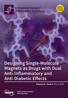
Cover Story (view full-size image): Novel cobalt coordination compounds based on bumetanide and indomethacin therapeutic agents have been designed and synthesized. The anti-inflammatory effects of metal complexes with this ligand were assayed in lipopolysaccharide stimulated RAW 264.7 macrophages. Both compounds show anti-diabetic activity with low in vitro cell toxicities. The formation of metal complexes with bioactive ligands is a new and promising strategy to find new compounds with high and enhanced biochemical properties. View this paper
  • Issues are regarded as officially published after their release is announced to the table of contents alert mailing list.
  • You may sign up for e-mail alerts to receive table of contents of newly released issues.
  • PDF is the official format for papers published in both, html and pdf forms. To view the papers in pdf format, click on the "PDF Full-text" link, and use the free Adobe Reader to open them.
Order results
Result details
Section
Select all
Export citation of selected articles as:
32 pages, 1088 KiB  
Review
Modeling Cardiovascular Diseases with hiPSC-Derived Cardiomyocytes in 2D and 3D Cultures
by Claudia Sacchetto, Libero Vitiello, Leon J. de Windt, Alessandra Rampazzo and Martina Calore
Int. J. Mol. Sci. 2020, 21(9), 3404; https://doi.org/10.3390/ijms21093404 - 11 May 2020
Cited by 44 | Viewed by 7173
Abstract
In the last decade, the generation of cardiac disease models based on human-induced pluripotent stem cells (hiPSCs) has become of common use, providing new opportunities to overcome the lack of appropriate cardiac models. Although much progress has been made toward the generation of [...] Read more.
In the last decade, the generation of cardiac disease models based on human-induced pluripotent stem cells (hiPSCs) has become of common use, providing new opportunities to overcome the lack of appropriate cardiac models. Although much progress has been made toward the generation of hiPSC-derived cardiomyocytes (hiPS-CMs), several lines of evidence indicate that two-dimensional (2D) cell culturing presents significant limitations, including hiPS-CMs immaturity and the absence of interaction between different cell types and the extracellular matrix. More recently, new advances in bioengineering and co-culture systems have allowed the generation of three-dimensional (3D) constructs based on hiPSC-derived cells. Within these systems, biochemical and physical stimuli influence the maturation of hiPS-CMs, which can show structural and functional properties more similar to those present in adult cardiomyocytes. In this review, we describe the latest advances in 2D- and 3D-hiPSC technology for cardiac disease mechanisms investigation, drug development, and therapeutic studies. Full article
(This article belongs to the Special Issue Disease Modeling Using Human Induced Pluripotent Stem Cells 2.0)
Show Figures

Figure 1

15 pages, 3163 KiB  
Article
Characteristic Analysis of Homo- and Heterodimeric Complexes of Human Mitochondrial Pyruvate Carrier Related to Metabolic Diseases
by Jinyi Lee, Zeyu Jin, Donghan Lee, Ji-Hye Yun and Weontae Lee
Int. J. Mol. Sci. 2020, 21(9), 3403; https://doi.org/10.3390/ijms21093403 - 11 May 2020
Cited by 13 | Viewed by 4309
Abstract
Human mitochondrial pyruvate carriers (hMPCs), which are required for the uptake of pyruvate into mitochondria, are associated with several metabolic diseases, including type 2 diabetes and various cancers. Yeast MPC was recently demonstrated to form a functional unit of heterodimers. However, human MPC-1 [...] Read more.
Human mitochondrial pyruvate carriers (hMPCs), which are required for the uptake of pyruvate into mitochondria, are associated with several metabolic diseases, including type 2 diabetes and various cancers. Yeast MPC was recently demonstrated to form a functional unit of heterodimers. However, human MPC-1 (hMPC-1) and MPC-2 (hMPC-2) have not yet been individually isolated for their detailed characterization, in particular in terms of their structural and functional properties, namely, whether they exist as homo- or heterodimers. In this study, hMPC-1 and hMPC-2 were successfully isolated in micelles and they formed stable homodimers. However, the heterodimer state was found to be dominant when both hMPC-1 and hMPC-2 were present. In addition, as heterodimers, the molecules exhibited a higher binding capacity to both substrates and inhibitors, together with a larger structural stability than when they existed as homodimers. Taken together, our results demonstrated that the hetero-dimerization of hMPCs is the main functional unit of the pyruvate metabolism, providing a structural insight into the transport mechanisms of hMPCs. Full article
(This article belongs to the Collection Feature Papers in Molecular Biophysics)
Show Figures

Figure 1

24 pages, 11436 KiB  
Review
NMR Investigations of Crystalline and Glassy Solid Electrolytes for Lithium Batteries: A Brief Review
by Daniel J Morales and Steven Greenbaum
Int. J. Mol. Sci. 2020, 21(9), 3402; https://doi.org/10.3390/ijms21093402 - 11 May 2020
Cited by 21 | Viewed by 5878
Abstract
The widespread use of energy storage for commercial products and services have led to great advancements in the field of lithium-based battery research. In particular, solid state lithium batteries show great promise for future commercial use, as solid electrolytes safely allow for the [...] Read more.
The widespread use of energy storage for commercial products and services have led to great advancements in the field of lithium-based battery research. In particular, solid state lithium batteries show great promise for future commercial use, as solid electrolytes safely allow for the use of lithium-metal anodes, which can significantly increase the total energy density. Of the solid electrolytes, inorganic glass-ceramics and Li-based garnet electrolytes have received much attention in the past few years due to the high ionic conductivity achieved compared to polymer-based electrolytes. This review covers recent work on novel glassy and crystalline electrolyte materials, with a particular focus on the use of solid-state nuclear magnetic resonance spectroscopy for structural characterization and transport measurements. Full article
(This article belongs to the Special Issue NMR Characterization of Amorphous and Disordered Materials)
Show Figures

Figure 1

15 pages, 4645 KiB  
Article
Fabrication of Mechanically Reinforced Gelatin/Hydroxyapatite Bio-Composite Scaffolds by Core/Shell Nozzle Printing for Bone Tissue Engineering
by Haeri Kim, Hanjun Hwangbo, YoungWon Koo and GeunHyung Kim
Int. J. Mol. Sci. 2020, 21(9), 3401; https://doi.org/10.3390/ijms21093401 - 11 May 2020
Cited by 27 | Viewed by 5944
Abstract
In tissue engineering, biocompatible scaffolds are used as 3D cell niches to provide a similar environment to that of native tissue for seeded cells to regenerate the target tissue. When engineering bone tissue, high mechanical strength and calcium phosphate composition are essential factors [...] Read more.
In tissue engineering, biocompatible scaffolds are used as 3D cell niches to provide a similar environment to that of native tissue for seeded cells to regenerate the target tissue. When engineering bone tissue, high mechanical strength and calcium phosphate composition are essential factors to consider. In this study, we fabricated biocompatible composite scaffolds composed of synthetic polymers (polycaprolactone (PCL) and poly (vinyl alcohol) (PVA)), natural polymers (gelatin and collagen) and bioceramic (hydroxyapatite; HA) for bone tissue engineering. The synthetic polymers were used to enhance the mechanical properties of the composite scaffolds while the natural protein-based polymers were used to enhance various cellular activities, such as cell adhesion and proliferation. Meanwhile, the bioceramic was introduced to promote osteogenic differentiation. Composite scaffolds were evaluated for their physical characteristics, such as mechanical, swelling and protein absorbing properties as well as biological properties (cell proliferation, alkaline phosphatase (ALP) activities and calcium deposition) with human osteoblast-like cells (MG63). Consequently, incorporation of hydroxyapatite into the gelatin/PVA (C-GPH) scaffold showed 5-fold and 1.5-fold increase in calcium deposition and ALP activities, respectively compared to gelatin/PVA scaffold (C-GP). Moreover, compressive modulus also increased 1.8-fold. Integration of PCL core into gelatin/PVA/hydroxyapatite scaffold (C-PGPH) further amplified the compressive modulus 1.5-fold. In conclusion, the scaffold that is reinforced with HA particles and integrated with PCL core of the struts showed significant potential in field of bone tissue engineering. Full article
(This article belongs to the Collection Feature Papers in Materials Science)
Show Figures

Figure 1

24 pages, 6945 KiB  
Article
Bicalutamide Elicits Renal Damage by Causing Mitochondrial Dysfunction via ROS Damage and Upregulation of HIF-1
by Kuan-Chou Chen, Chang-Rong Chen, Chang-Yu Chen, Kai-Yi Tzou, Chiung-Chi Peng and Robert Y. Peng
Int. J. Mol. Sci. 2020, 21(9), 3400; https://doi.org/10.3390/ijms21093400 - 11 May 2020
Cited by 7 | Viewed by 3685
Abstract
Combined androgen blockade using bicalutamide (Bic) is a therapeutic choice for treating prostate cancer (PCa). However, even at regular clinical dosages, Bic frequently shows adverse effects associated with cardiovascular and renal damage. Previously, we found that Bic selectively damaged mesangial cells compared to [...] Read more.
Combined androgen blockade using bicalutamide (Bic) is a therapeutic choice for treating prostate cancer (PCa). However, even at regular clinical dosages, Bic frequently shows adverse effects associated with cardiovascular and renal damage. Previously, we found that Bic selectively damaged mesangial cells compared to tubular cells and in an in vivo rat model, we also found renal damage caused by Bic. In the present study, a rat mesangial cell model was used to further the investigation. Results indicated that Bic enhanced lactate dehydrogenase release, reactive oxygen species (ROS) production, lysosome population and kidney injury molecule-1 and decreased N-cadherin. Bic elicited mitochondrial swelling and reduced the mitochondrial potential, resulting in severe suppression of the oxygen consumption rate (OCR), maximum respiration and ATP production. The hypoxia-inducible factor (HIF)-1 transcriptional activity and messenger RNA were significantly upregulated in dose-dependent manners. The HIF-1 protein reached a peak value at 24 h then rapidly decayed. BCL2/adenovirus E1B 19-kDa protein-interacting protein 3 and cleaved caspase-3 were dose-dependently upregulated by Bic (60 M) and that eventually led to cell apoptosis. It is suggested that Bic induces renal damage via ROS and modulates HIF-1 pathway and clinically, some protective agents like antioxidants are recommended for co-treatment. Full article
(This article belongs to the Special Issue Mitochondria-Targeted Approaches in Health and Disease)
Show Figures

Figure 1

11 pages, 700 KiB  
Article
Disease-Specific Derangement of Circulating Endocannabinoids and N-Acylethanolamines in Myeloproliferative Neoplasms
by Dorian Forte, Flaminia Fanelli, Marco Mezzullo, Martina Barone, Giulia Corradi, Giuseppe Auteri, Daniela Bartoletti, Marina Martello, Emanuela Ottaviani, Carolina Terragna, Antonio Curti, Uberto Pagotto, Francesca Palandri, Michele Cavo and Lucia Catani
Int. J. Mol. Sci. 2020, 21(9), 3399; https://doi.org/10.3390/ijms21093399 - 11 May 2020
Cited by 4 | Viewed by 2851
Abstract
Growing evidence highlights the endocannabinoid (EC) system involvement in cancer progression. Lipid mediators of this system are secreted by hematopoietic cells, including the ECs 2-arachidonoyl-glycerol (2AG) and arachidonoyl-ethanolamide (AEA), the 2AG metabolite 1AG, and members of N-acylethanolamine (NAE) family—palmitoyl-ethanolamide (PEA) and oleoyl-ethanolamide (OEA). [...] Read more.
Growing evidence highlights the endocannabinoid (EC) system involvement in cancer progression. Lipid mediators of this system are secreted by hematopoietic cells, including the ECs 2-arachidonoyl-glycerol (2AG) and arachidonoyl-ethanolamide (AEA), the 2AG metabolite 1AG, and members of N-acylethanolamine (NAE) family—palmitoyl-ethanolamide (PEA) and oleoyl-ethanolamide (OEA). However, the relevance of the EC system in myeloproliferative neoplasms (MPN) was never investigated. We explored the EC plasma profile in 55 MPN patients, including myelofibrosis (MF; n = 41), polycythemia vera (PV; n = 9), and essential thrombocythemia (ET; n = 5) subclasses and in 10 healthy controls (HC). AEA, PEA, OEA, 2AG, and 1AG plasma levels were measured by LC–MS/MS. Overall considered, MPN patients displayed similar EC and NAE levels compared to HC. Nonetheless, AEA levels in MPN were directly associated with the platelet count. MF patients showed higher levels of the sum of 2AG and 1AG compared to ET and PV patients, higher OEA/AEA ratios compared to HC and ET patients, and higher OEA/PEA ratios compared to HC. Furthermore, the sum of 2AG and 1AG positively correlated with JAK2V617F variant allele frequency and splenomegaly in MF and was elevated in high-risk PV patients compared to in low-risk PV patients. In conclusion, our work revealed specific alterations of ECs and NAE plasma profile in MPN subclasses and potentially relevant associations with disease severity. Full article
Show Figures

Figure 1

14 pages, 4823 KiB  
Article
Predictive Potential of Circulating Ube2h mRNA as an E2 Ubiquitin-Conjugating Enzyme for Diagnosis or Treatment of Alzheimer’s Disease
by Key-Hwan Lim and Jae-Yeol Joo
Int. J. Mol. Sci. 2020, 21(9), 3398; https://doi.org/10.3390/ijms21093398 - 11 May 2020
Cited by 14 | Viewed by 4279
Abstract
Neurodegenerative disorders are caused by neuronal cell death, miscommunications between synapse, and abnormal accumulations of proteins in the brain. Alzheimer’s disease (AD) is one of the age-related disorders, which are the most common degenerative disorders today, and strongly affects memory consolidation and cognitive [...] Read more.
Neurodegenerative disorders are caused by neuronal cell death, miscommunications between synapse, and abnormal accumulations of proteins in the brain. Alzheimer’s disease (AD) is one of the age-related disorders, which are the most common degenerative disorders today, and strongly affects memory consolidation and cognitive function in the brain. Amyloid-β and tau proteins are triggers for AD pathogenesis, and usually used as AD candidate biomarkers in the clinical research. Especially, clinical exam, brain imaging and molecular biological methods are being used to diagnosis for AD. Genome-wide association study (GWAS) is a new biomedical method, and its use contributes to understanding many human diseases, including brain diseases. Here, we identified ubiquitin conjugating enzyme E2 (Ube2) gene expression in neurons through GWAS. The subfamilies of Ube2’s genetic expression and inborn errors affect the ubiquitin proteasome system (UPS), leading to protein degradation in the brain. We found that only Ube2h mRNA transcription was significantly increased in the blood from AD, however we did not find any change of Ube2 subfamily genes’ expression in the blood and brain tissue. These data may provide information for diagnosis or clinical approach, and suggest that cell-free circulating Ube2h mRNA is a novel potential biomarker for AD. Full article
(This article belongs to the Special Issue Proteolytic Control of Deubiquitinating Enzymes in Health and Disease)
Show Figures

Figure 1

14 pages, 2549 KiB  
Article
Lysosome Dynamic Properties during Neuronal Stem Cell Differentiation Studied by Spatiotemporal Fluctuation Spectroscopy and Organelle Tracking
by William Durso, Manuella Martins, Laura Marchetti, Federico Cremisi, Stefano Luin and Francesco Cardarelli
Int. J. Mol. Sci. 2020, 21(9), 3397; https://doi.org/10.3390/ijms21093397 - 11 May 2020
Cited by 10 | Viewed by 3357
Abstract
We investigated lysosome dynamics during neuronal stem cell (NSC) differentiation by two quantitative and complementary biophysical methods based on fluorescence: imaging-derived mean square displacement (iMSD) and single-particle tracking (SPT). The former extracts the average dynamics and size of the whole population [...] Read more.
We investigated lysosome dynamics during neuronal stem cell (NSC) differentiation by two quantitative and complementary biophysical methods based on fluorescence: imaging-derived mean square displacement (iMSD) and single-particle tracking (SPT). The former extracts the average dynamics and size of the whole population of moving lysosomes directly from imaging, with no need to calculate single trajectories; the latter resolves the finest heterogeneities and dynamic features at the single-lysosome level, which are lost in the iMSD analysis. In brief, iMSD analysis reveals that, from a structural point of view, lysosomes decrement in size during NSC differentiation, from 1 μm average diameter in the embryonic cells to approximately 500 nm diameter in the fully differentiated cells. Concomitantly, iMSD analysis highlights modification of key dynamic parameters, such as the average local organelle diffusivity and anomalous coefficient, which may parallel cytoskeleton remodeling during the differentiation process. From average to local, SPT allows mapping heterogeneous dynamic responses of single lysosomes in different districts of the cells. For instance, a dramatic decrease of lysosomal transport in the soma is followed by a rapid increase of transport in the projections at specific time points during neuronal differentiation, an observation compatible with the hypothesis that lysosomal active mobilization shifts from the soma to the newborn projections. Our combined results provide new insight into the lysosome size and dynamics regulation throughout NSC differentiation, supporting new functions proposed for this organelle. Full article
Show Figures

Figure 1

21 pages, 966 KiB  
Review
Behavioral, Biochemical and Electrophysiological Changes in Spared Nerve Injury Model of Neuropathic Pain
by Francesca Guida, Danilo De Gregorio, Enza Palazzo, Flavia Ricciardi, Serena Boccella, Carmela Belardo, Monica Iannotta, Rosmara Infantino, Federica Formato, Ida Marabese, Livio Luongo, Vito de Novellis and Sabatino Maione
Int. J. Mol. Sci. 2020, 21(9), 3396; https://doi.org/10.3390/ijms21093396 - 11 May 2020
Cited by 62 | Viewed by 7544
Abstract
Neuropathic pain is a pathological condition induced by a lesion or disease affecting the somatosensory system, with symptoms like allodynia and hyperalgesia. It has a multifaceted pathogenesis as it implicates several molecular signaling pathways involving peripheral and central nervous systems. Affective and cognitive [...] Read more.
Neuropathic pain is a pathological condition induced by a lesion or disease affecting the somatosensory system, with symptoms like allodynia and hyperalgesia. It has a multifaceted pathogenesis as it implicates several molecular signaling pathways involving peripheral and central nervous systems. Affective and cognitive dysfunctions have been reported as comorbidities of neuropathic pain states, supporting the notion that pain and mood disorders share some common pathogenetic mechanisms. The understanding of these pathophysiological mechanisms requires the development of animal models mimicking, as far as possible, clinical neuropathic pain symptoms. Among them, the Spared Nerve Injury (SNI) model has been largely characterized in terms of behavioral and functional alterations. This model is associated with changes in neuronal firing activity at spinal and supraspinal levels, and induces late neuropsychiatric disorders (such as anxious-like and depressive-like behaviors, and cognitive impairments) comparable to an advanced phase of neuropathy. The goal of this review is to summarize current findings in preclinical research, employing the SNI model as a tool for identifying pathophysiological mechanisms of neuropathic pain and testing pharmacological agent. Full article
Show Figures

Graphical abstract

30 pages, 2879 KiB  
Review
Regulation of Iron Homeostasis and Use in Chloroplasts
by Gretchen E. Kroh and Marinus Pilon
Int. J. Mol. Sci. 2020, 21(9), 3395; https://doi.org/10.3390/ijms21093395 - 11 May 2020
Cited by 84 | Viewed by 7061
Abstract
Iron (Fe) is essential for life because of its role in protein cofactors. Photosynthesis, in particular photosynthetic electron transport, has a very high demand for Fe cofactors. Fe is commonly limiting in the environment, and therefore photosynthetic organisms must acclimate to Fe availability [...] Read more.
Iron (Fe) is essential for life because of its role in protein cofactors. Photosynthesis, in particular photosynthetic electron transport, has a very high demand for Fe cofactors. Fe is commonly limiting in the environment, and therefore photosynthetic organisms must acclimate to Fe availability and avoid stress associated with Fe deficiency. In plants, adjustment of metabolism, of Fe utilization, and gene expression, is especially important in the chloroplasts during Fe limitation. In this review, we discuss Fe use, Fe transport, and mechanisms of acclimation to Fe limitation in photosynthetic lineages with a focus on the photosynthetic electron transport chain. We compare Fe homeostasis in Cyanobacteria, the evolutionary ancestors of chloroplasts, with Fe homeostasis in green algae and in land plants in order to provide a deeper understanding of how chloroplasts and photosynthesis may cope with Fe limitation. Full article
(This article belongs to the Special Issue Iron and Sulfur in Plants)
Show Figures

Figure 1

22 pages, 4461 KiB  
Article
Different Roles of Auxins in Somatic Embryogenesis Efficiency in Two Picea Species
by Teresa Hazubska-Przybył, Ewelina Ratajczak, Agata Obarska and Emilia Pers-Kamczyc
Int. J. Mol. Sci. 2020, 21(9), 3394; https://doi.org/10.3390/ijms21093394 - 11 May 2020
Cited by 30 | Viewed by 3976
Abstract
The effects of auxins 2,4-D (2,4-dichlorophenoxyacetic acid), NAA (1-naphthaleneacetic acid) or picloram (4-amino-3,5,6-trichloropicolinic acid; 9 µM) and cytokinin BA (benzyloadenine; 4.5 µM) applied in the early stages of somatic embryogenesis (SE) on specific stages of SE in Picea abies and P. omorika were [...] Read more.
The effects of auxins 2,4-D (2,4-dichlorophenoxyacetic acid), NAA (1-naphthaleneacetic acid) or picloram (4-amino-3,5,6-trichloropicolinic acid; 9 µM) and cytokinin BA (benzyloadenine; 4.5 µM) applied in the early stages of somatic embryogenesis (SE) on specific stages of SE in Picea abies and P. omorika were investigated. The highest SE initiation frequency was obtained after 2,4-D application in P. omorika (22.00%) and picloram application in P. abies (10.48%). NAA treatment significantly promoted embryogenic tissue (ET) proliferation in P. abies, while 2,4-D treatment reduced it. This reduction was related to the oxidative stress level, which was lower with the presence of NAA in the proliferation medium and higher with the presence of 2,4-D. The reduced oxidative stress level after NAA treatment suggests that hydrogen peroxide (H2O2) acts as a signalling molecule and promotes ET proliferation. NAA and picloram in the proliferation medium decreased the further production and maturation of P. omorika somatic embryos compared with that under 2,4-D. The quality of the germinated P. abies embryos and their development into plantlets depended on the auxin type and were the highest in NAA-originated embryos. These results show that different auxin types can generate different physiological responses in plant materials during SE in both spruce species. Full article
(This article belongs to the Section Molecular Plant Sciences)
Show Figures

Figure 1

16 pages, 1831 KiB  
Article
Pan-Genome Analyses of Geobacillus spp. Reveal Genetic Characteristics and Composting Potential
by Mengmeng Wang, Han Zhu, Zhijian Kong, Tuo Li, Lei Ma, Dongyang Liu and Qirong Shen
Int. J. Mol. Sci. 2020, 21(9), 3393; https://doi.org/10.3390/ijms21093393 - 11 May 2020
Cited by 6 | Viewed by 3344
Abstract
The genus Geobacillus is abundant in ecological diversity and is also well-known as an authoritative source for producing various thermostable enzymes. Although it is clear now that Geobacillus evolved from Bacillus, relatively little knowledge has been obtained regarding its evolutionary mechanism, which [...] Read more.
The genus Geobacillus is abundant in ecological diversity and is also well-known as an authoritative source for producing various thermostable enzymes. Although it is clear now that Geobacillus evolved from Bacillus, relatively little knowledge has been obtained regarding its evolutionary mechanism, which might also contribute to its ecological diversity and biotechnology potential. Here, a statistical comparison of thirty-two Geobacillus genomes was performed with a specific focus on pan- and core genomes. The pan-genome of this set of Geobacillus strains contained 14,913 genes, and the core genome contained 940 genes. The Clusters of Orthologous Groups (COG) and Carbohydrate-Active Enzymes (CAZymes) analysis revealed that the Geobacillus strains had huge potential industrial application in composting for agricultural waste management. Detailed comparative analyses showed that basic functional classes and housekeeping genes were conserved in the core genome, while genes associated with environmental interaction or energy metabolism were more enriched in the pan-genome. Therefore, the evolution of Geobacillus seems to be guided by environmental parameters. In addition, horizontal gene transfer (HGT) events among different Geobacillus species were detected. Altogether, pan-genome analysis was a useful method for detecting the evolutionary mechanism, and Geobacillus’ evolution was directed by the environment and HGT events. Full article
(This article belongs to the Section Molecular Genetics and Genomics)
Show Figures

Figure 1

16 pages, 2563 KiB  
Article
Micheliolide Enhances Radiosensitivities of p53-Deficient Non-Small-Cell Lung Cancer via Promoting HIF-1α Degradation
by Peizhong Kong, K.N. Yu, Miaomiao Yang, Waleed Abdelbagi Almahi, Lili Nie, Guodong Chen and Wei Han
Int. J. Mol. Sci. 2020, 21(9), 3392; https://doi.org/10.3390/ijms21093392 - 11 May 2020
Cited by 12 | Viewed by 3662
Abstract
Micheliolide (MCL) has shown promising anti-inflammatory and anti-tumor efficacy. However, whether and how MCL enhances the sensitivity of non-small-cell lung cancer (NSCLC) to radiotherapy are still unknown. In the present paper, we found that MCL exerted a tumor cell killing effect on NSCLC [...] Read more.
Micheliolide (MCL) has shown promising anti-inflammatory and anti-tumor efficacy. However, whether and how MCL enhances the sensitivity of non-small-cell lung cancer (NSCLC) to radiotherapy are still unknown. In the present paper, we found that MCL exerted a tumor cell killing effect on NSCLC cells in a dose-dependent manner, and MCL strongly sensitized p53-deficient NSCLC cells, but not the cells with wild-type p53 to irradiation (IR). Meanwhile, MCL markedly inhibited the expression of hypoxia-inducible factor-1α (HIF-1α) after IR and hypoxic exposure in H1299 and Calu-1 cells rather than in H460 cells. Consistently, radiation- or hypoxia-induced expression of vascular endothelial growth factor (VEGF) was also significantly inhibited by MCL in H1299 and Calu-1 cells, but not in H460 cells. Therefore, inhibition of the HIF-1α pathway might, at least in part, contribute to the radiosensitizing effect of MCL. Further study showed that MCL could accelerate the degradation of HIF-1α through the ubiquitin-proteosome system. In addition, the transfection of wild-type p53 into p53-null cells (H1299) attenuated the effect of MCL on inhibiting HIF-1α expression. These results suggest MCL effectively sensitizes p53-deficient NSCLC cells to IR in a manner of inhibiting the HIF-1α pathway via promoting HIF-1α degradation, and p53 played a negative role in MCL-induced HIF-1α degradation. Full article
(This article belongs to the Section Molecular Pathology, Diagnostics, and Therapeutics)
Show Figures

Figure 1

17 pages, 1108 KiB  
Review
Colorectal Cancer Invasion and Atrophy of the Enteric Nervous System: Potential Feedback and Impact on Cancer Progression
by Janusz Godlewski and Zbigniew Kmiec
Int. J. Mol. Sci. 2020, 21(9), 3391; https://doi.org/10.3390/ijms21093391 - 11 May 2020
Cited by 21 | Viewed by 4454
Abstract
Colorectal cancer (CRC) invasion within the large intestine wall results in the replacement of normal tissue architecture by tumour mass. Cancer cells digest the extracellular matrix (ECM) by the release of proteolytic enzymes. The disintegration of matrix ground substance activates several deposited growth [...] Read more.
Colorectal cancer (CRC) invasion within the large intestine wall results in the replacement of normal tissue architecture by tumour mass. Cancer cells digest the extracellular matrix (ECM) by the release of proteolytic enzymes. The disintegration of matrix ground substance activates several deposited growth factors which stimulate cell proliferation. Stromal (mainly fibroblasts), immune and cancer cells dominate in this area and become involved in a network of multimodal interactions which significantly induce proliferation of colon cancer cells, inhibit their apoptosis and promote their spreading within the local tumour microenvironment. Cancer invasion destroys nerve fibres and neurons of the local enteric nervous system (ENS) and induces subsequent atrophy of the submucosal and myenteric plexuses in areas adjacent to the cancer boundary. Interestingly, the reduction of plexuses’ size is accompanied by the increased number of galanin-immunoreactive neurons and increased galanin content in parts of the colon located close to the tumour. Galanin, a neuroprotective peptide, may inhibit the extrinsic pathway of apoptosis and in this way promote cancer cell survival. The possible role of acetylcholine and some ENS neuropeptides was also discussed. Invasion of cancer cells spreads along nerve fibres with the involvement of locally-released neutrophins which promote, via their specific receptors, cancer cell proliferation and pro-survival signalling pathways. Thus, during CRC development cancer cells and neurons of the ENS release many neurotransmitters/neuropeptides which affect key cellular signalling pathways promoting cancer cell proliferation and pro-survival phenotype. The multiple interactions between ENS neurons, cancer cells and other cell types present in the colon wall increase cancer cell invasiveness and have a negative impact on the course of CRC. Full article
(This article belongs to the Special Issue Survival Pathways Involved in Resistance to Apoptosis in Cancer)
Show Figures

Figure 1

16 pages, 3813 KiB  
Article
The Bacterial Toxin CNF1 Protects Human Neuroblastoma SH-SY5Y Cells against 6-Hydroxydopamine-Induced Cell Damage: The Hypothesis of CNF1-Promoted Autophagy as an Antioxidant Strategy
by Sara Travaglione, Stefano Loizzo, Rosa Vona, Giulia Ballan, Roberto Rivabene, Danila Giordani, Marco Guidotti, Maria Luisa Dupuis, Zaira Maroccia, Monica Baiula, Roberto Rimondini, Gabriele Campana and Carla Fiorentini
Int. J. Mol. Sci. 2020, 21(9), 3390; https://doi.org/10.3390/ijms21093390 - 11 May 2020
Cited by 4 | Viewed by 3355
Abstract
Several chronic neuroinflammatory diseases, including Parkinson’s disease (PD), have the so-called ‘redox imbalance’ in common, a dynamic system modulated by various factors. Among them, alteration of the mitochondrial functionality can cause overproduction of reactive oxygen species (ROS) with the consequent induction of oxidative [...] Read more.
Several chronic neuroinflammatory diseases, including Parkinson’s disease (PD), have the so-called ‘redox imbalance’ in common, a dynamic system modulated by various factors. Among them, alteration of the mitochondrial functionality can cause overproduction of reactive oxygen species (ROS) with the consequent induction of oxidative DNA damage and apoptosis. Considering the failure of clinical trials with drugs that eliminate ROS directly, research currently focuses on approaches that counteract redox imbalance, thus restoring normal physiology in a neuroinflammatory condition. Herein, we used SH-SY5Y cells treated with 6-hydroxydopamine (6-OHDA), a neurotoxin broadly employed to generate experimental models of PD. Cells were pre-treated with the Rho-modulating Escherichia coli cytotoxic necrotizing factor 1 (CNF1), before the addition of 6-OHDA. Then, cell viability, mitochondrial morphology and dynamics, redox profile as well as autophagic markers expression were assessed. We found that CNF1 preserves cell viability and counteracts oxidative stress induced by 6-OHDA. These effects are accompanied by modulation of the mitochondrial network and an increase in macroautophagic markers. Our results confirm the Rho GTPases as suitable pharmacological targets to counteract neuroinflammatory diseases and evidence the potentiality of CNF1, whose beneficial effects on pathological animal models have been already proven to act against oxidative stress through an autophagic strategy. Full article
(This article belongs to the Special Issue Bacterial Protein Toxins: Enemies within or Unexpected Friends 2.0)
Show Figures

Figure 1

18 pages, 884 KiB  
Review
Rheumatic Manifestations in Patients Treated with Immune Checkpoint Inhibitors
by Konstantinos Melissaropoulos, Kalliopi Klavdianou, Alexandra Filippopoulou, Fotini Kalofonou, Haralabos Kalofonos and Dimitrios Daoussis
Int. J. Mol. Sci. 2020, 21(9), 3389; https://doi.org/10.3390/ijms21093389 - 11 May 2020
Cited by 19 | Viewed by 3955
Abstract
Immune checkpoint inhibitors (ICIs) are monoclonal antibodies that activate the immune system, aiming at enhancing antitumor immunity. Their clinical efficacy is well-documented, but the side effects associated with their use are still under investigation. These drugs cause several immune-related adverse events (ir-AEs), some [...] Read more.
Immune checkpoint inhibitors (ICIs) are monoclonal antibodies that activate the immune system, aiming at enhancing antitumor immunity. Their clinical efficacy is well-documented, but the side effects associated with their use are still under investigation. These drugs cause several immune-related adverse events (ir-AEs), some of which stand within the field of rheumatology. Herein, we present a literature review performed in an effort to evaluate all publicly available clinical data regarding rheumatic manifestations associated with ICIs. The most common musculoskeletal ir-AEs are inflammatory arthritis, polymyalgia rheumatica and myositis. Non-musculoskeletal rheumatic manifestations are less frequent, with the most prominent being sicca, vasculitides and sarcoidosis. Cases of systemic lupus erythematosus or scleroderma are extremely rare. The majority of musculoskeletal ir-AEs are of mild/moderate severity and can be managed with steroids with no need for ICI discontinuation. In severe cases, more intense immunosuppressive therapy and permanent ICI discontinuation may be employed. Oncologists should periodically screen patients receiving ICIs for new-onset inflammatory musculoskeletal complaints and seek a rheumatology consultation in cases of persisting symptoms. Full article
(This article belongs to the Special Issue Monoclonal Antibodies to Treat Cancer)
Show Figures

Figure 1

12 pages, 260 KiB  
Review
Special Issue about Head and Neck Cancers: HPV Positive Cancers
by Panagiota Economopoulou, Ioannis Kotsantis and Amanda Psyrri
Int. J. Mol. Sci. 2020, 21(9), 3388; https://doi.org/10.3390/ijms21093388 - 11 May 2020
Cited by 36 | Viewed by 4604
Abstract
The oropharynx has become the leading primary site for Human Papilloma Virus (HPV)-associated head and neck cancer. HPV positive oropharyngeal squamous cell carcinoma (HPV+ OSCC) has emerged as an epidemic not easily recognized by many physicians, resulting in delays in diagnosis and management. [...] Read more.
The oropharynx has become the leading primary site for Human Papilloma Virus (HPV)-associated head and neck cancer. HPV positive oropharyngeal squamous cell carcinoma (HPV+ OSCC) has emerged as an epidemic not easily recognized by many physicians, resulting in delays in diagnosis and management. HPV+ OSCC traditionally refers to younger, healthier patients with high economic status and high-risk sexual behavior and is related to improved prognosis. De-intensification strategies are being evaluated in ongoing clinical trials and if validated, might help spare severe morbidity associated with current cisplatin-based chemoradiotherapy, which is the standard of care for all patients with locally advanced head and neck cancer. On the other hand, whether HPV status represents an important prognostic factor for non-oropharyngeal sites remains to be elucidated. Full article
19 pages, 1001 KiB  
Review
The Interplay between MicroRNAs and the Components of the Tumor Microenvironment in B-Cell Malignancies
by Sherien M. El-Daly, Recep Bayraktar, Simone Anfossi and George A. Calin
Int. J. Mol. Sci. 2020, 21(9), 3387; https://doi.org/10.3390/ijms21093387 - 11 May 2020
Cited by 19 | Viewed by 4767
Abstract
An increased focus is being placed on the tumorigenesis and contexture of tumor microenvironment in hematopoietic and solid tumors. Despite recent clinical revolutions in adoptive T-cell transfer approaches and immune checkpoint blockade, tumor microenvironment is a major obstacle to tumor regression in B-cell [...] Read more.
An increased focus is being placed on the tumorigenesis and contexture of tumor microenvironment in hematopoietic and solid tumors. Despite recent clinical revolutions in adoptive T-cell transfer approaches and immune checkpoint blockade, tumor microenvironment is a major obstacle to tumor regression in B-cell malignancies. A transcriptional alteration of coding and non-coding RNAs, such as microRNAs (miRNAs), has been widely demonstrated in the tumor microenvironment of B-cell malignancies. MiRNAs have been associated with different clinical-biological forms of B-cell malignancies and involved in the regulation of B lymphocyte development, maturation, and function, including B-cell activation and malignant transformation. Additionally, tumor-secreted extracellular vesicles regulate recipient cell functions in the tumor microenvironment to facilitate metastasis and progression by delivering miRNA contents to neighboring cells. Herein, we focus on the interplay between miRNAs and tumor microenvironment components in the different B-cell malignancies and its impact on diagnosis, proliferation, and involvement in treatment resistance. Full article
Show Figures

Graphical abstract

17 pages, 3037 KiB  
Article
Probing Nanoelectroporation and Resealing of the Cell Membrane by the Entry of Ca2+ and Ba2+ Ions
by Wenfei Bo, Mantas Silkunas, Uma Mangalanathan, Vitalij Novickij, Maura Casciola, Iurii Semenov, Shu Xiao, Olga N. Pakhomova and Andrei G. Pakhomov
Int. J. Mol. Sci. 2020, 21(9), 3386; https://doi.org/10.3390/ijms21093386 - 11 May 2020
Cited by 22 | Viewed by 3211
Abstract
The principal bioeffect of the nanosecond pulsed electric field (nsPEF) is a lasting cell membrane permeabilization, which is often attributed to the formation of nanometer-sized pores. Such pores may be too small for detection by the uptake of fluorescent dyes. We tested if [...] Read more.
The principal bioeffect of the nanosecond pulsed electric field (nsPEF) is a lasting cell membrane permeabilization, which is often attributed to the formation of nanometer-sized pores. Such pores may be too small for detection by the uptake of fluorescent dyes. We tested if Ca2+, Cd2+, Zn2+, and Ba2+ ions can be used as nanoporation markers. Time-lapse imaging was performed in CHO, BPAE, and HEK cells loaded with Fluo-4, Calbryte, or Fluo-8 dyes. Ca2+ and Ba2+ did not change fluorescence in intact cells, whereas their entry after nsPEF increased fluorescence within <1 ms. The threshold for one 300-ns pulse was at 1.5–2 kV/cm, much lower than >7 kV/cm for the formation of larger pores that admitted YO-PRO-1, TO-PRO-3, or propidium dye into the cells. Ba2+ entry caused a gradual emission rise, which reached a stable level in 2 min or, with more intense nsPEF, kept rising steadily for at least 30 min. Ca2+ entry could elicit calcium-induced calcium release (CICR) followed by Ca2+ removal from the cytosol, which markedly affected the time course, polarity, amplitude, and the dose-dependence of fluorescence change. Both Ca2+ and Ba2+ proved as sensitive nanoporation markers, with Ba2+ being more reliable for monitoring membrane damage and resealing. Full article
(This article belongs to the Section Molecular Biophysics)
Show Figures

Figure 1

14 pages, 3279 KiB  
Article
Akkermansia muciniphila Aspartic Protease Amuc_1434* Inhibits Human Colorectal Cancer LS174T Cell Viability via TRAIL-Mediated Apoptosis Pathway
by Xin Meng, Jinrui Zhang, Hao Wu, Dahai Yu and Xuexun Fang
Int. J. Mol. Sci. 2020, 21(9), 3385; https://doi.org/10.3390/ijms21093385 - 11 May 2020
Cited by 35 | Viewed by 3748
Abstract
Mucin2 (Muc2) is the main component of the intestinal mucosal layer and is highly expressed in mucous colorectal cancer. Previous studies conducted by our lab found that the recombinant protein Amuc_1434 (expressed in Escherichia coli prokaryote cell system, hereinafter termed Amuc_1434*), derived from [...] Read more.
Mucin2 (Muc2) is the main component of the intestinal mucosal layer and is highly expressed in mucous colorectal cancer. Previous studies conducted by our lab found that the recombinant protein Amuc_1434 (expressed in Escherichia coli prokaryote cell system, hereinafter termed Amuc_1434*), derived from Akkermansia muciniphila, can degrade Muc2. Thus, the main objective of this study was to explore the effects of Amuc_1434* on LS174T in colorectal cancer cells expressing Muc2. Results from this study demonstrated that Amuc_1434* inhibited the proliferation of LS174T cells, which was related to its ability to degrade Muc2. Amuc_1434* also blocked the G0/G1 phase of the cell cycle of LS174T cells and upregulated the expression of tumor protein 53 (p53), which is a cell cycle-related protein. In addition, Amuc_1434* promoted apoptosis of LS174T cells and increased mitochondrial ROS levels in LS174T cells. The mitochondrial membrane potential of LS174T cells was also downregulated by Amuc_1434*. Amuc_1434* can activate the death receptor pathway and mitochondrial pathway of apoptosis by upregulating tumor-necrosis-factor-related apoptosis-inducing ligand (TRAIL). In conclusion, our study was the first to demonstrate that the protein Amuc_1434* derived from Akkermansia muciniphila suppresses LS174T cell viability via TRAIL-mediated apoptosis pathway. Full article
(This article belongs to the Section Molecular Biology)
Show Figures

Figure 1

16 pages, 932 KiB  
Article
A Data-Driven Review of the Genetic Factors of Pregnancy Complications
by Yury A. Barbitoff, Alexander A. Tsarev, Elena S. Vashukova, Evgeniia M. Maksiutenko, Liudmila V. Kovalenko, Larisa D. Belotserkovtseva and Andrey S. Glotov
Int. J. Mol. Sci. 2020, 21(9), 3384; https://doi.org/10.3390/ijms21093384 - 11 May 2020
Cited by 16 | Viewed by 5207
Abstract
Over the recent years, many advances have been made in the research of the genetic factors of pregnancy complications. In this work, we use publicly available data repositories, such as the National Human Genome Research Institute GWAS Catalog, HuGE Navigator, and the UK [...] Read more.
Over the recent years, many advances have been made in the research of the genetic factors of pregnancy complications. In this work, we use publicly available data repositories, such as the National Human Genome Research Institute GWAS Catalog, HuGE Navigator, and the UK Biobank genetic and phenotypic dataset to gain insights into molecular pathways and individual genes behind a set of pregnancy-related traits, including the most studied ones—preeclampsia, gestational diabetes, preterm birth, and placental abruption. Using both HuGE and GWAS Catalog data, we confirm that immune system and, in particular, T-cell related pathways are one of the most important drivers of pregnancy-related traits. Pathway analysis of the data reveals that cell adhesion and matrisome-related genes are also commonly involved in pregnancy pathologies. We also find a large role of metabolic factors that affect not only gestational diabetes, but also the other traits. These shared metabolic genes include IGF2, PPARG, and NOS3. We further discover that the published genetic associations are poorly replicated in the independent UK Biobank cohort. Nevertheless, we find novel genome-wide associations with pregnancy-related traits for the FBLN7, STK32B, and ACTR3B genes, and replicate the effects of the KAZN and TLE1 genes, with the latter being the only gene identified across all data resources. Overall, our analysis highlights central molecular pathways for pregnancy-related traits, and suggests a need to use more accurate and sophisticated association analysis strategies to robustly identify genetic risk factors for pregnancy complications. Full article
(This article belongs to the Special Issue OMICs, Data Integration, and Applications in Personalized Medicine)
Show Figures

Graphical abstract

13 pages, 1886 KiB  
Article
Caviar Extract and Its Constituent DHA Inhibits UVB-Irradiated Skin Aging by Inducing Adiponectin Production
by Kyung-Eun Lee, Youn-Hwa Nho, Seok Kyun Yun, Sung-Min Park, Seunghyun Kang and Hyeonju Yeo
Int. J. Mol. Sci. 2020, 21(9), 3383; https://doi.org/10.3390/ijms21093383 - 11 May 2020
Cited by 11 | Viewed by 6398
Abstract
In this study, caviar (sturgeon eggs) was used to elucidate its roles in adiponectin production and skin anti-aging. Recently, caviar has been largely used not only as a nutritional food, but also in cosmetic products. In particular, it has been reported that docosahexaenoic [...] Read more.
In this study, caviar (sturgeon eggs) was used to elucidate its roles in adiponectin production and skin anti-aging. Recently, caviar has been largely used not only as a nutritional food, but also in cosmetic products. In particular, it has been reported that docosahexaenoic acid (DHA), as one of the main phospholipid components of caviar extract, induces intracellular lipid accumulation and the expression of adiponectin in adipocytes. Although adipocytes are well known to be associated with the skin dermis by secreting various factors (e.g., adiponectin), the effects of caviar extract and DHA on the skin are not well studied. Here, we demonstrate the effects of caviar extract and DHA on adipocyte differentiation and adiponectin production, resulting in a preventive role in UV-irradiated skin aging. Caviar extract and DHA enhanced adipocyte differentiation and promoted the synthesis of transcription factors controlling adipocyte differentiation and adiponectin. In addition, the mRNA expression levels of matrix metalloproteinase-1 (MMP-1) were decreased in UVB-irradiated Hs68 fibroblasts that were cultured in conditioned medium from caviar extract or DHA-treated differentiated adipocytes. Taken together, these results indicate that caviar extract and DHA induce adipocyte differentiation and adiponectin production, thereby inhibiting UVB-induced premature skin aging via the suppression of MMP-1 production. Full article
(This article belongs to the Special Issue Molecular Mechanisms of Skin Aging)
Show Figures

Figure 1

17 pages, 2709 KiB  
Article
Effects of PDE3 Inhibitor Olprinone on the Respiratory Parameters, Inflammation, and Apoptosis in an Experimental Model of Acute Respiratory Distress Syndrome
by Petra Kosutova, Pavol Mikolka, Sona Balentova, Marian Adamkov, Andrea Calkovska and Daniela Mokra
Int. J. Mol. Sci. 2020, 21(9), 3382; https://doi.org/10.3390/ijms21093382 - 11 May 2020
Cited by 8 | Viewed by 2769
Abstract
This study aimed to investigate whether a selective phosphodiesterase-3 (PDE3) inhibitor olprinone can positively influence the inflammation, apoptosis, and respiratory parameters in animals with acute respiratory distress syndrome (ARDS) model induced by repetitive saline lung lavage. Adult rabbits were divided into 3 groups: [...] Read more.
This study aimed to investigate whether a selective phosphodiesterase-3 (PDE3) inhibitor olprinone can positively influence the inflammation, apoptosis, and respiratory parameters in animals with acute respiratory distress syndrome (ARDS) model induced by repetitive saline lung lavage. Adult rabbits were divided into 3 groups: ARDS without therapy (ARDS), ARDS treated with olprinone i.v. (1 mg/kg; ARDS/PDE3), and healthy ventilated controls (Control), and were oxygen-ventilated for the following 4 h. Dynamic lung–thorax compliance (Cdyn), mean airway pressure (MAP), arterial oxygen saturation (SaO2), alveolar-arterial gradient (AAG), ratio between partial pressure of oxygen in arterial blood to a fraction of inspired oxygen (PaO2/FiO2), oxygenation index (OI), and ventilation efficiency index (VEI) were evaluated every hour. Post mortem, inflammatory and oxidative markers (interleukin (IL)-6, IL-1β, a receptor for advanced glycation end products (RAGE), IL-10, total antioxidant capacity (TAC), 3-nitrotyrosine (3NT), and malondialdehyde (MDA) and apoptosis (apoptotic index and caspase-3) were assessed in the lung tissue. Treatment with olprinone reduced the release of inflammatory mediators and markers of oxidative damage decreased apoptosis of epithelial cells and improved respiratory parameters. The results indicate a future potential of PDE3 inhibitors also in the therapy of ARDS. Full article
Show Figures

Figure 1

24 pages, 3355 KiB  
Review
Linear Ubiquitin Code: Its Writer, Erasers, Decoders, Inhibitors, and Implications in Disorders
by Daisuke Oikawa, Yusuke Sato, Hidefumi Ito and Fuminori Tokunaga
Int. J. Mol. Sci. 2020, 21(9), 3381; https://doi.org/10.3390/ijms21093381 - 11 May 2020
Cited by 35 | Viewed by 6782
Abstract
The linear ubiquitin chain assembly complex (LUBAC) is a ubiquitin ligase composed of the Heme-oxidized IRP2 ubiquitin ligase-1L (HOIL-1L), HOIL-1L-interacting protein (HOIP), and Shank-associated RH domain interactor (SHARPIN) subunits. LUBAC specifically generates the N-terminal Met1-linked linear ubiquitin chain and regulates acquired and innate [...] Read more.
The linear ubiquitin chain assembly complex (LUBAC) is a ubiquitin ligase composed of the Heme-oxidized IRP2 ubiquitin ligase-1L (HOIL-1L), HOIL-1L-interacting protein (HOIP), and Shank-associated RH domain interactor (SHARPIN) subunits. LUBAC specifically generates the N-terminal Met1-linked linear ubiquitin chain and regulates acquired and innate immune responses, such as the canonical nuclear factor-κB (NF-κB) and interferon antiviral pathways. Deubiquitinating enzymes, OTULIN and CYLD, physiologically bind to HOIP and control its function by hydrolyzing the linear ubiquitin chain. Moreover, proteins containing linear ubiquitin-specific binding domains, such as NF-κB-essential modulator (NEMO), optineurin, A20-binding inhibitors of NF-κB (ABINs), and A20, modulate the functions of LUBAC, and the dysregulation of the LUBAC-mediated linear ubiquitination pathway induces cancer and inflammatory, autoimmune, and neurodegenerative diseases. Therefore, inhibitors of LUBAC would be valuable to facilitate investigations of the molecular and cellular bases for LUBAC-mediated linear ubiquitination and signal transduction, and for potential therapeutic purposes. We identified and characterized α,β-unsaturated carbonyl-containing chemicals, named HOIPINs (HOIP inhibitors), as LUBAC inhibitors. We summarize recent advances in elucidations of the pathophysiological functions of LUBAC-mediated linear ubiquitination and identifications of its regulators, toward the development of LUBAC inhibitors. Full article
(This article belongs to the Special Issue Ubiquitination in Health and Diseases)
Show Figures

Figure 1

15 pages, 3218 KiB  
Article
Characterization of FcXTH2, a Novel Xyloglucan Endotransglycosylase/Hydrolase Enzyme of Chilean Strawberry with Hydrolase Activity
by Luis Morales-Quintana, Dina Beltrán, Ángela Mendez-Yañez, Felipe Valenzuela-Riffo, Raúl Herrera and María Alejandra Moya-León
Int. J. Mol. Sci. 2020, 21(9), 3380; https://doi.org/10.3390/ijms21093380 - 11 May 2020
Cited by 9 | Viewed by 3111
Abstract
Xyloglucan endotransglycosylase/hydrolases (XTHs) are cell wall enzymes with hydrolase (XEH) and/or endotransglycosylase (XET) activities. As they are involved in the modification of the xyloglucans, a type of hemicellulose present in the cell wall, they are believed to be very important in different processes, [...] Read more.
Xyloglucan endotransglycosylase/hydrolases (XTHs) are cell wall enzymes with hydrolase (XEH) and/or endotransglycosylase (XET) activities. As they are involved in the modification of the xyloglucans, a type of hemicellulose present in the cell wall, they are believed to be very important in different processes, including growth, development, and fruit ripening. Previous studies suggest that XTHs might play a key role in development and ripening of Fragaria chiloensis fruit, and its characterization is pending. Therefore, in order to provide a biochemical characterization of the FcXTH2 enzyme to explain its possible role in strawberry development, the molecular cloning and the heterologous expression of FcXTH2 were performed. The recombinant FcXTH2 was active and displayed mainly XEH activity. The optimal pH and temperature are 5.5 and 37 °C, respectively. A KM value of 0.029 mg mL−1 was determined. Additionally, its protein structural model was built through comparative modeling methodology. The model showed a typically β-jelly-roll type folding in which the catalytic motif was oriented towards the FcXTH2 central cavity. Using molecular docking, protein-ligand interactions were explored, finding better interaction with xyloglucan than with cellulose. The data provided groundwork for understanding, at a molecular level, the enzymatic mechanism of FcXTH2, an important enzyme acting during the development of the Chilean strawberry. Full article
(This article belongs to the Special Issue Structural/Functional Characterization of Plant Proteins)
Show Figures

Graphical abstract

23 pages, 1724 KiB  
Review
Interplay between Cytokine Circuitry and Transcriptional Regulation Shaping Helper T Cell Pathogenicity and Plasticity in Inflammatory Bowel Disease
by Shin-Huei Fu, Ming-Wei Chien, Chao-Yuan Hsu, Yu-Wen Liu and Huey-Kang Sytwu
Int. J. Mol. Sci. 2020, 21(9), 3379; https://doi.org/10.3390/ijms21093379 - 11 May 2020
Cited by 15 | Viewed by 4748
Abstract
Inflammatory bowel disease (IBD) is a chronic disorder manifested as Crohn’s disease (CD) and ulcerative colitis (UC) characterized by intestinal inflammation and involves a dysregulated immune response against commensal microbiota through the activation of CD4 T helper cells. T helper cell differentiation to [...] Read more.
Inflammatory bowel disease (IBD) is a chronic disorder manifested as Crohn’s disease (CD) and ulcerative colitis (UC) characterized by intestinal inflammation and involves a dysregulated immune response against commensal microbiota through the activation of CD4 T helper cells. T helper cell differentiation to effector or regulatory phenotypes is controlled by cytokine networks and transcriptional regulators. Distinct polarized T helper cells are able to alter their phenotypes to adapt to diverse and fluctuating physiological environments. T helper cells exhibit intrinsic instability and flexibility to express cytokines of other lineages or transdifferentiate from one T helper cell type to another in response to various perturbations from physiological cytokine milieu as a means of promoting local immunity in response to injury or ensure tissue homeostasis. Furthermore, functional plasticity and diversity of T helper cells are associated with pathogenicity and are critical for immune homeostasis and prevention of autoimmunity. In this review, we provide deeper insights into the combinatorial extrinsic and intrinsic signals that control plasticity and transdifferentiation of T helper cells and also highlight the potential of exploiting the genetic reprogramming plasticity of T helper cells in the treatment of IBD. Full article
(This article belongs to the Special Issue Pathophysiology in Colonic Diseases)
Show Figures

Graphical abstract

13 pages, 284 KiB  
Review
Gender-Related Approach to Kidney Cancer Management: Moving Forward
by Mariangela Mancini, Marialaura Righetto and Giovannella Baggio
Int. J. Mol. Sci. 2020, 21(9), 3378; https://doi.org/10.3390/ijms21093378 - 10 May 2020
Cited by 28 | Viewed by 3888
Abstract
Men are more frequently diagnosed with kidney cancer than women, with a more aggressive histology, larger tumors, a higher grade and stage, and worse oncological outcomes. Smoking habits and sex steroid hormones seem to have a possible role in explaining these gender disparities. [...] Read more.
Men are more frequently diagnosed with kidney cancer than women, with a more aggressive histology, larger tumors, a higher grade and stage, and worse oncological outcomes. Smoking habits and sex steroid hormones seem to have a possible role in explaining these gender disparities. Moreover, the expression of genes involved in tumor growth and immune response in kidney cancer varies between men and women, having an impact on the gender-related response to oncological therapy, such as anti-angiogenic drugs and immunotherapy. Recent advances have been made in our understanding of the molecular and genetic mechanisms involved in kidney cancer, which could partially explain the gender differences, and they are summarized in this paper. However, other key mechanisms, which fully clarify the striking clinical gender-related differences observed in kidney cancer, are not completely understood at present. We reviewed and summarized the most relevant publications about the relationship between gender and kidney cancer. Efforts should be made to progress in bench and clinical research on gender-related signatures and disparities, and their impact on the clinical management of kidney cancer. Full article
(This article belongs to the Special Issue Gender Medicine: Pharmacogenetics and Personalised Medicine)
Show Figures

Graphical abstract

13 pages, 299 KiB  
Review
SARS-CoV-2 Inflammatory Syndrome. Clinical Features and Rationale for Immunological Treatment
by Marcella Prete, Elvira Favoino, Giacomo Catacchio, Vito Racanelli and Federico Perosa
Int. J. Mol. Sci. 2020, 21(9), 3377; https://doi.org/10.3390/ijms21093377 - 10 May 2020
Cited by 54 | Viewed by 7007
Abstract
The current pandemic coronavirus, SARS-CoV-2, is a global health emergency because of its highly contagious nature, the great number of patients requiring intensive care therapy, and the high fatality rate. In the absence of specific antiviral drugs, passive prophylaxis, or a vaccine, the [...] Read more.
The current pandemic coronavirus, SARS-CoV-2, is a global health emergency because of its highly contagious nature, the great number of patients requiring intensive care therapy, and the high fatality rate. In the absence of specific antiviral drugs, passive prophylaxis, or a vaccine, the treatment aim in these patients is to prevent the potent virus-induced inflammatory stimuli from leading to the acute respiratory distress syndrome (ARDS), which has a severe prognosis. Here, the mechanism of action and the rationale for employing immunological strategies, which range from traditional chemically synthesized drugs, anti-cytokine antibodies, human immunoglobulin for intravenous use, to vaccines, are reviewed. Full article
(This article belongs to the Special Issue Immunoglobulins in Inflammation)
12 pages, 2427 KiB  
Article
The Conserved Effector UvHrip1 Interacts with OsHGW and Infection of Ustilaginoidea virens Regulates Defense- and Heading Date-Related Signaling Pathway
by Songhong Wei, Yingling Wang, Jianming Zhou, Shibo Xiang, Wenxian Sun, Xunwen Peng, Jing Li, Yingfan Hai, Yan Wang and Shuai Li
Int. J. Mol. Sci. 2020, 21(9), 3376; https://doi.org/10.3390/ijms21093376 - 10 May 2020
Cited by 4 | Viewed by 2839
Abstract
Ustilaginoidea virens, which causes rice false smut (RFS), is one of the most detrimental rice fungal diseases and poses a severe threat to rice production and quality. Effectors in U. virens often act as a group of essential virulence factors that play [...] Read more.
Ustilaginoidea virens, which causes rice false smut (RFS), is one of the most detrimental rice fungal diseases and poses a severe threat to rice production and quality. Effectors in U. virens often act as a group of essential virulence factors that play crucial roles in the interaction between host and the pathogen. Thus, the functions of individual effectors in U. virens need to be further explored. Here, we demonstrated a small secreted hypersensitive response-inducing protein (hrip), named UvHrip1, which was highly conserved in U. virens isolates. UvHrip1 was also proven to suppress necrosis-like defense symptoms in N. benthamiana induced by the oomycete elicitor INF1. The localization of UvHrip1 was mainly in the nuclei and cytoplasm via monitoring the UvHrip1-GFP fusion protein in rice cells. Furthermore, Y2H and BiFC assay demonstrated that UvHrip1 interacted with OsHGW, which is a critical regulator in heading date and grain weight signaling pathways in rice. Expression patterns of defense- and heading date-related genes, OsPR1#051 and OsMYB21, were down-regulated over U. virens infection in rice. Collectively, our data provide a theory for gaining an insight into the molecular mechanisms underlying the UvHrip1 virulence function. Full article
(This article belongs to the Section Molecular Biology)
Show Figures

Figure 1

19 pages, 5872 KiB  
Article
A Novel Zinc Chelator, 1H10, Ameliorates Experimental Autoimmune Encephalomyelitis by Modulating Zinc Toxicity and AMPK Activation
by Bo Young Choi, Jeong Hyun Jeong, Jae-Won Eom, Jae-Young Koh, Yang-Hee Kim and Sang Won Suh
Int. J. Mol. Sci. 2020, 21(9), 3375; https://doi.org/10.3390/ijms21093375 - 10 May 2020
Cited by 6 | Viewed by 3596
Abstract
Previous studies in our lab revealed that chemical zinc chelation or zinc transporter 3 (ZnT3) gene deletion suppresses the clinical features and neuropathological changes associated with experimental autoimmune encephalomyelitis (EAE). In addition, although protective functions are well documented for AMP-activated protein [...] Read more.
Previous studies in our lab revealed that chemical zinc chelation or zinc transporter 3 (ZnT3) gene deletion suppresses the clinical features and neuropathological changes associated with experimental autoimmune encephalomyelitis (EAE). In addition, although protective functions are well documented for AMP-activated protein kinase (AMPK), paradoxically, disease-promoting effects have also been demonstrated for this enzyme. Recent studies have demonstrated that AMPK contributes to zinc-induced neurotoxicity and that 1H10, an inhibitor of AMPK, reduces zinc-induced neuronal death and protects against oxidative stress, excitotoxicity, and apoptosis. Here, we sought to evaluate the therapeutic efficacy of 1H10 against myelin oligodendrocyte glycoprotein 35-55-induced EAE. 1H10 (5 μg/kg) was intraperitoneally injected once per day for the entire experimental course. Histological evaluation was performed three weeks after the initial immunization. We found that 1H10 profoundly reduced the severity of the induced EAE and that there was a remarkable suppression of demyelination, microglial activation, and immune cell infiltration. 1H10 also remarkably inhibited EAE-associated blood-brain barrier (BBB) disruption, MMP-9 activation, and aberrant synaptic zinc patch formation. Furthermore, the present study showed that long-term treatment with 1H10 also reduced the clinical course of EAE. Therefore, the present study suggests that zinc chelation and AMPK inhibition with 1H10 may have great therapeutic potential for the treatment of multiple sclerosis. Full article
Show Figures

Figure 1

Previous Issue
Next Issue
Back to TopTop