Next Issue
Volume 21, April-1
Previous Issue
Volume 21, March-1
 
 
ijms-logo

Journal Browser

Journal Browser

Int. J. Mol. Sci., Volume 21, Issue 6 (March-2 2020) – 354 articles

Cover Story (view full-size image): 
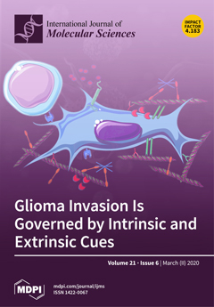
The dissemination of therapy-resistant glioma cancer cells into healthy brain parenchyma presents one of the greatest obstacles to curative therapy. Intensive research in the last decade, especially a multitude of expression sequencing studies, has provided a profound insight into the heterogeneity of this devastating disease. However, the interplay between tumor and surrounding cells as well as matrix components has only recently come into focus. The present review tries to summarize advances in understanding genetic as well as extrinsic mechanisms leading to GBM invasion. View this paper.
  • Issues are regarded as officially published after their release is announced to the table of contents alert mailing list.
  • You may sign up for e-mail alerts to receive table of contents of newly released issues.
  • PDF is the official format for papers published in both, html and pdf forms. To view the papers in pdf format, click on the "PDF Full-text" link, and use the free Adobe Reader to open them.
Order results
Result details
Section
Select all
Export citation of selected articles as:
1 pages, 167 KiB  
Correction
Correction: Li, P. et al. Mechanical Characteristics, In Vitro Degradation, Cytotoxicity, and Antibacterial Evaluation of Zn-4.0Ag Alloy as a Biodegradable Material. Int. J. Mol. Sci. 2018, 19, 755
by Ping Li, Christine Schille, Ernst Schweizer, Frank Rupp, Alexander Heiss, Claudia Legner, Ulrich E. Klotz, Jürgen Geis-Gerstorfer and Lutz Scheideler
Int. J. Mol. Sci. 2020, 21(6), 2258; https://doi.org/10.3390/ijms21062258 - 24 Mar 2020
Cited by 3 | Viewed by 2049
Abstract
The authors wish to make the following corrections to this paper [...] Full article
15 pages, 1113 KiB  
Review
Iron Metabolism in Cancer Progression
by Stefania Forciniti, Luana Greco, Fabio Grizzi, Alberto Malesci and Luigi Laghi
Int. J. Mol. Sci. 2020, 21(6), 2257; https://doi.org/10.3390/ijms21062257 - 24 Mar 2020
Cited by 58 | Viewed by 11490
Abstract
Iron is indispensable for cell metabolism of both normal and cancer cells. In the latter, several disruptions of its metabolism occur at the steps of tumor initiation, progression and metastasis. Noticeably, cancer cells require a large amount of iron, and exhibit a strong [...] Read more.
Iron is indispensable for cell metabolism of both normal and cancer cells. In the latter, several disruptions of its metabolism occur at the steps of tumor initiation, progression and metastasis. Noticeably, cancer cells require a large amount of iron, and exhibit a strong dependence on it for their proliferation. Numerous iron metabolism-related proteins and signaling pathways are altered by iron in malignancies, displaying the pivotal role of iron in cancer. Iron homeostasis is regulated at several levels, from absorption by enterocytes to recycling by macrophages and storage in hepatocytes. Mutations in HFE gene alter iron homeostasis leading to hereditary hemochromatosis and to an increased cancer risk because the accumulation of iron induces oxidative DNA damage and free radical activity. Additionally, the iron capability to modulate immune responses is pivotal in cancer progression. Macrophages show an iron release phenotype and potentially deliver iron to cancer cells, resulting in tumor promotion. Overall, alterations in iron metabolism are among the metabolic and immunological hallmarks of cancer, and further studies are required to dissect how perturbations of this element relate to tumor development and progression. Full article
Show Figures

Figure 1

16 pages, 2465 KiB  
Review
The Respiratory Phenotype of Pompe Disease Mouse Models
by Anna F. Fusco, Angela L. McCall, Justin S. Dhindsa, Lucy Zheng, Aidan Bailey, Amanda F. Kahn and Mai K. ElMallah
Int. J. Mol. Sci. 2020, 21(6), 2256; https://doi.org/10.3390/ijms21062256 - 24 Mar 2020
Cited by 9 | Viewed by 3869
Abstract
Pompe disease is a glycogen storage disease caused by a deficiency in acid α-glucosidase (GAA), a hydrolase necessary for the degradation of lysosomal glycogen. This deficiency in GAA results in muscle and neuronal glycogen accumulation, which causes respiratory insufficiency. Pompe disease mouse models [...] Read more.
Pompe disease is a glycogen storage disease caused by a deficiency in acid α-glucosidase (GAA), a hydrolase necessary for the degradation of lysosomal glycogen. This deficiency in GAA results in muscle and neuronal glycogen accumulation, which causes respiratory insufficiency. Pompe disease mouse models provide a means of assessing respiratory pathology and are important for pre-clinical studies of novel therapies that aim to treat respiratory dysfunction and improve quality of life. This review aims to compile and summarize existing manuscripts that characterize the respiratory phenotype of Pompe mouse models. Manuscripts included in this review were selected utilizing specific search terms and exclusion criteria. Analysis of these findings demonstrate that Pompe disease mouse models have respiratory physiological defects as well as pathologies in the diaphragm, tongue, higher-order respiratory control centers, phrenic and hypoglossal motor nuclei, phrenic and hypoglossal nerves, neuromuscular junctions, and airway smooth muscle. Overall, the culmination of these pathologies contributes to severe respiratory dysfunction, underscoring the importance of characterizing the respiratory phenotype while developing effective therapies for patients. Full article
Show Figures

Figure 1

26 pages, 1658 KiB  
Review
LncRNAs in HCV Infection and HCV-Related Liver Disease
by Juan P. Unfried and P. Fortes
Int. J. Mol. Sci. 2020, 21(6), 2255; https://doi.org/10.3390/ijms21062255 - 24 Mar 2020
Cited by 32 | Viewed by 4695
Abstract
Long non-coding RNAs (lncRNAs) are transcripts with poor coding capacity that may interact with proteins, DNA, or other RNAs to perform structural and regulatory functions. The lncRNA transcriptome changes significantly in most diseases, including cancer and viral infections. In this review, we summarize [...] Read more.
Long non-coding RNAs (lncRNAs) are transcripts with poor coding capacity that may interact with proteins, DNA, or other RNAs to perform structural and regulatory functions. The lncRNA transcriptome changes significantly in most diseases, including cancer and viral infections. In this review, we summarize the functional implications of lncRNA-deregulation after infection with hepatitis C virus (HCV). HCV leads to chronic infection in many patients that may progress to liver cirrhosis and hepatocellular carcinoma (HCC). Most lncRNAs deregulated in infected cells that have been described function to potentiate or block the antiviral response and, therefore, they have a great impact on HCV viral replication. In addition, several lncRNAs upregulated by the infection contribute to viral release. Finally, many lncRNAs have been described as deregulated in HCV-related HCC that function to enhance cell survival, proliferation, and tumor progression by different mechanisms. Interestingly, some HCV-related HCC lncRNAs can be detected in bodily fluids, and there is great hope that they could be used as biomarkers to predict cancer initiation, progression, tumor burden, response to treatment, resistance to therapy, or tumor recurrence. Finally, there is high confidence that lncRNAs could also be used to improve the suboptimal long-term outcomes of current HCC treatment options. Full article
Show Figures

Figure 1

14 pages, 3002 KiB  
Article
Lead (Pb) as a Factor Initiating and Potentiating Inflammation in Human THP-1 Macrophages
by Emilia Metryka, Patrycja Kupnicka, Patrycja Kapczuk, Donata Simińska, Maciej Tarnowski, Marta Goschorska, Izabela Gutowska, Dariusz Chlubek and Irena Baranowska-Bosiacka
Int. J. Mol. Sci. 2020, 21(6), 2254; https://doi.org/10.3390/ijms21062254 - 24 Mar 2020
Cited by 9 | Viewed by 3070
Abstract
The aim of this study was to assess the influence of lead (Pb) at low concentrations (imitating Pb levels in human blood in chronic environmental exposure to this metal) on interleukin 1β (IL-1β) and interleukin 6 (IL-6) concentrations and the activity and expression [...] Read more.
The aim of this study was to assess the influence of lead (Pb) at low concentrations (imitating Pb levels in human blood in chronic environmental exposure to this metal) on interleukin 1β (IL-1β) and interleukin 6 (IL-6) concentrations and the activity and expression of COX-1 and COX-2 in THP-1 macrophages. Macrophages were cultured in vitro in the presence of Pb at concentrations of: 1.25 μg/dL; 2.5 μg/dL; 5 μg/dL; 10 μg/dL. The first two concentrations of Pb were selected on the basis of our earlier study, which showed that Pb concentration in whole blood (PbB) of young women living in the northern regions of Poland and in the cord blood of their newborn children was within this range (a dose imitating environmental exposure). Concentrations of 5 μg/dL and 10 μg/dL correspond to the previously permissible PbB concentrations in children or pregnant women, and adults. Our results indicate that even low concentrations of Pb cause an increase in production of inflammatory interleukins (IL-1β and IL-6), increases expression of COX-1 and COX-2, and increases thromboxane B2 and prostaglandin E2 concentration in macrophages. This clearly suggests that the development of inflammation is associated not only with COX-2 but also with COX-1, which, until recently, had only been attributed constitutive expression. It can be concluded that environmental Pb concentrations are able to activate the monocytes/macrophages similarly to the manner observed during inflammation. Full article
(This article belongs to the Special Issue Molecular Mechanisms of Lead Neurotoxicity)
Show Figures

Graphical abstract

20 pages, 4458 KiB  
Review
Cancer Diagnosis through SERS and Other Related Techniques
by Maria Blanco-Formoso and Ramon A. Alvarez-Puebla
Int. J. Mol. Sci. 2020, 21(6), 2253; https://doi.org/10.3390/ijms21062253 - 24 Mar 2020
Cited by 53 | Viewed by 6175
Abstract
Cancer heterogeneity increasingly requires ultrasensitive techniques that allow early diagnosis for personalized treatment. In addition, they should preferably be non-invasive tools that do not damage surrounding tissues or contribute to body toxicity. In this context, liquid biopsy of biological samples such as urine, [...] Read more.
Cancer heterogeneity increasingly requires ultrasensitive techniques that allow early diagnosis for personalized treatment. In addition, they should preferably be non-invasive tools that do not damage surrounding tissues or contribute to body toxicity. In this context, liquid biopsy of biological samples such as urine, blood, or saliva represents an ideal approximation of what is happening in real time in the affected tissues. Plasmonic nanoparticles are emerging as an alternative or complement to current diagnostic techniques, being able to detect and quantify novel biomarkers such as specific peptides and proteins, microRNA, circulating tumor DNA and cells, and exosomes. Here, we review the latest ideas focusing on the use of plasmonic nanoparticles in coded and label-free surface-enhanced Raman scattering (SERS) spectroscopy. Moreover, surface plasmon resonance (SPR) spectroscopy, colorimetric assays, dynamic light scattering (DLS) spectroscopy, mass spectrometry or total internal reflection fluorescence (TIRF) microscopy among others are briefly examined in order to highlight the potential and versatility of plasmonics. Full article
(This article belongs to the Special Issue Development of Responsive Nanoparticles for Cancer Therapy)
Show Figures

Figure 1

22 pages, 2766 KiB  
Article
Vesicles Shed by Pathological Murine Adipocytes Spread Pathology: Characterization and Functional Role of Insulin Resistant/Hypertrophied Adiposomes
by Tamara Camino, Nerea Lago-Baameiro, Susana B. Bravo, Aurelio Sueiro, Iván Couto, Fernando Santos, Javier Baltar, Felipe F. Casanueva and María Pardo
Int. J. Mol. Sci. 2020, 21(6), 2252; https://doi.org/10.3390/ijms21062252 - 24 Mar 2020
Cited by 28 | Viewed by 4254
Abstract
Extracellular vesicles (EVs) have recently emerged as a relevant way of cell to cell communication, and its analysis has become an indirect approach to assess the cell/tissue of origin status. However, the knowledge about their nature and role on metabolic diseases is still [...] Read more.
Extracellular vesicles (EVs) have recently emerged as a relevant way of cell to cell communication, and its analysis has become an indirect approach to assess the cell/tissue of origin status. However, the knowledge about their nature and role on metabolic diseases is still very scarce. We have established an insulin resistant (IR) and two lipid (palmitic/oleic) hypertrophied adipocyte cell models to isolate EVs to perform a protein cargo qualitative and quantitative Sequential Window Acquisition of All Theoretical Mass Spectra (SWATH) analysis by mass spectrometry. Our results show a high proportion of obesity and IR-related proteins in pathological EVs; thus, we propose a panel of potential obese adipose tissue EV-biomarkers. Among those, lipid hypertrophied vesicles are characterized by ceruloplasmin, mimecan, and perilipin 1 adipokines, and those from the IR by the striking presence of the adiposity and IR related transforming growth factor-beta-induced protein ig-h3 (TFGBI). Interestingly, functional assays show that IR and hypertrophied adipocytes induce differentiation/hypertrophy and IR in healthy adipocytes through secreted EVs. Finally, we demonstrate that lipid atrophied adipocytes shed EVs promote macrophage inflammation by stimulating IL-6 and TNFα expression. Thus, we conclude that pathological adipocytes release vesicles containing representative protein cargo of the cell of origin that are able to induce metabolic alterations on healthy cells probably exacerbating the disease once established. Full article
(This article belongs to the Special Issue Extracellular Vesicles in Endocrine and Endocrine-Dependent Cancer)
Show Figures

Graphical abstract

20 pages, 1355 KiB  
Review
Stem Cells as a Target for the Delivery of Active Molecules to Skin by Topical Administration
by Hamid-Reza Ahmadi-Ashtiani, Parisa Bishe, Anna Baldisserotto, Piergiacomo Buso, Stefano Manfredini and Silvia Vertuani
Int. J. Mol. Sci. 2020, 21(6), 2251; https://doi.org/10.3390/ijms21062251 - 24 Mar 2020
Cited by 3 | Viewed by 4513
Abstract
Cutaneous stem cells, gained great attention in the field of regenerative medicine as a potential therapeutic target for the treatment of skin and hair disorders and various types of skin cancers. Cutaneous stem cells play a key role in several processes like the [...] Read more.
Cutaneous stem cells, gained great attention in the field of regenerative medicine as a potential therapeutic target for the treatment of skin and hair disorders and various types of skin cancers. Cutaneous stem cells play a key role in several processes like the renovation of skin structures in the condition of homeostasis and after injuries, the hair follicle growth and the reconstruction and production of melanocytes. Thus, gaining effective access to skin stem cells for therapeutic interventions that often involve active molecules with non-favorable characteristics for skin absorption is a valuable achievement. The topical route with high patient compliance and several other benefits is gaining increasing importance in basic and applied research. However, the major obstacle for topical drug delivery is the effective barrier provided by skin against penetration of the vast majority of exogenous molecules. The research in this field is focusing more and more on new strategies to circumvent and pass this barrier effectively. In this article the existing approaches are discussed considering physical and chemical methods along with utilization of novel drug delivery systems to enhance penetration of drugs to the skin. In particular, attention has been paid to studies finalized to the delivery of molecules to cutaneous stem cells with the aim of transferring signals, modulating their metabolic program, inducing physiological modifications and stem cell gene therapy. Full article
(This article belongs to the Section Molecular Biology)
Show Figures

Figure 1

14 pages, 2800 KiB  
Article
Norepinephrine-Induced DNA Damage in Ovarian Cancer Cells
by Rocio Lamboy-Caraballo, Carmen Ortiz-Sanchez, Arelis Acevedo-Santiago, Jaime Matta, Alvaro N.A. Monteiro and Guillermo N. Armaiz-Pena
Int. J. Mol. Sci. 2020, 21(6), 2250; https://doi.org/10.3390/ijms21062250 - 24 Mar 2020
Cited by 23 | Viewed by 4522
Abstract
Multiple studies have shown that psychological distress in epithelial ovarian cancer (EOC) patients is associated with worse quality of life and poor treatment adherence. This may influence chemotherapy response and prognosis. Moreover, although stress hormones can reduce cisplatin efficacy in EOC treatment, their [...] Read more.
Multiple studies have shown that psychological distress in epithelial ovarian cancer (EOC) patients is associated with worse quality of life and poor treatment adherence. This may influence chemotherapy response and prognosis. Moreover, although stress hormones can reduce cisplatin efficacy in EOC treatment, their effect on the integrity of DNA remains poorly understood. In this study, we investigated whether norepinephrine and epinephrine can induce DNA damage and modulate cisplatin-induced DNA damage in three EOC cell lines. Our data show that norepinephrine and epinephrine exposure led to increased nuclear γ-H2AX foci formation in EOC cells, a marker of double-strand DNA breaks. We further characterized norepinephrine-induced DNA damage by subjecting EOC cells to alkaline and neutral comet assays. Norepinephrine exposure caused DNA double-strand breaks, but not single-strand breaks. Interestingly, pre-treatment with propranolol abrogated norepinephrine-induced DNA damage indicating that its effects may be mediated by β-adrenergic receptors. Lastly, we determined the effects of norepinephrine on cisplatin-induced DNA damage. Our data suggest that norepinephrine reduced cisplatin-induced DNA damage in EOC cells and that this effect may be mediated independently of β-adrenergic receptors. Taken together, these results suggest that stress hormones can affect DNA integrity and modulate cisplatin resistance in EOC cells. Full article
(This article belongs to the Special Issue Recognition of DNA Lesions)
Show Figures

Figure 1

21 pages, 2430 KiB  
Article
AHR Signaling Dampens Inflammatory Signature in Neonatal Skin γδ T Cells
by Katja Merches, Alfonso Schiavi, Heike Weighardt, Swantje Steinwachs, Nadine Teichweyde, Irmgard Förster, Katrin Hochrath, Beatrix Schumak, Natascia Ventura, Patrick Petzsch, Karl Köhrer and Charlotte Esser
Int. J. Mol. Sci. 2020, 21(6), 2249; https://doi.org/10.3390/ijms21062249 - 24 Mar 2020
Cited by 11 | Viewed by 4448
Abstract
Background Aryl hydrocarbon receptor (AHR)-deficient mice do not support the expansion of dendritic epidermal T cells (DETC), a resident immune cell population in the murine epidermis, which immigrates from the fetal thymus to the skin around birth. Material and Methods In order to [...] Read more.
Background Aryl hydrocarbon receptor (AHR)-deficient mice do not support the expansion of dendritic epidermal T cells (DETC), a resident immune cell population in the murine epidermis, which immigrates from the fetal thymus to the skin around birth. Material and Methods In order to identify the gene expression changes underlying the DETC disappearance in AHR-deficient mice, we analyzed microarray RNA-profiles of DETC, sorted from the skin of two-week-old AHR-deficient mice and their heterozygous littermates. In vitro studies were done for verification, and IL-10, AHR repressor (AHRR), and c-Kit deficient mice analyzed for DETC frequency. Results We identified 434 annotated differentially expressed genes. Gene set enrichment analysis demonstrated that the expression of genes related to proliferation, ion homeostasis and morphology differed between the two mouse genotypes. Importantly, with 1767 pathways the cluster-group “inflammation” contained the majority of AHR-dependently regulated pathways. The most abundant cluster of differentially expressed genes was “inflammation.” DETC of AHR-deficient mice were inflammatory active and had altered calcium and F-actin levels. Extending the study to the AHRR, an enigmatic modulator of AHR-activity, we found approximately 50% less DETC in AHRR-deficient mice than in wild-type-littermates. Conclusion AHR-signaling in DETC dampens their inflammatory default potential and supports their homeostasis in the skin. Full article
(This article belongs to the Special Issue Aryl Hydrocarbon Receptor in Biology and Toxicology 2.0)
Show Figures

Figure 1

14 pages, 11386 KiB  
Article
Association of a Disrupted Dipping Pattern of Blood Pressure with Progression of Renal Injury during the Development of Salt-Dependent Hypertension in Rats
by Abu Sufiun, Asadur Rahman, Kazi Rafiq, Yoshihide Fujisawa, Daisuke Nakano, Hideki Kobara, Tsutomu Masaki and Akira Nishiyama
Int. J. Mol. Sci. 2020, 21(6), 2248; https://doi.org/10.3390/ijms21062248 - 24 Mar 2020
Cited by 6 | Viewed by 2517
Abstract
The aim of the present study is to investigate whether a disruption of the dipping pattern of blood pressure (BP) is associated with the progression of renal injury in Dahl salt-sensitive (DSS) hypertensive rats. Seven-week-old DSS rats were fed a high salt diet [...] Read more.
The aim of the present study is to investigate whether a disruption of the dipping pattern of blood pressure (BP) is associated with the progression of renal injury in Dahl salt-sensitive (DSS) hypertensive rats. Seven-week-old DSS rats were fed a high salt diet (HSD; 8% NaCl) for 10 weeks, followed by a transition to a normal salt diet (NSD; 0.3% NaCl) for 4 weeks. At baseline, NSD-fed DSS rats showed a dipper-type circadian rhythm of BP. By contrast, HSD for 5 days caused a significant increase in the difference between the active and inactive periods of BP with an extreme dipper type of BP, while proteinuria and renal tissue injury were not observed. Interestingly, HSD feeding for 10 weeks developed hypertension with a non-dipper pattern of BP, which was associated with obvious proteinuria and renal tissue injury. Four weeks after switching to an NSD, BP and proteinuria were significantly decreased, and the BP circadian rhythm returned to the normal dipper pattern. These data suggest that the non-dipper pattern of BP is associated with the progression of renal injury during the development of salt-dependent hypertension. Full article
(This article belongs to the Special Issue Crosstalk between Circadian Rhythm and Diseases)
Show Figures

Graphical abstract

15 pages, 1550 KiB  
Review
Lineage Decision-Making within Normal Haematopoietic and Leukemic Stem Cells
by Geoffrey Brown, Lucía Sánchez and Isidro Sánchez-García
Int. J. Mol. Sci. 2020, 21(6), 2247; https://doi.org/10.3390/ijms21062247 - 24 Mar 2020
Viewed by 3604
Abstract
To produce the wide range of blood and immune cell types, haematopoietic stem cells can “choose” directly from the entire spectrum of blood cell fate-options. Affiliation to a single cell lineage can occur at the level of the haematopoietic stem cell and these [...] Read more.
To produce the wide range of blood and immune cell types, haematopoietic stem cells can “choose” directly from the entire spectrum of blood cell fate-options. Affiliation to a single cell lineage can occur at the level of the haematopoietic stem cell and these cells are therefore a mixture of some pluripotent cells and many cells with lineage signatures. Even so, haematopoietic stem cells and their progeny that have chosen a particular fate can still “change their mind” and adopt a different developmental pathway. Many of the leukaemias arise in haematopoietic stem cells with the bulk of the often partially differentiated leukaemia cells belonging to just one cell type. We argue that the reason for this is that an oncogenic insult to the genome “hard wires” leukaemia stem cells, either through development or at some stage, to one cell lineage. Unlike normal haematopoietic stem cells, oncogene-transformed leukaemia stem cells and their progeny are unable to adopt an alternative pathway. Full article
(This article belongs to the Special Issue Cellular and Molecular Mechanisms of Hematopoiesis)
Show Figures

Graphical abstract

15 pages, 5552 KiB  
Article
Characterization, Expression Pattern and Antiviral Activities of Mx Gene in Chinese Giant Salamander, Andrias davidianus
by Yanan Liu, Yiqun Li, Yongze Zhou, Nan Jiang, Yuding Fan and Lingbing Zeng
Int. J. Mol. Sci. 2020, 21(6), 2246; https://doi.org/10.3390/ijms21062246 - 24 Mar 2020
Cited by 10 | Viewed by 3073
Abstract
Mx, Myxovirus resistance is an important interferon-stimulated protein that mediates antiviral responses. In this study, the expression and activities of Chinese giant salamander, Andrias davidianus Mx gene, AdMx, were investigated. The AdMx cDNA sequence contains an open reading frame (ORF) of 2112 nucleotides, [...] Read more.
Mx, Myxovirus resistance is an important interferon-stimulated protein that mediates antiviral responses. In this study, the expression and activities of Chinese giant salamander, Andrias davidianus Mx gene, AdMx, were investigated. The AdMx cDNA sequence contains an open reading frame (ORF) of 2112 nucleotides, encoding a putative protein of 703 aa. Meanwhile, AdMx possesses the conserved tripartite GTP binding motif and a dynamin family signature. qRT-PCR analysis revealed a broad expression of AdMx in vivo, with the highest expression levels in brain, kidney and spleen. The AdMx expression level in kidney, spleen and muscle significantly increased at 6 h after Chinese giant salamander iridovirus (GSIV) infection and peaked at 48 h, while that in muscle cell line (GSM) was not noticeably up-regulated until 72 h post infection. Additionally, a plasmid expressing AdMx was constructed and transfected into the Chinese giant salamander GSM cells. The virus load and gene copies in AdMx over-expressed cells were significantly reduced compared with those in the control cells. Moreover, compared to the control cells, a lower level of virus major capsid protein (MCP) synthesis in AdMx over-expressed cells was confirmed by Western blot. These results collectively suggest that Mx plays an important antiviral role in the immune responses against GSIV in Chinese giant salamander. Full article
(This article belongs to the Section Molecular Biology)
Show Figures

Figure 1

13 pages, 591 KiB  
Review
Transcriptome Analysis in Renal Transplant Biopsies Not Fulfilling Rejection Criteria
by Francesc Moreso, Joana Sellarès, María José Soler and Daniel Serón
Int. J. Mol. Sci. 2020, 21(6), 2245; https://doi.org/10.3390/ijms21062245 - 24 Mar 2020
Cited by 3 | Viewed by 2742
Abstract
The clinical significance of renal transplant biopsies displaying borderline changes suspicious for T-cell mediated rejection (TCMR) or interstitial fibrosis and tubular atrophy (IFTA) with interstitial inflammation has not been well defined. Molecular profiling to evaluate renal transplant biopsies using microarrays has been shown [...] Read more.
The clinical significance of renal transplant biopsies displaying borderline changes suspicious for T-cell mediated rejection (TCMR) or interstitial fibrosis and tubular atrophy (IFTA) with interstitial inflammation has not been well defined. Molecular profiling to evaluate renal transplant biopsies using microarrays has been shown to be an objective measurement that adds precision to conventional histology. We review the contribution of transcriptomic analysis in surveillance and indication biopsies with borderline changes and IFTA associated with variable degrees of inflammation. Transcriptome analysis applied to biopsies with borderline changes allows to distinguish patients with rejection from those in whom mild inflammation mainly represents a response to injury. Biopsies with IFTA and inflammation occurring in unscarred tissue display a molecular pattern similar to TCMR while biopsies with IFTA and inflammation in scarred tissue, apart from T-cell activation, also express B cell, immunoglobulin and mast cell-related genes. Additionally, patients at risk for IFTA progression can be identified by genes mainly reflecting fibroblast dysregulation and immune activation. At present, it is not well established whether the expression of rejection gene transcripts in patients with fibrosis and inflammation is the consequence of an alloimmune response, tissue damage or a combination of both. Full article
(This article belongs to the Special Issue Renal Disease and Immunity)
Show Figures

Figure 1

18 pages, 3621 KiB  
Article
Complex Size and Surface Charge Determine Nucleic Acid Transfer by Fusogenic Liposomes
by Marco Hoffmann, Nils Hersch, Sven Gerlach, Georg Dreissen, Ronald Springer, Rudolf Merkel, Agnes Csiszár and Bernd Hoffmann
Int. J. Mol. Sci. 2020, 21(6), 2244; https://doi.org/10.3390/ijms21062244 - 24 Mar 2020
Cited by 19 | Viewed by 4123
Abstract
Highly efficient, biocompatible, and fast nucleic acid delivery methods are essential for biomedical applications and research. At present, two main strategies are used to this end. In non-viral transfection liposome- or polymer-based formulations are used to transfer cargo into cells via endocytosis, whereas [...] Read more.
Highly efficient, biocompatible, and fast nucleic acid delivery methods are essential for biomedical applications and research. At present, two main strategies are used to this end. In non-viral transfection liposome- or polymer-based formulations are used to transfer cargo into cells via endocytosis, whereas viral carriers enable direct nucleic acid delivery into the cell cytoplasm. Here, we introduce a new generation of liposomes for nucleic acid delivery, which immediately fuse with the cellular plasma membrane upon contact to transfer the functional nucleic acid directly into the cell cytoplasm. For maximum fusion efficiency combined with high cargo transfer, nucleic acids had to be complexed and partially neutralized before incorporation into fusogenic liposomes. Among the various neutralization agents tested, small, linear, and positively charged polymers yielded the best complex properties. Systematic variation of liposomal composition and nucleic acid complexation identified surface charge as well as particle size as essential parameters for cargo-liposome interaction and subsequent fusion induction. Optimized protocols were tested for the efficient transfer of different kinds of nucleic acids like plasmid DNA, messenger RNA, and short-interfering RNA into various mammalian cells in culture and into primary tissues. Full article
(This article belongs to the Special Issue Membrane Fusion 2.0)
Show Figures

Graphical abstract

17 pages, 2962 KiB  
Article
Accurate Representation of Protein-Ligand Structural Diversity in the Protein Data Bank (PDB)
by Nicolas K. Shinada, Peter Schmidtke and Alexandre G. de Brevern
Int. J. Mol. Sci. 2020, 21(6), 2243; https://doi.org/10.3390/ijms21062243 - 24 Mar 2020
Cited by 3 | Viewed by 3552
Abstract
The number of available protein structures in the Protein Data Bank (PDB) has considerably increased in recent years. Thanks to the growth of structures and complexes, numerous large-scale studies have been done in various research areas, e.g., protein–protein, protein–DNA, or in drug discovery. [...] Read more.
The number of available protein structures in the Protein Data Bank (PDB) has considerably increased in recent years. Thanks to the growth of structures and complexes, numerous large-scale studies have been done in various research areas, e.g., protein–protein, protein–DNA, or in drug discovery. While protein redundancy was only simply managed using simple protein sequence identity threshold, the similarity of protein-ligand complexes should also be considered from a structural perspective. Hence, the protein-ligand duplicates in the PDB are widely known, but were never quantitatively assessed, as they are quite complex to analyze and compare. Here, we present a specific clustering of protein-ligand structures to avoid bias found in different studies. The methodology is based on binding site superposition, and a combination of weighted Root Mean Square Deviation (RMSD) assessment and hierarchical clustering. Repeated structures of proteins of interest are highlighted and only representative conformations were conserved for a non-biased view of protein distribution. Three types of cases are described based on the number of distinct conformations identified for each complex. Defining these categories decreases by 3.84-fold the number of complexes, and offers more refined results compared to a protein sequence-based method. Widely distinct conformations were analyzed using normalized B-factors. Furthermore, a non-redundant dataset was generated for future molecular interactions analysis or virtual screening studies. Full article
Show Figures

Graphical abstract

12 pages, 2159 KiB  
Article
Sustained Release of Decoy Wnt Receptor (sLRP6E1E2)-Expressing Adenovirus Using Gel-Encapsulation for Scar Remodeling in Pig Model
by Chae-Eun Yang, Sewoon Choi, Ju Hee Lee, Eun Hye Kang, Hyo Min Ahn, Tai Suk Roh, Chae-Ok Yun and Won Jai Lee
Int. J. Mol. Sci. 2020, 21(6), 2242; https://doi.org/10.3390/ijms21062242 - 24 Mar 2020
Cited by 6 | Viewed by 2424
Abstract
An adenoviral vector (Ad) expressing a Wnt decoy receptor (sLRP6E1E2) is known to induce an anti-fibrotic effect by inhibiting Wnt signaling. We evaluated its effects in vivo using pig models and attempted to introduce an alginate gel-matrix system to prolong the effect of [...] Read more.
An adenoviral vector (Ad) expressing a Wnt decoy receptor (sLRP6E1E2) is known to induce an anti-fibrotic effect by inhibiting Wnt signaling. We evaluated its effects in vivo using pig models and attempted to introduce an alginate gel-matrix system to prolong the effect of the Ad. Transduction efficiency as to the biological activity of Ad in different forms was evaluated. Then, 50 days after the formation of full-thickness skin defects on the backs of Yorkshire pigs, scars were treated with each form of Ad. Therapeutic efficacy and various factors influencing scar formation and collagen rearrangement were analyzed. Inflammatory cell infiltration within the scar tissues was also evaluated. Decoy Wnt receptor (sLRP6E1E2)-expressing adenovirus treatment improved scar quality in a pig model. Loading this construct in alginate gel allows sustained virus release into local tissues and prolongs Ad activity, thus maintaining its therapeutic effect longer in vivo. Full article
(This article belongs to the Section Molecular Pathology, Diagnostics, and Therapeutics)
Show Figures

Figure 1

19 pages, 3809 KiB  
Article
Insights into Interactions of Flavanones with Target Human Respiratory Syncytial Virus M2-1 Protein from STD-NMR, Fluorescence Spectroscopy, and Computational Simulations
by Hêmily M. R. Piva, Jéssica M. Sá, Artemiza S. Miranda, Ljubica Tasic, Marcelo A. Fossey, Fátima P. Souza and Ícaro P. Caruso
Int. J. Mol. Sci. 2020, 21(6), 2241; https://doi.org/10.3390/ijms21062241 - 24 Mar 2020
Cited by 12 | Viewed by 3218
Abstract
The human Respiratory Syncytial Virus (hRSV) is the most frequent agent of respiratory infections in infants and children with no currently approved vaccine. The M2-1 protein is an important transcriptional antitermination factor and a potential target for viral replication inhibitor development. Hesperetin [...] Read more.
The human Respiratory Syncytial Virus (hRSV) is the most frequent agent of respiratory infections in infants and children with no currently approved vaccine. The M2-1 protein is an important transcriptional antitermination factor and a potential target for viral replication inhibitor development. Hesperetin (HST) and hesperidin (HSD) are flavonoids from the flavanone group, naturally found in citrus and have, as one of their properties, antiviral activity. The present study reports on the interactions between hRSV M2-1 and these flavanones using experimental techniques in association with computational tools. STD-NMR results showed that HST and HSD bind to M2-1 by positioning their aromatic rings into the target protein binding site. Fluorescence quenching measurements revealed that HST had an interaction affinity greater than HSD towards M2-1. The thermodynamic analysis suggested that hydrogen bonds and van der Waals interactions are important for the molecular stabilization of the complexes. Computational simulations corroborated with the experimental results and indicated that the possible interaction region for the flavonoids is the AMP-binding site in M2-1. Therefore, these results point that HST and HSD bind stably to a critical region in M2-1, which is vital for its biological function, and thus might play a possible role antiviral against hRSV. Full article
(This article belongs to the Special Issue Recent Advances in Biomolecular Recognition)
Show Figures

Graphical abstract

18 pages, 1909 KiB  
Article
SIRT2 Affects Primary Cilia Formation by Regulating mTOR Signaling in Retinal Pigmented Epithelial Cells
by Jeaho Lim, Juyoung Son, Jaewook Ryu and Ja-Eun Kim
Int. J. Mol. Sci. 2020, 21(6), 2240; https://doi.org/10.3390/ijms21062240 - 24 Mar 2020
Cited by 12 | Viewed by 4199
Abstract
SIRT2, a member of the Class III HDAC family, participates in diverse cellular processes and regulates several pathological conditions. Although a few reports show that SIRT2 regulates the cell cycle, the causes and outcomes of SIRT2-dependent cell proliferation remain unclear. Here, we examined [...] Read more.
SIRT2, a member of the Class III HDAC family, participates in diverse cellular processes and regulates several pathological conditions. Although a few reports show that SIRT2 regulates the cell cycle, the causes and outcomes of SIRT2-dependent cell proliferation remain unclear. Here, we examined the effects of SIRT2 suppression in human RPE1 cells using siRNA targeting SIRT2, and AK-1, a SIRT2-specific inhibitor. The number of primary cilia in SIRT2-suppressed cells increased under serum-present conditions. Suppressing SIRT2 induced cell cycle arrest at G0/G1 phase by inactivating mammalian target of rapamycin (mTOR) signaling, possibly through mTORC1. Treatment with torin 1, an inhibitor of mTORC1/mTORC2, yielded results similar to those observed after SIRT2 suppression. However, SIRT2 suppression did not affect primary cilia formation or mTOR signaling following serum starvation. This suggests that SIRT2 acts as a critical sensor that links growth factor-dependent signal transduction and primary cilia formation by regulating the cell cycle. Full article
(This article belongs to the Section Molecular Biology)
Show Figures

Graphical abstract

15 pages, 1423 KiB  
Review
Recent Overview of the Use of iPSCs Huntington’s Disease Modeling and Therapy
by Maria Csobonyeiova, Stefan Polak and Lubos Danisovic
Int. J. Mol. Sci. 2020, 21(6), 2239; https://doi.org/10.3390/ijms21062239 - 24 Mar 2020
Cited by 36 | Viewed by 6695
Abstract
Huntington’s disease (HD) is an inherited, autosomal dominant, degenerative disease characterized by involuntary movements, cognitive decline, and behavioral impairment ending in death. HD is caused by an expansion in the number of CAG repeats in the huntingtin gene on chromosome 4. To date, [...] Read more.
Huntington’s disease (HD) is an inherited, autosomal dominant, degenerative disease characterized by involuntary movements, cognitive decline, and behavioral impairment ending in death. HD is caused by an expansion in the number of CAG repeats in the huntingtin gene on chromosome 4. To date, no effective therapy for preventing the onset or progression of the disease has been found, and many symptoms do not respond to pharmacologic treatment. However, recent results of pre-clinical trials suggest a beneficial effect of stem-cell-based therapy. Induced pluripotent stem cells (iPSCs) represent an unlimited cell source and are the most suitable among the various types of autologous stem cells due to their patient specificity and ability to differentiate into a variety of cell types both in vitro and in vivo. Furthermore, the cultivation of iPSC-derived neural cells offers the possibility of studying the etiopathology of neurodegenerative diseases, such as HD. Moreover, differentiated neural cells can organize into three-dimensional (3D) organoids, mimicking the complex architecture of the brain. In this article, we present a comprehensive review of recent HD models, the methods for differentiating HD–iPSCs into the desired neural cell types, and the progress in gene editing techniques leading toward stem-cell-based therapy. Full article
(This article belongs to the Special Issue Disease Modeling Using Human Induced Pluripotent Stem Cells 2.0)
Show Figures

Graphical abstract

9 pages, 527 KiB  
Review
AMPK Regulates Developmental Plasticity through an Endogenous Small RNA Pathway in Caenorhabditis elegans
by Christopher Wong and Richard Roy
Int. J. Mol. Sci. 2020, 21(6), 2238; https://doi.org/10.3390/ijms21062238 - 24 Mar 2020
Cited by 6 | Viewed by 4064
Abstract
Caenorhabditis elegans larvae can undergo developmental arrest upon entry into the dauer stage in response to suboptimal growth conditions. Dauer larvae can exit this stage in replete conditions with no reproductive consequence. During this diapause stage, the metabolic regulator AMP-activated protein kinase (AMPK) [...] Read more.
Caenorhabditis elegans larvae can undergo developmental arrest upon entry into the dauer stage in response to suboptimal growth conditions. Dauer larvae can exit this stage in replete conditions with no reproductive consequence. During this diapause stage, the metabolic regulator AMP-activated protein kinase (AMPK) ensures that the germ line becomes quiescent to maintain germ cell integrity. Animals that lack all AMPK signalling undergo germline hyperplasia upon entering dauer, while those that recover from this stage become sterile. Neuronal AMPK expression in otherwise AMPK-deficient animals is sufficient for germline quiescence and germ cell integrity and its effects are likely mediated through an endogenous small RNA pathway. Upon impairing small RNA biosynthesis, the post-dauer fertility is restored in AMPK mutants. These data suggest that AMPK may function in neurons to relay a message through small RNAs to the germ cells to alter their quiescence in the dauer stage, thus challenging the permeability of the Weismann barrier. Full article
(This article belongs to the Special Issue AMP-Activated Protein Kinase Signalling 2.0)
Show Figures

Figure 1

17 pages, 6388 KiB  
Article
Analysis of the Activity and Expression of Cyclooxygenases COX1 and COX2 in THP-1 Monocytes and Macrophages Cultured with BiodentineTM Silicate Cement
by Katarzyna Barczak, Mirona Palczewska-Komsa, Alicja Nowicka, Dariusz Chlubek and Jadwiga Buczkowska-Radlińska
Int. J. Mol. Sci. 2020, 21(6), 2237; https://doi.org/10.3390/ijms21062237 - 24 Mar 2020
Cited by 9 | Viewed by 3371
Abstract
BiodentineTM is a material based on hydrated calcium silicate with odontotropic properties. However, from the clinician’s perspective, every material used to fill a tooth—even those showing the optimal biochemical parameters—is in fact a foreign body introduced to the organism of the host. [...] Read more.
BiodentineTM is a material based on hydrated calcium silicate with odontotropic properties. However, from the clinician’s perspective, every material used to fill a tooth—even those showing the optimal biochemical parameters—is in fact a foreign body introduced to the organism of the host. Therefore, apart from the chemical parameters of such materials, equally important is the so-called biocompatibility of such materials. The aim of the study was to investigate whether BiodentineTM, used in the regeneration of the pulp-dentine complex, may affect the expression of the enzymes cyclooxygenase 1 (COX1) and cyclooxygenase 2 (COX2) in THP-1 monocytes/macrophages and the amount of prostanoids synthesized by these enzymes-precursors of biologically active prostanoids such as prostaglandin E2 (PGE2) and thromboxane (TXB2) which are mediators of inflammation. An original aspect of this research is the use of the THP-1 monocyte/macrophage cell model and the use of biomaterial in direct contact with cells. In this way we tried to reflect the clinical conditions of regenerative pulp and periodontal tissue treatment using BiodentineTM. The results of our study showed a lack of macrophage activation (measured by flow cytometry) and a lack of stimulation of the expression of the studied cyclooxygenase enzymes (measured by Western blotting and fluorescent microscopy), as well as a lack of increase in the concentration (measured by ELISA method) of their inflammatory mediators (PGE2 and TXB2) in vitro incubated with BiodentineTM. Full article
(This article belongs to the Section Materials Science)
Show Figures

Graphical abstract

11 pages, 4382 KiB  
Article
Enhancement of Neurite Outgrowth by Warming Biomaterial Ultrasound Treatment
by Jung-Chih Chen, Chao-Ming Su, Gin-Shin Chen, Chin-Chun Lai, Ching-Yun Chen, Kurt Ming-Chao Lin, Feng-Huei Lin and Guo-Chung Dong
Int. J. Mol. Sci. 2020, 21(6), 2236; https://doi.org/10.3390/ijms21062236 - 23 Mar 2020
Cited by 2 | Viewed by 3322
Abstract
Ultrasound is a method for enhancing neurite outgrowth because of its thermal effect. In order to reach the working temperature to enhance neurite outgrowth, long-time treatment by ultrasound is necessary, while acknowledging that the treatment poses a high risk of damaging nerve cells. [...] Read more.
Ultrasound is a method for enhancing neurite outgrowth because of its thermal effect. In order to reach the working temperature to enhance neurite outgrowth, long-time treatment by ultrasound is necessary, while acknowledging that the treatment poses a high risk of damaging nerve cells. To overcome this problem, we developed a method that shortens the ultrasonic treatment time with a warming biomaterial. In this study, we used Fe3O4 nanoparticle-embedded polycaprolactone (PCL) as a sonosensitized biomaterial, which has an excellent heating rate due to its high acoustic attenuation. With this material, the ultrasonic treatment time for enhancing neurite outgrowth could be effectively shortened. Ultrasonic treatment could also increase neuronal function combined with the warming biomaterial, with more promoter neuronal function than only ultrasound. Moreover, the risk of overexposure can be avoided by the use of the warming biomaterial by reducing the ultrasonic treatment time, providing better effectiveness. Full article
(This article belongs to the Special Issue Bio-Engineering and Nano-Medicine)
Show Figures

Graphical abstract

23 pages, 2310 KiB  
Article
Synthesis, Anticancer Evaluation and Structure-Activity Analysis of Novel (E)- 5-(2-Arylvinyl)-1,3,4-oxadiazol-2-yl)benzenesulfonamides
by Krzysztof Szafrański, Jarosław Sławiński, Łukasz Tomorowicz and Anna Kawiak
Int. J. Mol. Sci. 2020, 21(6), 2235; https://doi.org/10.3390/ijms21062235 - 23 Mar 2020
Cited by 6 | Viewed by 2933
Abstract
To learn more about the structure–activity relationships of (E)-3-(5-styryl-1,3,4-oxadiazol-2-yl)benzenesulfonamide derivatives, which in our previous research displayed promising in vitro anticancer activity, we have synthesized a group of novel (E)-5-[(5-(2-arylvinyl)-1,3,4-oxadiazol-2-yl)]-4-chloro-2-R1-benzenesulfonamides 736 as well as (E [...] Read more.
To learn more about the structure–activity relationships of (E)-3-(5-styryl-1,3,4-oxadiazol-2-yl)benzenesulfonamide derivatives, which in our previous research displayed promising in vitro anticancer activity, we have synthesized a group of novel (E)-5-[(5-(2-arylvinyl)-1,3,4-oxadiazol-2-yl)]-4-chloro-2-R1-benzenesulfonamides 736 as well as (E)-4-[5-styryl1,3,4-oxadiazol-2-yl]benzenesulfonamides 4750 and (E)-2-(2,4-dichlorophenyl)-5-(2-arylvinyl)-1,3,4-oxadiazols 5155. All target derivatives were evaluated for their anticancer activity on HeLa, HCT-116, and MCF-7 human tumor cell lines. The obtained results were analyzed in order to explain the influence of a structure of the 2-aryl-vinyl substituent and benzenesulfonamide scaffold on the anti-tumor activity. Compound 31, bearing 5-nitrothiophene moiety, exhibited the most potent anticancer activity against the HCT-116, MCF-7, and HeLa cell lines, with IC50 values of 0.5, 4, and 4.5 µM, respectively. Analysis of structure-activity relationship showed significant differences in activity depending on the substituent in position 3 of the benzenesulfonamide ring and indicated as the optimal meta position of the sulfonamide moiety relative to the oxadizole ring. In the next stage, chemometric analysis was performed basing on a set of computed molecular descriptors. Hierarchical cluster analysis was used to examine the internal structure of the obtained data and the quantitative structure–activity relationship (QSAR) analysis with multiple linear regression (MLR) method allowed for finding statistically significant models for predicting activity towards all three cancer cell lines. Full article
(This article belongs to the Section Physical Chemistry and Chemical Physics)
Show Figures

Graphical abstract

18 pages, 1845 KiB  
Review
Overcoming Shellfish Allergy: How Far Have We Come?
by Christine Y.Y. Wai, Nicki Y.H. Leung, Ka Hou Chu, Patrick S.C. Leung, Agnes S.Y. Leung, Gary W.K. Wong and Ting Fan Leung
Int. J. Mol. Sci. 2020, 21(6), 2234; https://doi.org/10.3390/ijms21062234 - 23 Mar 2020
Cited by 41 | Viewed by 7993
Abstract
Shellfish allergy caused by undesirable immunological responses upon ingestion of crustaceans and mollusks is a common cause of food allergy, especially in the Asia-Pacific region. While the prevalence of shellfish allergy is increasing, the mainstay of clinical diagnosis for these patients includes extract-based [...] Read more.
Shellfish allergy caused by undesirable immunological responses upon ingestion of crustaceans and mollusks is a common cause of food allergy, especially in the Asia-Pacific region. While the prevalence of shellfish allergy is increasing, the mainstay of clinical diagnosis for these patients includes extract-based skin prick test and specific IgE measurement while clinical management consists of food avoidance and as-needed use of adrenaline autoinjector should they develop severe allergic reactions. Such a standard of care is unsatisfactory to both patients and healthcare practitioners. There is a pressing need to introduce more specific diagnostic methods, as well as effective and safe therapies for patients with shellfish allergy. Knowledge gained on the identifications and defining the immuno-molecular features of different shellfish allergens over the past two decades have gradually translated into the design of new diagnostic and treatment options for shellfish allergy. In this review, we will discuss the epidemiology, the molecular identification of shellfish allergens, recent progress in various diagnostic methods, as well as current development in immunotherapeutic approaches including the use of unmodified allergens, hypoallergens, immunoregulatory peptides and DNA vaccines for the prevention and treatment of shellfish allergy. The prospect of a “cure “for shellfish allergy is within reach. Full article
(This article belongs to the Special Issue Molecular and Cellular Basis of Food Allergies)
Show Figures

Figure 1

12 pages, 2490 KiB  
Article
Forkhead Box P3 Methylation and Expression in Men with Obstructive Sleep Apnea
by David Sanz-Rubio, Arianne Sanz, Luis Varona, Rosa Bolea, Marta Forner, Ana V. Gil, Pablo Cubero, Marta Marin-Oto, Inmaculada Martin-Burriel and Jose M. Marin
Int. J. Mol. Sci. 2020, 21(6), 2233; https://doi.org/10.3390/ijms21062233 - 23 Mar 2020
Cited by 7 | Viewed by 2682
Abstract
Background: Epigenetic changes in obstructive sleep apnea (OSA) have been proposed as a mechanism for end-organ vulnerability. In children with OSA, Forkhead Box P3 (FOXP3) DNA methylation were associated with inflammatory biomarkers; however, the methylation pattern and its effect in the expression of [...] Read more.
Background: Epigenetic changes in obstructive sleep apnea (OSA) have been proposed as a mechanism for end-organ vulnerability. In children with OSA, Forkhead Box P3 (FOXP3) DNA methylation were associated with inflammatory biomarkers; however, the methylation pattern and its effect in the expression of this gene have not been tested in adults with OSA. Methods: Plasma samples from subjects without comorbid conditions other than OSA were analyzed (the Epigenetics Status and Subclinical Atherosclerosis in Obstructive Sleep Apnea (EPIOSA) Study: NCT02131610). In 16 patients with severe OSA (Apnea-Hypopnea Index—AHI- > 30 events/h) and seven matched controls (AHI < 5), methylation of FOXP3 gen was evaluated by PCR of the promoter and by pyrosequencing of the intron 1 Treg-specific demethylated region (TSDR). In another 74 patients with OSA (AHI > 10) and 31 controls, we quantified FOXP3 protein expression by ELISA and gene expression by quantitative real-time PCR. C-reactive protein (CRP) and plasma Treg cells were also evaluated. Results: Neither the levels of the promoter nor the TSDR demethylated region were different between controls and patients with OSA, whether they were grouped by normal or high CRP. FOXP3 protein and mRNA expression did not differ between groups. Conclusions: FOXP3 methylation or its expression is not altered in adults with OSA, whatever their inflammatory status. Full article
(This article belongs to the Special Issue Genetic Markers in Sleep Disorders)
Show Figures

Figure 1

17 pages, 1857 KiB  
Review
Light and Circadian Signaling Pathway in Pregnancy: Programming of Adult Health and Disease
by Chien-Ning Hsu and You-Lin Tain
Int. J. Mol. Sci. 2020, 21(6), 2232; https://doi.org/10.3390/ijms21062232 - 23 Mar 2020
Cited by 37 | Viewed by 13476
Abstract
Light is a crucial environmental signal that affects elements of human health, including the entrainment of circadian rhythms. A suboptimal environment during pregnancy can increase the risk of offspring developing a wide range of chronic diseases in later life. Circadian rhythm disruption in [...] Read more.
Light is a crucial environmental signal that affects elements of human health, including the entrainment of circadian rhythms. A suboptimal environment during pregnancy can increase the risk of offspring developing a wide range of chronic diseases in later life. Circadian rhythm disruption in pregnant women may have deleterious consequences for their progeny. In the modern world, maternal chronodisruption can be caused by shift work, jet travel across time zones, mistimed eating, and excessive artificial light exposure at night. However, the impact of maternal chronodisruption on the developmental programming of various chronic diseases remains largely unknown. In this review, we outline the impact of light, the circadian clock, and circadian signaling pathways in pregnancy and fetal development. Additionally, we show how to induce maternal chronodisruption in animal models, examine emerging research demonstrating long-term negative implications for offspring health following maternal chronodisruption, and summarize current evidence related to light and circadian signaling pathway targeted therapies in pregnancy to prevent the development of chronic diseases in offspring. Full article
(This article belongs to the Special Issue Molecular Research on Light's Effects on Animals and Humans)
Show Figures

Figure 1

17 pages, 2898 KiB  
Article
Effect of a Resveratrol/Quercetin Mixture on the Reversion of Hypertension Induced by a Short-Term Exposure to High Sucrose Levels Near Weaning and a Long-Term Exposure That Leads to Metabolic Syndrome in Rats
by Vicente Castrejón-Téllez, Mariana Villegas-Romero, María Esther Rubio-Ruiz, Israel Pérez-Torres, Elizabeth Carreón-Torres, Eulises Díaz-Díaz and Verónica Guarner-Lans
Int. J. Mol. Sci. 2020, 21(6), 2231; https://doi.org/10.3390/ijms21062231 - 23 Mar 2020
Cited by 12 | Viewed by 2727
Abstract
Hypertension is an important global public health problem. Excess sucrose during a short period near weaning (short sucrose period, SSP; sucrose during rat postnatal days 12 to 28) increases the risk of developing hypertension during adulthood and sucrose ingestion for 6 months after [...] Read more.
Hypertension is an important global public health problem. Excess sucrose during a short period near weaning (short sucrose period, SSP; sucrose during rat postnatal days 12 to 28) increases the risk of developing hypertension during adulthood and sucrose ingestion for 6 months after weaning also results in metabolic syndrome (MS) accompanied by hypertension. The aim of this study was to test if the mechanisms that lead to hypertension induced by SSP and MS are similarly modified by a resveratrol/quercetin mixture (RSV/QSC) that targets epigenetic cues. We studied the reversion of hypertension by an RSV/QSC mixture administered for 1 month (from month 6 to month 7 of age) in these two models, since it is effective against some signs of MS. RSV/QSC might determine Sirtuin 1 (SIRT1) and Sirtuin 3 (SIRT3) expression that modulates the expression of endothelial nitric oxide synthase (eNOS), which synthesizes nitric oxide (NO), and of superoxide dismutases (SOD1 and 2), which are antioxidant enzymes that have an impact on the NO levels. Short- (SSP) and long-term (MS) exposure to sucrose induced hypertension and RSV/QSC reversed it. It increased the insulin sensitivity, which may determine the eNOS expression. eNOS expression was decreased in aortas from SSP and MS rats and RSV/QSC only elevated its levels in aortas from MS rats. SIRT1 was also only increased in the MS aortas. Hypertension was accompanied by a decrease in total non-enzymatic antioxidant defenses in SSP and MS aortas, which improved with the RSV/QSC treatment. SOD1 expression was not modified by the sucrose treatments, but SOD2 expression was decreased in SSP and MS aortas. The RSV/QSC treatment increased SOD1 expression in MS aortas. SIRT3 was not modified by the sucrose or RSV/QSC treatments. In conclusion, SSP and MS lead to hypertension, but MS leads to more possible epigenetically- regulated mechanisms related to high blood pressure that could be targeted by the RSV/QSC mixture. Therefore, treatment has better effects on hypertension produced by MS. Full article
(This article belongs to the Special Issue Bioactive Phenolics and Polyphenols 2020)
Show Figures

Graphical abstract

19 pages, 3543 KiB  
Article
Fatty Acid Binding Protein 3 Enhances the Spreading and Toxicity of α-Synuclein in Mouse Brain
by Yasushi Yabuki, Kazuya Matsuo, Ichiro Kawahata, Naoya Fukui, Tomohiro Mizobata, Yasushi Kawata, Yuji Owada, Norifumi Shioda and Kohji Fukunaga
Int. J. Mol. Sci. 2020, 21(6), 2230; https://doi.org/10.3390/ijms21062230 - 23 Mar 2020
Cited by 30 | Viewed by 4685
Abstract
Oligomerization and/or aggregation of α-synuclein (α-Syn) triggers α-synucleinopathies such as Parkinson’s disease and dementia with Lewy bodies. It is known that α-Syn can spread in the brain like prions; however, the mechanism remains unclear. We demonstrated that fatty acid binding protein 3 (FABP3) [...] Read more.
Oligomerization and/or aggregation of α-synuclein (α-Syn) triggers α-synucleinopathies such as Parkinson’s disease and dementia with Lewy bodies. It is known that α-Syn can spread in the brain like prions; however, the mechanism remains unclear. We demonstrated that fatty acid binding protein 3 (FABP3) promotes propagation of α-Syn in mouse brain. Animals were injected with mouse or human α-Syn pre-formed fibrils (PFF) into the bilateral substantia nigra pars compacta (SNpc). Two weeks after injection of mouse α-Syn PFF, wild-type (WT) mice exhibited motor and cognitive deficits, whereas FABP3 knock-out (Fabp3−/−) mice did not. The number of phosphorylated α-Syn (Ser-129)-positive cells was significantly decreased in Fabp3−/− mouse brain compared to that in WT mice. The SNpc was unilaterally infected with AAV-GFP/FABP3 in Fabp3−/− mice to confirm the involvement of FABP3 in the development of α-Syn PFF toxicity. The number of tyrosine hydroxylase (TH)- and phosphorylated α-Syn (Ser-129)-positive cells following α-Syn PFF injection significantly decreased in Fabp3−/− mice and markedly increased by AAV-GFP/FABP3 infection. Finally, we confirmed that the novel FABP3 inhibitor MF1 significantly antagonized motor and cognitive impairments by preventing α-Syn spreading following α-Syn PFF injection. Overall, FABP3 enhances α-Syn spreading in the brain following α-Syn PFF injection, and the FABP3 ligand MF1 represents an attractive therapeutic candidate for α-synucleinopathy. Full article
(This article belongs to the Special Issue Precision Medicine and Therapy for Neurodegenerative Disorders)
Show Figures

Figure 1

16 pages, 2153 KiB  
Review
MiRNAs in the Peri-Implantation Period: Contribution to Embryo–Maternal Communication in Pigs
by Monika M. Kaczmarek, Joanna Najmula, Maria M. Guzewska and Emilia Przygrodzka
Int. J. Mol. Sci. 2020, 21(6), 2229; https://doi.org/10.3390/ijms21062229 - 23 Mar 2020
Cited by 31 | Viewed by 3959
Abstract
MicroRNAs (miRNAs) constitute a large family of noncoding RNAs, approximately 22 nucleotides long, which function as guide molecules in RNA silencing. Targeting most protein-coding transcripts, miRNAs are involved in nearly all developmental and pathophysiological processes in animals. To date, the regulatory roles of [...] Read more.
MicroRNAs (miRNAs) constitute a large family of noncoding RNAs, approximately 22 nucleotides long, which function as guide molecules in RNA silencing. Targeting most protein-coding transcripts, miRNAs are involved in nearly all developmental and pathophysiological processes in animals. To date, the regulatory roles of miRNAs in reproduction, such as fertilization, embryo development, implantation, and placenta formation, among others, have been demonstrated in numerous mammalian species, including domestic livestock such as pigs. Over the past years, it appeared that understanding the functions of miRNAs in mammalian reproduction can substantially improve our understanding of the biological challenges of successful reproductive performance. This review describes the current knowledge on miRNAs, specifically in relation to the peri-implantation period when the majority of embryonic mortality occurs in pigs. To present a broader picture of crucial peri-implantation events, we focus on the role of miRNA-processing machinery and miRNA–mRNA infarctions during the maternal recognition of pregnancy, leading to maintenance of the corpus luteum function and further embryo implantation. Furthermore, we summarize the current knowledge on cell-to-cell communication involving extracellular vesicles at the embryo–maternal interface in pigs. Finally, we discuss the potential of circulating miRNAs to serve as indicators of ongoing embryo–maternal crosstalk. Full article
(This article belongs to the Special Issue Embryo-Maternal Interactions Underlying Reproduction in Mammals)
Show Figures

Figure 1

Previous Issue
Next Issue
Back to TopTop