Next Issue
Volume 21, March-2
Previous Issue
Volume 21, February-2
 
 
ijms-logo

Journal Browser

Journal Browser

Int. J. Mol. Sci., Volume 21, Issue 5 (March-1 2020) – 355 articles

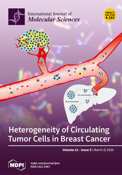
Cover Story (view full-size image): The entry of cancer cells into blood vessels and the appearance of circulating tumor cells (CTCs) seeding and colonizing distant tissues and organs is one of the key stages in the metastatic cascade. Only a small number of CTCs can survive in the bloodstream and form metastases. The identification of metastasis-initiating CTCs is a critical issue in developing therapeutic strategies against metastatic disease. According to current findings, metastatic CTCs in breast cancer are represented by clusters and individual cells with a specific molecular makeup. View this paper.
  • Issues are regarded as officially published after their release is announced to the table of contents alert mailing list.
  • You may sign up for e-mail alerts to receive table of contents of newly released issues.
  • PDF is the official format for papers published in both, html and pdf forms. To view the papers in pdf format, click on the "PDF Full-text" link, and use the free Adobe Reader to open them.
Order results
Result details
Section
Select all
Export citation of selected articles as:
19 pages, 4402 KiB  
Article
Identification and Functional Study of Chitin Metabolism and Detoxification-Related Genes in Glyphodes pyloalis Walker (Lepidoptera: Pyralidae) Based on Transcriptome Analysis
by Zuo-min Shao, Yi-jiangcheng Li, Xiao-rui Zhang, Jie Chu, Jia-hui Ma, Zhi-xiang Liu, Jun Wang, Sheng Sheng and Fu-an Wu
Int. J. Mol. Sci. 2020, 21(5), 1904; https://doi.org/10.3390/ijms21051904 - 10 Mar 2020
Cited by 16 | Viewed by 3462
Abstract
Glyphodes pyloalis Walker (Lepidoptera: Pyralididae) is a serious pest in the sericulture industry, which has caused damage and losses in recent years. With the widespread use of insecticides, the insecticide resistance of G. pyloalis has becomes increasingly apparent. In order to find other [...] Read more.
Glyphodes pyloalis Walker (Lepidoptera: Pyralididae) is a serious pest in the sericulture industry, which has caused damage and losses in recent years. With the widespread use of insecticides, the insecticide resistance of G. pyloalis has becomes increasingly apparent. In order to find other effective methods to control G. pyloalis, this study performed a transcriptome analysis of the midgut, integument, and whole larvae. Transcriptome data were annotated with KEGG and GO, and they have been shown to be of high quality by RT-qPCR. The different significant categories of differentially expressed genes between the midgut and the integument suggested that the transcriptome data could be used for next analysis. With the exception of Dda9 (GpCDA5), 19 genes were involved in chitin metabolism, most of which had close protein–protein interactions. Among them, the expression levels of 11 genes, including GpCHSA, GpCDA1, GpCDA2, GpCDA4, GPCHT1, GPCHT2a, GPCHT3a, GPCHT7, GpTre1, GpTre2, and GpRtv were higher in the integument than in the midgut, while the expression levels of the last eight genes, including GpCHSB, GpCDA5, GpCHT2b, GpCHT3b, GpCHT-h, GpPAGM, GpNAGK, and GpUAP, were higher in the midgut than in the integument. Moreover, 282 detoxification-related genes were identified and can be divided into 10 categories, including cytochrome P450, glutathione S-transferase, carboxylesterase, nicotinic acetylcholine receptor, aquaporin, chloride channel, methoprene-tolerant, serine protease inhibitor, sodium channel, and calcium channel. In order to further study the function of chitin metabolism-related genes, dsRNA injection knocked down the expression of GpCDA1 and GpCHT3a, resulting in the significant downregulation of its downstream genes. These results provide an overview of chitin metabolism and detoxification of G. pyloalis and lay the foundation for the effective control of this pest in the sericulture industry. Full article
(This article belongs to the Section Molecular Biology)
Show Figures

Figure 1

12 pages, 2938 KiB  
Review
Exposure to Dichlorodiphenyldichloroethylene (DDE) and Metallothionein Levels in Rats Fed with Normocaloric or High-Fat Diet: A Review
by Vincenzo Migliaccio, Lillà Lionetti, Rosalba Putti and Rosaria Scudiero
Int. J. Mol. Sci. 2020, 21(5), 1903; https://doi.org/10.3390/ijms21051903 - 10 Mar 2020
Cited by 13 | Viewed by 3541
Abstract
The growing number of studies on metallothioneins (MTs), cysteine-rich metal-binding proteins, have been disclosing new functions of these proteins. Thanks to their inducibility, they were considered to play a pivotal role in regulating trace metals homeostasis and in detoxification from heavy metals; nowadays, [...] Read more.
The growing number of studies on metallothioneins (MTs), cysteine-rich metal-binding proteins, have been disclosing new functions of these proteins. Thanks to their inducibility, they were considered to play a pivotal role in regulating trace metals homeostasis and in detoxification from heavy metals; nowadays, it is known that they are involved in various physiological and pathological processes, such as regulation of apoptosis, elimination of free radicals, and protection of nucleic acids against toxic insults. MT induction has been demonstrated following stress factors other than heavy metals, such as endocrine-disrupting chemicals, insecticides, and herbicides. However, retrieved data are often controversial: in some cases, xenobiotics elicit MT expression and synthesis; under different conditions, they lead to a decrease in cellular MT content. This review describes the MT response to dichlorodiphenyltrichloroethane (DDT) contamination in mammalian tissues. In particular, attention focuses on changes in MT expression, synthesis, and localization in rat liver, kidneys, and testes following oral administration of dichlorodiphenyldichloroethylene (DDE), the main metabolite of DDT, under normal dietary conditions or in combination with a high fat diet potentially able to increase the cellular uptake of this lipophilic pesticide. The potential connection between MT expression and synthesis, lipophilic substances and trace metals availability is also discussed. Full article
(This article belongs to the Special Issue Advances in Metal Metabolism Research)
Show Figures

Figure 1

30 pages, 2392 KiB  
Article
Gene Polymorphisms in Boar Spermatozoa and Their Associations with Post-Thaw Semen Quality
by Anna Mańkowska, Paweł Brym, Łukasz Paukszto, Jan P. Jastrzębski and Leyland Fraser
Int. J. Mol. Sci. 2020, 21(5), 1902; https://doi.org/10.3390/ijms21051902 - 10 Mar 2020
Cited by 21 | Viewed by 5342
Abstract
Genetic markers have been used to assess the freezability of semen. With the advancement in molecular genetic techniques, it is possible to assess the relationships between sperm functions and gene polymorphisms. In this study, variant calling analysis of RNA-Seq datasets was used to [...] Read more.
Genetic markers have been used to assess the freezability of semen. With the advancement in molecular genetic techniques, it is possible to assess the relationships between sperm functions and gene polymorphisms. In this study, variant calling analysis of RNA-Seq datasets was used to identify single nucleotide polymorphisms (SNPs) in boar spermatozoa and to explore the associations between SNPs and post-thaw semen quality. Assessment of post-thaw sperm quality characteristics showed that 21 boars were considered as having good semen freezability (GSF), while 19 boars were classified as having poor semen freezability (PSF). Variant calling demonstrated that most of the polymorphisms (67%) detected in boar spermatozoa were at the 3’-untranslated regions (3’-UTRs). Analysis of SNP abundance in various functional gene categories showed that gene ontology (GO) terms were related to response to stress, motility, metabolism, reproduction, and embryo development. Genomic DNA was isolated from sperm samples of 40 boars. Forty SNPs were selected and genotyped, and several SNPs were significantly associated with motility and membrane integrity of frozen-thawed (FT) spermatozoa. Polymorphism in SCLT1 gene was associated with significantly higher motility and plasma membrane integrity of FT spermatozoa from boars of the GSF group compared with those of the PSF group. Likewise, polymorphisms in MAP3K20, MS4A2, and ROBO1 genes were significantly associated with reduced cryo-induced lipid peroxidation and DNA damage of FT spermatozoa from boars of the GSF group. Candidate genes with significant SNP associations, including APPL1, PLBD1, FBXO16, EML5, RAB3C, OXSR1, PRICKLE1, and MAP3K20 genes, represent potential markers for post-thaw semen quality, and they might be relevant for future improvement in the selection procedure of boars for cryopreservation. The findings of this study provide evidence indicating that polymorphisms in genes expressed in spermatozoa could be considered as factors associated with post-thaw semen quality. Full article
(This article belongs to the Section Biochemistry)
Show Figures

Figure 1

17 pages, 2759 KiB  
Article
Primary Human Chondrocytes Affected by Cigarette Smoke—Therapeutic Challenges
by Tao Chen, Sabrina Ehnert, Gauri Tendulkar, Sheng Zhu, Christian Arnscheidt, Romina H. Aspera-Werz and Andreas K. Nussler
Int. J. Mol. Sci. 2020, 21(5), 1901; https://doi.org/10.3390/ijms21051901 - 10 Mar 2020
Cited by 10 | Viewed by 3515
Abstract
Although several researchers have attested deleterious effects of smoking to the musculoskeletal system, the association between smoking and the onset of osteoarthritis (OA) remains unclear. Here, we investigate the effect of cigarette smoke extract (CSE) on primary human chondrocytes. The present study demonstrates [...] Read more.
Although several researchers have attested deleterious effects of smoking to the musculoskeletal system, the association between smoking and the onset of osteoarthritis (OA) remains unclear. Here, we investigate the effect of cigarette smoke extract (CSE) on primary human chondrocytes. The present study demonstrates that physiological concentrations of CSE (0.1%–10%) inhibit the viability, proliferation, and matrix formation of chondrocytes in a dose- and time-dependent manner. Significant amounts of free radicals were generated by 10% of CSE and led to cell death. A clinical dosage (4 mg/mL) of dexamethasone (Dex) showed toxic effects on chondrocytes, and the long-time treatment by lower doses (4–400 μg/mL) induced hypertrophic changes in the chondrocytes. To substitute Dex, diclofenac (Dic, 1 μg/mL) and acetaminophen (Ace, 10 μg/mL) were tested and did not worsen the metabolic activity of CSE-exposed chondrocytes. Hyaluronic acid (HA, 5 mg/mL) combined with Dic or Ace significantly inhibited the oxidative stress and enhanced the viability and matrix formation of CSE-exposed chondrocytes. This study shows for the first time that CSE mediates the disruption of cartilage through inducing cell death by increasing oxidative stress, and that this effect is fortified by Dex. The deleterious effects of CSE on chondrocytes could be reversed by treatment with HA combined with first-line analgesic/anti-inflammatory agents. Full article
(This article belongs to the Special Issue The Future of Cartilage Repair in Complex Biological Situations)
Show Figures

Graphical abstract

29 pages, 14758 KiB  
Article
Cockle Shell-Derived Aragonite CaCO3 Nanoparticles for Co-Delivery of Doxorubicin and Thymoquinone Eliminates Cancer Stem Cells
by Kehinde Muibat Ibiyeye and Abu Bakar Zakaria Zuki
Int. J. Mol. Sci. 2020, 21(5), 1900; https://doi.org/10.3390/ijms21051900 - 10 Mar 2020
Cited by 17 | Viewed by 3659
Abstract
Cancer stem cells CSCs (tumour-initiating cells) are responsible for cancer metastasis and recurrence associated with resistance to conventional chemotherapy. This study generated MBA MD231 3D cancer stem cells enriched spheroids in serum-free conditions and evaluated the influence of combined doxorubicin/thymoquinone-loaded cockle-shell-derived aragonite calcium [...] Read more.
Cancer stem cells CSCs (tumour-initiating cells) are responsible for cancer metastasis and recurrence associated with resistance to conventional chemotherapy. This study generated MBA MD231 3D cancer stem cells enriched spheroids in serum-free conditions and evaluated the influence of combined doxorubicin/thymoquinone-loaded cockle-shell-derived aragonite calcium carbonate nanoparticles. Single loaded drugs and free drugs were also evaluated. WST assay, sphere forming assay, ALDH activity analysis, Surface marker of CD44 and CD24 expression, apoptosis with Annexin V-PI kit, cell cycle analysis, morphological changes using a phase contrast light microscope, scanning electron microscopy, invasion assay and migration assay were carried out; The combination therapy showed enhanced apoptosis, reduction in ALDH activity and expression of CD44 and CD24 surface maker, reduction in cellular migration and invasion, inhibition of 3D sphere formation when compared to the free drugs and the single drug-loaded nanoparticle. Scanning electron microscopy showed poor spheroid formation, cell membrane blebbing, presence of cell shrinkage, distortion in the spheroid architecture; and the results from this study showed that combined drug-loaded cockle-shell-derived aragonite calcium carbonate nanoparticles can efficiently destroy the breast CSCs compared to single drug-loaded nanoparticle and a simple mixture of doxorubicin and thymoquinone. Full article
(This article belongs to the Special Issue Development of Responsive Nanoparticles for Cancer Therapy)
Show Figures

Figure 1

13 pages, 2051 KiB  
Article
Functional Consequences of Low Activity of Transport System A for Neutral Amino Acids in Human Bone Marrow Mesenchymal Stem Cells
by Martina Chiu, Giuseppe Taurino, Massimiliano G. Bianchi, Erica Dander, Alessandra Fallati, Nicola Giuliani, Giovanna D’Amico and Ovidio Bussolati
Int. J. Mol. Sci. 2020, 21(5), 1899; https://doi.org/10.3390/ijms21051899 - 10 Mar 2020
Cited by 6 | Viewed by 2709
Abstract
In cultured human fibroblasts, SNAT transporters (System A) account for the accumulation of non-essential neutral amino acids, are adaptively up-regulated upon amino acid deprivation and play a major role in cell volume recovery upon hypertonic stress. No information is instead available on the [...] Read more.
In cultured human fibroblasts, SNAT transporters (System A) account for the accumulation of non-essential neutral amino acids, are adaptively up-regulated upon amino acid deprivation and play a major role in cell volume recovery upon hypertonic stress. No information is instead available on the expression and activity of SNAT transporters in human bone marrow mesenchymal stromal cells (MSC), although they are increasingly investigated for their staminal and immunomodulatory properties and used for several therapeutic applications. The uptake of glutamine and proline, two substrates of SNAT1 and SNAT2 transporters, was measured in primary human MSC and an MSC line. The amino acid analogue MeAIB, a specific substrate of these carriers, has been used to selectively inhibit SNAT-dependent transport of glutamine and, through its sodium-dependent transport, as an indicator of SNAT1/2 activity. SNAT1/2 expression and localization were assessed with RT-PCR and confocal microscopy, respectively. Cell volume was assessed from urea distribution space. In all these experiments, primary human fibroblasts were used as the positive control for SNAT expression and activity. Compared with fibroblasts, MSC have a lower SNAT1 expression and hardly detectable membrane localization of both SNAT1 and SNAT2. Moreover, they exhibit no sodium-dependent MeAIB uptake or MeAIB-inhibitable glutamine transport, and exhibit a lower ability to accumulate glutamine and proline than fibroblasts. MSC exhibited an only marginal increase in MeAIB transport upon amino acid starvation and did not recover cell volume after hypertonic stress. In conclusion, the activity of SNAT transporters is low in human MSC. MSC adaptation to amino acid shortage is expected to rely on intracellular synthesis, given the absence of an effective up-regulation of the SNAT transporters. Full article
(This article belongs to the Special Issue Amino Acids Transport and Metabolism 3.0)
Show Figures

Figure 1

26 pages, 4169 KiB  
Article
Melatonin Priming Alleviates Aging-Induced Germination Inhibition by Regulating β-oxidation, Protein Translation, and Antioxidant Metabolism in Oat (Avena sativa L.) Seeds
by Huifang Yan, Shangang Jia and Peisheng Mao
Int. J. Mol. Sci. 2020, 21(5), 1898; https://doi.org/10.3390/ijms21051898 - 10 Mar 2020
Cited by 28 | Viewed by 3878
Abstract
Although melatonin has been reported to play an important role in regulating metabolic events under adverse stresses, its underlying mechanisms on germination in aged seeds remain unclear. This study was conducted to investigate the effect of melatonin priming (MP) on embryos of aged [...] Read more.
Although melatonin has been reported to play an important role in regulating metabolic events under adverse stresses, its underlying mechanisms on germination in aged seeds remain unclear. This study was conducted to investigate the effect of melatonin priming (MP) on embryos of aged oat seeds in relation to germination, ultrastructural changes, antioxidant responses, and protein profiles. Proteomic analysis revealed, in total, 402 differentially expressed proteins (DEPs) in normal, aged, and aged + MP embryos. The downregulated DEPs in aged embryos were enriched in sucrose metabolism, glycolysis, β-oxidation of lipid, and protein synthesis. MP (200 μM) turned four downregulated DEPs into upregulated DEPs, among which, especially 3-ketoacyl-CoA thiolase-like protein (KATLP) involved in the β-oxidation pathway played a key role in maintaining TCA cycle stability and providing more energy for protein translation. Furthermore, it was found that MP enhanced antioxidant capacity in the ascorbate-glutathione (AsA-GSH) system, declined reactive oxygen species (ROS), and improved cell ultrastructure. These results indicated that the impaired germination and seedling growth of aged seeds could be rescued to a certain level by melatonin, predominantly depending on β-oxidation, protein translation, and antioxidant protection of AsA-GSH. This work reveals new insights into melatonin-mediated mechanisms from protein profiles that occur in embryos of oat seeds processed by both aging and priming. Full article
(This article belongs to the Section Molecular Plant Sciences)
Show Figures

Figure 1

13 pages, 2728 KiB  
Article
Pravastatin Alleviates Radiation Proctitis by Regulating Thrombomodulin in Irradiated Endothelial Cells
by Hyosun Jang, Seo-Young Kwak, Sunhoo Park, Kyuchang Kim, Young-heon Kim, Jiyoung Na, Hyewon Kim, Won-Suk Jang, Sun-Joo Lee, Min Jung Kim, Jae Kyung Myung and Sehwan Shim
Int. J. Mol. Sci. 2020, 21(5), 1897; https://doi.org/10.3390/ijms21051897 - 10 Mar 2020
Cited by 18 | Viewed by 3704
Abstract
Although radiotherapy plays a crucial in the management of pelvic tumors, its toxicity on surrounding healthy tissues such as the small intestine, colon, and rectum is one of the major limitations associated with its use. In particular, proctitis is a major clinical complication [...] Read more.
Although radiotherapy plays a crucial in the management of pelvic tumors, its toxicity on surrounding healthy tissues such as the small intestine, colon, and rectum is one of the major limitations associated with its use. In particular, proctitis is a major clinical complication of pelvic radiotherapy. Recent evidence suggests that endothelial injury significantly affects the initiation of radiation-induced inflammation. The damaged endothelial cells accelerate immune cell recruitment by activating the expression of endothelial adhesive molecules, which participate in the development of tissue damage. Pravastatin, a cholesterol lowering drug, exerts persistent anti-inflammatory and anti-thrombotic effects on irradiated endothelial cells and inhibits the interaction of leukocytes and damaged endothelial cells. Here, we aimed to investigate the effects of pravastatin on radiation-induced endothelial damage in human umbilical vein endothelial cell and a murine proctitis model. Pravastatin attenuated epithelial damage and inflammatory response in irradiated colorectal lesions. In particular, pravastatin improved radiation-induced endothelial damage by regulating thrombomodulin (TM) expression. In addition, exogenous TM inhibited leukocyte adhesion to the irradiated endothelial cells. Thus, pravastatin can inhibit endothelial damage by inducing TM, thereby alleviating radiation proctitis. Therefore, we suggest that pharmacological modulation of endothelial TM may limit intestinal inflammation after irradiation. Full article
(This article belongs to the Section Molecular Pharmacology)
Show Figures

Figure 1

17 pages, 2269 KiB  
Article
Improvement of Alcohol-Poisoning Symptoms in Mice by the Oral Administration of Live Lactobacillus plantarum SN13T Cells
by Masafumi Noda, Masafumi Maruyama, Narandalai Danshiitsoodol, Fumiko Higashikawa and Masanori Sugiyama
Int. J. Mol. Sci. 2020, 21(5), 1896; https://doi.org/10.3390/ijms21051896 - 10 Mar 2020
Cited by 4 | Viewed by 4165
Abstract
A clinical study carried out previously by our group has demonstrated that yogurt manufactured with a plant-derived lactic acid bacterium, Lactobacillus plantarum SN13T, significantly reduces the γ-glutamyl transpeptidase (γ-GTP) level as a liver-function parameter. In the present study, we show that with the [...] Read more.
A clinical study carried out previously by our group has demonstrated that yogurt manufactured with a plant-derived lactic acid bacterium, Lactobacillus plantarum SN13T, significantly reduces the γ-glutamyl transpeptidase (γ-GTP) level as a liver-function parameter. In the present study, we show that with the oral administration of live SN13T cells, alcohol-poisoning symptoms in mice are improved, and the condition does not become fatal. However, prior to the simultaneous administration with ethanol, when the cells were heat-killed or sonicated, the improvement was not observed, and almost all of the mice died. In addition, the dysbiosis of the intestinal microbiota observed in the mice administered with ethanol was restored by simultaneous administration with live SN13T cells. Furthermore, by analyzing the metabolites detected in contents from the mouse cecum, it was demonstrated that the increase in nonvolatile putrefactive amines observed in the ethanol-administration group was reduced by simultaneous administration with live SN13T cells. Judging from these results, the lactic acid bacterial cells capable of reaching the living bowels prevent ethanol-induced poisoning and restore the intestinal microbiota. Full article
(This article belongs to the Section Molecular Pathology, Diagnostics, and Therapeutics)
Show Figures

Figure 1

22 pages, 1840 KiB  
Review
Biopolymer-Based Microcarriers for Three-Dimensional Cell Culture and Engineered Tissue Formation
by Lixia Huang, Ahmed M.E. Abdalla, Lin Xiao and Guang Yang
Int. J. Mol. Sci. 2020, 21(5), 1895; https://doi.org/10.3390/ijms21051895 - 10 Mar 2020
Cited by 63 | Viewed by 7416
Abstract
The concept of three-dimensional (3D) cell culture has been proposed to maintain cellular morphology and function as in vivo. Among different approaches for 3D cell culture, microcarrier technology provides a promising tool for cell adhesion, proliferation, and cellular interactions in 3D space mimicking [...] Read more.
The concept of three-dimensional (3D) cell culture has been proposed to maintain cellular morphology and function as in vivo. Among different approaches for 3D cell culture, microcarrier technology provides a promising tool for cell adhesion, proliferation, and cellular interactions in 3D space mimicking the in vivo microenvironment. In particular, microcarriers based on biopolymers have been widely investigated because of their superior biocompatibility and biodegradability. Moreover, through bottom-up assembly, microcarriers have opened a bright door for fabricating engineered tissues, which is one of the cutting-edge topics in tissue engineering and regeneration medicine. This review takes an in-depth look into the recent advancements of microcarriers based on biopolymers—especially polysaccharides such as chitosan, chitin, cellulose, hyaluronic acid, alginate, and laminarin—for 3D cell culture and the fabrication of engineered tissues based on them. The current limitations and potential strategies were also discussed to shed some light on future directions. Full article
Show Figures

Figure 1

24 pages, 2763 KiB  
Article
The Polymorphic PolyQ Tail Protein of the Mediator Complex, Med15, Regulates the Variable Response to Diverse Stresses
by Jennifer E.G. Gallagher, Suk Lan Ser, Michael C. Ayers, Casey Nassif and Amaury Pupo
Int. J. Mol. Sci. 2020, 21(5), 1894; https://doi.org/10.3390/ijms21051894 - 10 Mar 2020
Cited by 7 | Viewed by 4115
Abstract
The Mediator is composed of multiple subunits conserved from yeast to humans and plays a central role in transcription. The tail components are not required for basal transcription but are required for responses to different stresses. While some stresses are familiar, such as [...] Read more.
The Mediator is composed of multiple subunits conserved from yeast to humans and plays a central role in transcription. The tail components are not required for basal transcription but are required for responses to different stresses. While some stresses are familiar, such as heat, desiccation, and starvation, others are exotic, yet yeast can elicit a successful stress response. 4-Methylcyclohexane methanol (MCHM) is a hydrotrope that induces growth arrest in yeast. We found that a naturally occurring variation in the Med15 allele, a component of the Mediator tail, altered the stress response to many chemicals in addition to MCHM. Med15 contains two polyglutamine repeats (polyQ) of variable lengths that change the gene expression of diverse pathways. The Med15 protein existed in multiple isoforms and its stability was dependent on Ydj1, a protein chaperone. The protein level of Med15 with longer polyQ tracts was lower and turned over faster than the allele with shorter polyQ repeats. MCHM sensitivity via variation of Med15 was regulated by Snf1 in a Myc-tag-dependent manner. Tagging Med15 with Myc altered its function in response to stress. Genetic variation in transcriptional regulators magnified genetic differences in response to environmental changes. These polymorphic control genes were master variators. Full article
(This article belongs to the Special Issue Small Molecules, Influence of Molecular Pathways)
Show Figures

Graphical abstract

21 pages, 3960 KiB  
Article
Valine Radiolysis by H+, He+, N+, and S15+ MeV Ions
by Cíntia A. P. da Costa, Gabriel S. Vignoli Muniz, Philippe Boduch, Hermann Rothard and Enio F. da Silveira
Int. J. Mol. Sci. 2020, 21(5), 1893; https://doi.org/10.3390/ijms21051893 - 10 Mar 2020
Cited by 12 | Viewed by 2823
Abstract
Radiolysis of biomolecules by fast ions has interest in medical applications and astrobiology. The radiolysis of solid D-valine (0.2–2 μm thick) was performed at room temperature by 1.5 MeV H+, He+, N+, and 230 MeV S15+ [...] Read more.
Radiolysis of biomolecules by fast ions has interest in medical applications and astrobiology. The radiolysis of solid D-valine (0.2–2 μm thick) was performed at room temperature by 1.5 MeV H+, He+, N+, and 230 MeV S15+ ion beams. The samples were prepared by spraying/dropping valine-water-ethanol solution on ZnSe substrate. Radiolysis was monitored by infrared spectroscopy (FTIR) through the evolution of the intensity of the valine infrared 2900, 1329, 1271, 948, and 716 cm−1 bands as a function of projectile fluence. At the end of sample irradiation, residues (tholins) presenting a brownish color are observed. The dependence of the apparent (sputtering + radiolysis) destruction cross section, σd, on the beam stopping power in valine is found to follow the power law σd = aSen, with n close to 1. Thus, σd is approximately proportional to the absorbed dose. Destruction rates due to the main galactic cosmic ray species are calculated, yielding a million year half-life for solid valine in space. Data obtained in this work aim a better understanding on the radioresistance of complex organic molecules and formation of radioproducts. Full article
(This article belongs to the Special Issue Radiation Damage in Biomolecules and Cells)
Show Figures

Figure 1

22 pages, 1948 KiB  
Article
Malignancy Grade-Dependent Mapping of Metabolic Landscapes in Human Urothelial Bladder Cancer: Identification of Novel, Diagnostic, and Druggable Biomarkers
by Aikaterini Iliou, Aristeidis Panagiotakis, Aikaterini F. Giannopoulou, Dimitra Benaki, Mariangela Kosmopoulou, Athanassios D. Velentzas, Ourania E. Tsitsilonis, Issidora S. Papassideri, Gerassimos E. Voutsinas, Eumorphia G. Konstantakou, Evagelos Gikas, Emmanuel Mikros and Dimitrios J. Stravopodis
Int. J. Mol. Sci. 2020, 21(5), 1892; https://doi.org/10.3390/ijms21051892 - 10 Mar 2020
Cited by 8 | Viewed by 3775
Abstract
Background: Urothelial bladder cancer (UBC) is one of the cancers with the highest mortality rate and prevalence worldwide; however, the clinical management of the disease remains challenging. Metabolomics has emerged as a powerful tool with beneficial applications in cancer biology and thus can [...] Read more.
Background: Urothelial bladder cancer (UBC) is one of the cancers with the highest mortality rate and prevalence worldwide; however, the clinical management of the disease remains challenging. Metabolomics has emerged as a powerful tool with beneficial applications in cancer biology and thus can provide new insights on the underlying mechanisms of UBC progression and/or reveal novel diagnostic and therapeutic schemes. Methods: A collection of four human UBC cell lines that critically reflect the different malignancy grades of UBC was employed; RT4 (grade I), RT112 (grade II), T24 (grade III), and TCCSUP (grade IV). They were examined using Nuclear Magnetic Resonance, Mass Spectrometry, and advanced statistical approaches, with the goal of creating new metabolic profiles that are mechanistically associated with UBC progression toward metastasis. Results: Distinct metabolic profiles were observed for each cell line group, with T24 (grade III) cells exhibiting the most abundant metabolite contents. AMP and creatine phosphate were highly increased in the T24 cell line compared to the RT4 (grade I) cell line, indicating the major energetic transformation to which UBC cells are being subjected during metastasis. Thymosin β4 and β10 were also profiled with grade-specific patterns of expression, strongly suggesting the importance of actin-cytoskeleton dynamics for UBC advancement to metastatic and drug-tolerant forms. Conclusions: The present study unveils a novel and putatively druggable metabolic signature that holds strong promise for early diagnosis and the successful chemotherapy of UBC disease. Full article
Show Figures

Figure 1

16 pages, 4899 KiB  
Article
Epigenetic Regulation of WNT3A Enhancer during Regeneration of Injured Cortical Neurons
by Chu-Yuan Chang, Jui-Hung Hung, Liang-Wei Huang, Joye Li, Ka Shing Fung, Cheng-Fu Kao and Linyi Chen
Int. J. Mol. Sci. 2020, 21(5), 1891; https://doi.org/10.3390/ijms21051891 - 10 Mar 2020
Cited by 3 | Viewed by 2850
Abstract
Traumatic brain injury is known to reprogram the epigenome. Chromatin immunoprecipitation-sequencing of histone H3 lysine 27 acetylation (H3K27ac) and tri-methylation of histone H3 at lysine 4 (H3K4me3) marks was performed to address the transcriptional regulation of candidate regeneration-associated genes. In this study, we [...] Read more.
Traumatic brain injury is known to reprogram the epigenome. Chromatin immunoprecipitation-sequencing of histone H3 lysine 27 acetylation (H3K27ac) and tri-methylation of histone H3 at lysine 4 (H3K4me3) marks was performed to address the transcriptional regulation of candidate regeneration-associated genes. In this study, we identify a novel enhancer region for induced WNT3A transcription during regeneration of injured cortical neurons. We further demonstrated an increased mono-methylation of histone H3 at lysine 4 (H3K4me1) modification at this enhancer concomitant with a topological interaction between sub-regions of this enhancer and with promoter of WNT3A gene. Together, this study reports a novel mechanism for WNT3A gene transcription and reveals a potential therapeutic intervention for neuronal regeneration. Full article
(This article belongs to the Special Issue Modifications of Molecular Structure and Interactions in Epigenome)
Show Figures

Figure 1

12 pages, 2716 KiB  
Article
Leucine-Rich Repeat Kinase 2 Controls Inflammatory Cytokines Production through NF-κB Phosphorylation and Antigen Presentation in Bone Marrow-Derived Dendritic Cells
by Makoto Kubo, Ryuichi Nagashima, Mitsue Kurihara, Fumitaka Kawakami, Tatsunori Maekawa, Koji Eshima, Etsuro Ohta, Hirotomo Kato and Fumiya Obata
Int. J. Mol. Sci. 2020, 21(5), 1890; https://doi.org/10.3390/ijms21051890 - 10 Mar 2020
Cited by 7 | Viewed by 3398
Abstract
Leucine-rich repeat kinase 2 (LRRK2) is the causal molecule of familial Parkinson’s disease. Although the characteristics of LRRK2 have gradually been revealed, its true physiological functions remain unknown. LRRK2 is highly expressed in immune cells such as B2 cells and macrophages, suggesting that [...] Read more.
Leucine-rich repeat kinase 2 (LRRK2) is the causal molecule of familial Parkinson’s disease. Although the characteristics of LRRK2 have gradually been revealed, its true physiological functions remain unknown. LRRK2 is highly expressed in immune cells such as B2 cells and macrophages, suggesting that it plays important roles in the immune system. In the present study, we investigate the roles of LRRK2 in the immune functions of dendritic cells (DCs). Bone marrow-derived DCs from both C57BL/6 wild-type (WT) and LRRK2 knockout (KO) mice were induced by culture with granulocyte/macrophage-colony stimulating factor (GM/CSF) in vitro. We observed the differentiation of DCs, the phosphorylation of the transcriptional factors NF-κB, Erk1/2, and p-38 after lipopolysaccharide (LPS) stimulation and antigen-presenting ability by flow cytometry. We also analyzed the production of inflammatory cytokines by ELISA. During the observation period, there was no difference in DC differentiation between WT and LRRK2-KO mice. After LPS stimulation, phosphorylation of NF-κB was significantly increased in DCs from the KO mice. Large amounts of inflammatory cytokines were produced by DCs from KO mice after both stimulation with LPS and infection with Leishmania. CD4+ T-cells isolated from antigen-immunized mice proliferated to a significantly greater degree upon coculture with antigen-stimulated DCs from KO mice than upon coculture with DCs from WT mice. These results suggest that LRRK2 may play important roles in signal transduction and antigen presentation by DCs. Full article
(This article belongs to the Special Issue Neuroimmunology)
Show Figures

Figure 1

18 pages, 3995 KiB  
Article
HSP Transcript and Protein Accumulation in Brassinosteroid Barley Mutants Acclimated to Low and High Temperatures
by Iwona Sadura, Marta Libik-Konieczny, Barbara Jurczyk, Damian Gruszka and Anna Janeczko
Int. J. Mol. Sci. 2020, 21(5), 1889; https://doi.org/10.3390/ijms21051889 - 10 Mar 2020
Cited by 23 | Viewed by 3641
Abstract
In temperature stress, the main role of heat-shock proteins (HSP) is to act as molecular chaperones for other cellular proteins. However, knowledge about the hormonal regulation of the production of the HSP is quite limited. Specifically, little is known about the role of [...] Read more.
In temperature stress, the main role of heat-shock proteins (HSP) is to act as molecular chaperones for other cellular proteins. However, knowledge about the hormonal regulation of the production of the HSP is quite limited. Specifically, little is known about the role of the plant steroid hormones—brassinosteroids (BR)—in regulating the HSP expression. The aim of our study was to answer the question of how a BR deficit or disturbances in its signaling affect the accumulation of the HSP90, HSP70, HSP18, and HSP17 transcripts and protein in barley growing at 20 °C (control) and during the acclimation of plants at 5 °C and 27 °C. In barley, the temperature of plant growth modified the expression of HSPs. Furthermore, the BR-deficient mutants (mutations in the HvDWARF or HvCPD genes) and BR-signaling mutants (mutation in the HvBRI1 gene) were characterized by altered levels of the transcripts and proteins of the HSP group compared to the wild type. The BR-signaling mutant was characterized by a decreased level of the HSP transcripts and heat-shock proteins. In the BR-deficient mutants, there were temperature-dependent cases when the decreased accumulation of the HSP70 and HSP90 transcripts was connected to an increased accumulation of these HSP. The significance of changes in the accumulation of HSPs during acclimation at 27 °C and 5 °C is discussed in the context of the altered tolerance to more extreme temperatures of the studied mutants (i.e., heat stress and frost, respectively). Full article
(This article belongs to the Special Issue Wheat and Barley: Acclimatization to Abiotic and Biotic Stress)
Show Figures

Figure 1

13 pages, 4582 KiB  
Article
Effects of Osiris9a on Silk Properties in Bombyx mori Determined by Transgenic Overexpression
by Tingcai Cheng, Xia Zhang, Zhangchuan Peng, Yinfeng Fan, Lin Zhang and Chun Liu
Int. J. Mol. Sci. 2020, 21(5), 1888; https://doi.org/10.3390/ijms21051888 - 10 Mar 2020
Cited by 8 | Viewed by 3200
Abstract
Osiris is an insect-specific gene family with multiple biological roles in development, phenotypic polymorphism, and protection. In the silkworm, we have previously identified twenty-five Osiris genes with high evolutionary conservation and remarkable synteny among several insects. Bombxy mori Osiris9a (BmOsi9a) is [...] Read more.
Osiris is an insect-specific gene family with multiple biological roles in development, phenotypic polymorphism, and protection. In the silkworm, we have previously identified twenty-five Osiris genes with high evolutionary conservation and remarkable synteny among several insects. Bombxy mori Osiris9a (BmOsi9a) is expressed only in the silk gland, particularly in the middle silk gland (MSG). However, the biological function of BmOsi9a is still unknown. In this study, we overexpressed BmOsi9a in the silk gland by germline transgene expression. BmOsi9a was overexpressed not only in the MSG but also in the posterior silk gland (PSG). Interestingly, BmOsi9a could be secreted into the lumen in the MSG but not in the PSG. In the silk fiber, overexpressed BmOsi9a interacted with Sericin1 in the MSG, as confirmed by a co-immunoprecipitation assay. The overexpression of BmOsi9a altered the secondary structure and crystallinity of the silk fiber, thereby changing the mechanical properties. These results provide insight into the mechanisms underlying silk proteins secretion and silk fiber formation. Full article
(This article belongs to the Section Materials Science)
Show Figures

Figure 1

18 pages, 4410 KiB  
Article
Altered Structural Expression and Enzymatic Activity Parameters in Quiescent Ulcerative Colitis: Are These Potential Normalization Criteria?
by Sebastian Kjærgaard, Morten M. B. Damm, Joan Chang, Lene B. Riis, Hanne B. Rasmussen, Rasmus Hytting-Andreasen, Susanne M. Krug, Jörg-Dieter Schulzke, Niels Bindslev and Mark Berner Hansen
Int. J. Mol. Sci. 2020, 21(5), 1887; https://doi.org/10.3390/ijms21051887 - 10 Mar 2020
Cited by 8 | Viewed by 3400
Abstract
Mucosal healing determined by endoscopy is currently the remission standard for ulcerative colitis (UC). However, new criteria for remission are emerging, such as histologic normalization, which appears to correlate better to the risk of relapse. Here, we study mucosal healing on a molecular [...] Read more.
Mucosal healing determined by endoscopy is currently the remission standard for ulcerative colitis (UC). However, new criteria for remission are emerging, such as histologic normalization, which appears to correlate better to the risk of relapse. Here, we study mucosal healing on a molecular and functional level in quiescent UC. We obtained endoscopic biopsies from 33 quiescent UC patients and from 17 controls. Histology was assessed using Geboes score. Protein and mRNA levels were evaluated for the tight junction proteins claudin-2, claudin-4, occludin, and tricellulin, as well as Cl/HCO3 exchanger DRA, and cyclo-oxygenase enzymes (COX-1, COX-2). The mucosal activity of COX-1 and COX-2 enzymes was assessed in modified Ussing chambers, measuring electrogenic ion transport (short-circuit current, SCC). Chronic inflammation was present in most UC patients. The protein level of claudin-4 was reduced, while mRNA-levels of claudin-2 and claudin-4 were upregulated in UC patients. Surprisingly, the mRNA level of COX-1 was downregulated, but was unaltered for COX-2. Basal ion transport was not affected, while COX-2 inhibition induced a two-fold larger decrease in SCC in UC patients. Despite being in clinical and endoscopic remission, quiescent UC patients demonstrated abnormal mucosal barrier properties at the molecular and functional level. Further exploration of mucosal molecular signature for revision of current remission standards should be considered. Full article
(This article belongs to the Special Issue Update on Basic and Molecular Research in Inflammatory Bowel Disease)
Show Figures

Figure 1

19 pages, 4336 KiB  
Article
At-Hook Motif Nuclear Localised Protein 18 as a Novel Modulator of Root System Architecture
by Marek Širl, Tereza Šnajdrová, Dolores Gutiérrez-Alanís, Joseph G. Dubrovsky, Jean Phillipe Vielle-Calzada, Ivan Kulich and Aleš Soukup
Int. J. Mol. Sci. 2020, 21(5), 1886; https://doi.org/10.3390/ijms21051886 - 10 Mar 2020
Cited by 20 | Viewed by 4158
Abstract
The At-Hook Motif Nuclear Localized Protein (AHL) gene family encodes embryophyte-specific nuclear proteins with DNA binding activity. They modulate gene expression and affect various developmental processes in plants. We identify AHL18 (At3G60870) as a developmental modulator of root system architecture and [...] Read more.
The At-Hook Motif Nuclear Localized Protein (AHL) gene family encodes embryophyte-specific nuclear proteins with DNA binding activity. They modulate gene expression and affect various developmental processes in plants. We identify AHL18 (At3G60870) as a developmental modulator of root system architecture and growth. AHL18 is involved in regulation of the length of the proliferation domain and number of dividing cells in the root apical meristem and thereby, cell production. Both primary root growth and lateral root development respond according to AHL18 transcription level. The ahl18 knock-out plants show reduced root systems due to a shorter primary root and a lower number of lateral roots. This change results from a higher number of arrested and non-developing lateral root primordia (LRP) rather than from a decreased LRP initiation. The over-expression of AHL18 results in a more extensive root system, longer primary roots, and increased density of lateral root initiation events. AHL18 is thus involved in the formation of lateral roots at both LRP initiation and their later development. We conclude that AHL18 participates in modulation of root system architecture through regulation of root apical meristem activity, lateral root initiation and emergence; these correspond well with expression pattern of AHL18. Full article
(This article belongs to the Special Issue Plant Cell and Organism Development)
Show Figures

Figure 1

20 pages, 2390 KiB  
Review
Promoting Roles of Embryonic Signals in Embryo Implantation and Placentation in Cooperation with Endocrine and Immune Systems
by Hiroshi Fujiwara, Masanori Ono, Yukiyasu Sato, Kazuhiko Imakawa, Takashi Iizuka, Kyosuke Kagami, Tomoko Fujiwara, Akihito Horie, Hirohiko Tani, Akira Hattori, Takiko Daikoku and Yoshihiko Araki
Int. J. Mol. Sci. 2020, 21(5), 1885; https://doi.org/10.3390/ijms21051885 - 10 Mar 2020
Cited by 26 | Viewed by 6407
Abstract
Embryo implantation in the uterus is an essential process for successful pregnancy in mammals. In general, the endocrine system induces sufficient embryo receptivity in the endometrium, where adhesion-promoting molecules increase and adhesion-inhibitory molecules decrease. Although the precise mechanisms remain unknown, it is widely [...] Read more.
Embryo implantation in the uterus is an essential process for successful pregnancy in mammals. In general, the endocrine system induces sufficient embryo receptivity in the endometrium, where adhesion-promoting molecules increase and adhesion-inhibitory molecules decrease. Although the precise mechanisms remain unknown, it is widely accepted that maternal–embryo communications, including embryonic signals, improve the receptive ability of the sex steroid hormone-primed endometrium. The embryo may utilize repulsive forces produced by an Eph–ephrin system for its timely attachment to and subsequent invasion through the endometrial epithelial layer. Importantly, the embryonic signals are considered to act on maternal immune cells to induce immune tolerance. They also elicit local inflammation that promotes endometrial differentiation and maternal tissue remodeling during embryo implantation and placentation. Additional clarification of the immune control mechanisms by embryonic signals, such as human chorionic gonadotropin, pre-implantation factor, zona pellucida degradation products, and laeverin, will aid in the further development of immunotherapy to minimize implantation failure in the future. Full article
(This article belongs to the Special Issue Embryo-Maternal Interactions Underlying Reproduction in Mammals)
Show Figures

Figure 1

13 pages, 696 KiB  
Review
Crosstalk Among Circadian Rhythm, Obesity and Allergy
by Kanami Orihara, Atsushi Haraguchi and Shigenobu Shibata
Int. J. Mol. Sci. 2020, 21(5), 1884; https://doi.org/10.3390/ijms21051884 - 10 Mar 2020
Cited by 19 | Viewed by 5229
Abstract
The circadian clock system works not only as a cellular time-keeper but also as a coordinator for almost all physiological functions essential to maintaining human health. Therefore, disruptions or malfunctions of this system can cause many diseases and pre-symptomatic conditions. Indeed, previous studies [...] Read more.
The circadian clock system works not only as a cellular time-keeper but also as a coordinator for almost all physiological functions essential to maintaining human health. Therefore, disruptions or malfunctions of this system can cause many diseases and pre-symptomatic conditions. Indeed, previous studies have indicated that disrupted clock gene expression rhythm is closely related to obesity, and that allergic diseases can be regulated by controlling peripheral clocks in organs and tissues. Moreover, recent studies have found that obesity can lead to immune disorders. Accordingly, in this review, we assess the connection between obesity and allergy from the point of view of the circadian clock system anew and summarize the relationships among the circadian clock system, obesity, and allergy. Full article
(This article belongs to the Special Issue Crosstalk between Circadian Rhythm and Diseases)
Show Figures

Figure 1

14 pages, 861 KiB  
Article
Bacteriophage-Insensitive Mutants of Antimicrobial-Resistant Salmonella Enterica are Altered in their Tetracycline Resistance and Virulence in Caco-2 Intestinal Cells
by Karen Fong, Kaiwen Mu, Jean-Guillaume Rheault, Roger C. Levesque, David D. Kitts, Pascal Delaquis, Lawrence Goodridge and Siyun Wang
Int. J. Mol. Sci. 2020, 21(5), 1883; https://doi.org/10.3390/ijms21051883 - 10 Mar 2020
Cited by 13 | Viewed by 3810
Abstract
Bacteriophages have shown promise as therapeutic alternatives to antibiotics for the control of infectious bacteria, including the human pathogen Salmonella. However, the development of effective phage-based applications requires the elucidation of key interactions between phages and target hosts, particularly since host resistance to [...] Read more.
Bacteriophages have shown promise as therapeutic alternatives to antibiotics for the control of infectious bacteria, including the human pathogen Salmonella. However, the development of effective phage-based applications requires the elucidation of key interactions between phages and target hosts, particularly since host resistance to phage is inevitable. Little is known about the alteration of host phenotypes following the development of resistance to phage. The aim of this study is to evaluate the antibiotic susceptibility and virulence of a Salmonella isolate following the development of resistance to bacteriophage SI1. We observed enhanced susceptibility to tetracycline and decreased invasion capacity in a differentiated Caco-2 intestinal cell line. Whole genome sequence analysis revealed an array of mutations, most notably, truncations in vgrG1_2, a core gene involved in Type VI secretion and mutations in the lipopolysaccharide, thereby indicating the plausible attachment site of phage SI1. These findings shed light on understanding the underlying mechanism for phage immunity within the host. Importantly, we reveal an associated genetic cost to the bacterial host with developing resistance to phages. Taken together, these results will aid in advancing strategies to delay or eliminate the development of host resistance when designing informed phage-based antimicrobials. Full article
Show Figures

Figure 1

19 pages, 16693 KiB  
Article
The mito-QC Reporter for Quantitative Mitophagy Assessment in Primary Retinal Ganglion Cells and Experimental Glaucoma Models
by Ines Rosignol, Beatriz Villarejo-Zori, Petra Teresak, Elena Sierra-Filardi, Xandra Pereiro, Natalia Rodríguez-Muela, Elena Vecino, Helena L. A. Vieira, Katharina Bell and Patricia Boya
Int. J. Mol. Sci. 2020, 21(5), 1882; https://doi.org/10.3390/ijms21051882 - 10 Mar 2020
Cited by 18 | Viewed by 6145
Abstract
Mitochondrial damage plays a prominent role in glaucoma. The only way cells can degrade whole mitochondria is via autophagy, in a process called mitophagy. Thus, studying mitophagy in the context of glaucoma is essential to understand the disease. Up to date limited tools [...] Read more.
Mitochondrial damage plays a prominent role in glaucoma. The only way cells can degrade whole mitochondria is via autophagy, in a process called mitophagy. Thus, studying mitophagy in the context of glaucoma is essential to understand the disease. Up to date limited tools are available for analyzing mitophagy in vivo. We have taken advantage of the mito-QC reporter, a recently generated mouse model that allows an accurate mitophagy assessment to fill this gap. We used primary RGCs and retinal explants derived from mito-QC mice to quantify mitophagy activation in vitro and ex vivo. We also analyzed mitophagy in retinal ganglion cells (RGCs), in vivo, using different mitophagy inducers, as well as after optic nerve crush (ONC) in mice, a commonly used surgical procedure to model glaucoma. Using mito-QC reporter we quantified mitophagy induced by several known inducers in primary RGCs in vitro, ex vivo and in vivo. We also found that RGCs were rescued from some glaucoma relevant stress factors by incubation with the iron chelator deferiprone (DFP). Thus, the mito-QC reporter-based model is a valuable tool for accurately analyzing mitophagy in the context of glaucoma. Full article
(This article belongs to the Special Issue Retinal Ganglion Cells)
Show Figures

Figure 1

26 pages, 2433 KiB  
Review
Flavodoxins as Novel Therapeutic Targets against Helicobacter pylori and Other Gastric Pathogens
by Sandra Salillas and Javier Sancho
Int. J. Mol. Sci. 2020, 21(5), 1881; https://doi.org/10.3390/ijms21051881 - 10 Mar 2020
Cited by 20 | Viewed by 9692
Abstract
Flavodoxins are small soluble electron transfer proteins widely present in bacteria and absent in vertebrates. Flavodoxins participate in different metabolic pathways and, in some bacteria, they have been shown to be essential proteins representing promising therapeutic targets to fight bacterial infections. Using purified [...] Read more.
Flavodoxins are small soluble electron transfer proteins widely present in bacteria and absent in vertebrates. Flavodoxins participate in different metabolic pathways and, in some bacteria, they have been shown to be essential proteins representing promising therapeutic targets to fight bacterial infections. Using purified flavodoxin and chemical libraries, leads can be identified that block flavodoxin function and act as bactericidal molecules, as it has been demonstrated for Helicobacter pylori (Hp), the most prevalent human gastric pathogen. Increasing antimicrobial resistance by this bacterium has led current therapies to lose effectiveness, so alternative treatments are urgently required. Here, we summarize, with a focus on flavodoxin, opportunities for pharmacological intervention offered by the potential protein targets described for this bacterium and provide information on other gastrointestinal pathogens and also on bacteria from the gut microbiota that contain flavodoxin. The process of discovery and development of novel antimicrobials specific for Hp flavodoxin that is being carried out in our group is explained, as it can be extrapolated to the discovery of inhibitors specific for other gastric pathogens. The high specificity for Hp of the antimicrobials developed may be of help to reduce damage to the gut microbiota and to slow down the development of resistant Hp mutants. Full article
(This article belongs to the Special Issue Biochemistry, Molecular Biology and Druggability of Proteins)
Show Figures

Figure 1

19 pages, 1552 KiB  
Article
Deciphering the Role of Multiple Thioredoxin Fold Proteins of Leptospirillum sp. in Oxidative Stress Tolerance
by Daniela González, Pamela Álamos, Matías Rivero, Omar Orellana, Javiera Norambuena, Renato Chávez and Gloria Levicán
Int. J. Mol. Sci. 2020, 21(5), 1880; https://doi.org/10.3390/ijms21051880 - 10 Mar 2020
Cited by 19 | Viewed by 3298
Abstract
Thioredoxin fold proteins (TFPs) form a family of diverse proteins involved in thiol/disulfide exchange in cells from all domains of life. Leptospirillum spp. are bioleaching bacteria naturally exposed to extreme conditions like acidic pH and high concentrations of metals that can contribute to [...] Read more.
Thioredoxin fold proteins (TFPs) form a family of diverse proteins involved in thiol/disulfide exchange in cells from all domains of life. Leptospirillum spp. are bioleaching bacteria naturally exposed to extreme conditions like acidic pH and high concentrations of metals that can contribute to the generation of reactive oxygen species (ROS) and consequently the induction of thiol oxidative damage. Bioinformatic studies have predicted 13 genes that encode for TFP proteins in Leptospirillum spp. We analyzed the participation of individual tfp genes from Leptospirillum sp. CF-1 in the response to oxidative conditions. Genomic context analysis predicted the involvement of these genes in the general thiol-reducing system, cofactor biosynthesis, carbon fixation, cytochrome c biogenesis, signal transduction, and pilus and fimbria assembly. All tfp genes identified were transcriptionally active, although they responded differentially to ferric sulfate and diamide stress. Some of these genes confer oxidative protection to a thioredoxin-deficient Escherichia coli strain by restoring the wild-type phenotype under oxidative stress conditions. These findings contribute to our understanding of the diversity and complexity of thiol/disulfide systems, and of adaptations that emerge in acidophilic microorganisms that allow them to thrive in highly oxidative environments. These findings also give new insights into the physiology of these microorganisms during industrial bioleaching operations. Full article
(This article belongs to the Section Molecular Microbiology)
Show Figures

Graphical abstract

20 pages, 2863 KiB  
Article
Major Latex Protein MdMLP423 Negatively Regulates Defense against Fungal Infections in Apple
by Shanshan He, Gaopeng Yuan, Shuxun Bian, Xiaolei Han, Kai Liu, Peihua Cong and Caixia Zhang
Int. J. Mol. Sci. 2020, 21(5), 1879; https://doi.org/10.3390/ijms21051879 - 10 Mar 2020
Cited by 26 | Viewed by 3882
Abstract
Major latex proteins (MLPs) play critical roles in plants defense and stress responses. However, the roles of MLPs from apple (Malus × domestica) have not been clearly identified. In this study, we focused on the biological role of MdMLP423, which [...] Read more.
Major latex proteins (MLPs) play critical roles in plants defense and stress responses. However, the roles of MLPs from apple (Malus × domestica) have not been clearly identified. In this study, we focused on the biological role of MdMLP423, which had been previously characterized as a potential pathogenesis-related gene. Phylogenetic analysis and conserved domain analysis indicated that MdMLP423 is a protein with a ‘Gly-rich loop’ (GXGGXG) domain belonging to the Bet v_1 subfamily. Gene expression profiles showed that MdMLP423 is mainly expressed in flowers. In addition, the expression of MdMLP423 was significantly inhibited by Botryosphaeria berengeriana f. sp. piricola (BB) and Alternaria alternata apple pathotype (AAAP) infections. Apple calli overexpressing MdMLP423 had lower expression of resistance-related genes, and were more sensitive to infection with BB and AAAP compared with non-transgenic calli. RNA-seq analysis of MdMLP423-overexpressing calli and non-transgenic calli indicated that MdMLP423 regulated the expression of a number of differentially expressed genes (DEGs) and transcription factors, including genes involved in phytohormone signaling pathways, cell wall reinforcement, and genes encoding the defense-related proteins, AP2-EREBP, WRKY, MYB, NAC, Zinc finger protein, and ABI3. Taken together, our results demonstrate that MdMLP423 negatively regulates apple resistance to BB and AAAP infections by inhibiting the expression of defense- and stress-related genes and transcription factors. Full article
(This article belongs to the Special Issue Plant Disease Resistance)
Show Figures

Figure 1

18 pages, 7517 KiB  
Article
TmSpz4 Plays an Important Role in Regulating the Production of Antimicrobial Peptides in Response to Escherichia coli and Candida albicans Infections
by Tariku Tesfaye Edosa, Yong Hun Jo, Maryam Keshavarz, Young Min Bae, Dong Hyun Kim, Yong Seok Lee and Yeon Soo Han
Int. J. Mol. Sci. 2020, 21(5), 1878; https://doi.org/10.3390/ijms21051878 - 09 Mar 2020
Cited by 20 | Viewed by 3066
Abstract
Spätzle family proteins activate the Toll pathway and induce antimicrobial peptide (AMP) production against microbial infections. However, the functional importance of Tmspätzle4 (TmSpz4) in the immune response of Tenebrio molitor has not been reported. Therefore, here, we have identified and [...] Read more.
Spätzle family proteins activate the Toll pathway and induce antimicrobial peptide (AMP) production against microbial infections. However, the functional importance of Tmspätzle4 (TmSpz4) in the immune response of Tenebrio molitor has not been reported. Therefore, here, we have identified and functionally characterized the role of TmSpz4 against bacterial and fungal infections. We showed that TmSpz4 expression was significantly induced in hemocytes at 6 h post-injection with Escherichia coli, Staphylococcus aureus, and Candida albicans. TmSpz4 knock-down significantly reduced larval survival against E. coli and C. albicans. To understand the reason for the survivability difference, the role of TmSpz4 in AMP production was examined in TmSpz4-silenced larvae following microbe injection. The AMPs that are active against Gram-negative bacteria, including TmTenecin-2, TmTenecin-4, TmAttacin-1a, TmDefensin-2, and TmCecropin-2, were significantly downregulated in response to E. coli in TmSpz4-silenced larvae. Similarly, the expression of TmTenecin-1, TmTenecin-3, TmThaumatin-like protein-1 and -2, TmDefensin-1, TmDefensin-2, and TmCecropin-2 were downregulated in response to C. albicans in TmSpz4-silenced larvae. In addition, the transcription factor NF-κB (TmDorX1 and TmDorX2) expression was significantly suppression in TmSpz4-silenced larvae. In conclusion, these results suggest that TmSpz4 plays a key role in regulating immune responses of T. molitor against to E. coli and C. albicans. Full article
(This article belongs to the Section Molecular Immunology)
Show Figures

Graphical abstract

23 pages, 1306 KiB  
Review
TRP Channels in Digestive Tract Cancers
by Paulina Stokłosa, Anna Borgström, Sven Kappel and Christine Peinelt
Int. J. Mol. Sci. 2020, 21(5), 1877; https://doi.org/10.3390/ijms21051877 - 09 Mar 2020
Cited by 37 | Viewed by 6278
Abstract
Cancers of the digestive tract are among the most prevalent types of cancer. These types of cancers are often diagnosed at a late stage, which results in a poor prognosis. Currently, many biomedical studies focus on the role of ion channels, in particular [...] Read more.
Cancers of the digestive tract are among the most prevalent types of cancer. These types of cancers are often diagnosed at a late stage, which results in a poor prognosis. Currently, many biomedical studies focus on the role of ion channels, in particular transient receptor potential (TRP) channels, in cancer pathophysiology. TRP channels show mostly non-selective permeability to monovalent and divalent cations. TRP channels are often dysregulated in digestive tract cancers, which can result in alterations of cancer hallmark functions, such as enhanced proliferation, migration, invasion and the inability to induce apoptosis. Therefore, TRP channels could serve as potential diagnostic biomarkers. Moreover, TRP channels are mostly expressed on the cell surface and ion channel targeting drugs do not need to enter the cell, making them attractive candidate drug targets. In this review, we summarize the current knowledge about TRP channels in connection to digestive tract cancers (oral cancer, esophageal cancer, liver cancer, pancreatic cancer, gastric cancer and colorectal cancer) and give an outlook on the potential of TRP channels as cancer biomarkers or therapeutic targets. Full article
Show Figures

Graphical abstract

14 pages, 2469 KiB  
Article
Silver Nanoparticles in Zebrafish (Danio rerio) Embryos: Uptake, Growth and Molecular Responses
by Liyuan Qiang, Zeinab H. Arabeyyat, Qi Xin, Vesselin N. Paunov, Imogen J. F. Dale, Richard I. Lloyd Mills, Jeanette M. Rotchell and Jinping Cheng
Int. J. Mol. Sci. 2020, 21(5), 1876; https://doi.org/10.3390/ijms21051876 - 09 Mar 2020
Cited by 34 | Viewed by 6221
Abstract
Silver nanoparticles (AgNPs) are widely used in commercial applications as antimicrobial agents, but there have recently been increasing concerns raised about their possible environmental and health impacts. In this study, zebrafish embryos were exposed to two sizes of AgNP, 4 and 10 nm, [...] Read more.
Silver nanoparticles (AgNPs) are widely used in commercial applications as antimicrobial agents, but there have recently been increasing concerns raised about their possible environmental and health impacts. In this study, zebrafish embryos were exposed to two sizes of AgNP, 4 and 10 nm, through a continuous exposure from 4 to 96 h post-fertilisation (hpf), to study their uptake, impact and molecular defense responses. Results showed that zebrafish embryos were significantly impacted by 72 hpf when continuously exposed to 4 nm AgNPs. At concentrations above 0.963 mg/L, significant in vivo uptake and delayed yolk sac absorption was evident; at 1.925 mg/L, significantly reduced body length was recorded compared to control embryos. Additionally, 4 nm AgNP treatment at the same concentration resulted in significantly upregulated hypoxia inducible factor 4 (HIF4) and peroxisomal membrane protein 2 (Pxmp2) mRNA expression in exposed embryos 96 hpf. In contrast, no significant differences in terms of larvae body length, yolk sac absorption or gene expression levels were observed following exposure to 10 nm AgNPs. These results demonstrated that S4 AgNPs are available for uptake, inducing developmental (measured as body length and yolk sac area) and transcriptional (specifically HIF4 and Pxmp2) perturbations in developing embryos. This study suggests the importance of particle size as one possible factor in determining the developmental toxicity of AgNPs in fish embryos. Full article
Show Figures

Figure 1

11 pages, 854 KiB  
Communication
Assessment of the Effect of Sorafenib on Omega-6 and Omega-3 Epoxyeicosanoid Formation in Patients with Hepatocellular Carcinoma
by Can G. Leineweber, Anne Pietzner, Ingrid W. Zhang, Usha B. Blessin, Michael Rothe, Eckart Schott, Nils H. Schebb and Karsten H. Weylandt
Int. J. Mol. Sci. 2020, 21(5), 1875; https://doi.org/10.3390/ijms21051875 - 09 Mar 2020
Cited by 20 | Viewed by 3018
Abstract
Hepatocellular carcinoma (HCC) is a leading cause of cancer death. The multikinase inhibitor sorafenib is widely used for systemic therapy in advanced HCC. Sorafenib might affect epoxyeicosanoids, as it is also a potent inhibitor of the soluble epoxide hydrolase (sEH), which catalyzes the [...] Read more.
Hepatocellular carcinoma (HCC) is a leading cause of cancer death. The multikinase inhibitor sorafenib is widely used for systemic therapy in advanced HCC. Sorafenib might affect epoxyeicosanoids, as it is also a potent inhibitor of the soluble epoxide hydrolase (sEH), which catalyzes the conversion of epoxides derived from long-chain polyunsaturated fatty acids (PUFAs), such as arachidonic acid (AA) and omega-3 docosahexaenoic acid (DHA), into their corresponding diols. Experimental studies with AA-derived epoxyeicosatrienoic acids (EETs) showed that they can promote tumor growth and metastasis, while DHA-derived 19,20-epoxydocosapentaenoic acid (19,20-EDP) was shown to have anti-tumor activity in mice. In this pilot study, we assessed the effect of sorafenib treatment on the presence of lipid mediators, such as EETs, in blood of the patients with HCC using the lipidomics technology. We found a significant increase in 11,12-EET and 14,15-EET levels in HCC patients treated with sorafenib. Furthermore, while not significant in this small sample set, the data presented indicate that sorafenib can also increase the level of omega-3 DHA-derived 19,20-EDP. While the effect on EETs might hamper the anti-tumor effect of sorafenib, we hypothesize that supplementation of DHA in sorafenib-treated HCC patients could increase the level of 19,20-EDP and thereby enhance its anti-tumor effect. Full article
(This article belongs to the Special Issue Omega-3 Fatty Acids in Health and Disease: New Knowledge 2019)
Show Figures

Figure 1

Previous Issue
Next Issue
Back to TopTop