Photonic Therapy in Periodontal Diseases an Overview with Appraisal of the Literature and Reasoned Treatment Recommendations


Abstract
:1. Introduction
2. Photonic Therapy and Devices
3. Photoablative Therapy
4. Low-Energy Photonic Therapy
5. Multi-Photonic Therapy
6. Cellular and Molecular Effects of Photonic Treatments
7. Traps and Tips of Photonic Therapy Protocols
8. Conclusions
Author Contributions
Funding
Conflicts of Interest
References
- Cobb, C.M. Lasers and the treatment of periodontitis: The essence and the noise. Periodontol 2000 2017, 75, 205–295. [Google Scholar] [CrossRef] [PubMed]
- Mills, M.P.; Rosen, P.S.; Chambrone, L.; Greenwell, H.; Kao, R.T.; Klokkevold, P.R.; McAllister, B.S.; Reynolds, M.A.; Romanos, G.E.; Wang, H.L. American Academy of Periodontology best evidence consensus statement on the efficacy of laser therapy used alone or as an adjunct to non-surgical and surgical treatment of periodontitis and peri-implant diseases. J. Periodontol. 2018, 89, 737–742. [Google Scholar] [CrossRef] [PubMed] [Green Version]
- Aoki, A.; Sasaki, K.M.; Watanabe, H.; Ishikawa, I. Lasers in nonsurgical periodontal therapy. Periodontol 2000 2004, 36, 59–97. [Google Scholar] [CrossRef] [PubMed]
- Schwarz, F.; Aoki, A.; Becker, J.; Sculean, A. Laser application in non-surgical periodontal therapy: A systematic review. J. Clin. Periodontol. 2008, 35, 29–44. [Google Scholar] [CrossRef] [PubMed]
- de Paula Eduardo, C.; de Freitas, P.M.; Esteves-Oliveira, M.; Aranha, A.C.; Ramalho, K.M.; Simões, A.; Bello-Silva, M.S.; Tunér, J. Laser phototherapy in the treatment of periodontal disease. Lasers Med. Sci. 2010, 25, 781–792. [Google Scholar] [CrossRef] [PubMed]
- Porteous, M.S.; Rowe, D.J. Adjunctive use of the diode laser in non-surgical periodontal therapy: Exploring the controversy. J. Dent. Hyg. 2014, 88, 78–86. [Google Scholar] [PubMed]
- Smiley, C.J.; Tracy, S.L.; Abt, E.; Michalowicz, B.S.; John, M.T.; Gunsolley, J.; Cobb, C.M.; Rossmann, J.; Harrel, S.K.; Forrest, J.L.; et al. Systematic review and meta-analysis on the nonsurgical treatment of chronic periodontitis by means of scaling and root planing with or without adjuncts. J. Am. Dent. Assoc. 2015, 146, 508–524. [Google Scholar] [CrossRef]
- Cheng, Y.; Chen, J.W.; Ge, M.K.; Zhou, Z.Y.; Yin, X.; Zou, S.J. Efficacy of adjunctive laser in non-surgical periodontal treatment: A systematic review and meta-analysis. Lasers Med. Sci. 2016, 31, 151–163. [Google Scholar] [CrossRef] [PubMed]
- Mizutani, K.; Aoki, A.; Coluzzi, D.; Yukna, R.; Wang, C.Y.; Pavlic, V.; Izumi, Y. Lasers in minimally invasive periodontal and peri-implant therapy. Periodontol 2000 2016, 71, 185–212. [Google Scholar] [CrossRef]
- Azaripour, A.; Dittrich, S.; van Noorden, C.J.F.; Willershausen, B. Efficacy of photodynamic therapy as adjunct treatment of chronic periodontitis: A systematic review and meta-analysis. Lasers Med. Sci. 2018, 33, 407–423. [Google Scholar] [CrossRef]
- Zein, R.; Selting, W.; Hamblin, M.R. Review of light parameters and photobiomodulation efficacy: Dive into complexity. J. Biomed. Opt. 2018, 23, 1–17. [Google Scholar] [CrossRef] [PubMed]
- Clayman, L.; Kuo, P. Lasers in Maxillofacial Surgery and Dentistry; Thieme: New York, NY, USA, 1997; pp. 1–9. [Google Scholar]
- Ishikawa, I.; Aoki, A.; Takasaki, A.A.; Mizutani, K.; Sasaki, K.M.; Izumi, Y. Application of lasers in periodontics: True innovation or myth? Periodontol 2000 2009, 50, 90–126. [Google Scholar] [CrossRef]
- Heiskanen, V.; Hamblin, M.R. Photobiomodulation: Lasers vs. light emitting diodes? Photochem. Photobiol. Sci. 2018, 17, 1003–1017. [Google Scholar] [CrossRef] [PubMed]
- Niemz, M.H. Laser–tissue Interaction. Fundamentals and Applications; Springer: Berlin, Germany, 1996; pp. 64–65. [Google Scholar]
- Schwarz, F.; Sculean, A.; Berakdar, M.; Georg, T.; Reich, E.; Becker, J. Periodontal treatment with an Er:YAG laser or scaling and root planing. A 2-year follow up split-mouth study. J. Periodontol. 2003, 74, 590–596. [Google Scholar] [CrossRef] [PubMed]
- Sasaki, K.M.; Aoki, A.; Ichinose, S.; Ishikawa, I. Ultrastructural analysis of bone tissue irradiated by Er:YAG laser. Lasers Surg. Med. 2002, 31, 322–332. [Google Scholar] [CrossRef] [PubMed]
- Walsh, J.T., Jr.; Flotte, T.J.; Deutsch, T.F. Er:YAG laser ablation of tissue: Effect of pulse duration and tissue type on thermal damage. Lasers Surg. Med. 1989, 9, 314–326. [Google Scholar] [CrossRef] [PubMed]
- Watanabe, H.; Ishikawa, I.; Suzuki, M.; Hasegawa, K. Clinical assessments of the erbium:YAG laser for soft tissue surgery and scaling. J. Clin. Laser Med. Surg. 1996, 14, 67–75. [Google Scholar] [CrossRef]
- Aoki, A.; Miura, M.; Akiyama, F.; Nakagawa, N.; Tanaka, J.; Oda, S.; Watanabe, H.; Ishikawa, I. In vitro evaluation of Er:YAG laser scaling of subgingival calculus in comparison with ultrasonic scaling. J. Periodont. Res. 2000, 35, 266–277. [Google Scholar] [CrossRef]
- Folwaczny, M.; Mehl, A.; Haffner, C.; Benz, C.; Hickel, R. Root substance removal with Er:YAG laser radiation at different parameters using a new delivery system. J. Periodontol. 2000, 71, 147–155. [Google Scholar] [CrossRef]
- Schwarz, F.; Sculean, A.; Georg, T.; Reich, E. Periodontal treatment with an Er:YAG laser compared to scaling and root planing. A controlled clinical study. J. Periodontol. 2001, 72, 361–367. [Google Scholar] [CrossRef]
- Schwarz, F.; Sculean, A.; Berakdar, M.; Georg, T.; Becker, J. In vivo and in vitro effects of an Er:YAG laser; a GaAlAs diode laser and scaling and root planing on periodontally diseased root surfaces. A comparative histologic study. Lasers Surg. Med. 2003, 32, 359–366. [Google Scholar] [CrossRef]
- Frentzen, M.; Braun, A.; Aniol, D. Er:YAG laser scaling of diseased root surfaces. J. Periodontol. 2002, 73, 524–530. [Google Scholar] [CrossRef]
- Akiyama, F.; Aoki, A.; Miura-Uchiyama, M.; Sasaki, K.M.; Ichinose, S.; Umeda, M.; Ishikawa, I.; Izumi, Y. In vitro studies of the ablation mechanism of periodontopathic bacteria and decontamination effect on periodontally diseased root surfaces by erbium: yttrium-aluminum-garnet laser. Lasers Med. Sci. 2011, 26, 193–204. [Google Scholar] [CrossRef]
- Schoop, U.; Moritz, A.; Maleschitz, P.; Goharkhay, K.; Kiuger, W.; Wernisch, J.; Sperr, W. The impact of Er:YAG laser irradiation on root surfaces: An in vitro evaluation. J. Oral Laser Appl. 2001, 1, 35–41. [Google Scholar]
- Crespi, R.; Barone, A.; Covani, U. Effect of Er:YAG laser on diseased root surfaces: An in vivo study. J. Periodontol. 2005, 76, 1386–1390. [Google Scholar] [CrossRef]
- Israel, M.; Cobb, C.M.; Rossmann, J.A.; Spencer, P. The effects of CO2, Nd:YAG and Er:YAG lasers with and without surface coolant on tooth root surfaces. An in vitro study. J. Clin. Periodontol. 1997, 24, 595–602. [Google Scholar] [CrossRef]
- Fujii, T.; Baehni, P.C.; Kawai, O.; Kawakami, T.; Matsuda, K.; Kowashi, Y. Scanning electron microscopic study of the effects of Er:YAG laser on root cementum. J. Periodontol. 1998, 69, 1283–1290. [Google Scholar] [CrossRef]
- Schwarz, F.; Sculean, A.; Berakdar, M.; Georg, T.; Reich, E.; Becker, J. Clinical evaluation of an Er:YAG laser combined with scaling and root planing for non-surgical periodontal treatment. A controlled; prospective clinical study. J. Clin. Periodontol. 2003, 30, 26–34. [Google Scholar] [CrossRef]
- Zhao, Y.; Yin, Y.; Tao, L.; Nie, P.; Tang, Y.; Zhu, M. Er:YAG laser versus scaling and root planing as alternative or adjuvant for chronic periodontitis treatment: a systematic review. J. Clin. Periodontol. 2014, 41, 1069–1079. [Google Scholar] [CrossRef]
- Sculean, A.; Schwarz, F.; Berakdar, M.; Arweiler, N.; Becker, J. Periodontal treatment with an Er:YAG laser compared to ultrasonic instrumentation. J. Periodontol. 2004, 75, 974–981. [Google Scholar] [CrossRef]
- Ando, Y.; Aoki, A.; Watanabe, H.; Ishikawa, I. Bactericidal effect of erbium YAG laser on periodontopathic bacteria. Lasers Surg. Med. 1996, 19, 190–200. [Google Scholar] [CrossRef]
- Yamaguchi, H.; Kobayashi, K.; Osada, R.; Sakuraba, E.; Nomura, T.; Arai, T.; Nakamura, J. Effects of irradiation of an erbium:YAG laser on root surfaces. J. Periodontol. 1997, 68, 1151–1155. [Google Scholar] [CrossRef]
- Krause, F.; Braun, A.; Frentzen, M. The possibility of detecting subgingival calculus by laser-fluorescence in vitro. Lasers Med. Sci. 2003, 18, 32–35. [Google Scholar] [CrossRef]
- Giannelli, M.; Formigli, L.; Lorenzini, L.; Bani, D. Combined photoablative and photodynamic diode laser therapy as an adjunct to non-surgical periodontal treatment: a randomized split-mouth clinical trial. J. Clin. Periodontol. 2012, 39, 962–970. [Google Scholar] [CrossRef]
- Rosa, D.S.; Aranha, A.C.; Eduardo Cde, P.; Aoki, A. Esthetic treatment of gingival melanin hyperpigmentation with Er:YAG laser: Short-term clinical observations and patient follow-up. J. Periodontol. 2007, 78, 2018–2025. [Google Scholar] [CrossRef]
- Lopes, B.M.; Theodoro, L.H.; Melo, R.F.; Thompson, G.M.; Marcantonio, R.A. Clinical and microbiologic follow-up evaluations after non-surgical periodontal treatment with erbium:YAG laser and scaling and root planing. J. Periodontol. 2010, 81, 682–691. [Google Scholar] [CrossRef]
- Soo, L.; Leichter, J.W.; Windle, J.; Monteith, B.; Williams, S.M.; Seymour, G.J.; Cullinan, M.P. A comparison of Er:YAG laser and mechanical debridement for the non-surgical treatment of chronic periodontitis: a randomized, prospective clinical study. J. Clin. Periodontol. 2012, 39, 537–545. [Google Scholar] [CrossRef]
- Birang, R.; Yaghini, J.; Nasri, N.; Noordeh, N.; Iranmanesh, P.; Saeidi, A.; Naghsh, N. Comparison of Er:YAG laser and ultrasonic scaler in the treatment of moderate chronic periodontitis: a randomized clinical trial. J. Lasers Med. Sci. 2017, 8, 51–55. [Google Scholar] [CrossRef]
- Grzech-Leśniak, K.; Sculean, A.; Gašpirc, B. Laser reduction of specific microorganisms in the periodontal pocket using Er:YAG and Nd:YAG lasers: a randomized controlled clinical study. Lasers Med. Sci. 2018, 33, 1461–1470. [Google Scholar] [CrossRef]
- Seka, W.; Featherstone, J.D.B.; Fried, D.; Visuri, S.R.; Walsh, J.T. Laser ablation of dental hard tissue: From explosive ablation to plasma-mediated ablation. Proc. SPIE 1996, 2672, 144–158. [Google Scholar]
- Gopin, B.W.; Cobb, C.M.; Rapley, J.W.; Killoy, W.J. Histologic evaluation of soft tissue attachment to CO2 laser-treated root surfaces: An in vitro study. Int. J. Periodont. Restor. Dent. 1997, 17, 317–325. [Google Scholar]
- Romanos, G.E. Clinical applications of the Nd:YAG laser in oral soft tissue surgery and periodontology. J. Clin. Laser Med. Surg. 1994, 12, 103–108. [Google Scholar] [CrossRef]
- Giannelli, M.; Bani, D.; Viti, C.; Tani, A.; Lorenzini, L.; Zecchi-Orlandini, S.; Formigli, L. Comparative evaluation of the effects of different photoablative laser irradiation protocols on the gingiva of periodontopathic patients. Photomed. Laser Surg. 2012, 30, 222–230. [Google Scholar] [CrossRef]
- Cobb, C.M.; McCawley, T.K.; Killoy, W.J. A preliminary study on the effects of the Nd:YAG laser on root surfaces and subgingival microflora in vivo. J. Periodontol. 1992, 63, 701–707. [Google Scholar] [CrossRef]
- Gregg, R.H.; McCarthy, D. Laser periodontal therapy for bone regeneration. Dent. Today 2002, 21, 54–59. [Google Scholar]
- Jha, A.; Gupta, V.; Adinarayan, R. LANAP, periodontics and beyond: A review. J. Lasers Med. Sci. 2018, 9, 76–81. [Google Scholar] [CrossRef]
- Miyazaki, A.; Yamaguchi, T.; Nishikata, J.; Okuda, K.; Suda, S.; Orima, K.; Kobayashi, T.; Yamazaki, K.; Yoshikawa, E.; Yoshie, H. Effects of Nd:YAG and CO2 laser treatment and ultrasonic scaling on periodontal pockets of chronic periodontitis patients. J. Periodontol. 2003, 74, 175–180. [Google Scholar] [CrossRef]
- Yukna, R.A.; Carr, R.L.; Evans, G.H. Histologic evaluation of an Nd:YAG laser-assisted new attachment procedure in humans. Int. J. Periodontics Restorative Dent. 2007, 27, 577–587. [Google Scholar]
- Qadri, T.; Poddani, P.; Javed, F.; Tuner, J.; Gustafsson, A. A short-term evaluation of Nd:YAG laser as an adjunct to scaling and root planing in the treatment of periodontal inflammation. J. Periodontol. 2010, 81, 1161–1166. [Google Scholar] [CrossRef]
- Eltas, A.; Orbak, R. Effect of 1,064-nm Nd:YAG laser therapy on GCF IL-1beta and MMP-8 levels in patients with chronic periodontitis. Lasers Med. Sci. 2012, 27, 543–550. [Google Scholar] [CrossRef]
- The Academy of Laser Dentistry (ALD). Featured wavelength: Diode—The diode laser in dentistry (Academy report). Wavelengths 2000, 8, 13. [Google Scholar]
- Giannelli, M.; Formigli, L.; Lorenzini, L.; Bani, D. Efficacy of combined photoablative-photodynamic diode laser therapy adjunctive to scaling and root planing in periodontitis: randomized split-mouth trial with 4-year follow-up. Photomed. Laser Surg. 2015, 33, 473–480. [Google Scholar] [CrossRef]
- Giannelli, M.; Materassi, F.; Fossi, T.; Lorenzini, L.; Bani, D. Treatment of severe periodontitis with a laser and light-emitting diode (LED) procedure adjunctive to scaling and root planing: A double-blind, randomized, single-center, split-mouth clinical trial investigating its efficacy and patient-reported outcomes at 1 year. Lasers Med. Sci. 2018, 33, 991–1002. [Google Scholar]
- Rastegar, S.; Jacques, S.L.; Motamedi, M.; Kim, B.M. Theoretical analysis of equivalency of high-power diode laser and Nd:YAG laser for coagulation. Proc. SPIE. 1992, 1646, 150–160. [Google Scholar]
- Kreisler, M.; Al Haj, H.; d’Hoedt, B. Clinical efficacy of semiconductor laser application as an adjunct to conventional scaling and root planing. Lasers Surg. Med. 2005, 37, 350–355. [Google Scholar] [CrossRef]
- Caruso, U.; Nastri, L.; Piccolomini, R.; d’Ercole, S.; Mazza, C.; Guida, L. Use of diode laser 980 nm as adjunctive therapy in the treatment of chronic periodontitis. A randomized controlled clinical trial. New Microbiol. 2008, 31, 513–518. [Google Scholar]
- De Micheli, G.; de Andrade, A.K.; Alves, V.T.; Seto, M.; Pannuti, C.M.; Cai, S. Efficacy of high intensity diode laser as an adjunct to non-surgical periodontal treatment: A randomized controlled trial. Lasers Med. Sci. 2011, 26, 43–48. [Google Scholar] [CrossRef]
- Dukić, W.; Bago, I.; Aurer, A.; Roguljić, M. Clinical effectiveness of diode laser therapy as an adjunct to non-surgical periodontal treatment: a randomized clinical study. J. Periodontol. 2013, 84, 1111–1117. [Google Scholar] [CrossRef]
- Saglam, M.; Kantarci, A.; Dundar, N.; Hakki, S.S. Clinical and biochemical effects of diode laser as an adjunct to nonsurgical treatment of chronic periodontitis: A randomized, controlled clinical trial. Lasers Med. Sci. 2014, 29, 37–46. [Google Scholar] [CrossRef]
- Matarese, G.; Ramaglia, L.; Cicciù, M.; Cordasco, G.; Isola, G. The Effects of diode laser therapy as an adjunct to scaling and root planing in the treatment of aggressive periodontitis: A 1-year randomized controlled clinical trial. Photomed. Laser Surg. 2017, 35, 702–709. [Google Scholar] [CrossRef]
- Frentzen, M.; Koort, H.J.; Thiensiri, I. Excimer lasers in dentistry: Future possibilities with advanced technology. Quintessence Int. 1992, 23, 117–133. [Google Scholar]
- Rechmann, P.; Henning, T. Selective ablation of sub- and supragingival calculus with a frequency-doubled Alexandrite laser. Proc. SPIE. 1995, 2394, 203–210. [Google Scholar]
- Harris, D.M.; Yessik, M. Therapeutic ratio quantifies laser antiseptis: Ablation of Porphyromonas gingivalis with dental lasers. Surg. Med. 2004, 35, 206–213. [Google Scholar] [CrossRef]
- Kamma, J.J.; Vasdekis, V.G.; Romanos, G. The effect of diode laser (980 nm) treatment on aggressive periodontitis: Evaluation of microbial and clinical parameters. Photomed. Laser Surg. 2009, 27, 11–19. [Google Scholar] [CrossRef]
- Goodson, J.M.; Gunsolley, J.C.; Grossi, S.G.; Bland, P.S.; Otomo-Corgel, J.; Doherty, F.; Comiskey, J. Minocycline HCl microspheres reduce red-complex bacteria in periodontal disease therapy. J. Periodontol. 2007, 78, 1568–1579. [Google Scholar] [CrossRef]
- Slot, D.E.; Jorritsma, K.H.; Cobb, C.M.; van der Weijden, F.A. The effect of the thermal diode laser (wavelength 808-980 nm) in non-surgical periodontal therapy: A systematic review and meta-analysis. J. Clin. Periodontol. 2014, 41, 681–692. [Google Scholar] [CrossRef]
- Barneck, M.D.; Rhodes, N.L.R.; de la Presa, M.; Allen, J.P.; Poursaid, A.E.; Nourian, M.M.; Firpo, M.A.; Langell, J.T. Violet 405-nm light: A novel therapeutic agent against common pathogenic bacteria. J. Surg. Res. 2016, 206, 316–324. [Google Scholar] [CrossRef]
- Gillespie, J.B.; Maclean, M.; Given, M.J.; Wilson, M.P.; Judd, M.D.; Timoshkin, I.V.; MacGregor, S.J. Efficacy of pulsed 405-nm light-emitting diodes for antimicrobial photodynamic inactivation: effects of intensity, frequency, and duty cycle. Photomed. Laser Surg. 2017, 35, 150–156. [Google Scholar] [CrossRef]
- Soukos, N.S.; Som, S.; Abernethy, A.D.; Ruggiero, K.; Dunham, J.; Lee, C.; Doukas, A.G.; Goodson, J.M. Phototargeting oral black-pigmented bacteria. Antimicrob. Agents Chemother. 2005, 49, 1391–1396. [Google Scholar] [CrossRef]
- Fontana, C.R.; Song, X.; Polymeri, A.; Goodson, J.M.; Wang, X.; Soukos, N.S. The effect of blue light on periodontal biofilm growth in vitro. Lasers Med. Sci. 2015, 30, 2077–2086. [Google Scholar] [CrossRef]
- Ramakrishnan, P.; Maclean, M.; MacGregor, S.J.; Anderson, J.G.; Grant, M.H. Cytotoxic responses to 405nm light exposure in mammalian and bacterial cells: Involvement of reactive oxygen species. Toxicol. In Vitro 2016, 33, 54–62. [Google Scholar] [CrossRef] [Green Version]
- Giannelli, M.; Landini, G.; Materassi, F.; Chellini, F.; Antonelli, A.; Tani, A.; Nosi, D.; Zecchi-Orlandini, S.; Rossolini, G.M.; Bani, D. Effects of photodynamic laser and violet-blue led irradiation on Staphylococcus aureus biofilm and Escherichia coli lipopolysaccharide attached to moderately rough titanium surface: In vitro study. Lasers Med. Sci. 2017, 32, 857–864. [Google Scholar] [CrossRef]
- Nair, S.P.; Meghji, S.; Wilson, M.; Reddi, K.; White, P.; Henderson, B. Bacterially induced bone destruction: Mechanisms and misconceptions. Infect. Immun. 1996, 64, 2371–2380. [Google Scholar]
- Coulthwaite, L.; Pretty, I.A.; Smith, P.W.; Higham, S.M.; Verran, J. The microbiological origin of fluorescence observed in plaque on dentures during QLF analysis. Caries Res. 2006, 40, 112–116. [Google Scholar] [CrossRef]
- Yin, R.; Hamblin, M.R. Antimicrobial photosensitizers: Drug discovery under the spotlight. Curr. Med. Chem. 2015, 22, 2159–2185. [Google Scholar] [CrossRef]
- Wainwright, M. The development of phenothiazinium photosensitisers. Photodiagnosis Photodyn. Ther. 2005, 2, 263–272. [Google Scholar] [CrossRef]
- Misba, L.; Zaidi, S.; Khan, A.U. Efficacy of photodynamic therapy against Streptococcus mutans biofilm: Role of singlet oxygen. J. Photochem. Photobiol. B 2018, 183, 16–21. [Google Scholar] [CrossRef]
- Castano, A.P.; Demidova, T.N.; Hamblin, M.R. Mechanisms in photodynamic therapy: Part one-photosensitizers, photochemistry and cellular localization. Photodiagnosis Photodyn. Ther. 2004, 1, 279–293. [Google Scholar] [CrossRef]
- Meimandi, M.; Talebi Ardakani, M.R.; Esmaeil Nejad, A.; Yousefnejad, P.; Saebi, K.; Tayeed, M.H. The effect of photodynamic therapy in the treatment of chronic periodontitis: A review of literature. J. Lasers Med. Sci. 2017, 8, S7–S11. [Google Scholar] [CrossRef]
- Usacheva, M.N.; Teichert, M.C.; Biel, M.A. The role of the methylene blue and toluidine blue monomers and dimers in the photoinactivation of bacteria. J. Photochem. Photobiol. B 2003, 71, 87–98. [Google Scholar] [CrossRef]
- Chan, Y.; Lai, C.H. Bactericidal effects of different laser wavelengths on periodontopathic germs in photodynamic therapy. Lasers Med. Sci. 2003, 18, 51–55. [Google Scholar] [CrossRef]
- Fontana, C.R.; Abernethy, A.D.; Som, S.; Ruggiero, K.; Doucette, S.; Marcantonio, R.C.; Boussios, C.I.; Kent, R.; Goodson, J.M.; Tanner, A.C.; et al. The antibacterial effect of photodynamic therapy in dental plaque-derived biofilms. J. Periodont. Res. 2009, 44, 751–759. [Google Scholar] [CrossRef]
- Giannelli, M.; Pini, A.; Formigli, L.; Bani, D. Comparative in vitro study among the effects of different laser and LED irradiation protocols and conventional chlorhexidine treatment for deactivation of bacterial lipopolysaccharide adherent to titanium surface. Photomed. Laser Surg. 2011, 29, 573–580. [Google Scholar] [CrossRef]
- Moreira, A.L.; Novaes, A.B., Jr.; Grisi, M.F.; Taba, M., Jr.; Souza, S.L.; Palioto, D.B.; de Oliveira, P.G.; Casati, M.Z.; Casarin, R.C.; Messora, M.R. Antimicrobial photodynamic therapy as an adjunct to non-surgical treatment of aggressive periodontitis: A split-mouth randomized controlled trial. J. Periodontol. 2015, 86, 376–386. [Google Scholar] [CrossRef]
- Cadore, U.B.; Reis, M.B.L.; Martins, S.H.L.; Invernici, M.M.; Novaes, A.B., Jr.; Taba, M., Jr.; Palioto, D.B.; Messora, M.R.; Souza, S.L.S. Multiple sessions of antimicrobial photodynamic therapy associated with surgical periodontal treatment in patients with chronic periodontitis. J. Periodontol. 2019, 90, 339–349. [Google Scholar] [CrossRef]
- Tardivo, J.P.; del Giglio, A.; de Oliveira, C.S.; Gabrielli, D.S.; Junqueira, H.C.; Tada, D.B.; Severino, D.; de Fátima Turchiello, R.; Baptista, M.S. Methylene blue in photodynamic therapy: From basic mechanisms to clinical applications. Photodiagnosis Photodyn. Ther. 2005, 2, 175–191. [Google Scholar] [CrossRef]
- Htet, M.; Madi, M.; Zakaria, O.; Miyahara, T.; Xin, W.; Lin, Z.; Aoki, K.; Kasugai, S. Decontamination of anodized implant surface with different modalities for peri-implantitis treatment: lasers and mechanical debridement with citric acid. J. Periodontol. 2016, 87, 953–961. [Google Scholar] [CrossRef]
- Gomes, T.F.; Pedrosa, M.M.; de Toledo, A.C.L.; Arnoni, V.W.; Dos Santos Monteiro, M.; Piai, D.C.; Sylvestre, S.H.Z.; Ferreira, B. Bactericide effect of methylene blue associated with low-level laser therapy in Escherichia coli bacteria isolated from pressure ulcers. Lasers Med. Sci. 2018, 33, 1723–1731. [Google Scholar] [CrossRef]
- Lambrechts, S.A.G.; Aalders, M.C.G.; Verbraak, F.D.; Lagerberg, J.W.M.; Dankert, J.; Schuitmaker, J.J. Effect of albumin on the photoinactivation of microorganisms by a cationic porphyrin. J. Photochem. Photobiol. B 2005, 79, 51–57. [Google Scholar] [CrossRef]
- Ronay, V.; Buchalla, W.; Sahrmann, P.; Attin, T.; Schmidlin, P.R. In vitro evaluation of the oxidation efficacy of transgingival photodynamic therapy. Acta Odontol. Scand. 2013, 71, 1216–1220. [Google Scholar] [CrossRef]
- Andersen, R.; Loebel, N.; Hammond, D.; Wilson, M. Treatment of periodontal photodisinfection compared to scaling and root planing. J. Clin. Dent. 2007, 18, 34–38. [Google Scholar]
- Lulic, M.; Leiggener Görög, I.; Salvi, G.E.; Ramseier, C.A.; Mattheos, N.; Lang, N.P. One-year outcomes of repeated adjunctive photodynamic therapy during periodontal maintenance: A proof-of-principle randomized-controlled clinical trial. J. Clin. Periodontol. 2009, 36, 661–666. [Google Scholar] [CrossRef]
- Kolbe, M.F.; Ribeiro, F.V.; Luchesi, V.H.; Casarin, R.C.; Sallum, E.A.; Nociti, F.H., Jr.; Ambrosano, G.M.; Cirano, F.R.; Pimentel, S.P.; Casati, M.Z. Photodynamic therapy during supportive periodontal care: Clinical, microbiologic, immunoinflammatory, and patient-centered performance in a split-mouth randomized clinical trial. J. Periodontol. 2014, 85, e277–e286. [Google Scholar] [CrossRef]
- Corrêa, M.G.; Oliveira, D.H.; Saraceni, C.H.; Ribeiro, F.V.; Pimentel, S.P.; Cirano, F.R.; Casarin, R.C. Short-term microbiological effects of photodynamic therapy in non-surgical periodontal treatment of residual pockets: A split-mouth RCT. Lasers Surg. Med. 2016, 48, 944–950. [Google Scholar]
- Alvarenga, L.H.; Gomes, A.C.; Carribeiro, P.; Godoy-Miranda, B.; Noschese, G.; Simões Ribeiro, M.; Tiemy Kato, I.; Kalil Bussadori, S.; Pavani, C.; Geraldo, Y.G.E.; et al. Parameters for antimicrobial photodynamic therapy on periodontal pocket. Randomized clinical trial. Photodiagnosis Photodyn. Ther. 2019. [Google Scholar] [CrossRef]
- Kreisler, M.B.; Haj, H.A.; Noroozi, N.; Willershausen, B. Efficacy of low level laser therapy in reducing postoperative pain after endodontic surgery—A randomized double blind clinical study. Int. J. Oral Maxillofac. Surg. 2004, 33, 38–41. [Google Scholar] [CrossRef]
- Qadri, T.; Miranda, L.; Tuner, J.; Gustafsson, A. The short-term effects of low-level lasers as adjunct therapy in the treatment of periodontal inflammation. J. Clin. Periodontol. 2005, 32, 714–719. [Google Scholar] [CrossRef]
- Merli, L.A.; Santos, M.T.; Genovese, W.J.; Faloppa, F. Effect of low-intensity laser irradiation on the process of bone repair. Photomed. Laser Surg. 2005, 23, 212–215. [Google Scholar] [CrossRef]
- Hochman, L. Photobiomodulation therapy in veterinary medicine: A review. Top Companion Anim. Med. 2018, 33, 83–88. [Google Scholar] [CrossRef]
- Karu, T. Mitochondrial mechanisms of photobiomodulation in context of new data about multiple roles of ATP. Photomed. Laser Surg. 2010, 28, 159–160. [Google Scholar] [CrossRef]
- Ball, K.A.; Castello, P.R.; Poyton, R.O. Low intensity light stimulates nitrite-dependent nitric oxide synthesis but not oxygen consumption by cytochrome c oxidase: implications for phototherapy. J. Photochem. Photobiol. B 2011, 102, 182–191. [Google Scholar] [CrossRef]
- Hamblin, M.R. Mechanisms and mitochondrial redox signaling in photobiomodulation. Photochem. Photobiol. 2018, 94, 199–212. [Google Scholar] [CrossRef]
- Nomura, K.; Yamaguchi, M.; Abiko, Y. Inhibition of interleukin-1beta production and gene expression in human gingival fibroblasts by low-energy laser irradiation. Lasers Med. Sci. 2001, 16, 218–223. [Google Scholar] [CrossRef]
- Safavi, S.M.; Kazemi, B.; Esmaeili, M.; Fallah, A.; Modarresi, A.; Mir, M. Effects of low-level He-Ne laser irradiation on the gene expression of IL-1beta, TNF-alpha, IFN-gamma, TGF-beta, bFGF, and PDGF in rat’s gingiva. Lasers Med. Sci. 2008, 23, 331–335. [Google Scholar] [CrossRef]
- Sassoli, C.; Chellini, F.; Squecco, R.; Tani, A.; Idrizaj, E.; Nosi, D.; Giannelli, M.; Zecchi-Orlandini, S. Low intensity 635 nm diode laser irradiation inhibits fibroblast-myofibroblast transition reducing TRPC1 channel expression/activity: New perspectives for tissue fibrosis treatment. Lasers Surg. Med. 2016, 48, 318–332. [Google Scholar] [CrossRef]
- Stein, E.; Koehn, J.; Sutter, W.; Wendtlandt, G.; Wanschitz, F.; Thurnher, D.; Baghestanian, M.; Turhani, D. Initial effects of low-level laser therapy on growth and differentiation of human osteoblast-like cells. Wien Klin. Wochenschr. 2008, 120, 112–117. [Google Scholar] [CrossRef]
- Tani, A.; Chellini, F.; Giannelli, M.; Nosi, D.; Zecchi-Orlandini, S.; Sassoli, C. Red (635 nm), near-infrared (808 nm) and violet-blue (405 nm) photobiomodulation potentiality on human osteoblasts and mesenchymal stromal cells: A morphological and molecular in vitro study. Int. J. Mol. Sci. 2018, 19, 1946. [Google Scholar] [CrossRef]
- Pamuk, F.; Lütfioğlu, M.; Aydoğdu, A.; Koyuncuoglu, C.Z.; Cifcibasi, E.; Badur, O.S. The effect of low-level laser therapy as an adjunct to non-surgical periodontal treatment on gingival crevicular fluid levels of transforming growth factor-beta 1, tissue plasminogen activator and plasminogen activator inhibitor 1 in smoking and non-smoking chronic periodontitis patients: A split-mouth, randomized control study. J. Periodontal Res. 2017, 52, 872–882. [Google Scholar]
- Ren, C.; McGrath, C.; Jin, L.; Zhang, C.; Yang, Y. The effectiveness of low-level laser therapy as an adjunct to non-surgical periodontal treatment: a meta-analysis. J. Periodont. Res. 2017, 52, 8–20. [Google Scholar] [CrossRef]
- Lamont, R.J.; Yilmaz, O. In or out: The invasiveness of oral bacteria. Periodontol 2000 2002, 30, 61–69. [Google Scholar] [CrossRef]
- Tribble, G.D.; Lamont, R.J. Bacterial invasion of epithelial cells and spreading in periodontal tissue. Periodontol 2000 2010, 52, 68–83. [Google Scholar] [CrossRef]
- Mombelli, A.; Schmid, B.; Rutar, A.; Lang, N.P. Persistence patterns of Porphyromonas gingivalis, Prevotella intermedia/nigrescens, and Actinobacillus actinomyetemcomitans after mechanical therapy of periodontal disease. J. Periodontol. 2000, 71, 14–21. [Google Scholar] [CrossRef]
- Johnson, J.D.; Chen, R.; Lenton, P.A.; Zhang, G.; Hinrichs, J.E.; Rudney, J.D. Persistence of extracrevicular bacterial reservoirs after treatment of aggressive periodontitis. J. Periodontol. 2008, 79, 2305–2312. [Google Scholar] [CrossRef]
- Ardila, C.M.; Granada, M.I.; Guzmán, I.C. Antibiotic resistance of subgingival species in chronic periodontitis patients. J. Periodont. Res. 2010, 45, 557–563. [Google Scholar] [CrossRef]
- Freitas de Freitas, L.; Hamblin, M.R. Proposed mechanisms of photobiomodulation or low-level light therapy. IEEE J. Sel. Top Quantum Electron. 2016, 22, 1–37. [Google Scholar] [CrossRef]
- Musstaf, R.A.; Jenkins, D.F.L.; Jha, A.N. Assessing the impact of low level laser therapy (LLLT) on biological systems: A review. Int. J. Radiat. Biol. 2019, 95, 120–143. [Google Scholar] [CrossRef]
- Prindeze, N.J.; Moffatt, L.T.; Shupp, J.W. Mechanisms of action for light therapy: A review of molecular interactions. Exp. Biol. Med. 2012, 237, 1241–1248. [Google Scholar] [CrossRef]
- Wu, S.; Zhou, F.; Wei, Y.; Chen, W.R.; Chen, Q.; Xing, D. Cancer phototherapy via selective photoinactivation of respiratory chain oxidase to trigger a fatal superoxide anion burst. Antioxid. Redox Signal. 2014, 20, 733–746. [Google Scholar] [CrossRef]
- Visser, M.B.; Ellen, R.P. New insights into the emerging role of oral spirochaetes in periodontal disease. Clin. Microbiol. Infect. 2011, 17, 502–512. [Google Scholar] [CrossRef] [Green Version]
- Bender, J.S.; Thang, H.; Glogauer, M. Novel rinse assay for the quantification of oral neutrophils and the monitoring of chronic periodontal disease. J. Periodont. Res. 2006, 41, 214–220. [Google Scholar] [CrossRef]
- Bhadbhade, S.J.; Acharya, A.B.; Thakur, S. Correlation between probing pocket depth and neutrophil counts in dental plaque, saliva, and gingival crevicular fluid. Quintessence Int. 2012, 43, 111–117. [Google Scholar]
- Filoche, S.K.; Coleman, M.J.; Angker, L.; Sissons, C.H. A fluorescence assay to determine the viable biomass of microcosm dental plaque biofilms. J. Microbiol. Methods 2007, 69, 489–496. [Google Scholar] [CrossRef]
- Müller Campanile, V.S.; Giannopoulou, C.; Campanile, G.; Cancela, J.A.; Mombelli, A. Single or repeated antimicrobial photodynamic therapy as adjunct to ultrasonic debridement in residual periodontal pockets: Clinical, microbiological, and local biological effects. Lasers Med. Sci. 2015, 30, 27–34. [Google Scholar] [CrossRef]
- Giannelli, M.; Formigli, L.; Lasagni, M.; Bani, D. A new thermographic and fluorescent method for tuning photoablative laser removal of the gingival epithelium in patients with chronic periodontitis and hyperpigmentation. Photomed. Laser Surg. 2013, 31, 212–218. [Google Scholar] [CrossRef]
- Shea, B.J.; Hamel, C.; Wells, G.A.; Bouter, L.M.; Kristjansson, A.; Grimshaw, J.; Grimshaw, J.; Henry, D.A.; Boers, M. AMSTAR is a reliable and valid measurement tool to assess the methodological quality of systematic reviews. J. Clin. Epidemiol. 2009, 62, 1013–1020. [Google Scholar] [CrossRef] [Green Version]


| Source (reference No.) | [16] | [38] | [39] | [40] | [41] | Overall suggestions | |
| Laser emission settings | |||||||
| λ (nm) | 2940 | 2940 | 2490 | 2490 | 2940 | 2490 | |
| Wave emission mode (continuous/pulsed) | pulsed | pulsed | pulsed | pulsed | pulsed | pulsed | |
| Pulse energy (mJ) | 160 | 100 (71) * | 160 (114–136) * | 160 | 40 | 40-160 |  |
| Frequency (Hz) | 10 | 10 | 10 | 10 | 40 | 40-10 | |
| Pulse width (ms) | 0.25–0.5 | 0.1 | ND | ||||
| Peak power (W) | 400–200 (284–142) * | 400 | ≤400 | ||||
| Average power (W) | 1.6 | 1.0 (0.71) * | 1,6 (1.14–1.36) * | 1.6 | 1.6 | ≤1.6 | |
| Applicator characteristics | |||||||
| Tip shape and size (mm) | chise l0.5 × 1.65 0.5 × 1.1 | chisel 1.1 × 0.5 | chisel 0.5 × 1.65 0.5 × 1.1 | chisel | round 0.4 | round 0.5 chisel 0.5–1.1 | |
| Beam divergence (degree) | ND | ||||||
| Fluence at tip level (J/cm2) | 19.39 29.91 | 18.18 (12.91) * | 18.8–14.5 | 31.83 | ≤50 | ||
| Laser application details | |||||||
| Application mode (contact/non-contact) | contact | contact | contact | contact | contact | contact | |
| Distance (mm) | 0 | ||||||
| Laser spot size (mm2) | ND | ||||||
| Beam inclination (degrees) | 15–20 | 30 | 15–20 | 15–20 | 15–20 | ||
| Power density (W/cm2) | ND | ||||||
| Energy density (J/cm2) | 12.9 | ND | |||||
| Treatment surface (cm2) | ND | ||||||
| Tip movement speed (mm/s) | ND | ||||||
| Total energy density (J/cm2) | ND | ||||||
| Treatment details | |||||||
| Treatment time (s) single (S)/multiple (M) dental roots | 600 S 960 M | 180–240 | till smooth surface | dependent on whole surface | |||
| No. of treatment | 1 | 1 | 1 | 1 | 1 | 1 | |
| Cooling system | water | water | water + air | water + air 28 mL/min | water + air ≥28 mL/min | ||
| Source (reference No.) | [49] | [50] | [51] | [52] | Overall suggestions | |
| Laser emission settings | ||||||
| λ (nm) | 1064 | 1064 | 1064 | 1064 | 1064 | |
| Wave emission mode (continuous/pulsed) | pulsed | pulsed | pulsed | pulsed | pulsed | |
| Pulse energy (mJ) | 100 | 180–200 | 80 | 100 | 80–200 | |
| Frequency (Hz) | 20 | 20 | 50 | 10 | 50–20 | |
| Pulse width (ms) | 0.1–0.15 0.55–0.65 | 0.35 | ND | |||
| Peak power (W) | 240 | ND | ||||
| Average power (W) | 2 | 3.6–4.0 | 5 | 1 | ≤5 | |
| Applicator characteristics | ||||||
| Tip type, diameter (mm)/area (mm2) | optic fiber | optic fiber 0.36/0.10 | optic fiber 0.6/0.28 | optic fiber 0.2/0.003 | optic fiber 0.3–0.6 | |
| Beam divergence (degree) | ND | |||||
| Fluence at tip level (J/cm2) | 176.83–196.48 | 28.29 | 318.31 | ≤200 | ||
| Beam average power density (W/cm2) | 3536–3930 | 1430 | 3183 | 1400–4000 | ||
| Beam peak power density (W/cm2) | 85.800 | ND | ||||
| Laser application details | ||||||
| Application mode (contact/non-contact) | Contact | Contact | Contact | Contact | Contact | |
| Distance (mm) | 0 | 0 | 0 | 0 | 0 | |
| Laser spot size (mm2) | ND | |||||
| Beam inclination (degree) | ND | |||||
| Power density (W/cm2) | 1430 | ≤1400 | ||||
| Energy density (J/cm2) | 12–17 | ND | ||||
| Energy (J) | 240–480 per tooth | 240–480 | ||||
| Treatment surface (cm2) | ND | |||||
| Tip movement speed (m/s) | slowly moved, circular | moved continuously | slowly moved sweeping | ND | ||
| Total energy density (J/m2) | ND | |||||
| Treatment details | ||||||
| Treatment time (s) | 60–120 per tooth | 120 | dependent on whole surface | |||
| No. of treatment | 1 | 1 | 1 | 1 | 1 | |
| Cooling system | water + air | air | ||||
| Source (reference No.) | [57] | [58] | [59] | [60] | [61] | [62] | [36,54,55] | Overall suggestions |
| Laser emission settings | ||||||||
| λ (nm) | 810 | 980 | 810 | 980 | 940 | 810 | 810 | 810–980 |
| Wave emission mode (continuous/pulsed) | continuous | pulsed | continuous | pulsed | pulsed | pulsed | continuous | Both* |
| Pulse width (ms) | 10 | 25 | 20 | 10–25 | ||||
| Frequency (Hz) | 30 | 13 | 25 | 50 | 10–30 | |||
| Peak power (W) | 1.5 | 2 | 1.5 | 2.5 | ||||
| Beam power (W) (average power if pulsed) | 1 | 1.5 | 1.5 | 0.66 | 1.5 | 1 | 1 | 1 |
| Applicator characteristics | ||||||||
| Tip type/diameter (mm) | optic fiber 0.6/0.28 | optic fiber 0.4/ | optic fiber 0.4/ | optic fiber 0.3 | optic fiber 0.3 | optic fiber 0.3 | optic fiber polyimide-coated silica 0.6 | 0.4–0.6 |
| Beam diameter (mm) | 0.28 | ND | ||||||
| Beam divergence (degree) | 16 | ND | ||||||
| Laser application details | ||||||||
| Application mode (contact/non-contact) ‘hot tip’ inizialization (Y/N) | Contact Y | Contact Y | Contact N | Contact Y | Contact Y | Contact Y | Contact Y | Contact Y |
| Laser spot size (mm2) | 0.28 | ND | ||||||
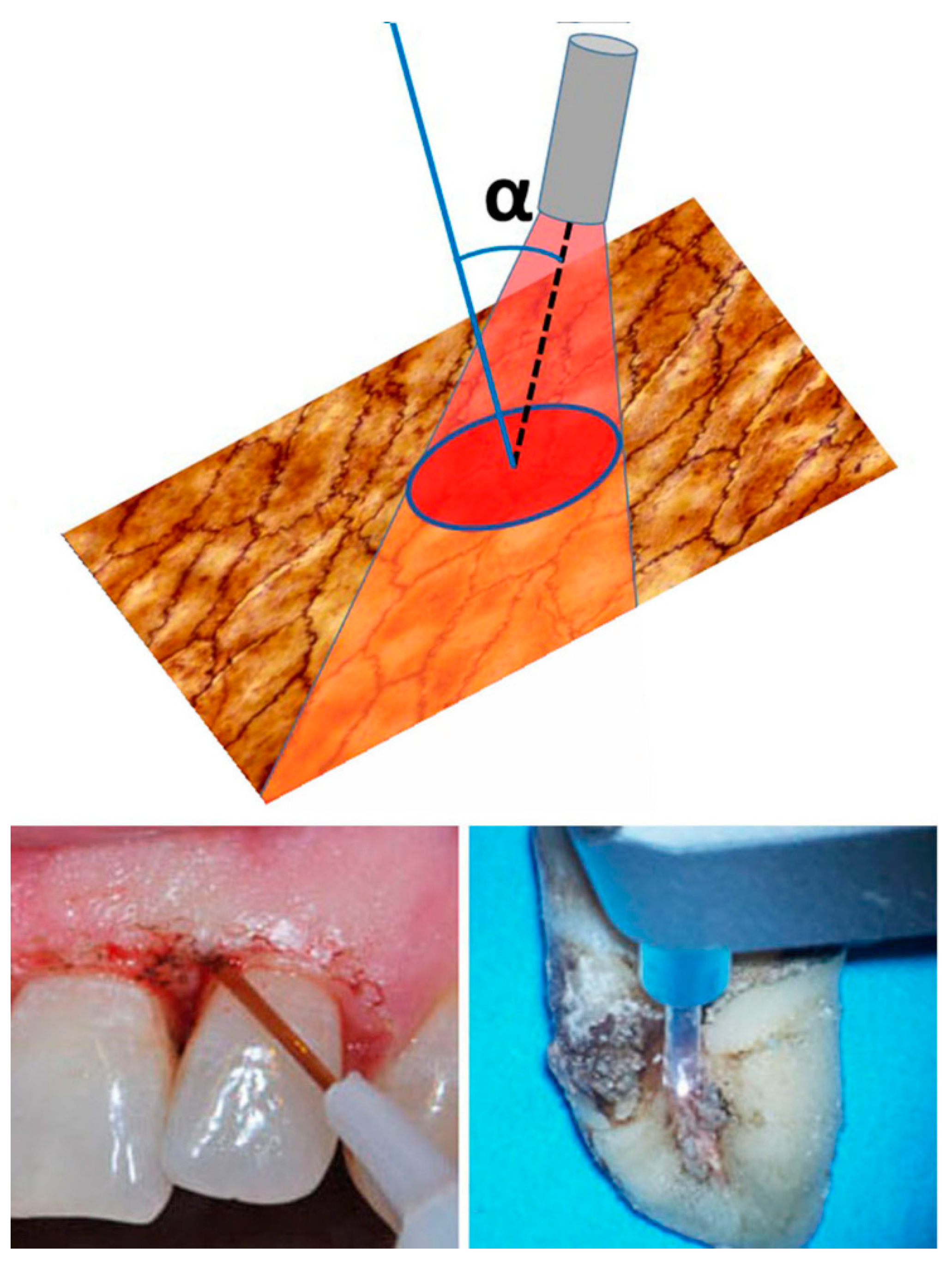
| Beam inclination (degree) | 20 | 15–20 | 15–20 | |||||
| Power density (W/cm2) | 1193.7 | 353.4 | 1193.7–353.4 | |||||
| Total energy density (fluence) (J/cm2) | 15 | 66.7 | 66.7 | |||||
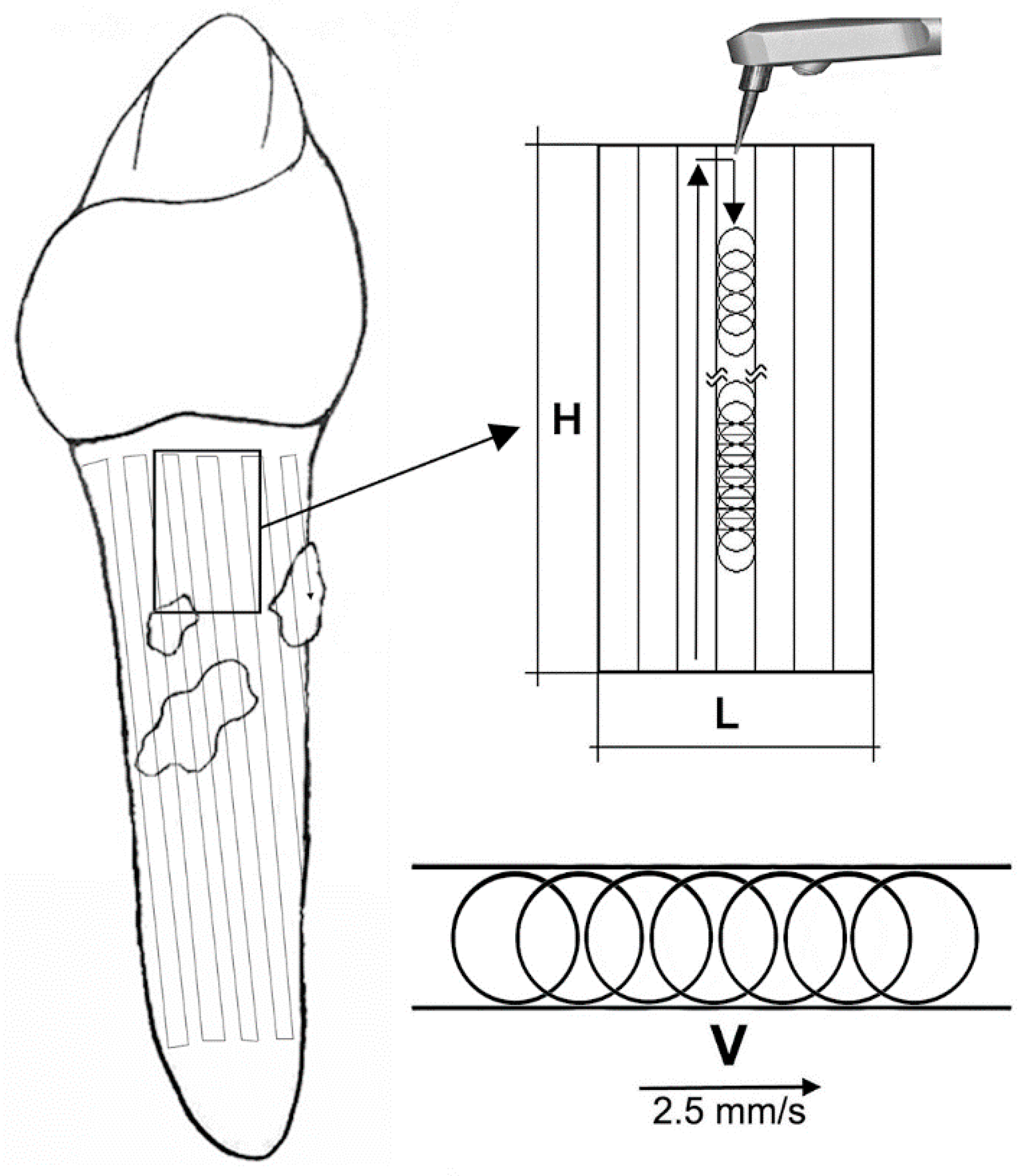
| Laser tip movement, - time per area (s/cm2) - linear speed (mm/s) | vertical and horizontal lines | vertical and horizontal lines | vertical and horizontal lines | vertical and horizontal lines | vertical and horizontal lines 20 s/cm2 | vertical and horizontal lines 2.5 mm/s | vertical and horizontal lines 2.5 mm/s | vertical and horizontal lines 2.5 mm/s |
| Treatment details | ||||||||
| Treatment time/tooth (s) | adjusted depending on the pocket’s surface area | 60 | 20 | 20 | 20 | 20 | adjusted depending on the pocket’s surface area | adjusted depending on the pocket’s surface area |
| No./Frequency of treatments | 1 | 1 | 2 | 3 | 1 | 1 | 1 | 1–3 |
| Cooling system | no | no | no | no | no | no | airflow | airflow |
| External power meter check | no | no | yes | no | no | no | yes | yes |
| Source (reference No.) | [93] | [94] | [95] | [86] | [96] | [55] | [97] | Overall suggestions |
| Laser emission settings | ||||||||
| λ (nm) | 670 | 670 | 660 | 670 | 660 | 635 | 660 | 635/660 |
| Wave emission mode (continuous/pulsed) | continuous | continuous | continuous | continuous | continuous | continuous | continuous | continuous |
| Applicator characteristics | ||||||||
| Tip type/diameter (mm) | optic fiber | optic fiber | optic fiber 0.6 | optic fiber 0.6 | optic fiber 0.6 | focalized zoom | focalized zoom/ optic fiber 0.6 | |
| Fluence at tip level (J/cm2) | 10–20 | 129 | 14.9 | 129 | 21 | 21–129 | ||
| Power output (mW) | 150 | 60 | 75 | 60 | 100 | 100 | 60–100 | |
| Power density (W/cm2) | 0.075 | 21.4 | 1.2 | 21.4 | 0.35 | 0.25 | 0.35–21.4 | |
| Laser application details | ||||||||
| Application mode | transgingival | subgingival | subgingival | subgingival | subgingival | transgingival | transgingival | transgingival |
| Distance (mm) | 7 | 0 | 0 | 0 | 30 | 30 | ||
| spot diameter(mm)/area (mm2) | 90 | 6/28,3 | ND | |||||
| Beam inclination (degree) | 90 | 90 | ||||||
| Energy density (J/cm2) | 2.4 | 21 | 75 | 21–75 | ||||
| Treatment surface (cm2) | 128 | 0.4 | ND | |||||
| Photosensitizer | ||||||||
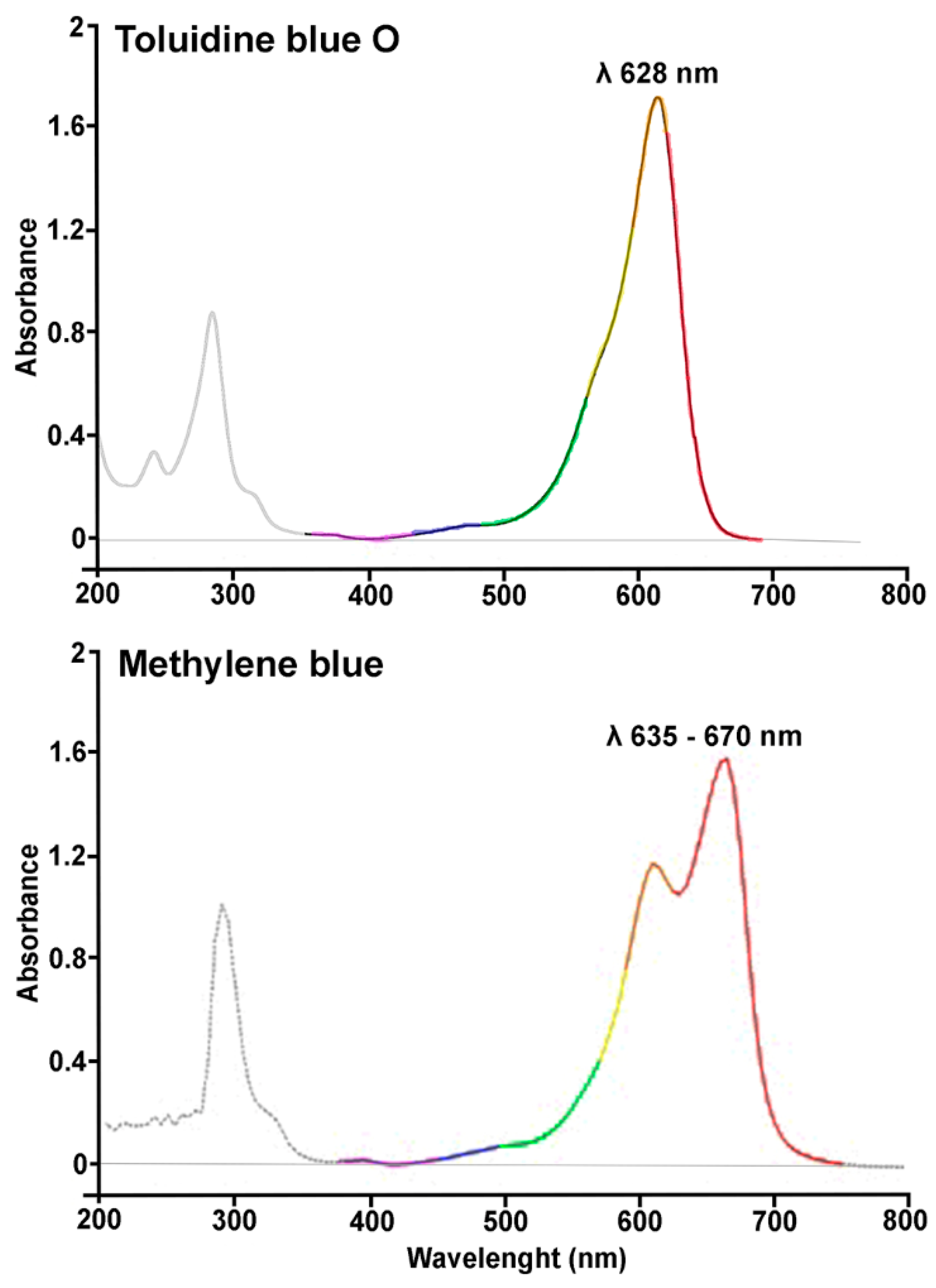
| Metyhylene blue, MB Toluidine blue, TB | MB | MB | MB | MB | MB | TB | MB | MB/TB |
| Solvent | PeriowaveTM solution | HelboTM solution | water | HelboTM solution | water | water | Na dodecyl-sulfate in water | water |
| Rinsing time pre-irradiation (min) | 3 | 1 | 1 | 1 | 1 | 1-3 | ||
| Concentration (mg/mL) | 0.005 | 10 * | 10 * | 10 * | 10 * | 0.001 | 0.003 | 0.001–0.003 without rinsing |
| Rinsing after application | yes | yes | yes | yes | no | no | NO | |
| Treatment details | ||||||||
| Treatment time per tooth (s) | 60 | 60 | 60 | 60 | 60 | 300 | 300 | adjusted depending on the area of each pocket |
| Treatment repetitions | 1 | 4 | 1 | 4 | 1 | 7–10 | 1 | 4–10 adjusted depending on healing markers ** |
| External power meter check | no | no | no | no | no | yes | no | yes |
| Photoablative Diode Laser | Phototherapy LED | PDT Diode Laser | |
|---|---|---|---|
| Laser emission settings | |||
| λ (nm) | 810 | 405 | 635 Toluidine blue (1 μg/mL) |
| Wave emission mode | continuous | continuous | continuous |
| Beam power (W) | 1 | 1 | 0.1 |
| Laser application details | |||
| Handpiece Diameter (mm) | Optic fiber 0.6 | Adjustable focus lens | Crystal lightpipe 10 |
| Application mode (contact/non-contact) | contact | non-contact | non-contact |
| Distance (mm) | 0 | 10 | 30 |
| Light spot size (mm2) | 0.28 | 95 | 28.3 |
| Power density (W/cm2) | 353.4 | 1.05 | 0.35 |
| Total energy density (fluence) (J/cm2) | 66.7 | 63 | 21 |
| Tip movement speed (mm/s) | 2.5 | ||
| Treatment details | |||
| No. of treatment | 1 | 1 | 4–10 adjusted depending on healing markers * |
| Cooling system | airflow | ||
© 2019 by the authors. Licensee MDPI, Basel, Switzerland. This article is an open access article distributed under the terms and conditions of the Creative Commons Attribution (CC BY) license (http://creativecommons.org/licenses/by/4.0/).
Share and Cite
Giannelli, M.; Lasagni, M.; Bani, D. Photonic Therapy in Periodontal Diseases an Overview with Appraisal of the Literature and Reasoned Treatment Recommendations. Int. J. Mol. Sci. 2019, 20, 4741. https://doi.org/10.3390/ijms20194741
Giannelli M, Lasagni M, Bani D. Photonic Therapy in Periodontal Diseases an Overview with Appraisal of the Literature and Reasoned Treatment Recommendations. International Journal of Molecular Sciences. 2019; 20(19):4741. https://doi.org/10.3390/ijms20194741
Chicago/Turabian StyleGiannelli, Marco, Massimo Lasagni, and Daniele Bani. 2019. "Photonic Therapy in Periodontal Diseases an Overview with Appraisal of the Literature and Reasoned Treatment Recommendations" International Journal of Molecular Sciences 20, no. 19: 4741. https://doi.org/10.3390/ijms20194741