Next Issue
Volume 20, August-1
Previous Issue
Volume 20, July-1
 
 
ijms-logo

Journal Browser

Journal Browser

Int. J. Mol. Sci., Volume 20, Issue 14 (July-2 2019) – 232 articles

Cover Story (view full-size image): 
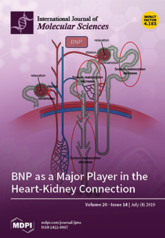
Although it is known that brain natriuretic peptide (BNP) levels are relatively higher in patients with chronic kidney disease, the mechanism remains unknown. Here, we review the functions and roles of BNP in heart–kidney interaction. In addition, we discuss the relevant molecular mechanisms that suggest BNP is protective against chronic kidney diseases and heart failure, especially in terms of the counterparts of the renin–angiotensin–aldosterone system. A better understanding of these processes will help to accelerate pharmacological treatments for heart–kidney disease. View this paper.
  • Issues are regarded as officially published after their release is announced to the table of contents alert mailing list.
  • You may sign up for e-mail alerts to receive table of contents of newly released issues.
  • PDF is the official format for papers published in both, html and pdf forms. To view the papers in pdf format, click on the "PDF Full-text" link, and use the free Adobe Reader to open them.
Order results
Result details
Section
Select all
Export citation of selected articles as:
15 pages, 4362 KiB  
Article
Infusing Mesenchymal Stromal Cells into Porcine Kidneys during Normothermic Machine Perfusion: Intact MSCs Can Be Traced and Localised to Glomeruli
by Merel Pool, Tim Eertman, Jesus Sierra Parraga, Nils ’t Hart, Marieke Roemeling-van Rhijn, Marco Eijken, Bente Jespersen, Marlies Reinders, Martin Hoogduijn, Rutger Ploeg, Henri Leuvenink and Cyril Moers
Int. J. Mol. Sci. 2019, 20(14), 3607; https://doi.org/10.3390/ijms20143607 - 23 Jul 2019
Cited by 46 | Viewed by 4794
Abstract
Normothermic machine perfusion (NMP) of kidneys offers the opportunity to perform active interventions, such as the addition of mesenchymal stromal cells (MSCs), to an isolated organ prior to transplantation. The purpose of this study was to determine whether administering MSCs to kidneys during [...] Read more.
Normothermic machine perfusion (NMP) of kidneys offers the opportunity to perform active interventions, such as the addition of mesenchymal stromal cells (MSCs), to an isolated organ prior to transplantation. The purpose of this study was to determine whether administering MSCs to kidneys during NMP is feasible, what the effect of NMP is on MSCs and whether intact MSCs are retained in the kidney and to which structures they home. Viable porcine kidneys were obtained from a slaughterhouse. Kidneys were machine perfused during 7 h at 37 °C. After 1 h of perfusion either 0, 105, 106 or 107 human adipose tissue derived MSCs were added. Additional ex vivo perfusions were conducted with fluorescent pre-labelled bone-marrow derived MSCs to assess localisation and survival of MSCs during NMP. After NMP, intact MSCs were detected by immunohistochemistry in the lumen of glomerular capillaries, but only in the 107 MSC group. The experiments with fluorescent pre-labelled MSCs showed that only a minority of glomeruli were positive for infused MSCs and most of these glomeruli contained multiple MSCs. Flow cytometry showed that the number of infused MSCs in the perfusion circuit steeply declined during NMP to approximately 10%. In conclusion, the number of circulating MSCs in the perfusate decreases rapidly in time and after NMP only a small portion of the MSCs are intact and these appear to be clustered in a minority of glomeruli. Full article
Show Figures

Figure 1

14 pages, 589 KiB  
Review
Mutations in Prion Protein Gene: Pathogenic Mechanisms in C-Terminal vs. N-Terminal Domain, a Review
by Livia Bernardi and Amalia C. Bruni
Int. J. Mol. Sci. 2019, 20(14), 3606; https://doi.org/10.3390/ijms20143606 - 23 Jul 2019
Cited by 16 | Viewed by 5896
Abstract
Inherited mutations in the Prion protein (PrP), encoded by the PRNP gene, have been associated with autosomal dominant neurodegenerative disorders, such as Creutzfeldt–Jacob disease (CJD), Gerstmann–Sträussler–Scheinker syndrome (GSS), and Fatal Familial Insomnia (FFI). Notably, PRNP mutations have also been described in clinical pictures [...] Read more.
Inherited mutations in the Prion protein (PrP), encoded by the PRNP gene, have been associated with autosomal dominant neurodegenerative disorders, such as Creutzfeldt–Jacob disease (CJD), Gerstmann–Sträussler–Scheinker syndrome (GSS), and Fatal Familial Insomnia (FFI). Notably, PRNP mutations have also been described in clinical pictures resembling other neurodegenerative diseases, such as frontotemporal dementia. Regarding the pathogenesis, it has been observed that these point mutations are located in the C-terminal region of the PRNP gene and, currently, the potential significance of the N-terminal domain has largely been underestimated. The purpose of this report is to review and provide current insights into the pathogenic mechanisms of PRNP mutations, emphasizing the differences between the C- and N-terminal regions and focusing, in particular, on the lesser-known flexible N-terminal, for which recent biophysical evidence has revealed a physical interaction with the globular C-terminal domain of the cellular prion protein (PrPC). Full article
(This article belongs to the Special Issue Protein Domain Evolution and Involvement in Diseases)
Show Figures

Figure 1

16 pages, 3273 KiB  
Article
Resveratrol Plays a Protective Role against Premature Ovarian Failure and Prompts Female Germline Stem Cell Survival
by Yu Jiang, Zhaoyuan Zhang, Lijun Cha, Lili Li, Dantian Zhu, Zhi Fang, Zhiqiang He, Jian Huang and Zezheng Pan
Int. J. Mol. Sci. 2019, 20(14), 3605; https://doi.org/10.3390/ijms20143605 - 23 Jul 2019
Cited by 43 | Viewed by 5993
Abstract
This study was designed to investigate the protective effect of resveratrol (RES) on premature ovarian failure (POF) and the proliferation of female germline stem cells (FGSCs) at the tissue and cell levels. POF mice were lavaged with RES, and POF ovaries were co-cultured [...] Read more.
This study was designed to investigate the protective effect of resveratrol (RES) on premature ovarian failure (POF) and the proliferation of female germline stem cells (FGSCs) at the tissue and cell levels. POF mice were lavaged with RES, and POF ovaries were co-cultured with RES and/or GANT61 in vitro. FGSCs were pretreated with Busulfan and RES and/or GANT61 and co-cultured with M1 macrophages, which were pretreated with RES. The weights of mice and their ovaries, as well as their follicle number, were measured. Ovarian function, antioxidative stress, inflammation, and FGSCs survival were evaluated. RES significantly increased the weights of POF mice and their ovaries as well as the number of follicles, while it decreased the atresia rate of follicles. Higher levels of Mvh, Oct4, SOD2, GPx, and CAT were detected after treatment with RES in vivo and in vitro. RES treatment resulted in significantly lower TNF-α and IL-6 concentrations and an obviously higher IL-10 concentration in the ovaries. In FGSCs, higher Mvh, Oct4, and SOD2 concentrations and lower TNF-α, IL-6, and MDA concentrations were measured in the RES group. Blockage of the Hh signaling pathway reversed the protective effect of RES on FGSCs. In conclusion, RES effectively improved the ovarian function of the POF model and the productive capacity of FGSCs via relieving oxidative stress and inflammation and a mechanism involving the Hh signaling pathway, suggesting that RES is a potential agent against POF and can aid in the survival of FGSCs. Full article
(This article belongs to the Section Bioactives and Nutraceuticals)
Show Figures

Graphical abstract

12 pages, 3710 KiB  
Article
Intragenic Antimicrobial Peptide Hs02 Hampers the Proliferation of Single- and Dual-Species Biofilms of P. aeruginosa and S. aureus: A Promising Agent for Mitigation of Biofilm-Associated Infections
by Lucinda J. Bessa, Julia R. Manickchand, Peter Eaton, José Roberto S. A. Leite, Guilherme D. Brand and Paula Gameiro
Int. J. Mol. Sci. 2019, 20(14), 3604; https://doi.org/10.3390/ijms20143604 - 23 Jul 2019
Cited by 17 | Viewed by 4278
Abstract
Pseudomonas aeruginosa and Staphylococcus aureus are two major pathogens involved in a large variety of infections. Their co-occurrence in the same site of infection has been frequently reported and is linked to enhanced virulence and difficulty of treatment. Herein, the antimicrobial and antibiofilm [...] Read more.
Pseudomonas aeruginosa and Staphylococcus aureus are two major pathogens involved in a large variety of infections. Their co-occurrence in the same site of infection has been frequently reported and is linked to enhanced virulence and difficulty of treatment. Herein, the antimicrobial and antibiofilm activities of an intragenic antimicrobial peptide (IAP), named Hs02, which was uncovered from the human unconventional myosin 1H protein, were investigated against several P. aeruginosa and S. aureus strains, including multidrug-resistant (MDR) isolates. The antibiofilm activity was evaluated on single- and dual-species biofilms of P. aeruginosa and S. aureus. Moreover, the effect of peptide Hs02 on the membrane fluidity of the strains was assessed through Laurdan generalized polarization (GP). Minimum inhibitory concentration (MIC) values of peptide Hs02 ranged from 2 to 16 μg/mL against all strains and MDR isolates. Though Hs02 was not able to hamper biofilm formation by some strains at sub-MIC values, it clearly affected 24 h preformed biofilms, especially by reducing the viability of the bacterial cells within the single- and dual-species biofilms, as shown by confocal laser scanning microscopy (CLSM) and atomic force microscopy (AFM) images. Laurdan GP values showed that Hs02 induces membrane rigidification in both P. aeruginosa and S. aureus. Peptide Hs02 can potentially be a lead for further improvement as an antibiofilm agent. Full article
(This article belongs to the Special Issue Peptides for Health Benefits 2019)
Show Figures

Figure 1

14 pages, 22087 KiB  
Article
Carbon Nanomaterials and LED Irradiation as Antibacterial Strategies against Gram-Positive Multidrug-Resistant Pathogens
by Lisa Elias, Rafael Taengua, Belén Frígols, Beatriz Salesa and Ángel Serrano-Aroca
Int. J. Mol. Sci. 2019, 20(14), 3603; https://doi.org/10.3390/ijms20143603 - 23 Jul 2019
Cited by 31 | Viewed by 4123
Abstract
Background: Due to current antibiotic resistance worldwide, there is an urgent need to find new alternative antibacterial approaches capable of dealing with multidrug-resistant pathogens. Most recent studies have demonstrated the antibacterial activity and non-cytotoxicity of carbon nanomaterials such as graphene oxide (GO) and [...] Read more.
Background: Due to current antibiotic resistance worldwide, there is an urgent need to find new alternative antibacterial approaches capable of dealing with multidrug-resistant pathogens. Most recent studies have demonstrated the antibacterial activity and non-cytotoxicity of carbon nanomaterials such as graphene oxide (GO) and carbon nanofibers (CNFs). On the other hand, light-emitting diodes (LEDs) have shown great potential in a wide range of biomedical applications. Methods: We investigated a nanotechnological strategy consisting of GO or CNFs combined with light-emitting diod (LED) irradiation as novel nanoweapons against two clinically relevant Gram-positive multidrug-resistant pathogens: methicillin-resistant Staphylococcus aureus (MRSA) and methicillin-resistant Staphylococcus epidermidis (MRSE). The cytotoxicity of GO and CNFs was studied in the presence of human keratinocyte HaCaT cells. Results: GO or CNFs exhibited no cytotoxicity and high antibacterial activity in direct contact with MRSE and MRSA cells. Furthermore, when GO or CNFs were illuminated with LED light, the MRSE and MRSA cells lost viability. The rate of decrease in colony forming units from 0 to 3 h, measured per mL, increased to 98.5 ± 1.6% and 95.8 ± 1.4% for GO and 99.5 ± 0.6% and 99.7 ± 0.2% for CNFs. Conclusions: This combined antimicrobial approach opens up many biomedical research opportunities and provides an enhanced strategy for the prevention and treatment of Gram-positive multidrug-resistant infections. Full article
(This article belongs to the Special Issue Graphene-Based Materials: Biological and Biomedical Applications)
Show Figures

Graphical abstract

21 pages, 1353 KiB  
Review
The Role of Cysteine Cathepsins in Cancer Progression and Drug Resistance
by Magdalena Rudzińska, Alessandro Parodi, Surinder M. Soond, Andrey Z. Vinarov, Dmitry O. Korolev, Andrey O. Morozov, Cenk Daglioglu, Yusuf Tutar and Andrey A. Zamyatnin, Jr.
Int. J. Mol. Sci. 2019, 20(14), 3602; https://doi.org/10.3390/ijms20143602 - 23 Jul 2019
Cited by 79 | Viewed by 7453
Abstract
Cysteine cathepsins are lysosomal enzymes belonging to the papain family. Their expression is misregulated in a wide variety of tumors, and ample data prove their involvement in cancer progression, angiogenesis, metastasis, and in the occurrence of drug resistance. However, while their overexpression is [...] Read more.
Cysteine cathepsins are lysosomal enzymes belonging to the papain family. Their expression is misregulated in a wide variety of tumors, and ample data prove their involvement in cancer progression, angiogenesis, metastasis, and in the occurrence of drug resistance. However, while their overexpression is usually associated with highly aggressive tumor phenotypes, their mechanistic role in cancer progression is still to be determined to develop new therapeutic strategies. In this review, we highlight the literature related to the role of the cysteine cathepsins in cancer biology, with particular emphasis on their input into tumor biology. Full article
Show Figures

Figure 1

20 pages, 6319 KiB  
Article
Apoptosis Induction and Alteration of Cell Adherence in Human Lung Cancer Cells under Simulated Microgravity
by Carlo Dietz, Manfred Infanger, Alexander Romswinkel, Florian Strube and Armin Kraus
Int. J. Mol. Sci. 2019, 20(14), 3601; https://doi.org/10.3390/ijms20143601 - 23 Jul 2019
Cited by 32 | Viewed by 5144
Abstract
Background: Lung cancer cells are known to change proliferation and migration under simulated microgravity. In this study, we sought to evaluate cell adherence, apoptosis, cytoskeleton arrangement, and gene expression under simulated microgravity. Methods: Human lung cancer cells were exposed to simulated microgravity in [...] Read more.
Background: Lung cancer cells are known to change proliferation and migration under simulated microgravity. In this study, we sought to evaluate cell adherence, apoptosis, cytoskeleton arrangement, and gene expression under simulated microgravity. Methods: Human lung cancer cells were exposed to simulated microgravity in a random-positioning machine (RPM). Cell morphology and adherence were observed under phase-contrast microscopy, cytoskeleton staining was performed, apoptosis rate was determined, and changes in gene and protein expression were detected by real-time PCR with western blot confirmation. Results: Three-dimensional (3D)-spheroid formation was observed under simulated microgravity. Cell viability was not impaired. Actin filaments showed a shift in alignment from longitudinal to spherical. Apoptosis rate was significantly increased in the spheroids compared to the control. TP53, CDKN2A, PTEN, and RB1 gene expression was significantly upregulated in the adherent cells under simulated microgravity with an increase in corresponding protein production for p14 and RB1. SOX2 expression was significantly upregulated in the adherent cells, but protein was not. Gene expressions of AKT3, PIK3CA, and NFE2L2 remained unaltered. Conclusion: Simulated microgravity induces alteration in cell adherence, increases apoptosis rate, and leads to upregulation of tumor suppressor genes in human lung cancer cells. Full article
(This article belongs to the Special Issue Microgravity and Cell Adherence)
Show Figures

Figure 1

11 pages, 1903 KiB  
Article
Revealing Adenosine A2A-Dopamine D2 Receptor Heteromers in Parkinson’s Disease Post-Mortem Brain through a New AlphaScreen-Based Assay
by Víctor Fernández-Dueñas, Maricel Gómez-Soler, Marta Valle-León, Masahiko Watanabe, Isidre Ferrer and Francisco Ciruela
Int. J. Mol. Sci. 2019, 20(14), 3600; https://doi.org/10.3390/ijms20143600 - 23 Jul 2019
Cited by 32 | Viewed by 5462
Abstract
Background: Several biophysical techniques have been successfully implemented to detect G protein-coupled receptors (GPCRs) heteromerization. Although these approaches have made it possible to ascertain the presence of GPCR heteromers in animal models of disease, no success has been accomplished in pathological human [...] Read more.
Background: Several biophysical techniques have been successfully implemented to detect G protein-coupled receptors (GPCRs) heteromerization. Although these approaches have made it possible to ascertain the presence of GPCR heteromers in animal models of disease, no success has been accomplished in pathological human post-mortem brains. The AlphaScreen technology has been consistently used to quantify small analyte accumulation or depletion, bimolecular interactions, and post-translational modifications. The high signal-to-background, dynamic range and sensitivity exhibited by this technology support that it may be suitable to detect GPCR heteromers even under non-optimal conditions. Methods: Here, we describe the development of a new AlphaScreen assay to detect GPCR oligomers in human post-mortem brain. Results: Adenosine A2A-dopamine D2 receptor (A2AR/D2R) heteromer formation was monitored in caudate from healthy and Parkinson’s disease (PD) subjects. The approach was first validated using striatal membranes from wild type and A2AR deficient mice. Secondly, we took advantage of the 6-hydroxydopamine hemiparkinsonian rat model to validate previous results. In addition, finally, A2AR/D2R heteromer formation was assessed in caudate membranes from human post-mortem brains. Importantly, our preliminary results revealed an increase in A2AR/D2R heteromer formation in PD brains. Conclusions: The new AlphaScreen assay allowed assessing GPCR heteromers in human post-mortem brains with high sensitivity. Full article
(This article belongs to the Special Issue G Protein-Coupled Adenosine Receptors: Molecular Aspects and Beyond)
Show Figures

Figure 1

20 pages, 1911 KiB  
Article
Dissecting Adaptation Mechanisms to Contrasting Solar Irradiance in the Mediterranean Shrub Cistus incanus
by Federico Sebastiani, Sara Torre, Antonella Gori, Cecilia Brunetti, Mauro Centritto, Francesco Ferrini and Massimiliano Tattini
Int. J. Mol. Sci. 2019, 20(14), 3599; https://doi.org/10.3390/ijms20143599 - 23 Jul 2019
Cited by 6 | Viewed by 3576
Abstract
Molecular mechanisms that are the base of the strategies adopted by Mediterranean plants to cope with the challenges imposed by limited or excessive solar radiation during the summer season have received limited attention. In our study, conducted on C. incanus plants growing in [...] Read more.
Molecular mechanisms that are the base of the strategies adopted by Mediterranean plants to cope with the challenges imposed by limited or excessive solar radiation during the summer season have received limited attention. In our study, conducted on C. incanus plants growing in the shade or in full sunlight, we performed measurements of relevant physiological traits, such as leaf water potential, gas exchange and PSII photochemistry, RNA-Seq with de-novo assembly, and the analysis of differentially expressed genes. We also identified and quantified photosynthetic pigments, abscisic acid, and flavonoids. Here, we show major mechanisms regulating light perception and signaling which, in turn, sustain the shade avoidance syndrome displayed by the ‘sun loving’ C. incanus. We offer clear evidence of the detrimental effects of excessive light on both the assembly and the stability of PSII, and the activation of a suite of both repair and effective antioxidant mechanisms in sun-adapted leaves. For instance, our study supports the view of major antioxidant functions of zeaxanthin in sunny plants concomitantly challenged by severe drought stress. Finally, our study confirms the multiple functions served by flavonoids, both flavonols and flavanols, in the adaptive mechanisms of plants to the environmental pressures associated to Mediterranean climate. Full article
(This article belongs to the Collection Feature Papers in Molecular Plant Sciences)
Show Figures

Figure 1

21 pages, 1761 KiB  
Review
Cell Death in the Kidney
by Giovanna Priante, Lisa Gianesello, Monica Ceol, Dorella Del Prete and Franca Anglani
Int. J. Mol. Sci. 2019, 20(14), 3598; https://doi.org/10.3390/ijms20143598 - 23 Jul 2019
Cited by 109 | Viewed by 12029
Abstract
Apoptotic cell death is usually a response to the cell’s microenvironment. In the kidney, apoptosis contributes to parenchymal cell loss in the course of acute and chronic renal injury, but does not trigger an inflammatory response. What distinguishes necrosis from apoptosis is the [...] Read more.
Apoptotic cell death is usually a response to the cell’s microenvironment. In the kidney, apoptosis contributes to parenchymal cell loss in the course of acute and chronic renal injury, but does not trigger an inflammatory response. What distinguishes necrosis from apoptosis is the rupture of the plasma membrane, so necrotic cell death is accompanied by the release of unprocessed intracellular content, including cellular organelles, which are highly immunogenic proteins. The relative contribution of apoptosis and necrosis to injury varies, depending on the severity of the insult. Regulated cell death may result from immunologically silent apoptosis or from immunogenic necrosis. Recent advances have enhanced the most revolutionary concept of regulated necrosis. Several modalities of regulated necrosis have been described, such as necroptosis, ferroptosis, pyroptosis, and mitochondrial permeability transition-dependent regulated necrosis. We review the different modalities of apoptosis, necrosis, and regulated necrosis in kidney injury, focusing particularly on evidence implicating cell death in ectopic renal calcification. We also review the evidence for the role of cell death in kidney injury, which may pave the way for new therapeutic opportunities. Full article
(This article belongs to the Special Issue Kidney Inflammation, Injury and Regeneration)
Show Figures

Figure 1

14 pages, 1076 KiB  
Review
Transposable Elements Adaptive Role in Genome Plasticity, Pathogenicity and Evolution in Fungal Phytopathogens
by Nurhani Mat Razali, Boon Huat Cheah and Kalaivani Nadarajah
Int. J. Mol. Sci. 2019, 20(14), 3597; https://doi.org/10.3390/ijms20143597 - 23 Jul 2019
Cited by 35 | Viewed by 8691
Abstract
Transposable elements (TEs) are agents of genetic variability in phytopathogens as they are a source of adaptive evolution through genome diversification. Although many studies have uncovered information on TEs, the exact mechanism behind TE-induced changes within the genome remains poorly understood. Furthermore, convergent [...] Read more.
Transposable elements (TEs) are agents of genetic variability in phytopathogens as they are a source of adaptive evolution through genome diversification. Although many studies have uncovered information on TEs, the exact mechanism behind TE-induced changes within the genome remains poorly understood. Furthermore, convergent trends towards bigger genomes, emergence of novel genes and gain or loss of genes implicate a TE-regulated genome plasticity of fungal phytopathogens. TEs are able to alter gene expression by revamping the cis-regulatory elements or recruiting epigenetic control. Recent findings show that TEs recruit epigenetic control on the expression of effector genes as part of the coordinated infection strategy. In addition to genome plasticity and diversity, fungal pathogenicity is an area of economic concern. A survey of TE distribution suggests that their proximity to pathogenicity genes TEs may act as sites for emergence of novel pathogenicity factors via nucleotide changes and expansion or reduction of the gene family. Through a systematic survey of literature, we were able to conclude that the role of TEs in fungi is wide: ranging from genome plasticity, pathogenicity to adaptive behavior in evolution. This review also identifies the gaps in knowledge that requires further elucidation for a better understanding of TEs’ contribution to genome architecture and versatility. Full article
(This article belongs to the Special Issue Transposable Elements)
Show Figures

Graphical abstract

16 pages, 20650 KiB  
Article
Loss of Wwox Causes Defective Development of Cerebral Cortex with Hypomyelination in a Rat Model of Lethal Dwarfism with Epilepsy
by Yuki Tochigi, Yutaka Takamatsu, Jun Nakane, Rika Nakai, Kentaro Katayama and Hiroetsu Suzuki
Int. J. Mol. Sci. 2019, 20(14), 3596; https://doi.org/10.3390/ijms20143596 - 23 Jul 2019
Cited by 15 | Viewed by 4515
Abstract
WW domain-containing oxidoreductase (Wwox) is a putative tumor suppressor. Several germline mutations of Wwox have been associated with infant neurological disorders characterized by epilepsy, growth retardation, and early death. Less is known, however, about the pathological link between Wwox mutations and these disorders [...] Read more.
WW domain-containing oxidoreductase (Wwox) is a putative tumor suppressor. Several germline mutations of Wwox have been associated with infant neurological disorders characterized by epilepsy, growth retardation, and early death. Less is known, however, about the pathological link between Wwox mutations and these disorders or the physiological role of Wwox in brain development. In this study, we examined age-related expression and histological localization of Wwox in forebrains as well as the effects of loss of function mutations in the Wwox gene in the immature cortex of a rat model of lethal dwarfism with epilepsy (lde/lde). Immunostaining revealed that Wwox is expressed in neurons, astrocytes, and oligodendrocytes. lde/lde cortices were characterized by a reduction in neurite growth without a reduced number of neurons, severe reduction in myelination with a reduced number of mature oligodendrocytes, and a reduction in cell populations of astrocytes and microglia. These results indicate that Wwox is essential for normal development of neurons and glial cells in the cerebral cortex. Full article
(This article belongs to the Section Molecular Pathology, Diagnostics, and Therapeutics)
Show Figures

Graphical abstract

14 pages, 3730 KiB  
Article
Dielectrophoresis of Amyloid-Beta Proteins as a Microfluidic Template for Alzheimer’s Research
by Salman Ali Al-Ahdal, Aminuddin Bin Ahmad Kayani, Mohd Anuar Md Ali, Jun Yuan Chan, Talal Ali, Norah Adnan, Muhamad Ramdzan Buyong, Ervina Efzan Mhd Noor, Burhanuddin Yeop Majlis and Sharath Sriram
Int. J. Mol. Sci. 2019, 20(14), 3595; https://doi.org/10.3390/ijms20143595 - 23 Jul 2019
Cited by 6 | Viewed by 4184
Abstract
We employed dielectrophoresis to a yeast cell suspension containing amyloid-beta proteins (Aβ) in a microfluidic environment. The Aβ was separated from the cells and characterized using the gradual dissolution of Aβ as a function of the applied dielectrophoretic parameters. We established the gradual [...] Read more.
We employed dielectrophoresis to a yeast cell suspension containing amyloid-beta proteins (Aβ) in a microfluidic environment. The Aβ was separated from the cells and characterized using the gradual dissolution of Aβ as a function of the applied dielectrophoretic parameters. We established the gradual dissolution of Aβ under specific dielectrophoretic parameters. Further, Aβ in the fibril form at the tip of the electrode dissolved at high frequency. This was perhaps due to the conductivity of the suspending medium changing according to the frequency, which resulted in a higher temperature at the tips of the electrodes, and consequently in the breakdown of the hydrogen bonds. However, those shaped as spheroidal monomers experienced a delay in the Aβ fibril transformation process. Yeast cells exposed to relatively low temperatures at the base of the electrode did not experience a positive or negative change in viability. The DEP microfluidic platform incorporating the integrated microtip electrode array was able to selectively manipulate the yeast cells and dissolve the Aβ to a controlled extent. We demonstrate suitable dielectrophoretic parameters to induce such manipulation, which is highly relevant for Aβ-related colloidal microfluidic research and could be applied to Alzheimer’s research in the future. Full article
(This article belongs to the Section Molecular Biophysics)
Show Figures

Figure 1

15 pages, 3435 KiB  
Article
Acute Cytotoxic Effects on Morphology and Mechanical Behavior in MCF-7 Induced by TiO2NPs Exposure
by Mariafrancesca Cascione, Valeria De Matteis, Giacomo Mandriota, Stefano Leporatti and Rosaria Rinaldi
Int. J. Mol. Sci. 2019, 20(14), 3594; https://doi.org/10.3390/ijms20143594 - 23 Jul 2019
Cited by 7 | Viewed by 3504
Abstract
The side effects induced by nanoparticle exposure at a cellular level are one of the priority research topics due to the steady increase in the use of nanoparticles (NPs). Recently, the focus on cellular morphology and mechanical behavior is gaining relevance in order [...] Read more.
The side effects induced by nanoparticle exposure at a cellular level are one of the priority research topics due to the steady increase in the use of nanoparticles (NPs). Recently, the focus on cellular morphology and mechanical behavior is gaining relevance in order to fully understand the cytotoxic mechanisms. In this regard, we have evaluated the morphomechanical alteration in human breast adenocarcinoma cell line (MCF-7) exposed to TiO2NPs at two different concentrations (25 and 50 µg/mL) and two time points (24 and 48 h). By using confocal and atomic force microscopy, we demonstrated that TiO2NP exposure induces significant alterations in cellular membrane elasticity, due to actin proteins rearrangement in cytoskeleton, as calculated in correspondence to nuclear and cytoplasmic compartments. In this work, we have emphasized the alteration in mechanical properties of the cellular membrane, induced by nanoparticle exposure. Full article
(This article belongs to the Special Issue Biomechanics of Cell Membrane)
Show Figures

Figure 1

22 pages, 2809 KiB  
Article
Partial Fluxes of Phosphoric Acid Anions through Anion-Exchange Membranes in the Course of NaH2PO4 Solution Electrodialysis
by Olesya Rybalkina, Kseniya Tsygurina, Ekaterina Melnikova, Semyon Mareev, Ilya Moroz, Victor Nikonenko and Natalia Pismenskaya
Int. J. Mol. Sci. 2019, 20(14), 3593; https://doi.org/10.3390/ijms20143593 - 23 Jul 2019
Cited by 32 | Viewed by 4399
Abstract
Electrodialysis (ED) with ion-exchange membranes is a promising method for the extraction of phosphates from municipal and other wastewater in order to obtain cheap mineral fertilizers. Phosphorus is transported through an anion-exchange membrane (AEM) by anions of phosphoric acid. However, which phosphoric acid [...] Read more.
Electrodialysis (ED) with ion-exchange membranes is a promising method for the extraction of phosphates from municipal and other wastewater in order to obtain cheap mineral fertilizers. Phosphorus is transported through an anion-exchange membrane (AEM) by anions of phosphoric acid. However, which phosphoric acid anions carry the phosphorus in the membrane and the boundary solution, that is, the mechanism of phosphorus transport, is not yet clear. Some authors report an unexpectedly low current efficiency of this process and high energy consumption. In this paper, we report the partial currents of H2PO4, HPO42−, and PO43− through Neosepta AMX and Fujifilm AEM Type X membranes, as well as the partial currents of H2PO4 and H+ ions through a depleted diffusion layer of a 0.02 M NaH2PO4 feed solution measured as functions of the applied potential difference across the membrane under study. It was shown that the fraction of the current transported by anions through AEMs depend on the total current density/potential difference. This was due to the fact that the pH of the internal solution in the membrane increases with the growing current due to the increasing concentration polarization (a lower electrolyte concentration at the membrane surface leads to higher pH shift in the membrane). The HPO42− ions contributed to the charge transfer even when a low current passed through the membrane; with an increasing current, the contribution of the HPO42− ions grew, and when the current was about 2.5 ilimLev (ilimLev was the theoretical limiting current density), the PO43− ions started to carry the charge through the membrane. However, in the feed solution, the pH was 4.6 and only H2PO4 ions were present. When H2PO4 ions entered the membrane, a part of them transformed into doubly and triply charged anions; the H+ ions were released in this transformation and returned to the depleted diffusion layer. Thus, the phosphorus total flux, jP (equal to the sum of the fluxes of all phosphorus-bearing species) was limited by the H2PO4 transport from the bulk of feed solution to the membrane surface. The value of jP was close to ilimLev/F (F is the Faraday constant). A slight excess of jP over ilimLev/F was observed, which is due to the electroconvection and exaltation effects. The visualization showed that electroconvection in the studied systems was essentially weaker than in systems with strong electrolytes, such as NaCl. Full article
(This article belongs to the Special Issue Ion and Molecule Transport in Membrane Systems)
Show Figures

Graphical abstract

18 pages, 3276 KiB  
Article
Integrated Metabolome and Transcriptome Analysis Provide Insights into the Effects of Grafting on Fruit Flavor of Cucumber with Different Rootstocks
by Li Miao, Qinghua Di, Tianshu Sun, Yansu Li, Ying Duan, Jun Wang, Yan Yan, Chaoxing He, Changlin Wang and Xianchang Yu
Int. J. Mol. Sci. 2019, 20(14), 3592; https://doi.org/10.3390/ijms20143592 - 23 Jul 2019
Cited by 28 | Viewed by 5819
Abstract
Rootstocks frequently exert detrimental effects on the fruit quality of grafted cucumber (Cucumis sativus L.) plants. To understand and ultimately correct this deficiency, a transcriptomic and metabolomic comparative analysis was performed among cucumber fruits from non-grafted plants (NG), and fruits from plants [...] Read more.
Rootstocks frequently exert detrimental effects on the fruit quality of grafted cucumber (Cucumis sativus L.) plants. To understand and ultimately correct this deficiency, a transcriptomic and metabolomic comparative analysis was performed among cucumber fruits from non-grafted plants (NG), and fruits from plants grafted onto different rootstocks of No.96 and No.45 (Cucurbita moschata. Duch), known to confer a different aroma and taste. We found remarkable changes in the primary metabolites of sugars, organic acids, amino acids, and alcohols in the fruit of the grafted cucumber plants with different rootstocks, compared to the non-grafted ones, especially No.45. We identified 140, 131, and 244 differentially expressed genes (DEGs) in the comparisons of GNo.96 vs. NG, GNo.45 vs. NG, and GNo.45 vs. GNo.96. The identified DEGs have functions involved in many metabolic processes, such as starch and sucrose metabolism; the biosynthesis of diterpenoid, carotenoid, and zeatin compounds; and plant hormone signal transduction. Members of the HSF, AP2/ERF-ERF, HB-HD-ZIP, and MYB transcription factor families were triggered in the grafted cucumbers, especially in the cucumber grafted on No.96. Based on a correlation analysis of the relationships between the metabolites and genes, we screened 10 candidate genes likely to be involved in sugar metabolism (Fructose-6-phosphate and trehalose), linoleic acid, and amino-acid (isoleucine, proline, and valine) biosynthesis in grafted cucumbers, and then confirmed the gene expression patterns of these genes by qRT-PCR. The levels of TPS15 (Csa3G040850) were remarkably increased in cucumber fruit with No.96 rootstock compared with No.45, suggesting changes in the volatile chemical production. Together, the results of this study improve our understanding of flavor changes in grafted cucumbers, and identify the candidate genes involved in this process. Full article
(This article belongs to the Collection Genetics and Molecular Breeding in Plants)
Show Figures

Figure 1

24 pages, 3638 KiB  
Article
Evolutionary and Comparative Expression Analyses of TCP Transcription Factor Gene Family in Land Plants
by Ming-Ming Liu, Mang-Mang Wang, Jin Yang, Jing Wen, Peng-Cheng Guo, Yun-Wen Wu, Yun-Zhuo Ke, Peng-Feng Li, Jia-Na Li and Hai Du
Int. J. Mol. Sci. 2019, 20(14), 3591; https://doi.org/10.3390/ijms20143591 - 23 Jul 2019
Cited by 43 | Viewed by 5614
Abstract
The plant-specific Teosinte-branched 1/Cycloidea/Proliferating (TCP) transcription factor genes are involved in plants’ development, hormonal pathways, and stress response but their evolutionary history is uncertain. The genome-wide analysis performed here for 47 plant species revealed 535 TCP candidates in terrestrial plants and none in [...] Read more.
The plant-specific Teosinte-branched 1/Cycloidea/Proliferating (TCP) transcription factor genes are involved in plants’ development, hormonal pathways, and stress response but their evolutionary history is uncertain. The genome-wide analysis performed here for 47 plant species revealed 535 TCP candidates in terrestrial plants and none in aquatic plants, and that TCP family genes originated early in the history of land plants. Phylogenetic analysis divided the candidate genes into Classes I and II, and Class II was further divided into CYCLOIDEA (CYC) and CINCINNATA (CIN) clades; CYC is more recent and originated from CIN in angiosperms. Protein architecture, intron pattern, and sequence characteristics were conserved in each class or clade supporting this classification. The two classes significantly expanded through whole-genome duplication during evolution. Expression analysis revealed the conserved expression of TCP genes from lower to higher plants. The expression patterns of Class I and CIN genes in different stages of the same tissue revealed their function in plant development and their opposite effects in the same biological process. Interaction network analysis showed that TCP proteins tend to form protein complexes, and their interaction networks were conserved during evolution. These results contribute to further functional studies on TCP family genes. Full article
Show Figures

Figure 1

21 pages, 1112 KiB  
Review
Molecular Mechanisms of p63-Mediated Squamous Cancer Pathogenesis
by Michael A. Moses, Andrea L. George, Nozomi Sakakibara, Kanwal Mahmood, Roshini M. Ponnamperuma, Kathryn E. King and Wendy C. Weinberg
Int. J. Mol. Sci. 2019, 20(14), 3590; https://doi.org/10.3390/ijms20143590 - 23 Jul 2019
Cited by 57 | Viewed by 7906
Abstract
The p63 gene is a member of the p53/p63/p73 family of transcription factors and plays a critical role in development and homeostasis of squamous epithelium. p63 is transcribed as multiple isoforms; ΔNp63α, the predominant p63 isoform in stratified squamous epithelium, is localized to [...] Read more.
The p63 gene is a member of the p53/p63/p73 family of transcription factors and plays a critical role in development and homeostasis of squamous epithelium. p63 is transcribed as multiple isoforms; ΔNp63α, the predominant p63 isoform in stratified squamous epithelium, is localized to the basal cells and is overexpressed in squamous cell cancers of multiple organ sites, including skin, head and neck, and lung. Further, p63 is considered a stem cell marker, and within the epidermis, ΔNp63α directs lineage commitment. ΔNp63α has been implicated in numerous processes of skin biology that impact normal epidermal homeostasis and can contribute to squamous cancer pathogenesis by supporting proliferation and survival with roles in blocking terminal differentiation, apoptosis, and senescence, and influencing adhesion and migration. ΔNp63α overexpression may also influence the tissue microenvironment through remodeling of the extracellular matrix and vasculature, as well as by enhancing cytokine and chemokine secretion to recruit pro-inflammatory infiltrate. This review focuses on the role of ΔNp63α in normal epidermal biology and how dysregulation can contribute to cutaneous squamous cancer development, drawing from knowledge also gained by squamous cancers from other organ sites that share p63 overexpression as a defining feature. Full article
(This article belongs to the Special Issue Molecular Aspects of Cutaneous Squamous Cell Carcinoma)
Show Figures

Graphical abstract

14 pages, 1993 KiB  
Article
Polymorphism in the Promoter Region of NFE2L2 Gene Is a Genetic Marker of Susceptibility to Cirrhosis Associated with Alcohol Abuse
by Kemper Nunes dos Santos, Rodrigo M. Florentino, Andressa França, Antônio Carlos Melo Lima Filho, Marcone Loiola dos Santos, Dabny Missiaggia, Matheus de Castro Fonseca, Igor Brasil Costa, Paula Vieira Teixeira Vidigal, Michael H. Nathanson, Fernanda de Oliveira Lemos and M. Fatima Leite
Int. J. Mol. Sci. 2019, 20(14), 3589; https://doi.org/10.3390/ijms20143589 - 23 Jul 2019
Cited by 16 | Viewed by 3482
Abstract
Alcoholic liver disease (ALD) is a highly prevalent spectrum of pathologies caused by alcohol overconsumption. Morbidity and mortality related to ALD are increasing worldwide, thereby demanding strategies for early diagnosis and detection of ALD predisposition. A potential candidate as a marker for ALD [...] Read more.
Alcoholic liver disease (ALD) is a highly prevalent spectrum of pathologies caused by alcohol overconsumption. Morbidity and mortality related to ALD are increasing worldwide, thereby demanding strategies for early diagnosis and detection of ALD predisposition. A potential candidate as a marker for ALD susceptibility is the transcription factor nuclear factor erythroid-related factor 2 (Nrf2), codified by the nuclear factor erythroid 2-related factor 2 gene (NFE2L2). Nrf2 regulates expression of proteins that protect against oxidative stress and inflammation caused by alcohol overconsumption. Here, we assessed genetic variants of NFE2L2 for association with ALD. Specimens from patients diagnosed with cirrhosis caused by ALD were genotyped for three NFE2L2 single nucleotide polymorphisms (SNP) (SNPs: rs35652124, rs4893819, and rs6721961). Hematoxylin & eosin and immunohistochemistry were performed to determine the inflammatory score and Nrf2 expression, respectively. SNPs rs4893819 and rs6721961 were not specifically associated with ALD, but analysis of SNP rs35652124 suggested that this polymorphism predisposes to ALD. Furthermore, SNP rs35652124 was associated with a lower level of Nrf2 expression. Moreover, liver samples from ALD patients with this polymorphism displayed more severe inflammatory activity. Together, these findings provide evidence that the SNP rs35652124 variation in the Nrf2-encoding gene NFE2L2 is a potential genetic marker for susceptibility to ALD. Full article
(This article belongs to the Special Issue The Nrf2 Pathway: Regulation, Functions, and Potential Applications)
Show Figures

Figure 1

21 pages, 3221 KiB  
Article
Sirtuin 1 Regulates Mitochondrial Biogenesis and Provides an Endogenous Neuroprotective Mechanism Against Seizure-Induced Neuronal Cell Death in the Hippocampus Following Status Epilepticus
by Yao-Chung Chuang, Shang-Der Chen, Shuo-Bin Jou, Tsu-Kung Lin, Shu-Fang Chen, Nai-Ching Chen and Chung-Yao Hsu
Int. J. Mol. Sci. 2019, 20(14), 3588; https://doi.org/10.3390/ijms20143588 - 23 Jul 2019
Cited by 33 | Viewed by 5194
Abstract
Status epilepticus may decrease mitochondrial biogenesis, resulting in neuronal cell death occurring in the hippocampus. Sirtuin 1 (SIRT1) functionally interacts with peroxisome proliferator-activated receptors and γ coactivator 1α (PGC-1α), which play a crucial role in the regulation of mitochondrial biogenesis. In Sprague-Dawley rats, [...] Read more.
Status epilepticus may decrease mitochondrial biogenesis, resulting in neuronal cell death occurring in the hippocampus. Sirtuin 1 (SIRT1) functionally interacts with peroxisome proliferator-activated receptors and γ coactivator 1α (PGC-1α), which play a crucial role in the regulation of mitochondrial biogenesis. In Sprague-Dawley rats, kainic acid was microinjected unilaterally into the hippocampal CA3 subfield to induce bilateral seizure activity. SIRT1, PGC-1α, and other key proteins involving mitochondrial biogenesis and the amount of mitochondrial DNA were investigated. SIRT1 antisense oligodeoxynucleotide was used to evaluate the relationship between SIRT1 and mitochondrial biogenesis, as well as the mitochondrial function, oxidative stress, and neuronal cell survival. Increased SIRT1, PGC-1α, and mitochondrial biogenesis machinery were found in the hippocampus following experimental status epilepticus. Downregulation of SIRT1 decreased PGC-1α expression and mitochondrial biogenesis machinery, increased Complex I dysfunction, augmented the level of oxidized proteins, raised activated caspase-3 expression, and promoted neuronal cell damage in the hippocampus. The results suggest that the SIRT1 signaling pathway may play a pivotal role in mitochondrial biogenesis, and could be considered an endogenous neuroprotective mechanism counteracting seizure-induced neuronal cell damage following status epilepticus. Full article
(This article belongs to the Special Issue Neuroprotective Strategies 2019)
Show Figures

Figure 1

14 pages, 2127 KiB  
Review
LOXL3 Function Beyond Amino Oxidase and Role in Pathologies, Including Cancer
by Talita de S. Laurentino, Roseli da S. Soares, Suely K. N. Marie and Sueli M. Oba-Shinjo
Int. J. Mol. Sci. 2019, 20(14), 3587; https://doi.org/10.3390/ijms20143587 - 23 Jul 2019
Cited by 20 | Viewed by 5810
Abstract
Lysyl oxidase like 3 (LOXL3) is a copper-dependent amine oxidase responsible for the crosslinking of collagen and elastin in the extracellular matrix. LOXL3 belongs to a family including other members: LOX, LOXL1, LOXL2, and LOXL4. Autosomal recessive mutations are rare and described in [...] Read more.
Lysyl oxidase like 3 (LOXL3) is a copper-dependent amine oxidase responsible for the crosslinking of collagen and elastin in the extracellular matrix. LOXL3 belongs to a family including other members: LOX, LOXL1, LOXL2, and LOXL4. Autosomal recessive mutations are rare and described in patients with Stickler syndrome, early-onset myopia and non-syndromic cleft palate. Along with an essential function in embryonic development, multiple biological functions have been attributed to LOXL3 in various pathologies related to amino oxidase activity. Additionally, various novel roles have been described for LOXL3, such as the oxidation of fibronectin in myotendinous junction formation, and of deacetylation and deacetylimination activities of STAT3 to control of inflammatory response. In tumors, three distinct roles were described: (1) LOXL3 interacts with SNAIL and contributes to proliferation and metastasis by inducing epithelial-mesenchymal transition in pancreatic ductal adenocarcinoma cells; (2) LOXL3 is localized predominantly in the nucleus associated with invasion and poor gastric cancer prognosis; (3) LOXL3 interacts with proteins involved in DNA stability and mitosis completion, contributing to melanoma progression and sustained proliferation. Here we review the structure, function and activity of LOXL3 in normal and pathological conditions and discuss the potential of LOXL3 as a therapeutic target in various diseases. Full article
(This article belongs to the Section Molecular Oncology)
Show Figures

Figure 1

12 pages, 662 KiB  
Review
Contribution of Aging, Obesity, and Microbiota on Tumor Immunotherapy Efficacy and Toxicity
by Regina E. M. Baiden-Amissah and Sandra Tuyaerts
Int. J. Mol. Sci. 2019, 20(14), 3586; https://doi.org/10.3390/ijms20143586 - 23 Jul 2019
Cited by 18 | Viewed by 4099
Abstract
Cancer immunotherapy has entered the forefront of cancer treatment, but major challenges still exist, such as the limited proportion of patients that respond to treatment and treatment-related toxicity. Therefore, biomarkers to predict which patients will benefit from therapy without major side effects are [...] Read more.
Cancer immunotherapy has entered the forefront of cancer treatment, but major challenges still exist, such as the limited proportion of patients that respond to treatment and treatment-related toxicity. Therefore, biomarkers to predict which patients will benefit from therapy without major side effects are of the utmost importance. Moreover, novel therapeutic targets to increase the proportion of responding patients on a given immunotherapy or to alleviate immunotherapy-induced toxicity could be a valuable adjunct to immunotherapy treatment. Host factors such as age, obesity, and the composition of the gut microbiome have considerable effects on immune responses and, hence, could have a large impact on the outcome of immunotherapies. Moreover, since these host factors differ considerably between preclinical mouse models and human cancer patients, it might be possible that these host factors account, in part, for the observed discrepancies in outcomes between mice experiments and clinical trials. In this review, we discuss the latest data on the influence of aging, obesity, and the gut microbiome on the anti-tumor immune response and immunotherapy and propose avenues to increase our knowledge on this topic in order to improve patient selection for cancer immunotherapy treatment. Full article
(This article belongs to the Special Issue Cancer Immunology and Immunotherapy)
Show Figures

Figure 1

16 pages, 854 KiB  
Review
Effect of Photodynamic Therapy on Microorganisms Responsible for Dental Caries: A Systematic Review and Meta-Analysis
by Analú Barros de Oliveira, Túlio Morandin Ferrisse, Raquel Souza Marques, Sarah Raquel de Annunzio, Fernanda Lourenção Brighenti and Carla Raquel Fontana
Int. J. Mol. Sci. 2019, 20(14), 3585; https://doi.org/10.3390/ijms20143585 - 23 Jul 2019
Cited by 61 | Viewed by 5280
Abstract
The aim of this study was to perform a systematic review of the literature followed by a meta-analysis about the efficacy of photodynamic therapy (PDT) on the microorganisms responsible for dental caries. The research question and the keywords were constructed according to the [...] Read more.
The aim of this study was to perform a systematic review of the literature followed by a meta-analysis about the efficacy of photodynamic therapy (PDT) on the microorganisms responsible for dental caries. The research question and the keywords were constructed according to the PICO strategy. The article search was done in Embase, Lilacs, Scielo, Medline, Scopus, Cochrane Library, Web of Science, Science Direct, and Pubmed databases. Randomized clinical trials and in vitro studies were selected in the review. The study was conducted according the PRISMA guideline for systematic review. A total of 34 articles were included in the qualitative analysis and four articles were divided into two subgroups to perform the meta-analysis. Few studies have achieved an effective microbial reduction in microorganisms associated with the pathogenesis of dental caries. The results highlight that there is no consensus about the study protocols for PDT against cariogenic microorganisms, although the results showed the PDT could be a good alternative for the treatment of dental caries. Full article
(This article belongs to the Special Issue Insights into Photodynamic Therapy)
Show Figures

Figure 1

2 pages, 592 KiB  
Correction
Correction: An, F., et al. A Conjugate of Pentamethine Cyanine and 18F as a Positron Emission Tomography/Near-Infrared Fluorescence Probe for Multimodality Tumor Imaging. Int. J. Mol. Sci. 2017, 18, 1214
by Fei-Fei An, Harikrishna Kommidi, Nandi Chen and Richard Ting
Int. J. Mol. Sci. 2019, 20(14), 3584; https://doi.org/10.3390/ijms20143584 - 22 Jul 2019
Viewed by 1931
Abstract
The authors wish to make the following corrections to this paper [...] Full article
(This article belongs to the Section Molecular Pathology, Diagnostics, and Therapeutics)
Show Figures

Figure 1

15 pages, 2306 KiB  
Article
Ureaplasma Species Modulate Cytokine and Chemokine Responses in Human Brain Microvascular Endothelial Cells
by Christine Silwedel, Christian P. Speer, Axel Haarmann, Markus Fehrholz, Heike Claus, Nicolas Schlegel and Kirsten Glaser
Int. J. Mol. Sci. 2019, 20(14), 3583; https://doi.org/10.3390/ijms20143583 - 22 Jul 2019
Cited by 6 | Viewed by 4139
Abstract
Ureaplasma species are common colonizers of the adult genitourinary tract and often considered as low-virulence commensals. Intraamniotic Ureaplasma infections, however, facilitate chorioamnionitis and preterm birth, and cases of Ureaplasma-induced neonatal sepsis, pneumonia, and meningitis raise a growing awareness of their clinical relevance. [...] Read more.
Ureaplasma species are common colonizers of the adult genitourinary tract and often considered as low-virulence commensals. Intraamniotic Ureaplasma infections, however, facilitate chorioamnionitis and preterm birth, and cases of Ureaplasma-induced neonatal sepsis, pneumonia, and meningitis raise a growing awareness of their clinical relevance. In vitro studies are scarce but demonstrate distinct Ureaplasma-driven impacts on immune mechanisms. The current study addressed cytokine and chemokine responses upon exposure of native or lipopolysaccharide (LPS) co-stimulated human brain microvascular endothelial cells (HBMEC) to Ureaplasma urealyticum or U. parvum, using qRT-PCR, RNA sequencing, multi-analyte immunoassay, and flow cytometry. Ureaplasma exposure in native HBMEC reduced monocyte chemoattractant protein (MCP)-3 mRNA expression (p < 0.01, vs. broth). In co-stimulated HBMEC, Ureaplasma spp. attenuated LPS-evoked mRNA responses for C-X-C chemokine ligand 5, MCP-1, and MCP-3 (p < 0.05, vs. LPS) and mitigated LPS-driven interleukin (IL)-1α protein secretion, as well as IL-8 mRNA and protein responses (p < 0.05). Furthermore, Ureaplasma isolates increased C-X-C chemokine receptor 4 mRNA levels in native and LPS co-stimulated HBMEC (p < 0.05). The presented results may imply immunomodulatory capacities of Ureaplasma spp. which may ultimately promote chronic colonization and long-term neuroinflammation. Full article
(This article belongs to the Special Issue Small Vessel Disease: A Whole Brain Disease)
Show Figures

Figure 1

15 pages, 3563 KiB  
Article
In Silico Analysis of Gene Expression Change Associated with Copy Number of Enhancers in Pancreatic Adenocarcinoma
by Rajesh Kumar, Sumeet Patiyal, Vinod Kumar, Gandharva Nagpal and Gajendra P.S. Raghava
Int. J. Mol. Sci. 2019, 20(14), 3582; https://doi.org/10.3390/ijms20143582 - 22 Jul 2019
Cited by 10 | Viewed by 6731
Abstract
Understanding the gene regulatory network governing cancer initiation and progression is necessary, although it remains largely unexplored. Enhancer elements represent the center of this regulatory circuit. The study aims to identify the gene expression change driven by copy number variation in enhancer elements [...] Read more.
Understanding the gene regulatory network governing cancer initiation and progression is necessary, although it remains largely unexplored. Enhancer elements represent the center of this regulatory circuit. The study aims to identify the gene expression change driven by copy number variation in enhancer elements of pancreatic adenocarcinoma (PAAD). The pancreatic tissue specific enhancer and target gene data were taken from EnhancerAtlas. The gene expression and copy number data were taken from The Cancer Genome Atlas (TCGA). Differentially expressed genes (DEGs) and copy number variations (CNVs) were identified between matched tumor-normal samples of PAAD. Significant CNVs were matched onto enhancer coordinates by using genomic intersection functionality from BEDTools. By combining the gene expression and CNV data, we identified 169 genes whose expression shows a positive correlation with the CNV of enhancers. We further identified 16 genes which are regulated by a super enhancer and 15 genes which have high prognostic potential (Z-score > 1.96). Cox proportional hazard analysis of these genes indicates that these are better predictors of survival. Taken together, our integrative analytical approach identifies enhancer CNV-driven gene expression change in PAAD, which could lead to better understanding of PAAD pathogenesis and to the design of enhancer-based cancer treatment strategies. Full article
(This article belongs to the Section Molecular Informatics)
Show Figures

Figure 1

17 pages, 1336 KiB  
Review
BNP as a Major Player in the Heart-Kidney Connection
by Ryuji Okamoto, Yusuf Ali, Ryotaro Hashizume, Noboru Suzuki and Masaaki Ito
Int. J. Mol. Sci. 2019, 20(14), 3581; https://doi.org/10.3390/ijms20143581 - 22 Jul 2019
Cited by 52 | Viewed by 10381
Abstract
Brain natriuretic peptide (BNP) is an important biomarker for patients with heart failure, hypertension and cardiac hypertrophy. Although it is known that BNP levels are relatively higher in patients with chronic kidney disease and no heart disease, the mechanism remains unknown. Here, we [...] Read more.
Brain natriuretic peptide (BNP) is an important biomarker for patients with heart failure, hypertension and cardiac hypertrophy. Although it is known that BNP levels are relatively higher in patients with chronic kidney disease and no heart disease, the mechanism remains unknown. Here, we review the functions and the roles of BNP in the heart-kidney interaction. In addition, we discuss the relevant molecular mechanisms that suggest BNP is protective against chronic kidney diseases and heart failure, especially in terms of the counterparts of the renin-angiotensin-aldosterone system (RAAS). The renal medulla has been reported to express depressor substances. The extract of the papillary tips from kidneys may induce the expression and secretion of BNP from cardiomyocytes. A better understanding of these processes will help accelerate pharmacological treatments for heart-kidney disease. Full article
Show Figures

Figure 1

21 pages, 1003 KiB  
Review
The Role of Leukotrienes as Potential Therapeutic Targets in Allergic Disorders
by Airi Jo-Watanabe, Toshiaki Okuno and Takehiko Yokomizo
Int. J. Mol. Sci. 2019, 20(14), 3580; https://doi.org/10.3390/ijms20143580 - 22 Jul 2019
Cited by 85 | Viewed by 11736
Abstract
Leukotrienes (LTs) are lipid mediators that play pivotal roles in acute and chronic inflammation and allergic diseases. They exert their biological effects by binding to specific G-protein-coupled receptors. Each LT receptor subtype exhibits unique functions and expression patterns. LTs play roles in various [...] Read more.
Leukotrienes (LTs) are lipid mediators that play pivotal roles in acute and chronic inflammation and allergic diseases. They exert their biological effects by binding to specific G-protein-coupled receptors. Each LT receptor subtype exhibits unique functions and expression patterns. LTs play roles in various allergic diseases, including asthma (neutrophilic asthma and aspirin-sensitive asthma), allergic rhinitis, atopic dermatitis, allergic conjunctivitis, and anaphylaxis. This review summarizes the biology of LTs and their receptors, recent developments in the area of anti-LT strategies (in settings such as ongoing clinical studies), and prospects for future therapeutic applications. Full article
(This article belongs to the Special Issue Molecular Research in Allergic Diseases)
Show Figures

Figure 1

13 pages, 2281 KiB  
Article
Behavior of Embedded Cation-Exchange Particles in a DC Electric Field
by Lucie Vobecká, Tomáš Belloň and Zdeněk Slouka
Int. J. Mol. Sci. 2019, 20(14), 3579; https://doi.org/10.3390/ijms20143579 - 22 Jul 2019
Cited by 13 | Viewed by 3371
Abstract
Electrodialysis and electrodeionization are separation processes whose performance depends on the quality and properties of ion-exchange membranes. One of the features that largely affects these properties is heterogeneity of the membranes both on the macroscopic and microscopic level. Macroscopic heterogeneity is an intrinsic [...] Read more.
Electrodialysis and electrodeionization are separation processes whose performance depends on the quality and properties of ion-exchange membranes. One of the features that largely affects these properties is heterogeneity of the membranes both on the macroscopic and microscopic level. Macroscopic heterogeneity is an intrinsic property of heterogeneous ion-exchange membranes. In these membranes, the functional ion-exchange component is dispersed in a non-conductive binder. The functional component is finely ground ion-exchange resin particles. The understanding of the effect of structure on the heterogeneous membrane properties and behavior is thus of utmost importance since it does not only affect the actual performance but also the cost and therefore competitiveness of the aforementioned separation processes. Here we study the electrokinetic behavior of cation-exchange resin particle systems with well-defined geometrical structure. This approach can be understood as a bottom up approach regarding the membrane preparation. We prepare a structured cation-exchange membrane by using its fundamental component, which is the ion exchange resin. We then perform an experimental study with four different experimental systems in which the number of used cation-exchange particles changes from 1 to 4. These systems are studied by means of basic electrochemical characterization measurements, such as measurement of current–voltage curves and direct optical observation of phenomena that occur at the interface between the ion-exchange system and the adjacent electrolyte. Our work aims at better understanding of the relation between the structure and the membrane properties and of how structure affects electrokinetic behavior of these systems. Full article
(This article belongs to the Special Issue Ion and Molecule Transport in Membrane Systems)
Show Figures

Graphical abstract

19 pages, 5844 KiB  
Review
The Cytoskeleton of the Retinal Pigment Epithelium: from Normal Aging to Age-Related Macular Degeneration
by Ioana-Sandra Tarau, Andreas Berlin, Christine A. Curcio and Thomas Ach
Int. J. Mol. Sci. 2019, 20(14), 3578; https://doi.org/10.3390/ijms20143578 - 22 Jul 2019
Cited by 46 | Viewed by 8427
Abstract
The retinal pigment epithelium (RPE) is a unique epithelium, with major roles which are essential in the visual cycle and homeostasis of the outer retina. The RPE is a monolayer of polygonal and pigmented cells strategically placed between the neuroretina and Bruch membrane, [...] Read more.
The retinal pigment epithelium (RPE) is a unique epithelium, with major roles which are essential in the visual cycle and homeostasis of the outer retina. The RPE is a monolayer of polygonal and pigmented cells strategically placed between the neuroretina and Bruch membrane, adjacent to the fenestrated capillaries of the choriocapillaris. It shows strong apical (towards photoreceptors) to basal/basolateral (towards Bruch membrane) polarization. Multiple functions are bound to a complex structure of highly organized and polarized intracellular components: the cytoskeleton. A strong connection between the intracellular cytoskeleton and extracellular matrix is indispensable to maintaining the function of the RPE and thus, the photoreceptors. Impairments of these intracellular structures and the regular architecture they maintain often result in a disrupted cytoskeleton, which can be found in many retinal diseases, including age-related macular degeneration (AMD). This review article will give an overview of current knowledge on the molecules and proteins involved in cytoskeleton formation in cells, including RPE and how the cytoskeleton is affected under stress conditions—especially in AMD. Full article
(This article belongs to the Special Issue Molecular Biology of Age-Related Macular Degeneration (AMD))
Show Figures

Graphical abstract

Previous Issue
Next Issue
Back to TopTop