Next Issue
Volume 20, June-1
Previous Issue
Volume 20, May-1
 
 
ijms-logo

Journal Browser

Journal Browser

Int. J. Mol. Sci., Volume 20, Issue 10 (May-2 2019) – 237 articles

Cover Story (view full-size image): 
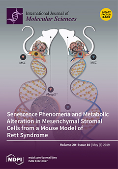
Chromatin modifiers play a crucial role in the regulation of stem cell biology. Among the epigenetic regulators, MeCP2 protein is particularly attractive. Mutations in the MECP2 gene are responsible for more than 90% of cases of Rett syndrome (RTT), a progressive neurodevelopmental disorder. Herein, we dissect the role of impaired MeCP2 function in triggering senescence along with other senescence-related aspects using MSCs from a mouse model of RTT. Our results support the idea that senescence and alteration in mitochondrial metabolic functions could play an important role in the pathogenesis of RTT. View this paper
  • Issues are regarded as officially published after their release is announced to the table of contents alert mailing list.
  • You may sign up for e-mail alerts to receive table of contents of newly released issues.
  • PDF is the official format for papers published in both, html and pdf forms. To view the papers in pdf format, click on the "PDF Full-text" link, and use the free Adobe Reader to open them.
Order results
Result details
Section
Select all
Export citation of selected articles as:
14 pages, 3286 KiB  
Article
Immunoglobulin Binding Protein 1 as a Potential Urine Biomarker in Patients with Lupus Nephritis
by Eun-Ju Lee, Oh Chan Kwon, Byeongzu Ghang, Doo-Ho Lim, Do Hoon Kim, Seokchan Hong, Chang-Keun Lee, Bin Yoo and Yong-Gil Kim
Int. J. Mol. Sci. 2019, 20(10), 2606; https://doi.org/10.3390/ijms20102606 - 27 May 2019
Cited by 12 | Viewed by 4103
Abstract
We evaluated the role of immunoglobulin binding protein 1 (IGBP1), a phosphoprotein associated with the B cell receptor (BCR) complex, as a urine biomarker in lupus nephritis (LN). The IGBP1 concentrations in plasma and urine of patients with LN, systemic lupus erythematosus (SLE) [...] Read more.
We evaluated the role of immunoglobulin binding protein 1 (IGBP1), a phosphoprotein associated with the B cell receptor (BCR) complex, as a urine biomarker in lupus nephritis (LN). The IGBP1 concentrations in plasma and urine of patients with LN, systemic lupus erythematosus (SLE) without nephritis and healthy controls were estimated by ELISA. IGBP1 expression in the kidneys of LN patients and transplantation donors was detected by immunohistochemistry. Microarray-based global gene expression profile of HK-2 cells with IGBP1 knock-down and fluorescence-activated cell sorting (FACS) for intracellular IGBP1 expression in human peripheral blood mononuclear cells (PBMCs) was performed. Urine IGBP1 levels were elevated significantly in LN patients, and it correlated with the clinical activity indices (complement 3 (C3) level, anti-dsDNA antibodies titer, SLE Disease Activity Index-2000 (SLEDAI-2K) and histological activity index. IGBP1 expression was increased in LN patients as compared to the donors and was detected mainly in the tubules by histopathology. In microarray analysis, several genes related to SLE pathogenesis (PPME1, ROCK2, VTCN1, IL-17R, NEU1, HLA-DM, and PTX3) responded to siRNA-mediated IGBP1 silencing. In FACS, IGBP1 was expressed mainly in the CD14+ cells. The overall expression of IGBP1 in PBMCs was higher in LN patients as compared with that in SLE patients without nephritis. Conclusively, urinary IGBP1 may be a novel biomarker reflecting the clinical and histological activities in LN. Full article
(This article belongs to the Section Molecular Immunology)
Show Figures

Figure 1

26 pages, 6056 KiB  
Review
Selection of Biophysical Methods for Characterisation of Membrane Proteins
by Tristan O. C. Kwan, Rosana Reis, Giuliano Siligardi, Rohanah Hussain, Harish Cheruvara and Isabel Moraes
Int. J. Mol. Sci. 2019, 20(10), 2605; https://doi.org/10.3390/ijms20102605 - 27 May 2019
Cited by 21 | Viewed by 8307
Abstract
Over the years, there have been many developments and advances in the field of integral membrane protein research. As important pharmaceutical targets, it is paramount to understand the mechanisms of action that govern their structure–function relationships. However, the study of integral membrane proteins [...] Read more.
Over the years, there have been many developments and advances in the field of integral membrane protein research. As important pharmaceutical targets, it is paramount to understand the mechanisms of action that govern their structure–function relationships. However, the study of integral membrane proteins is still incredibly challenging, mostly due to their low expression and instability once extracted from the native biological membrane. Nevertheless, milligrams of pure, stable, and functional protein are always required for biochemical and structural studies. Many modern biophysical tools are available today that provide critical information regarding to the characterisation and behaviour of integral membrane proteins in solution. These biophysical approaches play an important role in both basic research and in early-stage drug discovery processes. In this review, it is not our objective to present a comprehensive list of all existing biophysical methods, but a selection of the most useful and easily applied to basic integral membrane protein research. Full article
(This article belongs to the Special Issue Structure and Function of Membrane Proteins)
Show Figures

Figure 1

18 pages, 1357 KiB  
Article
Assessing the Impact of Single-Cell Stimulation on Local Networks in Rat Barrel Cortex—A Feasibility Study
by Beate Knauer and Maik C. Stüttgen
Int. J. Mol. Sci. 2019, 20(10), 2604; https://doi.org/10.3390/ijms20102604 - 27 May 2019
Cited by 2 | Viewed by 4120
Abstract
In contrast to the long-standing notion that the role of individual neurons in population activity is vanishingly small, recent studies have shown that electrical activation of only a single cortical neuron can have measurable effects on global brain state, movement, and perception. Although [...] Read more.
In contrast to the long-standing notion that the role of individual neurons in population activity is vanishingly small, recent studies have shown that electrical activation of only a single cortical neuron can have measurable effects on global brain state, movement, and perception. Although highly important for understanding how neuronal activity in cortex is orchestrated, the cellular and network mechanisms underlying this phenomenon are unresolved. Here, we first briefly review the current state of knowledge regarding the phenomenon of single-cell induced network modulation and discuss possible underpinnings. Secondly, we show proof of principle for an experimental approach to elucidate the mechanisms of single-cell induced changes in cortical activity. The setup allows simultaneous recordings of the spiking activity of multiple neurons across all layers of the cortex using a multi-electrode array, while manipulating the activity of one individual neuron in close proximity to the array. We demonstrate that single cells can be recorded and stimulated reliably for hundreds of trials, conferring high statistical power even for expectedly small effects of single-neuron spiking on network activity. Preliminary results suggest that single-cell stimulation on average decreases the firing rate of local network units. We expect that characterization of the spatiotemporal spread of single-cell evoked activity across layers and columns will yield novel insights into intracortical processing. Full article
(This article belongs to the Special Issue Rhine-Main Neuroscience Network: rmn^2-Oberwesel 2018)
Show Figures

Graphical abstract

11 pages, 1225 KiB  
Review
Role of Mast Cell-Derived Adenosine in Cancer
by Yaara Gorzalczany and Ronit Sagi-Eisenberg
Int. J. Mol. Sci. 2019, 20(10), 2603; https://doi.org/10.3390/ijms20102603 - 27 May 2019
Cited by 19 | Viewed by 3752
Abstract
Accumulating evidence has highlighted the accumulation of mast cells (MCs) in tumors. However, their impact on tumor development remained controversial. Indeed, cumulative data indicate an enigmatic role for MCs in cancer, whereby depending on the circumstances, which still need to be resolved, MCs [...] Read more.
Accumulating evidence has highlighted the accumulation of mast cells (MCs) in tumors. However, their impact on tumor development remained controversial. Indeed, cumulative data indicate an enigmatic role for MCs in cancer, whereby depending on the circumstances, which still need to be resolved, MCs function to promote or restrict tumor growth. By responding to multiple stimuli MCs release multiple inflammatory mediators, that contribute to the resolution of infection and resistance to envenomation, but also have the potency to promote or inhibit malignancy. Thus, MCs seem to possess the power to define tumor projections. Given this remarkable plasticity of MC responsiveness, there is an urgent need of understanding how MCs are activated in the tumor microenvironment (TME). We have recently reported on the direct activation of MCs upon contact with cancer cells by a mechanism involving an autocrine formation of adenosine and signaling by the A3 adenosine receptor. Here we summarized the evidence on the role of adenosine signaling in cancer, in MC mediated inflammation and in the MC-cancer crosstalk. Full article
(This article belongs to the Special Issue Mast Cells in Health and Disease)
Show Figures

Graphical abstract

14 pages, 4169 KiB  
Article
An R1R2R3 MYB Transcription Factor, MnMYB3R1, Regulates the Polyphenol Oxidase Gene in Mulberry (Morus notabilis)
by Dan Liu, Shuai Meng, Zhonghuai Xiang, Guangwei Yang and Ningjia He
Int. J. Mol. Sci. 2019, 20(10), 2602; https://doi.org/10.3390/ijms20102602 - 27 May 2019
Cited by 13 | Viewed by 4298
Abstract
The aim of this study was to determine how the mulberry (Morus notabilis) polyphenol oxidase 1 gene (MnPPO1) is regulated during plant stress responses by exploring the interaction between its promoter region and regulatory transcription factors. First, we analyzed [...] Read more.
The aim of this study was to determine how the mulberry (Morus notabilis) polyphenol oxidase 1 gene (MnPPO1) is regulated during plant stress responses by exploring the interaction between its promoter region and regulatory transcription factors. First, we analyzed the cis-acting elements in the MnPPO1 promoter. Then, we used the MnPPO1 promoter region [(1268 bp, including an MYB3R-binding cis-element (MSA)] as a probe to capture proteins in DNA pull-down assays. These analyses revealed that the MYB3R1 transcription factor in M. notabilis (encoded by MnMYB3R1) binds to the MnPPO1 promoter region. We further explored the interaction between the MnPPO1 promoter and MYB3R1 with the dual luciferase reporter, yeast one-hybrid, and chromatin immunoprecipitation assays. These analyses verified that MnMYB3R1 binds to the MSA in the MnPPO1 promoter region. The overexpression of MnMYB3R1 in tobacco upregulated the expression of the tobacco PPO gene. This observation as well as the quantitative real-time PCR results implied that MnMYB3R1 and PPO are involved in the abscisic acid-responsive stress response pathway. Full article
Show Figures

Figure 1

13 pages, 5406 KiB  
Article
Differences in Liver TFAM Binding to mtDNA and mtDNA Damage between Aged and Extremely Aged Rats
by Guglielmina Chimienti, Anna Picca, Flavio Fracasso, Emanuele Marzetti, Riccardo Calvani, Christiaan Leeuwenburgh, Francesco Russo, Angela Maria Serena Lezza and Vito Pesce
Int. J. Mol. Sci. 2019, 20(10), 2601; https://doi.org/10.3390/ijms20102601 - 27 May 2019
Cited by 16 | Viewed by 3799
Abstract
While mitochondrial dysfunction is acknowledged as a major feature of aging, much less is known about the role of mitochondria in extended longevity. Livers from aged (28-month-old) and extremely aged (32-month-old) rats were analyzed for citrate synthase activity, mitochondrial transcription factor A (TFAM) [...] Read more.
While mitochondrial dysfunction is acknowledged as a major feature of aging, much less is known about the role of mitochondria in extended longevity. Livers from aged (28-month-old) and extremely aged (32-month-old) rats were analyzed for citrate synthase activity, mitochondrial transcription factor A (TFAM) amount, mitochondrial DNA (mtDNA), and 4.8 Kb “common deletion” contents. None of the assayed parameters differed significantly between age groups. TFAM-binding to mtDNA and the incidence of 8-oxo-deoxyguanosine in specific mtDNA regions, encompassing the origins of mtDNA replication (D-loop and Ori-L) and the 16-bp long direct repeat 1 (DR1) of the 4.8 Kb deletion, were determined. A decrease in TFAM binding was unveiled at all regions in extremely aged in comparison with aged rats. Reduced incidence of oxidized purines at all assayed regions was detected in 32-month-old rats compared with the 28-month-old group. A significant positive correlation between the incidence of 8-oxo-deoxoguanosine and TFAM-bound mtDNA was found at D-Loop and Ori-L regions only in 28-month-old rats. The absence of such correlation in 32-month-old rats indicates a different, fine-tuned regulation of TFAM binding in the two age groups and supports the existence of two different paces in aging and extended aging. Full article
(This article belongs to the Special Issue mtDNA and Mitochondrial Stress Signaling in Human Diseases)
Show Figures

Figure 1

12 pages, 1206 KiB  
Review
Disturbance of Intracerebral Fluid Clearance and Blood–Brain Barrier in Vascular Cognitive Impairment
by Masaki Ueno, Yoichi Chiba, Ryuta Murakami, Koichi Matsumoto, Ryuji Fujihara, Naoya Uemura, Ken Yanase and Masaki Kamada
Int. J. Mol. Sci. 2019, 20(10), 2600; https://doi.org/10.3390/ijms20102600 - 27 May 2019
Cited by 20 | Viewed by 4118
Abstract
The entry of blood-borne macromolecular substances into the brain parenchyma from cerebral vessels is blocked by the blood–brain barrier (BBB) function. Accordingly, increased permeability of the vessels induced by insult noted in patients suffering from vascular dementia likely contributes to the cognitive impairment. [...] Read more.
The entry of blood-borne macromolecular substances into the brain parenchyma from cerebral vessels is blocked by the blood–brain barrier (BBB) function. Accordingly, increased permeability of the vessels induced by insult noted in patients suffering from vascular dementia likely contributes to the cognitive impairment. On the other hand, blood-borne substances can enter extracellular spaces of the brain via endothelial cells at specific sites without the BBB, and can move to brain parenchyma, such as the hippocampus and periventricular areas, adjacent to specific sites, indicating the contribution of increased permeability of vessels in the specific sites to brain function. It is necessary to consider influx and efflux of interstitial fluid (ISF) and cerebrospinal fluid (CSF) in considering effects of brain transfer of intravascular substances on brain function. Two pathways of ISF and CSF are recently being established. One is the intramural peri-arterial drainage (IPAD) pathway of ISF. The other is the glymphatic system of CSF. Dysfunction of the two pathways could also contribute to brain dysfunction. We review the effects of several kinds of insult on vascular permeability and the failure of fluid clearance on the brain function. Full article
Show Figures

Figure 1

18 pages, 3154 KiB  
Article
Characterization of Histone Deacetylase Expression Within In Vitro and In Vivo Bladder Cancer Model Systems
by Jenna M. Buckwalter, Wilson Chan, Lauren Shuman, Thomas Wildermuth, Justine Ellis-Mohl, Vonn Walter, Joshua I. Warrick, Xue-Ru Wu, Matt Kaag, Jay D. Raman and David J. DeGraff
Int. J. Mol. Sci. 2019, 20(10), 2599; https://doi.org/10.3390/ijms20102599 - 27 May 2019
Cited by 14 | Viewed by 4754
Abstract
Epigenetic aberrations are prominent in bladder cancer (BC) and contribute to disease pathogenesis. We characterized histone deacetylase (HDAC) expression, a family of deacetylation enzymes, in both in vitro and in vivo BC model systems and analyzed expression data from The Cancer Genome Atlas [...] Read more.
Epigenetic aberrations are prominent in bladder cancer (BC) and contribute to disease pathogenesis. We characterized histone deacetylase (HDAC) expression, a family of deacetylation enzymes, in both in vitro and in vivo BC model systems and analyzed expression data from The Cancer Genome Atlas (TCGA). Quantitative real-time polymerase chain reaction (qRT-PCR) and western blotting analysis was used to determine the expression status of Class I and II HDACs in ten human BC cell lines, while qRT-PCR was used to determine HDAC expression in 24 human tumor specimens. The TCGA cohort consists of 408 muscle invasive BC (MIBC) clinical samples and analysis of this data set identified expression of HDAC4 and -9 as being associated with basal–squamous disease. These findings agree with qRT-PCR results identifying increased expression of HDAC4, -7, and -9 in basal BC cell lines (p < 0.05; Kruskal–Wallis test) and in clinical specimens with invasive bladder cancer (not statistically significant). We also observed increased expression in Hdac4, -7, and -9 in commonly used BC mouse models. Here, we identify suitable preclinical model systems for the study of HDACs, and show increased expression of Class IIa HDACs, specifically HDAC4 and HDAC9, in basal BC cell lines and in invasive clinical specimens. These results suggest this class of HDACs may be best suited for targeted inhibition in patients with basal BC. Full article
(This article belongs to the Special Issue Epigenetics of Urological Cancers)
Show Figures

Figure 1

12 pages, 1914 KiB  
Article
Rice Genome Resequencing Reveals a Major Quantitative Trait Locus for Resistance to Bakanae Disease Caused by Fusarium fujikuroi
by Do-Yu Kang, Kyeong-Seong Cheon, Jun Oh, Hyoja Oh, Song Lim Kim, Nyunhee Kim, Eungyeong Lee, Inchan Choi, Jeongho Baek, Kyung-Hwan Kim, Nam-Jin Chung and Hyeonso Ji
Int. J. Mol. Sci. 2019, 20(10), 2598; https://doi.org/10.3390/ijms20102598 - 27 May 2019
Cited by 24 | Viewed by 3642
Abstract
Bakanae disease (BD), caused by the fungal pathogen Fusarium fujikuroi, has become a serious threat in rice-cultivating regions worldwide. In the present study, quantitative trait locus (QTL) mapping was performed using F2 and F3 plants derived after crossing a BD-resistant and a [...] Read more.
Bakanae disease (BD), caused by the fungal pathogen Fusarium fujikuroi, has become a serious threat in rice-cultivating regions worldwide. In the present study, quantitative trait locus (QTL) mapping was performed using F2 and F3 plants derived after crossing a BD-resistant and a BD-susceptible Korean japonica rice variety, ‘Samgwang’ and ‘Junam’, respectively. Resequencing of ‘Junam’ and ‘Samgwang’ genomes revealed 151,916 DNA polymorphisms between the two varieties. After genotyping 188 F2 plants, we constructed a genetic map comprising 184 markers, including 175 kompetitive allele-specific PCR markers, eight cleaved amplified polymorphic sequence (CAPS) markers, and a derived CAPS (dCAPS) marker. The degree of BD susceptibility of each F2 plant was evaluated on the basis of the mortality rate measured with corresponding F3 progeny seedlings by in vitro screening. Consequently, qFfR9, a major QTL, was discovered at 30.1 centimorgan (cM) on chromosome 9 with a logarithm of the odds score of 60.3. For the QTL interval, 95% probability lay within a 7.24–7.56 Mbp interval. In this interval, we found that eight genes exhibited non-synonymous single nucleotide polymorphisms (SNPs) by comparing the ‘Junam’ and ‘Samgwang’ genome sequence data, and are possibly candidate genes for qFfR9; therefore, qFfR9 could be utilized as a valuable resource for breeding BD-resistant rice varieties. Full article
(This article belongs to the Special Issue Mapping Plant Genes that Confer Resistance to Biotic Stress)
Show Figures

Figure 1

17 pages, 4116 KiB  
Article
Engineering of Nanobodies Recognizing the Human Chemokine Receptor CCR7
by Barbara D. Jakobs, Lisa Spannagel, Vladimir Purvanov, Edith Uetz-von Allmen, Christoph Matti and Daniel F. Legler
Int. J. Mol. Sci. 2019, 20(10), 2597; https://doi.org/10.3390/ijms20102597 - 27 May 2019
Cited by 11 | Viewed by 4261
Abstract
The chemokine receptor CCR7 plays a pivotal role in health and disease. In particular, CCR7 controls homing of antigen-bearing dendritic cells and T cells to lymph nodes, where adaptive immune responses are initiated. However, CCR7 also guides T cells to inflamed synovium and [...] Read more.
The chemokine receptor CCR7 plays a pivotal role in health and disease. In particular, CCR7 controls homing of antigen-bearing dendritic cells and T cells to lymph nodes, where adaptive immune responses are initiated. However, CCR7 also guides T cells to inflamed synovium and thereby contributes to rheumatoid arthritis and promotes cancer cell migration and metastasis formation. Nanobodies have recently emerged as versatile tools to study G-protein-coupled receptor functions and are being tested in diagnostics and therapeutics. In this study, we designed a strategy to engineer novel nanobodies recognizing human CCR7. We generated a nanobody library based on a solved crystal structure of the nanobody Nb80 recognizing the β2-adrenergic receptor (β2AR) and by specifically randomizing two segments within complementarity determining region 1 (CDR1) and CDR3 of Nb80 known to interact with β2AR. We fused the nanobody library to one half of split-YFP in order to identify individual nanobody clones interacting with CCR7 fused to the other half of split-YFP using bimolecular fluorescence complementation. We present three novel nanobodies, termed Nb1, Nb5, and Nb38, that recognize human CCR7 without interfering with G-protein-coupling and downstream signaling. Moreover, we were able to follow CCR7 trafficking upon CCL19 triggering using Nb1, Nb5, and Nb38. Full article
(This article belongs to the Special Issue Chemokines in Cancer and Inflammatory Diseases)
Show Figures

Graphical abstract

19 pages, 3626 KiB  
Article
Proteomic Landscape of the Mature Roots in a Rubber-Producing Grass Taraxacum Kok-saghyz
by Quanliang Xie, Guohua Ding, Liping Zhu, Li Yu, Boxuan Yuan, Xuan Gao, Dan Wang, Yong Sun, Yang Liu, Hongbin Li and Xuchu Wang
Int. J. Mol. Sci. 2019, 20(10), 2596; https://doi.org/10.3390/ijms20102596 - 27 May 2019
Cited by 14 | Viewed by 5151
Abstract
The rubber grass Taraxacum kok-saghyz (TKS) contains large amounts of natural rubber (cis-1,4-polyisoprene) in its enlarged roots and it is an alternative crop source of natural rubber. Natural rubber biosynthesis (NRB) and storage in the mature roots of TKS is a cascade process [...] Read more.
The rubber grass Taraxacum kok-saghyz (TKS) contains large amounts of natural rubber (cis-1,4-polyisoprene) in its enlarged roots and it is an alternative crop source of natural rubber. Natural rubber biosynthesis (NRB) and storage in the mature roots of TKS is a cascade process involving many genes, proteins and their cofactors. The TKS genome has just been annotated and many NRB-related genes have been determined. However, there is limited knowledge about the protein regulation mechanism for NRB in TKS roots. We identified 371 protein species from the mature roots of TKS by combining two-dimensional gel electrophoresis (2-DE) and mass spectrometry (MS). Meanwhile, a large-scale shotgun analysis of proteins in TKS roots at the enlargement stage was performed, and 3545 individual proteins were determined. Subsequently, all identified proteins from 2-DE gel and shotgun MS in TKS roots were subject to gene ontology and Kyoto Encyclopedia of Genes and Genomes (KEGG) enrichment analyses and most proteins were involved in carbon metabolic process with catalytic activity in membrane-bounded organelles, followed by proteins with binding ability, transportation and phenylpropanoid biosynthesis activities. Fifty-eight NRB-related proteins, including eight small rubber particle protein (SRPP) and two rubber elongation factor(REF) members, were identified from the TKS roots, and these proteins were involved in both mevalonate acid (MVA) and methylerythritol phosphate (MEP) pathways. To our best knowledge, it is the first high-resolution draft proteome map of the mature TKS roots. Our proteomics of TKS roots revealed both MVA and MEP pathways are important for NRB, and SRPP might be more important than REF for NRB in TKS roots. These findings would not only deepen our understanding of the TKS root proteome, but also provide new evidence on the roles of these NRB-related proteins in the mature TKS roots. Full article
Show Figures

Figure 1

14 pages, 1723 KiB  
Review
Two Old Dogs, One New Trick: A Review of RNA Polymerase and Ribosome Interactions during Transcription-Translation Coupling
by Adam B. Conn, Stephen Diggs, Timothy K. Tam and Gregor M. Blaha
Int. J. Mol. Sci. 2019, 20(10), 2595; https://doi.org/10.3390/ijms20102595 - 27 May 2019
Cited by 15 | Viewed by 7149
Abstract
The coupling of transcription and translation is more than mere translation of an mRNA that is still being transcribed. The discovery of physical interactions between RNA polymerase and ribosomes has spurred renewed interest into this long-standing paradigm of bacterial molecular biology. Here, we [...] Read more.
The coupling of transcription and translation is more than mere translation of an mRNA that is still being transcribed. The discovery of physical interactions between RNA polymerase and ribosomes has spurred renewed interest into this long-standing paradigm of bacterial molecular biology. Here, we provide a concise presentation of recent insights gained from super-resolution microscopy, biochemical, and structural work, including cryo-EM studies. Based on the presented data, we put forward a dynamic model for the interaction between RNA polymerase and ribosomes, in which the interactions are repeatedly formed and broken. Furthermore, we propose that long intervening nascent RNA will loop out and away during the forming the interactions between the RNA polymerase and ribosomes. By comparing the effect of the direct interactions between RNA polymerase and ribosomes with those that transcription factors NusG and RfaH mediate, we submit that two distinct modes of coupling exist: Factor-free and factor-mediated coupling. Finally, we provide a possible framework for transcription-translation coupling and elude to some open questions in the field. Full article
(This article belongs to the Special Issue Structure, Function and Evolution of the Ribosome)
Show Figures

Figure 1

9 pages, 3952 KiB  
Article
Efficient Agrobacterium-Mediated Transformation of the Commercial Hybrid Poplar Populus Alba × Populus glandulosa Uyeki
by Chengwei Song, Liang Lu, Yayu Guo, Huimin Xu and Ruili Li
Int. J. Mol. Sci. 2019, 20(10), 2594; https://doi.org/10.3390/ijms20102594 - 27 May 2019
Cited by 23 | Viewed by 4871
Abstract
Transgenic technology is a powerful tool for gene functional characterization, and poplar is a model system for genetic transformation of perennial woody plants. However, the poplar genetic transformation system is limited to a number of model genotypes. Herein, we developed a transformation system [...] Read more.
Transgenic technology is a powerful tool for gene functional characterization, and poplar is a model system for genetic transformation of perennial woody plants. However, the poplar genetic transformation system is limited to a number of model genotypes. Herein, we developed a transformation system based on efficient Agrobacterium-mediated transformation for the hybrid poplar Populus Alba × Populus glandulosa Uyeki, which is a fast-growing poplar species that is suitably grown in the northern part of China. Importantly, we optimized many independent factors and showed that the transformation efficiency was improved significantly using juvenile leaf explants. Explants were infected by an Agrobacterium suspension with the OD600 = 0.6 for 15 min and then co-cultured in dark conditions for 3 days. Using the improved transformation system, we obtained the transgenic poplar with overexpression of β-glucuronidase (GUS) via direct organogenesis without callus induction. Furthermore, we analyzed the GUS gene in the transgenic poplars using PCR, qRT-PCR, and GUS staining. These analyses revealed that the GUS gene was efficiently transformed, and it exhibited various expression levels. Taken together, these results represent a simple, fast, and efficient transformation system of hybrid poplar plants. Our findings may facilitate future studies of gene functions in perennial woody plants and tree breeding via transgenic technology assisted design. Full article
(This article belongs to the Section Molecular Plant Sciences)
Show Figures

Figure 1

16 pages, 2508 KiB  
Communication
Heme Oxygenase-1 Inhibition Sensitizes Human Prostate Cancer Cells towards Glucose Deprivation and Metformin-Mediated Cell Death
by Marco Raffaele, Valeria Pittalà, Veronica Zingales, Ignazio Barbagallo, Loredana Salerno, Giovanni Li Volti, Giuseppe Romeo, Giuseppe Carota, Valeria Sorrenti and Luca Vanella
Int. J. Mol. Sci. 2019, 20(10), 2593; https://doi.org/10.3390/ijms20102593 - 27 May 2019
Cited by 25 | Viewed by 4192
Abstract
High levels of heme oxygenase (HO)-1 have been frequently reported in different human cancers, playing a major role in drug resistance and regulation of cancer cell redox homeostasis. Metformin (MET), a drug widely used for type 2 diabetes, has recently gained interest for [...] Read more.
High levels of heme oxygenase (HO)-1 have been frequently reported in different human cancers, playing a major role in drug resistance and regulation of cancer cell redox homeostasis. Metformin (MET), a drug widely used for type 2 diabetes, has recently gained interest for treating several cancers. Recent studies indicated that the anti-proliferative effects of metformin in cancer cells are highly dependent on glucose concentration. The present work was directed to determine whether use of a specific inhibitor of HO-1 activity, alone or in combination with metformin, affected metastatic prostate cancer cell viability under different concentrations of glucose. MTT assay and the xCELLigence system were used to evaluate cell viability and cell proliferation in DU145 human prostate cancer cells. Cell apoptosis and reactive oxygen species were analyzed by flow cytometry. The activity of HO-1 was inhibited using a selective imidazole-based inhibitor; genes associated with antioxidant systems and cell death were evaluated by qRT-PCR. Our study demonstrates that metformin suppressed prostate cancer growth in vitro and increased oxidative stress. Disrupting the antioxidant HO-1 activity, especially under low glucose concentration, could be an attractive approach to potentiate metformin antineoplastic effects and could provide a biochemical basis for developing HO-1-targeting drugs against solid tumors. Full article
(This article belongs to the Special Issue Protective and Detrimental Role of Heme Oxygenase-1)
Show Figures

Figure 1

16 pages, 4153 KiB  
Article
Graptopetalum paraguayense Inhibits Liver Fibrosis by Blocking TGF-β Signaling In Vivo and In Vitro
by Wei-Hsiang Hsu, Se-Chun Liao, Yau-Jan Chyan, Kai-Wen Huang, Shih-Lan Hsu, Yi-Chen Chen, Ma-Li Siu, Chia-Chuan Chang, Yuh-Shan Chung and Chi-Ying F. Huang
Int. J. Mol. Sci. 2019, 20(10), 2592; https://doi.org/10.3390/ijms20102592 - 27 May 2019
Cited by 11 | Viewed by 4152
Abstract
Background and Aims: Liver fibrosis is the excessive accumulation of extracellular matrix proteins, including collagen, which occurs in most types of chronic liver diseases. Advanced liver fibrosis results in cirrhosis, liver failure, and portal hypertension. Activated hepatic perivascular stellate cells, portal fibroblasts, and [...] Read more.
Background and Aims: Liver fibrosis is the excessive accumulation of extracellular matrix proteins, including collagen, which occurs in most types of chronic liver diseases. Advanced liver fibrosis results in cirrhosis, liver failure, and portal hypertension. Activated hepatic perivascular stellate cells, portal fibroblasts, and myofibroblasts of bone marrow origin have been identified as major collagen-producing cells in the injured liver. These cells are activated by fibrogenic cytokines, such as TGF-β1. The inhibition of TGF-β1 function or synthesis is a major target for the development of antifibrotic therapies. Our previous study showed that the water and ethanol extracts of Graptopetalum paraguayense (GP), a Chinese herbal medicine, can prevent dimethylnitrosamine (DMN)-induced hepatic inflammation and fibrosis in rats. Methods: We used rat hepatic stellate HSC-T6 cells and a diethylnitrosamine (DEN)-induced rat liver injury model to test the potential mechanism of GP extracts and its fraction, HH-F3. Results: We demonstrated that GP extracts and HH-F3 downregulated the expression levels of extracellular matrix (ECM) proteins and inhibited the proliferation and migration via suppression of the TGF-β1 pathway in rat hepatic stellate HSC-T6 cells. Moreover, the HH-F3 fraction decreased hepatic collagen content and reduced plasma AST, ALT, and γ-GT activities in a DEN-induced rat liver injury model, suggesting that GP/HH-F3 has hepatoprotective effects against DEN-induced liver fibrosis. Conclusion: These findings indicate that GP/HH-F3 may be a potential therapeutic agent for the treatment of liver fibrosis. The inhibition of TGF-β-mediated fibrogenesis may be a central mechanism by which GP/HH-F3 protects the liver from injury. Full article
(This article belongs to the Special Issue TGF-Beta Super Family Signaling 2.0)
Show Figures

Figure 1

14 pages, 2155 KiB  
Article
Therapeutic Effects of rAAV-Mediated Concomittant Gene Transfer and Overexpression of TGF-β and IGF-I on the Chondrogenesis of Human Bone-Marrow-Derived Mesenchymal Stem Cells
by Stephanie Morscheid, Ana Rey-Rico, Gertrud Schmitt, Henning Madry, Magali Cucchiarini and Jagadeesh Kumar Venkatesan
Int. J. Mol. Sci. 2019, 20(10), 2591; https://doi.org/10.3390/ijms20102591 - 27 May 2019
Cited by 9 | Viewed by 3745
Abstract
Application of chondroreparative gene vectors in cartilage defects is a powerful approach to directly stimulate the regenerative activities of bone-marrow-derived mesenchymal stem cells (MSCs) that repopulate such lesions. Here, we investigated the ability of combined recombinant adeno-associated virus (rAAV) vector-mediated delivery of the [...] Read more.
Application of chondroreparative gene vectors in cartilage defects is a powerful approach to directly stimulate the regenerative activities of bone-marrow-derived mesenchymal stem cells (MSCs) that repopulate such lesions. Here, we investigated the ability of combined recombinant adeno-associated virus (rAAV) vector-mediated delivery of the potent transforming growth factor beta (TGF-β) and insulin-like growth factor I (IGF-I) to enhance the processes of chondrogenic differentiation in human MSCs (hMSCs) relative to individual candidate treatments and to reporter (lacZ) gene condition. The rAAV-hTGF-β and rAAV-hIGF-I vectors were simultaneously provided to hMSC aggregate cultures (TGF-β/IGF-I condition) in chondrogenic medium over time (21 days) versus TGF-β/lacZ, IGF-I/lacZ, and lacZ treatments at equivalent vector doses. The cultures were then processed to monitor transgene (co)-overexpression, the levels of biological activities in the cells (cell proliferation, matrix synthesis), and the development of a chondrogenic versus osteogenic/hypertrophic phenotype. Effective, durable co-overexpression of TGF-β with IGF-I via rAAV enhanced the proliferative, anabolic, and chondrogenic activities in hMSCs versus lacZ treatment and reached levels that were higher than those achieved upon single candidate gene transfer, while osteogenic/hypertrophic differentiation was delayed over the period of time evaluated. These findings demonstrate the potential of manipulating multiple therapeutic rAAV vectors as a tool to directly target bone-marrow-derived MSCs in sites of focal cartilage defects and to locally enhance the endogenous processes of cartilage repair. Full article
Show Figures

Figure 1

22 pages, 4613 KiB  
Review
Ion Transporters, Channelopathies, and Glucose Disorders
by Huseyin Demirbilek, Sonya Galcheva, Dogus Vuralli, Sara Al-Khawaga and Khalid Hussain
Int. J. Mol. Sci. 2019, 20(10), 2590; https://doi.org/10.3390/ijms20102590 - 27 May 2019
Cited by 21 | Viewed by 6534
Abstract
Ion channels and transporters play essential roles in excitable cells including cardiac, skeletal and smooth muscle cells, neurons, and endocrine cells. In pancreatic beta-cells, for example, potassium KATP channels link the metabolic signals generated inside the cell to changes in the beta-cell [...] Read more.
Ion channels and transporters play essential roles in excitable cells including cardiac, skeletal and smooth muscle cells, neurons, and endocrine cells. In pancreatic beta-cells, for example, potassium KATP channels link the metabolic signals generated inside the cell to changes in the beta-cell membrane potential, and ultimately regulate insulin secretion. Mutations in the genes encoding some ion transporter and channel proteins lead to disorders of glucose homeostasis (hyperinsulinaemic hypoglycaemia and different forms of diabetes mellitus). Pancreatic KATP, Non-KATP, and some calcium channelopathies and MCT1 transporter defects can lead to various forms of hyperinsulinaemic hypoglycaemia (HH). Mutations in the genes encoding the pancreatic KATP channels can also lead to different types of diabetes (including neonatal diabetes mellitus (NDM) and Maturity Onset Diabetes of the Young, MODY), and defects in the solute carrier family 2 member 2 (SLC2A2) leads to diabetes mellitus as part of the Fanconi–Bickel syndrome. Variants or polymorphisms in some ion channel genes and transporters have been reported in association with type 2 diabetes mellitus. Full article
Show Figures

Graphical abstract

17 pages, 275 KiB  
Review
Crucial Role of Extracellular Vesicles in Bronchial Asthma
by Tatsuya Nagano, Masahiro Katsurada, Ryota Dokuni, Daisuke Hazama, Tatsunori Kiriu, Kanoko Umezawa, Kazuyuki Kobayashi and Yoshihiro Nishimura
Int. J. Mol. Sci. 2019, 20(10), 2589; https://doi.org/10.3390/ijms20102589 - 27 May 2019
Cited by 29 | Viewed by 4424
Abstract
Extracellular vesicles (EVs) are circulating vesicles secreted by various cell types. EVs are classified into three groups according to size, structural components, and generation process of vesicles: exosomes, microvesicles, and apoptotic bodies. Recently, EVs have been considered to be crucial for cell-to-cell communications [...] Read more.
Extracellular vesicles (EVs) are circulating vesicles secreted by various cell types. EVs are classified into three groups according to size, structural components, and generation process of vesicles: exosomes, microvesicles, and apoptotic bodies. Recently, EVs have been considered to be crucial for cell-to-cell communications and homeostasis because they contain intracellular proteins and nucleic acids. Epithelial cells from mice suffering from bronchial asthma (BA) secrete more EVs and suppress inflammation-induced EV production. Moreover, microarray analyses of bronchoalveolar lavage fluid have revealed that several microRNAs are useful novel biomarkers of BA. Mesenchymal stromal cell-derived EVs are possible candidates of novel BA therapy. In this review, we highlight the biologic roles of EVs in BA and review novel EV-targeted therapy to help understanding by clinicians and biologists. Full article
(This article belongs to the Special Issue Intercellular Communication in Health and Disease)
Show Figures

Graphical abstract

18 pages, 1452 KiB  
Review
The Anti-inflammatory Effects of Dietary Anthocyanins against Ulcerative Colitis
by Shiyu Li, Binning Wu, Wenyi Fu and Lavanya Reddivari
Int. J. Mol. Sci. 2019, 20(10), 2588; https://doi.org/10.3390/ijms20102588 - 27 May 2019
Cited by 87 | Viewed by 13016
Abstract
Ulcerative colitis (UC), which is a major form of inflammatory bowel disease (IBD), is a chronic relapsing disorder of the gastrointestinal tract affecting millions of people worldwide. Alternative natural therapies, including dietary changes, are being investigated to manage or treat UC since current [...] Read more.
Ulcerative colitis (UC), which is a major form of inflammatory bowel disease (IBD), is a chronic relapsing disorder of the gastrointestinal tract affecting millions of people worldwide. Alternative natural therapies, including dietary changes, are being investigated to manage or treat UC since current treatment options have serious negative side effects. There is growing evidence from animal studies and human clinical trials that diets rich in anthocyanins, which are pigments in fruits and vegetables, protect against inflammation and increased gut permeability as well as improve colon health through their ability to alter bacterial metabolism and the microbial milieu within the intestines. In this review, the structure and bioactivity of anthocyanins, the role of inflammation and gut bacterial dysbiosis in UC pathogenesis, and their regulation by the dietary anthocyanins are discussed, which suggests the feasibility of dietary strategies for UC mitigation. Full article
(This article belongs to the Special Issue Health Promoting Effects of Phytochemicals)
Show Figures

Figure 1

15 pages, 459 KiB  
Review
Prostate Cancer and Bone Metastases: The Underlying Mechanisms
by Sok Kuan Wong, Nur-Vaizura Mohamad, Tijjani Rabiu Giaze, Kok-Yong Chin, Norazlina Mohamed and Soelaiman Ima-Nirwana
Int. J. Mol. Sci. 2019, 20(10), 2587; https://doi.org/10.3390/ijms20102587 - 27 May 2019
Cited by 102 | Viewed by 8705
Abstract
Patients with advanced prostate cancer often develop bone metastases, leading to bone pain, skeletal fracture, and increased mortality. Bone provides a hospitable microenvironment to tumor cells. The disease manifestation is driven by the interaction between invading tumor cells, bone-forming osteoblasts, and bone-resorbing osteoclasts. [...] Read more.
Patients with advanced prostate cancer often develop bone metastases, leading to bone pain, skeletal fracture, and increased mortality. Bone provides a hospitable microenvironment to tumor cells. The disease manifestation is driven by the interaction between invading tumor cells, bone-forming osteoblasts, and bone-resorbing osteoclasts. The increased level of osteoclast-activating factor (parathyroid hormone-related peptide, PTHrP) is believed to induce bone resorption by upregulating receptor activator of nuclear factor-kappa B ligand (RANKL) and the release of various growth factors into the bone microenvironment to enhance cancer cell growth. However, the underlying molecular mechanisms remain poorly understood. This review outlines the possible molecular mechanisms involved in governing bone metastases driven by prostate cancer, which further provide the basis in searching for new molecular targets for the development of potential therapy. Full article
(This article belongs to the Section Molecular Pathology, Diagnostics, and Therapeutics)
Show Figures

Figure 1

27 pages, 814 KiB  
Review
The Biology of the Nuclear Envelope and Its Implications in Cancer Biology
by Maria Alvarado-Kristensson and Catalina Ana Rosselló
Int. J. Mol. Sci. 2019, 20(10), 2586; https://doi.org/10.3390/ijms20102586 - 27 May 2019
Cited by 24 | Viewed by 5283
Abstract
The formation of the nuclear envelope and the subsequent compartmentalization of the genome is a defining feature of eukaryotes. Traditionally, the nuclear envelope was purely viewed as a physical barrier to preserve genetic material in eukaryotic cells. However, in the last few decades, [...] Read more.
The formation of the nuclear envelope and the subsequent compartmentalization of the genome is a defining feature of eukaryotes. Traditionally, the nuclear envelope was purely viewed as a physical barrier to preserve genetic material in eukaryotic cells. However, in the last few decades, it has been revealed to be a critical cellular component in controlling gene expression and has been implicated in several human diseases. In cancer, the relevance of the cell nucleus was first reported in the mid-1800s when an altered nuclear morphology was observed in tumor cells. This review aims to give a current and comprehensive view of the role of the nuclear envelope on cancer first by recapitulating the changes of the nuclear envelope during cell division, second, by reviewing the role of the nuclear envelope in cell cycle regulation, signaling, and the regulation of the genome, and finally, by addressing the nuclear envelope link to cell migration and metastasis and its use in cancer prognosis. Full article
(This article belongs to the Special Issue Molecular Regulation of the Endomembrane System)
Show Figures

Figure 1

22 pages, 4395 KiB  
Article
Salt Tolerance Improvement in Rice through Efficient SNP Marker-Assisted Selection Coupled with Speed-Breeding
by Md Masud Rana, Takeshi Takamatsu, Marouane Baslam, Kentaro Kaneko, Kimiko Itoh, Naoki Harada, Toshie Sugiyama, Takayuki Ohnishi, Tetsu Kinoshita, Hiroki Takagi and Toshiaki Mitsui
Int. J. Mol. Sci. 2019, 20(10), 2585; https://doi.org/10.3390/ijms20102585 - 26 May 2019
Cited by 80 | Viewed by 10585
Abstract
Salinity critically limits rice metabolism, growth, and productivity worldwide. Improvement of the salt resistance of locally grown high-yielding cultivars is a slow process. The objective of this study was to develop a new salt-tolerant rice germplasm using speed-breeding. Here, we precisely introgressed the [...] Read more.
Salinity critically limits rice metabolism, growth, and productivity worldwide. Improvement of the salt resistance of locally grown high-yielding cultivars is a slow process. The objective of this study was to develop a new salt-tolerant rice germplasm using speed-breeding. Here, we precisely introgressed the hst1 gene, transferring salinity tolerance from “Kaijin” into high-yielding “Yukinko-mai” (WT) rice through single nucleotide polymorphism (SNP) marker-assisted selection. Using a biotron speed-breeding technique, we developed a BC3F3 population, named “YNU31-2-4”, in six generations and 17 months. High-resolution genotyping by whole-genome sequencing revealed that the BC3F2 genome had 93.5% similarity to the WT and fixed only 2.7% of donor parent alleles. Functional annotation of BC3F2 variants along with field assessment data indicated that “YNU31-2-4” plants carrying the hst1 gene had similar agronomic traits to the WT under normal growth condition. “YNU31-2-4” seedlings subjected to salt stress (125 mM NaCl) had a significantly higher survival rate and increased shoot and root biomasses than the WT. At the tissue level, quantitative and electron probe microanalyzer studies indicated that “YNU31-2-4” seedlings avoided Na+ accumulation in shoots under salt stress. The “YNU31-2-4” plants showed an improved phenotype with significantly higher net CO2 assimilation and lower yield decline than WT under salt stress at the reproductive stage. “YNU31-2-4” is a potential candidate for a new rice cultivar that is highly tolerant to salt stress at the seedling and reproductive stages, and which might maintain yields under a changing global climate. Full article
(This article belongs to the Special Issue Plant Genomics 2019)
Show Figures

Graphical abstract

17 pages, 18303 KiB  
Review
Involvement of Extracellular Vesicles in Vascular-Related Functions in Cancer Progression and Metastasis
by Shinsuke Kikuchi, Yusuke Yoshioka, Marta Prieto-Vila and Takahiro Ochiya
Int. J. Mol. Sci. 2019, 20(10), 2584; https://doi.org/10.3390/ijms20102584 - 26 May 2019
Cited by 48 | Viewed by 7055
Abstract
The primary cause of mortality among patients with cancer is the progression of the tumor, better known as cancer invasion and metastasis. Cancer progression involves a series of biologically important steps in which the cross-talk between cancer cells and the cells in the [...] Read more.
The primary cause of mortality among patients with cancer is the progression of the tumor, better known as cancer invasion and metastasis. Cancer progression involves a series of biologically important steps in which the cross-talk between cancer cells and the cells in the surrounding environment is positioned as an important issue. Notably, angiogenesis is a key tumorigenic phenomenon for cancer progression. Cancer-related extracellular vesicles (EVs) commonly contribute to the modulation of a microenvironment favorable to cancer cells through their function of cell-to-cell communication. Vascular-related cells such as endothelial cells (ECs) and platelets activated by cancer cells and cancer-derived EVs develop procoagulant and proinflammatory statuses, which help excite the tumor environment, and play major roles in tumor progression, including in tumor extravasation, tumor cell microthrombi formation, platelet aggregation, and metastasis. In particular, cancer-derived EVs influence ECs, which then play multiple roles such as contributing to tumor angiogenesis, loss of endothelial vascular barrier by binding to ECs, and the subsequent endothelial-to-mesenchymal transition, i.e., extracellular matrix remodeling. Thus, cell-to-cell communication between cancer cells and ECs via EVs may be an important target for controlling cancer progression. This review describes the current knowledge regarding the involvement of EVs, especially exosomes derived from cancer cells, in EC-related cancer progression. Full article
(This article belongs to the Special Issue Tumor Cell Invasion and Metastases)
Show Figures

Figure 1

16 pages, 3402 KiB  
Article
Impact of Periprosthetic Fibroblast-Like Cells on Osteoclastogenesis in Co-Culture with Peripheral Blood Mononuclear Cells Varies Depending on Culture System
by Miriam I. Koehler, Eliza S. Hartmann, Sabine Schluessel, Felicitas Beck, Julia I. Redeker, Baerbel Schmitt, Marina Unger, Martijn van Griensven, Burkhard Summer, Andreas Fottner and Susanne Mayer-Wagner
Int. J. Mol. Sci. 2019, 20(10), 2583; https://doi.org/10.3390/ijms20102583 - 26 May 2019
Cited by 5 | Viewed by 3771
Abstract
Co-culture studies investigating the role of periprosthetic fibroblasts (PPFs) in inflammatory osteoclastogenesis reveal contrary results, partly showing an osteoprotective function of fibroblasts and high OPG expression in monolayer. These data disagree with molecular analyses of original periosteolytic tissues. In order to find a [...] Read more.
Co-culture studies investigating the role of periprosthetic fibroblasts (PPFs) in inflammatory osteoclastogenesis reveal contrary results, partly showing an osteoprotective function of fibroblasts and high OPG expression in monolayer. These data disagree with molecular analyses of original periosteolytic tissues. In order to find a more reliable model, PPFs were co-cultivated with peripheral blood mononuclear cells (PBMCs) in a transwell system and compared to conventional monolayer cultures. The gene expression of key regulators of osteoclastogenesis (macrophage colony-stimulating factor (MCSF), receptor activator of NF-κB ligand (RANK-L), osteoprotegerin (OPG), and tumor necrosis factor alpha (TNFα)) as well as the ability of bone resorption were analyzed. In monolayer co-cultures, PPFs executed an osteoprotective function with high OPG-expression, low RANK-L/OPG ratios, and a resulting inhibition of osteolysis even in the presence of MCSF and RANK-L. For transwell co-cultures, profound changes in gene expression, with a more than hundredfold decrease of OPG and a significant upregulation of TNFα were observed. In conclusion, we were able to show that a change of culture conditions towards a transwell system resulted in a considerably more osteoclastogenic gene expression profile, being closer to findings in original periosteolytic tissues. This study therefore presents an interesting approach for a more reliable in vitro model to examine the role of fibroblasts in periprosthetic osteoclastogenesis in the future. Full article
(This article belongs to the Special Issue Biomaterials for Musculoskeletal System)
Show Figures

Graphical abstract

15 pages, 3590 KiB  
Article
Transcriptome Profile Analysis Reveals that CsTCP14 Induces Susceptibility to Foliage Diseases in Cucumber
by Xuyang Zheng, Jingjing Yang, Tengxue Lou, Jian Zhang, Wenjin Yu and Changlong Wen
Int. J. Mol. Sci. 2019, 20(10), 2582; https://doi.org/10.3390/ijms20102582 - 26 May 2019
Cited by 7 | Viewed by 3472
Abstract
Foliage diseases are prevalent in cucumber production and cause serious yield reduction across the world. Identifying resistance or susceptible genes under foliage-disease stress is essential for breeding resistant varieties, of which leaf-specific expressed susceptible genes are extremely important but rarely studied in crops. [...] Read more.
Foliage diseases are prevalent in cucumber production and cause serious yield reduction across the world. Identifying resistance or susceptible genes under foliage-disease stress is essential for breeding resistant varieties, of which leaf-specific expressed susceptible genes are extremely important but rarely studied in crops. This study performed an in-depth mining of public transcriptome data both in different cucumber tissues and under downy mildew (DM) inoculation, and found that the expression of leaf-specific expressed transcription factor CsTCP14 was significantly increased after treatment with DM, as well as being upregulated under stress from another foliage disease, watermelon mosaic virus (WMV), in susceptible cucumbers. Furthermore, the Pearson correlation analysis identified genome-wide co-expressed defense genes with CsTCP14. A potential target CsNBS-LRR gene, Csa6M344280.1, was obtained as obviously reduced and was negatively correlated with the expression of the susceptible gene CsTCP14. Moreover, the interaction experiments of electrophoretic mobility shift assay (EMSA) and yeast one-hybrid assay (Y1H) were successfully executed to prove that CsTCP14 could transcriptionally repress the expression of the CsNBS-LRR gene, Csa6M344280.1, which resulted in inducing susceptibility to foliage diseases in cucumber. As such, we constructed a draft model showing that the leaf-specific expressed gene CsTCP14 was negatively regulating the defense gene Csa6M344280.1 to induce susceptibility to foliage diseases in cucumber. Therefore, this study explored key susceptible genes in response to foliage diseases based on a comprehensive analysis of public transcriptome data and provided an opportunity to breed new varieties that can resist foliage diseases in cucumber, as well as in other crops. Full article
(This article belongs to the Special Issue Transcriptional and Post-transcriptional Gene Regulation in Plants)
Show Figures

Figure 1

16 pages, 1856 KiB  
Review
Role of Nephronectin in Pathophysiology of Silicosis
by Suni Lee, Machiko Honda, Shoko Yamamoto, Naoko Kumagai-Takei, Kei Yoshitome, Yasumitsu Nishimura, Nagisa Sada, Shigeyuki Kon and Takemi Otsuki
Int. J. Mol. Sci. 2019, 20(10), 2581; https://doi.org/10.3390/ijms20102581 - 26 May 2019
Cited by 12 | Viewed by 6493
Abstract
Silicosis is a typical form of pneumoconiosis and is characterized as a type of lung fibrosis. Silica particles are captured and recognized upon by alveolar macrophages via the macrophage receptor with collagenous structure (MARCO) scavenger receptor, and thereafter the inflammasome is activated. Thereafter, [...] Read more.
Silicosis is a typical form of pneumoconiosis and is characterized as a type of lung fibrosis. Silica particles are captured and recognized upon by alveolar macrophages via the macrophage receptor with collagenous structure (MARCO) scavenger receptor, and thereafter the inflammasome is activated. Thereafter, various chemokines/cytokines play their roles to eventually form fibrosis. Additionally, silica particles chronically activate T helper cells which sets the background for the formation of silicosis-associated autoimmune disturbances. The occurrence and progression of lung fibrosis, the extracellular matrix-related molecules such as integrins and their ligands including fibronectin, vitronectin, laminin, and collagens, all play important roles. Here, the roles of these molecules in silicosis-related lung fibrosis are reviewed from the literature. Additionally, the measurement of serum nephronectin (Npnt), a new member of the integrin family of ligands, is discussed, together with investigations attempting to delineate the role of Npnt in silica-induced lung fibrosis. Serum Npnt was found to be higher in silicosis patients compared to healthy volunteers and seems to play a role in the progression of fibrosis with other cytokines. Therefore, serum Npnt levels may be employed as a suitable marker to monitor the progression of fibrosis in silicosis patients. Full article
(This article belongs to the Special Issue Macrophages in Inflammation 2019)
Show Figures

Figure 1

12 pages, 2323 KiB  
Article
Knockdown of NANOG Reduces Cell Proliferation and Induces G0/G1 Cell Cycle Arrest in Human Adipose Stem Cells
by Maria Pitrone, Giuseppe Pizzolanti, Antonina Coppola, Laura Tomasello, Stefania Martorana, Gianni Pantuso and Carla Giordano
Int. J. Mol. Sci. 2019, 20(10), 2580; https://doi.org/10.3390/ijms20102580 - 26 May 2019
Cited by 10 | Viewed by 4421
Abstract
The core components of regenerative medicine are stem cells with high self-renewal and tissue regeneration potentials. Adult stem cells can be obtained from many organs and tissues. NANOG, SOX2 and OCT4 represent the core regulatory network that suppresses differentiation-associated genes, maintaining the [...] Read more.
The core components of regenerative medicine are stem cells with high self-renewal and tissue regeneration potentials. Adult stem cells can be obtained from many organs and tissues. NANOG, SOX2 and OCT4 represent the core regulatory network that suppresses differentiation-associated genes, maintaining the pluripotency of mesenchymal stem cells. The roles of NANOG in maintaining self-renewal and undifferentiated status of adult stem cells are still not perfectly established. In this study we define the effects of downregulation of NANOG in maintaining self-renewal and undifferentiated state in mesenchymal stem cells (MSCs) derived from subcutaneous adipose tissue (hASCs). hASCs were expanded and transfected in vitro with short hairpin Lentivirus targeting NANOG. Gene suppressions were achieved at both transcript and proteome levels. The effect of NANOG knockdown on proliferation after 10 passages and on the cell cycle was evaluated by proliferation assay, colony forming unit (CFU), qRT-PCR and cell cycle analysis by flow-cytometry. Moreover, NANOG involvement in differentiation ability was evaluated. We report that downregulation of NANOG revealed a decrease in the proliferation and differentiation rate, inducing cell cycle arrest by increasing p27/CDKN1B (Cyclin-dependent kinase inhibitor 1B) and p21/CDKN1A (Cyclin-dependent kinase inhibitor 1A) through p53 and regulate DLK1/PREF1. Furthermore, NANOG induced downregulation of DNMT1, a major DNA methyltransferase responsible for maintaining methylation status during DNA replication probably involved in cell cycle regulation. Our study confirms that NANOG regulates the complex transcription network of plasticity of the cells, inducing cell cycle arrest and reducing differentiation potential. Full article
(This article belongs to the Special Issue Adipose Stem Cells 2019)
Show Figures

Figure 1

15 pages, 3160 KiB  
Article
Effects of Sapindus mukorossi Seed Oil on Skin Wound Healing: In Vivo and in Vitro Testing
by Chang-Chih Chen, Chia-Jen Nien, Lih-Geeng Chen, Kuen-Yu Huang, Wei-Jen Chang and Haw-Ming Huang
Int. J. Mol. Sci. 2019, 20(10), 2579; https://doi.org/10.3390/ijms20102579 - 26 May 2019
Cited by 21 | Viewed by 5560 | Correction
Abstract
Sapindus mukorossi seed oil is commonly used as a source for biodiesel fuel. Its phytochemical composition is similar to the extracted oil from Sapindus trifoliatus seeds, which exhibit beneficial effects for skin wound healing. Since S. mukorossi seed shows no cyanogenic property, it [...] Read more.
Sapindus mukorossi seed oil is commonly used as a source for biodiesel fuel. Its phytochemical composition is similar to the extracted oil from Sapindus trifoliatus seeds, which exhibit beneficial effects for skin wound healing. Since S. mukorossi seed shows no cyanogenic property, it could be a potential candidate for the treatment of skin wounds. Thus, we evaluated the effectiveness of S. mukorossi seed oil in the treatment of skin wounds. We characterized and quantified the fatty acids and unsaponifiable fractions (including β-sitosterol and δ-tocopherol) contained in S. mukorossi seed-extracted oil by GC-MS and HPLC, respectively. Cell proliferation and migratory ability were evaluated by cell viability and scratch experiments using CCD-966SK cells treated with S. mukorossi oil. The anti-inflammatory effects of the oil were evaluated by measuring the nitric oxide (NO) production in lipopolysaccharide-treated RAW 264.7 cells. Antimicrobial activity tests were performed with Propionibacterium acnes, Staphylococcus aureus, and Candida albicans using a modified Japanese Industrial Standard procedure. Uniform artificial wounds were created on the dorsum of rats. The wounds were treated with a carboxymethyl cellulose (CMC)/hyaluronic acid (HA)/sodium alginate (SA) hydrogel for releasing the S. mukorossi seed oil. The wound sizes were measured photographically for 12 days and were compared to wounds covered with analogous membranes containing a saline solution. Our results showed that the S. mukorossi seed oil used in this study contains abundant monounsaturated fatty acids, β-sitosterol, and δ-tocopherol. In the in vitro tests, S. mukorossi seed oil prompted cell proliferation and migration capability. Additionally, the oil had significant anti-inflammatory and anti-microbial activities. In the in vivo animal experiments, S. mukorossi seed oil-treated wounds revealed acceleration of sequential skin wound healing events after two days of healing. The size of oil-treated wound decreased to half the size of the untreated control after eight days of healing. The results suggest that S. mukorossi seed oil could be a potential source for promoting skin wound healing. Full article
(This article belongs to the Special Issue Chemopreventive Activities of Phytochemicals)
Show Figures

Graphical abstract

10 pages, 2084 KiB  
Case Report
Remarkable Alteration of PD-L1 Expression after Immune Checkpoint Therapy in Patients with Non-Small-Cell Lung Cancer: Two Autopsy Case Reports
by Toshiaki Takahashi, Akiko Tateishi, Andrey Bychkov and Junya Fukuoka
Int. J. Mol. Sci. 2019, 20(10), 2578; https://doi.org/10.3390/ijms20102578 - 26 May 2019
Cited by 30 | Viewed by 4757
Abstract
Pembrolizumab is an immune checkpoint inhibitor (ICI), currently recommended as the first-line treatment for patients with advanced non-small-cell lung cancer (NSCLC) showing ≥50% expression of programmed death-ligand 1 (PD-L1). Previously it was reported that platinum-based chemotherapy may change PD-L1 expression in solid cancers. [...] Read more.
Pembrolizumab is an immune checkpoint inhibitor (ICI), currently recommended as the first-line treatment for patients with advanced non-small-cell lung cancer (NSCLC) showing ≥50% expression of programmed death-ligand 1 (PD-L1). Previously it was reported that platinum-based chemotherapy may change PD-L1 expression in solid cancers. However, no reports addressing alteration of PD-L1 expression after ICI therapy in NSCLC are available so far. The patients were Japanese males 83 and 87 years old, who were diagnosed with NSCLC based on the transbronchial lung biopsies showing sarcomatoid feature with high PD-L1 expression. They received Pembrolizumab, however, passed away with disease progression on day 27 and day 9, respectively. PD-L1, PD1, and CD8 antibodies were applied to pretreatment tumor biopsies and autopsy specimens. Immunoexpression of all the markers was evaluated using Aperio ImageScope. We found that PD-L1 expression decreased significantly from 75.6% to 13.2% and from 100% to 58.8%, in patients 1 and 2, respectively. This alteration was less prominent in the perinecrotic tumor area. A considerable decrease of PD-L1 score was linked with a little effect of Pembrolizumab in our patients. This association might be one of the contributing mechanisms of resistance to ICI and needs further investigation in large-scale studies. Full article
(This article belongs to the Special Issue PD-L1, A Master Regulator of Immunity)
Show Figures

Figure 1

9 pages, 2620 KiB  
Communication
Expanding the Zebrafish Genetic Code through Site-Specific Introduction of Azido-lysine, Bicyclononyne-lysine, and Diazirine-lysine
by Junetha Syed, Saravanan Palani, Scott T. Clarke, Zainab Asad, Andrew R. Bottrill, Alexandra M.E. Jones, Karuna Sampath and Mohan K. Balasubramanian
Int. J. Mol. Sci. 2019, 20(10), 2577; https://doi.org/10.3390/ijms20102577 - 26 May 2019
Cited by 7 | Viewed by 5429
Abstract
Site-specific incorporation of un-natural amino acids (UNAA) is a powerful approach to engineer and understand protein function. Site-specific incorporation of UNAAs is achieved through repurposing the amber codon (UAG) as a sense codon for the UNAA, using a tRNACUA that base pairs [...] Read more.
Site-specific incorporation of un-natural amino acids (UNAA) is a powerful approach to engineer and understand protein function. Site-specific incorporation of UNAAs is achieved through repurposing the amber codon (UAG) as a sense codon for the UNAA, using a tRNACUA that base pairs with an UAG codon in the mRNA and an orthogonal amino-acyl tRNA synthetase (aaRS) that charges the tRNACUA with the UNAA. Here, we report an expansion of the zebrafish genetic code to incorporate the UNAAs, azido-lysine (AzK), bicyclononyne-lysine (BCNK), and diazirine-lysine (AbK) into green fluorescent protein (GFP) and glutathione-s-transferase (GST). We also present proteomic evidence for UNAA incorporation into GFP. Our work sets the stage for the use of AzK, BCNK, and AbK introduction into proteins as a means to investigate and engineer their function in zebrafish. Full article
(This article belongs to the Special Issue Expanding and Reprogramming the Genetic Code)
Show Figures

Figure 1

Previous Issue
Next Issue
Back to TopTop