Article

Structural (XRD) Characterization and an Analysis of H-Bonding Motifs in Some Tetrahydroxidohexaoxidopentaborate(1-) Salts of N-Substituted Guanidinium Cations †

1
School of Natural Sciences, Bangor University, Bangor, Gwynedd LL57 2UW, UK
2
Chemistry Department, University of Southampton, Southampton SO17 1BJ, UK
*
Author to whom correspondence should be addressed.
Dedicated to Professor John D. Kennedy on the occasion of his 80th birthday.
Molecules 2023, 28(7), 3273; https://doi.org/10.3390/molecules28073273
Submission received: 21 March 2023 / Revised: 4 April 2023 / Accepted: 5 April 2023 / Published: 6 April 2023

Abstract

:
The synthesis and characterization of six new substituted guanidium tetrahydroxidohexaoxidopentaborate(1-) salts are reported: [C(NH2)2(NHMe)][B5O6(OH)4]·H2O (1), [C(NH2)2(NH{NH2})][B5O6(OH)4] (2), [C(NH2)2(NMe2)][B5O6(OH)4] (3), [C(NH2)(NMe2)2][B5O6(OH)4] (4), [C(NHMe)(NMe2)2][B5O6(OH)4]·B(OH)3 (5), and [TBDH][B5O6(OH)4] (6) (TBD = 1,5,7-triazabicyclo [4.4.0]dec-5-ene). Compounds 16 were prepared as crystalline salts from basic aqueous solution via self-assembly processes from B(OH)3 and the appropriate substituted cation. Compounds 16 were characterized by spectroscopic (NMR and IR) and by single-crystal XRD studies. A thermal (TGA) analysis on compounds 13 and 6 demonstrated that they thermally decomposed via a multistage process to B2O3 at >650 °C. The low temperature stage (<250 °C) was endothermic and corresponded to a loss of H2O. Reactant stoichiometry, solid-state packing, and H-bonding interactions are all important in assembling these structures. An analysis of H-bonding motifs in known unsubstituted guanidinium salts [C(NH2)3]2[B4O5(OH)4]·2H2O, [C(NH2)3][B5O6(OH)4]·H2O, and [C(NH2)3]3[B9O12(OH)6] and in compounds 16 revealed that two important H-bonding R22(8) motifs competed to stabilize the observed structures. The guanidinium cation formed charge-assisted pincer cation–anion H-bonded rings as a major motif in [C(NH2)3]2[B4O5(OH)4]·2H2O and [C(NH2)3]3[B9O12(OH)6], whereas the anion–anion ring motif was dominant in [C(NH2)3][B5O6(OH)4]·H2O and in compounds 16. This behaviour was consistent with the stoichiometry of the salt and packing effects also strongly influencing their solid-state structures.

1. Introduction

Poly(hydroxidooxidoborate) compounds are well-known and are represented by many naturally occurring minerals [1,2,3,4,5,6] and by synthetic analogues [7,8,9,10,11]. These compounds are structurally diverse and are comprised of hydroxidooxidoborate anions paired with various cationic units. Structural features include simple neutral transition-metal complexes [11], insular salts (main group cations, transition-metal complexes and organic cations) [8,9,10,11], and many more highly condensed species comprised of 2-D chains or 3-D networks [1,2,3,4,5,6,7]. Some of these compounds, e.g., Na2B4O5(OH)4·8H2O (borax) and the zinc borate ZnB3O4(OH)3, are of great importance for many industrial applications [12,13,14]. Other compounds, e.g., β-BaB2O4 (BBO) have more specialized small-scale applications [4] and the physicochemical properties of others are currently being actively investigated for new potential applications [10,11].
The synthesis of new polyborate [15] salts is easily achieved through simple aqueous synthetic procedures or through solvothermal/hydrothermal methods [10,11]. The nitrogen-rich base guanidine, (H2N)2C = NH, is a strong base (pKaH of its conjugate acid = ca. 13.6 [16]) and organic bases of this strength readily form, when protonated, polyborate salts. Usually, solid-state interactions in polyborate chemistry favour specific cation–anion combinations [10] and therefore it is surprising to find that three guanidinium polyborate salts of differing stoichiometries have been structurally characterized by single-crystal X-ray diffraction studies: [C(NH2)3]2[B4O5(OH)4]·2H2O [17], [C(NH2)3][B5O6(OH)4]·H2O [18], and [C(NH2)3]3[B9O12(OH)6] [8,19]. The tetraborate(2-) salt, [C(NH2)3]2[B4O5(OH)4]·2H2O, was first reported in 1921 [20] and crystallographically characterized in 1985 [17]. Likewise, pentaborate(1-) salts of various degrees of hydration, [C(NH2)3][B5O6(OH)4]·xH2O have been described in early literature [21] but [C(NH2)3][B5O6(OH)4]·H2O was crystallographically characterized in 2020 together with its spectral, optical, thermal, and third-order NLO properties [18]. The large-scale high temperature synthesis of a salt containing the structurally rare nonaborate(3-) anion, [C(NH2)3]3[B9O12(OH)6], was reported in 2000 [19]. [C(NH2)3]3[B9O12(OH)6] has been used as a precursor to BN powders [22].
The guanidinium cation is therefore particularly well-suited in stabilizing polyborate anions in the solid state, whereas other nonmetal cations are in general less adaptable. The small size, high symmetry (D3h), and high H-bond donor capacity may be contributing factors for this [23,24]. In this manuscript, we investigated the synthesis of new polyborate salts by crystallization from aqueous solutions containing B(OH)3 and the substituted guanidinium cations. Six new tetrahydroxidohexaoxidopentaborate(1-) salts were obtained (see Figure 1 for schematic structures) and herein we report their synthesis and their solid-state structures as determined by single-crystal XRD studies. Their solid-state H-bond interactions were analysed together with those found in the polyborate salts of the unsubstituted guanidinium cations as a potential means of accounting for this behaviour [25,26].

2. Results and Discussion
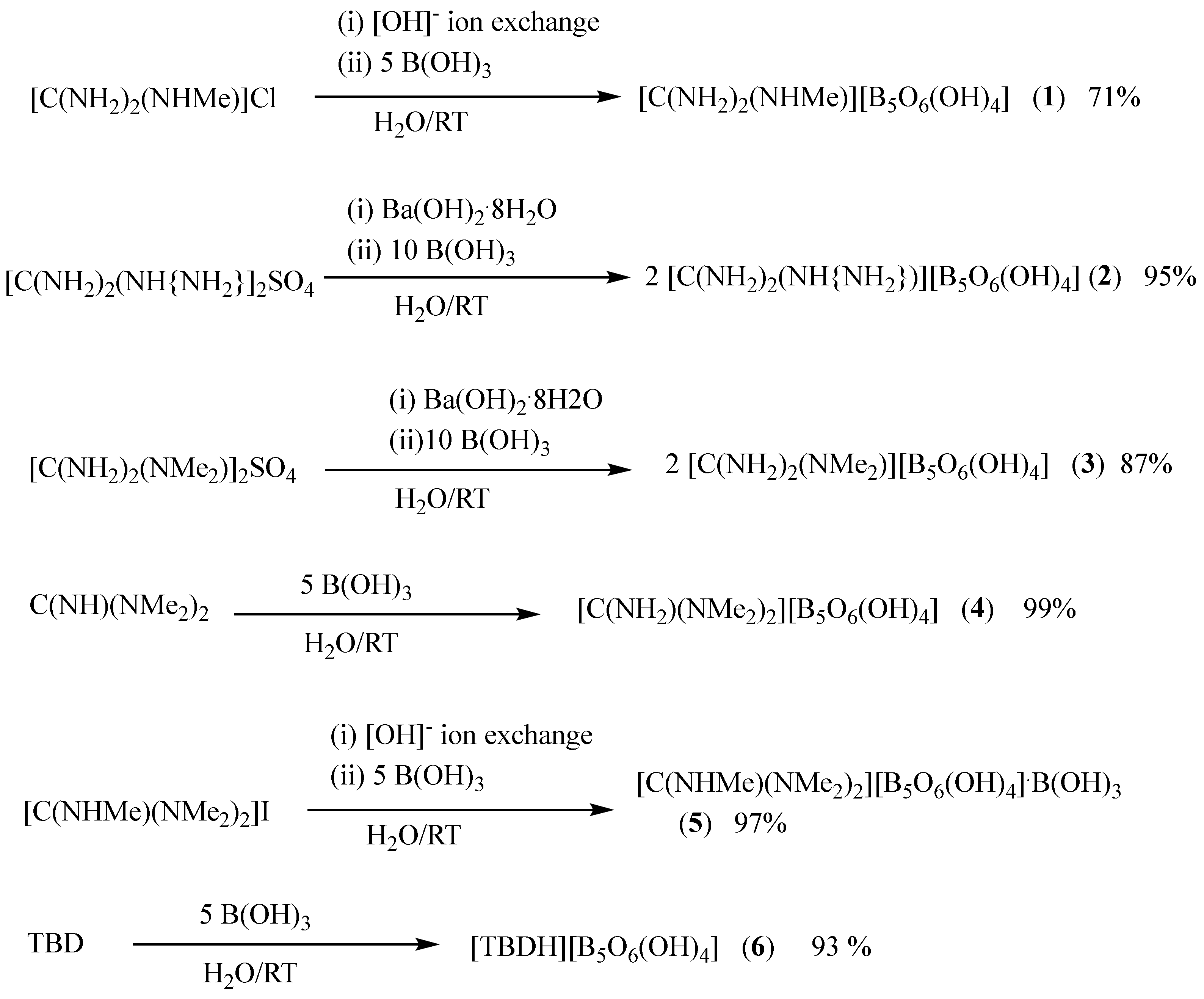
2.1. Synthesis

The three structurally characterized guanidinium polyborate salts [C(NH2)3]2[B4O5(OH)4]·2H2O [17], [C(NH2)3][B5O6(OH)4]·H2O [18], and [C(NH2)3]3[B9O12(OH)6] [19] were all prepared by crystallization from aqueous solutions containing [C(NH2)3]2CO3 and B(OH)3 at various temperatures and ratios [8]. Two alternative syntheses of [C(NH2)3]3[B9O12(OH)6] have been reported using either (i) [C(NH2)3]Cl, Na2B4O7·5H2O and B(OH)3 or (ii) [C(NH2)3]2[B4O5(OH)4]·2H2O and B(OH)3 in appropriate ratios [19]. It is well-known that B(OH)3, and other borate salts, exists in alkaline aqueous solution as equilibrium mixtures of numerous polyborate anions [27,28]. The guanidinium cation that is present templates the crystallization of specific products under the reaction conditions in what can be described as self-assembly processes [29,30].
The N-substituted guanidinium starting materials used in this study were all commercially available and were non-carbonate species. We have previously synthesized many nonmetal cation polyborate salts by a room temperature crystallization of aqueous solutions originally primed with an organic free base, or its protonated cation (prepared in situ as its [OH] salt by a metathesis reaction) and B(OH)3 [10,11]. Based on this strategy we prepared six new N-substituted guanidinium salts as shown in Scheme 1. Crude yields of these compounds ranged from 71% to near quantitative. The recrystallization of samples from H2O gave crystals suitable for single-crystal X-ray diffraction studies.
Thermal and spectroscopic data (Section 2.2) and elemental analysis data (Section 3.3, Section 3.4, Section 3.5, Section 3.6, Section 3.7, Section 3.8 and Section 3.9) on the crude products 16 were consistent with formulating these materials as N-substituted guanidinium pentaborate salts and these formulations were confirmed by single-crystal XRD studies (Section 2.3). All compounds were colourless and stable in the solid state, insoluble in organic solvents but soluble in H2O with decomposition.

2.2. Thermal and Spectroscopic Properties

Thermal gravimetric analysis (TGA) data (in air) for nonmetal cation polyborate salts are often reported with a thermal decomposition leading to B2O3 via multistage processes involving the loss of interstitial H2O (100–200 °C), the condensation of hydroxy groups bound to boron with the loss of a further two H2O molecules (250–400 °C), and then the oxidation of organics (400–700 °C) [10,31,32]. A TGA was undertaken on compounds 13 and 6 as representative examples of the new substituted guanidinium pentaborate(1-) salts. Compounds 2, 3, and 6 did not have interstitial H2O and their TGA curves showed the expected loss of two H2O molecules between 240 and 320 °C. Compound 1 followed this expected behaviour but there was no clear distinction between the two lower temperature processes that occurred (100–275 °C) with the loss of three H2O molecules. The glassy solids that remained after heating to 700 °C for compounds 13 and 6 had residual masses consistent with 2.5 B2O3.
1H and 13C NMR were obtained for compounds 1 and 36 dissolved in D2O and these all gave spectra consistent with the appropriate guanidinium cations being present, e.g., the 1H spectrum of compound 4 gave a signal for the N-Me groups as a singlet at 2.85 ppm with exchangeable H atoms with HOD (4.7 ppm). The 13C{1H} spectrum of compound 4 gave two signals at 36.98 and 160.2 ppm for the CH3 and CN3 carbons, respectively, with the downfield signal being weak. The presence of boron in these compounds was confirmed by 11B NMR (in D2O) for all compounds. All 11B spectra all showed three “signature” signals [31,33], arising through the decomposition of a pentaborate, with relative intensities appropriate for a sample that had attained polyborate/aqueous equilibrium [27]. Thus, compound 4 gave three signals at 1.0, 12.8, and 19.1 ppm with approximate relative intensities of 5%, 10%, and 85%, respectively. These signals have been previously assigned, moving downfield, to [B5O6(OH)4] (tetrahedral B), [B3O4(OH)4], and B(OH)3/[B(OH)4] [27]. Three signals were observed as the sample was relatively concentrated rather than one signal at +16.1, which would have been expected at infinite dilution [33].
All samples were characterized by FTIR spectroscopy. Strong and potentially diagnostic absorptions were to be expected for the cation ν(NH) at ca. 3400 cm−1 and ν(CN) at ca. 1650 cm−1 [34] and the polyborate anion ν(O-H) at 3500 cm−1 and the ν(B-O) stretches grouped between 1450 and 740 cm−1 [35]. In particular, a strong adsorption at ca. 925 cm−1 in the (Btrig-O)sym stretching region has previously been described as diagnostic for the [B5O6(OH)4] anion [36]. This strong absorption, amongst other strong B-O stretches, together with the strong adsorptions associated with the cation (1671–1625 cm−1), was present in all samples.

2.3. Single-Crystal XRD Studies

Compounds 16 were characterized by single-crystal XRD studies. All the compounds contained the expected insular N-substituted guanidinium(1+) cations and insular tetrahydroxidohexaoxidopentaborate(1-) anions within the asymmetric unit. Additionally, compound 1 had one H2O of crystallization per cation/anion and compound 5 was cocrystallized with a molecule of B(OH)3 per cation/anion. Crystallographically, compounds 15 were free of disorder, whilst compound 6 was modulated, but was only refined using the subcell with all atoms disordered over two positions of equal population. Brief crystallographic details for each compound are given in the experimental section, and atomic numbering schemes for compounds 16 are shown in Figure 2. The full crystallographic information is available as Supplementary Materials. Generally, the gross structures, bond angles, and bond distances within the guanidinium [17,37] and pentaborate [31,32,33,36] units were within the expected ranges for these ions and need no further comment. The N-substituted guanidinium cations had a variable number of potential H-bond donor sites (one to seven in this study) that were dependent on the extent of the substitution. Each pentaborate(1-) unit had four potential H-bonds donor sites and ten potential H-bond acceptor sites in locations described as α, β, or γ, as defined elsewhere [10,32], and they are illustrated in Figure 1. Since solid-state H-bonding is likely to be important in stabilizing these structures [23,24,25,26], the H-bond interactions in compounds 16 were analysed and are described in detail using an Etter graph set terminology [38].
Compounds 2, 3, 4, and 6 had the extended anion–anion H-bonded lattices that are found in many nonmetal cation pentaborate salts [10], with the cations filling the “voids” and “channels” and forming additional H-bonds to the anions. In these structures, each pentaborate(1-) anion donates four H-bond to four neighbouring pentaborate(1-) anions. The extended lattice structures are 3-D because the central B atom in each pentaborate(1-) is tetrahedral with its two associated boroxole rings perpendicular to each other (D2d for heavy atoms).
Thus, in compound 4, the H-bonds originating from each pentaborate were to three α and one β (or α,α,α,β) acceptor sites of its neighbours (see Figure 1 for acceptor site positions). This particular anion–anion giant structure is commonly found in many nonmetal cation pentaborate salts [10] and has be described as a “brick wall” with three R22(8) [38] reciprocal-α interactions forming a “layer”, resembling a brick wall, and the fourth interaction, at a β-site, linking these layers by infinite C(8) [38] chains. These R22(8) reciprocal interactions are known to be strong and are particularly common in polyborate chemistry [10,31,32,33,36,39]. R22(8) interactions are also prevalent elsewhere, e.g., carboxylic acid and boronic acid dimers [26]. In compound 4 these donors were O7-H7, O9-H9, and O10-H10 and the acceptors were O1, O4, and O6, respectively, on three different neighbouring pentaborate anions. The C(8) chain that linked layers arose through O8H8⋯O10′. The [C(NH2)(NMe2)2]+ cations in compound 4 were in the channels of the brick-wall structure (when viewed along the b axis) and each formed two donor H-bonds from the amino group to the γ (N1H1A⋯O2) and α (N1H1B⋯O3) positions of two neighbouring pentaborate(1-) anions.
Compounds 2 and 3 had similar, and previously observed [10], anion–anion H-bond arrangements (α,α,α,γ acceptor sites) as three R22(8) reciprocal-α interactions and one R22(8) reciprocal-γ interaction. The reciprocal-α H-bond donor interactions originated from O7-H7, O8-H8, and O10-H10 in compound 2 and from O8-H8, O9-H9, and O10-H10 in compound 3. The reciprocal-γ interactions for compound 2 were -O9-H9⋯O5′-B4′-O9′-H9′⋯O5-B4- and those for compound 3 were -O7-H7⋯O2′-B2′-O7′-H7′⋯O2-B2-. The [C(NH2)2(NMe2)]+ cation in compound 3 formed four H-bonds to three pentaborates of the anionic lattice. These interactions are shown in Figure 3a. The two interactions starting at N2-H2B and N3-H3A were both part of a dimeric centrosymmetric R44(20) ring comprised of two anions and two cations. The H-bonds originating from N2-H2A and N3-H3B were also of interest since these amino-hydrogens participated in a charge-assisted pincer R22(8) ring [23,40], as a double donor to an α-site (O1) and a β-site (O7) of a single pentaborate(1-) anion. Each [C(NH2)2(NH{NH2})]+ cation in compound 2 had seven potential H-bond donor sites and all were involved in H-bonds to five neighbouring pentaborate anions in a complex manner. However, the H-bond motifs described for compound 3 were also readily discerned in compound 2. Thus, “simple” interactions involved N1-H1A and N4-H4A with O8 (β) and O4 (α), respectively. N4-H4B was involved in a bifurcated H-bond bridging two pentaborates at the O5 (γ) sites. The four remaining H-bond donors, N1-H1B, N2-H2B, N2-H2A, and N3-H3, participated in two pincer-type R22(8) rings to two neighbouring pentaborates at O9 (β)/O4 (α) and O2 (γ)/O7 (β), respectively.
Compound 6 also had a previously observed anion–anion giant lattice with α,α,α,β acceptor sites (also observed in compound 4) but now arranged as three reciprocal-α rings and one R22(12) reciprocal-β ring [10]. The reciprocal-α H-bonds donor originated from O17-H17, O19-H19, and O20-H20 and the R22(12) reciprocal-β ring involved O18-H18⋯O17′ and O18′-H18′⋯O17. The bicyclic [TBDH]+ cation in compound 6 had two H-bonds donor sites, N12-H12 and N13-H13. Both were involved in a pincer-type R22(8) ring at O20 (β) and O15 (γ) acceptor sites, respectively, of a single pentaborate anion, Figure 3b.
Compounds 1 and 5 had additional cocrystallized molecules with the cation and anion. Cocrystallized molecules in H-bonded anionic polyborate lattices usually function either by (i) helping to fill space within the areas where the cations sit or by (ii) expanding the anionic lattices to help accommodate larger cations [10,41].
Compound 1 had a pentaborate lattice structure that was very similar to compound 3 with α,α,α,γ acceptor sites involved in four R22(8) rings. In compound 1, these donors were O7-H7, O8-H8, O9-H9, and O10-H10, and the acceptors were O3 (α), O1 (α), O5 (γ), and O6 (α) on four different neighbouring pentaborate anions. The structure of compound 1 differed from that of compound 3 in the detail of their H-bonded R22(8) rings with H-bonds originating from O10-H10 and O9-H9 reciprocal and those originating from O7-H7 and O8-H8 paired. The [C(NH2)2(NHMe)]+ cation in compound 1 had five H-bond donor sites. N2-H2A and N3-H3B were both part of a pincer-like R22(8) ring to O4 (α) and O9 (β) acceptor sites, respectively. N3-H3A and N2-H2B both donated H-bonds to a β-sites (O7 and O8). The H-bond from N2-H2B had a N2-H2B-O8 angle of 141.1(13)° and a H2B⋯O8 distance of 2.322(15) Å. The H-bond arising from N2-H2B may be considered as bifurcated since O11 (H2O) had a N2-H2B-O11 angle of 138.8(13)° with a long, potentially H-bonding H2B⋯O11 distance of 2.661(15) Å. This H2O molecule was also involved with closer and stronger acceptor interaction from N1-H1 (H1⋯O11, 1.943(15) Å and an angle of N1-H1⋯O11 170.2(14)°. This interaction together with that from N2-H2B formed a pincer R21(6) ring. The H2O molecule formed two donor bonds to two pentaborates at α,α (O1/O4, bifurcated) and β (O10) sites. The anion–anion lattice in compound 1 was largely unaffected by the presence of the cocrystallized H2O molecule. Since the cation in compound 1 was relatively small, the H2O positioned itself to fill space within the lattice reserved for the cations and further stabilized the structure by these H-bond interactions.
Compound 5 had B(OH)3 cocrystallized with the pentaborate salt and consequentially had a unique anionic pentaborate/boric acid lattice. However, the R22(8) building motifs seen in most polyborate structures were also readily identified here. Thus, one “plane” was comprised of chains of pentaborates each linked via two reciprocal-α rings. These reciprocal-α interactions originated from O7-H7 and O8-H8 with O3 and O1 acceptors, respectively. The other two H-bond interactions originating from each pentaborate (O9-H9 and O10-H10) are shown in Figure 4. The interactions shown here were approximately perpendicular to those just described an originating from O7-H7 and O-H8. The “horizontal” chains in Figure 4 were comprised of alternating pentaborate(1-)/boric acid moieties with the H-atom positions of the B(OH)3 acid arranged to maximise R22(8) interactions. Thus, O10-H10 on the pentaborate and O12-H12 on the B(OH)3 were involved in a standard borate–borate interaction and O11-H11 and O13-H13 were involved in a “pincer” double H-bond to the pentaborate at O4 (α) and O9 (β) sites. Each B(OH)3 formed three donor H-bonds to two α sites and one β site of neighbouring pentaborates. The fourth and final H-bond originating from each pentaborate is also shown in Figure 4. Here, O9-H9 formed an H-bond to a β-acceptor site (O8) of its neighbour, cross-linking the pentaborate(1-)/boric acid chains into the “plane” shown in Figure 4 by the formation of C(8) chains. The sterically demanding [C(NHMe)(NMe2)2]+ cation in compound 5 had only one amino group (N1-H1) available for H-bonding and this was utilized in the H-bonding to O11 of the B(OH)3 (Figure 4). The B(OH)3 in this structure served to expand the anionic pentaborate lattice to enable the sterically demanding cation more space. This behaviour has been observed before in several polyborate [36,41,42,43] salts including [N(nPr)4][B5O6(OH)4][B(OH)3]2 and [HPS][B5O6(OH)4].B(OH)3 (PS = proton sponge, 1,8-bis(dimethylamino)naphthalene).

2.4. Discussion on H-Bonding in Guanidinium Polyborates

The anions in polyborate salts, [BaOb(OH)c]n, are invariably rich in H-bond donor sites (there are c donor BOH groups) and oxygen-atom H-bond acceptor sites with acceptor sites (b + c) outnumbering the donor sites and enabling the opportunity for H-bond donor cations to contribute to the H-bonding networks. R22(8) are important stabilizing interactions [10,33] and the maximum number of borate–borate R22(8) rings available to a polyborate anion is limited to the lower value of b or c, since bridging oxygen atoms are acceptors in these R22(8) interactions. In actual structures, packing/steric effects may reduce further this maximum number.
The guanidinium (G) cation is well-known as a multi-H-bond donor in solid-state structures and several H-bond motifs are commonly observed [23,24,37,44,45,46,47,48]. These include a donor H-bond to an electronegative atom from a single N-H site (often this simple motif is incorporated into larger more complex rings and/or chains), and two pincer-type doner ring motifs to either a single acceptor site, R21(6), or to two acceptor sites, R22(8). The structure of GCl has Cl ions bridging G cations as part of alternating anion–cation chains linked by H-bonded R21(6) rings [44]. Oxoanions (e.g., borate esters [24], carboxylate [45,46], sulfonates [47,48], phosphonates [48]), are good R22(8) pincer H-bond acceptors and these interactions are strong and have been described as “‘charge-assisted” [45,49]. The structure of G[RSO3] is self-assembled and templated into layers with each G cation forming three such pincer R22(8) rings with three organosulfonate anions [47]. Cubic hydrogen-bonded borate networks containing four pincer R22(8) rings per borate have been observed in G4[B(OMe)4]3Cl·5H2O [24].
As noted in the introduction, the G cation was found in three polyborate structures. These “oxoanions” are ideally set up to accept the pincer R22(8) rings, and we examined, focussing on the polyborate anion, the extent of such interactions as they are in direct competition (see below) with borate–borate R22(8) rings in stabilizing these structures. We also examined how N-substitution affected this R22(8) balance and how this affected the observed structures.
When considering solid-state H-bonding interactions the generalised guanidinium (either N-substituted or unsubstituted) polyborate salt can be represented as (xG)n[BaOb(OH)c] where x represents the maximum number of potential donor pincer R22(8) motifs available to the cation. Each unsubstituted G cation has the potential to form a maximum of three donor pincer R22(8) motifs (i.e., x = 3), whilst substituted G cations will have fewer opportunities (x = 0, 1, or 2) with the value of x dependent on the extent and position of the substitution. Within guanidinium polyborate salts, the maximum number of R22(8) acceptor interactions (both pincer and normal) is b, since bridging oxygen atoms generally can only be used once and are required for both types of these R22(8) interactions. Usually, borate–borate interactions are approximately coplanar with boroxole rings. Packing/steric effects may reduce the maximum number for both types of interactions.
This analysis focused on an analysis of two ratios. Firstly, a value of >1 for (xGn + c)/b would indicate borate–borate and pincer R22(8) motifs were in direct competition for the available borate acceptor sites. Secondly, potential and actual (i.e., observed) (xGn)/c ratios for each compound were also of interest since a difference in these values may indicate a structural preference for one or the other of these H-bonding motifs.
Using this approach, G2B4O5(OH)4, GB5O6(OH)4, and G3B9O12(OH)6, had (xGn + c)/b ratios of 2.0, 1.17, and 1.25, respectively, confirming that pincer/borate donor rings were in competition for the available acceptor sites. The potential donor ratios, (xGn)/c, for the three compounds were 1.5, 0.75, and 1.5.
The examination of the structure of G2[B4O5(OH)4]·2H2O [17] revealed two independent G cations. Two molecules of H2O within this formula unit also affected the structure but did not affect this analysis. All five bridging oxygen atoms of the tetraborate(2-) anion were involved in R22(8) rings with four cation–borate pincer interactions and only one borate–borate interaction. Thus, within this structure, 80% of these rings involved G cations and the observed (xGn)/c ratio was 4.0. This value was much higher than would be expected (1.5) based on the number of potential donor sites. The higher-than-expected observed (xGn)/c ratio would indicate that the G cations were either able to replace the strong borate–borate interactions with stronger pincer motifs or that additional factors were also important. Such factors may include the compound’s stoichiometry and that the three-fold symmetry of G cations (D3d) were a good packing match for the tetraborate(2-) anion (C2v). H-bonding in tetraborate(2-) structures not containing the G cation have been recently reviewed [50] and their structural architectures are, as expected, dominated by borate–borate interactions with the majority of structures having at least two R22(8) rings.
Within the structure of G3[B9O12(OH)6] [19], all twelve bridging oxygen acceptor sites were involved in eleven R22(8) H-bonded rings with seven pincer charge-assisted interactions involving the three G cations and four borate–borate interactions. Interestingly, the four bridging oxygen atoms joined to the central boron were involved, solely amongst themselves, in three pincer rings; the two oxygens that bridged two four-coordinate borons were both involved in two pincer rings. These rings were not coplanar with boroxole rings. In this salt, 64% of the R22(8) rings involved the G cations and 36% were borate–borate. This observed ratio of 1.78 was higher than that expected (1.5) based on total potential donor sites and would again indicate that strong cation–anion pincer interactions were able to replace the strong borate–borate interactions, but again, stoichiometry and packing considerations were also important.
The H-bonded structure of G[B5O6(OH)4]·H2O [18] utilizes only four of the six available acceptor bridging oxygen atoms in R22(8) interactions. Here, and in contrast to the tetraborate(2-) and nonaborate(3-) structures, 75% of the H-bond ring interactions were borate–borate R22(8) rings with the charge-assisted pincer rings only accounting for 25%. The observed (xGn)/c ratio of 0.33 was lower than would be expected (0.75) from the number of potential donor sites and confirmed that the borate–borate interactions strongly influenced that structure and that the templating influence of G was minimal. These structures were strongly stabilized by these anion–anion interactions with any additional cation–anion interactions helping to further stabilize the anionic lattice [10]. Many nonmetal cation pentaborate(1-) salts show three such borate–borate interactions [10]. This was also the likely situation in this salt since the three-fold symmetry of the cation did not pack well with the D2d symmetry of the insular pentaborate(1-) anion and its perpendicularly H-bonded giant lattice.
Adding substituents to the G cations affects their structure directing potential in three ways: it reduces their ability to form H-bonds, increases their steric bulk, and disrupts their three-fold symmetry. The first factor also correspondingly lowers x, reduces the (xGn)/c ratio, and hence encourages more borate–borate H-bonds. The structures of the N-substituted guanidinium cation pentaborate(1-) salts 16 were described in detail in Section 2.2, but further general comments based on the analysis above would now be appropriate. The numbers of potential pincer H-bond donor sites (x) available in compounds 16 were 2, 2, 1, 0, 0, and 1, and the (xGn)/c ratios were lower than that for G[B5O6(OH)4] (0.75) at 0.5, 0.5, 0.25, 0, 0, and 0.25, respectively. This analysis would indicate borate–borate interactions should dominate the H-bond interactions in compounds 16, with additional charge-assisted pincer H-bonds forming wherever possible. Unsurprisingly, three or four borate–borate R22(8) rings were observed in compounds 16 and these interactions were clearly important in stabilizing their solid-state structures. Pincer interactions were unavailable for compounds 4 and 5 and the amino-H atoms formed simple H-bonds to borate/boric acid acceptor sites. Compounds 3 and 6 both had the potential to form one pincer bond to stabilize the lattice and each had one such pincer bond. Compound 2 had the potential to form two pincer bonds, and both were formed. Compound 1 had potentially two pincer bonds to the borate but here, only one was observed. However, this was a direct consequence of the cocrystallized H2O since the H2O was part of an alternative R21(6) pincer ring motif. Compound 5 had a molecule of B(OH)3 cocrystallized and no opportunity to form a pincer ring. As noted in Section 2.2, the [C(NHMe)(NMe2)2]+ in compound 5 was relatively bulky and the B(OH)3 served to extend the anionic lattice, by positioning itself between two pentaborate(1-) anions. It is interesting to note that the H-atom positions in B(OH)3 were asymmetric (see Figure 4) and therefore allowed it to partake in an R22(8) donor pincer interaction to one pentaborate(1-) and a standard R22(8) borate interaction to the other.

3. Materials and Experimental Methods

3.1. General

Reagents were all obtained commercially. FTIR spectra were obtained as KBr pellets on a Perkin-Elmer 100FTIR spectrometer (Perkin-Elmer, Seer Green, UK). 1H, 11B, and 13C{1H} NMR spectra were obtained on a Bruker Ultrashield Plus 400 spectrometer (Bruker, Coventry, UK) on samples dissolved in D2O at 400, 128, and 100 MHz, respectively. Chemical shifts are in ppm with positive values to the high frequency (downfield) of TMS (1H, 13C) or BF3.OEt2 (11B). TGA and DSC were performed on an SDT Q600 V4 instrument (TA Instruments, New Castle, DE, USA) using Al2O3 crucibles with a temperature ramp-rate of 10 °C per minute (20–700 °C in air). X-ray crystallography was performed at the EPSRC national crystallography service centre at Southampton University. Drawings in this manuscript have been generated using Mercury software (CCDC, Cambridge, UK). CHN analyses were obtained from OEA Laboratories (Callingham, Cornwall, UK).

3.2. X-ray Crystallography

Crystallographic data for compounds 13, 5, and 6 were obtained, with crystals kept at T = 100(2) K during data collection, on either a Rigaku FRE+ diffractometer equipped with VHF Varimax confocal mirrors and an AFC12 goniometer and Hypix 6000 detector (1, 3) or a Rigaku 007HF equipped with HF Varimax confocal mirrors and an AFC11 goniometer and HyPix 6000 detector (2, 5, 6) using CrysAlisPro [51]. Structures (13, 5, 6) were solved with the ShelXT [52] structure solution programme using the dual-solution structure method (1, 5) or the intrinsic-phasing solution method (2, 3, 6).The crystallographic data collection for compound 4 was obtained at 120(2) K on a Bruker-Nonius FR591 diffractometer with a Nonius KappaCCD detector using Collect [53] and Denzo [54] and SHELXS97 [55] was used for the structure solution. By using Olex2 [56] as the graphical interface, all structure models were refined with ShelXL [57] using a least-squares minimisation. Structure refinement details and ORTEP diagrams with 50% probability displacement ellipsoids for compounds 16 are given in the Supplementary Materials and selected crystallographic data are given for each compound in Section 3.3, Section 3.4, Section 3.5, Section 3.6, Section 3.7, Section 3.8 and Section 3.9.

3.3. Preparation of [C(NH2)2(NHMe)][B5O6(OH)4]·H2O (1)

DOWEX 550A (OH) ion exchange resin (18 g) was added to a solution of [C(NH2)2(NHMe)]Cl (0.5 g, 4.6 mmol) in H2O (25 mL) and the mixture was stirred for 18 h. The resin was removed by filtration and B(OH)3 (1.4 g, 23 mmol) was added to the filtrate. The solution was stirred for a further 2 h, evaporated under reduced pressure to yield 1.1 g of a white powder as a crude product (71%). Recrystallization of 0.3 g of the product from warm H2O afforded a few crystals suitable for XRD studies. TGA: 100–275 °C, condensation of pentaborate with loss of 3 H2O 17.4% (17.4% calc.); 275–700 °C, oxidation of organic residue leaving residual 2.5 B2O3 55.9% (56.1% calc.). NMR/ppm: δH: 2.69 (s, CH3) 4.70 (HOD); δB: 1.1 (3%), 13.2 (24%), 18.1 (73%); δC: 36.98 (CH3). FTIR (KBr, cm−1): 3435(s), 3400(s), 2958(m), 2924(m), 1664(s), 1439(m), 1347(m), 1103(m), 1025(m), 925(s), 782(m), 696(m). XRD crystallographic data: C2H14B5N3O11, Mr = 310.21, monoclinic, P21/c (no. 14), a = 9.9962(2) Å, b = 10.9047(2) Å, c = 11.7215(2) Å, β = 96.101(2), α = γ = 90°, V = 1270.47(4) Å3, T = 100(2) K, Z = 4, Z′ = 1; 17,592 reflections were measured, 2893 unique (Rint = 0.0115), which were used in all calculations. The final wR2 was 0.0743 (all data) and R1 was 0.0267 (I > 2σ(I)).

3.4. Preparation of [C(NH2)2(NH{NH2})][B5O6(OH)4] (2)

[C(NH2)2(NH{NH2})]2SO4 (1 g, 4.0 mol) was dissolved in H2O (25 mL) and to this was added Ba(OH)2.8H2O (1.3 g, 4.1 mmol) in H2O (5 mL). The resulting cloudy solution was stirred for 1 h and the solid barium sulphate was filtered from the solution. B(OH)3 (2.3 g, 37 mmol) was added to the clear solution, and stirred with gentle heating for 3 h. The solution was then evaporated under reduced pressure and dried in an oven to afford 2.0 g of a white powder as crude product (95%). A sample (0.3 g) of this solid was redissolved in 10 mL deionised water and left to crystallise over a few days to yield a few crystals suitable for XRD studies. CH11B5N4O10. Anal. Calc.: C = 4.1%, H = 3.8%, N = 19.1%. Found: C = 4.2%, H = 3.7%, N = 18.7%. TGA: 240–320 °C, condensation of pentaborate(1-) anions with loss of 2 H2O 11.9% (12.3% calc.); 320–700 °C, oxidation of organic residue leaving residual 2.5 B2O3 59.3% (59.3% calc.). NMR/ppm: δB: 1.2 (10%), 13.3 (9%), 19.2 (81%). FTIR (KBr, cm−1) 3451(s), 3372(s), 3295(s), 3235(m), 1672(s), 1657(s), 1437(s), 1361(s), 1252(m), 1196(m), 1103(s), 1026(s), 925(s), 782(s), 698(s). XRD crystallographic data: CH11B5N4O10, Mr = 293.19, triclinic, P-1 (no. 2), a = 7.4870(2) Å, b = 8.5076(2) Å, c = 9.6502(2) Å, α = 93.906(2)°, β = 98.470(2)°, γ = 96.457(2)°, V = 601.88(3) Å3, T = 100(2) K, Z = 2, Z′ = 1; 11,885 reflections were measured, 2142 unique (Rint = 0.0404), which were used in all calculations. The final wR2 was 0.0827 (all data) and R1 was 0.0294 (I > 2σ(I)).

3.5. Preparation of [C(NH2)2(NMe2)][B5O6(OH)4] (3)

An aqueous solution (5 mL) containing Ba(OH)2·8H2O (1.2 g, 3.8 mmol) was added to an aqueous solution (25 mL) of [C(NH2)2(NMe2)]2SO4 (1.0 g, 3.7 mmol). The mixture was stirred for 1 h and the BaSO4 was removed from the solution by gravity filtration and washed with H2O. B(OH)3 (2.2 g, 36 mmol) was added to the filtrate and the solution was stirred under gentle warming for 1 h. The solvent was removed under reduced pressure to yield 2.0 g of a white powder as the crude product (87%). This solid was characterised by FT-IR and NMR (1H, 11B, 13C) studies. A sample (0.3 g) of the crude product was redissolved in warm H2O and left to recrystallize. A few white single crystals suitable for XRD studies were obtained. C3H14B5N3O10. Anal. Calc.: C = 11.8%, H = 4.6%, N = 13.8%. Found: C = 11.9%, H = 4.6%, N = 13.5%. TGA: 240–275 °C, condensation of pentaborate(1-) anions with loss of 2 H2O 12.3% (11.8% calc.); 275–700 °C, oxidation of organic residue leaving residual 2.5 B2O3 55.3 (56.8% calc.). NMR/ppm δH: 2.89 (s, CH3N), 4.70 (HOD); δB: 1.0 (13%), 13.2 (11%), 18.3 (77%); δC: 37.34 (CH3). FTIR (KBr, cm−1): 3370(s), 3249(m), 3200(m), 1688(m), 1671(s), 1634(s), 1435(vs), 1393(s), 136 (s), 1313(s), 1114(s), 1033(s), 926(vs), 780(s), 699(m). XRD crystallographic data: C3H14B5N3O10, Mr = 306.22, monoclinic, P21/c, a = 9.9747(2) Å, b = 11.2563(3) Å, c = 11.6174(3) Å, α = γ = 90°, β = 96.084(2)°, V = 1297.03(5) Å3, T = 100(2) K, Z = 4, Z′ = 1; 18,168 reflections were measured, 2969 unique (Rint = 0.0191), which were used in calculations. The final ωR2 was 0.0811 (all data) and R1 was 0.0280 (I > 2σ(I)).

3.6. XRD Data for [C(NH2)(NMe2)2][B5O6(OH)4] (4)

B(OH)3 (6.2 g, 100 mmol) was dissolved in H2O (100 mL) and [C(NH)(NMe2)2] (1.2 g, 20 mmol) was added with stirring. The solution was stirred at RT for 1 h and evaporated to dryness to yield 6.6 g (99%) of the crude product. NMR/ppm δH: 2.85 (s, CH3N), 4.70 (HOD); δB: 1.0 (5%), 12.8 (10%), 19.1 (85%); δC: 38.77 (CH3), 161.3 (CN3). FTIR (KBr cm−1): 3164(m), 1669(m), 1377(s), 1291(s), 1041(m), 917(s), 772(m), 725(m). A sample (0.3 g) of this product was recrystallized from H2O to afford crystals suitable for sc-XRD studies. C5H18B5N3O13, Mr = 334.27, triclinic, P-1 (no.2), a = 9.5035(3) Å, b = 9.5151(3)Å, c = 10.4386(3) Å, α = 65.3810(10)°, β = 69.049(2), γ = 88.603(2), V = 792.72(4) Å3, T = 100(2) K, Z = 2; 16,517 reflections were measured, 3638 unique (Rint = 0.0406), which were used in all calculations. The final wR2 was 0.0935 (all data) and R1 was 0.0373 (I > 2σ(I)).

3.7. Preparation of [C(NHMe)(NMe2)2]I

C(NH)(NMe2) (2.1 g, 2.3 mL, 18.2 mmol) was added to an Ar charged two-necked flask, and to this, CH3CN (10 mL) was added. The sealed vessel was then cooled to 0 °C using an ice bath. MeI (2.9 g, 20.4 mmol) was then added to CH3CN (8 mL) and this solution was added dropwise to the cold C(NH)(NMe2) solution. The mixture was then stirred for 12 h and left to slowly equilibrate to room temperature. The solvents were then removed via rotary evaporation. The resulting oil was then washed with EtOAc (3 × 25 mL) and dried under vacuum at room temperature to yield a white crystalline product [C(NHMe)(NMe2)2]I (3.2 g, 63%). NMR/ppm: δH: 2.73 (3H, s, CH3N), 2.80 (12H, s, CH3N); δC: 38.91 (CH3).

3.8. Preparation of [C(NHMe)(NMe2)2][B5O6(OH)4]·B(OH)3 (5)

[C(NHMe)(NMe2)2]I (1.0 g, 4.0 mmol) was dissolved in H2O (25 mL) along with a DOWEX 550A (OH) ion exchange resin (18 g) and stirred for 18 h. The resin was removed by filtration and the filtrate was added to B(OH)3 (1.3 g, 21 mmol). The solution was stirred for 2 h and evaporated under reduced pressure to yield 1.4 g of a white powder as a crude product (97%). A 0.3 g sample of the product was redissolved in H2O (15 mL) and white crystals suitable for X-ray diffraction studies were obtained after a few days. C6H23B6N3O13. Anal. Calc.: C = 17.6%, H = 5.7%, N = 10.2%. Found: C = 17.7%, H = 5.5%, N = 10.1%. NMR/ppm: δH: 2.73 (3H, s, CH3N), 2.80 (12H, s, CH3N), 2.82 (1H, s), 4.70 (HOD); δC: 38.89 (CH3). δB: 1.1 (9%), 13.2 (13%), 18.8 (78%); FTIR (KBr, cm−1): 3370(s), 3236(s), 1625(s), 1592(m), 1426(s), 1398(m), 1367(s), 1324(m), 1156(m), 1022(m), 922(vs), 779(m), 709(m). XRD crystallographic data: C6H23B6N3O13, Mr = 410.13, monoclinic, P21/c (no. 14), a = 9.49570(10) Å, b = 11.44900(10) Å, c = 16.84590(10) Å, β = 98.0710(10), α = γ = 90, V = 1813.28(3) Å3, T = 100(2) K, Z = 4, Z′ = 1; 20,493 reflections were measured, 3281 unique (Rint = 0.0196), which were used in all calculations. The final wR2 was 0.0674 (all data) and R1 was 0.0263 (I > 2σ(I)).

3.9. Preparation of [TBDH][B5O6(OH)4] (6)

1,5,7-Triazabicyclo[4.4.0]dec-5-ene (TBD, 1.0 g, 7.2 mmol) was dissolved in H2O (20 mL) and to this was added B(OH)3 (2.2 g, 36 mmol). The resulting solution was stirred under gentle heating to fully dissolve the B(OH)3 and left for 3 h. The solution was then evaporated under reduced pressure to yield a crude white product (2.4 g, 6.7 mmol, 93%).
A small sample of this solid (0.3 g) was redissolved in H2O (20 mL) and left to recrystallize over 7 d to afford a small number of white crystals, suitable for X-ray diffraction studies. C7H18B5N3O10. Anal. Calc.: C = 23.5%, H = 5.1%, N = 11.7%. Found: C = 23.7%, H = 5.0%, N = 11.5%. TGA: 245–295 °C, condensation of pentaborate with the loss of 2 H2O, 11.0% (10.0% calc.); 295–700 °C, oxidation of organic residue leaving residual 2.5 B2O3 49.0% (48.5% calc.). NMR/ppm: δH: 1.84 (4H, quin, CH2), 3.12 (4H, t, CH2N), 3.20 (4H, t, CH2N), 4.70 (HOD); δC: 20.12 (CH2), 37.71 (CH2), 46.38 (CH2). δB: 0.8 (1%), 13.0 (29%), 17.7 (70%). FTIR (KBr/cm−1): 3238(m), 1630(m),1408(m), 1294(s), 1144(m), 1086(m), 1015(m), 910(vs), 816(m), 772(s), 723(m), 706(s). XRD crystallographic data: C7H18B5N3O10, Mr = 358.29, triclinic, P-1 (no. 2), a = 9.3096(6) Å, b = 9.3175(3) Å, c = 9.3733(6) Å, α = 76.598(5)°, β = 85.611(5)°, γ = 79.947(4)°, V = 778.22(8) Å3, T = 100(2) K, Z = 2, Z′ = 1; 13,382 reflections were measured, 2836 unique (Rint = 0.0443), which were used in all calculations. The final wR2 was 0.1747 (all data) and R1 was 0.0561 (I > 2σ(I)).

4. Conclusions

The synthesis and structural characterization of six N-substituted guanidinium pentaborate(1-) salts was accomplished. Reactant stoichiometry, solid-state packing, and H-bonding interactions are all important factors in the self-assembly of these structures. An analysis of the H-bonding motifs in these salts and in related unsubstituted guanidinium polyborates was undertaken to evaluate this contribution. This analysis indicated that strong charge-assisted pincer R22(8) motifs originating from the cation were in competition with the strong borate–borate R22(8) motifs for the available borate acceptor sites. The ratios of potential cationic pincer-type H-bond donors to borate H-bond donors were determined for all compounds and compared with the ratios observed in their solid-state structures. Differences in these values were indicative of a structural preference for one or the other of these H-bonding motifs. The salts [C(NH2)3]2[B4O5(OH)4]·2H2O and [C(NH2)3]3[B9O12(OH)6] had potential ratios of >1 with the observed ratios higher than their potential ratios, indicating that the cation–anion pincer H-bond motifs were favoured. In contrast, [C(NH2)3][B5O6(OH)4].H2O and the six new substituted guanidinium pentaborates described within this manuscript all had potential ratios of <1 and the observed ratios were generally lower than their potential ratios, indicating favourable borate–borate H-bond interactions. However, as both types of H-bond motifs were expected to be equally strong, other factors such as stoichiometry and crystal packing were strongly influential in determining the observed structures.

Supplementary Materials

The following supporting information can be downloaded at: https://www.mdpi.com/article/10.3390/molecules28073273/s1, Crystallographic data for compounds 16 are available as Supplementary Materials. CCDC 2249242-7 also contain the supplementary crystallographic data for this paper. These CCDC data can be obtained free of charge via http://www.ccdc.cam.ac.uk/conts/retrieving.html (or from CCDC, 12 Union Road, Cambridge, CB2 1EZ. Fax: +44 1223 336033; E-mail: [email protected]).

Author Contributions

M.A.B. conceived the research strategy and drafted this manuscript, supervision; T.A.R., experimental synthetic procedures and initial draft writing (Ph.D. thesis); P.N.H. and S.J.C., experimental crystallography and structure determinations; M.A.B., P.N.H. and T.A.R. manuscript review and editing; S.J.C., funding acquisition. All authors have read and agreed to the published version of the manuscript.

Funding

This research received no external funding.

Institutional Review Board Statement

Not applicable.

Informed Consent Statement

Not applicable.

Data Availability Statement

Crystallographic available from CCDC, UK, see Supplementary Materials for details. CIF files for published unsubstituted guanidinium polyborate salts were also obtained from CCDC: 146622 (WEHFAQ), 1132636 (CUGNAT), and 1824332 (QUFJAF).

Acknowledgments

Crystals of compound 4 were kindly provided by J. Morris. We thank the EPSRC for the NCS X-ray crystallographic service (Southampton).

Conflicts of Interest

The authors declare no conflict of interest.

Sample Availability

Samples of the compounds are not available from the authors.

References

  1. Farmer, J.B. Metal borates. Adv. Inorg. Chem Radiochem. 1982, 25, 187–237. [Google Scholar]
  2. Christ, C.L.; Clark, J.R. A crystal-chemical classification of borate structures with emphasis on hydrated borates. Phys. Chem. Miner. 1977, 2, 59–87. [Google Scholar] [CrossRef]
  3. Heller, G. A survey of structural types of borates and polyborates. Top. Curr. Chem. 1986, 131, 39–98. [Google Scholar]
  4. Becker, P. Borate materials in nonlinear optics. Adv. Mater. 1998, 10, 979–992. [Google Scholar] [CrossRef]
  5. Burns, P.C.; Grice, J.D.; Hawthorne, F.C. Borate minerals I. Polyhedral clusters and fundamental building blocks. Can. Mineral. 1995, 33, 1131–1151. [Google Scholar]
  6. Grice, J.D.; Burns, P.C.; Hawthorne, F.C. Borate minerals II. A hierarchy of structures based upon the borate fundamental building block. Can. Mineral. 1999, 37, 731–762. [Google Scholar]
  7. Touboul, M.; Penin, N.; Nowogrocki, G. Borates: A survey of main trends concerning crystal chemistry, polymorphism and dehydration process of alkaline and pseudo-alkaline borates. Solid State Sci. 2003, 5, 1327–1342. [Google Scholar] [CrossRef]
  8. Schubert, D.M.; Smith, R.A.; Visi, M.Z. Studies of crystalline non-metal borates. Glass Technol. 2003, 44, 63–70. [Google Scholar]
  9. Schubert, D.M.; Knobler, C.B. Recent studies of polyborate anions. Phys. Chem. Glasses Eur. J. Glass Sci. Technol. B 2009, 50, 71–78. [Google Scholar]
  10. Beckett, M.A. Recent Advances in crystalline hydrated borates with non-metal or transition-metal complex cations. Coord. Chem. Rev. 2016, 323, 2–14. [Google Scholar] [CrossRef] [Green Version]
  11. Xin, S.-S.; Zhou, M.-H.; Beckett, M.A.; Pan, C.-Y. Recent advances in crystalline oxidopolyborate complexes of d-block or p-block metals: Structural aspects, synthesis, and physical properties. Molecules 2021, 26, 3815. [Google Scholar] [CrossRef]
  12. Schubert, D.M. Borates in industrial use. Struct. Bond. 2003, 105, 1–40. [Google Scholar]
  13. Schubert, D.M. Boron oxide, boric acid, and borates. In Kirk-Othmer Encyclopedia of Chemical Technology, 5th ed.; John and Wiley and Sons: Hoboken, NJ, USA, 2011; pp. 1–68. [Google Scholar]
  14. Schubert, D.M. Hydrated zinc borates and their industrial use. Molecules 2019, 24, 2419. [Google Scholar] [CrossRef] [Green Version]
  15. Beckett, M.A.; Brellocks, B.; Chizhevsky, I.T.; Damhus, T.; Hellwich, K.-H.; Kennedy, J.D.; Laitinen, R.; Powell, W.H.; Rabinovich, D.; Vinas, C.; et al. Nomenclature for boranes and related species (IUPAC Recommendations 2019). Pure Appl. Chem. 2020, 92, 355–381. [Google Scholar] [CrossRef] [Green Version]
  16. Shaw, J.W.; Grayson, D.H.; Rozas, I. Topics in Heterocyclic Chemistry; Springer: Berlin/Heidelberg, Germany, 2015; pp. 1–51. [Google Scholar]
  17. Weakley, T.J.R. Guanidinium tetraborate(2-) dihydrate, (CH6N3)2[B4O5(OH)4]·2H2O. Acta Cryst. 1985, C41, 377–399. [Google Scholar] [CrossRef] [Green Version]
  18. Dhatchaiyini, M.K.; Rajasekar, G.; Mohideen, M.N.; Bhaskaran, A. Investigation on structural, spectral, optical, thermal studies and third order NLO properties of guanidinium pentaborate monohydrate single crystal. J. Mol. Sci. 2020, 1210, 128065. [Google Scholar] [CrossRef]
  19. Schubert, D.M.; Visi, M.Z.; Knobler, C.B. Guanidinium and imidazolium borates containing the first examples of an isolated nonaborate oxoanion: [B9O12(OH)6](3−). Inorg. Chem. 2000, 39, 2250–2251. [Google Scholar] [CrossRef]
  20. Rosenheim, A.; Lyser, F. Über Polyborate in wäßriger lösung. (Zur kenntnis der iso-und heteropolysäuren. XVII. Mitteilung). Z. Anorg. Allg. Chem. 1921, 119, 1–38. [Google Scholar] [CrossRef] [Green Version]
  21. Bowden, G.H. Supplement to Mellor’s Comprehensive Treatise on Inorganic and Theoretical Chemistry; Boron, Part A: Boron-Oxygen Compounds; Longman Group Ltd.: London, UK, 1980; Volume 5. [Google Scholar]
  22. Wood, G.L.; Janik, J.F.; Visi, M.Z.; Schubert, D.M.; Paine, R.T. New borate precursors for boron nitride powder synthesis. Chem Mater. 2005, 17, 1855–1859. [Google Scholar] [CrossRef]
  23. Blondeau, P.; Segura, M.; Perez-Fernandez, R.; de Mendoza, J. Molecular recognition of oxoanions based on guanidinium receptors. Chem. Soc. Rev. 2007, 26, 198–210. [Google Scholar] [CrossRef]
  24. Abrahams, B.F.; Haywood, M.G.; Robson, R. Guanidinium ion as a symmetrical template in the formation of cubic hydrogen-bonded borate networks with the boracite topology. J. Am. Chem. Soc. 2005, 127, 816–817. [Google Scholar] [CrossRef] [PubMed]
  25. Desiraju, G.R. Supramolecular synthons in crystal engineering—A new organic synthesis. Angew. Chem. Int. Ed. Engl. 1995, 34, 2311–2327. [Google Scholar] [CrossRef]
  26. Dunitz, J.D.; Gavezzotti, A. Supramolecular synthons: Validation and ranking of intermolecular interaction energies. Cryst. Growth Des. 2012, 12, 5873–5877. [Google Scholar] [CrossRef]
  27. Salentine, G. High-field 11B NMR of alkali borate. Aqueous polyborate equilibria. Inorg. Chem. 1983, 22, 3920–3924. [Google Scholar] [CrossRef]
  28. Anderson, J.L.; Eyring, E.M.; Whittaker, M.P. Temperature jump rate studies of polyborate formation in aqueous boric acid. J. Phys. Chem. 1964, 68, 1128–1132. [Google Scholar] [CrossRef]
  29. Corbett, P.T.; Leclaire, J.; Vial, L.; West, K.R.; Wietor, J.-L.; Sanders, J.K.M.; Otto, S. Dynamic combinatorial chemistry. Chem. Rev. 2006, 106, 3652–3711. [Google Scholar] [CrossRef]
  30. Sola, J.; Lafuente, M.; Atcher, J.; Alfonso, I. Constitutional self-selection from dynamic combinatorial libraries in aqueous solution through supramolecular interactions. Chem. Commun. 2014, 50, 4564–4566. [Google Scholar] [CrossRef] [Green Version]
  31. Beckett, M.A.; Horton, P.N.; Hursthouse, M.B.; Knox, D.A.; Timmis, J.L. Structural (XRD) and thermal (DSC, TGA) and BET analysis of materials derived from non-metal cation pentaborate salts. Dalton Trans. 2010, 39, 3944–3951. [Google Scholar] [CrossRef] [PubMed]
  32. Visi, M.Z.; Knobler, C.B.; Owen, J.J.; Khan, M.I.; Schubert, D.M. Structures of self-assembled nonmetal borates derived from α,ω-diaminoalkanes. Cryst. Growth Des. 2006, 6, 538–545. [Google Scholar] [CrossRef]
  33. Beckett, M.A.; Coles, S.J.; Davies, R.A.; Horton, P.N.; Jones, C.L. Pentaborate(1−) salts templated by substituted pyrrolidinium cations: Synthesis, structural characterization, and modelling of solid-state H-bond interactions by DFT calculations. Dalton Trans. 2015, 44, 7032–7040. [Google Scholar] [CrossRef] [PubMed] [Green Version]
  34. Bonner, O.D.; Jordan, C.F. The infrared and Raman spectra of guanidinium salts. SpectroChim. Acta Part A Mol. Spectrosc. 1976, 32, 1243–1246. [Google Scholar] [CrossRef]
  35. Li, J.; Xia, S.; Gao, S. FT-IR and Raman spectroscopic study of hydrated borates. Spectrochim. Acta 1995, 51, 519–532. [Google Scholar]
  36. Beckett, M.A.; Horton, P.N.; Coles, S.J.; Kose, D.A.; Kreuziger, A.-M. Structural and thermal studies of non-metal cation pentaborate salts with cations derived from 1,5-diazobicyclo[4.3.0]non-5-ene, 1,8-diazobicyclo[5.4.0]undec-7-ene and 1,8-bis(dimethylamino)naphthalene. Polyhedron 2012, 38, 157–161. [Google Scholar] [CrossRef]
  37. Drozd, M. Molecular structure and infrared spectra of guanidinium cation a combined theoretical and spectroscopic study. Mat. Sci. Eng. B 2007, 6, 20–28. [Google Scholar] [CrossRef]
  38. Etter, M.C. Encoding and decoding hydrogen-bond patterns of organic chemistry. Acc. Chem. Res. 1990, 23, 120–126. [Google Scholar] [CrossRef]
  39. Durka, K.; Jarzembska, K.N.; Kaminski, R.; Lulinski, S.; Serwatowski, J.; Wozniak, K. Structure and energetic landscapes of fluorinated 1,4-phenylboronic acids. Cryst. Growth Design 2012, 12, 3720–3734. [Google Scholar] [CrossRef]
  40. Heller, G. Die hydrolyse von borsäuretrimethylester in gegenwart organischer basen. J. Inorg. Nucl. Chem. 1968, 30, 2743–2754. [Google Scholar] [CrossRef]
  41. Beckett, M.A.; Coles, S.J.; Horton, P.N.; Jones, C.L. Polyborate anions partnered with large non-metal cations: Triborate(1-), pentaborate(1-) and heptaborate(2-) salts. Eur. J. Inorg. Chem. 2017, 2017, 4510–4518. [Google Scholar] [CrossRef] [Green Version]
  42. Freyhardt, C.C.; Wiebcke, M.; Felsche, J.; Englehardt, G. N(nPr4)[B5O6(OH)4][B(OH)3]2 and N(nBu4)[B5O6(OH)4][B(OH)3]2: Clathrates with a diamondoid arrangement of hydrogen bonded pentaborate anions. J. Inclusion Phenom. Mol. Recogn. Chem. 1994, 18, 161–175. [Google Scholar] [CrossRef]
  43. Beckett, M.A.; Horton, P.N.; Hursthouse, M.B.; Timmis, J.L.; Varma, K.S. Templated heptaborate and pentaborate salts of cyclo-alkylammonium cations: Structural and thermal properties. Dalton Trans. 2012, 41, 4396–4403. [Google Scholar] [CrossRef]
  44. Hass, D.J.; Harris, R.R.; Mills, H.H. The crystal structure of guanidinium chloride. Acta Cryst. 1965, 19, 676–679. [Google Scholar] [CrossRef] [PubMed]
  45. Smith, G.; Wermuth, U.D. Three-dimensional hydrogen-bonded structures in guanidinium salts of the monocyclic dicarboxylic acids rac-trans-cyclohexane-1,2-dicarboxylic acid (2:1, anhydrous) isophthalic (1:1, monohydrate) and terephthalic acid (2:1, trihydrate). Acta Cryst. Sect. C 2010, C66, o575–o580. [Google Scholar] [CrossRef] [Green Version]
  46. Vadivel, S.; Sultan, B.A.; Samad, S.A.; Shunmuganarayanan, A.; Muthu, R. Synthesis, structural elucidation, thermal, mechanical, linear and nonlinear optical properties of hydrogen bonded organic single crystal guanidinium propionate for optoelectronic device application. Chem Phys. Lett. 2018, 707, 165–171. [Google Scholar] [CrossRef]
  47. Russell, V.A.; Etter, M.C.; Ward, M.D. Layered materials by design: Structural enforcement by hydrogen bonding in guanidinium alkane- and arene-sulfonates. J. Am. Chem Soc. 1994, 116, 1941–1952. [Google Scholar] [CrossRef]
  48. Schug, K.A.; Lindner, W. Noncovalent Binding between Guanidinium and Anionic Groups: Focus on Biological- and Synthetic-Based Arginine/Guanidinium Interactions with Phosph[on]ate and Sulf[on]ate Residues. Chem. Rev. 2005, 105, 67–113. [Google Scholar] [CrossRef]
  49. Thomas, M.; Lagones, T.A.; Judd, M.; Morshedi, M.; White, N.G. Hydrogen bond-driven self-assembly between amidiniums and carboxylates: A combined molecular dynamics, NMR ppectroscopy, and single crystal X-ray diffraction study. Chem. Asian J. 2017, 12, 1587. [Google Scholar] [CrossRef] [PubMed]
  50. Avdeeva, V.V.; Malinina, E.A.; Vologzhanina, A.V.; Sivaev, I.B.; Kuznetsov, N.T. Formation of oxidoborates in destruction of the [B11H14] anion promoted by transition metals. Inorg. Chim. Acta 2020, 509, 119693. [Google Scholar] [CrossRef]
  51. CrysAlisPro Software System; Rigaku Oxford Diffraction; Agilent Technologies UK Ltd: Yarnton, UK, 2019.
  52. Sheldrick, G.M. ShelXT—intergrated space-group and crystal structure determination. Acta Cryst. 2015, A71, 3–8. [Google Scholar] [CrossRef] [Green Version]
  53. Hooft, R.; Nonius, B.V. COLLECT, Data Collection Software; Nonius BV: Delft, The Netherlands, 1998. [Google Scholar]
  54. Otwinowski, Z.; Minor, W. Processing of X-ray diffraction data collected in oscillation mode. Meth. Enzymol. 1997, 276, 307–326. [Google Scholar]
  55. Sheldrick, G.M. A short history of SHELX. Acta Cryst. A 2008, A64, 112–122. [Google Scholar] [CrossRef] [Green Version]
  56. Dolomanov, O.V.; Bourhis, L.J.; Gildea, R.J.; Howard, J.A.K.; Puschmann, H. Olex2: A complete structure solution, refinement and analysis program. J. Appl. Cryst. 2009, 42, 339–341. [Google Scholar] [CrossRef]
  57. Sheldrick, G.M. Crystal structure refinement with ShelXL. Acta Cryst. 2015, C27, 3–8. [Google Scholar]
Figure 1. Schematic drawings of the cations and anions found in compounds 16: (a) N-methyl guanidinium(+1) in 1; (b) N-aminoguanidinium(+1) in 2; (c) N,N-dimethylguanidinium(+1) in 3; (d) N,N,N′,N′-tetramethylguanidinium(1+) in 4; (e) N,N,N′,N′,N″-pentamethylguanidinium(1+) in 5; (f) TBDH (TBD = 1,5,7-triazabicyclo[4.4.0]dec-5-ene) in 6; (g) tetrahydroxidohexaoxidopentaborate(1-) found in compounds 16 with H-bond acceptor sites labelled as α, β, or γ.
Molecules 28 03273 g001
Scheme 1. Synthetic procedure for the synthesis of compounds 16 (TBD = 1,5,7-triazabicyclo[4.4.0]dec-5-ene and the structure of TBDH is illustrated in Figure 1f).
Molecules 28 03273 sch001
Figure 2. Drawings of the crystals structures of (a) [C(NH2)2(NHMe)][B5O6(OH)4]·H2O (1), (b) [C(NH2)2(NH{NH2})][B5O6(OH)4] (2), (c) [C(NH2)2(NMe2)][B5O6(OH)4] (3), (d) [C(NH2)(NMe2)2][B5O6(OH)4] (4), (e) [C(NHMe)(NMe2)2][B5O6(OH)4]·B(OH)3 (5), and (f) [TBDH][B5O6(OH)4] (6) (TBD = 1,5,7-triazabicyclo[4.4.0]dec-5-ene) showing atomic numbering. Compound 6 is modulated and just one component of the disordered anion and cation is shown in (f).
Molecules 28 03273 g002
Figure 3. H-bonds originating from the N-substituted guanidinium centres present in compounds (a) 2, and (b) 6, illustrating the “pincer”-like R22(8) rings.
Molecules 28 03273 g003
Figure 4. In [C(NHMe)(NMe2)2][B5O6(OH)4]·B(OH)3 (5) the “horizontal” chains (three partially shown) of alternating H-bonded R22(8) pentaborate(1-)/boric acid units are cross-linked by H-bonded C(8) “vertical” chains (O9H9⋯O8′) into planes. For clarity, H atoms are not labelled but are numbered the same as the oxygen/nitrogen atom to which they are attached. The [C(NHMe)(NMe2)2]+ cations (one shown) fill the “voids” and are H-bonded to the B(OH)3 units (N1H1⋯O11).
Molecules 28 03273 g004
Disclaimer/Publisher’s Note: The statements, opinions and data contained in all publications are solely those of the individual author(s) and contributor(s) and not of MDPI and/or the editor(s). MDPI and/or the editor(s) disclaim responsibility for any injury to people or property resulting from any ideas, methods, instructions or products referred to in the content.

