Article

Study on the Controllable Preparation of Nd3+ Doped in Fe3O4 Nanoparticles for Magnetic Protective Fabrics

1
Fujian Key Laboratory of Novel Functional Textile Fibers and Materials, Minjiang University, Fuzhou 350108, China
2
Faculty of Clothing and Design, Minjiang University, Fuzhou 350108, China
3
College of Textile and Clothing, Jiangnan University, Wuxi 214122, China
4
College of Fashion and Design, Donghua University, Shanghai 201620, China
*
Author to whom correspondence should be addressed.
Molecules 2023, 28(7), 3175; https://doi.org/10.3390/molecules28073175
Submission received: 6 March 2023 / Revised: 31 March 2023 / Accepted: 31 March 2023 / Published: 3 April 2023

Abstract

:
Magnetic protective fabrics with fine wearability and great protective properties are highly desirable for aerospace, national defense, and wearable protective applications. The study of the controllable preparation method of Nd3+ doped in Fe3O4 nanoparticles with supposed magnetic properties remains a challenge. The characterization of the microstructure, elemental composition, and magnetic properties of NdFe2O4 nanoparticles was verified. Then, the surface of NdFe2O4 was treated with glyceric acid to provide sufficient –OH. Subsequently, the connection of the nanoparticle by the succinimide group was studied and then grafted onto cotton fabrics as its bridging effect. The optimal loading rate of the functional fabrics with nanoparticles of an average size of 230 nm was 1.37% after a 25% alkali pretreatment. The color fatness to rubbing results showed better stability after washing and drying. The corresponding hysteresis loop indicated that the functional fabrics exhibited typical magnetism behavior with a closed “S” shape and a magnetic saturation value of 17.61 emu.g−1 with a particle size of 230 nm. However, the magnetic saturation value of the cotton fabric of 90 nm was just 4.89 emu.g−1, exhibiting controllable preparation for the aimed electromagnetic properties and great potential in radiation protective fields. The electrochemical properties of the functional fabrics exhibited extremely weak electrical conductivity caused by the movement of the magnetic dipole derived from the NdFe2O4 nanoparticles.

1. Introduction

The rapid development of electronic information technology, military science, and intelligent manufacturing technology in the 21st century has provided many conveniences and high−quality services [1,2]. However, the increasing electromagnetic radiation pollution woul damage the environmental climate, the smooth operation of precision instruments, and eventually human health, especially with the promotion of 5G networks and an increase in related products. These concerns have raised great attention in both scientific and industrial circles [3,4,5,6]. Since the electromagnetic characteristics of functional fabrics involve dielectric and magnetic loss, it is necessary to give much consideration to studies on electromagnetic wave absorption and recurrent reflex design [7]. Therefore, urgently developing a novel electromagnetic protective lightweight fabric with high−performance has become a research hot-spot in recent decades.
Recently, multicomponent dielectric and magnetic loss materials with honeycomb porous, core−shell, hollow, multilayer, or snowflake structures have been developed as effective electromagnetic wave radiation products [8,9,10]. The dielectric and magnetic loss materials, as well as the microstructures, can endow the micro/nano functional units with excellent impedance matching and synergetic electromagnetic losses. Xu et al. [11] reported on high−performance electromagnetic interference shielding graphene materials with honeycomb porous structures, simultaneously with ultralow density, excellent flexibility, and good mechanical properties using a laser scribing technology. Zhang et al. [12] studied a multilayer structure of the growth of Fe3O4 nanoparticles onto the Ti3C2Tx MXene surface and interlayer nanocomposites for enhancing microwave absorption properties. Lei et al. [13] revealed a simple freeze−drying route for designing a thermoplastic polyurethane composite with a snowflake structure, consisting of silver fractal dendrites, carbon nanotubes, and thermoplastic polyurethane for final studies on electromagnetic interference shielding properties.
High magnetic permeability is the primary factor determining the magnetic shielding properties of a material, which is favorable for a multi-reflection loss mechanism [14,15]. Among them, monodispersed Fe3O4 and Fe2O3 nanomaterials with superparamagnetic properties have been prepared for magnetic protective and medical drug conduction materials using ethanol and aqueous media. However, pure Fe3O4 nanoparticles (NPs) show high a saturation magnetization (80 emu.g−1) and a high coercive force (55 Oe), and they are widely used in magnetic protective materials and medical fields [16]. Many doping plans in the Fe3O4 crystal structure, such as Ni, La, Co, and Mn, have attracted much attention for their usability in magnetization and functional stability. Doping spherical NPs of M (M = Ni, Cu, Co, Zn, Au) into the Fe3O4 crystal can efficiently improve the magnetic performances of naked Fe3O4 at different degrees [17]. For instance, CoFe2O4@MgFe2O4 NPs have excellent magnetic saturation properties based on external spectroscopy in the magnetic hyperthermia field [18]. Neodymium exhibits excellent reactive properties, thermal stability, and effective paramagnetism when Nd3+ is doped into Fe3O4 during the crystal formation process, and the size, shape, and magnetic properties of the obtained particles can be improved [19]. Considering the loss of dielectric, magnetic, and reflection properties during the development of protective products, the conductivity and permeability performance of the functional materials must be revealed. At low frequencies, the magnetic losses are dominated by high permeability materials, which could be caused saturation with an increase in magnetic field strength. However, dielectric loss was mainly contributed to by fine conductivity materials at high frequencies. Ji and his team members [20] successfully prepared multilayer energy loss materials for clarifying the mechanism of compound electromagnetic loss with a thick and heavy structure.
However, considering the ubiquitous difficulty of easily aggregating, surface modification is required for Fe3O4−based NPs to improve their dispersion and stability. For instance, a fine composite of molybdenum disulfide@polypyrrole decorated with modified doping Fe3O4 NPs was shown to improve stability in the sensing field as an electromagnetic matching material. The core−shell structure of Fe3O4@SiO2 NPs with fine dispersion in thermosensitive poly (N−isopropylacrylamide) and luminescent lanthanide polyoxometalates was previously described for wearable flexibility materials [21,22,23].
According to the Schelkunoffs transmission theory [24,25], the electromagnetic interference of shielding materials with SE reflection (SER) and absorption (SEA), is mainly correlated with their electrical conductivity at high frequencies and magnetic permeability at low frequencies. However, reflection occurs once the incident waves reach the surface of the shielding materials, which is undesirable due to the risk of secondary radiation contamination [26]. Therefore, the loss in the process of multi reflection and absorption, which is attenuated by the movement and transferring of electric and magnetic dipoles in the shielding products under certain electric and magnetic fields, is greatly dependent on the materials’ magnetic permeability, electrical conductivity, and thickness [27]. Xu et al. [28] studied a waterborne polyurethane composite film with multilayer rGO@Fe3O4 structures, exhibiting an excellent electromagnetic shielding and a low reflection. A similar approach was taken by Duan and his team members [29] for designing an asymmetric conductive polymer composite foam with extremely low reflection characteristics as a shielding material.
However, the X−band frequency range, from 8.2 to 12.4 GHz, is widely used in radar detection and camouflage applications, and it is applicable to the flexible sample test via the rectangular waveguide method [30,31]. To obtain efficient magnetic protective fabrics, this work studied the effects of particle size on the magnetic performance of functional fabrics in the X−band. Firstly, a controllable-sized NdFe2O4 NP was developed using the solvent thermal synthesis method with a microwave synthesizer. The as−prepared NdFe2O4 NPs with average particle sizes of 90 nm and 230 nm were respectively studied for related structures and magnetic properties. Then, the functional groups on the surface of the NPs after modification were connected with rich −OH on cotton fabrics using a bridging agent for durable interface graft bonding. By using the above method, NdFe2O4 NPs with spherical shapes, fine dispersions, magnetic properties, and final functional fabrics with magnetic protective properties were developed. The structure and relevant properties of the as−prepared NPs and final fabrics were systematically characterized and analyzed.

2. Results and Discussion

2.1. Illustration of the Preparation Process

The detailed preparation process of the functional fabrics is diagrammatically exhibited in Figure 1, which includes the solvothermal synthesis and surface modification for reliable magnetic protective fabrics. First, a typical synthesis reaction was successfully performed to prepare controllable NdFe2O4 NPs. Glyceric acid was then used to improve the reaction capacity of NPs for further interface bonding. The structural formula of glyceric acid contains one carboxyl group and two hydroxyl groups. The carboxyl group of glyceric acid connects with the hydroxyl group on the surface of the particles through dehydration and condensation to form an ester group, thus obtaining the hydroxylation product of the nanoparticles. The cotton fabrics were fabricated with a fine appearance and performance through a weaving machine in our laboratory, and the samples were treated with a NaOH solution at a concentration of 25% to improve the surface activity for rich hydrophilic groups. Subsequently, the proper number of moles of DSC were added into the NdFe2O4 NPs dispersion, which acted as a bridge agent between the final fabrics and the NPs. The carbonated group on one side of the succinamide combined with the hydroxyl group on the surface of the particle through a dehydration condensation reaction to form an ester bond at a low temperature (40 °C). Meanwhile, the oxygen on the other side of the succinamide carbonate combined with the hydrogen proton in the hydroxyl group on the surface of the cotton fiber at a high temperature (80 °C). Then, the reliable cotton fabrics with durable magnetic protective properties were obtained.

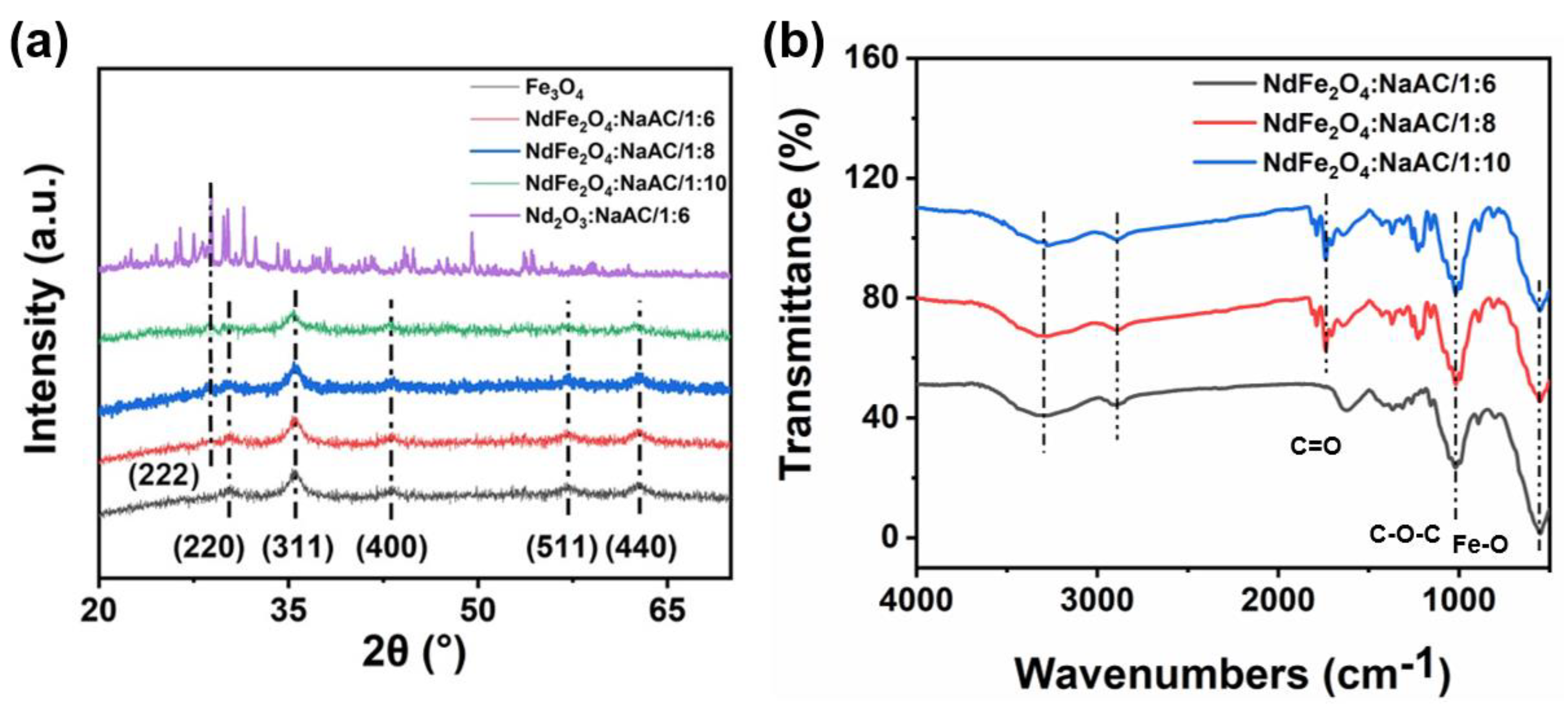
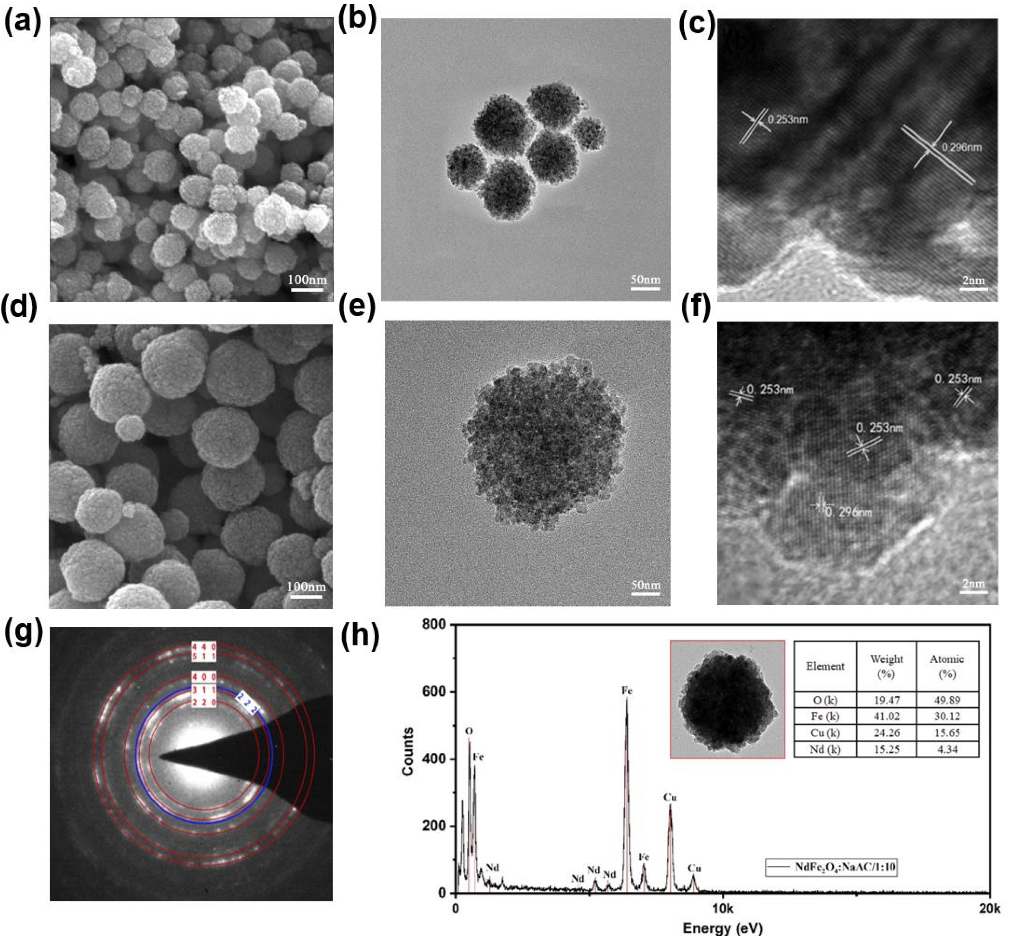
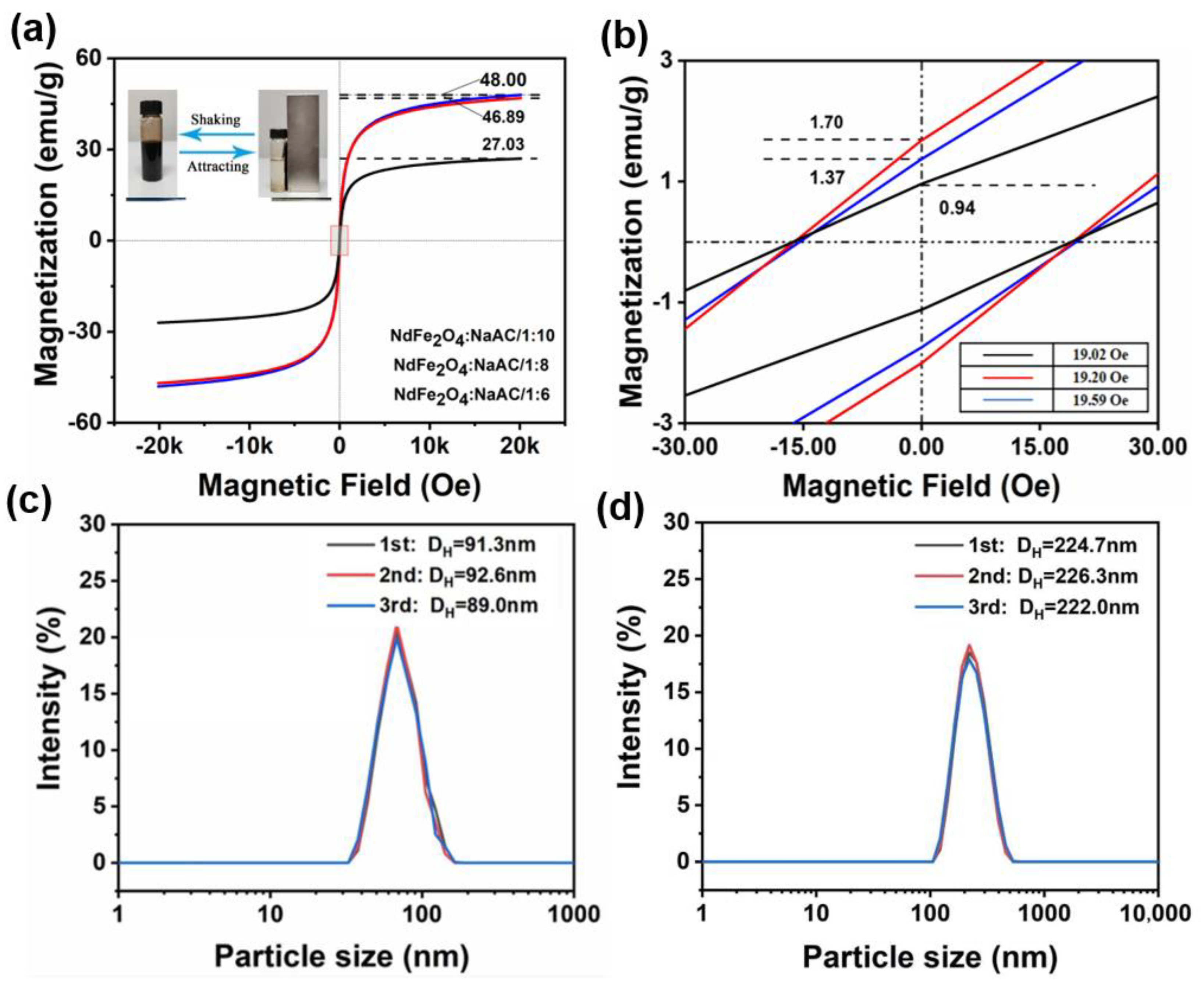
2.2. Characteristics and Magnetic Properties of NdFe2O4 NPs

The crystal forms of Fe3O4, Nd2O3, and NdFe2O4 NPs with different mole ratios of NaAC were detected though XRD, as shown in Figure 2a. The typical XRD pattern peaks of Fe3O4 were observed at 30.1°, 35.4°, 43.0°, 56.9°, and 62.5°, corresponding to the (220), (311), (400), (511), and (440) planes, respectively, which were consistent with the Joint Committee on Powder Diffraction Standards of Fe3O4 (JCPDS Card no: 65−3107) [32]. Furthermore, the typical lattice plane (222) of Nd2O3 at 27.891° (JCPDS Card no: 65−3187) appeared in the corresponding doping curves. Thus, the solvent thermal synthesis of Nd doping in Fe3O4 was successful in different mole ratios of NaAC, which supports previous reports. As the NaAC concentration increased from 1;6 to 1;10, the crystallite sizes were 13.2 dm, 13.6 nm, and 14.5 nm, respectively. The trend of the data was not obvious at this stage. Figure 2b shows a distinct absorption peak located at 582 cm−1 in the patterns, which could be attributed to the vibration of the Fe−O group that was obtained from the NdFe2O4 crystal structure [33]. Furthermore, the common peaks at 1046 and 1627 cm−1 corresponded to the hierarchical ether group (−C−O−C−) and carbonyl group (C=O), respectively [34]. However, the carbonyl group peaks among the three samples were similar because of the oxidation of hydroxy from PEG in the reducing reaction of partial Fe3+ to Fe2+ [35]. Considering the influence of the small size effect, some adsorption peaks showed a slight shift, as shown in the spectrum. However, the characteristic peak of the Nd−O group was located at 345 cm−1, which was out of this test range and further analyzed by XPS.
The elemental composition and the corresponding chemical binding energies of each element were investigated by examining the Fe3O4 and NdFe2O4 NPs at different mole ratios of NaAC via XPS in Figure 3. The survey scan patterns (Figure 3a) showed the binding energies of C and O in all the curves, which were located at 286.4 and 529.8 eV, respectively, while Nd 4d was only observed in the curves of NdFe2O4. The high−resolution peak of Fe 2p and Nd 3d for Fe3O4 and NdFe2O4 NPs were further confirmed in Figure 3b−d. Two obvious peaks were centered at 710.1 and 724.2 eV, which corresponded to Fe 2p1 and Fe 2p3 derived from Fe−O bonds, respectively [36]. In addition, the Nd 3d narrow scan peaks were detected at 974.12, 981.6, 994.4, and 1,003.5 eV, which could be assigned to Nd 3d5A, Nd 3d5B, Nd 3d3A, and Nd 3d3B, respectively [37]. The atomic percent data shown in Figure 3e indicated the elemental composition of the as-prepared NPs, exhibiting the successful doping of Nd in the Fe3O4 crystal structure without changing the valence state of the Fe. The XPS analysis above provides favorable evidence for the successful preparation and structural confirmation of the Nd doped in Fe3O4 crystal, which were consistent with the above XRD observations.
Apart from the element and crystal analysis of the sample, the surface morphology was also investigated. Figure 4 presents the TEM and SEM images of Fe3O4 and NdFe2O4 NPs prepared under different parameters together with the high-magnification lattice fringes and electron diffraction results. The as−prepared NdFe2O4 NPs had regular spherical structures at major particle sizes of 70−110 nm (Figure 4a,b) in 1:6 NaAc. However, larger NPs were obtained with a major size range of 200−270 nm (Figure 4d,e) in 1:10 NaAC, which can be attributed to the different reduction ability in different mole ratios of the Fe source to NaAC for the formation of the magnetic crystal [38]. Subsequently, the lattice fringe spacing showed common fringe spacings of 0.253 and 0.296 nm, corresponding to the (311) and (220) lattice planes from JCPDS Card no: 65−3107, respectively (Figure 4d,f). The electron diffraction pattern (Figure 4g) of the product displayed evident (220), (311), (400), (511), and (440) crystal plane electron diffraction signals, which belonged to the Fe3O4 crustal [39]. In addition, the blue electron diffraction signal of the (222) crystal plane coincided with the Nd product of JCPDS Card no: 65−3187, which was consistent with the XRD and XPS results of the above representations [40]. The body elements of NdFe2O4 were Fe, O, Cu, and some Nd in an atomic percentage (Figure 4h). In the measured area, the atomic percentage of Cu was associated with the test condition of the Cu net for the weight tray. The atomic percentage of Nd was only 4.34%, whereas that of Fe was 30.12% and that of O was 49.89%. This result can be attributed to the larger ionic radius of Nd3+ than that of Fe2+ [41], which reduced the crystallization capacity of Nd in Fe3O4. Thus, the production of Nd was successfully doped into the Fe3O4 crystal structure, which was consistent with the above XPS observations.
The magnetization performances of the obtained NdFe2O4 NPs were measured using VSM, and their hysteresis loops with closed “S” shapes are shown in Figure 5a. The saturation magnetization values of the samples were 27.03, 46.89, and 48.00 emu.g−1, indicating the differences in magnetization capacity [42]. Combined with the corresponding TEM results, NPs with a small particle size (at a mole ratio of 1:6) showed low saturation magnetization value of 27.03 emu.g−1, but it was close to 48 emu.g−1 for samples prepared at higher exposure times to NaAC, possibly because of the effect of particle size in different mole ratio of the iron source to NaAC. Thus, the particle size plays a key role in determining magnetism properties [43]. As shown in the inset picture, the prepared NPs could realize the rapid switch between good dispersion and absorption on the wall with and without the action of the magnet [44]. Thus, the as-prepared NPs were well-dispersed in water at room temperature, thereby promoting their medical and biological protective applications. The residual magnetization value and coercive force obtained from the enlarged curves (Figure 5b) are relatively close, which was beneficial for enhancing the following functional fabrics’ electromagnetic protective performances for the prospective applications [45].
Particle size analysis was carried out to measure the DLS distribution curves of the NPs at a 1:6 and 1:10 ratio for three times (Figure 5c,d). The tested particle size was mainly distributed at approximately 90 and 230 nm, respectively, which was generally consistent with the SEM and TEM results [46]. However, the polymer dispersity index values of the obtained NdFe2O4 NPs were between 0.071, 0.111, and 0.121, 0.183, respectively. This finding indicates a good shape and controllable particle size, and a slightly larger dispersion rate in 1:10 samples than that of the 1:6 samples, which support the above VSM results.

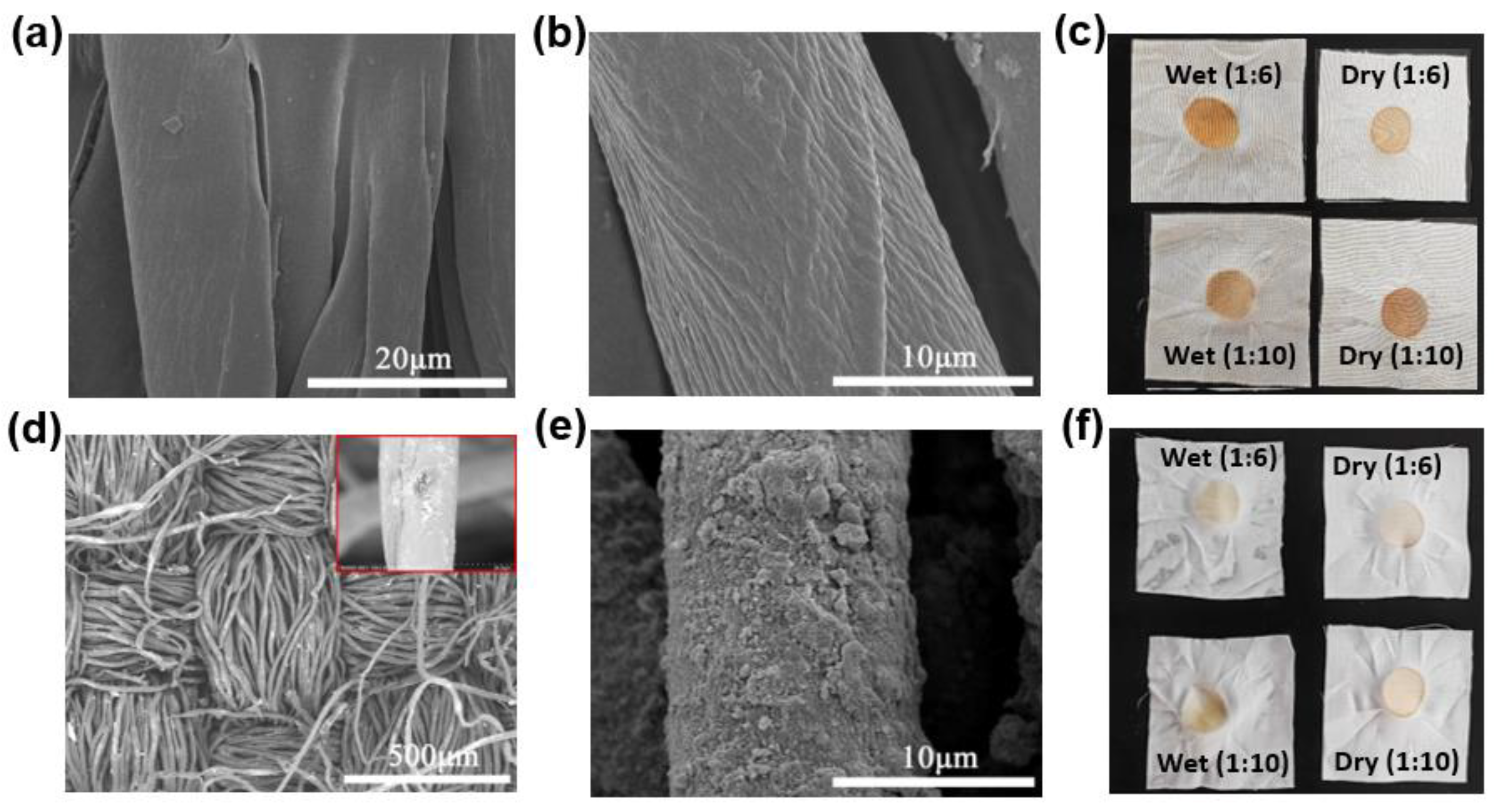
2.3. Characteristics and Magnetic protective of the Functional Fabrics

Glyceric acid and DSC were selected for the grafting of NdFe2O4 NPs onto cotton fabrics for durable wearability. The best load rate of the functional fabrics was 1.37% at room temperature, which can be attributed to the best water absorption of cotton fabrics following a 25% alkali treatment. The SEM images of the cotton fabrics before and after alkali treatment at a concentration of 25% are shown in Figure 6a,b. The surface of the fiber was quite smooth, a rough and etching shape of the cellulose fiber was obtained after the alkali treatment, and the surface activity was improved, which was useful for the following grafting of NPs. The sample grafted with a proper amount of the prepared NdFe2O4 NPs is shown in Figure 6d,e. The as−grafted fabric possessed a uniformly thin layer of nanosheet structure and showed an adequate grafting response to cellulose fibers. The durability of the functional fabrics was verified by rubbing the sample thrice and then washing it. The color fatness to rubbing was observed using a fatness tester, and the results are shown in Figure 6c,f. Generally, the grade of the color fatness of the grafting fabrics from Figure 6c was obviously higher than that of Figure 6f after washing and drying three times, and the specific fatness values of the samples are shown in the corner tables. The grade of color fatness was close to level-3 in Figure 6f, which is consistent with the ordinary outwear clothing fabrics’ value based on the ISO105/A03−1993 and GB/T251-2008 standards [47]. The permanent and reliable interface grafting is a safeguard for the subsequent fastness problem in the wearing and washing process of the protective fabrics.
In general, when the prepared magnetic nanoparticles were grafted onto the surface of the cotton fabrics in a gradient, their small particles were evenly scattered. This process allows for their use in electric and magnetic property testing and analysis. When the test frequency of electromagnetic wave radiation was incident upon the surface of the functional fabric, its magnetic energy was partially reflected in the fabric’s interior into the air as heat energy. Furthermore, the second part was absorbed by the multiple reflections on the functional component through dielectric and magnetic loss in the interior of the fabrics [48]. Finally, the transmitted radiation was evidently attenuated to a low value for the electromagnetic protective materials, as shown in Figure 7a. Figure 7b presents the electromagnetic property parameters of the obtained sample under the frequency range of 8.2−14.2 GHz in the X band, including the real and imaginary part, and the dielectric and magnetic loss were also calculated [49]. In addition, the imaginary parts would be transferred to each other until the end of the loss by the functional body through dielectric and magnetic loss [50]. These losses are often verified based on several electromagnetic parameters, such as permeability and permittivity. Furthermore, the real permeability ε’ and permittivity µ’ are connected to the dispassion capacity of the test fabric, and the imaginary parts µ’’ and ε’’ are related to the degradation capability of the electromagnetic energy, respectively [51]. The dielectric loss (tanδe = ε’’/ε’) and magnetic loss (tanδm = µ’’/µ’) curves of the fabric were generated (Figure 7c). The ε’ values were near 2.5, and the µ’ values floated around 2. When the frequency was under 9 GHz, the µ’ and µ’’ values showed a downtrend, which could be attributed to the magnetic dipoles’ movement for the magnetic loss property of NdFe2O4 [52]. However, the ε’’ and µ’’ values were relatively low, indicating a difficulty in transferring the electric and magnetic field forces [53]. The as-prepared fabrics exhibited a weak conductive ability, as shown in Figure 7d,e, which could be attributed to the magnetite NdFe2O4 NPs’ low interior motion of the magnetic dipole in varying magnetic fields for transferring electric fields, thus supporting the results in Figure 7b,c [54]. In comparison with the existing samples, the fabrics with different NPs had little difference in their specific capacitance (Cp) values and dielectric constants. The best Cp value was 0.0551 F/g, which was recorded from the fabric with NPs at 1:6 NaAC, and this value could be calculated based on the area of the curve through the CV results in Figure 7d. Furthermore, Figure 7e exhibits a different dielectric capacity for the obtained samples with a slight discrepancy in the curve radian. The double Ohm curve for such fabric with NPs at 1:6 NaAC also showed the lowest curve radian for the best dielectric property, which was consistent with the results in Figure 7d [55,56].
The stability of the functional fabrics at high temperatures was evaluated based on their magnetic susceptibility under a changing temperature M−T curve and magnetizing M−H curve. The samples exhibited fine magnetic properties and magnetic loss performances as the temperature increased from 300 K to 800 K (Figure 7f), and the magnetization ability decreased sharply from 3.3 emu.g−1 to close to 0 emu.g−1, thus confirming the loss velocity of magnetic properties in hot environments [57]. Furthermore, the three as-prepared samples exhibited obvious differences in magnetic properties from Figure 7g and the enlarged picture Figure 7h. The fine magnetic value of the functional fabric with NdFe2O4 NPs at 1:10 NaAC was 17.61 emu.g−1, which was higher than 12.67 emu.g−1 for the sample with 1:8 NaAC and 4.89 emu.g−1 for the sample with 1:6 NaAC, which belonged to the reduction action of different mole ratios of NaAC in the synthesis process [58,59]. The as-prepared fabrics with NdFe2O4 NPs showed fine magnetic properties with weak dielectric properties that need to be improved, proving the effect of the obtained particle size on the electromagnetic properties for the finally functional protective fabrics.

3. Experiment

3.1. Materials

All the reagents, including ferric trichloride (FeCl3·6H2O), neodymium trichloride (NdCl3·6H2O), polyethylene glycol (PEG), ethylene glycol (EG), sodium acetate (CH3COONa, NaAC), glyceric acid (C3H6O4), N,N′−dissuccinimide carbonate (C9H8N2O7, DSC), and alkali (NaOH) were obtained from Aladdin Industrial and Sinopharm Reagent Corporation. Ultrapure water with a conductivity of 18.33 Ω/cm was prepared using a deionizing water purification system (PT−10T, Hitech Instruments Co., Ltd., Shanghai, China). Natural cotton yarn with a fitness of 2 × 32S was provided by Haian Country Lianci Textile Co., Ltd. (Nantong, China).

3.2. Synthesis of Nd3+-doped Fe3O4 NPs

Spherical Fe3O4 NPs with uniform structures were prepared using a repeated synthesis method similar to the process described in our previous study [9]. Approximately 0.15 moles of NdCl3·6H2O were added to the FeCl3 solution, and the mixture was homogeneously dispersed and placed into a Teflon−lined container for the solvothermal reaction. Then, the NdFe2O4 NPs were obtained. The mole ratio of NaAC, a key reduction agent, was varied at different times: 1:6, 1:8, and 1:10 times as much as iron. Furthermore, a solvothermal reaction time of 12 h resulted in controllable sized NdFe2O4 NPs.

3.3. Preparation of Functional Fabrics with Modified NPs

First, plain weave fabrics with 2 × 32S cotton yarns were finished through a proofing rapier loom (Y300S, Automatic Rapier Loom Machine, Nantong, China) at 240 yarns per 10 cm centimeters in both warp and weft directions. The cotton fabrics were then subjected to alkali treatment for the optimization of water absorption for the subsequent functional finishing. The alkali treatment was conducted at concentrations of 15%, 20%, 25%, and 30%. The best alkali treatment concentration on the cotton fabric was found to be 25%, resulting in 10.37% water absorption. The obtained NdFe2O4 NPs were modified using glyceric acid, allowing a connection with −OH on the cotton fabric through a bridging agent, DSC. The prepared samples were washed thrice and dried to remove the residual reagent.

3.4. Characterizations

The morphology of the NPs and functional fabrics were observed using a TM4000Plus scanning electron microscope (SEM, Hitachi, Japan) and a JEM−2100Plus transmission electron microscope (TEM, Jeol, Japan) connected with an energy dispersive X−ray (EDS, Jeol, Japan). X−ray powder diffraction (XRD, Bruker−D8, 10−70°, Germany), IS50 Fourier infrared spectroscopy (FT−IR, Nicolet 6700, America) with KBr pellets, dynamic light scattering (DLS, Vasco, Portugal), and X−ray photoelectron spectroscopy (XPS, Thenno, ESCALAB250, America) were carried out. The magnetic properties of the obtained nanoparticles and fabrics were analyzed using the Lake Shore 7307 vibrating sample magnetometer (VSM) from −20 KOe to 20 KOe at room temperature and a physical property measurement system (PPMS, Quantum Design PPMS 9) at 300−800 K. The color fatness to the rubbing of fabrics was detected using an instrument (YG5711−II, Meibon Instruments Co., Ltd., Quanzhou, China). The electrical properties of the fabrics were tested using an electrochemical working station measurement (PGSTAT302N, Metrohm Autolab, Kanaalweg, The Netherlands). The electromagnetic wave absorption performance of the samples was measured using an N5234A vector network analyzer (VNA, Agilent, Santa Clara, CA, America) based on a wave−guide method from 8.2 GHz to 12.4 GHz.

4. Conclusions

Novel Nd3+−doped Fe3O4 nanoparticles based on a microwave synthesis method and a surface modification were grafted onto cotton fabrics, and their magnetic protective properties were examined. The structural characterization of the obtained doping nanoparticles showed a spherical shape and a good dispersion for fine magnetic properties that can be attributed to the contribution of nano−structural spinel ferrite. In addition, the controllable particle size was confirmed for the electromagnetic properties of the obtained nanoparticles and final fabrics. The surface modification on NdFe2O4 nanoparticles and alkali treatment on cotton fabrics for rich hydrophilic groups were further determined using a bridging agent for durable interface bonding, and the samples showed fine color fatness to rubbing at level−3 after washing. The final structure and obvious hysteresis loop results of the obtained fabrics had a maximum magnetic saturation value of 17.61 emu.g−1 and weak electric properties for magnetic protective applications. However, the synthesis and surface modification method, which involved a grafting reaction, can also be applied for the preparation of other similar controllable nanoparticles for further functional fabric studies and product development, showing good universality.

Author Contributions

X.S. and Y.L. conceived and designed the experiments. C.X., W.Y., and J.W. performed the related experiments. X.S. and Q.W. analysed the data results. X.S. and X.F. wrote and revised the manuscript. All authors have read and agreed to the published version of the manuscript.

Funding

This research was funded by the National Science Foundation of Fujian Province (No. 2020J01849), the Major Science and Technology Project of Fuzhou (No. 2021ZD298), the Science and Technology Projects of Fujian Province (No. 2021H0037), and the Research Project of the Fashu Foundation (No. MFK23004).

Data Availability Statement

Not applicable.

Conflicts of Interest

The authors declare no conflict of interest.

Sample Availability

Samples of the compounds mangetic protective fabrics are available from the authors.

References

  1. Qi, Y.; Yin, P.; Zhang, L.; Wang, J.; Feng, X.; Wang, K.; Zhao, L.; Sun, X.; Dai, J. Novel microwave absorber of NixMn1-xFe2O4/carbonized chaff (x = 0.3, 0.5 and 0.7) based on biomass. ACS Omega 2019, 4, 12376–12384. [Google Scholar] [CrossRef] [PubMed] [Green Version]
  2. Zhou, B.; Su, M.; Yang, D.-X.; Han, G.; Feng, Y.; Wang, B.; Ma, J.; Liu, C.; Shen, C. Flexible MXene/siver nanowire-based transparent conductive film with electromagnetic interference shielding and electro-photo-thermal performance. ACS Appl. Mater. Interfaces 2020, 12, 40859–40869. [Google Scholar] [CrossRef] [PubMed]
  3. Zhan, Y.; Long, Z.; Wan, X.; Zhang, J.; He, S.; He, Y. 3D carbon fiber mats/nano-Fe3O4 hybrid material with high electromagnetic shielding performance. Appl. Surf. Sci. 2018, 444, 710–720. [Google Scholar] [CrossRef]
  4. Liang, L.-Y.; Li, Q.-M.; Yan, X.; Feng, Y.-Z.; Wang, Y.-M.; Zhang, H.-B.; Zhou, X.-P.; Liu, C.-T.; Shen, C.-Y.; Xie, X.-L. Multifunctional magnetic Ti3C2Tx MXene/ graphene aerogel with superior electromagnetic wave absorption performance. ACS Nano 2021, 15, 6622–6632. [Google Scholar] [CrossRef]
  5. Lv, H.; Yang, Z.; Wang, P.-L.; Ji, G.; Song, J.; Zheng, L.; Zeng, H.; Xu, Z.-J. A voltage-boosting strategy enabling a low-frequency, flexible electromagnetic wave absorption device. Adv. Mater. 2018, 30, 1706343. [Google Scholar] [CrossRef]
  6. Zhang, H.; Guo, Y.; Zhang, X.; Wang, X.; Wang, H.; Shi, C.; He, F. Enhanced shielding performance of layered carbon fiber composites filled with carbonyl iron and carbon nanotubes in the koch curve fractal method. Molecules 2020, 25, 969. [Google Scholar] [CrossRef] [Green Version]
  7. Singh, A.-K.; Shishkin, A.; Koppel, T.; Gupta, N. A review of porous lightweight composite materials for electromagnetic interference shielding. Compos. Part B 2018, 149, 188–197. [Google Scholar] [CrossRef]
  8. Zhang, X.-J.; Zhu, J.-Q.; Yin, P.-G.; Guo, A.-P.; Huang, A.-P.; Guo, L.; Wang, G.-S. Tunable high-performance microwave absorption of Co1-xS hollow spheres constructed by nanosheets within ultralow filler loading. Adv. Funct. Mater. 2018, 28, 1800761. [Google Scholar] [CrossRef]
  9. Liu, Q.; Cao, Q.; Bi, H.; Liang, C.; Yuan, K.; Yang, Y.; Che, R. CoNi@SiO2@TiO2 and CoNi@Air@TiO2 microspheres with strong wideband microwave absorption. Adv. Mater. 2016, 28, 486–490. [Google Scholar] [CrossRef]
  10. Qiao, M.; Lei, X.; Ma, Y.; Tian, L.; He, X.; Su, K.; Zhang, Q. Application of yolk-shell Fe3O4@N-doped carbon nanochains as highly effective microwave-absorption material. Nano Res. 2018, 11, 1500–1519. [Google Scholar] [CrossRef] [Green Version]
  11. Xu, J.-D.; Li, R.-S.; Ji, S.-R.; Zhao, B.-C.; Cui, T.-R.; Tan, X.-C.; Gou, G.-Y.; Jian, J.-M.; Xu, H.-K.; Qiao, Y.-C.; et al. Multifunctional graphene microstructures inspired by honeycomb for ultrahigh performance electromagnetic interference shielding and wearable application. ACS Nano 2021, 15, 8907–8918. [Google Scholar] [CrossRef] [PubMed]
  12. Song, P.; Liang, C.-B.; Wang, L.; Qiu, H.; Gu, H.-B.; Kong, J.; Gu, J.-W. Obviously improved electromagnetic interference shielding performances for epoxy composites via constructing honeycomb structural reduced graphene oxide. Compos. Sci. Technol. 2019, 8, 107698. [Google Scholar] [CrossRef]
  13. Sridhar, V.; Lee, I.; Park, H. Metal organic frameworks derived Fe-N-C nanostructures as high-performance electrodes for sodium ion batteries and electromagnetic interference (EMI) shielding. Molecules 2021, 26, 1018. [Google Scholar] [CrossRef] [PubMed]
  14. Zhang, X.; Zhang, S.; Zhang, K.; Yan, F.; Zhu, C.; Yuan, H.; Zhang, X.; Chen, Y. Interface-induced enhanced electromagnetic wave absorption property of metal-organic frameworks wrapped by graphene sheets. J. Alloys Compd. 2019, 780, 718–726. [Google Scholar] [CrossRef]
  15. Giannakoudakis, D.-A.; Bandosz, T.-J. Building MOF nanocomposites with oxidized graphitic carbon nitride nanospheres: The effect of framework geometry on the structural heterogeneity. Molecules 2019, 24, 4529. [Google Scholar] [CrossRef] [Green Version]
  16. Song, X.-L.; Wu, Y.-L.; Zhang, S.-R.; Chen, Z.; Li, Y.-G. NdFe2O4 nanoparticles: Synthesis, characterization and magnetic properties. Sci. Adv. Mater. 2020, 12, 810–814. [Google Scholar] [CrossRef]
  17. Chen, X.; Shi, T.; Wu, G.; Lu, Y. Design of molybdenum disulfide@polypyrrole compsite decorated with Fe3O4 and superior electromagnetic wave absorption performance. J. Colloid Interface Sci. 2020, 572, 227–235. [Google Scholar] [CrossRef]
  18. Adam, A.; Parkhomenko, K. Orienting the pore morphology of core-shell magnetic mesoporous silica with the sol-gel temperature. influence on MRI and magnetic hyperthermia properties. Molecules 2021, 26, 971. [Google Scholar] [CrossRef]
  19. Wei, Q.; Pei, S.; Qian, X.; Liu, H.; Liu, Z.; Zhang, W.; Zhou, T.; Zhang, Z.; Zhang, X.; Cheng, H.-M. Superhigh electromagnetic interference shielding of ultrathin aligned pristine graphene nanosheets film. Adv. Mater. 2020, 32, 1907411. [Google Scholar] [CrossRef]
  20. Ji, B.; Fan, S.-W.; Kou, S. Microwave absorption properties of multilayer impedance gradient absorber consisting of Ti3C2TX MXene/polymer films. Carbon 2021, 181, 130–142. [Google Scholar] [CrossRef]
  21. Zirak, M.; Abdollahiyan, A.; Eftekhari, S.-B.; Saraei, M. Carboxymethyl cellulose coated Fe3O4@SiO2 core-shell magnetic nanoparticles for methylene blue removal: Equilibrium, kinetic, and thermodynamic studies. Cellulose 2017, 25, 503–515. [Google Scholar] [CrossRef]
  22. Ebenezer, C.N.; Peter, A.A. Multifunctional magnetic oxide nanoparticle (MNP) core-shell: Review of synthesis, structural studies and application for wastewater treatment. Molecules 2020, 25, 4110. [Google Scholar] [CrossRef] [PubMed]
  23. Liu, S.; Mei, J.; Zhang, C.; Zhang, J.; Shi, R. Synthesis and magnetic properties of shuriken-like nickel nanoparticles. J. Mater. Sci. Technol. 2018, 34, 836–841. [Google Scholar] [CrossRef]
  24. Iqbal, A.; Symbyal, P.; Koo, C.-M. 2D MXenes for electromagnetic shielding: A review. Adv. Func. Mater. 2020, 30, 2000833. [Google Scholar] [CrossRef]
  25. Sankaran, S.; Deshmukh, K.; Ahamed, M.-B.; Pasha, S.-K.-K. Recent advances in electromagnetic interference shielding properties of metal and carbon filler reinforced flexible polymer composites. Compos. Part A-Appl. S 2018, 114, 49–71. [Google Scholar] [CrossRef]
  26. Lei, Z.-M.; Tian, D.-K.; Liu, X.-B.; Wei, J.-H.; Rajavel, K.; Zhao, T.; Hu, Y.-G. Electrically conductive gradient structure design of thermoplastic polyurethane composite foams for efficient electromagnetic interference shielding and ultra-low microwave reflectivity. Chem. Eng. J. 2021, 424, 130365. [Google Scholar] [CrossRef]
  27. Jia, L.-C.; Yan, D.-X.; Liu, X.-F.; Ma, R.-J.; Wu, H.-Y.; Li, Z.-M. Highly efficient and reliable transparent electromagnetic interference shielding. ACS Appl. Mater. Interfaces 2018, 10, 11941–11949. [Google Scholar] [CrossRef]
  28. Xu, Y.; Yang, Y.; Yan, D.; Duan, H.; Zhao, G.; Liu, Y. Gradient structure design of flexible waterborne polyurethane conductive films for ultraefficient electromagnetic shielding with low reflection characteristic. ACS Appl. Mater. Interface 2018, 10, 19143–19152. [Google Scholar] [CrossRef]
  29. Duan, H.; Zhu, H.; Gao, J.; Yan, D.; Dai, K.; Yang, Y.; Zhao, G.; Liu, Y.; Li, Z. Asymmetric conductive polymer composite foam for absorption dominated ultra-efficient electromagnetic interference shielding with extremely low reflection characteristics. J. Mater. Chem. A 2020, 8, 9146–9159. [Google Scholar] [CrossRef]
  30. Abbasi, H.; Antunes, M.; Velasco, J.-I. Recent advances in carbon-based polymer nanocomposites for electromagnetic interference shielding. Prog. Mater. Sci. 2019, 103, 319–373. [Google Scholar] [CrossRef]
  31. Gupta, S.; Tai, N. Carbon materials and their composites for electromagnetic interference shielding effectiveness in X-band. Carbon 2019, 152, 159–187. [Google Scholar] [CrossRef]
  32. Adebayo, L.-L.; Soleimani, H.; Yahya, N.; Abbas, Z.; Wahaab, F.-A.; Ayinla, R.-T.; Ali, H. Recent advances in the development OF Fe3O4-BASED microwave absorbing materials. Ceram. Int. 2020, 46, 1249–1268. [Google Scholar] [CrossRef]
  33. Mi, H.-Y.; Li, H.; Jing, X.; Zhang, Q.; Feng, P.-Y.; Ping, H.; Liu, Y.-J. Superhydrophobic cellulose nanofibril/silica fiber/Fe3O4 nanocomposite aerogel for magnetically driven selective oil absorption. Cellulose 2020, 27, 8909–8922. [Google Scholar] [CrossRef]
  34. Luo, X.; Li, H.-F.; Deng, D.-D.; Zheng, L.; Wu, Y.-B.; Luo, W.-J.; Zhang, M.-J.; Gong, R.-Z. Preparation and excellent electromagnetic absorption properties of dendritic structured Fe3O4@PANI composites. J. Alloys Compd. 2022, 891, 161922. [Google Scholar] [CrossRef]
  35. Xu, L.; Zhong, W.-D.; Yang, C.; Zhao, R.; Wu, J.; Li, X.; Yang, N. Tailoring interfacial electron redistribution of Ni/Fe3O4 electrocatalysts for superior overall water splitting. J. Energy Chem. 2022, 73, 330–338. [Google Scholar] [CrossRef]
  36. Bui, D.-P.; Nguyen, T.-D.; Vo, T.-T.-L.; Cao, T.-M.; You, S.-J.; Pham, V.-V. SnO2-x nanoparticles decorated on graphitic carbon nitride as S-scheme photocatalysts for activation of peroxymonosulfate. ACS Appl. Nano Mater. 2021, 4, 9333–9343. [Google Scholar] [CrossRef]
  37. Almessiere, M.-A.; Khan, F.-A.; Auwal, I.-A.; Sertkol, M.; Tashkandi, N.; Rehan, I.; Baykal, A. Green synthesis, characterization and anti-cancer capability of Co0.5Ni0.5Nd0.02Fe1.98O4 nanocomposites. Arab. J. Chem. 2022, 15, 103564. [Google Scholar] [CrossRef]
  38. Sahar, B.-K.; Samuel, R.-J.; Sonia, S.; Stephen, E.-H.; Andrew, D.-W. Structure-based virtual screening, synthesis and biological evaluation of potential FAK-FAT domain inhibitors for treatment of metastatic cancer. Molecules 2020, 25, 3488. [Google Scholar] [CrossRef]
  39. Yang, Y.; Yang, F.; Wang, H.; Zhou, B.; Hao, S. Amine-promoted Ru1/Fe3O4 encapsulated in hollow periodic mesoporous organosilica sphere as a highly selective and stable catalyst for aqueous levulinic acid hydrogenation. J. Colloid Interface Sci. 2021, 581, 167–176. [Google Scholar] [CrossRef]
  40. Li, P.-L.; Zhang, S.; Zhu, Y.; Fan, H.; Ma, W.; Dong, P.; Wang, W.-Z.; Liu, T. Polyimide-based graphene composite foams with hierarchical impedance gradient for efficient electromagnetic absorption. J. Mater. Chem. C 2021, 9, 2086–2094. [Google Scholar] [CrossRef]
  41. Sandhiya, M.; Subramani, K.; Sathish, M. Augmenting the electrochemical performance of NiMn2O4 by doping of transition metal ions and compositing with rGO. J. Colloid Interface Sci. 2021, 598, 409–418. [Google Scholar] [CrossRef] [PubMed]
  42. Geng, L.; Liu, Q.; Zhao, J. In situ visualization of hierarchical agglomeration growth during electrochemical deposition of Cu nanocrystals in an open ionic liquid cell. Mater. Today Nano 2022, 18, 2–9. [Google Scholar] [CrossRef]
  43. Ma, J.; Wang, T.; Yu, S.; Zhang, Y.; Lyu, B. Preparation and application of dialdehyde nanocellulose reinforced jatropha oil based polymer emulsions as leather fatliquors. Cellulose 2020, 28, 331–346. [Google Scholar] [CrossRef]
  44. Elshypany, R.; Selim, H.; Zakaria, K.; Houstafa, A.-H. Magnetic ZnO crystal nanoparticle growth on reduced graphene oxide for enhanced photocatalytic performance under visible light irradiation. Molecules 2021, 26, 2269. [Google Scholar] [CrossRef] [PubMed]
  45. Kuwa, M.; Harada, M.; Sato, R.; Teranishi, T. Ligand-stabilized CoO and NiO nanoparticles for spintronic devices with antiferromagnetic insulators. ACS Appl. Nano Mater. 2020, 3, 2745–2755. [Google Scholar] [CrossRef]
  46. Zeynizadeh, B.; Mohammadzadeh, I.; Shokri, Z.; Ali, H.-S. Synthesis and characterization of NiFe2O4@Cu nanoparticles as a magnetically recoverable catalyst for reduction of nitroarenes to arylamines with NaBH4. J. Colloid Interface Sci. 2017, 500, 285–293. [Google Scholar] [CrossRef]
  47. Joanna, O.; Jadwiga, S.-L.; Anetta, W.; Anna, A.; Katarzyna, S.-C.; Jakub, Z.; Teofil, J. Antimicrobial activity and barrier properties against UV radiation of alkaline and enzymatically treated linen woven fabrics coated with inorganic hybrid material. Molecules 2020, 25, 5701. [Google Scholar] [CrossRef]
  48. Ding, K.; Liu, Z.; Xiao, C.-F. Fabrication of a novel one-step coating hyper-hydrophobic fluorine-free TiO2 decorated hollow composite membrane for use in longer-term VMD with enhanced flux, rejection, anti-wetting and anti-fouling performances. Nanoscale 2021, 13, 12342–12355. [Google Scholar] [CrossRef]
  49. Zhang, Y.; Fulajtárová, K.; Kub, M.; Mazur, M.; Shamzhy, M.; Hronec, M. Controlling dispersion and accessibility of Pd nanoparticles via 2D-to-3D zeolite transformation for shape-selective catalysis: Pd@MWW case. Mater. Today Nano 2019, 8, 100056. [Google Scholar] [CrossRef]
  50. Li, G.-H.; Ma, S.-P. High-quality ferromagnet Fe3GeTe2 for high-efficiency electromagnetic wave absorption and shielding with wideband radar cross section reduction. ACS Nano 2022, 16, 7861–7879. [Google Scholar] [CrossRef]
  51. Sun, S.; Wang, D.; Feng, Z.; Tan, W. Highly efficient unidirectional forward scattering induced by resonant interference in a metal-dielectric heterodimer. Nanoscale 2020, 12, 22289–22297. [Google Scholar] [CrossRef] [PubMed]
  52. Huang, H.; Gao, Y.; Fang, C.-F.; Wu, A.-M.; Dong, X.-L.; Kim, B.-S.; Byun, J.-H.; Zhang, G.-F. Spray granulation of Fe and C nanoparticles and their impedance match for microwave absorption. J. Mater. Sci. Technol. 2018, 34, 496–502. [Google Scholar] [CrossRef]
  53. Zhang, C.-J.; McKeon, L.; Kremer, M.-P.; Park, S.-H.; Ronan, O.; Seral-Ascaso, A.; Barwich, S.; Coileain, C.-O.; McEvoy, N.; Nerl, H.-C. Additive-free MXene inks and direct printing of micro-supercapacitors. Nat. Commun. 2019, 10, 1795. [Google Scholar] [CrossRef] [Green Version]
  54. Xia, S.-H.; Wei, C.-Y.; Tang, J.-C.; Yan, J.-H. Tensile stress-gated electromagnetic interference shielding fabrics with real-time adjustable shielding efficiency. ACS Sustainable Chem. Eng. 2021, 9(42), 13999–14005. [Google Scholar] [CrossRef]
  55. Ye, Y.; Jiang, Z.; Zou, Y.; Chen, H.; Guo, S.; Yang, Q.; Chen, L. Evaluation of the inhibition behavior of carbon dots on carbon steel in HCl and NaCl solutions. J. Mater. Sci. Technol. 2020, 43, 144–153. [Google Scholar] [CrossRef]
  56. Qu, Z.; Wang, Y.; Yang, P.; Zheng, W.; Li, N.; Bai, J.; Zhang, Y.; Li, K.; Wang, D. Enhanced electromagnetic wave absorption properties of ultrathin MnO2 nanosheet-decorated spherical flower-shaped carbonyl iron powder. Molecules 2022, 27, 135. [Google Scholar] [CrossRef]
  57. Yuan, B.-G.; Li, J.; Xia, M.-M.; Zhang, Y.; Lei, R.-Y.; Zhao, P.; Li, X. Synthesis and electrochemical performance of hollow-structured NiO + Ni nanofibers wrapped by graphene as anodes for Li-ion batteries. Nanotechnology 2021, 32, 335603. [Google Scholar] [CrossRef]
  58. Haider, W.-A.; Tahir, M.; He, L.; Mirza, H.-A.; Zhu, R.; Han, Y.; Mai, L. Structural engineering and coupling of two-dimensional transition metal compounds for micro-supercapacitor electrodes. ACS Cent. Sci. 2020, 6, 1901–1915. [Google Scholar] [CrossRef]
  59. Han, M.; Shuck, C.-E.; Rakhmanov, R.; Parchment, D.; Anasori, B.; Koo, C.-M.; Friedman, G.; Gogotsi, Y. Beyond Ti3C2Tx: MXenes for electromagnetic interference shielding. ACS Nano 2020, 14, 5008–5016. [Google Scholar] [CrossRef]
Figure 1. Illustration of the preparation process of the reliable magnetic protective fabrics.
Molecules 28 03175 g001
Figure 2. (a) XRD patterns, and (b) FT−IR patterns of Fe3O4 and NdFe2O4 NPs.
Molecules 28 03175 g002
Figure 3. XPS survey spectra and high-resolution spectra of Fe3O4 and NdFe2O4 NPs. (a) XPS full spectrum; (b) Fe spectrum of Fe3O4 NPs; (c) Fe spectrum of NdFe2O4 NPs; (d) Nd spectrum of NdFe2O4 NPs; (e) Atomic percent of the as-prepared NPs.
Molecules 28 03175 g003
Figure 4. Characterization of NdFe2O4 NPs. (a) SEM image, (b) TEM image, and (c) HR−TEM image of NdFe2O4 NPs at 1:6 NaAC; (d) SEM image, (e) TEM image, (f) HR−TEM image, (g) electron diffraction, and (h) EDS pattern of NdFe2O4 NPs at 1:10 NaAC.
Molecules 28 03175 g004
Figure 5. Magnetic performance of NdFe2O4 NPs. (a) M−H curves; (b) enlarged M−H curves; DLS data at 1:6 (c), and 1:10 (d) of NdFe2O4 NPs.
Molecules 28 03175 g005
Figure 6. Characterization of the functional fabrics. SEM images of (a) before and (b) after alkali treatment; SEM images of the grafting fabrics (d) before and (e) after washing for three times; color fatness to rubbing results of (c) before and (f) after washing the fabrics for three times.
Molecules 28 03175 g006
Figure 7. Illustration and electromagnetic properties of the functional fabrics. (a) The graphic mechanism of the functional fabrics; (b) electromagnetic parameters and (c) loss of the electromagnetic parameters; (d) CV curves and (e) double Ohm curves of the electrochemistry performance; (f) M−T curve, (g) M−H curves, and (h) enlarged M−H curves of the magnetic properties.
Molecules 28 03175 g007
Disclaimer/Publisher’s Note: The statements, opinions and data contained in all publications are solely those of the individual author(s) and contributor(s) and not of MDPI and/or the editor(s). MDPI and/or the editor(s) disclaim responsibility for any injury to people or property resulting from any ideas, methods, instructions or products referred to in the content.

