
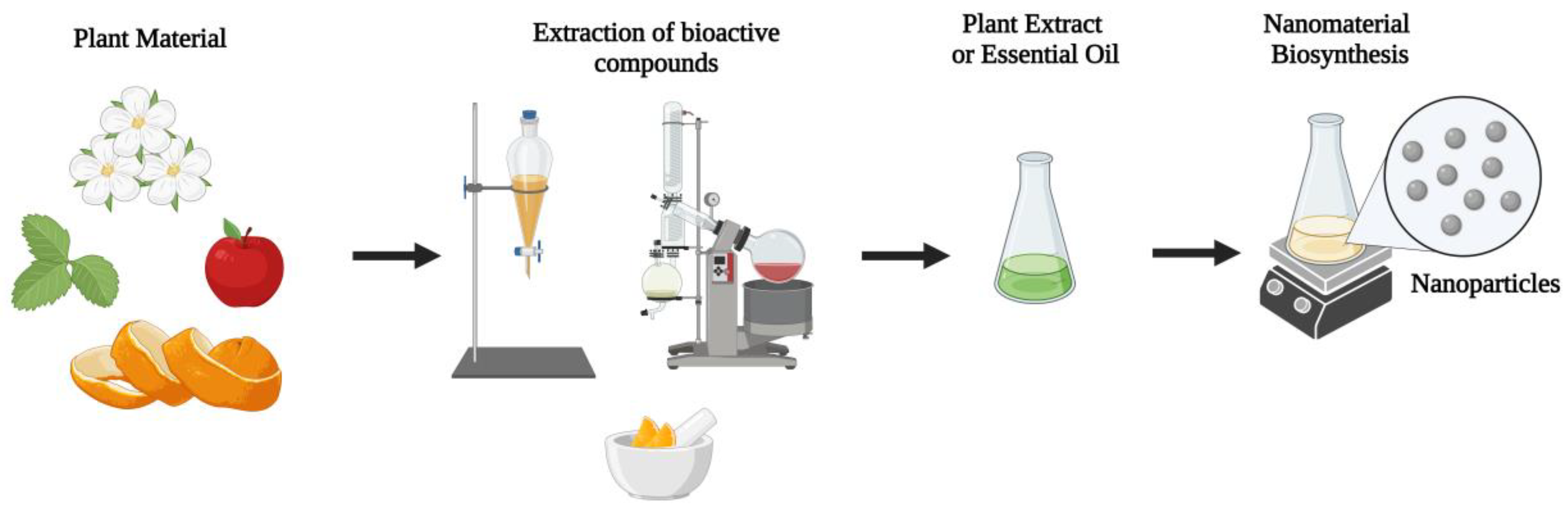
Biosynthesis of Nanoparticles Using Plant Extracts and Essential Oils
 , , , , , ,
Abstract
:1. Introduction
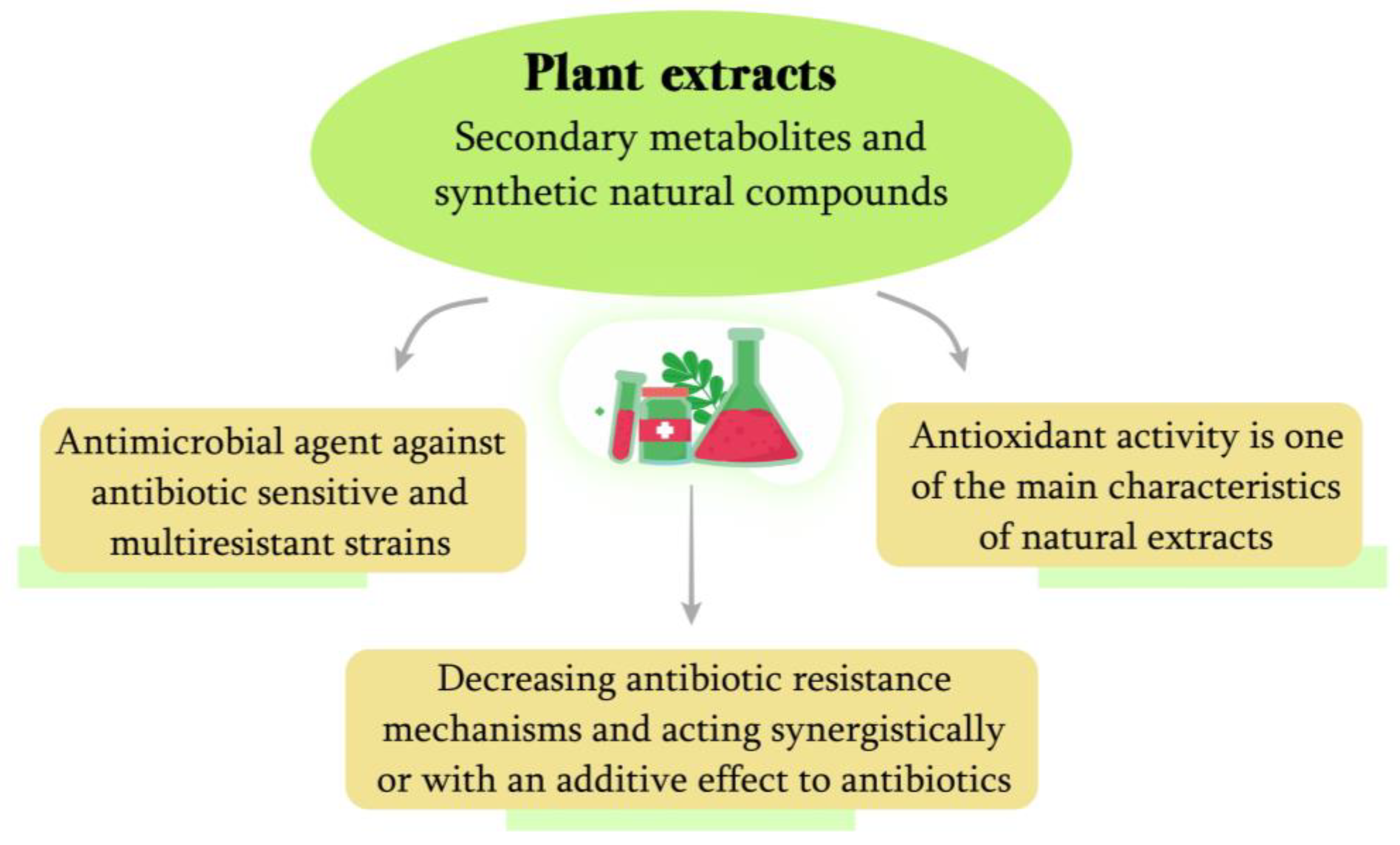
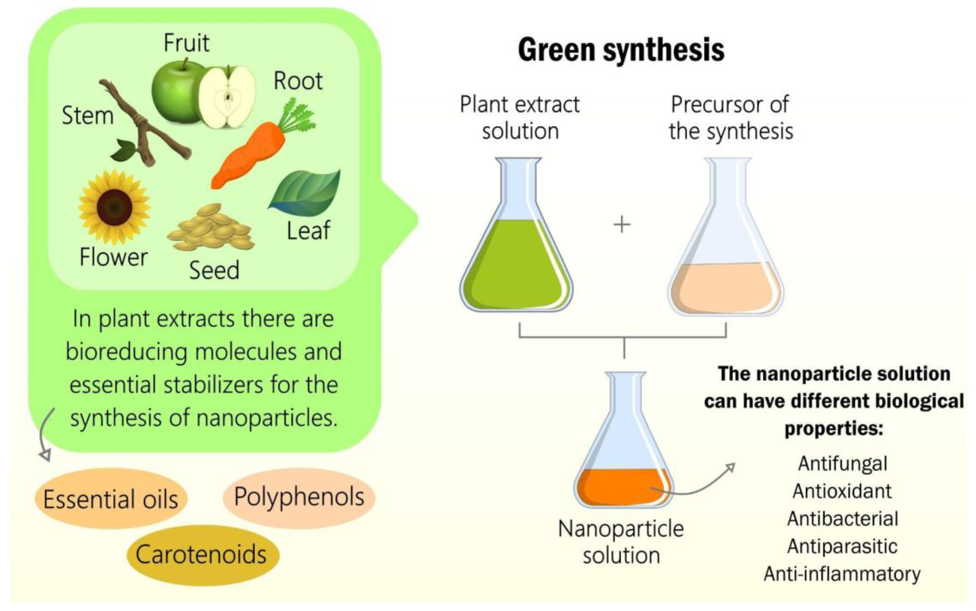
2. Plant Extracts
2.1. Polyphenols
2.1.1. Polyphenol Extraction
Conventional Techniques Used for the Extraction of Polyphenols
Modern Techniques Used for the Extraction of Polyphenols
2.1.2. Techniques Used for Polyphenol Analysis
2.2. Carotenoids
2.2.1. Carotenoid Extraction
- 1.
- Room temperature and atmospheric pressure: maceration process;
- 2.
- Boiling temperature of the solvents and atmospheric pressure: extraction of carotenoids using a Soxhlet technique in the presence of the proper solvent;
- 3.
- Low temperature and high pressure: accelerated solvent extraction (ASE), also known as pressurized liquid extraction (PLE); supercritical fluid extraction (SFE)—occurs with minimal use of a co-solvent, such as ethanol;
- 4.
- Ultrasound and microwaves, high voltage pulses that facilitate the release of intracellular carotenoids: microwave-assisted extraction; ultrasound-assisted extraction (UAE); pulsed electric field-assisted extraction (PEF); enzyme-assisted extraction (EAE).
2.2.2. Carotenoid Analysis
2.3. Essential Oils
2.3.1. Essential Oil Extraction
Conventional Extraction Methods
Modern Extraction Methods
2.3.2. Essential Oils Evaluation
2.4. Antioxidant Activity
2.4.1. Methods for TAC Assessment According to Mechanism
2.4.2. Methods Used for Evaluation of TAC
2.5. Antibacterial Activity
3. Plant-Based Antibacterial Green Nanomaterials
4. Conclusions
Author Contributions
Funding
Institutional Review Board Statement
Informed Consent Statement
Data Availability Statement
Acknowledgments
Conflicts of Interest
References
- Süntar, I. Importance of ethnopharmacological studies in drug discovery: Role of medicinal plants. Phytochem. Rev. 2020, 19, 1199–1209. [Google Scholar] [CrossRef]
- Alasmari, A. Phytomedicinal Potential Characterization of Medical Plants (Rumex nervosus and Dodonaea viscose). J. Biochem. Technol. 2020, 11, 113–121. [Google Scholar]
- Horstmanshoff, M. Ancient medicine between hope and fear: Medicament, magic and poison in the Roman Empire. Eur. Rev. 1999, 7, 37–51. [Google Scholar] [CrossRef]
- Sargin, S.A. Potential anti-influenza effective plants used in Turkish folk medicine: A review. J. Ethnopharmacol. 2021, 265, 113319. [Google Scholar] [CrossRef]
- Mahady, G.B. Medicinal Plants for the Prevention and Treatment of Bacterial Infections. Curr. Pharm. Des. 2005, 11, 2405–2427. [Google Scholar] [CrossRef]
- Stéphane, F.F.Y.; Jules, B.K.J.; Batiha, G.E.-S.; Ali, I.; Bruno, L.N. Extraction of Bioactive Compounds from Medicinal Plants and Herbs. Nat. Med. Plants 2022. [Google Scholar] [CrossRef]
- Hao, D.C.; Gu, X.J.; Xiao, P.G. Medicinal Plants: Chemistry, Biology and Omics; Woodhead Publishing: Sawston, UK, 2015. [Google Scholar]
- Ninkuu, V.; Zhang, L.; Yan, J.; Fu, Z.; Yang, T.; Zeng, H. Biochemistry of Terpenes and Recent Advances in Plant Protection. Int. J. Mol. Sci. 2021, 22, 5710. [Google Scholar] [CrossRef] [PubMed]
- Naboulsi, I.; Aboulmouhajir, A.; Kouisni, L.; Bekkaoui, F.; Yasri, A. Plants extracts and secondary metabolites, their extraction methods and use in agriculture for controlling crop stresses and improving productivity: A review. Acad. J. Med. Plants 2018, 6, 223–240. [Google Scholar] [CrossRef]
- Eddin, L.; Jha, N.; Meeran, M.; Kesari, K.; Beiram, R.; Ojha, S. Neuroprotective Potential of Limonene and Limonene Containing Natural Products. Molecules 2021, 26, 4535. [Google Scholar] [CrossRef] [PubMed]
- Vieira, A.; Beserra, F.; Souza, M.; Totti, B.; Rozza, A. Limonene: Aroma of innovation in health and disease. Chem. Biol. Interact. 2018, 283, 97–106. [Google Scholar] [CrossRef] [PubMed] [Green Version]
- Exposito, O.; Bonfill, M.; Moyano, E.; Onrubia, M.; Mirjalili, M.H.; Cusido, R.M.; Palazon, J. Biotechnological Production of Taxol and Related Taxoids: Current State and Prospects. Anti-Cancer Agents Med. Chem. 2012, 9, 109–121. [Google Scholar] [CrossRef] [PubMed] [Green Version]
- Kamatou, G.P.; Vermaak, I.; Viljoen, A.M.; Lawrence, B.M. Menthol: A simple monoterpene with remarkable biological properties. Phytochemistry 2013, 96, 15–25. [Google Scholar] [CrossRef] [PubMed]
- Kurek, J. Alkaloids—Their Importance in Nature and Human Life; Intechopen: London, UK, 2019; Available online: https://www.intechopen.com/chapters/66742 (accessed on 17 January 2023).
- Bhambhani, S.; Kondhare, K.; Giri, A. Diversity in Chemical Structures and Biological Properties of Plant Alkaloids. Molecules 2021, 26, 3374. [Google Scholar] [CrossRef]
- Dias, M.C.; Pinto, D.C.G.A.; Silva, A.M.S. Plant Flavonoids: Chemical Characteristics and Biological Activity. Molecules 2021, 26, 5377. [Google Scholar] [CrossRef]
- Brodowska, K.; Brodowska, K.M. Natural Flavonoids: Classification, Potential Role, and Application of Flavonoid Analogues. Eur. J. Biol. Res. 2017, 7, 108–123. [Google Scholar]
- Afify, A.E.-M.M.R.; El-Beltagi, H.S.; El-Salam, S.M.A.; Omran, A.A. Biochemical changes in phenols, flavonoids, tannins, vitamin E, β–carotene and antioxidant activity during soaking of three white sorghum varieties. Asian Pac. J. Trop. Biomed. 2012, 2, 203–209. [Google Scholar] [CrossRef] [PubMed] [Green Version]
- Serrano, J.; Puupponen-Pimiä, R.; Dauer, A.; Aura, A.-M.; Saura-Calixto, F. Tannins: Current knowledge of food sources, intake, bioavailability and biological effects. Mol. Nutr. Food Res. 2009, 53, S310–S329. [Google Scholar] [CrossRef] [Green Version]
- Alcantara, R.G.L.; Joaquim, R.H.V.T.; Sampaio, S.F. Plantas Medicinais: O Conhecimento e Uso Popular. Rev. APS—Atenção Primária Saúde 2015, 18, 1–13. [Google Scholar]
- Fitzgerald, M.; Heinrich, M.; Booker, A. Medicinal Plant Analysis: A Historical and Regional Discussion of Emergent Complex Techniques. Front. Pharmacol. 2019, 10, 1480. [Google Scholar] [CrossRef]
- Barbosa, F.E.S.; Guimarães, M.B.L.; Dos Santos, C.R.; Bezerra, A.F.B.; Tesser, C.D.; De Sousa, I.M.C. Oferta de Práticas Integrativas e Complementares em Saúde na Estratégia Saúde da Família no Brasil. Cad. Saude Publica 2020, 36, e00208818. [Google Scholar] [CrossRef] [PubMed] [Green Version]
- Filho, S.A.; Backx, B.P. Nanotecnologia e seus impactos na sociedade. Rev. Tecnol. Soc. 2020, 16, 1–15. [Google Scholar] [CrossRef]
- Backx, B.P. Green Nanotechnology: Only the Final Product That Matters? Nat. Prod. Res. 2022, 36, 3507–3509. [Google Scholar] [CrossRef]
- Raja, R.K.; Hazir, S.; Balasubramani, G.; Sivaprakash, G.; Obeth, E.S.J.; Boobalan, T.; Pugazhendhi, A.; Raj, R.H.K.; Arun, A. Green Nanotechnology for the Environment. In Handbook of Microbial Nanotechnology; Elsevier: Amsterdam, The Netherlands, 2022. [Google Scholar]
- Srivastava, S.; Bhargava, A. Tools and Techniques Used in Nanobiotechnology. In Green Nanoparticles: The Future of Nanobiotechnology; Springer: Singapore, 2022; pp. 29–55. [Google Scholar]
- Backx, B.P.; dos Santos, M.S.; dos Santos, O.A.; Filho, S.A. The Role of Biosynthesized Silver Nanoparticles in Antimicrobial Mechanisms. Curr. Pharm. Biotechnol. 2021, 22, 762–772. [Google Scholar] [CrossRef] [PubMed]
- Li, A.N.; Li, S.; Zhang, Y.J.; Xu, X.R.; Chen, Y.M.; Li, H. bin Resources and Biological Activities of Natural Polyphenols. Nutrients 2014, 6, 6020–6047. [Google Scholar] [CrossRef] [Green Version]
- Deng, G.-F.; Xu, X.-R.; Zhang, Y.; Li, D.; Gan, R.-Y.; Li, H.-B. Phenolic Compounds and Bioactivities of Pigmented Rice. Crit. Rev. Food Sci. Nutr. 2013, 53, 296–306. [Google Scholar] [CrossRef]
- Guo, Y.-J.; Deng, G.-F.; Xu, X.-R.; Wu, S.; Li, S.; Xia, E.-Q.; Li, F.; Chen, F.; Ling, W.-H.; Li, H.-B. Antioxidant capacities, phenolic compounds and polysaccharide contents of 49 edible macro-fungi. Food Funct. 2012, 3, 1195–1205. [Google Scholar] [CrossRef]
- Li, H.-B.; Cheng, K.-W.; Wong, C.-C.; Fan, K.-W.; Chen, F.; Jiang, Y. Evaluation of antioxidant capacity and total phenolic content of different fractions of selected microalgae. Food Chem. 2007, 102, 771–776. [Google Scholar] [CrossRef]
- Li, H.-B.; Chen, F. Preparative isolation and purification of astaxanthin from the microalga Chlorococcum sp. by high-speed counter-current chromatography. J. Chromatogr. A 2001, 925, 133–137. [Google Scholar] [CrossRef]
- Xia, E.-Q.; Wang, B.-W.; Xu, X.-R.; Zhu, L.; Song, Y.; Li, H.-B. Microwave-Assisted Extraction of Oleanolic Acid and Ursolic Acid from Ligustrum lucidum Ait. Int. J. Mol. Sci. 2011, 12, 5319–5329. [Google Scholar] [CrossRef]
- Song, F.-L.; Gan, R.-Y.; Zhang, Y.; Xiao, Q.; Kuang, L.; Li, H.-B. Total Phenolic Contents and Antioxidant Capacities of Selected Chinese Medicinal Plants. Int. J. Mol. Sci. 2010, 11, 2362–2372. [Google Scholar] [CrossRef]
- Fu, L.; Xu, B.-T.; Gan, R.-Y.; Zhang, Y.; Xu, X.-R.; Xia, E.-Q.; Li, H.-B. Total Phenolic Contents and Antioxidant Capacities of Herbal and Tea Infusions. Int. J. Mol. Sci. 2011, 12, 2112–2124. [Google Scholar] [CrossRef] [Green Version]
- Li, F.; Li, S.; Li, H.-B.; Deng, G.-F.; Ling, W.-H.; Xu, X.-R. Antiproliferative activities of tea and herbal infusions. Food Funct. 2013, 4, 530–538. [Google Scholar] [CrossRef] [PubMed]
- Xia, E.-Q.; Ai, X.-X.; Zang, S.-Y.; Guan, T.-T.; Xu, X.-R.; Li, H.-B. Ultrasound-assisted extraction of phillyrin from Forsythia suspensa. Ultrason. Sonochem. 2011, 18, 549–552. [Google Scholar] [CrossRef]
- Li, S.; Deng, G.-F.; Li, A.-N.; Xu, X.-R.; Wu, S.; Li, H.-B. Effect of Ultrasound-Assisted Extraction on Antioxidant Activity of Rose (Rosa hybrida) Petals. Int. J. Modern Biol. Med. 2012, 2, 91–100. [Google Scholar]
- Harborne, J.; Williams, C. Advances in flavonoid research since 1992. Phytochemistry 2000, 55, 481–504. [Google Scholar] [CrossRef]
- Manach, C.; Scalbert, A.; Morand, C.; Rémésy, C.; Jiménez, L. Polyphenols: Food sources and bioavailability. Am. J. Clin. Nutr. 2004, 79, 727–747. [Google Scholar] [CrossRef] [Green Version]
- Mukherjee, C.; Chakraborty, S. Study of dietary polyphenols from natural herbal sources for providing protection against human degenerative disorders. Biocatal. Agric. Biotechnol. 2021, 33, 101956. [Google Scholar] [CrossRef]
- Singla, R.K.; Dubey, A.K.; Garg, A.; Sharma, R.K.; Fiorino, M.; Ameen, S.M.; Haddad, M.A.; Al-Hiary, M. Natural Polyphenols: Chemical Classification, Definition of Classes, Subcategories, and Structures. J. AOAC Int. 2019, 102, 1397–1400. [Google Scholar] [CrossRef]
- Bertelli, A.; Biagi, M.; Corsini, M.; Baini, G.; Cappellucci, G.; Miraldi, E. Polyphenols: From Theory to Practice. Foods 2021, 10, 2595. [Google Scholar] [CrossRef] [PubMed]
- D’Archivio, M.; Filesi, C.; di Benedetto, R.; Gargiulo, R.; Giovannini, C.; Masella, R. Polyphenols, Dietary Sources and Bioavailability. Ann. Ist. Super. Sanita 2007, 43, 348. [Google Scholar]
- Ajila, C.; Brar, S.; Verma, M.; Tyagi, R.; Godbout, S.; Valéro, J. Extraction and Analysis of Polyphenols: Recent trends. Crit. Rev. Biotechnol. 2011, 31, 227–249. [Google Scholar] [CrossRef]
- Li, H.; Chen, B.; Yao, S. Application of ultrasonic technique for extracting chlorogenic acid from Eucommia ulmodies Oliv. (E. ulmodies). Ultrason. Sonochem. 2005, 12, 295–300. [Google Scholar] [CrossRef]
- Brglez Mojzer, E.; Knez Hrnčič, M.; Škerget, M.; Knez, Ž.; Bren, U. Polyphenols: Extraction Methods, Antioxidative Action, Bioavailability and Anticarcinogenic Effects. Molecules 2016, 21, 901. [Google Scholar] [CrossRef]
- Tapia-Quirós, P.; Montenegro-Landívar, M.F.; Reig, M.; Vecino, X.; Cortina, J.L.; Saurina, J.; Granados, M. Recovery of Poly-phenols from Agri-Food By-Products: The Olive Oil and Winery Industries Cases. Foods 2022, 11, 362. [Google Scholar] [CrossRef]
- Wang, L.; Weller, C.L. Recent advances in extraction of nutraceuticals from plants. Trends Food Sci. Technol. 2006, 17, 300–312. [Google Scholar] [CrossRef]
- Palaiogiannis, D.; Athanasiadis, V.; Bozinou, E.; Chatzimitakos, T.; Makris, D.P.; Lalas, S.I. Extraction of Polyphenolic and Volatile Compounds from Cistus creticus Using Deep Eutectic Solvents and Pulsed Electric Fields. Compounds 2022, 2, 26. [Google Scholar] [CrossRef]
- Ignat, I.; Volf, I.; Popa, V.I. A critical review of methods for characterisation of polyphenolic compounds in fruits and vegetables. Food Chem. 2011, 126, 1821–1835. [Google Scholar] [CrossRef] [PubMed]
- Lichtenthaler, R.N. Gerd Brunner: Gas Extraction—An Introduction to Fundamentals of Supercritical Fluids and the Application to Separation Processes. Topics in Physical Chemistry, Vol. 4; Baumgärtel, H., Franck, E.U., Grünbein. W., Eds.; Steinkopff: Darmstadt, Germany; Springer: New York, NY, USA. Ber. Bunsenges. Phys. Chem. 1996, 100, 1090–1091. [Google Scholar] [CrossRef]
- Brunner, G.J. Supercritical fluids: Technology and application to food processing. J. Food Eng. 2005, 67, 21–33. [Google Scholar] [CrossRef]
- Díaz-Reinoso, B.; Moure, A.; Domínguez, H.; Parajó, J.C. Supercritical CO2 Extraction and Purification of Compounds with Antioxidant Activity. J. Agric. Food Chem. 2006, 54, 2441–2469. [Google Scholar] [CrossRef]
- Castañeda-Ovando, A.; Pacheco-Hernandez, M.D.L.; Páez-Hernández, M.E.; Rodríguez, J.A.; Galán-Vidal, C.A. Chemical studies of anthocyanins: A review. Food Chem. 2009, 113, 859–871. [Google Scholar] [CrossRef]
- Abascal, K.; Ganora, L.; Yarnell, E. The effect of freeze-drying and its implications for botanical medicine: A review. Phytotherapy Res. 2005, 19, 655–660. [Google Scholar] [CrossRef] [PubMed]
- Stalikas, C.D. Extraction, separation, and detection methods for phenolic acids and flavonoids. J. Sep. Sci. 2007, 30, 3268–3295. [Google Scholar] [CrossRef] [PubMed]
- Qiu, Y.; Liu, Q.; Beta, T. Antioxidant properties of commercial wild rice and analysis of soluble and insoluble phenolic acids. Food Chem. 2010, 121, 140–147. [Google Scholar] [CrossRef]
- Metivier, R.P.; Francis, F.J.; Clydesdale, F.M. Solvent Extraction of Anthocyanins from Wine Pomace. J. Food Sci. 1980, 45, 1099–1100. [Google Scholar] [CrossRef]
- Prior, R.L.; Lazarus, S.A.; Cao, G.; Muccitelli, H.; Hammerstone, J.F. Identification of Procyanidins and Anthocyanins in Blueberries and Cranberries (Vaccinium spp.) Using High-Performance Liquid Chromatography/Mass Spectrometry. J. Agric. Food Chem. 2001, 49, 1270–1276. [Google Scholar] [CrossRef]
- Guyot, S.; Marnet, N.; Drilleau, J.-F. Thiolysis−HPLC Characterization of Apple Procyanidins Covering a Large Range of Polymerization States. J. Agric. Food Chem. 2001, 49, 14–20. [Google Scholar] [CrossRef] [PubMed]
- Labarbe, B.; Cheynier, V.; Brossaud, F.; Souquet, J.-M.; Moutounet, M. Quantitative Fractionation of Grape Proanthocyanidins According to Their Degree of Polymerization. J. Agric. Food Chem. 1999, 47, 2719–2723. [Google Scholar] [CrossRef] [PubMed]
- Vieira, G.S.; Cavalcanti, R.N.; Meireles, M.A.A.; Hubinger, M.D. Chemical and economic evaluation of natural antioxidant extracts obtained by ultrasound-assisted and agitated bed extraction from jussara pulp (Euterpe edulis). J. Food Eng. 2013, 119, 196–204. [Google Scholar] [CrossRef] [Green Version]
- Vázquez, M.B.; Comini, L.; Martini, R.; Montoya, S.N.; Bottini, S.; Cabrera, J. Comparisons between conventional, ultrasound-assisted and microwave-assisted methods for extraction of anthraquinones from Heterophyllaea pustulata Hook f. (Rubiaceae). Ultrason. Sonochem. 2014, 21, 478–484. [Google Scholar] [CrossRef] [PubMed]
- Knorr, D. Impact of Non-Thermal Processing on Plant Metabolites. J. Food Eng. 2003, 56, 131–134. [Google Scholar] [CrossRef]
- Rostagno, M.A.; Palma, M.; Barroso, C.G. Ultrasound-Assisted Extraction of Soy Isoflavones. J. Chromatogr. A 2003, 1012, 119–128. [Google Scholar] [CrossRef] [PubMed]
- Medina-Torres, N.; Ayora-Talavera, T.; Espinosa-Andrews, H.; Sánchez-Contreras, A.; Pacheco, N. Ultrasound Assisted Extraction for the Recovery of Phenolic Compounds from Vegetable Sources. Agronomy 2017, 7, 47. [Google Scholar] [CrossRef]
- Moret, S.; Conchione, C.; Srbinovska, A.; Lucci, P. Microwave-Based Technique for Fast and Reliable Extraction of Organic Contaminants from Food, with a Special Focus on Hydrocarbon Contaminants. Foods 2019, 8, 503. [Google Scholar] [CrossRef] [Green Version]
- Letellier, M.; Budzinski, H.; Charrier, L.; Capes, S.; Dorthe, A.M. Optimization by factorial design of focused microwave assisted extraction of polycyclic aromatic hydrocarbons from marine sediment. Fresenius J. Anal. Chem. 1999, 364, 228–237. [Google Scholar] [CrossRef]
- Letellier, M.; Budzinski, H. Microwave assisted extraction of organic compounds. Analusis 1999, 27, 259–270. [Google Scholar] [CrossRef]
- Talebi, M.; Ghassempour, A.; Talebpour, Z.; Rassouli, A.; Dolatyari, L. Optimization of the extraction of paclitaxel from Taxus baccata L. by the use of microwave energy. J. Sep. Sci. 2004, 27, 1130–1136. [Google Scholar] [CrossRef]
- King, M.B.; Bott, T.R. Extraction of Natural Products Using Near-Critical Solvents; Springer: Berlin/Heidelberg, Germany, 1993. [Google Scholar]
- McHugh, M.A.; Val, J. Krukonis Supercritical Fluid Extraction: Principles and Practice; Butterworth Publishers: Stoneham, MA, USA, 1986; Volume 66, p. 7. [Google Scholar] [CrossRef]
- Palenzuela, B.; Arce, L.; Macho, A. Bioguided extraction of polyphenols from grape marc by using an alternative supercritical-fluid extraction method based on a liquid solvent trap. Anal. Bioanal. Chem. 2004, 378, 2021–2027. [Google Scholar] [CrossRef]
- Ranjha, M.M.A.N.; Kanwal, R.; Shafique, B.; Arshad, R.N.; Irfan, S.; Kieliszek, M.; Kowalczewski, P.; Irfan, M.; Khalid, M.Z.; Roobab, U.; et al. A Critical Review on Pulsed Electric Field: A Novel Technology for the Extraction of Phytoconstituents. Molecules 2021, 26, 4893. [Google Scholar] [CrossRef]
- Bleve, M.; Ciurlia, L.; Erroi, E.; Lionetto, G.; Longo, L.; Rescio, L.; Schettino, T.; Vasapollo, G. An innovative method for the purification of anthocyanins from grape skin extracts by using liquid and sub-critical carbon dioxide. Sep. Purif. Technol. 2008, 64, 192–197. [Google Scholar] [CrossRef]
- Elkacmi, R.; Boulmal, N.; Kamil, N.; Bennajah, M. Techno-economical evaluation of a new technique for olive mill wastewater treatment. Sustain. Prod. Consum. 2017, 10, 38–49. [Google Scholar] [CrossRef]
- Lafka, T.-I.; Lazou, A.E.; Sinanoglou, V.J.; Lazos, E.S. Phenolic and antioxidant potential of olive oil mill wastes. Food Chem. 2011, 125, 92–98. [Google Scholar] [CrossRef]
- Caldas, T.W.; Mazza, K.E.L.; Teles, A.S.C.; Mattos, G.N.; Brígida, A.I.S.; Conte-Junior, C.A.; Borguini, R.G.; Godoy, R.L.O.; Cabral, L.M.C.; Tonon, R.V. Phenolic compounds recovery from grape skin using conventional and non-conventional extraction methods. Ind. Crop. Prod. 2018, 111, 86–91. [Google Scholar] [CrossRef]
- Tao, Y.; Wu, D.; Zhang, Q.-A.; Sun, D.-W. Ultrasound-assisted extraction of phenolics from wine lees: Modeling, optimization and stability of extracts during storage. Ultrason. Sonochem. 2014, 21, 706–715. [Google Scholar] [CrossRef] [PubMed]
- Bai, X.-L.; Yue, T.-L.; Yuan, Y.-H.; Zhang, H.-W. Optimization of microwave-assisted extraction of polyphenols from apple pomace using response surface methodology and HPLC analysis. J. Sep. Sci. 2010, 33, 3751–3758. [Google Scholar] [CrossRef] [PubMed]
- Ferri, M.; Vannini, M.; Ehrnell, M.; Eliasson, L.; Xanthakis, E.; Monari, S.; Sisti, L.; Marchese, P.; Celli, A.; Tassoni, A. From winery waste to bioactive compounds and new polymeric biocomposites: A contribution to the circular economy concept. J. Adv. Res. 2020, 24, 1–11. [Google Scholar] [CrossRef]
- Štambuk, P.; Tomašković, D.; Tomaz, I.; Maslov, L.; Stupić, D.; Kontić, J.K. Application of pectinases for recovery of grape seeds phenolics. 3 Biotech 2016, 6, 224. [Google Scholar] [CrossRef] [PubMed] [Green Version]
- Zuorro, A.; Lavecchia, R.; González-Delgado, D.; García-Martinez, J.B.; L’Abbate, P. Optimization of Enzyme-Assisted Extraction of Flavonoids from Corn Husks. Processes 2019, 7, 804. [Google Scholar] [CrossRef] [Green Version]
- Singleton, V.L.; Orthofer, R.; Lamuela-Raventós, R.M. Analysis of total phenols and other oxidation substrates and antioxidants by means of folin-ciocalteu reagent. Methods Enzymol. 1999, 299, 152–178. [Google Scholar]
- Dai, J.; Mumper, R.J. Plant Phenolics: Extraction, Analysis and Their Antioxidant and Anticancer Properties. Molecules 2010, 15, 7313–7352. [Google Scholar] [CrossRef]
- Delpino-Rius, A.; Eras, J.; Vilaró, F.; Cubero, M.; Balcells, M.; Canela-Garayoa, R. Characterisation of phenolic compounds in processed fibres from the juice industry. Food Chem. 2015, 172, 575–584. [Google Scholar] [CrossRef] [PubMed]
- Capriotti, A.L.; Cavaliere, C.; Foglia, P.; Piovesana, S.; Ventura, S. Chromatographic Methods Coupled to Mass Spectrometry Detection for the Determination of Phenolic Acids in Plants and Fruits. J. Liq. Chromatogr. Relat. Technol. 2014, 38, 353–370. [Google Scholar] [CrossRef]
- Pyrzynska, K.; Sentkowska, A. Recent Developments in the HPLC Separation of Phenolic Food Compounds. Crit. Rev. Anal. Chem. 2015, 45, 41–51. [Google Scholar] [CrossRef]
- Ross, K.; Beta, T.; Arntfield, S. A comparative study on the phenolic acids identified and quantified in dry beans using HPLC as affected by different extraction and hydrolysis methods. Food Chem. 2009, 113, 336–344. [Google Scholar] [CrossRef]
- Liu, Q.; Cai, W.; Shao, X. Determination of seven polyphenols in water by high performance liquid chromatography combined with preconcentration. Talanta 2008, 77, 679–683. [Google Scholar] [CrossRef]
- Lapornik, B.; Prošek, M.; Wondra, A.G. Comparison of extracts prepared from plant by-products using different solvents and extraction time. J. Food Eng. 2005, 71, 214–222. [Google Scholar] [CrossRef]
- Corrales, M.; García, A.F.; Butz, P.; Tauscher, B. Extraction of anthocyanins from grape skins assisted by high hydrostatic pressure. J. Food Eng. 2009, 90, 415–421. [Google Scholar] [CrossRef]
- Mustafa, A.M.; Abouelenein, D.; Angeloni, S.; Maggi, F.; Navarini, L.; Sagratini, G.; Santanatoglia, A.; Torregiani, E.; Vittori, S.; Caprioli, G. A New HPLC-MS/MS Method for the Simultaneous Determination of Quercetin and Its Derivatives in Green Coffee Beans. Foods 2022, 11, 3033. [Google Scholar] [CrossRef]
- Fernandes, A.S.; Nascimento, T.C.D.; Jacob-Lopes, E.; De Rosso, V.V.; Zepka, L.Q. Introductory Chapter: Carotenoids—A Brief Overview on Its Structure, Biosynthesis, Synthesis, and Applications. Prog. Carotenoid Res. 2018, 1, 1–17. [Google Scholar] [CrossRef] [Green Version]
- Kiokias, S.; Proestos, C.; Varzakas, T. A Review of the Structure, Biosynthesis, Absorption of Carotenoids-Analysis and Properties of their Common Natural Extracts. Curr. Res. Nutr. Food Sci. J. 2016, 4, 25–37. [Google Scholar] [CrossRef]
- Kritchevsky, S.B. γ-Carotene, Carotenoids and the Prevention of Coronary Heart Disease. J. Nutr. 1999, 129, 5–8. [Google Scholar] [CrossRef] [Green Version]
- Kiokias, S.; Varzakas, T. Activity of flavonoids and β-carotene during the auto-oxidative deterioration of model food oil-in water emulsions. Food Chem. 2014, 150, 280–286. [Google Scholar] [CrossRef]
- Akoh, C.C.; Min, D.B. Food Lipids: Chemistry, Nutrition, and Biotechnology, 3rd ed.; CRC Press: Boca Raton, FL, USA, 2008. [Google Scholar]
- van den Berg, H.; Faulks, R.; Granado, H.F.; Hirschberg, J.; Olmedilla, B.; Sandmann, G.; Southon, S.; Stahl, W. The Potential for the Improvement of Carotenoid Levels in Foods and the Likely Systemic Effects. J. Sci. Food Agric. 2000, 80, 880–912. [Google Scholar] [CrossRef]
- Astley, S. ANTIOXIDANTS|Role of Antioxidant Nutrients in Defense Systems. In Encyclopedia of Food Sciences and Nutrition; Academic Press: Cambridge, MA, USA, 2003; pp. 282–289. [Google Scholar] [CrossRef]
- US Department of Agriculture, Agricultural Research Service. SR28—Page Reports: USDA ARS. Available online: https://www.ars.usda.gov/northeast-area/beltsville-md-bhnrc/beltsville-human-nutrition-research-center/methods-and-application-of-food-composition-laboratory/mafcl-site-pages/sr28-page-reports/ (accessed on 11 February 2023).
- Rebecca, L.J.; Sharmila, S.; Das, M.P.; Seshiah, C. Extraction and Purification of Carotenoids from Vegetables. J. Chem. Pharm. Res. 2014, 6, 594–598. [Google Scholar]
- Saini, R.K.; Keum, Y.-S. Carotenoid extraction methods: A review of recent developments. Food Chem. 2018, 240, 90–103. [Google Scholar] [CrossRef]
- Kopec, R.E.; Cooperstone, J.L.; Cichon, M.J.; Schwartz, S.J. Analysis Methods of Carotenoids. In Analysis of Antioxidant-Rich Phytochemicals; John Wiley & Sons, Ltd.: Hoboken, NJ, USA, 2012; pp. 105–148. ISBN 9781118229378. [Google Scholar]
- Hussain, A.; Larsson, H.; Johansson, E. Carotenoid Extraction from Locally and Organically Produced Cereals Using Saponification Method. Processes 2021, 9, 783. [Google Scholar] [CrossRef]
- Mezzomo, N.; Maestri, B.; dos Santos, R.L.; Maraschin, M.; Ferreira, S.R. Pink shrimp (P. brasiliensis and P. paulensis) residue: Influence of extraction method on carotenoid concentration. Talanta 2011, 85, 1383–1391. [Google Scholar] [CrossRef]
- Poojary, M.M.; Passamonti, P. Optimization of extraction of high purity all-trans-lycopene from tomato pulp waste. Food Chem. 2015, 188, 84–91. [Google Scholar] [CrossRef]
- Pour Hosseini, S.R.; Tavakoli, O.; Sarrafzadeh, M.H. Experimental optimization of SC-CO2 extraction of carotenoids from Dunaliella salina. J. Supercrit. Fluids 2017, 121, 89–95. [Google Scholar] [CrossRef]
- Goula, A.M.; Ververi, M.; Adamopoulou, A.; Kaderides, K. Green ultrasound-assisted extraction of carotenoids from pomegranate wastes using vegetable oils. Ultrason. Sonochem. 2017, 34, 821–830. [Google Scholar] [CrossRef] [PubMed]
- Yara-Varón, E.; Fabiano-Tixier, A.S.; Balcells, M.; Canela-Garayoa, R.; Bily, A.; Chemat, F. Is it possible to substitute hexane with green solvents for extraction of carotenoids? A theoretical versus experimental solubility study. RSC Adv. 2016, 6, 27750–27759. [Google Scholar] [CrossRef] [Green Version]
- Butnariu, M. Methods of Analysis (Extraction, Separation, Identification and Quantification) of Carotenoids from Natural Products. J. Ecosyst. Ecogr. 2016, 6, 193. [Google Scholar] [CrossRef]
- Machado, C.A.; Oliveira, F.O.; de Andrade, M.A.; Hodel, K.V.S.; Lepikson, H.; Machado, B.A.S. Steam Distillation for Essential Oil Extraction: An Evaluation of Technological Advances Based on an Analysis of Patent Documents. Sustainability 2022, 14, 7119. [Google Scholar] [CrossRef]
- Geramitcioski, T.; Mitrevski, V.; Mijakovski, V. Design of a Small Press for Extracting Essential Oil According VDI 2221. In IOP Conference Series: Materials Science and Engineering; IOP Publishing: Bristol, UK, 2018; Volume 393, p. 012131. [Google Scholar]
- Le, X.D.; Thi, N.M.P.; Cam, T.I.; Do, H.N.; Thi, H.V.N.; Thang, T.D.; Thao, L.P.P.; Do, T.S.; Nguyen, T.D.; Pham, Q.L.; et al. Optimization of the Essential Oil Extraction Process from Dong Van Marjoram (E. winitiana var. dongvanensis Phuong.) by Using Microwave Assisted Hydrodistillation, and the Bioactivities of the Oil Against Some Cancer Cell Lines and Bacteria. Nat. Prod. Commun. 2021, 16, 1934578X211054235. [Google Scholar] [CrossRef]
- Zeleke, Z.Z. Extraction of Essential Oil from Lemon and Orange Peel by Clevenger Apparatus: Comparative GC_MS Analysis of Chemical Composition, from Debre Berehan Market Town Amahara Region Ethiopia. Ann. Biotechnol. 2022, 5, 1026. [Google Scholar] [CrossRef]
- Ribeiro, B.S.; Ferreira, M.D.F.; Moreira, J.L.; Santos, L. Simultaneous Distillation–Extraction of Essential Oils from Rosmarinus officinalis L. Cosmetics 2021, 8, 117. [Google Scholar] [CrossRef]
- Kawacka, I.; Olejnik-Schmidt, A.; Schmidt, M.; Sip, A. Natural Plant-Derived Chemical Compounds as Listeria monocytogenes Inhibitors In Vitro and in Food Model Systems. Pathogens 2021, 10, 12. [Google Scholar] [CrossRef]
- Stratakos, A.C.; Koidis, A. Methods for Extracting Essential Oils. In Essential Oils in Food Preservation, Flavor and Safety; Academic Press: Cambridge, MA, USA, 2016; pp. 31–38. [Google Scholar] [CrossRef]
- Kant, R.; Kumar, A. Review on essential oil extraction from aromatic and medicinal plants: Techniques, performance and economic analysis. Sustain. Chem. Pharm. 2022, 30, 100829. [Google Scholar] [CrossRef]
- Olivas, N.A.; Bejarano, C.V.; Soto, G.A.; Ortega, M.Z.; Salas, F.S.; Chávez, E.S.; Ochoa, L.H. Bioactive compounds and antioxidant activity of essential oils of Origanum dictamnus from Mexico. AIMS Agric. Food 2020, 5, 387–394. [Google Scholar] [CrossRef]
- Mahawer, S.K.; Himani; Arya, S.; Kumar, R.; Prakash, O. Extractions Methods and Biological Applications of Essential Oils. In Essential Oils; de Oliveira, M.S., de Aguiar Andrade, E.H., Eds.; IntechOpen: Rijeka, Croatia, 2022. [Google Scholar]
- Jyotirmayee, B.; Mahalik, G. Essential Oils Extracted from CO. In Aromatherapy and Its Benefits; Preetha, B., Sagarika, P., Eds.; Renu Publishers: New Delhi, India, 2021; ISBN 9788194094371. [Google Scholar]
- Nordin, S.; Broman, D.A.; Olofsson, J.K.; Wulff, M. A Longitudinal Descriptive Study of Self-reported Abnormal Smell and Taste Perception in Pregnant Women. Chem. Senses 2004, 29, 391–402. [Google Scholar] [CrossRef]
- Sankarikutty, B.; Narayanan, C.S. ESSENTIAL OILS: Isolation and Production. In Encyclopedia of Food Sciences and Nutrition; Academic Press: Cambridge, MA, USA, 2003. [Google Scholar]
- Aizpurua-Olaizola, O.; Ormazabal, M.; Vallejo, A.; Olivares, M.; Navarro, P.; Etxebarria, N.; Usobiaga, A. Optimization of Supercritical Fluid Consecutive Extractions of Fatty Acids and Polyphenols from Vitis vinifera Grape Wastes. J. Food Sci. 2015, 80, E101–E107. [Google Scholar] [CrossRef]
- Rassem, H.H.A.; Nour, A.H.; Yunus, R.M. Techniques for Extraction of Essential Oils from Plants: A Review. Aust. J. Basic Appl. Sci. 2016, 10, 117–127. [Google Scholar]
- Tongnuanchan, P.; Benjakul, S. Essential Oils: Extraction, Bioactivities, and Their Uses for Food Preservation. J. Food Sci. 2014, 79, R1231–R1249. [Google Scholar] [CrossRef] [PubMed]
- Babu, K.G.D.; Kaul, V.K. Variation in essential oil composition of rose-scented geranium (Pelargonium sp.) distilled by different distillation techniques. Flavour Fragr. J. 2005, 20, 222–231. [Google Scholar] [CrossRef]
- Afzal, A.; Munir, A.; Ghafoor, A.; Alvarado, J.L. Development of hybrid solar distillation system for essential oil extraction. Renew. Energy 2017, 113, 22–29. [Google Scholar] [CrossRef]
- Kulturel, Y.; Tarhan, S. Performance of a Solar Distillery of Essential Oils with Compound Parabolic Solar Collectors. J. Sci. Ind. Res. 2016, 75, 691–696. [Google Scholar]
- Radwan, M.N.; Morad, M.; Ali, M.; Wasfy, K.I. A solar steam distillation system for extracting lavender volatile oil. Energy Rep. 2020, 6, 3080–3087. [Google Scholar] [CrossRef]
- Gavahian, M.; Chu, Y.-H. Ohmic accelerated steam distillation of essential oil from lavender in comparison with conventional steam distillation. Innov. Food Sci. Emerg. Technol. 2018, 50, 34–41. [Google Scholar] [CrossRef]
- Sahraoui, N.; Vian, M.A.; Bornard, I.; Boutekedjiret, C.; Chemat, F. Improved microwave steam distillation apparatus for isolation of essential oils: Comparison with conventional steam distillation. J. Chromatogr. A 2008, 1210, 229–233. [Google Scholar] [CrossRef] [PubMed]
- Gavahian, M.; Farahnaky, A. Ohmic-assisted hydrodistillation technology: A review. Trends Food Sci. Technol. 2018, 72, 153–161. [Google Scholar] [CrossRef]
- Aziz, Z.A.A.; Ahmad, A.; Setapar, S.H.M.; Karakucuk, A.; Azim, M.M.; Lokhat, D.; Rafatullah, M.; Ganash, M.; Kamal, M.A.; Ashraf, G.M. Essential Oils: Extraction Techniques, Pharmaceutical and Therapeutic Potential—A Review. Curr. Drug Metab. 2018, 19, 1100–1110. [Google Scholar] [CrossRef]
- Taban, A.; Saharkhiz, M.J.; Niakousari, M. Sweet bay (Laurus nobilis L.) essential oil and its chemical composition, antioxidant activity and leaf micromorphology under different extraction methods. Sustain. Chem. Pharm. 2018, 9, 12–18. [Google Scholar] [CrossRef]
- Drinić, Z.; Pljevljakušić, D.; Živković, J.; Bigović, D.; Šavikin, K. Microwave-assisted extraction of O. vulgare L. spp. hirtum essential oil: Comparison with conventional hydro-distillation. Food Bioprod. Process. 2020, 120, 158–165. [Google Scholar] [CrossRef]
- Elyemni, M.; Louaste, B.; Nechad, I.; Elkamli, T.; Bouia, A.; Taleb, M.; Chaouch, M.; Eloutassi, N. Extraction of Essential Oils of Rosmarinus officinalis L. by Two Different Methods: Hydrodistillation and Microwave Assisted Hydrodistillation. Sci. World J. 2019, 2019, 3659432. [Google Scholar] [CrossRef] [PubMed] [Green Version]
- Golmakani, M.-T.; Rezaei, K. Comparison of microwave-assisted hydrodistillation withthe traditional hydrodistillation method in the extractionof essential oils from Thymus vulgaris L. Food Chem. 2008, 109, 925–930. [Google Scholar] [CrossRef] [PubMed]
- Wang, Z.; Ding, L.; Li, T.; Zhou, X.; Wang, L.; Zhang, H.; Liu, L.; Li, Y.; Liu, Z.; Wang, H.; et al. Improved solvent-free microwave extraction of essential oil from dried Cuminum cyminum L. and Zanthoxylum bungeanum Maxim. J. Chromatogr. A 2006, 1102, 11–17. [Google Scholar] [CrossRef] [PubMed]
- Gavahian, M.; Farahnaky, A.; Javidnia, K.; Majzoobi, M. Comparison of ohmic-assisted hydrodistillation with traditional hydrodistillation for the extraction of essential oils from Thymus vulgaris L. Innov. Food Sci. Emerg. Technol. 2012, 14, 85–91. [Google Scholar] [CrossRef]
- Vian, M.A.; Fernandez, X.; Visinoni, F.; Chemat, F. Microwave hydrodiffusion and gravity, a new technique for extraction of essential oils. J. Chromatogr. A 2008, 1190, 14–17. [Google Scholar] [CrossRef] [PubMed]
- Li, Y.; Fabiano-Tixier, A.-S.; Chemat, F. Essential Oils: From Conventional to Green Extraction. In Essential Oils as Reagents in Green Chemistry; Springer: Berlin/Heidelberg, Germany, 2014; pp. 9–20. [Google Scholar] [CrossRef]
- Bousbia, N.; Vian, M.A.; Ferhat, M.A.; Meklati, B.Y.; Chemat, F. A new process for extraction of essential oil from Citrus peels: Microwave hydrodiffusion and gravity. J. Food Eng. 2009, 90, 409–413. [Google Scholar] [CrossRef]
- Bousbia, N.; Vian, M.A.; Ferhat, M.A.; Petitcolas, E.; Meklati, B.Y.; Chemat, F. Comparison of two isolation methods for essential oil from rosemary leaves: Hydrodistillation and microwave hydrodiffusion and gravity. Food Chem. 2009, 114, 355–362. [Google Scholar] [CrossRef]
- Farhat, A.; Fabiano-Tixier, A.-S.; El Maataoui, M.; Maingonnat, J.-F.; Romdhane, M.; Chemat, F. Microwave steam diffusion for extraction of essential oil from orange peel: Kinetic data, extract’s global yield and mechanism. Food Chem. 2011, 125, 255–261. [Google Scholar] [CrossRef]
- Dawidowicz, A.L.; Rado, E.; Wianowska, D.; Mardarowicz, M.; Gawdzik, J. Application of PLE for the determination of essential oil components from Thymus vulgaris L. Talanta 2008, 76, 878–884. [Google Scholar] [CrossRef] [PubMed]
- Li, X.-M.; Tian, S.-L.; Pang, Z.-C.; Shi, J.-Y.; Feng, Z.-S.; Zhang, Y.-M. Extraction of Cuminum cyminum essential oil by combination technology of organic solvent with low boiling point and steam distillation. Food Chem. 2009, 115, 1114–1119. [Google Scholar] [CrossRef]
- Kusuma, H.; Mahfud, M. Kinetic studies on extraction of essential oil from sandalwood (Santalum album) by microwave air-hydrodistillation method. Alex. Eng. J. 2018, 57, 1163–1172. [Google Scholar] [CrossRef]
- Kusuma, H.S.; Mahfud, M. The extraction of essential oils from patchouli leaves (Pogostemon cablin Benth) using a microwave air-hydrodistillation method as a new green technique. RSC Adv. 2017, 7, 1336–1347. [Google Scholar] [CrossRef] [Green Version]
- Lucchesi, M.E.; Chemat, F.; Smadja, J. An original solvent free microwave extraction of essential oils from spices. Flavour Fragr. J. 2004, 19, 134–138. [Google Scholar] [CrossRef]
- Lucchesi, M.E.; Chemat, F.; Smadja, J. Solvent-free microwave extraction of essential oil from aromatic herbs: Comparison with conventional hydro-distillation. J. Chromatogr. A 2004, 1043, 323–327. [Google Scholar] [CrossRef] [PubMed]
- Filly, A.; Fernandez, X.; Minuti, M.; Visinoni, F.; Cravotto, G.; Chemat, F. Solvent-free microwave extraction of essential oil from aromatic herbs: From laboratory to pilot and industrial scale. Food Chem. 2014, 150, 193–198. [Google Scholar] [CrossRef] [PubMed] [Green Version]
- Eikani, M.H.; Golmohammad, F.; Rowshanzamir, S. Subcritical water extraction of essential oils from coriander seeds (Coriandrum sativum L.). J. Food Eng. 2007, 80, 735–740. [Google Scholar] [CrossRef]
- Basile, A.; Jiménez-Carmona, M.M.; Clifford, A.A. Extraction of Rosemary by Superheated Water. J. Agric. Food Chem. 1998, 46, 5205–5209. [Google Scholar] [CrossRef]
- Herrero, M.; Cifuentes, A.; Ibanez, E. Sub- and supercritical fluid extraction of functional ingredients from different natural sources: Plants, food-by-products, algae and microalgae: A review. Food Chem. 2006, 98, 136–148. [Google Scholar] [CrossRef] [Green Version]
- Özel, M.; Göğüş, F.; Lewis, A. Comparison of direct thermal desorption with water distillation and superheated water extraction for the analysis of volatile components of Rosa damascena Mill. using GCxGC-TOF/MS. Anal. Chim. Acta 2006, 566, 172–177. [Google Scholar] [CrossRef]
- Luque-García, J.L.; Luque De Castro, M.D. Ultrasound: A Powerful Tool for Leaching. TrAC—Trends Anal. Chem. 2003, 22, 41–47. [Google Scholar] [CrossRef]
- Cameron, M.; McMaster, L.D.; Britz, T.J. Impact of ultrasound on dairy spoilage microbes and milk components. Dairy Sci. Technol. 2009, 89, 83–98. [Google Scholar] [CrossRef]
- Vilkhu, K.; Mawson, R.; Simons, L.; Bates, D. Applications and opportunities for ultrasound assisted extraction in the food industry—A review. Innov. Food Sci. Emerg. Technol. 2008, 9, 161–169. [Google Scholar] [CrossRef]
- Trovati, G.; Sanches, E.A.; Chierice, G.O. TLC Separation and Identification of the Essential Oil Constituents from Aloysia gratissima. J. Liq. Chromatogr. Relat. Technol. 2009, 32, 890–895. [Google Scholar] [CrossRef]
- Llorens-molina, J.A.; Castell, V.; Vacas, S.; Verdeguer, M. TLC-GC/MS Method for Identifying and Selecting Valuable Essential Oil Chemotypes from Wild Populations of Mentha Longifolia L. Nat. Volatiles Essent. Oils 2017, 4, 49–61. [Google Scholar]
- Quereshi, S.; Upadhyay, A.; Singh, R.; Khan, N.A.; Mani, A.; Patel, J. GC Analysis of Essential Oils, TLC Profiling of Pigments and DNA Extraction from Eucalyptus Species. Curr. Bot. 2011, 2, 23–26. [Google Scholar]
- Sabriu-Haxhijaha, A.; Popovska, O.; Mustafa, Z. Thin-Layer Chromatography Analysis of Nigella sativa L. Essential Oil. J. Hyg. Eng. Des. 2020, 31, 152–156. [Google Scholar]
- Chokeprasert, P.; Charles, A.L.; Sue, K.-H.; Huang, T.-C. Volatile components of the leaves, fruits and seeds of wampee [Clausena lansium (Lour.) Skeels]. J. Food Compos. Anal. 2007, 20, 52–56. [Google Scholar] [CrossRef]
- Agatonovic-Kustrin, S.; Ristivojevic, P.; Gegechkori, V.; Litvinova, T.M.; Morton, D.W. Essential Oil Quality and Purity Evaluation via FT-IR Spectroscopy and Pattern Recognition Techniques. Appl. Sci. 2020, 10, 7294. [Google Scholar] [CrossRef]
- Truzzi, E.; Marchetti, L.; Bertelli, D.; Benvenuti, S. Attenuated total reflectance–Fourier transform infrared (ATR–FTIR) spectroscopy coupled with chemometric analysis for detection and quantification of adulteration in lavender and citronella essential oils. Phytochem. Anal. 2021, 32, 907–920. [Google Scholar] [CrossRef] [PubMed]
- Truzzi, E.; Durante, C.; Bertelli, D.; Catellani, B.; Pellacani, S.; Benvenuti, S. Rapid Classification and Recognition Method of the Species and Chemotypes of Essential Oils by ATR-FTIR Spectroscopy Coupled with Chemometrics. Molecules 2022, 27, 5618. [Google Scholar] [CrossRef] [PubMed]
- Shafey, A.M. el Green Synthesis of Metal and Metal Oxide Nanoparticles from Plant Leaf Extracts and Their Applications: A Review. Green Process. Synth. 2020, 9, 304–339. [Google Scholar]
- Apak, R.; Özyürek, M.; Güçlü, K.; Çapanoʇlu, E. Antioxidant Activity/Capacity Measurement. 1. Classification, Physico-chemical Principles, Mechanisms, and Electron Transfer (ET)-Based Assays. J. Agric. Food Chem. 2016, 64, 997–1027. [Google Scholar] [CrossRef]
- Prior, R.L.; Wu, X.; Schaich, K. Standardized Methods for the Determination of Antioxidant Capacity and Phenolics in Foods and Dietary Supplements. J. Agric. Food Chem. 2005, 53, 4290–4302. [Google Scholar] [CrossRef]
- Pisoschi, A.M.; Negulescu, G.P. Methods for Total Antioxidant Activity Determination: A Review. Biochem. Anal. Biochem. 2012, 1, 1000106. [Google Scholar] [CrossRef] [Green Version]
- de Kraker, M.E.A.; Stewardson, A.J.; Harbarth, S. Will 10 Million People Die a Year due to Antimicrobial Resistance by 2050? PLoS Med. 2016, 13, e1002184. [Google Scholar] [CrossRef] [PubMed] [Green Version]
- Murray, C.J.; Ikuta, K.S.; Sharara, F.; Swetschinski, L.; Aguilar, G.R.; Gray, A.; Han, C.; Bisignano, C.; Rao, P.; Wool, E.; et al. Global burden of bacterial antimicrobial resistance in 2019: A systematic analysis. Lancet 2022, 399, 629–655. [Google Scholar] [CrossRef] [PubMed]
- Hemeg, H.A.; Moussa, I.M.; Ibrahim, S.; Dawoud, T.M.; Alhaji, J.H.; Mubarak, A.S.; Kabli, S.A.; Alsubki, R.A.; Tawfik, A.M.; Marouf, S.A. Antimicrobial effect of different herbal plant extracts against different microbial population. Saudi J. Biol. Sci. 2020, 27, 3221–3227. [Google Scholar] [CrossRef] [PubMed]
- Bouarab-Chibane, L.; Forquet, V.; Lantéri, P.; Clément, Y.; Léonard-Akkari, L.; Oulahal, N.; Degraeve, P.; Bordes, C. Antibacterial Properties of Polyphenols: Characterization and QSAR (Quantitative Structure–Activity Relationship) Models. Front. Microbiol. 2019, 10, 829. [Google Scholar] [CrossRef] [PubMed] [Green Version]
- Lambert, R.; Skandamis, P.; Coote, P.; Nychas, G.-J. A study of the minimum inhibitory concentration and mode of action of oregano essential oil, thymol and carvacrol. J. Appl. Microbiol. 2001, 91, 453–462. [Google Scholar] [CrossRef] [Green Version]
- La Storia, A.; Ercolini, D.; Marinello, F.; Di Pasqua, R.; Villani, F.; Mauriello, G. Atomic force microscopy analysis shows surface structure changes in carvacrol-treated bacterial cells. Res. Microbiol. 2011, 162, 164–172. [Google Scholar] [CrossRef] [PubMed]
- Inouye, S.; Yamaguchi, H.; Takizawa, T. Screening of the antibacterial effects of a variety of essential oils on respiratory tract pathogens, using a modified dilution assay method. J. Infect. Chemother. 2001, 7, 251–254. [Google Scholar] [CrossRef]
- Gonelimali, F.D.; Lin, J.; Miao, W.; Xuan, J.; Charles, F.; Chen, M.; Hatab, S.R. Antimicrobial Properties and Mechanism of Action of Some Plant Extracts against Food Pathogens and Spoilage Microorganisms. Front. Microbiol. 2018, 9, 1639. [Google Scholar] [CrossRef] [PubMed]
- Chen, C.; Liu, C.-H.; Cai, J.; Zhang, W.; Qi, W.-L.; Wang, Z.; Liu, Z.-B.; Yang, Y. Broad-spectrum antimicrobial activity, chemical composition and mechanism of action of garlic (Allium sativum) extracts. Food Control. 2018, 86, 117–125. [Google Scholar] [CrossRef]
- Su, P.-W.; Yang, C.-H.; Yang, J.-F.; Su, P.-Y.; Chuang, L.-Y. Antibacterial Activities and Antibacterial Mechanism of Polygonum cuspidatum Extracts against Nosocomial Drug-Resistant Pathogens. Molecules 2015, 20, 11119–11130. [Google Scholar] [CrossRef] [PubMed] [Green Version]
- Tagousop, C.N.; Tamokou, J.-D.; Ekom, S.E.; Ngnokam, D.; Voutquenne-Nazabadioko, L. Antimicrobial activities of flavonoid glycosides from Graptophyllum grandulosum and their mechanism of antibacterial action. BMC Complement. Altern. Med. 2018, 18, 252. [Google Scholar] [CrossRef]
- Zhang, L.-L.; Zhang, L.-F.; Xu, J.-G. Chemical composition, antibacterial activity and action mechanism of different extracts from hawthorn (Crataegus pinnatifida Bge.). Sci. Rep. 2020, 10, 8876. [Google Scholar] [CrossRef]
- Álvarez-Martínez, F.; Barrajón-Catalán, E.; Herranz-López, M.; Micol, V. Antibacterial plant compounds, extracts and essential oils: An updated review on their effects and putative mechanisms of action. Phytomedicine 2021, 90, 153626. [Google Scholar] [CrossRef]
- Takó, M.; Kerekes, E.B.; Zambrano, C.; Kotogán, A.; Papp, T.; Krisch, J.; Vágvölgyi, C. Plant Phenolics and Phenolic-Enriched Extracts as Antimicrobial Agents against Food-Contaminating Microorganisms. Antioxidants 2020, 9, 165. [Google Scholar] [CrossRef] [PubMed] [Green Version]
- Wu, T.; Zang, X.; He, M.; Pan, S.; Xu, X. Structure–Activity Relationship of Flavonoids on Their Anti-Escherichia coli Activity and Inhibition of DNA Gyrase. J. Agric. Food Chem. 2013, 61, 8185–8190. [Google Scholar] [CrossRef] [PubMed]
- Peterson, E.; Kaur, P. Antibiotic Resistance Mechanisms in Bacteria: Relationships Between Resistance Determinants of Antibiotic Producers, Environmental Bacteria, and Clinical Pathogens. Front. Microbiol. 2018, 9, 2928. [Google Scholar] [CrossRef] [Green Version]
- Miklasińska-Majdanik, M.; Kępa, M.; Wojtyczka, R.D.; Idzik, D.; Wąsik, T.J. Phenolic Compounds Diminish Antibiotic Re-sistance of Staphylococcus aureus Clinical Strains. Int. J. Environ. Res. Public Health 2018, 15, 2321. [Google Scholar] [CrossRef] [PubMed] [Green Version]
- Nascimento, G.G.F.; Locatelli, J.; Freitas, P.C.; Silva, G.L. Antibacterial activity of plant extracts and phytochemicals on antibiotic-resistant bacteria. Braz. J. Microbiol. 2000, 31, 247–256. [Google Scholar] [CrossRef]
- Embaby, M.A.; El-Raey, M.A.; Zaineldain, M.; Almaghrabi, O.; Marrez, D.A. Synergistic effect and efflux pump inhibitory activity of Ficus nitida phenolic extract with tetracycline against some pathogenic bacteria. Toxin Rev. 2019, 40, 1187–1197. [Google Scholar] [CrossRef]
- Lu, W.-J.; Huang, Y.-J.; Lin, H.-J.; Chang, C.-J.; Hsu, P.-H.; Ooi, G.-X.; Huang, M.-Y.; Lin, H.-T.V. Phenolic Compound Ethyl 3,4-Dihydroxybenzoate Retards Drug Efflux and Potentiates Antibiotic Activity. Antibiotics 2022, 11, 497. [Google Scholar] [CrossRef]
- Belabdelli, F.; Bekhti, N.; Piras, A.; Benhafsa, F.M.; Ilham, M.; Adil, S.; Anes, L. Chemical composition, antioxidant and antibacterial activity of Crataegus monogyna leaves’ extracts. Nat. Prod. Res. 2022, 36, 3234–3239. [Google Scholar] [CrossRef] [PubMed]
- Stefani, T.; Garza-González, E.; Rivas-Galindo, V.M.; Rios, M.Y.; Alvarez, L.; Camacho-Corona, M.D.R. Hechtia glomerata Zucc: Phytochemistry and Activity of Its Extracts and Major Constituents Against Resistant Bacteria. Molecules 2019, 24, 3434. [Google Scholar] [CrossRef] [Green Version]
- Ríos, J.L.; Recio, M.C. Medicinal plants and antimicrobial activity. J. Ethnopharmacol. 2005, 100, 80–84. [Google Scholar] [CrossRef]
- Oulahal, N.; Degraeve, P. Phenolic-Rich Plant Extracts with Antimicrobial Activity: An Alternative to Food Preservatives and Biocides? Front. Microbiol. 2022, 12, 3906. [Google Scholar] [CrossRef]
- Ocsoy, I.; Tasdemir, D.; Mazicioglu, S.; Tan, W. Nanotechnology in Plants. In Advances in Biochemical Engineering/Biotechnology; Springer International Publishing AG: Berlin/Heidelberg, Germany, 2018; Volume 164. [Google Scholar]
- Nasrollahzadeh, M.; Sajadi, S.M.; Sajjadi, M.; Issaabadi, Z. Chapter 1: An Introduction to Nanotechnology. In An Introduction to Green Nanotechnology; Interface Science and Technology; Nasrollahzadeh, M., Sajadi, S.M., Sajjadi, M., Issaabadi, Z., Atarod, M., Eds.; Elsevier: Amsterdam, The Netherlands, 2019; Volume 28, pp. 113–143. ISBN 978-0-12-813586-0. [Google Scholar]
- Ali, S.; Shafique, O.; Mahmood, T.; Hanif, M.A.; Ahmed, I.; Khan, B.A. A Review about Perspectives of Nanotechnology in Agriculture. Pak. J. Agric. Res. 2018, 31, 116–121. [Google Scholar] [CrossRef]
- Hischier, R.; Walser, T. Life cycle assessment of engineered nanomaterials: State of the art and strategies to overcome existing gaps. Sci. Total Environ. 2012, 425, 271–282. [Google Scholar] [CrossRef] [PubMed]
- Salieri, B.; Turner, D.A.; Nowack, B.; Hischier, R. Life Cycle Assessment of Manufactured Nanomaterials: Where Are We? NanoImpact 2018, 10, 108–120. [Google Scholar] [CrossRef]
- Dos Santos, M.S.; Backx, B.P. Fatores que influenciam a estabilidade das nanopartículas de prata dispersas em própolis. Acta Apic. Bras. 2020, 8, e7805. [Google Scholar] [CrossRef]
- Behzad, F.; Naghib, S.M.; Kouhbanani, M.A.J.; Tabatabaei, S.N.; Zare, Y.; Rhee, K.Y. An overview of the plant-mediated green synthesis of noble metal nanoparticles for antibacterial applications. J. Ind. Eng. Chem. 2020, 94, 92–104. [Google Scholar] [CrossRef]
- Parveen, K.; Banse, V.; Ledwani, L. Green synthesis of nanoparticles: Their advantages and disadvantages. In AIP Conference Proceedings; AIP Publishing LLC: Melville, NY, USA, 2016; Volume 1724, p. 020048. [Google Scholar] [CrossRef]
- Jadoun, S.; Arif, R.; Jangid, N.K.; Meena, R.K. Green synthesis of nanoparticles using plant extracts: A review. Environ. Chem. Lett. 2020, 19, 355–374. [Google Scholar] [CrossRef]
- Mubayi, A.; Chatterji, S.; Rai, P.K.; Watal, G. Evidence Based Green Synthesis of Nanoparticles. Adv. Mater. Lett. 2012, 3, 519–525. [Google Scholar] [CrossRef]
- Dos Santos, M.S.; Filho, S.A.; Backx, B.P. Bionanotechnology in Agriculture: A One Health Approach. Life 2023, 13, 509. [Google Scholar] [CrossRef] [PubMed]
- Dos Santos, M.S.; Dos Santos, O.A.L.; Filho, S.A.; Santana, J.C.D.S.; De Souza, F.M.; Backx, B.P. Can Green Synthesis of Nanoparticles be Efficient all Year Long? Nanomater. Chem. Technol. 2019, 1, 32–36. [Google Scholar] [CrossRef]
- Gour, A.; Jain, N.K. Advances in green synthesis of nanoparticles. Artif. Cells Nanomed. Biotechnol. 2019, 47, 844–851. [Google Scholar] [CrossRef] [Green Version]
- Sant’Anna-Santos, B.; Da Silva, L.C.; Azevedo, A.A.; Aguiar, R. Effects of simulated acid rain on leaf anatomy and micromorphology of Genipa americana L. (Rubiaceae). Braz. Arch. Biol. Technol. 2006, 49, 313–321. [Google Scholar] [CrossRef]
- Piccolella, S.; Crescente, G.; Pacifico, F.; Pacifico, S. Wild Aromatic Plants Bioactivity: A Function of Their (Poly)Phenol Sea-sonality? A Case Study from Mediterranean Area. Phytochem. Rev. 2018, 17, 785–799. [Google Scholar] [CrossRef]
- Almeida, C.D.L.; Xavier, R.M.; Borghi, A.A.; dos Santos, V.F.; Sawaya, A.C.H.F. Effect of seasonality and growth conditions on the content of coumarin, chlorogenic acid and dicaffeoylquinic acids in Mikania laevigata Schultz and Mikania glomerata Sprengel (Asteraceae) by UHPLC–MS/MS. Int. J. Mass Spectrom. 2017, 418, 162–172. [Google Scholar] [CrossRef]
- Heffernan, N.; Smyth, T.; FitzGerald, R.J.; Vila-Soler, A.; Mendiola, J.A.; Ibáñez, E.; Brunton, N. Comparison of extraction methods for selected carotenoids from macroalgae and the assessment of their seasonal/spatial variation. Innov. Food Sci. Emerg. Technol. 2016, 37, 221–228. [Google Scholar] [CrossRef] [Green Version]
- Choi, S.; Johnston, M.; Wang, G.-S.; Huang, C. A seasonal observation on the distribution of engineered nanoparticles in municipal wastewater treatment systems exemplified by TiO2 and ZnO. Sci. Total Environ. 2018, 625, 1321–1329. [Google Scholar] [CrossRef] [PubMed]
- Al-Rifai, A.; Aqel, A.; Al-Warhi, T.; Wabaidur, S.M.; Al-Othman, Z.A.; Badjah-Hadj-Ahmed, A.Y. Antibacterial, Antioxidant Activity of Ethanolic Plant Extracts of Some Convolvulus Species and Their DART-ToF-MS Profiling. Evid.-Based Complement. Altern. Med. 2017, 2017, 1–9. [Google Scholar] [CrossRef] [Green Version]
- Chingwaru, C.; Bagar, T.; Chingwaru, W. Aqueous extracts of Flacourtia indica, Swartzia madagascariensis and Ximenia caffra are strong antibacterial agents against Shigella spp., Salmonella typhi and Escherichia coli O157. S. Afr. J. Bot. 2020, 128, 119–127. [Google Scholar] [CrossRef]
- Efenberger-Szmechtyk, M.; Nowak, A.; Czyzowska, A. Plant extracts rich in polyphenols: Antibacterial agents and natural preservatives for meat and meat products. Crit. Rev. Food Sci. Nutr. 2021, 61, 149–178. [Google Scholar] [CrossRef] [PubMed]
- de Carvalho, J.J.V.; Boaventura, F.G.; Silva, A.D.C.R.D.; Ximenes, R.L.; Rodrigues, L.K.C.; Nunes, D.A.D.A.; de Souza, V.K.G. Bactérias multirresistentes e seus impactos na saúde pública: Uma responsabilidade social. Res. Soc. Dev. 2021, 10, e58810616303. [Google Scholar] [CrossRef]
- Hamelian, M.; Hemmati, S.; Varmira, K.; Veisi, H. Green synthesis, antibacterial, antioxidant and cytotoxic effect of gold nanoparticles using Pistacia Atlantica extract. J. Taiwan Inst. Chem. Eng. 2018, 93, 21–30. [Google Scholar] [CrossRef]
- Amina, M.; Al Musayeib, N.M.; Alarfaj, N.A.; El-Tohamy, M.F.; Oraby, H.F.; Al Hamoud, G.A.; Bukhari, S.I.; Moubayed, N.M.S. Biogenic green synthesis of MgO nanoparticles using Saussurea costus biomasses for a comprehensive detection of their antimicrobial, cytotoxicity against MCF-7 breast cancer cells and photocatalysis potentials. PloS ONE 2020, 15, e0237567. [Google Scholar] [CrossRef]
- Chen, H.; Wang, J.; Huang, D.; Chen, X.; Zhu, J.; Sun, D.; Huang, J.; Li, Q. Plant-mediated synthesis of size-controllable Ni nanoparticles with alfalfa extract. Mater. Lett. 2014, 122, 166–169. [Google Scholar] [CrossRef]
- Król, A.; Railean-Plugaru, V.; Pomastowski, P.; Buszewski, B. Phytochemical investigation of Medicago sativa L. extract and its potential as a safe source for the synthesis of ZnO nanoparticles: The proposed mechanism of formation and antimicrobial activity. Phytochem. Lett. 2019, 31, 170–180. [Google Scholar] [CrossRef]
- Aadil, K.R.; Pandey, N.; Mussatto, S.I.; Jha, H. Green synthesis of silver nanoparticles using acacia lignin, their cytotoxicity, catalytic, metal ion sensing capability and antibacterial activity. J. Environ. Chem. Eng. 2019, 7, 103296. [Google Scholar] [CrossRef]
- Anandan, M.; Poorani, G.; Boomi, P.; Varunkumar, K.; Anand, K.; Chuturgoon, A.A.; Saravanan, M.; Prabu, H.G. Green synthesis of anisotropic silver nanoparticles from the aqueous leaf extract of Dodonaea viscosa with their antibacterial and anticancer activities. Process. Biochem. 2019, 80, 80–88. [Google Scholar] [CrossRef]
- Santana, J.; Antunes, S.; Bonelli, R.; Backx, B. Development of Antimicrobial Dressings by the Action of Silver Nanoparticles Based on Green Nanotechnology. Lett. Appl. NanoBioScience 2023, 12, 35. [Google Scholar] [CrossRef]
- Anandalakshmi, K.; Venugobal, J.; Ramasamy, V. Characterization of silver nanoparticles by green synthesis method using Pedalium murex leaf extract and their antibacterial activity. Appl. Nanosci. 2016, 6, 399–408. [Google Scholar] [CrossRef] [Green Version]
- dos Santos, O.A.L.; de Araujo, I.; da Silva, F.D.; Sales, M.N.; Christoffolete, M.A.; Backx, B.P. Surface modification of textiles by green nanotechnology against pathogenic microorganisms. Curr. Res. Green Sustain. Chem. 2021, 4, 100206. [Google Scholar] [CrossRef]
- Nayem, S.M.A.; Sultana, N.; Haque, M.; Miah, B.; Hasan, M.; Islam, T.; Awal, A.; Uddin, J.; Aziz, M.; Ahammad, A. Green synthesis of gold and silver nanoparticles by using Amorphophallus paeoniifolius tuber extract and evaluation of their antibacterial activity. Molecules 2020, 25, 4773. [Google Scholar] [CrossRef]
- Suriyakala, G.; Sathiyaraj, S.; Babujanarthanam, R.; Alarjani, K.M.; Hussein, D.S.; Rasheed, R.A.; Kanimozhi, K. Green synthesis of gold nanoparticles using Jatropha integerrima Jacq. Flower extract and their antibacterial activity. J. King Saud Univ. Sci. 2022, 34, 101830. [Google Scholar] [CrossRef]
- Yuan, C.; Huo, C.; Gui, B.; Cao, W. Green synthesis of gold nanoparticles using Citrus maxima peel extract and their catalytic/antibacterial activities. IET Nanobiotechnol. 2017, 11, 523–530. [Google Scholar] [CrossRef]
- Ghotekar, S. Plant extract mediated biosynthesis of Al2O3 nanoparticles- a review on plant parts involved, characterization and applications. Nanochem. Res. 2019, 4, 163–169. [Google Scholar] [CrossRef]
- Ansari, M.A.; Khan, H.M.; Alzohairy, M.A.; Jalal, M.; Ali, S.G.; Pal, R.; Musarrat, J. Green synthesis of Al2O3 nanoparticles and their bactericidal potential against clinical isolates of multi-drug resistant Pseudomonas aeruginosa. World J. Microbiol. Biotechnol. 2015, 31, 153–164. [Google Scholar] [CrossRef]
- Pugazhendhi, A.; Prabhu, R.; Muruganantham, K.; Shanmuganathan, R.; Natarajan, S. Anticancer, antimicrobial and photocatalytic activities of green synthesized magnesium oxide nanoparticles (MgONPs) using aqueous extract of Sargassum wightii. J. Photochem. Photobiol. B Biol. 2018, 190, 86–97. [Google Scholar] [CrossRef]
- Sharma, S.K.; Khan, A.U.; Khan, M.; Gupta, M.; Gehlot, A.; Park, S.; Alam, M. Biosynthesis of MgO nanoparticles using Annona squamosa seeds and its catalytic activity and antibacterial screening. Micro Nano Lett. 2020, 15, 30–34. [Google Scholar] [CrossRef]
- Singh, A.; Joshi, N.C.; Ramola, M. Magnesium oxide Nanoparticles (MgONPs): Green Synthesis, Characterizations and Antimicrobial activity. Res. J. Pharm. Technol. 2019, 12, 4644. [Google Scholar] [CrossRef]
- Ghosh, M.; Nallal, V.U.; Prabha, K.; Muthupandi, S.; Razia, M. Synergistic antibacterial potential of plant-based Zinc oxide Nanoparticles in combination with antibiotics against Pseudomonas aeruginosa. Mater. Today Proc. 2021, 49, 2632–2635. [Google Scholar] [CrossRef]
- Awwad, A.M.A. Green Synthesis of Zinc Oxide Nanoparticles (ZnO-NPs) Using Ailanthus Altissima Fruit Extracts and Antibacterial Activity. Chem. Int. 2020, 6, 151–159. [Google Scholar]
- Santhoshkumar, T.; Rahuman, A.A.; Jayaseelan, C.; Rajakumar, G.; Marimuthu, S.; Kirthi, A.V.; Velayutham, K.; Thomas, J.; Venkatesan, J.; Kim, S.-K. Green synthesis of titanium dioxide nanoparticles using Psidium guajava extract and its antibacterial and antioxidant properties. Asian Pac. J. Trop. Med. 2014, 7, 968–976. [Google Scholar] [CrossRef] [PubMed] [Green Version]
- Ahmad, W.; Jaiswal, K.K.; Soni, S. Green synthesis of titanium dioxide (TiO2) nanoparticles by using Mentha arvensis leaves extract and its antimicrobial properties. Inorg. Nano-Metal Chem. 2020, 50, 1032–1038. [Google Scholar] [CrossRef]
- Subhapriya, S.; Gomathipriya, P. Green synthesis of titanium dioxide (TiO2) nanoparticles by Trigonella foenum-graecum extract and its antimicrobial properties. Microb. Pathog. 2018, 116, 215–220. [Google Scholar] [CrossRef]
- Thakur, B.; Kumar, A.; Kumar, D. Green synthesis of titanium dioxide nanoparticles using Azadirachta indica leaf extract and evaluation of their antibacterial activity. S. Afr. J. Bot. 2019, 124, 223–227. [Google Scholar] [CrossRef]
- Manimaran, K.; Loganathan, S.; Prakash, D.G.; Natarajan, D. Antibacterial and anticancer potential of mycosynthesized titanium dioxide (TiO2) nanoparticles using Hypsizygus ulmarius. Biomass-Convers. Biorefinery 2022, 1–9. [Google Scholar] [CrossRef]
- AbuDalo, M.; Jaradat, A.; Albiss, B.; Al-Rawashdeh, N.A. Green synthesis of TiO2 NPs/pristine pomegranate peel extract nanocomposite and its antimicrobial activity for water disinfection. J. Environ. Chem. Eng. 2019, 7, 103370. [Google Scholar] [CrossRef]
- Shaikh, S.; Nazam, N.; Rizvi, S.M.D.; Ahmad, K.; Baig, M.H.; Lee, E.J.; Choi, I. Mechanistic Insights into the Antimicrobial Actions of Metallic Nanoparticles and Their Implications for Multidrug Resistance. Int. J. Mol. Sci. 2019, 20, 2468. [Google Scholar] [CrossRef] [Green Version]
- Rajeshkumar, S.; Bharath, L.V.; Geetha, R. Broad Spectrum Antibacterial Silver Nanoparticle Green Synthesis: Characteriza-tion, and Mechanism of Action. In Green Synthesis, Characterization and Applications of Nanoparticles; Elsevier: Amsterdam, The Netherlands, 2018. [Google Scholar]
- Moghadam, N.C.Z.; Jasim, S.A.; Ameen, F.; Alotaibi, D.H.; Nobre, M.A.L.; Sellami, H.; Khatami, M. Nickel oxide nanoparticles synthesis using plant extract and evaluation of their antibacterial effects on Streptococcus mutans. Bioprocess Biosyst. Eng. 2022, 45, 1201–1210. [Google Scholar] [CrossRef]
- Ahmad, A.; Wei, Y.; Syed, F.; Tahir, K.; Rehman, A.U.; Khan, A.; Ullah, S.; Yuan, Q. The effects of bacteria-nanoparticles interface on the antibacterial activity of green synthesized silver nanoparticles. Microb. Pathog. 2017, 102, 133–142. [Google Scholar] [CrossRef]
- Das, B.; Dash, S.K.; Mandal, D.; Ghosh, T.; Chattopadhyay, S.; Tripathy, S.; Das, S.; Dey, S.K.; Das, D.; Roy, S. Green synthesized silver nanoparticles destroy multidrug resistant bacteria via reactive oxygen species mediated membrane damage. Arab. J. Chem. 2017, 10, 862–876. [Google Scholar] [CrossRef] [Green Version]
- Akintelu, S.A.; Folorunso, A.S.; Folorunso, F.A.; Oyebamiji, A.K. Green synthesis of copper oxide nanoparticles for biomedical application and environmental remediation. Heliyon 2020, 6, e04508. [Google Scholar] [CrossRef] [PubMed]
- Álvarez-Chimal, R.; García-Pérez, V.I.; Álvarez-Pérez, M.A.; Arenas-Alatorre, J. Green synthesis of ZnO nanoparticles using a Dysphania ambrosioides extract. Structural characterization and antibacterial properties. Mater. Sci. Eng. C 2021, 118, 111540. [Google Scholar] [CrossRef] [PubMed]
- Ahmad, B.; Chang, L.; Satti, U.Q.; Rehman, S.U.; Arshad, H.; Mustafa, G.; Shaukat, U.; Wang, F.; Tong, C. Phyto-Synthesis, Characterization, and In Vitro Antibacterial Activity of Silver Nanoparticles Using Various Plant Extracts. Bioengineering 2022, 9, 779. [Google Scholar] [CrossRef] [PubMed]
- Gupta, A.; Mumtaz, S.; Li, C.-H.; Hussain, I.; Rotello, V.M. Combatting antibiotic-resistant bacteria using nanomaterials. Chem. Soc. Rev. 2019, 48, 415–427. [Google Scholar] [CrossRef] [PubMed]


| Extraction Techniques | Vegetal Materials | Solvent Extraction Ratio | Extraction Conditions | References |
|---|---|---|---|---|
| Liquid–liquid extraction | Olive mill wastewater | - | Ethyl acetate Temperature: 25 °C Extraction time: 20 min Four extraction cycles | [77] |
| Solid–liquid extraction | Olive pomace | Solvent-to-feed ratio: 1:5 (w/v) Ethanol, pH 2 | Temperature: 25 °C Time: 180 min | [78] |
| UAE | Jussara pulp | Solvent-to-feed ratio: 20 mL/g 50% ethanol in distilled water pH 3 | Time: 20 min Temperature: 25 °C | [63] |
| UAE | Grape skins | Solvent-to-feed ratio: 1:10 (w/v) Ethanol: water 50:50 v/v | Temperature: 25 °C Time: 9 min | [79] |
| UAE | Wine lees | Solvent-to-feed ratio: 1:60 (w/v) Ethanol 43.9% | Temperature: 60 °C Time: 25 min | [80] |
| MAE | Apple pomace | Solvent-to-feed ratio: 22.9:1 Ethanol concentration: 62.1% | Microwave power: 650.4 W Extraction time: 53.7 s Temperature: 70 °C | [81] |
| PLE | Grape pomace | - | Solvent extraction: ethanol: water 50:50 v/v Temperature: 80 °C Time: 50 min Pressure: 100 bar | [82] |
| EAE | Grape seeds | Enzyme dosage: 20 mg/g Water | pH 3.55 Lallzyme EX-V Enzyme dosage: 20 mg g−1 Temperature: 48 °C Time: 2.60 h | [83] |
| EAE | Corn husk | 1 g plant to 10 mL of 0.1 M PCB at pH 5.0 Cellulase | Incubation: 40 °C Inactivation of the enzyme in boiling water for 5 min Solvent extraction: ethanol Time: 2 h Temperature: 80 °C | [84] |
| PEF | Cistus creticus | 1 g plant in 10 mL solvent | PEF treatment time: 20 min Following extraction step: magnetic stirrer (500 rpm) and heated at 50 °C (in an oil bath) for 3 h | [50] |
| Compounds | Stationary Phase | Column Temperature (°C) | Mobile Phase | Flow Rate of the Mobile Phase (mL/min) | Wavelength (nm) | References |
|---|---|---|---|---|---|---|
| Phenolic acids | C18 (4.6 mm × 150 mm, 5 μm) | 25 | (A) 0.1% formic acid (B) 100% methanol | 0.7 | 270 | [90] |
| Phenols | C18 (2.1 mm × 150 mm, 5 μm) | 30 | (A) H2O (0.1% v/v HFor) (B) MeOH 100% (0.1% v/v HAcO) Gradient elution | 0.5 | 320, 280 | [87] |
| Polyphenols Flavonoids Phenolic acids | C18 (4.6 mm × 150 mm, 5 μm) | - | (A) Methanol (B) Acetic acid:water (1:99, v/v) | 1 | 210–400 | [91] |
| Anthocyanins Polyphenols | Superspher 100 RP, (250 × 4.6 mm 18.5 μm) | 30 | (A) 10% formic acid in water (B) Methanol:water:formic acid (45:45:10, v/v/v) | 0.8 | 530 | [92] |
| Anthocyanins | C18 | 20 | (A) Water:formic acid:ACN (87:10:3, v/v/v) (B) Water:formic acid:CAN (40:10:50, v/v/v) | 1 | 520 | [93] |
| Polyphenols | C18 (4.6 mm × 250 mm, 5 μm) | 40 | (A) water containing 0.5% v/v formic acid (B) acetonitrile:water (6:4) containing 0.5% v/v formic acid Gradient elution | 1 | 220–360 | [50] |
| Flavonols | Polar-RP 80A (150 mm × 4.6 mm 5 μm) | 30 | (A) water with 0.1% formic acid (B) methanol with 0.1% formic acid Gradient elution | 0.8 | [94] |
| Nanomaterials | Plant-Based Green Synthesis of Nanoparticles | Antibacterial Properties | References |
|---|---|---|---|
| AgNPs | Acacia lignin | Antibacterial activity against Escherichia coli, Pseudomonas aeruginosa, Bacillus subtilis Staphylococcus aureus, Bacillus circulam, and Ralstonia eutropha. | [224] |
| AgNPs | Dodonaea viscosa | Antibacterial activity against Streptococcus pyogene. | [225] |
| AgNPs | Euterpe oleracea | Antibacterial dressings against Staphylococcus aureus and Escherichia coli. | [226] |
| AgNPs | Pedalium murex | Antibacterial activity against Bacillus subtilis, Staphylococcus aureus, Escherichia coli, Micrococcus flavus, Pseudomonas aeruginosa, Klebsiella pheumoniae, and Bacillus pumilus. | [227] |
| AgNPs | Beta vulgaris | Antibacterial textiles against Staphylococcus aureus, Staphylococcus epidermidis, and Escherichia coli. | [228] |
| AuNPs | Pistacia atlantica | Antibacterial activity against Staphylococcus aureus, Bacillus subtilis, Escherichia coli, and Pseudomonas aeruginosa. | [220] |
| AuNPs | Amorphophallus paeoniifolius | Antibacterial activity against Escherichia coli, Citrobacter freundii, Bacillus subtilis, Pseudomonas aeruginosa, Salmonella typhimurium, and Staphylococcus aureus. | [229] |
| AuNPs | Jatropha integerrima Jacq. | Antibacterial activity against Bacillus subtilis, Staphylococcus aureus, Escherichia coli, and klebsiella pneumoniae. | [230] |
| AuNPs | Citrus maxima | Antibacterial activity against Staphylococcus aureus, and Escherichia coli. | [231] |
| Al2O3NPs | Prunus xyedonesis | Antibacterial activity against Pseudomonas aeruginosa. | [232] |
| Al2O3NPs | Cymbopogan citratus | Antibacterial activity against Pseudomonas aeruginosa. | [233] |
| MgONPs | Sargassum wightii | Antibacterial activity against Streptococcus pneumonia, Escherichia coli, Pseudomonas aeruginosa, and Aeromonas baumannii. | [234] |
| MgONPs | Annona squamosa | Antibacterial activity against Pactobacterium carotovorume. | [235] |
| MgONPs | Rhododendron arboreum | Antibacterial activity against Escherichia coli, Spectrococous mutans, and Proteus vulgaris. | [236] |
| MgONPs | Saussurea costus | Antibacterial activity against Staphylococcus aureus, Bacillus subtilis, Escherichia coli, and Pseudomonas aeruginosa. | [221] |
| ZnONPs | Pongamia pinnata | Antibacterial activity against Pseudomonas aeruginosa. | [237] |
| ZnONPs | Ailanthus altissima | Antibacterial activity against Staphylococcus aureus and Escherichia coli. | [238] |
| ZnONPs | Medicago sativa L. | Antibacterial activity against Staphylococcus epidermidis, Lactococcus lactis, and Lactobacillus casei. | [223] |
| TiO2NPs | Psidium guajava | Antibacterial activity against Staphylococcus aureus and Escherichia coli. | [239] |
| TiO2NPs | Mentha arvensis | Antibacterial activity against Escherichia coli, Proteus vulgaris, and Staphylococcus aureus. | [240] |
| TiO2NPs | Trigonella foenum-graecum | Antibacterial activity against Staphylococcus aureus, Enterococcus faecalis, Klebsiella pneumoniae, Streptococcus faecalis, Pseudomonas aeruginosa, Escherichia coli, Proteus vulgaris, Bacillus subtilis, and Yersinia enterocolitica. | [241] |
| TiO2NPs | Azadirachta indica | Antibacterial activity against Salmonella typhi and Escherichia coli. | [242] |
| TiO2NPs | Hypsizygus ulmarius | Antibacterial activity against Escherichia coli, Staphylococcus aureus, klebsiella pneumoniae, and Bacillus cereus. | [243] |
| TiO2NPs | Pristine pomegranate peel extract | Bacterial disinfection against Escherichia coli, Staphylococcus aureus, and Pseudomonas aeruginosa. | [244] |
Disclaimer/Publisher’s Note: The statements, opinions and data contained in all publications are solely those of the individual author(s) and contributor(s) and not of MDPI and/or the editor(s). MDPI and/or the editor(s) disclaim responsibility for any injury to people or property resulting from any ideas, methods, instructions or products referred to in the content. |
© 2023 by the authors. Licensee MDPI, Basel, Switzerland. This article is an open access article distributed under the terms and conditions of the Creative Commons Attribution (CC BY) license (https://creativecommons.org/licenses/by/4.0/).
Share and Cite
Antunes Filho, S.; dos Santos, M.S.; dos Santos, O.A.L.; Backx, B.P.; Soran, M.-L.; Opriş, O.; Lung, I.; Stegarescu, A.; Bououdina, M. Biosynthesis of Nanoparticles Using Plant Extracts and Essential Oils. Molecules 2023, 28, 3060. https://doi.org/10.3390/molecules28073060
Antunes Filho S, dos Santos MS, dos Santos OAL, Backx BP, Soran M-L, Opriş O, Lung I, Stegarescu A, Bououdina M. Biosynthesis of Nanoparticles Using Plant Extracts and Essential Oils. Molecules. 2023; 28(7):3060. https://doi.org/10.3390/molecules28073060
Chicago/Turabian StyleAntunes Filho, Sérgio, Mayara Santana dos Santos, Otávio Augusto L. dos Santos, Bianca Pizzorno Backx, Maria-Loredana Soran, Ocsana Opriş, Ildiko Lung, Adina Stegarescu, and Mohamed Bououdina. 2023. "Biosynthesis of Nanoparticles Using Plant Extracts and Essential Oils" Molecules 28, no. 7: 3060. https://doi.org/10.3390/molecules28073060