Article

Catalytic, Theoretical, and Biological Investigations of Ternary Metal (II) Complexes Derived from L-Valine-Based Schiff Bases and Heterocyclic Bases

by
Gopalakrishnan Sasikumar
1,
Annadurai Subramani
2,
Ramalingam Tamilarasan
3,
Punniyamurthy Rajesh
4,
Ponnusamy Sasikumar
5,*,
Salim Albukhaty
6,
Mustafa K. A. Mohammed
7,
Subramani Karthikeyan
8,
Zaidon T. Al-aqbi
9,
Faris A. J. Al-Doghachi
10 and
Yun Hin Taufiq-Yap
11,12,*
1
Department of Chemistry, St. Joseph’s College of Engineering, Chennai 600 119, Tamil Nadu, India
2
Department of biochemistry, Dwaraka Doss Goverdhan Doss Vaishnav College, Chennai 600 106, Tamil Nadu, India
3
Department of Chemistry, Vel Tech Multi Tech Dr. Rangarajan Dr. Sakunthala Engineering College, Chennai 600 062, Tamil Nadu, India
4
Department of Physics, Vels Institute of Science, Technology and Advance Studies of Basic Science, Chennai 600 017, Tamil Nadu, India
5
Department of Physics, Saveetha School of Engineering, SIMATS, Chennai 602 701, Tamil Nadu, India
6
Department of Chemistry, College of Science, University of Misan, Maysan 62001, Misan, Iraq
7
Radiological Techniques Department, Al-Mustaqbal University College, Hillah 51001, Babylon, Iraq
8
Department of Physics, Periyar University Centre for Post Graduate and Research Studies, Dharmapuri 636 701, Tamil Nadu, India
9
College of Agriculture, University of Misan, Al-Amara, Amarah 62001, Misan, Iraq
10
Department of Chemistry, Faculty of Science, University of Basrah, Basra 61004, Basrah, Iraq
11
Catalysis Science and Technology Research Centre, Faculty of Science, Universiti Putra Malaysia, Serdang 43400, Selangor, Malaysia
12
Faculty of Science and Natural Resources, University Malaysia Sabah, Kota Kinabalu 88400, Sabah, Malaysia
*
Authors to whom correspondence should be addressed.
Molecules 2023, 28(7), 2931; https://doi.org/10.3390/molecules28072931
Submission received: 25 January 2023 / Revised: 25 February 2023 / Accepted: 20 March 2023 / Published: 24 March 2023
(This article belongs to the Topic Advanced Structural Crystals)

Abstract

:
A new series of ternary metal complexes, including Co(II), Ni(II), Cu(II), and Zn(II), were synthesized and characterized by elemental analysis and diverse spectroscopic methods. The complexes were synthesized from respective metal salts with Schiff’s-base-containing amino acids, salicylaldehyde derivatives, and heterocyclic bases. The amino acids containing Schiff bases showed promising pharmacological properties upon complexation. Based on satisfactory elemental analyses and various spectroscopic techniques, these complexes revealed a distorted, square pyramidal geometry around metal ions. The molecular structures of the complexes were optimized by DFT calculations. Quantum calculations were performed with the density functional method for which the LACVP++ basis set was used to find the optimized molecular structure of the complexes. The metal complexes were subjected to an electrochemical investigation to determine the redox behavior and oxidation state of the metal ions. Furthermore, all complexes were utilized for catalytic assets of a multi-component Mannich reaction for the preparation of -amino carbonyl derivatives. The synthesized complexes were tested to determine their antibacterial activity against E. coli, K. pneumoniae, and S. aureus bacteria. To evaluate the cytotoxic effects of the Cu(II) complexes, lung cancer (A549), cervical cancer (HeLa), and breast cancer (MCF-7) cells compared to normal cells, cell lines such as human dermal fibroblasts (HDF) were used. Further, the docking study parameters were supported, for which it was observed that the metal complexes could be effective in anticancer applications.

1. Introduction

The discovery of cisplatin (cis-dichlorodiammine platinum(II)) and its subsequent use as a drug for the treatment of numerous human tumors sparked the development of advanced medicinal inorganic chemistry, which, in turn, was facilitated by inorganic chemists’ extensive knowledge of the coordination and redox characteristics of metal ions [1,2]. Schiff bases [3,4] have been used in countless organic syntheses and analyses of biomedical activities, such as anticancer [3], anti-inflammatory [4], anti-tubercular [5], antioxidant [6], and antibacterial [7], due to the occurrence of a bonding interaction with core metal ions upon complexation. The presence of azomethane bonds has demonstrated a good ability to coordinate metal ions, thereby yielding metal complexes with interesting structural and electronic properties. Additionally, it has been noted that a variety of processes have benefited from the mixed ligand complexes of amino-acid-based Schiff bases and heterocyclic bases with transition metal ions [8]. These processes have improved their functionality and product selectivity. More efficient methods of synthesis and the thermal stability of these complexes have greatly contributed to their potential biological uses as metal complexes in applications involving antibacterial activity and in vitro cytotoxicity. Significant efforts have been made in the field of catalysis to achieve Mannich reactions in water using various Lewis or Bronsted acid catalysts, such as Bi(OTf) 3.4H2O [9], scandium tris(dodecyl sulfate) [10], HBF4 [11], dodecylbenzenesulfonic acid [12], acidic liquids [13], SO3H-functionalized liquids [14], and amino acids [15]. This study aims to present conventional and sustainable ways of creating and utilizing metal (II) complex salts as catalysts. We report the synthesis of Cu(II), Ni(II), Co(II), and Zn(II) complexes using Schiff bases (L-valine with 5-bromosalicylaldehyde) and heterocyclic bases (1,10-phenanthroline or 2,2′-bipyridyl). To further comprehend the structural properties of these complexes, a wide range of physical techniques were used for their characterization. Herein, Schiff base metal complexes were used as antibacterial agents against bacterial cells at various concentrations. Similarly, in the quest for obtaining more effective and selective antitumor agents, this study investigated the cytotoxic effects of the developed Cu(II) complexes on three distinct human cancer cell lines. Nontumorigenic human dermal fibroblast cells were used as a normal cell line to test the toxicity of the complexes. Consequently, theoretical research was conducted on all metal(II) complexes to examine their orbitals. In addition, molecular docking investigations were used to determine how well the complexes bind to the active site of human thymidylate synthase. Therefore, through the abovementioned contributions, we report Co(II), Ni(II), Cu(II), and Zn(II) complexes containing a tridentate-based potassium (E)-2-((5-bromo-2-hydroxy benzylidene)amino)-3-methylbutanoate (HL) ligand, which also supports some other coligands. Furthermore, the molecular structures of the complexes were optimized by DFT calculations. The synthesized metal complexes have been utilized for catalytic and biological applications.

2. Materials and Methods

2.1. Experimental Section

2.1.1. Reagents and Characterization

The chemical compound 5-bromosalicylaldehyde was obtained from Alfa Aesar, and the AnalaR-grade products of L-valine, metal(II) salts, 1,10-phenanthroline, and 2,2′-bipyridine were obtained from Sisco Research Laboratories. The solvents utilized for synthesis were dried by a traditional procedure [16]. A Perkin-Elmer 2400 elemental analyzer was used to evaluate the complexes’ CHN. PerkinElmer FT-IR spectroscopy was used to acquire FT-IR spectra at 4000 and 400 cm−1, and UV–Vis patterns of samples were studied (Perkin-Elmer). X-ray diffraction patterns were acquired using BRUKER D8 with CuKα source. Cyclic voltammograms were acquired at room temperature under N2 atmosphere using a CHI600A electrochemical analyzer. At room temperature, EPR spectra were obtained using a JES-X310 EPR spectrometer.

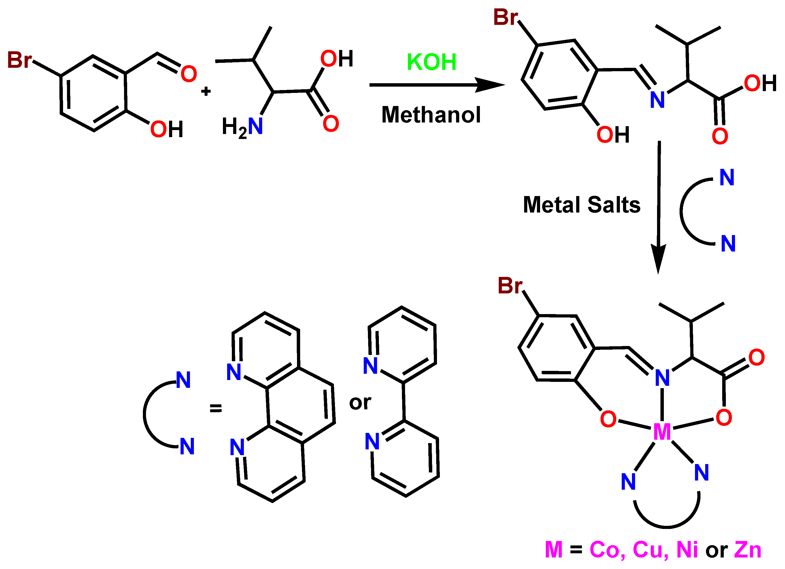
2.1.2. Preparation of Potassium (E)-2-((5-bromo-2-hydroxy benzylidene)amino)-3-methylbutanoate (HL)

An L-valine solution in methanol (0.1 mol in 50 mL) was refluxed with KOH at 50 °C for 60 min. The filtered, transparent solution was then refluxed with a methanolic solution of 5-bromosalicylaldehyde (0.1 mol in 50 mL). This process was completed after one hour of stirring at 60 °C. Subsequently, the mixture was crystallized by adding an excessive concentration of diethyl ether and then recrystallized, filtered, and dried. Yield: (80%). Color: yellow. M.p.110 °C. Anal. Calc for C12H13BrKNO3: found (calc.) (%): C, 48.56(48.18); H, 4.12(4.32); N, 4.38(4.68); 1H NMR (DMSO-d6,δ, PPM) 9.88(s, 1H, Ar-OH), 8.24 (s, 1H, N=CH), 7.54 (s, 1H, Ar-H), 6.54–7.37 (m, 1H, Ar-H), 3.62–3.68 (q,1H, valine C-H, 0.99 Hz), and 1.35–1.37 (m, 1H, valine C-H, 9.00 Hz). (d,3H,valine CH-(CH3)2, 2.99 Hz). IR (ν, cm−1): 3380 (OH); 1643 (C=N); 1594 (COO); 1405 (COO); (λmax/nm (ε/M−1cm−1dm3) in MeOH: 409(500), 353(180), and 264(1590).

2.2. Preparation of Complexes

2.2.1. Preparation of [Co(L)(phen)] (1a)

A heated methanolic liquid of potassium (E)-2-((5-bromo-2-hydroxy benzylidene)amino)-3-methylbutanoate (HL) (0.1 mol) was progressively introduced with steady mixing of CoCl2.6H20 (0.1 mol), and the solution was refluxed for 3 h. Then, the solution was refluxed for three hours with 1,10 phenanthroline (0.1 mol). After cooling to ambient temperature, the precipitate was collected, filtered, and repeatedly washed with methanol. Yield: 0.49 g (80%). Color: brown. M.p. > 300 °C (dec.). Anal. Calc for C24H20BrCoN3O3: found (calc.) (%): C, 53.63(53.65); H, 3.72(3.75); N, 7.81(7.82); selected IR data (ν, cm−1): 1636 (C=N), 1587 (COO), 1341(COO), 549 (M-O), and 457 (M-N). UV–Vis in MeOH [λmax/nm (ε/M−1cm−1)]: 228(33,895), 269(25,834), 390(1209), 477(152), and 615(58). ESI-MS (m/z): [Co(L)(phen)]+ (537.08).

2.2.2. Preparation of [Ni(L)(phen)] (1b)

Complexes 1b1d were prepared using the same method introduced for complex-1a. Complex-1b from NiCl2.6H2O (0.1 mole). Yield: 0.47 g (80%). Color: pale green. M.p. > 300 °C (dec.). Anal. Calc for C24H20BrN3NiO3: found (calc.) (%): C, 53.66(53.68); H, 3.73(3.75); N, 7.80(7.82); selected IR data (ν, cm−1): 1646 (C=N), 1590 (COO), 1325 (COO), 541 (M-O), and 454 (M-N). UV–Vis in MeOH [λmax/nm (ε/M−1cm−1)]: 254(57302), 288(36331), 389(3657), and 623(5.3). ESI-MS (m/z): [Ni(L)(phen)]+ (536.92).

2.2.3. Preparation of [Cu(L)(phen)] (1c)

Complex-1c from CuCl2.2H2O (0.1 mole). Yield: 0.56 g (88%). Color: dark green. M.p. > 300 °C (dec.). Anal. Calc for C24H20BrCuN3O3: found (calc.) (%): C, 53.19 (53.20); H, 3.70(3.72); N, 7.72(7.75); selected IR data (ν, cm−1): 1627 (C=N), 1592 (COO), 1325 (COO), 554 (M-O), and 455 (M-N). UV–Vis in MeOH [λmax/nm (ε/M−1cm−1)]: 259(86,422), 284(58,665), 372(1502), and 637(17). ESI-MS (m/z): [Cu(L)(phen)]+ (541.78).

2.2.4. Preparation of [Zn(L)(phen)] (1d)

Complex-1d from Zn(CH3COOH)2.2H2O (0.1 mole). Yield: 0.42 g (67%). Color: Pale yellow. M.p. > 300 °C (dec.). Anal. Calc for C24H20BrN3O3Zn: found (calc.) (%): C, 52.98(53.02); H, 3.69(3.71); N, 7.72(7.73); selected IR data (ν, cm−1): 1622 (C=N), 1574 (COO), 1343 (COO), 549 (M-O), and 510 (M-N). UV–Vis in MeOH [λmax/nm (ε/M−1cm−1)]: 270(35,064), 292(9154), and 373(4019)). ESI-MS (m/z): [Zn(L)(phen)]+ (543.28).

2.2.5. Preparation of [Co(L)(bpy)] (1e)

A similar procedure used to create complex 1a was utilized to develop complexes 1e1h, but instead of 1,10–phenanthroline, 0.1 mole of 2,2′–bipyridyl was used. Complex-1e from CoCl2.6H20 (0.1 mole.). Yield: 0.48 g (84%). Color: Brown. M.p. > 300 °C (dec.). Anal. Calc for C22H20BrCoN3O3: found (calc.) (%): C, 51.46(51.48); H, 3.92(3.93); N, 8.18(8.19); selected IR data (ν, cm−1): 1623 (C=N), 1592 (COO), 1368 (COO), 562 (M-O), and 488 (MνN). UV–Vis in MeOH [λmax/nm (ε/M−1cm−1)]: 249(19,579), 301(8048), 389(854), 463(127), and 619(65). ESI-MS (m/z): [Co(L)(bpy)]+ (513.03).

2.2.6. Preparation of [Ni(L)(bpy)] (1f)

Complex-1f from NiCl2.6H2O (0.1 mole). Yield: 0.46 g (79%). Color: Pale green. M.p. > 300 °C (dec.); Anal. Calc for C22H20BrN3NiO3: found (calc.) (%): C, 51.49(51.51); H, 3.91(3.93); N, 8.17(8.19); selected IR data (ν, cm−1): 1647 (C=N), 1591 (COO), 1326 (COO), 542 (M-O), and 503 (M-N). UV–Vis in MeOH [λmax/nm (ε/M−1cm−1)]: 238(4056), 298(2094), 379(920), and 625(3.4). ESI-MS (m/z): [Ni(L)(bpy)]+ (512.89).

2.2.7. Preparation of [Cu(L)(bpy)] (1g)

Complex-1g from CuCl2.2H2O (0.1 mole). Yield: 0.52 g (83%). Color: Green. M.p. > 300 °C (dec.). Anal. Calc for C22H20BrCuN3O3: found (calc.) (%): C, 51.00(51.02); H, 3.88(3.89); N, 8.09(8.11); selected IR data (ν, cm−1): 1627 (C=N), 1593(COO), 1325 (COO), 551(M-O), and 503 (M-N). UV–Vis in MeOH [λmax/nm (ε/M−1cm−1)]: 240(13846), 300(11085), 375(144), and 642(24). ESI-MS (m/z): [Cu(L)(bpy)]+ (517.69).

2.2.8. Preparation of [Zn(L)(bpy)] (1h)

Complex-1h from Zn(CH3COOH)2.2H2O (0.1 mole). Yield: 0.41 g (67%). Color: Pale yellow. M.p. > 300 °C (dec.). Anal. Calc for C22H20BrN3O3Zn: found (calc.) (%): C, 50.81(50.84); H, 3.86(3.88); N, 8.06(8.09); selected IR data (ν,cm−1): 1635 (C=N), 1595 (COO), 1314 (COO), 548 (M-O), and 505 (M-N).UV–Vis in MeOH [λmax/nm (ε/M−1cm−1)]: 274(20198), 294(16748), and 374(8425). ESI-MS (m/z): [Zn(L)(bpy)]+ (519.38).

2.3. In Vitro MTT Assay

According to the previous literature [17,18], the cytotoxic activity of compounds [Cu(L)(phen)] 1c and [Cu(L)(bpy)] 1g was assessed with respect to three tumor cell lines, including the A549, HeLa, and MCF-7 lines, as well as NHDF normal cells. The medium was switched to DMEM with 1% FBS after one day of growth in Dulbecco’s Modified Eagle Medium with 10% FBS. After one day, 2, 5, 10, 25, 50, and 100 µL doses of the produced compounds diluted in DMSO were applied to the cells, which were then incubated. Each well was then filled with 10 µL of MTT (5 mg/mL) and maintained in this state for 3 h. Farmazone crystals were produced and dispersed in 100 µL of DMSO; then, the absorbance at 570 nm was calculated using an ELISA spectrometer. The proportion of viable cells was determined using the following formula [17,19,20]:
Cell viability (%) = (A570 nm of treated samples/A570 nm of control samples) × 100

2.4. Molecular Modeling

Complexes were modeled molecularly by Maestro software coupled with the Schrodinger equation. The complex 1(ah) 3D structures were first sketched in the maestro builder panel before being optimized in the Ligprep system. The complexes’ tailored structures were suitable for docking with receptors. The receptor thymidylate synthase (PDB ID: 1HZW) was chosen from the protein data bank (http://www.rcsb.org, accessed on 22 October 2022). Utilizing the force field OPLS-2005, the receptor was also extensively tuned in the protein preparation wizard. The grid parameters were generated by supplying coordinates of −23.213, 43.011, and 21.67 Å and maintaining a diameter of 20 × 20 × 20 Å. Finally, docking programs were run in the maestro workspace for optimized complexes with receptor grids and binding interactions (3D and 2D).

2.5. Computational Analysis

Jaguar software, which is included in the Schrödinger suite 2017, was used to run the computational program employed in this study. Density functional theory (DFT) and the B3LYP/LACVP++ basis set were used to examine the optimal geometries and perform molecular orbital analyses (HOMO–LUMO) of the complexes.

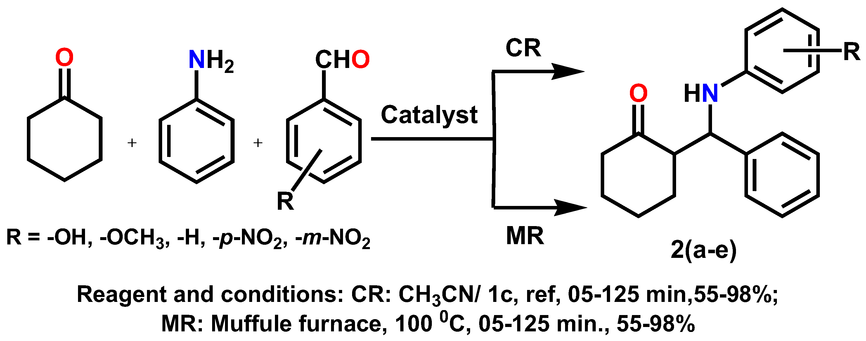
2.6. The General Process for the Synthesis of β-Amino Carbonyl Derivative

Simple/substituted aryl aldehyde (9.423 × 10⁻³ mmol; 1.0 equiv.) is mixed with aniline (9.894 × 10⁻³ mmol; 1.05 equiv.) and cyclohexanone (1.038 × 10⁻² mmol; 1.05 equiv.), and optimized catalyst concentration of substitued2,2-bipyridyl/phenanthroline metal (II) salts 1(ah) (5.44 × 10⁻² mmol) is added in the presence of 40 mL of dry CH3CN for 5–125 min under refluxing conditions to yield substituted β-Amino carbonyl derivatives 2(ae) at 55–98% after purification. All the synthesized β-Amino carbonyl derivatives are thoroughly analyzed via spectral and analytical tools and verified through an examination of the literature.

3. Results and Discussion

3.1. Results and Discussion

The metal(II) complexes 1(ah) were synthesized by a stoichiometric (1:1:1) template reaction of a methanolic solution containing ligand potassium (E)-2-((5-bromo-2-hydroxybenzylidene) amino)-3-methylbutanoate (HL), heterocyclic bases (1,10-phenanthroline or 2,2′-bipyridyl), and hydrated metal(II) salts (Scheme 1). Table 1 depicts the physical and elemental analyses of the metal(II) complexes. The complexes presented high yields and stability at ambient temperatures. The complexes were highly soluble in many solvents, such as methanol, acetonitrile, DMSO, and DMF, and were partially soluble in water.

3.2. Spectral Characterization

3.2.1. FT-IR Spectral Analysis

As shown in Table 2, the FT-IR spectra of the mixed ligand complexes 1(ah) were measured in the 450–4000 cm−1 wavelength range (Figure S1 from Supplementary Materials). A broad peak was observed in the ligand 3300–3500 cm−1 region due to the -OH stretching vibration of the ligand (HL), which was not present in the complexes for which the coordination mode of the deprotonated hydroxyl group with metal ions was confirmed. The C=N- (azomethine) stretching frequency of the ligand showed a strong absorbance peak at 1643 cm−1; a shift toward the higher wavelength region (1610–1635 cm−1) of the metal(II) complexes revealed that the azomethine nitrogen had coordinated with metal ions (Table 2) [21]. The bands at (1570–1590 cm−1) and (1350–1380 cm−1) were attributed to the asymmetric and symmetric vibrational frequencies, respectively, of the carboxyl groups present in the amino acid [22,23,24,25]. The absence of a band in the 1700–1750 cm−1 region suggested that the COO group of L-valine was coordinated with the central metal ion and that the difference in the frequency value between the asymmetric and symmetric stretching vibrations of the complexes lay between 180 and 216 cm−1, for which the latter is practically larger than that of a free carboxylate ion [26]. These findings supported the notion of a monodentate coordination of the carboxyl group of L-valine with metal ions. The spectra of all the complexes showed a strong band at 725–727 cm−1, thus indicating that –CH was out of the plane in the center ring of 1,10-phenanthroline. The lower frequency regions of the metal(II) complexes around 540–565 cm−1 and 450–505 cm−1, which are attributed to the (M-O) and (M-N) bands, respectively, confirmed the coordination of ligands with metal ions [27,28]. Hence, all the above facts are in good agreement with the complexes coordinated as a tridentate mode.

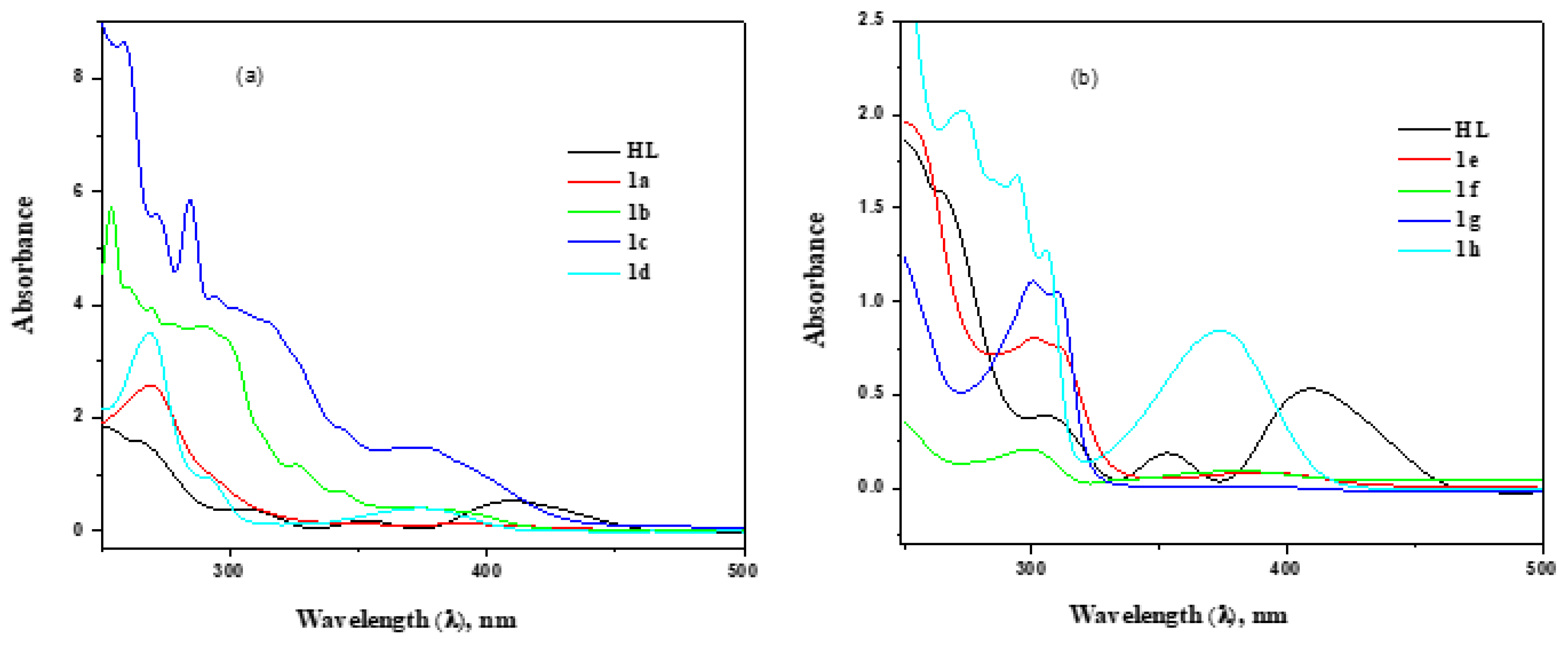
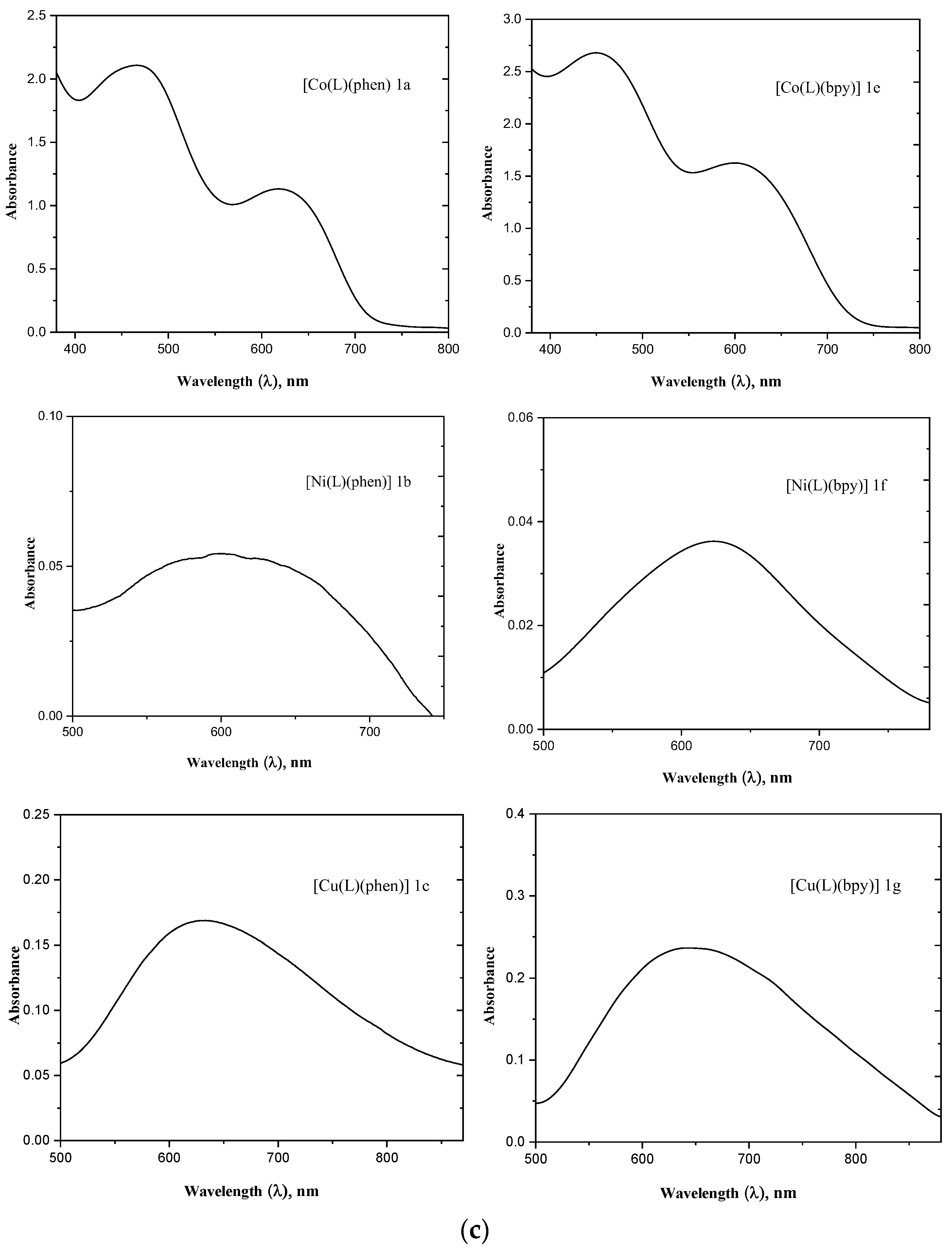
3.2.2. UV–Visible Spectral Analysis

The electronic spectra of metal(II) complex 1(ah) were measured using methanol solution (Figure 1a–c) and are presented in Table 2. A series of intense bands were observed for the ligand (HL) at 264, 353, and 409 nm, which were assigned to π–π* and n–π* transitions involved in azomethine nitrogen and aromatic ring systems, and this transition showed a decrease in wavelength at 230–300 nm upon complexation [29]. A moderately intense band at around 330–390 nm corresponds to the ligand-to-metal charge transfer transition [30]. In the visible region, cobalt(II) complexes 1a and 1e showed two weak absorption bands at 494 and 634 nm as well as 463 and 619 nm, respectively, suggesting a distorted trigonal bipyramidal geometry around the metal ion [31]. Whereas the nickel(II) complexes 1b and 1f and copper(II) complexes 1c and 1g showed one weak broad absorption peak in the 620–650 nm region due to the distorted square pyramidal geometry around the metal(II)complexes [32]. Moreover, the zinc(II) complexes 1d and 1h did not present any bands in the visible region due to their diamagnetic behavior and the d10 electronic configuration of the metal ion [33].

3.2.3. Mass Spectral Analysis

The ESI+ mass spectra of complexes 1a and 1b (Figure S2) were determined to have molecular peaks at m/z = 536.50 and 536.20, which were attributed to the [C24H20BrCoN3O3)]+ and [C24H20BrNiN3O3)]+ fragment ions, respectively. These complexes show a base peak at m/z = 355.19 and 354.11 due to the fragmentation of [C12H12BrCoNO3]2+ and [C12H12BrNiNO3]2+ ions (Figures S1 and S2). In the same way, other metal(II) complexes (1c1h) exhibited molecular ion peaks at m/z = 541.78, 543.28, 513.03, 512.89, 517.69, and 519.38, which corresponded to [M1c−1h (L)(diimine)]+ ions (M=Co, Ni, Cu, and Zn). The resulting spectral data of the complexes showed the formation of the suggested molecular structure.

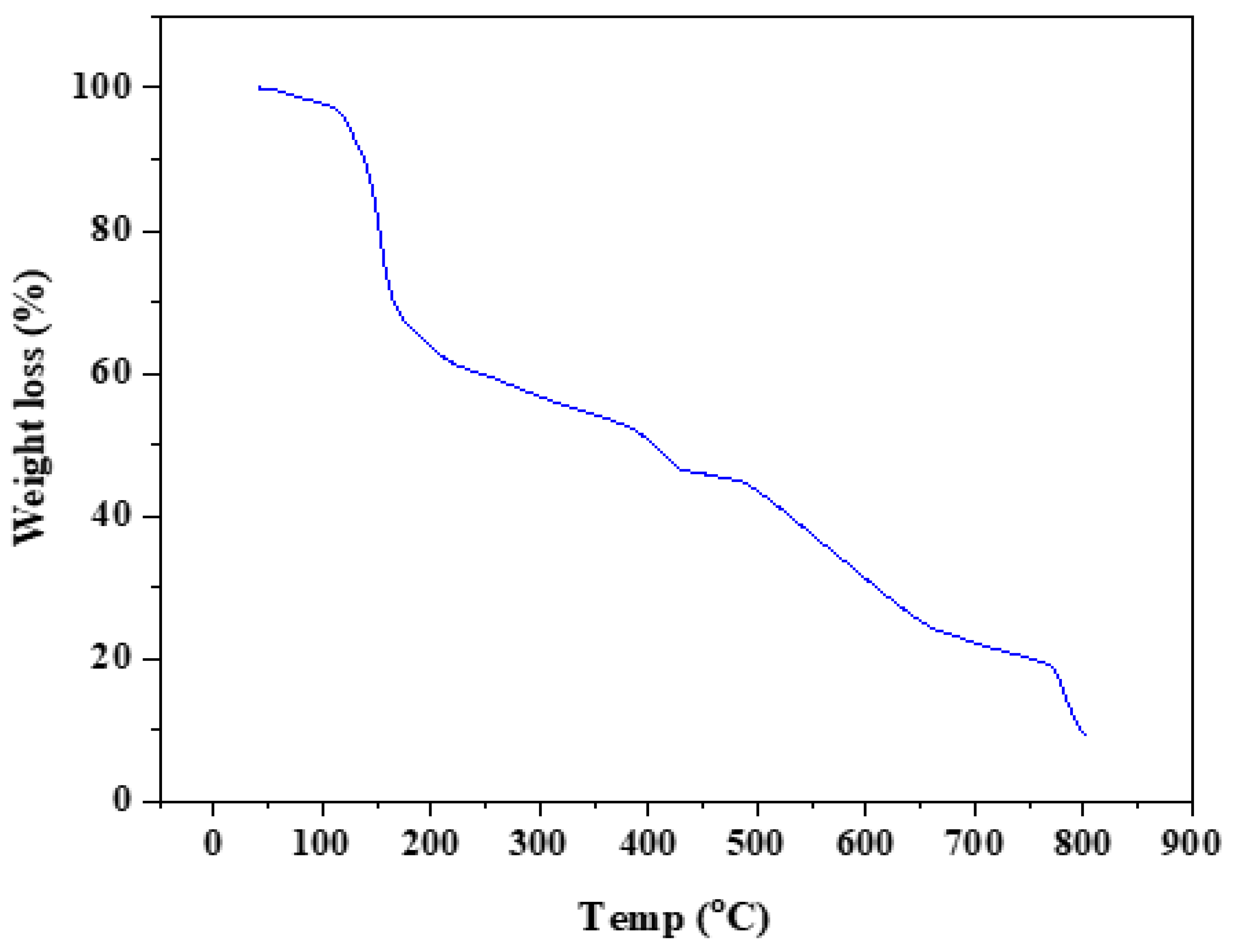
3.2.4. Thermal Analysis

The thermal behaviors of the metal complexes were determined under a nitrogen atmosphere, as shown in Figure 2. Upon comparison, it is clear that the weight loss of the metal(II) complexes correlates with different compositions at specific temperatures. The [Cu(L)(bpy)] 1g complex displays a weight decrement in three steps from 38 to 800 °C. The first one is related to the decrement in the outer lattice dehydration of a single H2O molecule at 30–120 °C. The second step temperature range of 120–270 °C is due to the loss of organic molecules of C10H8N2 (2,2′-bipyridyl). A peak corresponding to a weight loss of 24.52% (calcd. 24.46%) at 420–510 °C was related to the ligand moiety in the metal complexes in the third step and the subsequent complex, thus leaving CuO as a residue. The same trend was traced in the TGA plots of the other metal complexes.

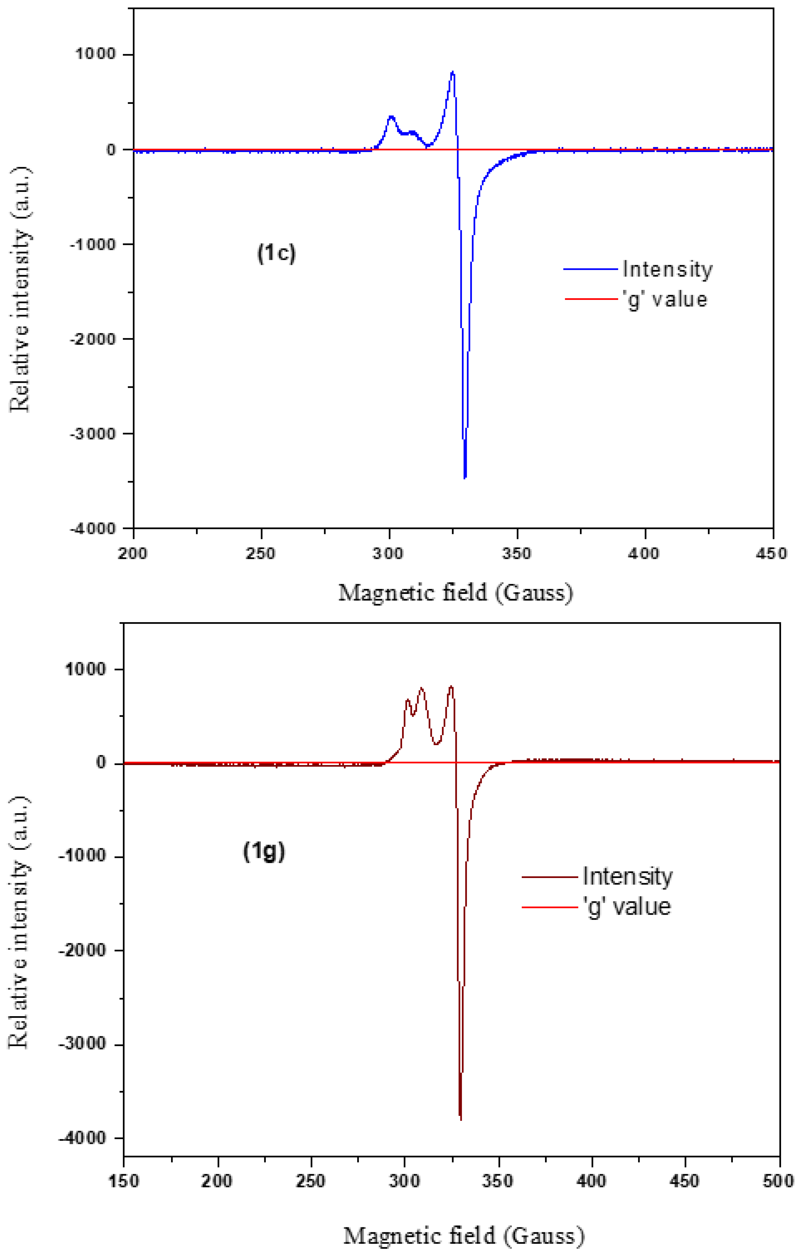
3.2.5. EPR Spectral Analysis

The EPR spectra of the copper(II) complexes [Cu(L)(phen)] 1c and [Cu(L)(bpy)] 1g were measured in polycrystalline conditions at 298 K. The hyperfine splitting patterns of the EPR spectra revealed that the discrete g|| and g values obtained correspond to axial spectral features (Figure 3). The spectral data suggested that unpaired electrons of the copper(II) complex lay in the dx2–y2 molecular orbital having a ground state((2B1g) with g|| > g > 2.0023. The g|| magnitudes elucidated the types of bonds (ionic or covalent) included in the observed metal-to-ligand coordination process. Usually, ionic bonds showed g||> 2.4 and covalent bonds indicated g|| < 2.4. The g||values of complexes 1c and 1g were 2.184 and 2.186, thus confirming the covalent nature of the copper(II) complexes. The attained g values confirmed the square pyramidal geometry of the copper complexes reported in previous studies [34]. According to Hathway [35], the geometric parameter ‘G’ indicates an exchange interaction between the copper–copper centers and is calculated as follows:
G = (g|| − 2)/(g− 2) for axial spectra
The G values calculated for complexes 1c and 1g were 2.61 and 3.064, respectively. As these G values were found to be around 3.0, the presence of a dx2–y2 ground state of a square pyramidal geometry and a substantial exchange interaction in the polycrystalline state were confirmed [36,37].

3.2.6. XRD Analysis

The purity, crystallite size, and crystallinity of the synthesized complexes were analyzed by Powder XRD [38,39,40,41]. The diffractograms of complexes 1a, 1b, 1f, and 1h are shown in Figure 4a,b. In the region 5–50° (2θ), complexes 1(ac), and 1g showed sharp diffraction patterns, suggesting that the complexes exist in a crystalline structure. While complexes 1(df), and 1h showed very few diffraction peaks, they indicated that the complexes were between crystalline and amorphous phases [42]. The crystallite sizes of the samples were evaluated using Scherrer’s equation [43,44]. From the calculated data, the average crystallite size of complexes is in the range of 50–60 nm.

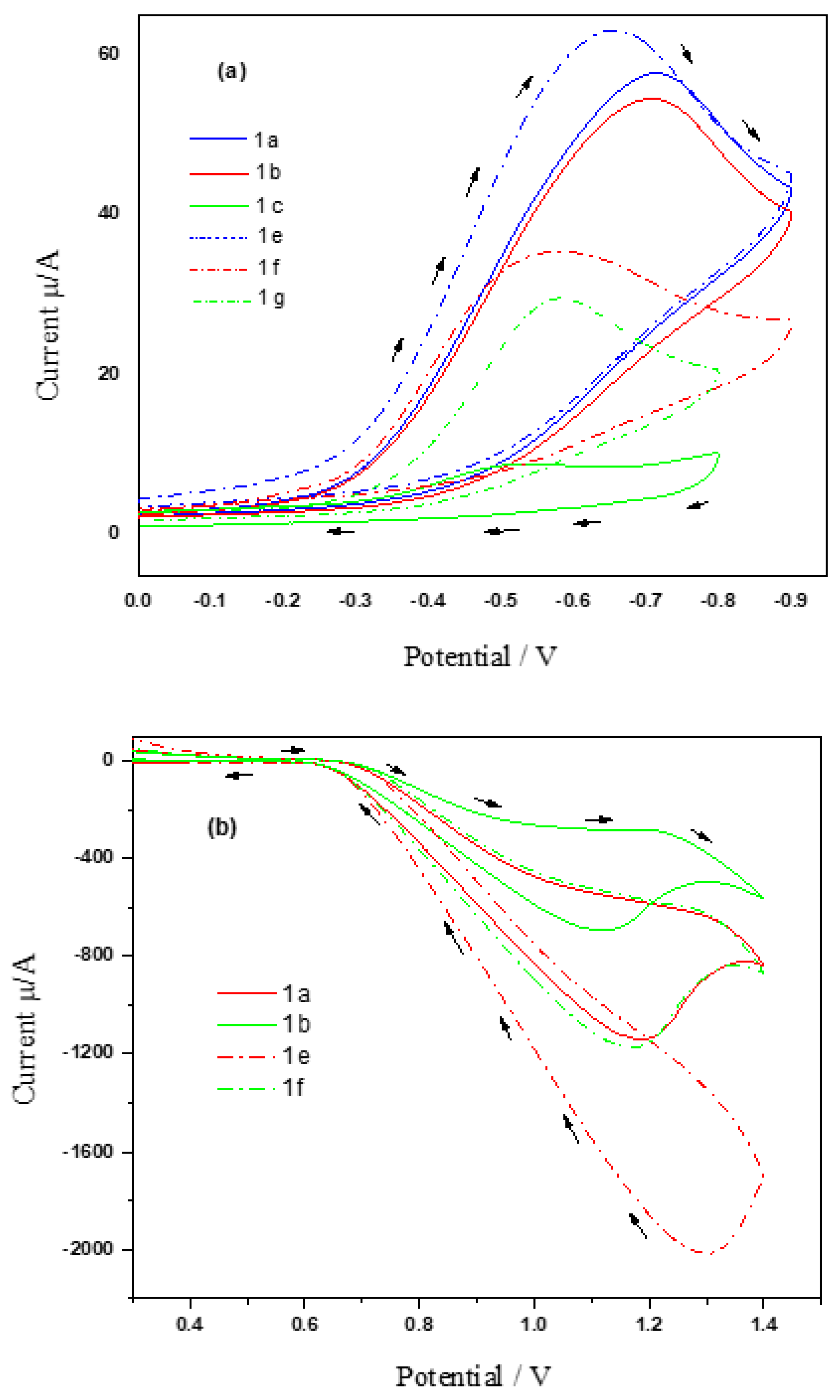
3.3. Electrochemical Studies

The redox potential for metal(II) complexes 1(ah) was calculated via cyclic voltammetry in DMF containing 0.1 M of tetra(n-butyl) ammonium perchlorate and at a scan rate of 100 mVs−1, for which the results are listed in Table 3. The absence of a cyclic voltammogram for complexes 1d and 1h was due to the electrochemically inactive nature of the zinc(II) ion. The anodic and cathodic peak current ratios (Ipc/Ipa) were found to be less than unity, thereby suggesting that the metal(II) complexes were irreversible during the redox process.

3.3.1. Reduction Process

The cyclic voltammograms of the Co(II), Ni(II), and Cu(II) metals were studied at the cathodic region from 0 to –1.4 V (Figure 5a). The voltammogram peaks of cobalt(II) complex 1a, nickel(II) complex 1b, and copper(II) complex 1c showed irreversible one-electron transfer reduction at −0.71 V, −0.71 V, and −0.54 V. Likewise, the absence of counter-reduction patterns in the reverse scan corroborated the irreversible character of the metal in complexes 1(eg) [45,46]. These reduction potential peaks were observed at −0.66 V, −0.58 V, and −0.58 V, respectively. It was suggested that the following one-electron reduction method should be used:
[ M II ( L ) ( phen   or   bpy ) ] e [ M I ( L ) ( phen   or   bpy ) ]

3.3.2. Oxidation Process

The cyclic voltammograms of the Co(II), Ni(II), and Cu(II) metals were studied at the cathodic region from 0 to –1.4 V (Figure 5b). The cyclic voltammograms for cobalt(II) complex 1a and nickel(II) complex 1b revealed irreversible transfer oxidation at +1.19 V and +1.12 V, respectively. Likewise, the cobalt(II) complex 1e and nickel(II) complex 1f showed oxidation potential at +1.30 and +1.17, respectively; finally, the absence of a counter-oxidation peak in the reverse pattern verified the irreversible nature of the metallic ions [47]. The oxidation step at the anodic region is defined as follows:
[ M II ( L ) ( phen   or   bpy ) ] e [ M III ( L ) ( phen   or   bpy ) ]

3.4. Catalytic Activity

Subsequently, all metal(II) complexes utilized for multi-component reactions in a single pot were examined at various concentrations with or without a solvent (Scheme 2). To improve the catalytic performance of Schiff base metal(II) complex 1c, substituted β-amino carbonyl derivatives were synthesized in one pot repeatedly at different concentrations, including 0.0384 mmol, 0.0769 mmol, 0.1154 mmol, and 0.1539 mmol. While floating the amount of the catalyst from 0.1154 mmol to 0.1539 mmol, the yield rate and reaction time remained unaffected.
Various derivatives of substituted β-amino carbonyl compounds were synthesized using conventional and solid-phase techniques, for which reactions were continued at multiple varying concentrations. These results are shown in Table 4 and Table 5. In the conventional technique, the zinc enolate is produced by linkage, and the catalyst then deprotonates the alkynyl ketone. However, the reaction time and percentage of yield are notable for the pricey Zn-Pro Phenol complex catalyst. β-Amino carbonyl groups were synthesized with less than 0.5 mmol of a nanocomposite catalyst, for which more than 10 h were required to complete the investigation. Furthermore, 60 mg of metal (II) salt was used for 5 min under the silica-supported method.
The synthesized metal (II) complexes were utilized as a model catalyst for the acceleration of the Mannich reaction. Under the same reaction conditions, the catalyst was used up to four times in the synthesis of substituted β-amino carbonyl compounds. The final product was the same as when it was first used, even after the fourth recycling phase, as shown in Table 6.

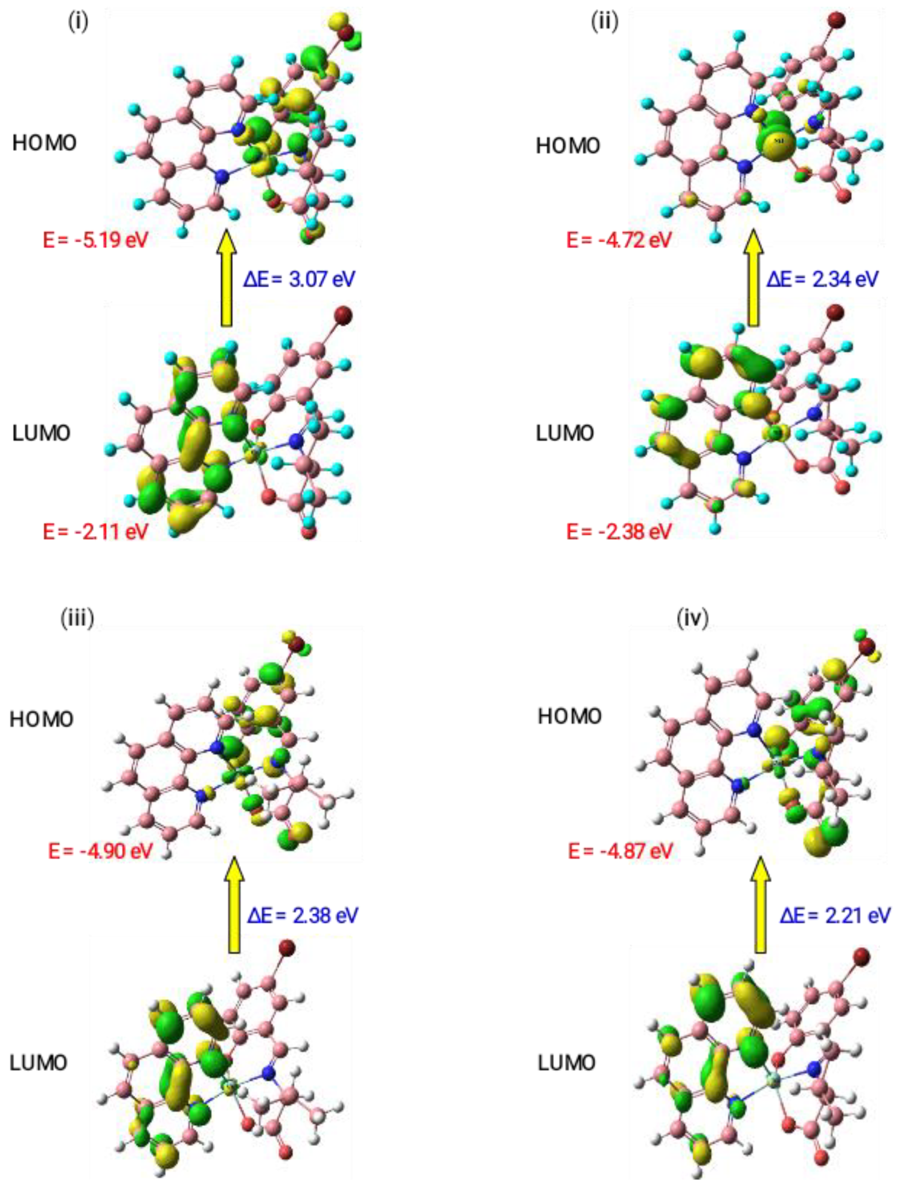
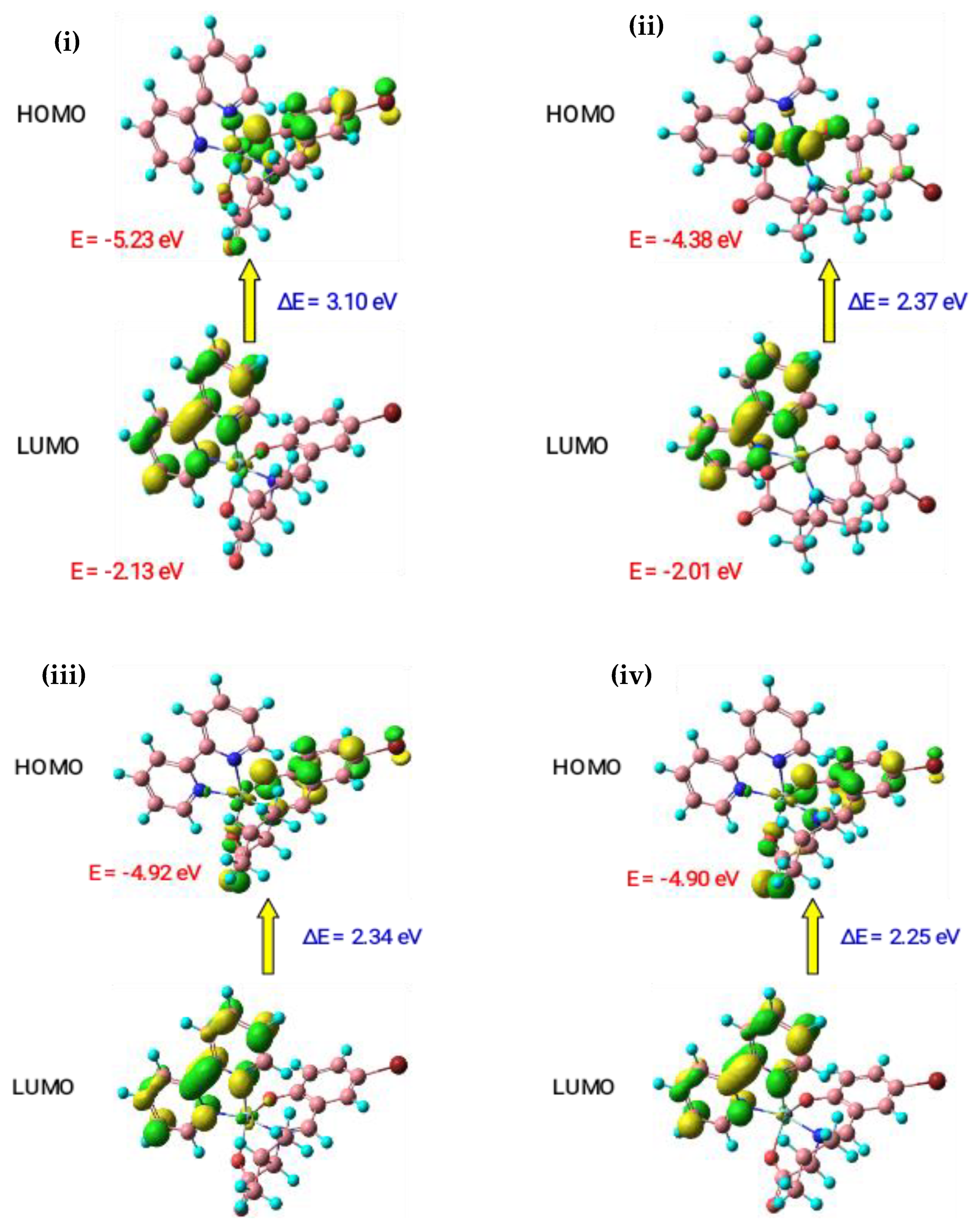
3.5. Theoretical Studies

3.5.1. Geometry Optimization

With the help of the Jaguar 8.8 software, which is based on DFT with the B3LYP and LACVP++ basis sets, the optimal geometry for the metal(II) complexes was obtained. The chosen bond angles and bond lengths are shown in Table 7 and Table 8, and the molecular structures of the metal(II) complexes are illustrated in Figure 6 and Figure 7. It was discovered that the computed values of bond angles and bond lengths were more suitable for forecasting the geometry of the metal(II) complexes. The geometric parameter ‘τ’ determined the molecular structure of the complex for the pentacoordinate system, and it was computed using the following formula [48].
τ = (βα)/60
In the formula presented above, α and β are axial and equatorial bond angles, respectively. When τ ≈ 0, the geometry becomes square pyramidal, and it becomes closer to 1 for a perfect trigonal bipyramidal structure. For a trigonal bipyramidal geometry, the τ values lie between 0 and 0.5. In this study, the calculated bond angles for complexes 1(ah), were 171.2, 156.9, 154.9, 151.1, 172.8, 162.2, 152.7, and 151.9°, respectively, which correspond to β (axial bond angles), and the values 126.2, 153.4, 149.1, 135.7, 125.1, 158.5, 148.6, and 134.8°, respectively, which correspond to α (equatorial bond angles). The calculated τ values for the metal(II) complexes were 0.75, 0.05, 0.09, 0.26, 0.79, 0.06, 0.06, and 0.28, respectively. Complexes 1a and 1e and were, as suggested, of a trigonal bipyramidal geometry due to their τ values of 0.75 and 0.79, respectively. Complexes 1b, 1c, 1f, and 1g were suggested to possess a square pyramidal geometry because their τ values were very close to 0 (0.05–0.09). Finally, complexes 1d and 1h were suggested to possess a distorted trigonal bipyramidal geometry due to their τ values of 0.26 and 0.28, respectively.

3.5.2. Molecular Orbital Analysis

The molecular orbitals for the synthesized metal(II) complexes were theoretically analyzed by frontier molecular orbital theory. This theory states that the interaction between the frontier orbitals (HOMO and LUMO) of the reacting molecules generate an electric change. The energies of the primary orbitals were used to determine whether a compound would be stable or chemically reactive at the lowest unoccupied molecular orbital (LUMO) and the highest occupied molecular orbital (HOMO). A tendency to donate an electron corresponds to the HOMO, while a tendency to withdraw an electron corresponds to the LUMO [49]. In many molecular systems, the energy difference (ΔE) between the HOMO and LUMO is thought to be a crucial stability index for highlighting structural and conformational barriers. Charge transfer between the HOMO and LUMO happens when ΔE is low. The increased activity of the molecule is influenced by the lower ΔE value.
In this study, the significant quantum parameters, such as the HOMO–LUMO energy gap (ΔE), chemical potentials (Pi), absolute electronegativities (χ), absolute softness ((σ), absolute hardness (η), global electrophilicity (ω), global softness (S), and additional electronic charge (ΔNmax), were calculated in accordance with Koopman’s hypothesis using the following equations [50]:
χ = −(EHOMO + ELUMO)/2
η = ELUMOEHOMO/2
ΔE = ELUMOEHOMO
σ = 1/η
S = 1/2η
Pi = −χ
Ω = Pi2/2η
ΔNmax = −Pi/η
The obtained parameters for the complexes are given in Table 9. The ΔE values of complex 1(ah) were found to be 3.07, 2.34, 2.38, 2.21, 3.10, 2.37, 2.34, and 2.25 eV, respectively. Particularly, the zinc(II) complexes 1d and 1h have the lowest energy band gap, suggesting higher activity and lesser stability. The lowest energy gap value of the zinc(II) complex 1d demonstrated an undemanding electronic transition between the HOMO and LUMO, which may be reason for the higher bioactivity observed.

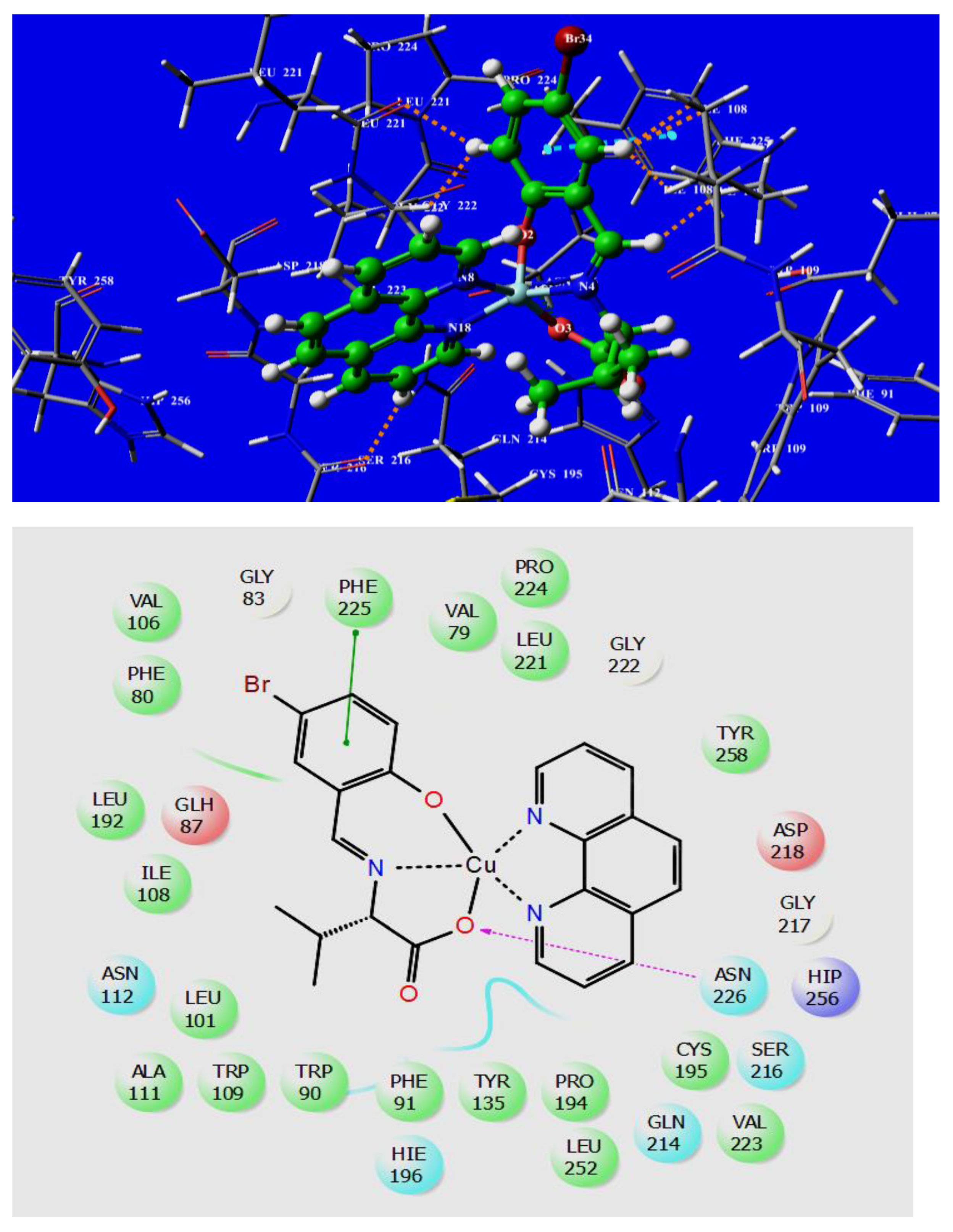
3.6. Molecular Docking Studies

Validation of the Active Site of Thymidylate Synthase

Thymidylate synthase (TS) plays a decisive role in the biosynthesis of DNA precursors. An increased level of thymidylate synthase activity has been observed in colorectal, breast, cervical, kidney, and lung tumors. Many researchers have encountered difficulties when attempting to find a better way to regulate thymidylate synthase action for both the development of therapeutic strategies and tumor prevention. Hence, the prepared complexes were assessed with respect to the binding affinity of the thymidylate synthase receptor to understand the interactive behavior of the studied complexes with the TS receptor and thus develop a strategy for their optimization. By analyzing the metal(II) complexes 1(ah) applied for docking with thymidylate synthase (PDB ID: 1HZW) and their binding affinity values, it was revealed that the complexes were more active on that site. The calculated docking scores and active sites of TS with various modes of interaction are shown in Table 10 (Figures S3 and S4 from Supplementary Materials). The docking study’s results revealed that all the complexes were located within the hydrophobic site of the TS receptor. The docking scores for the metal(II) complexes with a TS receptor were found to be −5.70, −5.615, −5.791, −5.367, −5.49, −4.97, −5.026, and −5.223 kcal mol−1. In addition, it was found that complex 1c had the highest docking score due to binding interaction with the TS receptor via π–π stacking, hydrogen bonding, and hydrophobic interactions, as shown in Figure 8. This complex showed one hydrogen bond (distance of 2.33 Å) interaction between an amino hydrogen of residue ASN 226 with the carboxylate oxygen of the complex. Further, complex 1c showed π–π stacking interaction between the residue PHE 225 and a phenolate ring with an interaction distance of 4.01 Å. Furthermore, the complex showed an abundance of hydrophobic interactions with different amino acid residues, such as VAL 79, PHE 80, TRP 90, PHE 91, LEU 101, VAL 106, ILE 108, TRP 109, ALA 111, TYR 135, LEU 192, PRO 194, CYS 195, PRO 194, LEU 221, VAL 223, PRO 224, PHE 225, and TYR 258. The prepared metal(II) complexes showed better activity compared to that presented in our previously published article [51]. The highest docking score value of complex 1c was obtained with thymidylate synthase, which encouraged us to study the cytotoxicity of the metal complexes experimentally.

3.7. Biological Evaluation

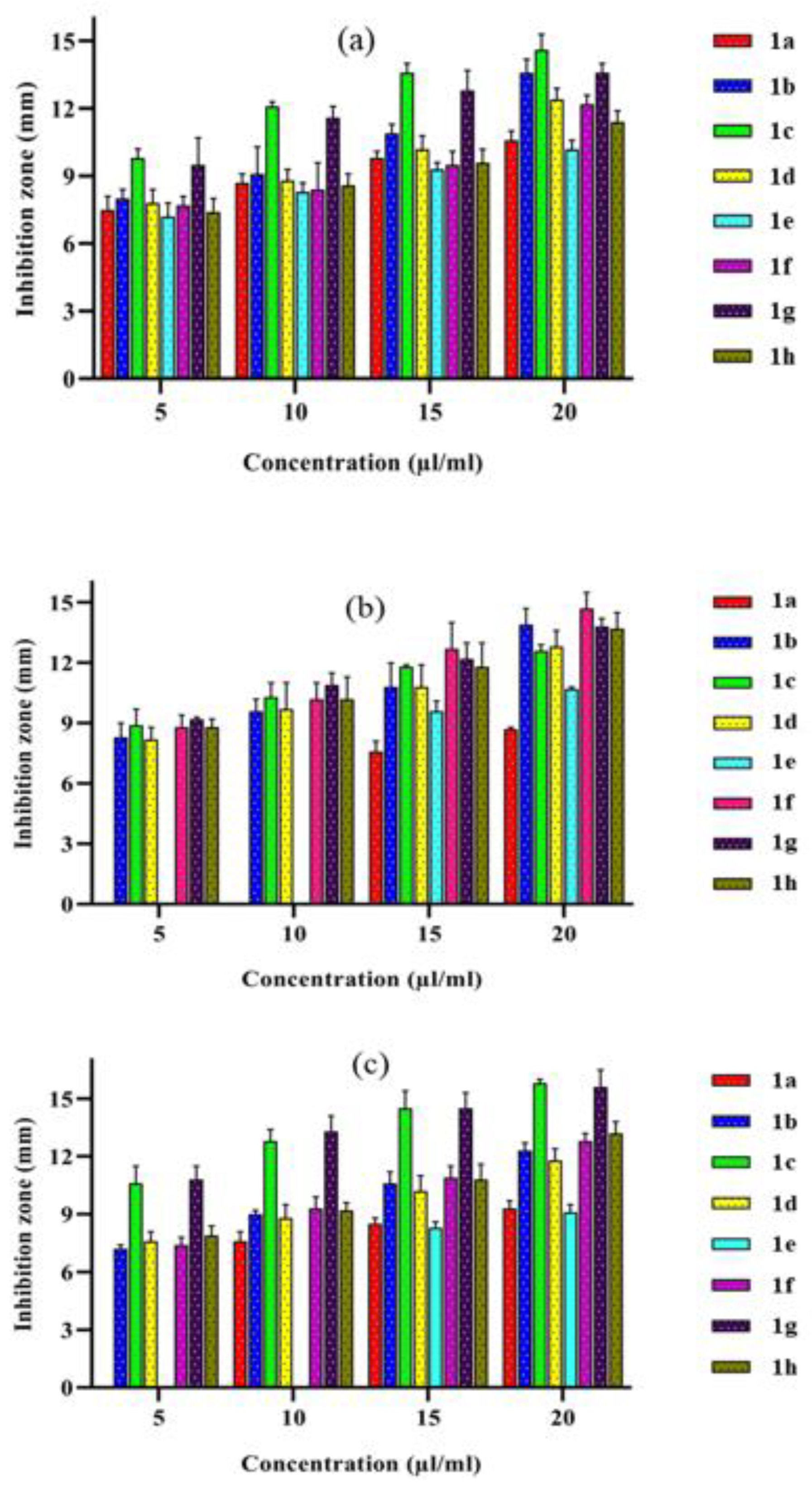
3.7.1. In Vitro Antibacterial Assay

In vitro antibacterial activity for metal(II) complex 1(ah) was checked against two Gram-negative bacteria (Escherichia coli and Klebsiella pneumonia) and one Gram-positive bacterium (Staphylococcus aureus) in different concentration ranges from 5–20 µL/mL using the agar diffusion method [21,52,53]. The gradient changes were illustrated as graphs (Figure 9a–c), and their data are shown in Table 11. From the data, it is evident that the metal complexes showed better activity upon an increase in concentration.
The results showed that all the metal complexes have considerable activity at a concentration of 20 µL/mL. Among them, complexes 1c and 1f had higher activity at this concentration against the microbial test strains, specifically with respect to copper(II) complex 1c against Escherichia coli, a group of Gram-negative bacteria, and Staphylococcus aureus, a Gram-positive bacterium, for which there were greater inhibition zones at 14.6 and 15.8 mL, respectively. Further, complex 1f was more active against Klebsiella pneumonia (a Gram-negative bacterium), having a higher inhibition zone at 14.7 ± 0.8 µL/mL. From the above particulars, it is evident that the metal ions coordinated with diamine play a key role in antimicrobial activity, and we conclude that metalation is significantly related to the inhibitory activity of complexes as explained by Tweedy’s chelation theory [54]. This increasing activity showed that chelation significantly improved the lipophilicity of the chemicals, resulting in their absorption through the lipoid layer of the microbe’s cell membrane and DNA destruction [55,56]. These results are in agreement with those presented in previous studies involving L-alanine-incorporated metal(II) complexes [56,57].

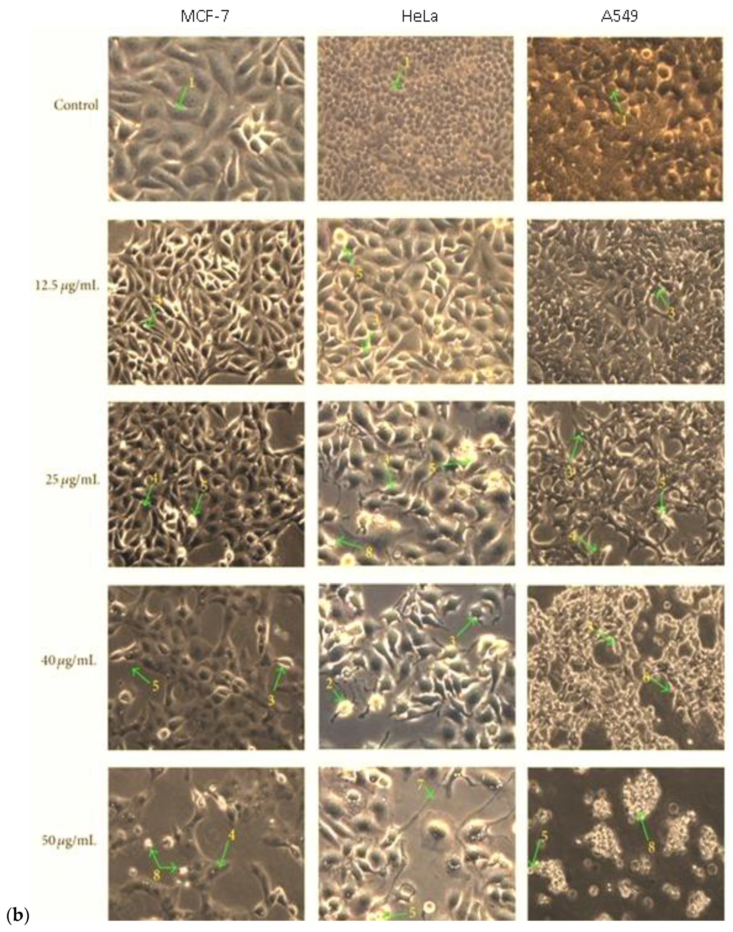
3.7.2. In Vitro Anticancer Activity

The metal Cu(II) complexes 1c and 1g were used in the MTT procedure to measure the vitality of the analyzed cells. Different cancer cell lines were used for the MTT assay, such as an A549 lung cancer cell line, a HeLa cervical cancer cell line, an MCF-7 breast cancer cell line, and NHDF cell lines, while employing cis-platin as a control. Their IC50 values for each complex were determined up to 24 h alongside an increase in complex concentration. The data have been presented in a graph (Figure 10a) and in Table 12. The IC50 value of complex 1c showed 25.95 ± 1.82, 26.26 ± 1.06, and 17.13 ± 0.74 μg/mL for the A549, HeLa, and MCF-7 cell lines, respectively. From the results, complex 1c showed a higher anticancer efficacy than complex 1g, and its value was found to be very close to that of cis-platin. The photomicrographs of complex 1c against MCF-7 showed different cytotoxic morphologies of condensed nuclei, cell shrinkage, membrane blebbing, apoptotic bodies, bubbling, and echinoid spikes (Figure 10b). The toxicity results also suggested that complex 1c is less toxic than complex 1g with respect to the NHDF cell line, for which the following order was observed: MCF-7 > A549 > HeLa. The extended aromatic conjugation of the phenanthroline moiety and strong hydrophobic interaction of complex 1c showed higher potential anticancer activities compared to the bipyridyl co-ligand.

4. Conclusions

In this work, Schiff base ligands (HL) and heterocyclic bases (1,10-phenanthroline or 2,2′-bipyridyl) were used to synthesize mixed-ligand metal(II) complexes 1(ah). Based on the spectral results, it was determined that the Co(II), Ni(II), Cu(II), and Zn(II) complexes assumed the geometry of a distorted square pyramidal shape. With regard to the electrochemical behavior of the complexes, it was observed that one electron was irreversibly transferred. The results regarding catalytic activity showed that the muffle furnace approach reached the target molecule much faster than the conventional method. Metal(II) complex 1c revealed significant antibacterial activity against Escherichia coli and Staphylococcus aureus at a 20 µg/mL concentration applied using the agar diffusion method. Similarly, the in vitro cytotoxicity of complex [Cu(L)(phen)] 1c showed promising anticancer activity against MCF-7 (17.13 ± 0.74), A549 (25.95 ± 1.82), and HeLa (26.26 ± 1.06) cancer cell lines, whose IC50 values were very close in terms of activity to that of cis-platin. The frontier orbital (HOMO–LUMO) analyses revealed that the complex [Zn(L)(phen)] 1d exhibited a lower band gap (2.21 eV) than the other complexes. The smaller energy gap led to an easing of the transition occurring between energy levels and precipitated the higher biological activity of the metal(II) complexes. Finally, the docking score of complex 1c (−5.791 kcal mol−1) showed better binding interactions with the thymidylate synthase receptor, viz., several hydrophobic, stacking, and hydrogen bonding interactions. Further investigation and developments in the area of Schiff base complexes of transition metal ions would be highly beneficial for industries and drug-related research.

Supplementary Materials

The following supporting information can be downloaded at: https://www.mdpi.com/article/10.3390/molecules28072931/s1, Figure S1: FT-IR spectra of metal(II) complexes 1a1h; Figure S2: ESI-Mass spectra of metal(II) complexes [Co(L)(phen)] 1a and [Ni(L)(phen)] 1b; Figure S3: Optimized molecular structure of (i) cobalt(II) complex 1a, (ii) nickel(II) complex 1b, (iii) copper(II) complex 1c and (iv) zinc(II) complex 1d using B3LYP/LACVP++ basis set; Figure S4: Optimized molecular structure of (v) cobalt(II) complex 1e, (vi) nickel(II) complex 1f, (vii) copper(II) complex 1g and (viii) zinc(II) complex 1h using B3LYP/LACVP++ basis set.

Author Contributions

Conceptualization, G.S., S.K. and A.S.; methodology, R.T.; software, P.R.; validation, S.A., M.K.A.M. and Z.T.A.-a.; formal analysis, F.A.J.A.-D.; investigation, Y.H.T.-Y.; resources, P.S.; data curation, S.A.; writing—original draft preparation, G.S.; writing—review and editing, G.S.; visualization, Y.H.T.-Y.; supervision, P.S.; project administration, A.S.; funding acquisition, R.T. All authors have read and agreed to the published version of the manuscript.

Funding

This research received no external funding.

Institutional Review Board Statement

Not applicable.

Informed Consent Statement

Not applicable.

Data Availability Statement

Not applicable.

Acknowledgments

All authors thank the Central Leather Research Institute in Chennai for their assistance with EPR spectral analysis, the SRM Institute of Science and Technology in Chennai for their assistance with powder XRD spectra and biological studies, and the Indian Institute of Technology Madras (IIT-M), Chennai-600 036, for their assistance with ESI-mass spectral data collection.

Conflicts of Interest

The authors declare no conflict of interest.

Abbreviations

bpy2,2′-Bipyridyl
DMFN,N-Dimethylformamide
DPPH2,2′-Diphenyl-1-picrylhydrazyl
DMEMDulbecco’s Modified Eagle’s Medium
DFTDensity functional theory
EPRElectron paramagnetic resonance spectroscopy
ESI-MSElectrospray ionization mass spectroscopy
FBSFetal bovine serum
FMOFrontier molecular orbital
HOMOHighest occupied molecular orbital
IC5050% of inhibitory concentration
LUMOLowest unoccupied molecular orbital
MTT3-(4,5-Dimethyl-2-thiazolyl)-2,5-diphenyltetrazolium bromide
NHDFNontumorigenic human dermal fibroblasts
OPLSOptimized potentials for liquid simulations
PDBProtein data bank
phen1,10-Phenanthroline
RMSDRoot mean square deviation
TBAPTetra(n-butyl)ammonium perchlorate
UV–VisUltraviolet–visible
XRDX-ray diffraction
MCF-7Michigan Cancer Foundation
HeLaHenrietta Lacks

References

  1. Rosenberg, B.; VanCamp, L. The successful regression of large solid sarcoma 180 tumors by platinum compounds. Cancer Res. 1970, 30, 1799–1802. [Google Scholar]
  2. Rosenberg, B.; Vancamp, L.; Trosko, J.E.; Mansour, V.H. Platinum compounds: A new class of potent antitumour agents. Nature 1969, 222, 385–386. [Google Scholar] [CrossRef] [PubMed]
  3. Creaven, B.S.; Duff, B.; Egan, D.A.; Kavanagh, K.; Rosair, G.; Thangella, V.R.; Walsh, M. Anticancer and antifungal activity of copper (II) complexes of quinolin-2 (1H)-one-derived Schiff bases. Inorg. Chim. Acta 2010, 363, 4048–4058. [Google Scholar] [CrossRef] [Green Version]
  4. Sondhi, S.M.; Singh, N.; Kumar, A.; Lozach, O.; Meijer, L. Synthesis, anti-inflammatory, analgesic and kinase (CDK-1, CDK-5 and GSK-3) inhibition activity evaluation of benzimidazole/benzoxazole derivatives and some Schiff’s bases. Bioorganic Med. Chem. 2006, 14, 3758–3765. [Google Scholar] [CrossRef] [PubMed]
  5. Aboul-Fadl, T.; Bin-Jubair, F.A.; Aboul-Wafa, O. Schiff bases of indoline-2,3-dione (isatin) derivatives and nalidixic acid carbohydrazide, synthesis, antitubercular activity and pharmacophoric model building. Eur. J. Med. Chem. 2010, 45, 4578–4586. [Google Scholar] [CrossRef]
  6. Mohammed, D.F.; Madlool, H.A.; Faris, M.; Shalan, B.H.; Hasan, H.H.; Azeez, N.F.; Abbas, F.H. Harnessing inorganic nanomaterials for chemodynamic cancer therapy. Nanomedicine 2022, 17, 1891–1906. [Google Scholar] [CrossRef]
  7. Obaid, R.F.; Nada, K.K.H.; Samah, A.K.; Lubab, A.J.A.; Abduladheem, T.J. Antibacterial activity, anti-adherence and anti-biofilm activities of plants extracts against Aggregatibacter actinomycetemcomitans: An in vitro study in Hilla City, Iraq. Caspian J. Environ. Sci. 2022, 20, 367–372. [Google Scholar]
  8. Aboul-Fadl, T.; Mohammed, F.A.-H.; Hassan, E.A.-S. Synthesis, antitubercular activity and pharmacokinetic studies of some Schiff bases derived from 1-alkylisatin and isonicotinic acid hydrazide (INH). Arch. Pharmacal Res. 2003, 26, 778–784. [Google Scholar] [CrossRef]
  9. Ollevier, T.; Nadeau, E.; Guay-Bégin, A.-A. Direct-type catalytic three-component Mannich reaction in aqueous media. Tetrahedron Lett. 2006, 47, 8351–8354. [Google Scholar] [CrossRef]
  10. Manabe, K.; Mori, Y.; Wakabayashi, T.; Nagayama, S.; Kobayashi, S. Organic synthesis inside particles in water: Lewis acid− surfactant-combined catalysts for organic reactions in water using colloidal dispersions as reaction media. J. Am. Chem. Soc. 2000, 122, 7202–7207. [Google Scholar] [CrossRef]
  11. Akiyama, T.; Takaya, J.; Kagoshima, H. One-pot Mannich-type reaction in water: HBF4 catalyzed condensation of aldehydes, amines, and silyl enolates for the synthesis of β-amino carbonyl compounds. Synlett 1999, 1999, 1426–1428. [Google Scholar] [CrossRef]
  12. Manabe, K.; Mori, Y.; Kobayashi, S. Three-component carbon–carbon bond-forming reactions catalyzed by a Brønsted acid–surfactant-combined catalyst in water. Tetrahedron 2001, 57, 2537–2544. [Google Scholar] [CrossRef]
  13. Zhao, G.; Jiang, T.; Gao, H.; Han, B.; Huang, J.; Sun, D. Mannich reaction using acidic ionic liquids as catalysts and solvents. Green Chem. 2004, 6, 75–77. [Google Scholar] [CrossRef]
  14. Chang, T.; He, L.; Bian, L.; Han, H.; Yuan, M.; Gao, X. Brønsted acid-surfactant-combined catalyst for the Mannich reaction in water. RSC Adv. 2014, 4, 727–731. [Google Scholar] [CrossRef]
  15. Jin, Y.; Chen, D.; Zhang, X.R. Direct Asymmetric anti-Mannich-Type Reactions Catalyzed by Cinchona Alkaloid Derivatives. Chirality 2014, 26, 801–805. [Google Scholar] [CrossRef] [PubMed]
  16. Wiemer, B. DD Perrin and WLF Armarego: Purification of Laboratory Chemicals. 3. Aufl., Oxford.; Pergamon Press, 1988, 391 S., zahlr. Tab., $37,50, ISBN 0-08-034714-2; Wiley Online Library: New York, NY, USA, 1989. [Google Scholar]
  17. Safat, S.; Buazar, F.; Albukhaty, S.; Matroodi, S. Enhanced sunlight photocatalytic activity and biosafety of marine-driven synthesized cerium oxide nanoparticles. Sci. Rep. 2021, 11, 14734. [Google Scholar] [CrossRef] [PubMed]
  18. Ahmed, D.S.; Mohammed, M.K. Studying the bactericidal ability and biocompatibility of gold and gold oxide nanoparticles decorating on multi-wall carbon nanotubes. Chem. Pap. 2020, 74, 4033–4046. [Google Scholar] [CrossRef]
  19. Mahmood, R.I.; Kadhim, A.A.; Ibraheem, S.; Albukhaty, S.; Mohammed-Salih, H.S.; Abbas, R.H.; Jabir, M.S.; Mohammed, M.K.; Nayef, U.M.; AlMalki, F.A. Biosynthesis of copper oxide nanoparticles mediated Annona muricata as cytotoxic and apoptosis inducer factor in breast cancer cell lines. Sci. Rep. 2022, 12, 16165. [Google Scholar] [CrossRef]
  20. Alhomaidi, E.; Jasim, S.A.; Amin, H.I.M.; Lima Nobre, M.A.; Khatami, M.; Jalil, A.T.; Hussain Dilfy, S. Biosynthesis of silver nanoparticles using Lawsonia inermis and their biomedical application. IET Nanobiotechnol. 2022, 16, 284–294. [Google Scholar] [CrossRef]
  21. Alijani, H.Q.; Fathi, A.; Amin, H.I.M.; Lima Nobre, M.A.; Akbarizadeh, M.R.; Khatami, M.; Jalil, A.T.; Naderifar, M.; Dehkordi, F.S.; Shafiee, A. Biosynthesis of core–shell α-Fe2O3@ Au nanotruffles and their biomedical applications. Biomass Convers. Biorefinery 2022, 1–15. [Google Scholar] [CrossRef]
  22. Choudhary, M.I.; Thomsen, W.J. Bioassay Techniques for Drug Development; CRC Press: Boca Raton, FL, USA, 2001. [Google Scholar]
  23. Alyamani, A.A.; Al-Musawi, M.H.; Albukhaty, S.; Sulaiman, G.M.; Ibrahim, K.M.; Ahmed, E.M.; Jabir, M.S.; Al-Karagoly, H.; Aljahmany, A.A.; Mohammed, M.K. Electrospun Polycaprolactone/Chitosan Nanofibers Containing Cordia myxa Fruit Extract as Potential Biocompatible Antibacterial Wound Dressings. Molecules 2023, 28, 2501. [Google Scholar] [CrossRef]
  24. Faris, A.H.; Hamid, K.J.; Naji, A.M.; Mohammed, M.K.; Nief, O.A.; Jabir, M.S. Novel Mo-doped WO3/ZnO nanocomposites loaded with polyvinyl alcohol towards efficient visible-light-driven photodegradation of methyl orange. Mater. Lett. 2022, 334, 133746. [Google Scholar] [CrossRef]
  25. Olegovich Bokov, D.; Jalil, A.T.; Alsultany, F.H.; Mahmoud, M.Z.; Suksatan, W.; Chupradit, S.; Qasim, M.T.; Delir Kheirollahi Nezhad, P. Ir-decorated gallium nitride nanotubes as a chemical sensor for recognition of mesalamine drug: A DFT study. Mol. Simul. 2022, 48, 438–447. [Google Scholar] [CrossRef]
  26. Jihad, M.A.; Noori, F.T.M.; Jabir, M.S.; Albukhaty, S.; AlMalki, F.A.; Alyamani, A.A. Polyethylene Glycol Functionalized Graphene Oxide Nanoparticles Loaded with Nigella sativa Extract: A Smart Antibacterial Therapeutic Drug Delivery System. Molecules 2021, 26, 3067. [Google Scholar] [CrossRef]
  27. Zhong, G.-Q.; Zhong, Q. Solid–solid synthesis, characterization, thermal decomposition and antibacterial activities of zinc (II) and nickel (II) complexes of glycine–vanillin Schiff base ligand. Green Chem. Lett. Rev. 2014, 7, 236–242. [Google Scholar] [CrossRef] [Green Version]
  28. Shivankar, V.; Vaidya, R.; Dharwadkar, S.; Thakkar, N. Synthesis, characterization, and biological activity of mixed ligand Co (II) complexes of 8-hydroxyquinoline and some amino acids. Synth. React. Inorg. Met.-Org. Chem. 2003, 33, 1597–1622. [Google Scholar] [CrossRef]
  29. Trzesowska-Kruszynska, A. Copper complex of glycine Schiff base: In situ ligand synthesis, structure, spectral, and thermal properties. J. Mol. Struct. 2012, 1017, 72–78. [Google Scholar] [CrossRef]
  30. Al Rugaie, O.; Jabir, M.S.; Mohammed, M.K.; Abbas, R.H.; Ahmed, D.S.; Sulaiman, G.M.; Mohammed, S.A.; Khan, R.A.; Al-Regaiey, K.A.; Alsharidah, M. Modification of SWCNTs with hybrid materials ZnO–Ag and ZnO–Au for enhancing bactericidal activity of phagocytic cells against Escherichia coli through NOX2 pathway. Sci. Rep. 2022, 12, 17203. [Google Scholar] [CrossRef]
  31. Ghosh, T.; Mondal, B.; Ghosh, T.; Sutradhar, M.; Mukherjee, G.; Drew, M.G. Synthesis, structure, solution chemistry and the electronic effect of para substituents on the vanadium center in a family of mixed-ligand [VVO (ONO)(ON)] complexes. Inorg. Chim. Acta 2007, 360, 1753–1761. [Google Scholar] [CrossRef]
  32. Somov, N.; Chausov, F.; Zakirova, R.; Fedotova, I. Synthesis and structure of cobalt (II) complexes with nitrilotris (methylenephosphonic) acid [Co(H2O)3{NH (CH2PO3H)3}] and Na4[Co{N(CH2PO3)3}]·13H2O. Russ. J. Coord. Chem. 2015, 41, 798–804. [Google Scholar] [CrossRef]
  33. Kunishita, A.; Doi, Y.; Kubo, M.; Ogura, T.; Sugimoto, H.; Itoh, S. Ni (II)/H2O2 reactivity in bis [(pyridin-2-yl)methyl] amine tridentate ligand system. Aromatic hydroxylation reaction by bis (μ-oxo) dinickel (III) complex. Inorg. Chem. 2009, 48, 4997–5004. [Google Scholar] [CrossRef] [PubMed]
  34. Ravichandran, J.; Gurumoorthy, P.; Karthick, C.; Rahiman, A.K. Mononuclear zinc (II) complexes of 2-((2-(piperazin-1-yl)ethylimino)methyl)-4-substituted phenols: Synthesis, structural characterization, DNA binding and cheminuclease activities. J. Mol. Struct. 2014, 1062, 147–157. [Google Scholar] [CrossRef]
  35. Billing, D.; Dudley, R.; Hathaway, B.; Tomlinson, A. Single-crystal electronic and electron spin resonance spectra of dichloroaquo-(2,9-dimethyl-1,10-phenanthroline) copper (II). J. Chem. Soc. A Inorg. Phys. Theor. 1971, 691–696. [Google Scholar] [CrossRef]
  36. Hathaway, B.; Tomlinson, A. Copper (II) ammonia complexes. Coord. Chem. Rev. 1970, 5, 1–43. [Google Scholar] [CrossRef]
  37. Maki, A.; McGarvey, B. Electron spin resonance in transition metal chelates. I. Copper (II) bis-acetylacetonate. J. Chem. Phys. 1958, 29, 31–34. [Google Scholar] [CrossRef]
  38. Wasson, J.R.; Trapp, C. Electron spin resonance study of copper (II) O-alkyl-1-amidinourea complexes. Properties of the CuN42-chromophore. J. Phys. Chem. 1969, 73, 3763–3772. [Google Scholar] [CrossRef]
  39. Mohammed, M.M.; Ali, N.S.M.; Alalwan, H.A.; Alminshid, A.H.; Aljaafari, H.A. Synthesis of ZnO-CoO/Al2O3 nanoparticles and its application as a catalyst in ethanol conversion to acetone. Results Chem. 2021, 3, 100249. [Google Scholar] [CrossRef]
  40. Naji, A.M.; Kareem, S.H.; Faris, A.H.; Mohammed, M.K. Polyaniline polymer-modified ZnO electron transport material for high-performance planar perovskite solar cells. Ceram. Int. 2021, 47, 33390–33397. [Google Scholar] [CrossRef]
  41. Jasim, S.A.; Hachem, K.; Abed Hussein, S.; Turki Jalil, A.; Hameed, N.M.; Dehno Khalaji, A. New chitosan modified with epichlohydrin and bidentate Schiff base applied to removal of Pb2+ and Cd2+ ions. J. Chin. Chem. Soc. 2022, 69, 1051–1059. [Google Scholar] [CrossRef]
  42. Hammadi, A.H.; Jasim, A.M.; Abdulrazzak, F.H.; Al-Sammarraie, A.M.; Cherifi, Y.; Boukherroub, R.; Hussein, F.H. Purification for carbon nanotubes synthesized by flame fragments deposition via hydrogen peroxide and acetone. Materials 2020, 13, 2342. [Google Scholar] [CrossRef]
  43. El-Sonbati, A.; Diab, M.; El-Bindary, A.; Eldesoky, A.; Morgan, S.M. Correlation between ionic radii of metals and thermal decomposition of supramolecular structure of azodye complexes. Spectrochim. Acta Part A Mol. Biomol. Spectrosc. 2015, 135, 774–791. [Google Scholar] [CrossRef] [PubMed]
  44. Mohammed, M.K.; Al-Mousoi, A.K.; Khalaf, H.A. Deposition of multi-layer graphene (MLG) film on glass slide by flame synthesis technique. Optik 2016, 127, 9848–9852. [Google Scholar] [CrossRef]
  45. Al-Mousoi, A.K.; Mohammed, M.K.; Khalaf, H.A. Preparing and characterization of indium arsenide (InAs) thin films by chemical spray pyrolysis (CSP) technique. Optik 2016, 127, 5834–5840. [Google Scholar] [CrossRef]
  46. Benzekri, A.; Dubourdeaux, P.; Latour, J.-M.; Rey, P.; Laugier, J. Binuclear copper (II) complexes of a new sulphur-containing binucleating ligand: Structural and physicochemical properties. J. Chem. Soc. Dalton Trans. 1991, 12, 3359–3365. [Google Scholar] [CrossRef]
  47. Santra, B.K.; Reddy, P.A.; Nethaji, M.; Chakravarty, A.R. Structural model for the CuB site of dopamine β-hydroxylase: Crystal structure of a copper (II) complex showing N3OS coordination with an axial sulfur ligation. Inorg. Chem. 2002, 41, 1328–1332. [Google Scholar] [CrossRef]
  48. Azevedo, F.; de CT Carrondo, M.A.; de Castro, B.; Convery, M.; Domingues, D.; Freire, C.; Duarte, M.T.; Nielsen, K.; Santos, I.C. Electrochemical and structural studies of nickel (II) complexes with N2O2 Schiff base ligands 2. Crystal and molecular structure of N,N′-l,2-ethane-1,2-diyl-bis(2-hydroxyacetophenonylideneiminate) nickel (II), N, N′-1,2-cis cyclohexane-1,2-diyl-bis(2-hydroxyacetophenonylideneiminate)-nickel (II) and N,N′-1,2-benzene-1,2-diyl-bis(3,5-dichlorosalicylideneiminate) nickel (II). Inorg. Chim. Acta 1994, 219, 43–54. [Google Scholar]
  49. Addison, A.W.; Rao, T.N.; Reedijk, J.; van Rijn, J.; Verschoor, G.C. Synthesis, structure, and spectroscopic properties of copper (II) compounds containing nitrogen–sulphur donor ligands; the crystal and molecular structure of aqua [1, 7-bis (N-methylbenzimidazol-2′-yl)-2, 6-dithiaheptane] copper (II) perchlorate. J. Chem. Soc. Dalton Trans. 1984, 7, 1349–1356. [Google Scholar] [CrossRef]
  50. Kivelson, D.; Neiman, R. ESR studies on the bonding in copper complexes. J. Chem. Phys. 1961, 35, 149–155. [Google Scholar] [CrossRef]
  51. El-Ghamaz, N.; Ghoneim, M.; El-Sonbati, A.; Diab, M.; El-Bindary, A.; Abd El-Kader, M. Synthesis and optical properties studies of antipyrine derivatives thin films. J. Saudi Chem. Soc. 2017, 21, S339–S348. [Google Scholar] [CrossRef] [Green Version]
  52. Sasikumar, G.; Arulmozhi, S.; Ashma, A.; Sudha, A. Mixed ligand ternary complexes of Co (II), Ni (II), Cu (II) and Zn (II) and their structural characterization, electrochemical, theoretical and biological studies. J. Mol. Struct. 2019, 1187, 108–120. [Google Scholar] [CrossRef]
  53. Al-Awsi, G.R.L.; Alsudani, A.A.; Omran, F.K. The Antibacterial Activity of Althaea Officinalis L. Methanolic Extract against Some Nosocomial Pathogens In Vitro and In Vivo; IOP Conference Series: Earth and Environmental Science; IOP Publishing: Bristol, UK, 2021; p. 012013. [Google Scholar]
  54. Alsudani, A.A.; Mohammed, G.J.; Al-Awsi, G.R.L. In Vitro, the Antimicrobial Activity of Some Medicinal Plant Extracts on the Growth of Some Bacterial and Fungal Pathogens. J. Phys. Conf. Ser. 2019, 1294, 062099. [Google Scholar] [CrossRef]
  55. Tweedy, B. Plant extracts with metal ions as potential antimicrobial agents. Phytopathology 1964, 55, 910–914. [Google Scholar]
  56. Erkkila, K.E.; Odom, D.T.; Barton, J.K. Recognition and reaction of metallointercalators with DNA. Chem. Rev. 1999, 99, 2777–2796. [Google Scholar] [CrossRef] [PubMed]
  57. Ji, L.-N.; Zou, X.-H.; Liu, J.-G. Shape-and enantioselective interaction of Ru (II)/Co (III) polypyridyl complexes with DNA. Coord. Chem. Rev. 2001, 216, 513–536. [Google Scholar] [CrossRef]
Scheme 1. Synthetic route of Co(II), Ni(II), Cu(II), and Zn(II) complexes with tridentate ligand (HL) and bidentate diamines (1,10-phenanthroline or 2,2′-bipyridyl).
Molecules 28 02931 sch001
Figure 1. UV spectra of (a) complexes 1a1d and (b) complexes 1e1h and (c) visible spectra of complexes 1a1h in methanol.
Molecules 28 02931 g001aMolecules 28 02931 g001b
Figure 2. TGA curve of [Cu(L)(bpy)] 1g complex.
Molecules 28 02931 g002
Figure 3. X−band EPR spectrum of complex [Cu(L)(phen)] 1c and [Cu(L)(bpy)] 1g at room temperature.
Molecules 28 02931 g003
Figure 4. Powdered X-ray patterns of (a) complexes 1a1d and (b) complexes 1e1h.
Molecules 28 02931 g004
Figure 5. Cyclic voltammograms of metal(II) complexes 1ah. (a) Reduction process at cathodic region. (b) Oxidation process at anodic region in DMF containing 0.1M of tetra(n−butyl)ammoniumperchlorate. The scan rate applied was 100 mVs−1.
Molecules 28 02931 g005
Scheme 2. Catalytic one-pot three-component Mannich reaction through multiple approaches.
Molecules 28 02931 sch002
Figure 6. Frontier molecular orbitals of investigated complexes, namely, (i) cobalt(II) complex 1a, (ii) nickel(II) complex 1b, (iii) copper(II) complex 1c, and (iv) zinc(II) complex 1d, using B3LYP/LACVP++ basis set.
Molecules 28 02931 g006
Figure 7. Frontier molecular orbitals of investigated complexes, namely, (i) cobalt(II) complex 1e, (ii) nickel(II) complex 1f, (ii) copper(II) complex 1g, and (iv) zinc(II) complex 1h, using B3LYP/LACVP++ basis set.
Molecules 28 02931 g007
Figure 8. The 3D and 2D interactions of [Cu(L)(phen)] complex 1c located in hydrophobic sites of thymidylate synthase receptor.
Molecules 28 02931 g008
Figure 9. Antibacterial activity of metal(II) complexes (1a1h) tested against (a) Escherichia coli, (b) Klebsiella pneumonia, and (c) Staphylococcus aureus bacteria at different concentrations.
Molecules 28 02931 g009
Figure 10. (a) In vitro anticancer activity of copper(II) complexes 1c and 1g against A549, HeLa, and MCF-7 cancer cell lines. (b) MTT-based antiproliferative photomicrographs of copper(II) complex 1c on MCF7 HeLa and A549 cancer cell lines after 24 h incubation under a phase contrast microscope. Arrows indicate (1) control cell, (2) condensed nuclei, (3) cell shrinkage, (4) membrane blebbing, (5) apoptotic bodies, (6) bubbling, and (7) echinoid spikes.
Molecules 28 02931 g010aMolecules 28 02931 g010b
Table 1. Physical and analytical data of metal(II) complexes 1(ah).
ComplexesFormulaColorMol.Wt.
gm/mol
M.p.
(°C)
Yield
(%)
Found (calc.) (%)
CHN
[Co(L)(phen)] 1aC24H20BrCoN3O3Brown537.08>3008053.63 (53.65)3.72 (3.75)7.81 (7.82)
[Ni(L)(phen)] 1bC24H20BrN3NiO3Pale green536.92>3008053.66 (53.68)3.73 (3.75)7.80 (7.82)
[Cu(L)(phen)] 1cC24H20BrCuN3O3green541.78>3008853.19 (53.20)3.70 (3.72)7.72 (7.75)
[Zn(L)(phen)] 1dC24H20BrN3O3ZnPale yellow543.28>3006752.98 (53.02)3.69 (3.71)7.72 (7.73)
[Co(L)(bpy)] 1eC22H20BrCoN3O3Brown513.03>3008451.46 (51.48)3.92 (3.93)8.18 (8.19)
[Ni(L)(bpy)] 1fC22H20BrN3NiO3Pale green512.89>3007951.49 (51.51)3.91 (3.93)8.17 (8.19)
[Cu(L)(bpy)] 1gC22H20BrCuN3O3green517.69>3008351.00 (51.02)3.88 (3.89)8.09 (8.11)
[Zn(L)(bpy) ] 1hC22H20BrN3O3ZnPale yellow519.38>3006750.81 (50.84)3.86 (3.88)8.06 (8.09)
Table 2. FT-IR and UV–visible spectral data of mixed ligand metal(II) complexes 1(ah).
ComplexesFT-IR Spectral Data (cm−1)UV–Visible Spectral Data (λmax/nm (ε/M−1cm−1dm3)
ν(-C=N-)νas(COO)νs(COO)ν(M-O)ν(M-N)d-dCharge Transfer
HL164315941405------409 (500), 353 (180), 264 (1590)
[Co(L)(phen)] 1a163615871371549457494 (152)
634 (58)
228 (33,895) (π–π*)
269 (25,834) (n–π*)
337 (1209) (LMCT)
[Ni(L)(phen)] 1b161215861380542460623 (6)254 (57,302) (π–π*)
288 (36,331) (n–π*)
389 (3657) (LMCT)
[Cu(L)(phen)] 1c161115621356554486637 (17)259 (864,222) (π–π*)
284 (58,665) (n–π*)
372 (1502) (LMCT)
[Zn(L)(phen)] 1d162215571376549502--270 (35,064) (π–π*)
292 (9154) (n–π*)
373 (4019) (LMCT
[Co(L)(bpy)] 1e162315751369562488463 (127)
619 (65)
249 (19,579)(π–π*)
301 (8048) (n–π*)
389 (854) (LMCT)
[Ni(L)(bpy)] 1f163415701379542485625 (4)238 (4056) (π–π*)
298 (2094) (n–π*)
379 (920) (LMCT)
[Cu(L)(bpy)] 1g159415681358553461642 (24)240 (13,846) (π–π*)
300 (11,085) (n–π*)
375 (144) (LMCT
[Zn(L3)(bpy)] 3h163515771370548497--274 (20,198) (π–π*)
294 (16,748) (n–π*)
374 (8425) (LMCT)
Table 3. Electrochemical data of metal(II) complexes in DMF.
ComplexesEpc (V)Epa (V)∆Ep (V)Ipc (μA)Ipa (μA)Ipc/Ipa
[Co(L)(phen)] 1a−0.71+1.191.9057.63−11380.050
[Ni(L)(phen)] 1b−0.71+1.121.8354.50−6940.078
[Cu(L)(phen)] 1c−0.54----8.90----
[Zn(L)(phen)] 1dNA *NA--------
[Co(L)(bpy)] 1e−0.66+1.301.9662.97−20150.031
[Ni(L)(bpy)] 1f−0.58+1.171.7535.40−11740.030
[Cu(L)(bpy)] 1g−0.58----29.55----
[Zn(L)(bpy)] 1hNANA--------
*: Not applicable.
Table 4. Synthesis of β-Amino carbonyl derivatives assisted by various concentrations of metal complex 1(ad).
S.NoCatalystDeri-
vative
Concentration of Substituted Phenanthroline Metal Salts
0.0367 mmol0.0735 mmol0.1103 mmol
CRMRCRMRCRMR
TYTYTYTYTYTY
1Substituted phenanthroline metal (II) salts1a2a856540757576308265882097
2b956445718574357975862594
2c756635757077308260922090
2d657330806083258450911593
2e557625804588208740901095
21b2a956250718574408075863094
2b1056055739571457485833591
2c856145728075408070903088
2d757140787078357760882589
2e657235765582308550862091
31c2a1056060699570507885824092
2b11557657010567557195804588
2c955955689072507780824090
2d856750798076457870823589
2e757245766582408560863091
41d2a11559706410569607295805090
2b125577570115676571105805588
2c1055955659070507580804088
2d956550778073457570813588
2e757045726579408360823090
Table 5. Synthesis of β-Amino carbonyl derivatives assisted by various concentrations of complex 1(eh).
S.NoCatalystDeri-
vative
Concentration of Substituted Pyridine Metal Salts
0.0384 mmol0.0769 mmol0.1154 mmol
CRMRCRMRCRMR
TYTYTYTYTYTY
1Substituted 2,2-bipyidyl metal (II) salts1e2a756835787079208555911596
2b856740748077258265892092
2c656930796580208550951595
2d557625835586158740941097
4e457920844091109030930598
21f2a856545747577308365892592
2b956350778573357775863090
2c756440757078308360932591
2d657435816081258150912089
2e557530794585208840891591
31g2a956355718573408175853590
2b1056060739570457485834089
2c856250718075408070853589
2d757045827079258160863090
2e657540795585308850892592
41h2a1056365689572507585854591
2b11561707310571557495835090
2c956250658073407870843589
2d856945777076358160863092
2e657340725582308760852590
Table 6. Reusability of catalysts 1a1h.
CatalystPercentage of Recyclability of Catalyst
First CycleSecond CycleThird CycleFourth Cycle
1a91919089
1b89898787
1c87878785
1d92919190
1e90898988
1f88888886
1g90898988
1h89888886
Table 7. Selected optimized geometric parameters of metal(II) complex 1(ad) determined by B3LYP method using LACVP++ basis set.
Bond Angle (deg)
B3LYP/LACVP++
[Co(L)(phen)] 1a[Ni(L)(phen)] 1b[Cu(L)(phen)] 1c[Zn(L1)(phen)] 1d
N(18)-Co(1)-N(8)84.536N(18)-Ni(1)-N(8)85.326N(18)-Cu(1)-N(8)81.547N(18)-Zn(1)-N(8)79.945
N(18)-Co(1)-N(4)171.246N(18)-Ni(1)-N(4)153.435N(18)-Cu(1)-N(4)154.944N(18)-Zn(1)-N(4)151.161
N(18)-Co(1)-O(3)85.923N(18)-Ni(1)-O(3)86.310N(18)-Cu(1)-O(3)85.065N(18)-Zn(1)-O(3)81.925
N(18)-Co(1)-O(2)98.447N(18)-Ni(1)-O(2)113.717N(18)-Cu(1)-O(2)108.729N(18)-Zn(1)-O(2)113.265
N(8)-Co(1)-N(4)98.118N(8)-Ni(1)-N(4)94.750N(8)-Cu(1)-N(4)95.945N(8)-Zn(1)-N(4)98.121
N(8)-Co(1)-O(3)126.249N(8)-Ni(1)-O(3)156.955N(8)-Cu(1)-O(3)149.119N(8)-Zn(1)-O(3)135.705
N(8)-Co(1)-O(2)101.038N(8)-Ni(1)-O(2)94.179N(8)-Cu(1)-O(2)95.100N(8)-Zn(1)-O(2)103.250
N(4)-Co(1)-O(3)84.163N(4)-Ni(1)-O(3)83.387N(4)-Cu(1)-O(3)82.697N(4)-Zn(1)-O(3)79.707
N(4)-Co(1)-O(2)92.301N(4)-Ni(1)-O(2)92.797N(4)-Cu(1)-O(2)94.313N(4)-Zn(1)-O(2)95.274
O(3)-Co(1)-O(2)123.606O(3)-Ni(1)-O(2)108.843O(3)-Cu(1)-O(2)112.778O(3)-Zn(1)-O(2)121.039
Bond distance (Å)
Co(1)-N(18)1.870Ni(1)-N(18)1.836Cu(1)-N(18)1.934Zn(1)-N(18)2.029
Co(1)-N(8)1.918Ni(1)-N(8)1.884Cu(1)-N(8)2.059Zn(1)-N(8)2.049
Co(1)-N(4)1.871Ni(1)-N(4)1.845Cu(1)-N(4)1.912Zn(1)-N(4)2.004
Co(1)-O(3)1.837Ni(1)-O(3)1.822Cu(1)-O(3)1.882Zn(1)-O(3)1.937
Co(1)-O(2)1.918Ni(1)-O(2)2.014Cu(1)-O(2)1.932Zn(1)-O(2)1.873
Table 8. Selected optimized geometry parameters of metal(II) complexes 1(eh) by B3LYP method using LACVP++ basis set.
Bond Angle (deg)
B3LYP/LACVP++
[Co(L)(bpy)] 1e[Ni(L)(bpy)] 1f[Cu(L)(bpy)] 1g[Zn(L)(bpy)] 1h
N(16)-Co(1)-N(8)83.244N(16)-Ni(1)-N(8)78.400N(16)-Cu(1)-N(8)80.539N(16)-Zn(1)-N(8)78.477
N(16)-Co(1)-N(4)172.888N(16)-Ni(1)-N(4)120.752N(16)-Cu(1)-N(4)152.716N(16)-Zn(1)-N(4)151.914
N(16)-Co(1)-O(3)86.817N(16)-Ni(1)-O(3)79.803N(16)-Cu(1)-O(3)85.620N(16)-Zn(1)-O(3)82.922
N(16)-Co(1)-O(2)97.946N(16)-Ni(1)-O(2)105.384N(16)-Cu(1)-O(2)113.072N(16)-Zn(1)-O(2)112.970
N(8)-Co(1)-N(4)99.500N(8)-Ni(1)-N(4)158.557N(8)-Cu(1)-N(4)96.460N(8)-Zn(1)-N(4)98.792
N(8)-Co(1)-O(3)125.104N(8)-Ni(1)-O(3)87.110N(8)-Cu(1)-O(3)148.646N(8)-Zn(1)-O(3)134.815
N(8)-Co(1)-O(2)101.888N(8)-Ni(1)-O(2)88.367N(8)-Cu(1)-O(2)97.421N(8)-Zn(1)-O(2)104.122
N(4)-Co(1)-O(3)83.670N(4)-Ni(1)-O(3)87.159N(4)-Cu(1)-O(3)81.407N(4)-Zn(1)-O(3)79.510
N(4)-Co(1)-O(2)92.045N(4)-Ni(1)-O(2)94.828N(4)-Cu(1)-O(2)94.212N(4)-Zn(1)-O(2)94.892
O(3)-Co(1)-O(2)122.850O(3)-Ni(1)-O(2)162.290O(3)-Cu(1)-O(2)117.930O(3)-Zn(1)-O(2)121.051
Bond distance (Å)
Co(1)-N(16)1.855Ni(1)-N(16)2.289Cu(1)-N(16)1.929Zn(1)-N(16)2.026
Co(1)-N(8)1.897Ni(1)-N(8)1.865Cu(1)-N(8)2.002Zn(1)-N(8)2.030
Co(1)-N(4)1.880Ni(1)-N(4)1.790Cu(1)-N(4)1.928Zn(1)-N(4)2.013
Co(1)-O(3)1.840Ni(1)-O(3)1.805Cu(1)-O(3)1.890Zn(1)-O(3)1.933
Co(1)-O(2)1.917Ni(1)-O(2)1.804Cu(1)-O(2)1.938Zn(1)-O(2)1.873
Table 9. The calculated quantum parameters for the metal(II) complex 1(ah).
ComplexesHOMO (eV)LUMO (eV)ΔE (eV)χησPiSωΔN Max
[Co(L)(phen)] 1a−5.19−2.113.073.651.540.65−3.650.3310.24−2.38
[Ni(L)(phen)] 1b−4.72−2.382.343.551.170.85−3.550.437.36−3.03
[Cu(L)(phen)] 1c−4.90−2.522.383.711.190.84−3.710.428.18−3.11
[Zn(L)(phen)] 1d−4.87−2.672.213.771.100.91−3.770.457.85−3.41
[Co(L)(bpy)] 1e−5.23−2.133.103.681.550.64−3.680.3210.52−2.37
[Ni(L)(bpy)] 1f−4.38−2.012.373.191.190.84−3.190.426.05−2.69
[Cu(L)(bpy)] 1g−4.92−2.582.343.751.170.85−3.750.438.25−3.20
[Zn(L)(bpy)] 1h−4.90−2.662.253.781.120.89−3.780.458.02−3.37
Table 10. Molecular docking parameters of complex 1(a–h) with TS receptor.
ComplexesDocking Score
kcal.mol−1
Active Sites with a Mode of Interaction
H-bondΠ–π StackingHydrophobic Interactions
(Cutoff at 5Å)
[Co(L)(phen)] 1a−5.700--PHE 225PHE 80, PHE 91, ILE 108, TRP 109, TYR 135, LEU 192, CYS 195, LEU 221, VAL 223, PRO 224, PHE 225, TYR 258
[Ni(L)(phen)] 1b−5.615--PHE 225PHE 80, PHE 91, ILE 108, TRP 109, TYR 135, LEU 192, CYS 195, LEU 221, VAL 223, PRO 224, PHE 225, TYR 258
[Cu(L)(phen)] 1c−5.791ASN 226PHE 225PHE 80, PHE 91, ILE 108, TRP 109, TYR 135, LEU 192, PRO 193, PRO 194, CYS 195, VAL 223, PHE 225, VAL 238, TYR 258
[Zn(L)(phen)] 1d−5.367ASN 226--PHE 80, PHE 91, ILE 108, TRP 109, TYR 135, LEU 192, CYS 195, LEU 221, VAL 223, PHE 225, TYR 258
[Co(L)(bpy)] 1e−5.49ASN 226--PHE 80, TRP 90, LEU 101, ALA 111 ILE 108, TRP 109, ALA 111, TYR 135, LEU 192, CYS 195, LEU 221, VAL 223, PHE 225
[Ni(L)(bpy)] 1f−4.97--TRP 109PHE 80, TRP 90, LEU 101, ALA 111 ILE 108, TRP 109, ALA 111, TYR 135, LEU 192, PRO 193, PRO 194, CYS 195, LEU 221, VAL 223, PHE 225
[Cu(L)(bpy)] 1g−5.026--PHE 225PHE 80, PHE 91, ILE 108, TRP 109, ALA 111, TYR 135, LEU 192, CYS 195, LEU 221, VAL 223, PRO 224, PHE 225, TYR 258
[Zn(L)(bpy)] 1h−5.223ASN 226--PHE 80, PHE 91, ILE 108, TRP 109, TYR 135, LEU 192, CYS 195, LEU 221, VAL 223, PHE 225, TYR 258
Table 11. Antibacterial activity of metal (II) complexes 1(ah) against pathogenic bacteria tested at various concentrations by an agar diffusion method.
ComplexesInhibition Zone Measured (mm)
Escherichia coliKlebsiella pneumoniaStaphylococcus aureus
Concentration (µL/mL)
510152051015205101520
[Co(L)(phen)] 1a7.5 ± 0.68.7 ± 0.49.8 ± 0.310.6 ± 0.4----7.6 ± 0.58.7 ± 0.1--7.6 ± 0.58.5 ± 0.39.3 ± 0.4
[Ni(L)(phen)] 1b8.0 ± 0.49.1 ± 1.210.9 ± 0.413.6 ± 0.68.3 ± 0.79.6 ± 0.610.8 ± 1.213.9 ± 0.87.2 ± 0.29.0 ± 0.210.6 ± 0.612.3 ± 0.4
[Cu(L)(phen)] 1c9.8 ± 0.412.1 ± 0.213.6 ± 0.414.6 ± 0.78.9 ± 0.810.3 ± 0.711.8 ± 0.112.6 ± 0.310.6 ± 0.912.8 ± 0.614.5 ± 0.915.8 ± 0.2
[Zn(L)(phen)] 1d7.8 ± 0.68.8 ± 0.510.2 ± 0.612.4 ± 0.58.2 ± 0.69.7 ± 1.310.8 ± 1.112.8 ± 0.87.6 ± 0.58.8 ± 0.710.2 ± 0.811.8 ± 0.6
[Co(L)(bpy)] 1e7.2 ± 0.68.3 ± 0.49.3 ± 0.310.2 ± 0.4----9.6 ± 0.510.7 ± 0.1----8.3 ± 0.39.1 ± 0.4
[Ni(L)(bpy)] 1f7.7 ± 0.48.4 ± 1.29.5 ± 0.612.2 ± 0.48.8 ± 0.610.2 ± 0.812.7 ± 1.314.7 ± 0.87.4 ± 0.49.3 ± 0.610.9 ± 0.612.8 ± 0.4
[Cu(L)(bpy)] 1g9.5 ± 1.211.6 ± 0.512.8 ± 0.913.6 ± 0.49.2 ± 0.110.9 ± 0.612.2 ± 0.813.8 ± 0.410.8 ± 0.713.3 ± 0.814.5 ± 0.815.6 ± 0.9
[Zn(L)(bpy)] 1h7.4 ± 0.68.6 ± 0.59.6 ± 0.611.4 ± 0.58.8 ± 0.410.2 ± 1.111.8 ± 1.213.7 ± 0.87.9 ± 0.59.2 ± 0.410.8 ± 0.813.2 ± 0.6
Table 12. In vitro anticancer activity of metal(II) complexes 1c and 1g against A549, HeLa, and MCF-7 cancer lines.
S. NoComplexesCell lines Tested
IC50 (μM) *
A549HeLaMCF-7NHDF
1.[Cu(L)(phen)] 1c25.95 ± 1.8226.26 ± 1.0617.13 ± 0.7486.46
2.[Cu(L)(bpy)] 1g39.18 ± 0.4343.14 ± 0.6533.18 ± 1.1471.73
3.cisplatin17.91 ± 0.1216.13 ± 0.1613.01 ± 0.4494.12
*: is the concentration of a drug needed to inhibit MCF-7 cells by 50%.
Disclaimer/Publisher’s Note: The statements, opinions and data contained in all publications are solely those of the individual author(s) and contributor(s) and not of MDPI and/or the editor(s). MDPI and/or the editor(s) disclaim responsibility for any injury to people or property resulting from any ideas, methods, instructions or products referred to in the content.

