
Phytochemical Composition and Antimicrobial Activity of Essential Oil from the Leaves of Artemisia vulgaris L.

 ,
,
Abstract
:1. Introduction
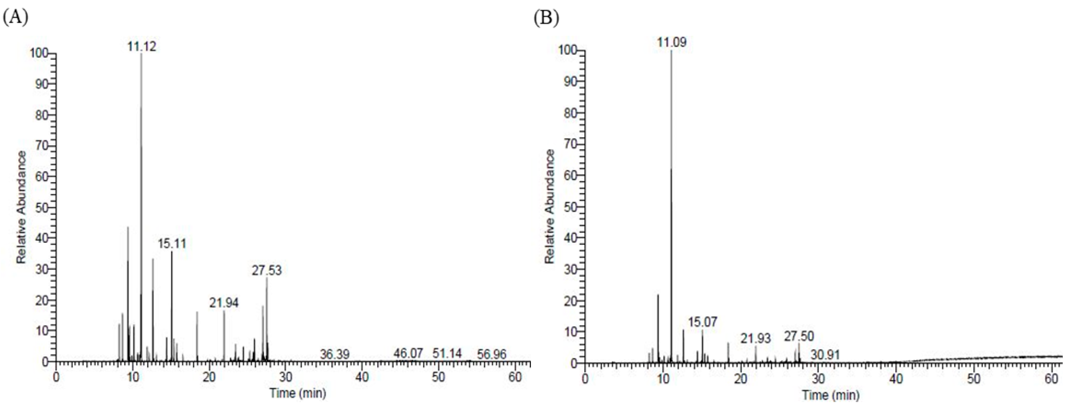
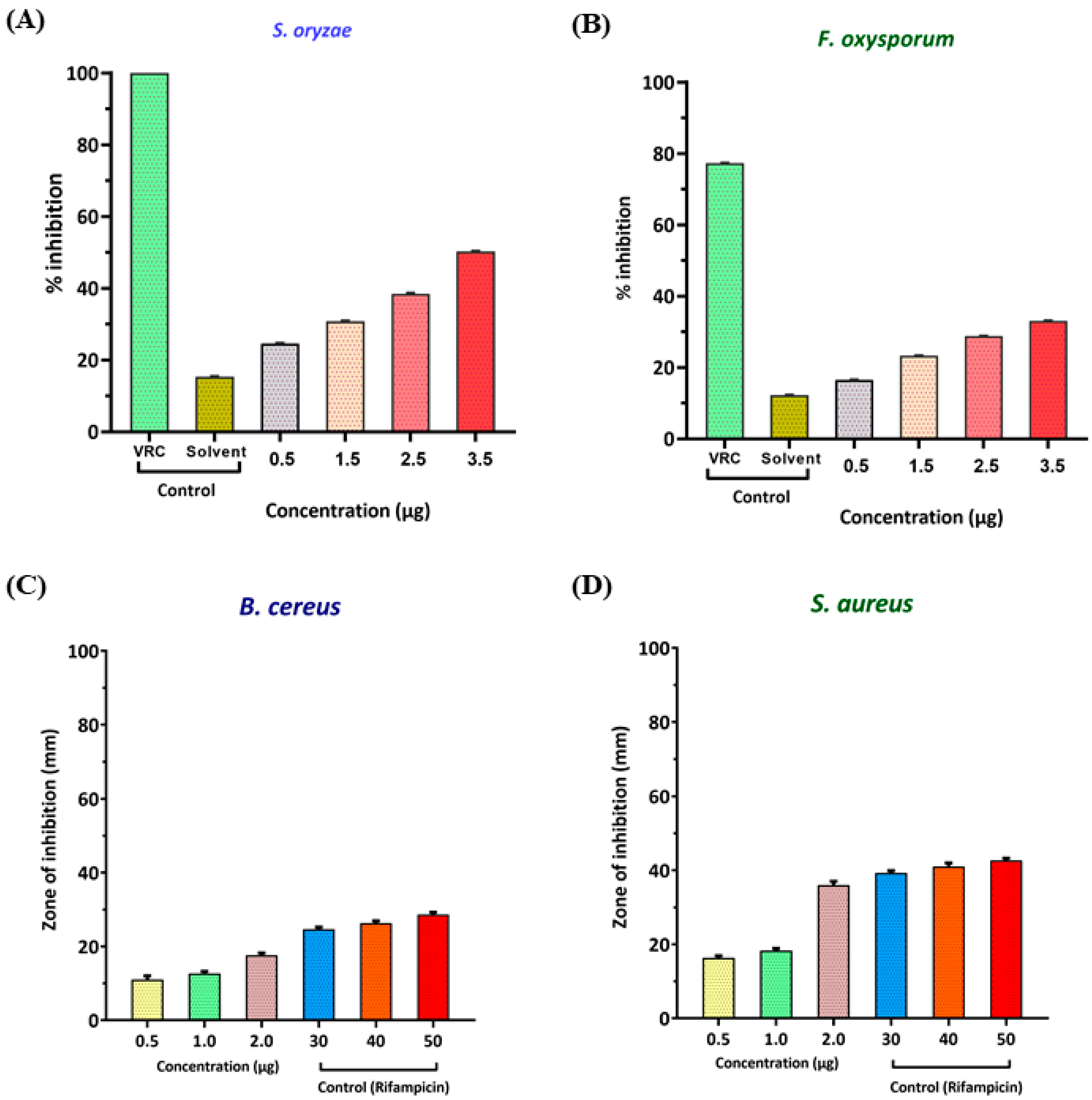
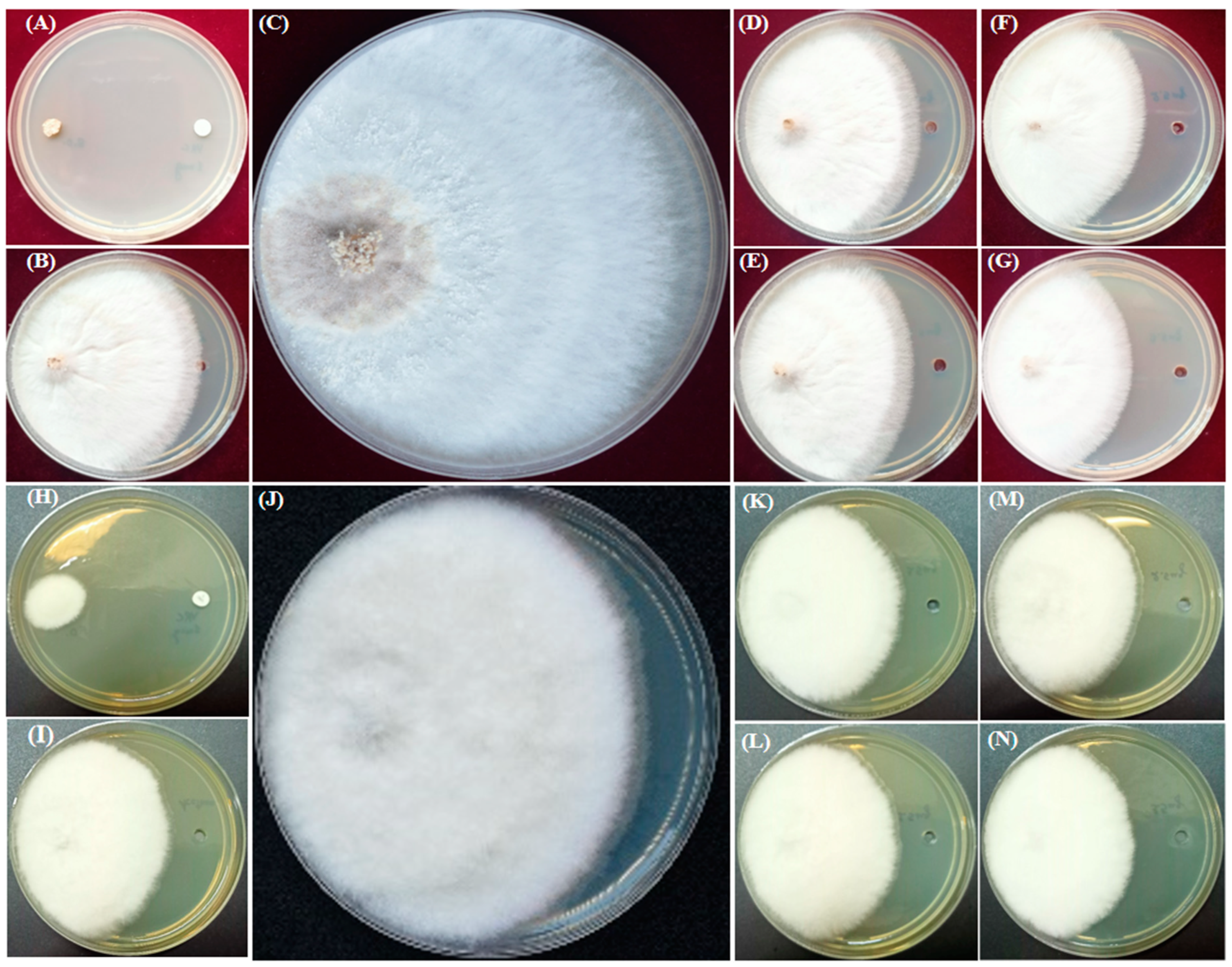
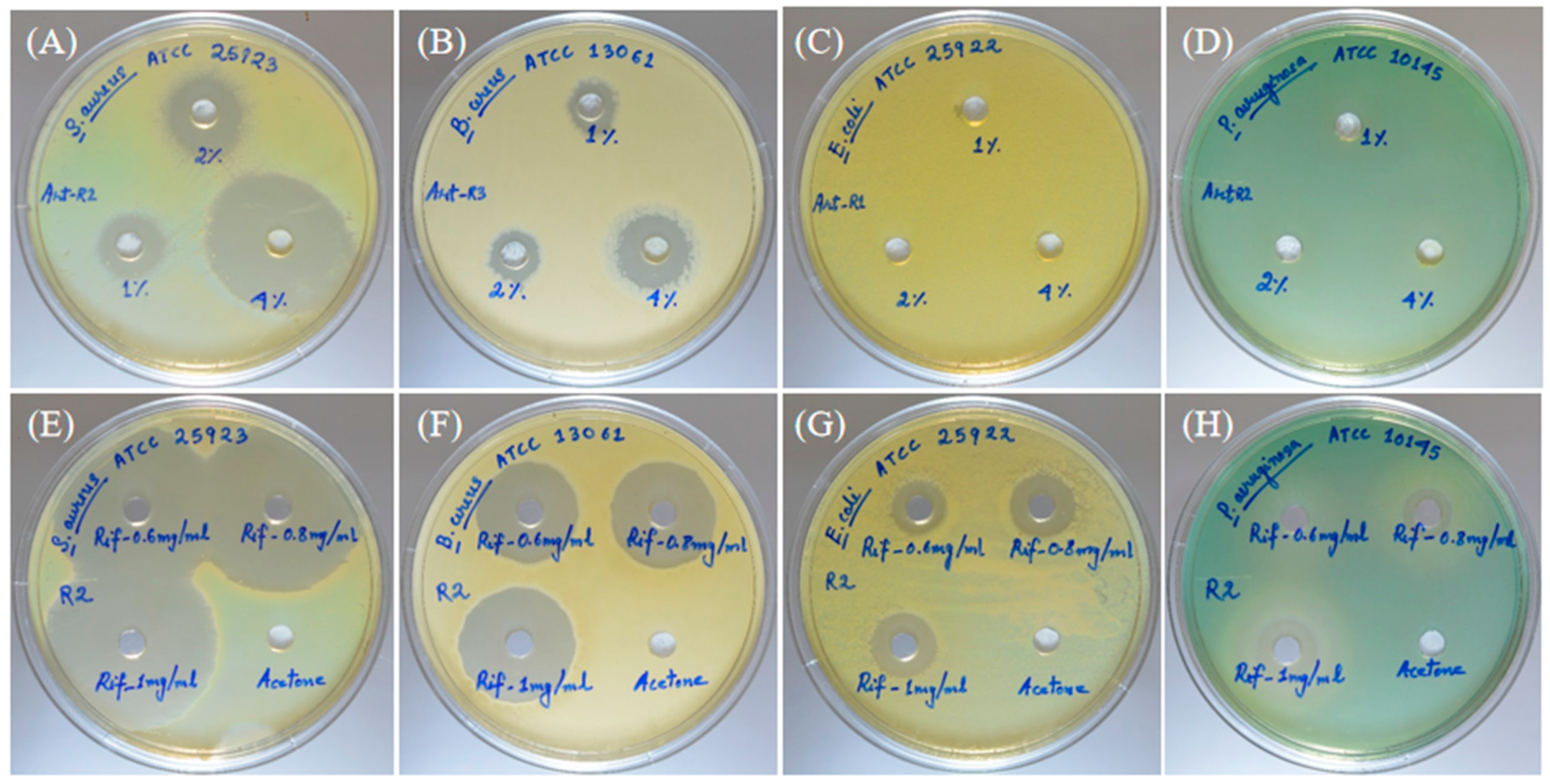
2. Results
3. Discussion
4. Materials and Methods
4.1. Plant Material
4.2. Hydro-Distillation Apparatus and Methods
4.3. Headspace SPME Extraction
4.4. GC and GC-MS Analysis
4.5. Test Organisms
4.6. Antifungal Activity of Essential Oil
4.7. Antibacterial Activity of the Essential Oil
4.8. Minimum Inhibitory Concentration (MIC) and Minimum Bactericidal Concentration (MBC) of the Essential Oil
5. Conclusions
Author Contributions
Funding
Acknowledgments
Conflicts of Interest
References
- Barney, J.N.; Hay, A.G.; Weston, L.A. Isolation and characterization of allelopathic volatiles from mugwort (Artemisia vulgaris). J. Chem. Ecol. 2005, 31, 247–265. [Google Scholar] [CrossRef] [PubMed]
- Gurib-Fakim, A. Medicinal plants: Traditions of yesterday and drugs of tomorrow. Mol. Aspects Med. 2006, 27, 1–93. [Google Scholar] [CrossRef] [PubMed]
- Khan, K.A. A preclinical antihyperlipidemic evaluation of Artemisia vulgaris root in diet induced hyperlipidemic animal model. Int. J. Pharmacol. Res. 2015, 5, 110–114. [Google Scholar]
- Obistioiu, D.; Cristina, R.T.; Schmerold, I.; Chizzola, R.; Stolze, K.; Nichita, I.; Chiurciu, V. Chemical characterization by GC-MS and in vitro activity against Candida albicans of volatile fractions prepared from Artemisia dracunculus, Artemisia abrotanum, Artemisia absinthium and Artemisia vulgaris. Chem. Cent. J. 2014, 8, 1–11. [Google Scholar] [CrossRef] [PubMed]
- Ekiert, H.; Pajor, J.; Klin, P.; Rzepiela, A.; Ślesak, H.; Szopa, A. Significance of Artemisia vulgaris L.(Common Mugwort) in the history of medicine and its possible contemporary applications substantiated by phytochemical and pharmacological studies. Molecules 2020, 25, 4415. [Google Scholar] [CrossRef]
- Abad, M.J.; Bedoya, L.M.; Apaza, L.; Bermejo, P. The Artemisia L. genus: A review of bioactive essential oils. Molecules 2012, 17, 2542–2566. [Google Scholar] [CrossRef]
- Siwan, D.; Nandave, D.; Nandave, M. Artemisia vulgaris Linn: An updated review on its multiple biological activities. Future J. Pharm. Sci. 2022, 8, 1–14. [Google Scholar] [CrossRef]
- Hiremath, S.K.; Kolume, D.G.; Muddapur, U.M. Antimicrobial activity of Artemisia vulgaris Linn. (Damanaka). Int. J. Ayurveda Res. 2011, 2, 1674–1675. [Google Scholar]
- Nurlybekova, A.; Kudaibergen, A.; Kazymbetova, A.; Amangeldi, M.; Baiseitova, A.; Ospanov, M.; Aisa, H.A.; Ye, Y.; Ibrahim, M.A.; Jenis, J. Traditional Use, Phytochemical Profiles and Pharmacological Properties of Artemisia Genus from Central Asia. Molecules 2022, 27, 5128. [Google Scholar] [CrossRef]
- Dikshit, K.R.; Dikshit, J.K. Weather and Climate of North-East India. In North-East India: Land, People and Economy; Springer: Dordrecht, The Netherlands, 2014; pp. 149–173. [Google Scholar]
- Harish, B.S.; Dandin, S.B.; Umesha, K.; Sasanur, A. Impact of climate change on Medicinal Plants—A review. Anc. Sci. Life 2012, 32, 23. [Google Scholar] [CrossRef]
- Swamy, M.K.; Akhtar, M.S.; Sinniah, U.R. Antimicrobial properties of plant essential oils against human pathogens and their mode of action: An updated review. Evid. Based Complement. Altern. Med. 2016, 2016, 3012462. [Google Scholar] [CrossRef]
- Mehani, M.; Segni, L.; Terzi, V.; Morcia, C.; Ghizzoni, R.; Goudgil, B.; Benchikh, S. Antifungal activity of Artemisia herba-alba on various Fusarium. Phytothérapie 2018, 16, 87–90. [Google Scholar] [CrossRef]
- Farzaneh, M.; Ahmadzadeh, M.; Hadian, J.; Tehrani, A.S. Chemical composition and antifungal activity of the essential oils of three species of Artemisia on some soil-borne phytopathogens. Commun. Agric. Appl. Biol. 2006, 71, 1327–1333. [Google Scholar]
- Zhigzhitzhapova, S.V.; Radnaeva, L.D.; Gao, Q.; Chen, S.; Zhang, F. Chemical composition of volatile organic compounds of Artemisia vulgaris L. (Asteraceae) from the Qinghai–Tibet Plateau. Ind. Crops Prod. 2016, 83, 462–469. [Google Scholar] [CrossRef]
- Haider, F.; Dwivedi, P.D.; Naqvi, A.A.; Bagchi, G.D. Essential oil composition of Artemisia vulgaris harvested at different growth periods under Indo-Gangetic plain conditions. J. Essent. Oil. Res. 2003, 15, 376–378. [Google Scholar] [CrossRef]
- Said-Al Ahl, H.H.; Hussein, M.S.; Tkachenko, K.G.; Nkomo, M.; Mudau, F. Essential oil composition of Artemisia vulgaris grown in Egypt. Int. J. Pharm. Pharm. Sci. 2016, 1302, 1–7030. [Google Scholar]
- Pandey, B.P.; Thapa, R.; Upreti, A. Chemical composition, antioxidant and antibacterial activities of essential oil and methanol extract of Artemisia vulgaris and Gaultheria fragrantissima collected from Nepal. Asian Pac. J. Trop. Med. 2017, 10, 952–959. [Google Scholar] [CrossRef]
- Labarrere, B.; Prinzing, A.; Dorey, T.; Chesneau, E.; Hennion, F. Variations of secondary metabolites among natural populations of sub-Antarctic ranunculus species suggest functional redundancy and versatility. Plants 2019, 8, 234. [Google Scholar] [CrossRef]
- Singh, B.R.; Singh, V.; Singh, R.K.; Toppo, S.; Haque, N.; Ebibeni, N. Antimicrobial effect of Artemisia vulgaris essential oil. Nat. Prod. 2011, 7, 5–12. [Google Scholar]
- Munda, S.; Pandey, S.K.; Dutta, S.; Baruah, J.; Lal, M. Antioxidant activity, antibacterial activity and chemical composition of essential oil of Artemisia vulgaris L. leaves from Northeast India. J. Essent. Oil Bear. Plants. 2019, 22, 368–379. [Google Scholar] [CrossRef]
- Sharma, K.R.; Adhikari, S. Phytochemical analysis and biological activities of Artemisia vulgaris grown in different altitudes of Nepal. Int. J. Food Prop. 2023, 26, 414–427. [Google Scholar] [CrossRef]
- Malik, S.; de Mesquita, L.S.S.; Silva, C.R.; de Mesquita, J.W.C.; de Sá Rocha, E.; Bose, J.; Abiri, R.; de Maria Silva Figueiredo, P.; Costa-Júnior, L.M. Chemical profile and biological activities of essential oil from Artemisia vulgaris L. cultivated in Brazil. Pharmaceuticals 2019, 12, 49. [Google Scholar] [CrossRef]
- Ivanov, M.; Kannan, A.; Stojković, D.S.; Glamočlija, J.; Calhelha, R.C.; Ferreira, I.C.; Sanglard, D.; Soković, M. Camphor and eucalyptol—Anticandidal spectrum, antivirulence effect, efflux pumps interference and cytotoxicity. Int. J. Mol. Sci. 2021, 22, 483. [Google Scholar] [CrossRef] [PubMed]
- Arunkumar, R.; Nair, S.A.; Rameshkumar, K.B.; Subramoniam, A. The essential oil constituents of Zorniadiphylla (L.) Pers, and anti-inflammatory and antimicrobial activities of the oil. Rec. Nat. Prod. 2014, 8, 385. [Google Scholar]
- Cao, B.; Ni, H.-Y.; Li, J.; Zhou, Y.; Bian, X.-L.; Tao, Y.; Cai, C.-Y.; Qin, C.; Wu, H.-Y.; Chang, L.; et al. (+)-Borneol suppresses conditioned fear recall and anxiety-like behaviors in mice. Biochem. Biophys. Res. Commun. 2018, 495, 1588–1593. [Google Scholar] [CrossRef]
- Selestino Neta, M.C.; Vittorazzi, C.; Guimarães, A.C.; Martins, J.D.L.; Fronza, M.; Endringer, D.C.; Scherer, R. Effects of β-caryophyllene and Murrayapaniculata essential oil in the murine hepatoma cells and in the bacteria and fungi 24-h time–kill curve studies. Pharm. Biol. 2017, 55, 190–197. [Google Scholar] [CrossRef]
- Jassal, K.; Kaushal, S.; Rashmi; Rani, R. Antifungal potential of guava (Psidium guajava) leaves essential oil, major compounds: Beta-caryophyllene and caryophyllene oxide. Arch. Phytopathol. Pflanzenschutz. 2021, 54, 2034–2050. [Google Scholar] [CrossRef]
- Kim, S.; Lee, S.; Hong, C.; Cho, S.; Park, M.; Choi, I. Antifungal effect of elemol and eudesmol from Cryptomeria japonica essential oil against Trichophyton rubrum. Acad. J. Agric. Res. 2016, 4, 511–517. [Google Scholar]
- Ozil, O.; Diler, O.; Nazıroğlu, M. Antifungal activity of some essential oil nanoemulsions against Saprolegniasis in rainbow trout (Oncorhynchus mykiss) eggs: Antifungal activity of essential oil nanoemulsions. Aquac. Int. 2022, 30(5), 2201–2212. [Google Scholar] [CrossRef]
- Lingan, K. Antifungal activity of Artemisia Nilagirica essential oil from the Western Ghats Nilgiris against food-borne fungi. Int. J. Appl. Biol. 2021, 2, 3. [Google Scholar] [CrossRef]
- Fang, D.; Liu, X.; Chen, X.; Yan, W.; He, Y.; Cheng, Y.; Chen, J.; Li, Z.; Guo, L.; Wang, T.; et al. Fusarium species and Fusarium oxysporum species complex genotypes associated with yam wilt in South-Central China. Front. Microbiol. 2020, 11, 1964. [Google Scholar]
- Gordon, T.R. Fusarium oxysporum and the Fusarium wilt syndrome. Annu. Rev. Phytopathol. 2017, 55, 23–39. [Google Scholar] [CrossRef]
- Rao, V.N.; Sastry, R.K.; Craufurd, P.; Meinke, H.; Parsons, D.; Rego, T.J.; Rathore, A. Cropping systems strategy for effective management of Fusarium wilt in safflower. Field Crops Res. 2014, 156, 191–198. [Google Scholar]
- Kumar, A.; Ram, S.; Jalali, B. Management of stem rot of rice with resistance inducing chemicals and fungicides. Indian Phytopathol. 2003, 56, 266–269. [Google Scholar]
- Adley, C.; Ryan, M. The nature and extent of foodborne disease. In Antimicrobial Food Packaging; Academic Press: Cambridge, MA, USA, 2016; pp. 1–10. [Google Scholar]
- Kadariya, J.; Smith, T.C.; Thapaliya, D. Staphylococcus aureus and staphylococcal food-borne disease: An ongoing challenge in public health. BioMed Res. Int. 2014, 2014. [Google Scholar] [CrossRef]
- Ahmadizadeh, C.; Monadi, A.; Rezaie, A.; Rad, M.G.; Jafari, B. Antibacterial activity of methanolic extract and essence of Sagebrush (Artemisia vulgaris) against pathogenic bacteria. Life Sci. J. 2018, 15, 69–73. [Google Scholar]
- Devi, M.A.; Nameirakpam, B.; Devi, T.B.; Mayanglambam, S.; Singh, K.D.; Sougrakpam, S.; Shadia, S.; Tongbram, M.; Singh, S.D.; Sahoo, D.; et al. Chemical compositions and insecticidal efficacies of four aromatic essential oils on rice weevil Sitophilus oryzae L. Int. J. Trop. Insect Sci. 2020, 40, 549–559. [Google Scholar] [CrossRef]
- Veiga, A.; da Graça, T.T.M.; Rossa, L.S.; Mengarda, M.; Stofella, N.C.; Oliveira, L.J.; Gonçalves, A.G.; Murakami, F.S. Colorimetric microdilution assay: Validation of a standard method for determination of MIC, IC50%, and IC90% of antimicrobial compounds. J. Microbiol. Methods 2019, 162, 50–61. [Google Scholar] [CrossRef]


| Direct AVEO Injection Method | AVEO SPME Method | |||||||||
|---|---|---|---|---|---|---|---|---|---|---|
| S No. | a RT | Compound | b RSI | C RA% | Mol. Wt. | a RT | Compound | b RSI | C RA% | Mol. Wt. |
| 1 | 8.23 | α-Pinene | 948 | 2.24 | 136 | 8.23 | α-Pinene | 949 | 1.45 | 136 |
| 2 | 8.65 | Camphene | 967 | 3.03 | 136 | 8.65 | Camphene | 964 | 2.14 | 136 |
| 3 | 9.38 | Sabinene | 950 | 8.44 | 136 | 9.37 | Sabinene | 938 | 8.86 | 136 |
| 4 | 9.47 | p-mentha-1(7) | 923 | 1.27 | 136 | 9.46 | p-mentha-1(7) | 927 | 1.34 | 136 |
| 5 | 9.57 | 1-Octen-3-ol | 920 | 2.1 | 128 | 9.56 | 1-Octen-3-ol | 894 | 0.78 | 128 |
| 6 | 9.88 | β-Myrcene | 867 | 0.45 | 136 | 9.88 | β-Myrcene | 867 | 0.41 | 136 |
| 7 | 10.16 | Yomogi alcohol | 916 | 2.34 | 154 | 10.15 | Yomogi alcohol | 911 | 0.93 | 154 |
| 8 | 10.63 | 1,3-Cyclohexadiene,1-methyl-4-(1-methylethyl)- | 934 | 0.53 | 136 | 10.62 | 1,3-Cyclohexadiene,1-methyl-4-(1-methylethyl)- | 931 | 0.63 | 136 |
| 9 | 10.87 | o-Cymene | 967 | 0.43 | 134 | 10.87 | o-Cymene | 967 | 0.52 | 134 |
| 10 | 11.00 | Cyclohexene,1-methyl-5-(1-methylethenyl)-,(R)- | 910 | 0.81 | 136 | 10.99 | Cyclohexene,1-methyl-5-(1-methylethenyl)-,(R)- | 907 | 1.06 | 136 |
| 11 | 11.12 | Eucalyptol | 923 | 29.91 | 154 | 11.08 | Eucalyptol | 937 | 47.30 | 154 |
| 12 | 11.88 | γ-Terpinene | 923 | 0.85 | 136 | 11.88 | γ-Terpinene | 923 | 1.12 | 136 |
| 13 | 12.15 | 5-Isopropyl-2-methylbicyclo [3.1.0]hexan-2-ol | 925 | 0.45 | 154 | 12.15 | 5-Isopropyl-2-methylbicyclo [3.1.0]hexan-2-ol | 921 | 0.29 | 154 |
| 14 | 12.65 | 2,7-Dimethyl-2,6-octadien-4-ol | 916 | 6.76 | 154 | 12.63 | 2,7-Dimethyl-2,6-octadien-4-ol | 918 | 4.24 | 154 |
| 15 | 12.76 | Cyclohexene,1-methyl-4-(1-methylethylidene)- | 955 | 0.15 | 136 | 12.76 | Cyclohexene,1-methyl-4-(1-methylethylidene)- | 952 | 0.27 | 136 |
| 16 | 13.09 | Linalool | 892 | 0.4 | 154 | 13.09 | Linalool | 875 | 0.28 | 154 |
| 17 | 14.29 | 1,7-Octadien-3-one,2-methyl-6-methylene- | 858 | 0.1 | 150 | 14.29 | 1,7-Octadien-3-one,2-methyl-6-methylene- | 860 | 0.16 | 150 |
| 18 | 14.43 | (-)-Alcanfor | 965 | 1.37 | 152 | 14.43 | (-)-Alcanfor | 966 | 1.78 | 152 |
| 19 | 14.81 | Isobornyl formate | 865 | 0.12 | 154 | 14.81 | Isobornyl formate | 884 | 0.18 | 182 |
| 20 | 15.00 | trans-Verbenol | 887 | 0.22 | 152 | 14.97 | trans-Verbenol | 844 | 0.28 | 152 |
| 21 | 15.11 | endo-Borneol | 939 | 8.24 | 154 | 15.07 | endo-Borneol | 946 | 4.76 | 154 |
| 22 | 15.39 | Terpinen-4-ol | 896 | 1.21 | 154 | 15.38 | Terpinen-4-ol | 906 | 1.32 | 154 |
| 23 | 15.77 | α-Terpineol | 942 | 0.91 | 154 | 15.76 | α-Terpineol | 951 | 0.83 | 154 |
| 24 | 16.57 | trans-Carveol | 886 | 0.26 | 152 | 16.56 | trans-Carveol | 889 | 0.34 | 152 |
| 25 | 18.41 | Bornyl acetate | 955 | 2.92 | 196 | 18.40 | Bornyl acetate | 950 | 2.74 | 196 |
| 26 | 19.76 | Cyclohexene,1,5,5-trimethyl-3-methylene- | 903 | 0.11 | 136 | 19.76 | Cyclohexene,1,5,5-trimethyl-3-methylene- | 900 | 0.16 | 136 |
| 27 | 20.14 | Tricyclo [5.4.0.0(2,8)]undec-9-ene, 2,6,6,9-tetramethyl-,(1R,2S,7R,8R)- | 866 | 0.11 | 204 | 20.14 | Tricyclo [5.4.0.0(2,8)]undec-9-ene, 2,6,6,9-tetramethyl-,(1R,2S,7R,8R)- | 871 | 0.28 | 204 |
| 28 | 20.78 | Copaene | 871 | 0.21 | 204 | 20.79 | Copaene | 885 | 0.36 | 204 |
| 29 | 21.15 | cis-β-Copaene | 912 | 0.10 | 204 | 21.15 | cis-β-Copaene | 900 | 0.17 | 204 |
| 30 | 21.69 | 1H-Cycloprop[e]azulene,1a,2,3,4,4a,5,6,7b-octahydro-1,1,4,7-tetramethyl-, | 904 | 0.13 | 204 | 21.68 | 1H-Cycloprop[e]azulene,1a,2,3,4,4a,5,6,7b-octahydro-1,1,4,7-tetramethyl-, | 909 | 0.24 | 204 |
| 31 | 21.94 | Caryophyllene | 204 | 2.95 | 945 | 21.93 | Caryophyllene | 945 | 2.48 | 204 |
| 32 | 22.79 | Humulene | 204 | 0.26 | 204 | 22.78 | Humulene | 900 | 0.27 | 204 |
| 33 | 23.33 | 1-Methyl-4-(6-methylhept-5-en-2-yl)cyclohexa-1,3-diene | 892 | 0.13 | 204 | 23.33 | 1-Methyl-4-(6-methylhept-5-en-2-yl)cyclohexa-1,3-diene | 885 | 0.11 | 204 |
| 34 | 23.46 | Germacrene D | 952 | 1.01 | 204 | 23.46 | Germacrene D | 951 | 0.74 | 204 |
| 35 | 23.74 | cis-Muurola-4(15),5-diene | 944 | 0.15 | 204 | 23.74 | cis-Muurola-4(15),5-diene | 925 | 0.18 | 204 |
| 36 | 23.85 | Bicyclogermacrene | 906 | 0.43 | 204 | 23.84 | Bicyclogermacrene | 912 | 0.49 | 204 |
| 37 | 24.45 | 1-Isopropyl-4,7-dimethyl-1,2,3,5,6,8a-hexahydronaphthalene | 927 | 0.84 | 204 | 24.44 | 1-Isopropyl-4,7-dimethyl-1,2,3,5,6,8a-hexahydronaphthalene | 923 | 0.53 | 204 |
| 38 | 25.31 | 1,6,10-Dodecatrien-3-ol,3,7,11-trimethyl-, (E)- | 923 | 0.65 | 222 | 25.31 | 1,6,10-Dodecatrien-3-ol,3,7,11-trimethyl- | 914 | 0.28 | 222 |
| 39 | 25.75 | 5,10-Pentadecadiyn-1-ol | 953 | 0.77 | 220 | 25.75 | 5,10-Pentadecadiyn-1-ol | 951 | 0.33 | 220 |
| 40 | 25.93 | Caryophyllene oxide | 920 | 1.38 | 220 | 25.92 | Caryophyllene oxide | 912 | 0.61 | 220 |
| 41 | 26.39 | (-)-Globulol | 902 | 0.18 | 222 | 26.39 | (-)-Globulol | 895 | 0.11 | 222 |
| 42 | 26.89 | 2-Methyl-3-(3-methyl-but-2-enyl)-2-(4-methyl-pent-3-enyl)-oxetane | 930 | 0.35 | 222 | 26.87 | 2-Methyl-3-(3-methyl-but-2-enyl)-2-(4-methyl-pent-3-enyl)-oxetane | 937 | 0.15 | 222 |
| 43 | 27.02 | Cedren-13-ol, 8- | 870 | 4.12 | 220 | 27.01 | Cedren-13-ol, 8- | 875 | 1.77 | 220 |
| 44 | 27.14 | 11,11-Dimethyl-4,8-dimethylenebicyclo [7.2.0]undecan-3-ol | 220 | 0.36 | 220 | 27.13 | 11,11-Dimethyl-4,8-dimethylenebicyclo [7.2.0]undecan-3-ol | 901 | 0.22 | 220 |
| 45 | 27.24 | Dihydro-cis-à-copaene-8-ol | 943 | 0.28 | 222 | 27.23 | Dihydro-cis-à-copaene-8-ol | 945 | 0.19 | 222 |
| 46 | 27.53 | 10-epi-γ-Eudesmol | 949 | 6.50 | 222 | 27.5 | 10-epi-γ-Eudesmol | 940 | 3.09 | 222 |
| 47 | 27.67 | 10-Epijuneol | 920 | 1.13 | 264 | 27.65 | 10-Epijuneol | 919 | 0.58 | 264 |
| Total area % = 97.66 | Total area % = 97.35 | |||||||||
| S No. | Sample | Conc. | Amount of E.O. per 50 µL Volume | Percent Inhibition (Mean ± SD) | |
|---|---|---|---|---|---|
| S. oryzae ITCC 4107 | F. oxysporum MTCC 9913 | ||||
| 1. | Essential oil (E.O.) | 1% | 0.5 µL | 24.6 ± 0.09 | 16.56 ± 0.06 |
| 3% | 1.5 µL | 30.8 ± 0.09 | 23.31 ± 0.06 | ||
| 5% | 2.5 µL | 38.50 ± 0.09 | 28.83 ± 0.06 | ||
| 7% | 3.5 µL | 50.3 ± 0.03 | 33.13 ± 0.06 | ||
| 2. | Vorioconazole | 1 µg | - | 100 ± 0.0 | 77.8 ± 0.07 |
| 3. | Acetone | - | - | 15.4 ± 0.03 | 13.0 ± 0.1 |
| S No. | Sample | Conc. | Amount of AVEO/Antibiotic per 50 µL Volume | Zone of Inhibition (mm), Mean ± SD | |||
|---|---|---|---|---|---|---|---|
| S. aureus ATCC 25923 | B. cereus ATCC 13061 | E. coli ATCC 25922 | P. aeruginosa ATCC 10145 | ||||
| 1. | Essential oil (E.O.) | 1% | 0.5 µL | 16.33 ± 0.58 | 11.00 ± 1.00 | Nil | Nil |
| 2% | 1 µL | 18.33 ± 0.58 | 12.67 ± 0.58 | Nil | Nil | ||
| 4% | 2 µL | 36.00 ± 1.00 | 17.67 ± 0.58 | Nil | Nil | ||
| 2. | Rifampicin | 0.6 mg/ml | 30 µg | 39.33 ± 0.58 | 24.67 ± 0.58 | 14.33 ± 0.58 | 8.00 ± 0.00 |
| 0.8 mg/ml | 40 µg | 41.00 ± 1.00 | 26.33 ± 0.58 | 16.00 ± 0.00 | 11.67 ± 0.58 | ||
| 1.0 mg/ml | 50 µg | 43.00 ± 0.00 | 28.67 ± 0.58 | 17.00 ± 0.00 | 15.00± 0.00 | ||
| 3. | Acetone | - | - | Nil | Nil | Nil | Nil |
| S No. | Sample | Microorganisms | MIC * | MBC ** |
|---|---|---|---|---|
| 1. | Essential oil | B. cereus S. aureus | 0.31% 0.63% | 0.63% 2.5% |
| 2. | Rifampicin | B. cereus S. aureus | ≤0.49 µg/mL ≤0.49 µg/mL | 1.95 µg/mL ≤0.49 µg/mL |
Disclaimer/Publisher’s Note: The statements, opinions and data contained in all publications are solely those of the individual author(s) and contributor(s) and not of MDPI and/or the editor(s). MDPI and/or the editor(s) disclaim responsibility for any injury to people or property resulting from any ideas, methods, instructions or products referred to in the content. |
© 2023 by the authors. Licensee MDPI, Basel, Switzerland. This article is an open access article distributed under the terms and conditions of the Creative Commons Attribution (CC BY) license (https://creativecommons.org/licenses/by/4.0/).
Share and Cite
Singh, N.B.; Devi, M.L.; Biona, T.; Sharma, N.; Das, S.; Chakravorty, J.; Mukherjee, P.K.; Rajashekar, Y. Phytochemical Composition and Antimicrobial Activity of Essential Oil from the Leaves of Artemisia vulgaris L. Molecules 2023, 28, 2279. https://doi.org/10.3390/molecules28052279
Singh NB, Devi ML, Biona T, Sharma N, Das S, Chakravorty J, Mukherjee PK, Rajashekar Y. Phytochemical Composition and Antimicrobial Activity of Essential Oil from the Leaves of Artemisia vulgaris L. Molecules. 2023; 28(5):2279. https://doi.org/10.3390/molecules28052279
Chicago/Turabian StyleSingh, Nameirakpam Bunindro, Moirangthem Lakshmipriyari Devi, Thokchom Biona, Nanaocha Sharma, Sudripta Das, Jharna Chakravorty, Pulok Kumar Mukherjee, and Yallappa Rajashekar. 2023. "Phytochemical Composition and Antimicrobial Activity of Essential Oil from the Leaves of Artemisia vulgaris L." Molecules 28, no. 5: 2279. https://doi.org/10.3390/molecules28052279