Article

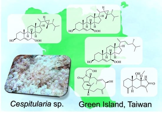
New Verticillene Diterpenoids, Eudesmane Sesquiterpenoids, and Hydroperoxysteroids from the Further Chemical Investigation of a Taiwanese Soft Coral Cespitularia sp.

1
Department of Marine Biotechnology and Resources, National Sun Yat-sen University, Kaohsiung 804, Taiwan
2
Doctoral Degree Program in Marine Biotechnology, National Sun Yat-sen University, Kaohsiung 804, Taiwan
3
Graduate Institute of Natural Products, Kaohsiung Medical University, Kaohsiung 807, Taiwan
4
Institute of Biomedical Sciences, National Chung Hsing University, Taichung 402, Taiwan
5
Institute of Oceanography, National Taiwan University, Taipei 106, Taiwan
6
Department of Medical Research, China Medical University Hospital, China Medical University, Taichung 404, Taiwan
*
Author to whom correspondence should be addressed.
Molecules 2023, 28(4), 1521; https://doi.org/10.3390/molecules28041521
Submission received: 17 January 2023 / Revised: 3 February 2023 / Accepted: 3 February 2023 / Published: 4 February 2023

Abstract

:
An investigation of the chemical composition of a Formosan soft coral Cespitularia sp. led to the discovery of one new verticillene-type diterpenoid, cespitulactam M (1); one new eudesmane sesquiterpenoid, cespilamide F (2); and three new hydroperoxysteroids (35) along with twelve known analogous metabolites (617). In addition, one new derivative, cespitulactam M-6,2′-diacetate (1a), was prepared from compound 1. The structures were determined by detailed spectroscopic analyses, particularly HRESIMS and NMR techniques. Moreover, the in vitro cytotoxicity, anti-inflammatory, and antibacterial activity of 117 and 1a were evaluated.

Graphical Abstract

1. Introduction

In the past three decades, a number of studies have shown that the genus of soft coral, Cespitularia (a phylum of Xeniidae), elaborates diterpenoids with the verticillene skeleton [1,2,3,4,5,6,7,8,9,10,11,12,13,14,15,16,17] and eudesmane sesquiterpenoids [3,7,17] as major characteristic metabolites, mainly isolated from the species C. hypotentaculata and C. taeniata, which demonstrated cytotoxic [1,2,3,6,7,8,10,11,13], anti-inflammatory [5,16], antiviral [12], and antibacterial [11] activities. In our previous study, we investigated a series of bioactive verticillene diterpenoids as promising compounds for further marine anti-inflammatory drug development [16]. Herein, this continuous chemical investigation of the Formosan Cespitularia sp. collected in Green Island led to one new verticillene-type diterpenoid, one new eudesmane sesquiterpenoid, three new hydroperoxysteroids, and twelve known metabolites. Compounds 117 were evaluated for anti-inflammatory activity and cytotoxicity against human lung adenocarcinoma (A549), human hepatocellular liver carcinoma (HepG2), and human breast adenocarcinoma (MDA-MB-231) cancer cell lines, and tested for ten species of pathogenic microbes.

2. Results and Discussion

The extract of Cespitularia sp. was separated by column chromatography and HPLC to afford five new secondary metabolites (15) and twelve related known compounds, which were identified as 7α-hydroperoxycampesterol (6) [18], 7β-hydroperoxycholesterol (7) [19], 7α-hydroperoxycholesterol (8) [19], cespitulactam D (9) [11], cespitulactam F (10) [11], cespitulin S (11) [16], cespitularin D (12) [1], cespitularin O (13) [8], cespitulactone B (14) [10], cespihypotin Q (15) [13], atractylenolide II (16) [20], and atractylenolide III (17) [20] (Figure 1). (Supplementary materials, Figures S1–S68.)
Cespitulactam M (1) was obtained as an amorphous solid and displayed HRESIMS (m/z 382.2350 [M + Na]+, calculated for C22H33NO3Na, 382.2353) consistent with the molecular formula C22H33NO3, implying seven degrees of unsaturation. The IR spectrum revealed the presence of amide (1656 cm−1) and hydroxy (3388 cm−1) groups. Subsequently, the 1H NMR and 13C NMR spectroscopic data (Table 1 and Table 2) demonstrated signals of three methyls, eight sp3 methylenes, one sp2 methylene, three sp3 methines, one sp2 methine, one sp3, and five sp2 quaternary carbons (including a carbonyl carbon appearing at δC 172.5 ppm). The above data accounted for four of the seven degrees of unsaturation, resulting in compound 1 with a tricyclic structure. The COSY spectrum, recorded in CDCl3, showed five proton sequences from H-1 to H2-3, H2-5 to H-7, H2-9 to H-10, H2-13 to H2-14, and H2-1′ to H2-2′. Furthermore, key HMBC correlations of H2-3 to C-4; H2-5 to C-4; H-7 to C-9; H2-14 to C-12; H3-16 to C-1, C-11, and C-15; H3-17 to C-1, C-11, and C-15; H2-18 to C-3 and C-5; H3-19 to C-7, C-8, and C-9 confirmed the connection of the carbon skeleton. Based on the above analysis, the planar structure of 1 was established (Figure 2).
With the planar structure of 1 determined, the relative stereochemistry of the three stereogenic centers, 1R*, 6S*, and 10R*, of 1, was assigned via the analysis of the NOESY spectrum (Figure 3). It was found that H-1 displayed NOE interactions with H3-16 and H3-17, and H-10 also demonstrated NOE interactions with H3-16, H3-17, and one proton of H2-9 (δH 2.74, br d, J = 14.4 Hz). Based on previous studies, all naturally occurring verticillane diterpenoids are assigned H-1 as β-oriented, as shown in verticillene-type derivatives [1,2,3,4,5,6,7,8,9,10,11,12,13,14,15,16,17]. Hence, H-1, one proton of H2-9 (δH 2.74, br d, J = 14.4 Hz), as well as H-10, would be positioned on the β face. On the other hand, H-6 (δH 4.35, m) exhibited NOE correlations with the other proton of H2-9 (δH 2.68, dd, J = 15.0, 4.2 Hz), revealing the α-orientation of proton H-6. On the basis of the NOESY spectral analysis and MM2 force field analysis, the relative structure of cespitulactam M (1) was determined. The absolute configuration of 1 was suggested as 1R, 6S, and 10R by the proposed biosynthetic pathway as an intermediate from cespitularin C to cespitulamide C [7].
Furthermore, upon acetylation, compound 1 afforded the diacetate, cespitulactam M-6,2′-diacetate (1a), which exhibited two additional three-proton acetyl singlets at δH 2.01 and δH 2.04. Through the comparison of 1H and 13C NMR spectra of 1 and 1a, the deshielding of H2-2′ from δH 3.85 to 4.31 and 4.12 suggested the location of an acetyl group at C-2′, while the deshielding of H-6 from δH 4.35 to 5.29 also indicated the location of an acetyl group at C-6. In addition, the HRESIMS of 1a revealed a molecular ion at m/z 466.2565, [M + Na]+ (calculated for C22H33NO3Na, 466.2564).
Cespilamide F (2) was obtained as a white powder. The HRESIMS (m/z 300.1572 [M + Na]+, calculated for C16H23NO3Na, 300.1570) of 2 established the molecular formula C16H23NO3, appropriate for six degrees of unsaturation. Its IR spectrum also revealed the presence of amide (1693 cm−1) and hydroxy (3418 cm−1) groups. The 13C NMR and HSQC spectroscopic data (Table 1 and Table 2) illustrated signals of three methyls (including one methoxyl group appearing at δH 3.09 and δC 49.6 ppm), four sp3 methylenes, one sp2 methylene, two sp3 methines, two sp3, and four sp2 quaternary carbons (including one carbonyl carbon appearing at δC 174.3). The above data accounted for three of the six degrees of unsaturation, indicating a tricyclic structure for 2. From the COSY spectrum measured in CDCl3, we established two proton sequences from H2-1 to H2-3 and H-5 to H2-6. The key HMBC correlations of H2-6 to C-7 and C-8; H-9 to C-7, C-8, and C-10; H2-13 to C-7, C-11, and C-12; H3-14 to C-1, C-5, C-9, and C-10; H3-15 to C-3 and C-5; and H3-16 to C-8 permitted the connection of the carbon skeleton (supplementary materials, S12–S20). Based on the above analysis, the planar structure of 2 was established (Figure 2).
The relative configuration of 2 was determined on the basis of the NOESY experiment and compared with the published compound, taenialactams A, which was isolated from C. taeniata in 2009 [3]. Assuming that H-5 (δH 2.28, d, J = 12.0 Hz) possesses an α-orientation similar to that of taenialactams A, it was found that H-5 (δH 2.28, d, J = 12.0 Hz) demonstrated NOE interactions with one proton of H2-6 (δH 2.54, dd, J = 13.2, 3.0 Hz); therefore, H-5, and one proton of H2-6 (δH 2.54, dd, J = 13.2, 3.0 Hz) should also be positioned on the α face. On the contrary, the other one proton of H2-6 (δH 2.16, m) exhibited NOE correlations with H3-14 (δH 1.00, s) and H3-8-OMe (δH 3.09, s). Moreover, H-9 (δH 3.51, s) also showed NOE correlations with H3-14 (δH 1.00, s) and H3-8-OMe (δH 3.09, s), revealing the β-orientation of H2-6β (δH 2.16, m), H3-8-OMe (δH 3.09, s), H-9 (δH 3.51, s), and H3-14 (δH 1.00, s). On the basis of the NOESY spectral analysis and MM2 force field analysis, the relative structure of 2 was determined as 5S*, 8R*, 9S*, and 10S* (Figure 3).
7β-Hydroperoxygorgosterol (3) was also obtained as a white powder. The HRESIMS (m/z 481.3651 [M + Na]+) of 3 confirmed the molecular formula C30H50O3, implying 6 degrees of unsaturation. The presence of the hydroxy (3380 cm−1) group was shown on the IR spectrum. The 1D 13C NMR and DEPT spectroscopic data (Table 1 and Table 2) showed signals of seven methyls, eight sp3 methylenes, ten sp3 methines, one sp2 methine, three sp3, and one sp2 quaternary carbons. The above data accounted for one of the six degrees of unsaturation, indicating a pentacyclic structure for 3 (supplementary materials, S21–S28). From the COSY spectrum measured in CDCl3, it was possible to establish five proton sequences from H2-1 to H2-4, H-7 to H2-11, H-14 to H-17, H-17 to H-20, H-22 to H2-29, and H-24 to H3-28. Key HMBC correlations of H2-4 to C-5; H-6 to C-4, C-8, and C-10; H-9 to C-5; H3-18 to C-12, C-13, C-14, and C-17; H3-19 to C-1, C-5, C-9, and C-10; H3-21 to C-17, C-20, and C-22; H3-28 to C-23; and H3-30 to C-22, C-23, C-24, and C-29 permitted the connection of the carbon skeleton. On the basis of the above analysis, the planar structure of 3 was established (Figure 2).
The relative configuration of 3 was elucidated on the basis of the observed key NOE correlations (Figure 3). In particular, the stereo center of C-7 was the most significant result in this compound; thus, it was compared with the reported compounds 7β-hydroperoxycholesterol and its stereoisomer 7α-hydroperoxycholesterol [19]. In terms of the absolute configuration of the side chain, the 1D NMR spectra of compound 3 were compared with those of gorgosterol [21] and 7-oxogorgosterol [22] from previous research. It turned out that the chemical shifts of compound 3 were similar to that of 7-oxogorgosterol as 20R, 22R, 23R, and 24R. Thus, the absolute configuration of 7β-hydroperoxygorgosterol (3) was proposed.
7α-Hydroperoxygorgosterol (4) was isolated as a white powder. The HRESIMS exhibited a [M + Na]+ ion peak at 481.3652 m/z, establishing a molecular formula of C30H50O3. By 2D NMR spectroscopy data, including HSQC, COSY, and HMBC (supplementary materials, S29–S36), compound 4 was displayed to possess the same molecular framework as that of 3. The molecular formula of 3 and 4 indicates that 4 is an isomer of 3. On the basis of the above references and its NOE correlations, compound 4 was revealed to be the C-7 epimer of 3, namely 7α-hydroperoxygorgosterol (4).
The HRESIMS of 7β-hydroperoxycampesterol (5) showed that it possesses the molecular formula C28H48O3 (m/z 455.3495 [M + Na]+). The IR spectrum of 5 showed the absorption of a hydroxy group (3383 cm−1). Comparison of the 1H and 13C NMR spectroscopic data of compound 5 with known compound 7α-hydroperoxycampesterol (6) suggested that the planar structure of 5 was the same as 7α-hydroperoxycampesterol [18].
Owing to the NMR data of 5 and the reported compound 7α-hydroperoxycampesterol (6), this suggests that 5 is an isomer of 6. Compound 5 was also compared with the known compounds 7β-hydroperoxycholesterol and 7α-hydroperoxycholesterol in order to define the stereo center C-7 [19]. In terms of the chiral center C-24 on the side chain, the 1D NMR data were compared with those of (24R)-campesterol and (24S)-methylcholesterol [23,24]. Based on the previous literature and NOE correlations, the absolute configuration of 5 was proposed to be 3S, 7R, and 24S.
Compounds 116 and 1a were also evaluated for their cytotoxicity to human lung adenocarcinoma (A549), human hepatocellular carcinoma (HepG2), and human breast adenocarcinoma (MDA-MB-231) cancer cell lines by using the Almar Blue assay [25,26]. The results showed that only 7β-hydroperoxycampesterol (5) exhibited cytotoxicity (IC50 = 15.40, 18.74 μg/mL) toward the cell lines MDA-MB-231 and A549, compared with the positive control, doxorubicin (IC50 0.30, 0.15 μg/mL), respectively, while others did not exhibit cytotoxicity within 20 μg/mL.
Furthermore, the antibacterial activities of 37, 9, and 12 were tested against the growth of a limited panel of bacteria strains, including Bacillus subtilis, Enterobacter aerogenes, Escherichia coli, Klebsiella pneumoniae, Salmonella enteritidis, Salmonella typhimurium, Serratia marcescens, Shigella sonnei, Staphylococcus aureus, and Yersinia enterocolitica. As a result, compound 5 exhibited antibacterial activities against S. enteritidis (inhibition zone: 6.0 mm) and K. pneumoniae (inhibition zone: 5.0 mm), and compound 9 showed an inhibition zone of 9.0 mm against K. pneumoniae at the dosage of 25 μg/disk by the disc diffusion method, compared with the positive control, ampicillin, against S. enteritidis (inhibition zone: 10.0 mm) and K. pneumoniae (inhibition zone: 5.0 mm) at the same dosage of 25 μg/disk, while others did not show activities to these bacteria strains.
In order to discover bioactive compounds with anti-inflammatory activities by inhibiting TNF-α, PGE2, and NO overproduction, 110, 1a, 14, and 15 isolated from this extract were assayed as previously described [16]. At a concentration of 100 µM, cespihypotin Q (15) could weakly inhibit TNF-α expression and PGE2 by 23.6 ± 2.5% and 21.2 ± 0.9%, respectively, relative to the control cells treated with LPS only. In addition, compounds 3, 4, 14, and 15 inhibited NO release by 33.8 ± 1.5, 34.9 ± 3.9, 24.8 ± 1.4, and 35.0 ± 3.7%, respectively, at a concentration of 100 µM for compounds 3, 14, and 15, and at 25 µM for compound 4 (supplementary materials, Tables S1–S3).

3. Materials and Methods

3.1. General Experimental Procedures

Optical rotations were measured on a JASCO P-1020 digital polarimeter (Jasco Corporation, Tokyo, Japan). IR spectra were recorded on a JASCO P-1020 FT-IR-4100 (Jasco Corporation, Tokyo, Japan) and Nicolet iS5 FT-IR infrared spectrophotometers (Thermo Fisher Scientific Inc., Waltham, MA, USA). The NMR spectra were recorded on a JEOL ECZ600R FT-NMR (JEOL Ltd., Tokyo, Japan) at 600 and 150 MHz for 1H and 13C, respectively, or on a Varian Unity Inova 500 FT-NMR (Varian Inc., Palo Alto, CA, USA) at 500 and 125 MHz for 1H and 13C, respectively, or on a Varian 400 FT-NMR at 400 and 100 MHz for 1H and 13C, respectively. All NMR experiments were performed at room temperature using CDCl3 as solvent. ESIMS and HRESIMS data were obtained with a Bruker APEX II mass spectrometer (Bruker, Bremen, Germany). Silica gel (200–400 mesh, Merck, Darmstadt, Germany), reversed-phase silica gel (C18; 230–400 mesh, Merck, Darmstadt, Germany), or Sephadex LH-20 gel (particle size: 18–111 μg, GE Healthcare, Chicago, IL, USA) were used for column chromatography (C.C.). Precoated silica gel plates (Kieselgel 60 F254, 0.2 mm, Merck, Darmstadt, Germany) were also used for analytical thin-layer chromatography (TLC). HPLC was performed on a Hitachi diode array detector L-2455 system and a pump L-2130 system equipped with a Supelco C18 column (5 µm, 250 × 21.2 mm; Merck, Darmstadt, Germany).

3.2. Animal Material

The soft coral, Cespitularia sp., was collected by hand using SCUBA from the coast of Green Island, Taiwan, in June 2007, at a depth of 10–15 m and stored in a –20 °C freezer until extraction. The soft coral was identified by Professor Chang-Feng Dai, Institute of Oceanography, National Taiwan University. A voucher sample was deposited at the Department of Marine Biotechnology and Resources, National Sun Yat-sen University.

3.3. Extraction and Isolation

The frozen bodies of soft coral Cespitularia sp. (880 g, wet weight) were minced and extracted with EtOAc (1 L × 5) and further extracted exhaustively with MeOH (1 L × 5). Afterward, the EtOAc extract (4.26 g) was chromatographed by silica gel open column chromatography with solution EtOAc in n-hexane (0–100%, gradient) and then substituted for MeOH in EtOAc (0–100%, gradient) to yield 14 fractions. Fraction 6 was further separated via n-hexane/EtOAc (6:1–2:1, gradient) to afford seven subfractions (6-1–6-7). In the next step, subfraction 6-7 was purified by reversed-phase HPLC with MeOH/H2O (19:1) to afford six hydroperoxysterols; that is, novel chemical structures 3 (2.2 mg), 4 (1.2 mg), 5 (2.6 mg), and known compounds 6 (1.7 mg), 7 (1.8 mg) and 8 (0.7 mg). In terms of known compound 16 (1.9 mg), this compound was isolated from subfraction 6-2 via reversed-phase HPLC with ACN/H2O (7:4).
On the other hand, the MeOH extract of this soft coral, Cespitularia sp., was partitioned by CH2Cl2 and H2O in order to separate the CH2Cl2 soluble fraction for further study. Initially, the CH2Cl2 extract (3.83 g) was chromatographed by silica gel open column chromatography and eluted with EtOAc in n-hexane (0–100%, gradient), and then replaced by acetone, MeOH in EtOAc (0–100%, gradient) to yield 17 fractions. Subsequently, fraction 7 was eluted with n-hexane/EtOAc (6:1–3:1, gradient) so as to afford five subfractions (7-1–7-5). Afterward, subfraction 7-3 was separated with n-hexane/EtOAc (5:1) and further purified by reversed-phase HPLC with MeOH/H2O (2:1) to obtain known compounds 15 (1.7 mg) and 17 (11.6 mg). Similarly, subfraction 7-4 was separated with n-hexane/EtOAc (4:1) and eluted via reversed-phase HPLC with ACN/H2O (1:1) to obtain known verticillene diterpenoids 11 (1.5 mg) and 14 (1.2 mg). In the next fraction (Fr. 8), this sample was eluted by Sephadex LH-20 column with MeOH, which belongs to size-exclusion chromatography, to yield 6 fractions. Afterward, subfraction 8-5 was purified by RP-HPLC with ACN/H2O (1:1) in an effort to obtain a new sesquiterpenoid 2 (1.0 mg) as well as two known verticillene diterpenoids 12 (2.2 mg) and 13 (0.9 mg). Subsequently, a similar method to that used for fraction 8 was used for fractions 14 and 15, except for different solvent systems in RP-HPLC at the last step. To be more specific, known cespitulactam 9 (2.7 mg) and 10 (1.6 mg) were isolated from fraction 14 by reversed-phase HPLC with MeOH/H2O (3:2); a new cespitulactam 1 (2.8 mg) was eluted with ACN/H2O (1:2) from fraction 15.
Cespitulactam M (1): Amorphous powder; [α]25D −165 (c 0.05, CHCl3); UV (MeOH) λmax (log ε) 241 (3.5) nm; IR (neat) vmax 3388, 2921, 2362, 1656 and 1450 cm−1; 1H (600 MHz, CDCl3) (see Table 1) and 13C NMR (150 MHz, CDCl3) data (see Table 2); HRESIMS m/z 382.2350 [M + Na]+ (calcd for C22H33NO3Na, 382.2353).
Cespilamide F (2): Amorphous powder; [α]25D +40 (c 0.04, CHCl3); UV (MeOH) λmax (log ε) 230 (3.4) nm; IR (neat) vmax 3418, 2924, 2854, 2362, and 1693 cm−1; 1H (600 MHz, CDCl3) (see Table 1) and 13C NMR (150 MHz, CDCl3) data (see Table 2); HRESIMS m/z 300.1572 [M + Na]+ (calcd for C16H23NO3Na, 300.1570).
7β-Hydroperoxygorgosterol (3): White solid; [α]25D +12 (c 0.11, CHCl3); UV (MeOH) λmax (log ε) 213 (3.5) nm; IR (neat) vmax 3380, 2933, 2850, 2362, and 1459 cm−1; 1H (600 MHz, CDCl3) (see Table 1) and 13C NMR (150 MHz, CDCl3) data (see Table 2); HRESIMS m/z 481.3651 [M + Na]+ (calculated for C30H50O3Na, 481.3652).
7α-Hydroperoxygorgosterol (4): White solid; [α]25D −93 (c 0.07, CHCl3); UV (MeOH) λmax (log ε) 213 (3.5) nm; IR (neat) vmax 3384, 2933, 2871, 2360, and 1457 cm−1; 1H (600 MHz, CDCl3) (see Table 1) and 13C NMR (150 MHz, CDCl3) data (see Table 2); HRESIMS m/z 481.3652 [M + Na]+ (calculated for C30H50O3Na, 481.3652).
7β-Hydroperoxycampesterol (5): White solid; [α]25D +17 (c 0.12, CHCl3); UV (MeOH) λmax (log ε) 239 (3.5) and 215 (3.5) nm; IR (neat) vmax 3383, 2933, 2868, 2360, and 1457 cm−1; 1H (600 MHz, CDCl3) (see Table 1) and 13C NMR (150 MHz, CDCl3) data (see Table 2); HRESIMS m/z 455.3495 [M + Na]+ (calculated for C28H48O3Na, 455.3496).
7α-Hydroperoxycampesterol (6): White solid; [α]25D −134 (c 0.05, CHCl3); 1H (400 MHz, CDCl3) and 13C NMR (100 MHz, CDCl3) data (supplementary materials, Figures S45 and S46); ESIMS m/z 455 [M + Na]+, molecular formula C28H48O3.
7β-Hydroperoxycholesterol (7): White solid; [α]25D +43 (c 0.05, CHCl3); 1H (400 MHz, CDCl3) and 13C NMR (100 MHz, CDCl3) data (supplementary materials, Figures S47 and S48); ESIMS m/z 441 [M + Na]+, molecular formula C27H46O3.
7α-Hydroperoxycholesterol (8): White solid; [α]25D −97 (c 0.03, CHCl3); 1H (400 MHz, CDCl3) and 13C NMR (100 MHz, CDCl3) data (supplementary materials, Figures S49 and S50); ESIMS m/z 441 [M + Na]+, molecular formula C27H46O3.
Cespitulactam D (9): White solid; [α]25D −86 (c 0.05, CHCl3); 1H (400 MHz, CDCl3) and 13C NMR (100 MHz, CDCl3) data (supplementary materials, Figures S51 and S52); ESIMS m/z 338 [M + Na]+, molecular formula C20H29NO2.
Cespitulactam F (10): White solid; [α]25D −168 (c 0.02, CHCl3); 1H (400 MHz, CDCl3) and 13C NMR (100 MHz, CDCl3) data (supplementary materials, Figures S53 and S54); ESIMS m/z 354 [M + Na]+, molecular formula C20H29NO3.
Cespitulin S (11): White solid; [α]25D +13 (c 0.06, CHCl3); 1H (600 MHz, CDCl3) and 13C NMR (150 MHz, CDCl3) data (supplementary materials, Figures S55 and S56); ESIMS m/z 387 [M + Na]+, molecular formula C21H32O5.
Cespitularin D (12): White solid; [α]25D −67 (c 0.03, CHCl3); 1H (400 MHz, CDCl3) and 13C NMR (100 MHz, CDCl3) data (supplementary materials, Figures S57 and S58); ESIMS m/z 355 [M + Na]+, molecular formula C20H28O4.
Cespitularin O (13): White solid; [α]25D −25 (c 0.02, CH2Cl2); 1H (400 MHz, CDCl3) and 13C NMR (100 MHz, CDCl3) data (supplementary materials, Figures S59 and S60); ESIMS m/z 339 [M + Na]+, molecular formula C20H28O3.
Cespitulactone B (14): White solid; [α]25D −148 (c 0.03, CHCl3); 1H (400 MHz, CDCl3) and 13C NMR (100 MHz, CDCl3) data (supplementary materials, Figures S61 and S62); ESIMS m/z 369 [M + Na]+, molecular formula C21H30O4.
Cespihypotin Q (15): White solid; [α]25D −36 (c 0.06, CH2Cl2); 1H (400 MHz, CDCl3) and 13C NMR (100 MHz, CDCl3) data (supplementary materials, Figures S63 and S64); ESIMS m/z 385 [M + Na]+.
Atractylenolide II (16): White solid; [α]25D +190 (c 0.08, CHCl3); 1H (400 MHz, CDCl3) and 13C NMR (100 MHz, CDCl3) data (supplementary materials, Figures S65 and S66); ESIMS m/z 255 [M + Na]+.
Atractylenolide III (17): White solid; [α]25D +244 (c 0.41, CHCl3); 1H (400 MHz, CDCl3) and 13C NMR (100 MHz, CDCl3) data (supplementary materials, Figures S67 and S68); ESIMS m/z 271 [M + Na]+.
Cespitulactam M-6,2′-diacetate (1a): Cespitulactam M (1) (1.2 mg) in pyridine was mixed with Ac2O, and the mixture was stirred at room temperature for 24 h. After evaporation of excess reagent, the acetyl derivative of 1a (1.0 mg) was yielded as a white solid. [α]25D −100 (c 0.02, CHCl3); 1H (600 MHz, CDCl3) and 13C NMR (150 MHz, CDCl3) data (supplementary materials, Figures S9–S11); HRESIMS m/z 466.2565 [M + Na]+ (calcd for C22H33NO3Na, 466.2564).

3.4. Cytotoxicity Assay

Cell lines were purchased from the American Type Culture Collection (ATCC). Cytotoxicity of compounds 117 and 1a were assayed using the Almar Blue assay [25,26]. Doxorubicin, employed as positive control, showed cytotoxic activity toward HepG2, MDA-MB231, and A549 cell lines with IC50 = 0.37, 0.30, and 0.15 μg/mL, respectively.

3.5. In Vitro Antibacterial Assay

The antibacterial assay of compounds 117 and 1a was evaluated against B. subtilis (ATCC 6051), E. aerogenes (ATCC 13048), E. coli (ATCC 25922), K. pneumoniae (ATCC 10031), S. enteritidis (ATCC 13076), S. typhimurium (ATCC 14028), S. marcescens (ATCC 25419), S. sonnei (ATCC 11060), S. aureus (ATCC 9144), and Y. enterocolitica (ATCC 23715), by the procedures described previously [27].

3.6. In Vitro Anti-Inflammatory Assay

3.6.1. Measurement of Cytokine Production by Dendritic Cells (DCs)

The experiment for measuring cytokine was tested by enzyme-link immunosorbent assay (ELISA) from the previously reported method [28,29]. The DCs were manipulated with lipopolysaccharide (LPS, 100 ng/mL) from Escherichia coli 055:B5 and the following treatment with the isolated compounds for 24 h. The optical density of the production of TNF-α was measured at 450 nm using the ELISA reader.

3.6.2. Measurement of Nitric Oxide (NO) Production by DCs

DC cells were seeded in 24-well plates at a density of 1 × 10⁶ cells/mL. DCs were treated with each compound for 1 h and then stimulated with 100 ng/mL LPS for 24 h. The nitrite concentration in the medium was measured as an indicator of NO production through the Griess reaction. Briefly, 100 µL of cell culture supernatant was reacted with 100 µL of Griess reagent (1:1 mixture of 2% sulfanilamide and 0.2% N-(1-naphthyl)ethylenediamine dihydrochloride in water) in 96-well plate at room temperature for 10 min, and absorbance at 540 nm was recorded using sandwich ELISA assays [28,29].

3.6.3. Statistical Analysis

The results are expressed as the mean ± SEM, and comparisons were made using one-way ANOVA by Tukey’s post hoc test (GraphPad Prism 5.0, GraphPad Software, San Diego, CA, USA). A probability value of 0.05 or less was considered significant. The software Sigma Plot was used for the statistical analysis.

4. Conclusions

In conclusion, a new nitrogen-containing verticillene diterpenoid, cespitulactam M (1); one new eudesmane sesquiterpenoid, cespilamide F (2); and three new hydroperoxysteroids (35) along with twelve known analogous metabolites (617) were isolated from a Formosan soft coral, Cespitularia sp. Subsequently, one new acetyl-derivative, cespitulactam M-6,2′-diacetate (1a), was prepared from compound 1, and its bioactivities were evaluated. Furthermore, hydroperoxysteroids (38) were discovered in the genus of Cespitularia for the first time; in particular, 7β-hydroperoxygorgosterol (3) and 7α-hydroperoxygorgosterol (4) showed anti-inflammatory activities. Moreover, 7β-hydroperoxycampesterol (5) exhibited weak cytotoxicity and antibacterial activities. In this study, soft coral Cespitularia sp., with abundant natural product resources, resulted in a wide variety of chemical structures as well as diverse bioactivities for further research.

Supplementary Materials

The following supporting information can be downloaded at: https://www.mdpi.com/article/10.3390/molecules28041521/s1, Figures S1–S68: ESIMS and NMR spectra of compounds 117, Tables S1–S3: the results of cytotoxic, antibacterial, and anti-inflammatory activities.

Author Contributions

J.-H.S. conceptualized and guided the experiment; C.-W.F. and Y.-C.L. purified, analyzed, and elucidated the structures of compounds; J.-H.S. and C.-W.F. prepared the manuscript; C.-W.F., S.-F.C., S.-L.C., C.-C.L. and H.-C.W. performed data acquisition and bioassays; C.-F.D. identified the species of soft coral. All authors have read and agreed to the published version of the manuscript.

Funding

This study was funded by the Ministry of Science and Technology (MOST 107-2320-B-110-001-MY3, and MOST 111-2320-B-110-010) of Taiwan.

Institutional Review Board Statement

Not applicable.

Informed Consent Statement

Not applicable.

Data Availability Statement

Data from the present study are available in the article and Supplementary Materials.

Conflicts of Interest

The authors declare no conflict of interest.

Sample Availability

Samples of the compounds are not available from the authors.

References

  1. Duh, C.-Y.; El-Gamal, A.A.H.; Wang, S.-K.; Dai, C.-F. Novel Terpenoids from the Formosan Soft Coral Cespitularia hypotentaculata. J. Nat. Prod. 2002, 65, 1429–1433. [Google Scholar] [CrossRef]
  2. Shen, Y.-C.; Lin, Y.-S.; Kuo, Y.-H.; Cheng, Y.-B. Cespitulactams A, B, and C, Three New Nitrogen-Containing Diterpenes from Cespitularia taeniata May. Tetrahedron Lett. 2005, 46, 7893–7897. [Google Scholar] [CrossRef]
  3. Cheng, Y.-B.; Chen, C.-Y.; Kuo, Y.-H.; Shen, Y.-C. New Nitrogen-Containing Sesquiterpenoids from the Taiwanese Soft Coral Cespitularia taeniata May. Chem. Biodivers. 2009, 6, 1266–1272. [Google Scholar] [CrossRef]
  4. Cheng, S.-Y.; Lin, E.-H.; Wen, Z.-H.; Chiang, M.Y.-N.; Duh, C.-Y. Two New Verticillane-Type Diterpenoids from the Formosan Soft Coral Cespitularia hypotentaculata. Chem. Pharm. Bull. 2010, 58, 848–851. [Google Scholar] [CrossRef]
  5. Chang, J.-Y.; Fazary, A.-E.; Lin, Y.-C.; Hwang, T.-L.; Shen, Y.-C. New Verticillane Diterpenoids from Cespitularia taeniata. Chem. Biodivers. 2012, 9, 654–661. [Google Scholar] [CrossRef]
  6. Lin, Y.-C.; Wang, S.-S.; Chen, C.-H.; Kuo, Y.-H.; Shen, Y.-C. Cespitulones A and B, Cytotoxic Diterpenoids of a New Structure Class from the Soft Coral Cespitularia taeniata. Mar. Drugs 2014, 12, 3477–3486. [Google Scholar] [CrossRef]
  7. Wang, S.-S.; Cheng, Y.-B.; Lin, Y.-C.; Liaw, C.-C.; Chang, J.-Y.; Kuo, Y.-H.; Shen, Y.-C. Nitrogen-Containing Diterpenoids, Sesquiterpenoids, and Nor-Diterpenoids from Cespitularia taeniata. Mar. Drugs 2015, 13, 5796–5814. [Google Scholar] [CrossRef]
  8. Duh, C.-Y.; Li, C.-H.; Wang, S.-K.; Dai, C.-F. Diterpenoids, Norditerpenoids, and Secosteroids from the Formosan Soft Coral Cespitularia hypotentaculata. J. Nat. Prod. 2006, 69, 1188–1192. [Google Scholar] [CrossRef]
  9. Shen, Y.-C.; Lin, J.-J.; Wu, Y.-R.; Chang, J.-Y.; Duh, C.-Y.; Lo, K.-L. New Norditerpenoids from Cespitularia hypotentaculata. Tetrahedron Lett. 2006, 47, 6651–6655. [Google Scholar] [CrossRef]
  10. Shen, Y.-C.; Ho, C.-J.; Kuo, Y.-H.; Lin, Y.-S. Cespitulactones A and B, New Diterpenoids from Cespitularia taeniata. Bioorganic Med. Chem. Lett. 2006, 16, 2369–2372. [Google Scholar] [CrossRef] [PubMed]
  11. Shen, Y.-C.; Cheng, Y.-B.; Kobayashi, J.; Kubota, T.; Takahashi, Y.; Mikami, Y.; Ito, J.; Lin, Y.-S. Nitrogen-Containing Verticillene Diterpenoids from the Taiwanese Soft Coral Cespitularia taeniata. J. Nat. Prod. 2007, 70, 1961–1965. [Google Scholar] [CrossRef] [PubMed]
  12. Shen, Y.-C.; Wu, Y.-R.; Lin, J.-J.; Lo, K.-L.; Kuo, Y.-C.; Khalil, A.-T. Eight New Diterpenoids from Soft Coral Cespitularia hypotentaculata. Tetrahedron 2007, 63, 10914–10920. [Google Scholar] [CrossRef]
  13. Shen, Y.-C.; Lo, K.-L.; Kuo, Y.-H.; Kuo, Y.-C.; Chen, C.-H.; Khalil, A.-T. Cespihypotins Q-V, Verticillene Diterpenoids from Cespitularia hypotentaculata. J. Nat. Prod. 2008, 71, 1993–1997. [Google Scholar] [CrossRef]
  14. Cheng, Y.-B.; Lo, K.-L.; Chen, C.-Y.; Khalil, A.-T.; Shen, Y.-C. New Verticillane-Type Diterpenoids from the Taiwanese Soft Coral Cespitularia hypotentaculata. Helv. Chim. Acta 2008, 91, 2308–2315. [Google Scholar] [CrossRef]
  15. Chang, J.-Y.; Abd El-Razek, M.H.; Shen, Y.-C. Verticillane and Norverticillane Diterpenoids from the Formosan Soft Coral Cespitularia hypotentaculata. Helv. Chim. Acta 2009, 92, 2146–2154. [Google Scholar] [CrossRef]
  16. Lin, Y.-C.; Lin, C.-C.; Chu, Y.-C.; Fu, C.-W.; Sheu, J.-H. Bioactive Diterpenes, Norditerpenes, and Sesquiterpenes from a Formosan Soft Coral Cespitularia sp. Pharmaceuticals 2021, 14, 1252. [Google Scholar] [CrossRef]
  17. Lin, Y.-C.; Abd El-Razek, M.H.; Shen, Y.-C. Verticillane-Type Diterpenoids and an Eudesmanolide-Type Sesquiterpene from the Formosan Soft Coral Cespitularia hypotentaculata. Helv. Chim. Acta 2010, 93, 1238. [Google Scholar] [CrossRef]
  18. Pinto, F.C.L.; Almeida, J.G.; Silveira, E.R.; Costa, A.M.; Guimarães, L.A.; Wilke, D.V.; Costa-Lotufo, L.V.; Torres, M.C.M.; Pessoa, O.D.L. Steroids from the Brazilian Zoanthids Palythoa caribaeorum and Palythoa variabilis. J. Braz. Chem. Soc. 2017, 28, 485–491. [Google Scholar] [CrossRef]
  19. Sung, P.-J.; Lin, M.-R.; Chen, J.-J.; Lin, S.-F.; Wu, Y.-C.; Hwang, T.-L.; Fang, L.-S. Hydroperoxysterols from the Tunicate Eudistoma sp. Chem. Pharm. Bull. 2007, 55, 666–668. [Google Scholar] [CrossRef]
  20. Duan, J.-A.; Wang, L.; Qian, S.; Su, S.; Tang, Y. A New Cytotoxic Prenylated Dihydrobenzofuran Derivative and Other Chemical Constituents from the Rhizomes of Atractylodes lancea DC. Arch. Pharm. Res. 2008, 31, 965–969. [Google Scholar] [CrossRef]
  21. Thanh, N.V.; Ngoc, N.T.; Anh, H.L.T.; Thung, D.C.; Thao, D.T.; Cuong, N.X.; Nam, N.H.; Kiem, P.V.; Minh, C.V. Steroid Constituents from the Soft Coral Sinularia microspiculata. J. Asian Nat. Prod. Res. 2016, 18, 938–944. [Google Scholar] [CrossRef] [PubMed]
  22. Al-Lihaibi, S.S.; Abdel-Lateff, A.; Alarif, W.M.; Alorfi, H.S.; Nogata, Y.; Okino, T. Environmentally Friendly Antifouling Metabolites from Red Sea Organisms. J. Chem. 2019, 2019, 3278394. [Google Scholar] [CrossRef]
  23. Fan, F.; Li, G.-Q.; Li, Z.-J.; Zhang, J.; Yuan, E.; Wu, L.; Ma, G.-Q.; Bae, Y.-S. Steroidal Compounds from Roots of Cinnamomum camphora. Chem. Nat. Compd. 2020, 56, 177–179. [Google Scholar] [CrossRef]
  24. Rahelivao, M.P.; Lübken, T.; Gruner, M.; Kataeva, O.; Ralambondrahety, R.; Andriamanantoanina, H.; Checinski, M.P.; Bauer, I.; Knölker, H.J. Isolation and Structure Elucidation of Natural Products of Three Soft Corals and a Sponge from the Coast of Madagascar. Org. Biomol. Chem. 2017, 15, 2593–2608. [Google Scholar] [CrossRef] [PubMed]
  25. Nakayama, G.R.; Caton, M.C.; Nova, M.P.; Parandoosh, Z. Assessment of the Alamar Blue Assay for Cellular Growth and Viability In Vitro. J. Immunol. Methods 1997, 204, 205–208. [Google Scholar] [CrossRef] [PubMed]
  26. O’Brien, J.; Wilson, I.; Orton, T.; Pognan, F. Investigation of the Alamar Blue (Resazurin) Fluorescent Dye for the Assessment of Mammalian Cell Cytotoxicity. Eur. J. Biochem. 2000, 267, 5421–5426. [Google Scholar] [CrossRef]
  27. Lin, Y.-C.; Chao, C.-H.; Fu, C.-W.; Chiou, S.-F.; Huang, T.-Y.; Yang, Y.-J.; Wu, S.-H.; Chen, S.-L.; Wang, H.-C.; Yu, M.-C.; et al. Computationally Assisted Structure Elucidation of New 2-Guanidinoethanesulfonyl Sesquiterpenoid Alkaloids: Agelasidines G–I from the Marine Sponge Agelas nakamurai. Tetrahedron 2022, 126, 133077. [Google Scholar] [CrossRef]
  28. Lin, M.-K.; Yu, Y.-L.; Chen, K.-C.; Chang, W.-T.; Lee, M.-S.; Yang, M.-J.; Cheng, H.-C.; Liu, C.-H.; Chen, D.-C.; Chu, C.-L. Kaempferol from Semen cuscutae Attenuates the Immune Function of Dendritic Cells. Immunobiology 2011, 216, 1103–1109. [Google Scholar] [CrossRef]
  29. Lai, K.-H.; You, W.-J.; Lin, C.-C.; El-Shazly, M.; Liao, Z.-J.; Su, J.-H. Anti-Inflammatory Cembranoids from the Soft Coral Lobophytum crassum. Mar. Drugs 2017, 15, 327. [Google Scholar] [CrossRef] [Green Version]
Figure 1. Structures of compounds 117.
Molecules 28 01521 g001
Figure 2. Selected 1H–1H COSY and HMBC correlations of 15.
Molecules 28 01521 g002
Figure 3. Selected NOE correlations of 15.
Molecules 28 01521 g003
Table 1. 1H NMR spectroscopic data of compounds 15.
Position12345
δHaδHaδHaδHaδHa
11.68 m2.00 m
1.31 m
1.10 td (12.0, 4.2)1.84 m
1.14 m
1.85 m
1.06 m
22.35 m
2.20 m
1.64 m1.55 m1.86 m1.87 m
1.55 m
32.34 m2.33 m
1.94 m
3.56 quint (4.8)3.63 quint (5.4) 3.57 m
4 2.40 ddd (13.2, 4.8, 2.4)
2.29 tt (11.4, 2.4)
2.41 ddd (15.0, 5.4, 1.8)
2.33 tt (11.4, 2.4)
2.40 ddd (13.2, 4.8, 2.4)
2.29 tt (9.0, 2.4)
52.36 m2.28 d (12.0)
64.35 m2.54 dd (13.2, 3.0)
2.16 m
5.58 t (1.8)5.73 dd (5.4, 1.8)5.58 t (2.4)
75,41 d (8.4) b 4.15 dt (8.4, 1.8)4.17 td (4.8, 1.8)4.15 dt (9.0, 2.4)
8 1.59 m1.61 m1.65 m
92.74 br d (14.4)
2.68 dd (14.4, 4.2)
3.51 s1.09 m1.41 m1.09 m
104.32 br s
11 1.55 m
1.46 m
1.48 m1.56 m
12 2.03 m
1.18 m
1.98 m
1.18 m
2.02 dt (12.6, 3.6)
1.14 m
131.57 m1.85 s
141.69 m1.00 s1.10 m1.47 m1.10 m
15 4.86 s
4.59 s
1.37 m1.89 m
1.12 m
1.77 m
1.35 m
161.17 s 1.36 m2.10 m
1.33 m
1.87 m
1.29 m
171.40 s 1.23 m1.31 m1.17 m
184.82 d (6.0) 0.67 s0.65 s0.69 s
191.43 s 1.04 s1.00 s1.05 s
20 1.60 m1.02 m1.37 m
21 1.01 d (4.8)1.04 br s0.92 d (6.6)
22 0.18 m0.18 m1.40 m
0.95 m
23 1.37 m
0.93 m
24 0.24 m0.24 m1.18 m
25 1.56 m1.54 m1.57 m
26 0.95 d (7.8)0.96 d (6.6)0.86 d (7.2)
27 0.85 d (5.4)0.85 d (7.2)0.79 d (6.6)
28 0.94 d (7.8)0.94 d (7.2)0.78 d (6.6)
29 0.46 dd (9.0, 4.2)
−0.12 dd (6.0, 4.2)
0.46 dd (9.6, 4.2)
−0.13 dd (6.0, 4.2)
30 0.90 s0.91 s
1′3.92 ddd (15.0, 7.8, 3.0)
3.34 ddd (15.0, 7.8, 3.0)
2′3.85 m
7−OOH 7.45 s7.59 s7.48 s
8−OMe 3.09 s
a Spectrum recorded at 600 MHz in CDCl3. b J values (in Hz) in parentheses.
Table 2. 13C NMR spectroscopic data of compounds 15.
Position12345
δCaδCaδCaδCaδCa
143.1 (CH) b34.8 (CH2)36.8 (CH2)36.7 (CH2)36.8 (CH2)
218.2 (CH2)22.2 (CH2)31.6 (CH2)31.3 (CH2)31.6 (CH2)
332.5 (CH2)36.1 (CH2)71.3 (CH)71.4 (CH)71.3 (CH)
4146.5 (C)No detected (C)41.9 (CH2)42.2 (CH2)41.8 (CH2)
543.8 (CH2)43.9 (CH)146.0 (C)148.9 (C)146.1 (C)
668.3 (CH)24.3 (CH2)121.5 (CH)119.9 (CH)121.5 (CH)
7134.2 (CH)151.8 (C)86.6 (CH)78.5 (CH)86.6 (CH)
8133.4 (C)93.1 (C)34.6 (CH)37.1 (CH)34.5 (CH)
938.5 (CH2)78.7 (CH)48.7 (CH)43.5 (CH)48.7 (CH)
1062.4 (CH)40.5 (C)36.4 (C)37.4 (C)36.4 (C)
11161.3 (C)130.0 (C)21.3 (CH2)20.9 (CH2)21.3 (CH2)
12131.6 (C)174.3 (C)39.6 (CH2)39.1 (CH2)39.5 (CH2)
1332.0 (CH2)8.1 (CH3)43.3 (C)42.8 (C)42.8 (C)
1424.3 (CH2)16.2 (CH3)55.8 (CH)48.9 (CH)55.4 (CH)
1537.1 (C)106.6 (CH2)26.2 (CH2)24.7 (CH2)26.0 (CH2)
1635.1 (CH3) 28.3 (CH2)28.2 (CH2)28.3 (CH2)
1725.3 (CH3) 57.4 (CH)57.5 (CH)55.9 (CH)
18113.8 (CH2) 11.9 (CH3)11.3 (CH3)11.8 (CH3)
1917.2 (CH3) 18.8 (CH3)18.2 (CH3)18.8 (CH3)
20172.5 (C) 35.2 (CH)35.4 (CH)36.1 (CH)
21 21.2 (CH3)21.2 (CH3)18.9 (CH3)
22 32.0 (CH)32.0 (CH)33.7 (CH2)
23 25.8 (C)25.8 (C)30.6 (CH2)
24 50.8 (CH)50.8 (CH)39.1 (CH)
25 32.1 (CH)32.2 (CH)31.4 (CH)
26 22.2 (CH3)22.2 (CH3)20.5 (CH3)
27 21.5 (CH3)21.5 (CH3)17.6 (CH3)
28 15.4 (CH3)15.5 (CH3)15.4 (CH3)
29 21.3 (CH2)21.3 (CH2)
30 14.3 (CH3)14.3 (CH3)
1′44.5 (CH2)
2′62.2 (CH2)
8−OMe 49.6 (CH3)
a Spectrum recorded at 150 MHz in CDCl3. b Attached protons were deduced by the DEPT experiment.
Disclaimer/Publisher’s Note: The statements, opinions and data contained in all publications are solely those of the individual author(s) and contributor(s) and not of MDPI and/or the editor(s). MDPI and/or the editor(s) disclaim responsibility for any injury to people or property resulting from any ideas, methods, instructions or products referred to in the content.

