
Chitosan-Based Hemostatic Hydrogels: The Concept, Mechanism, Application, and Prospects
 and
and
Abstract
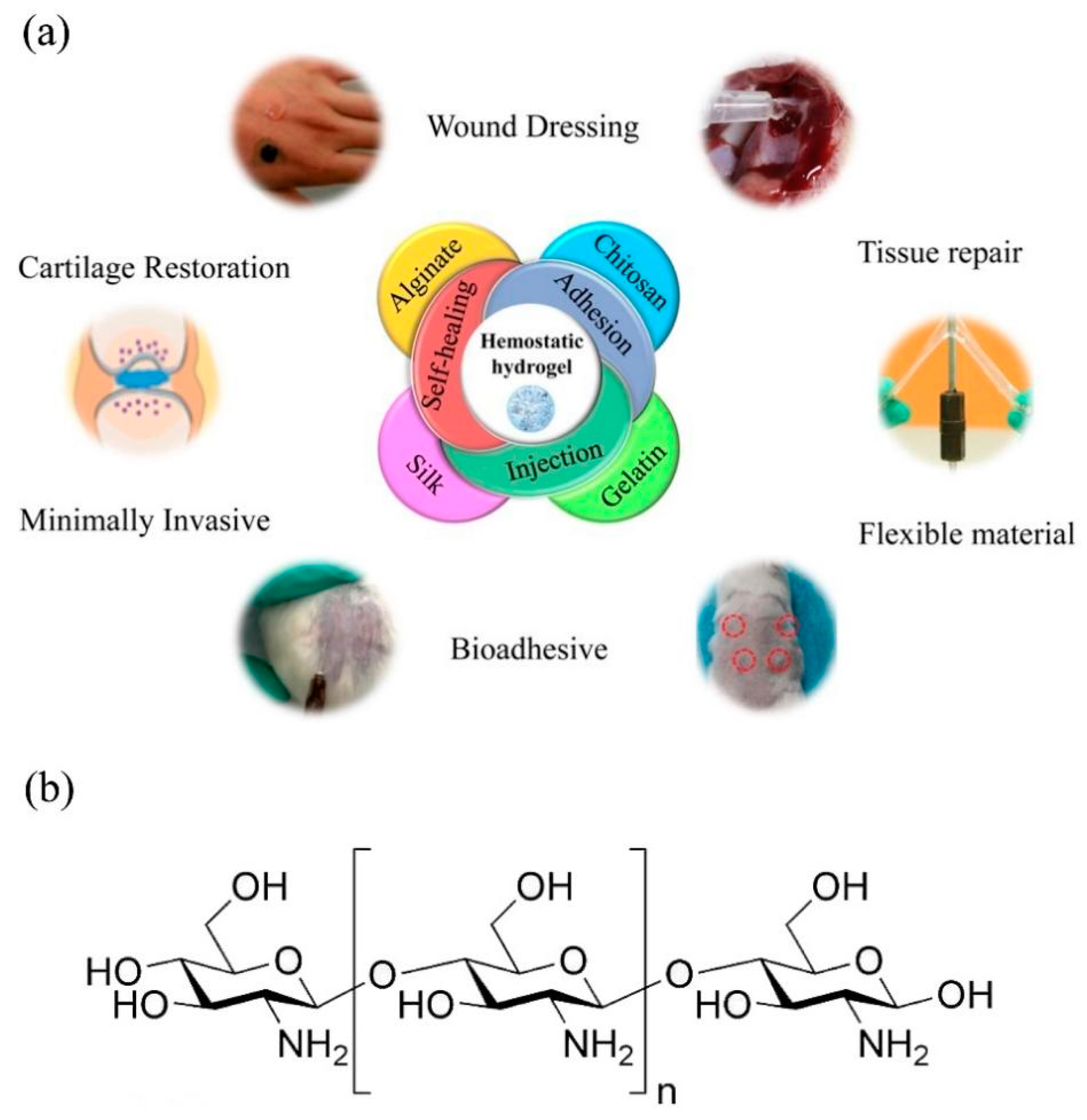
:1. Introduction
2. Hemostatic Mechanism of CS Hydrogels
2.1. The Principle of Hemostasis
2.2. The Hemostatic Mechanism of CS
2.3. Effect of CS Deacetylation Degree and Relative Molecular Weight on Hemostasis
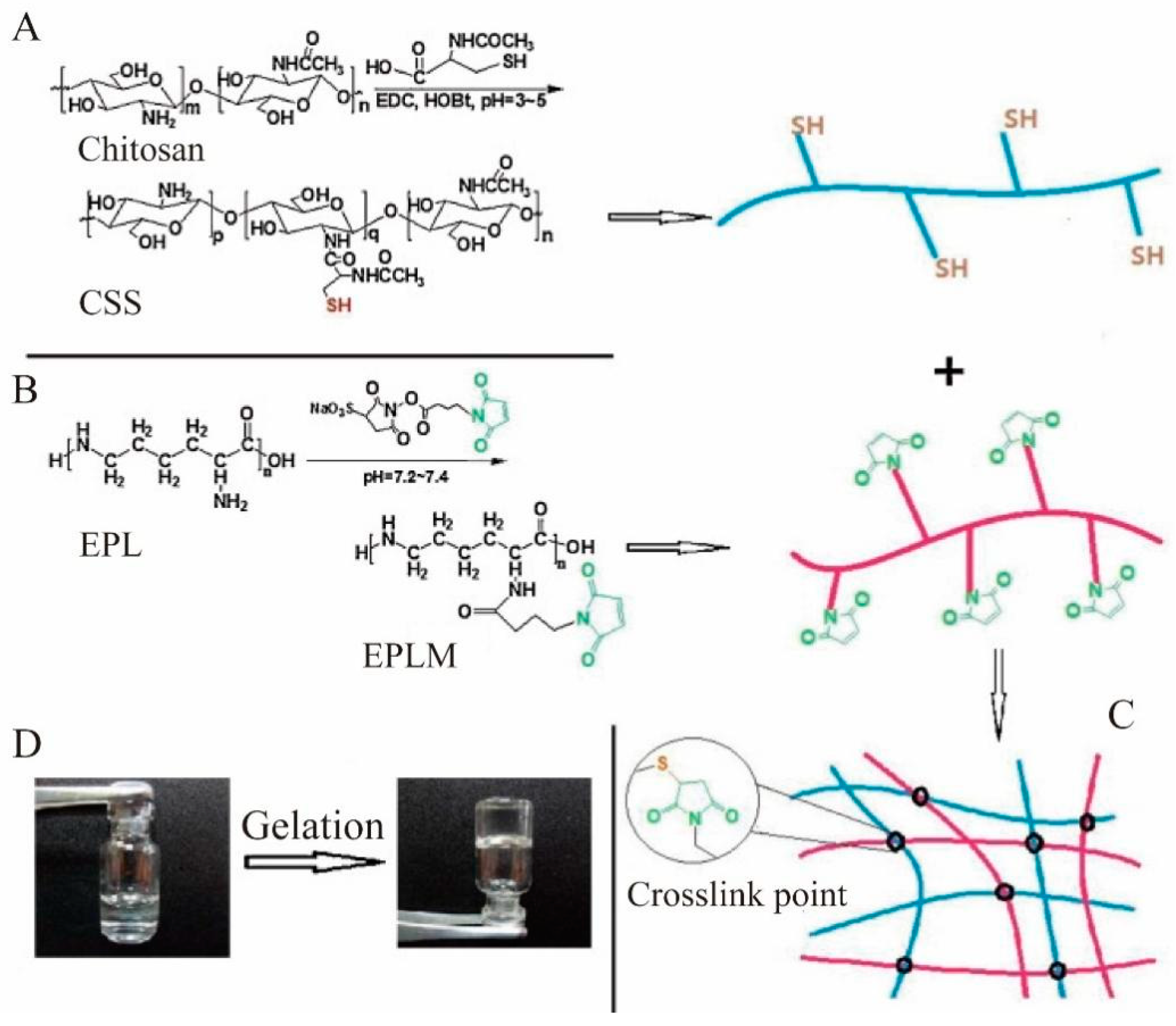
2.4. Effects of Chemical Modifications on Hemostasis
2.4.1. N-alkyl Modification
2.4.2. Carboxyl Modification
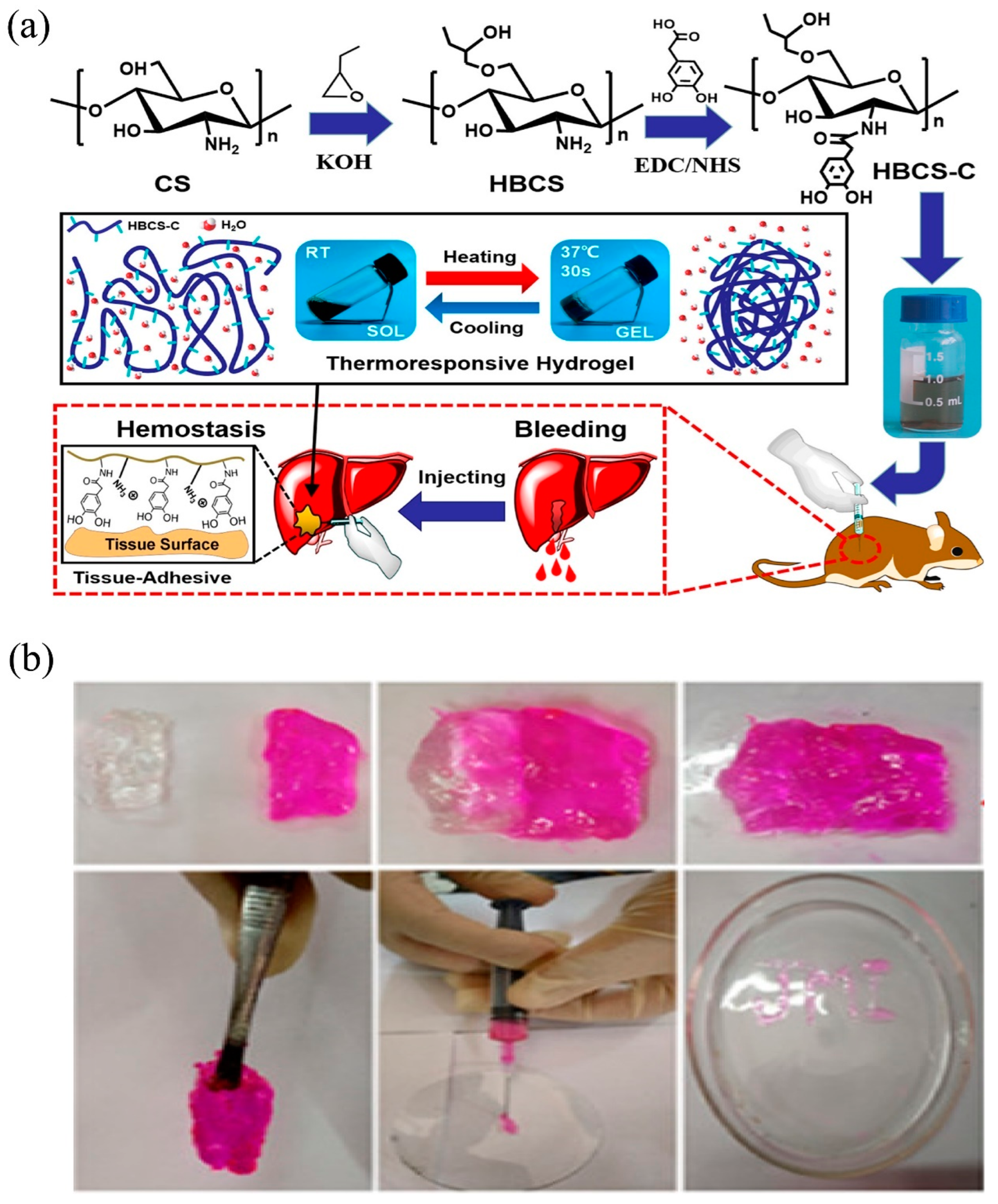
2.4.3. Hydroxybutyl Modification
2.4.4. Succinic Anhydride Modification
2.4.5. Other Modifications

| Materials | Advantages | References |
|---|---|---|
| Catechol-functionalized CS | Superior mechanical performance and stability, strong adhesiveness, excellent hemostatic property, and injectable and thermosensitive properties | [108] |
| N-alkyl-functionalized CS | Hydrophobic properties, excellent hemostasis property and high adhesion strength | [95,96,97] |
| Carboxyl-functionalized CS | Good mechanical properties, swelling properties, antibacterial, hemostatic and pro-healing properties | [98,99] |
| Hydroxybutyl-functionalized CS | Thermosensitive properties, high adhesion strength and hemostatic property | [100] |
| Succinic anhydride-functionalized CS | Non-toxicity, hemostatic properties, and good mechanical and swelling properties | [101,102,103] |
| Thiol-functionalized CS | Non-toxicity, excellent hemostatic property, and hemostatic and high adhesion strength | [81] |

3. Preparation of CS Hydrogel and Its Application as Hemostatic Materials
3.1. Preparation Methods of CS Hydrogel
3.1.1. Chemical Cross-Linking
The Schiff Base Reaction
Glutaraldehyde Reaction
Other Chemical Reactions
3.1.2. Physical Cross-Linking
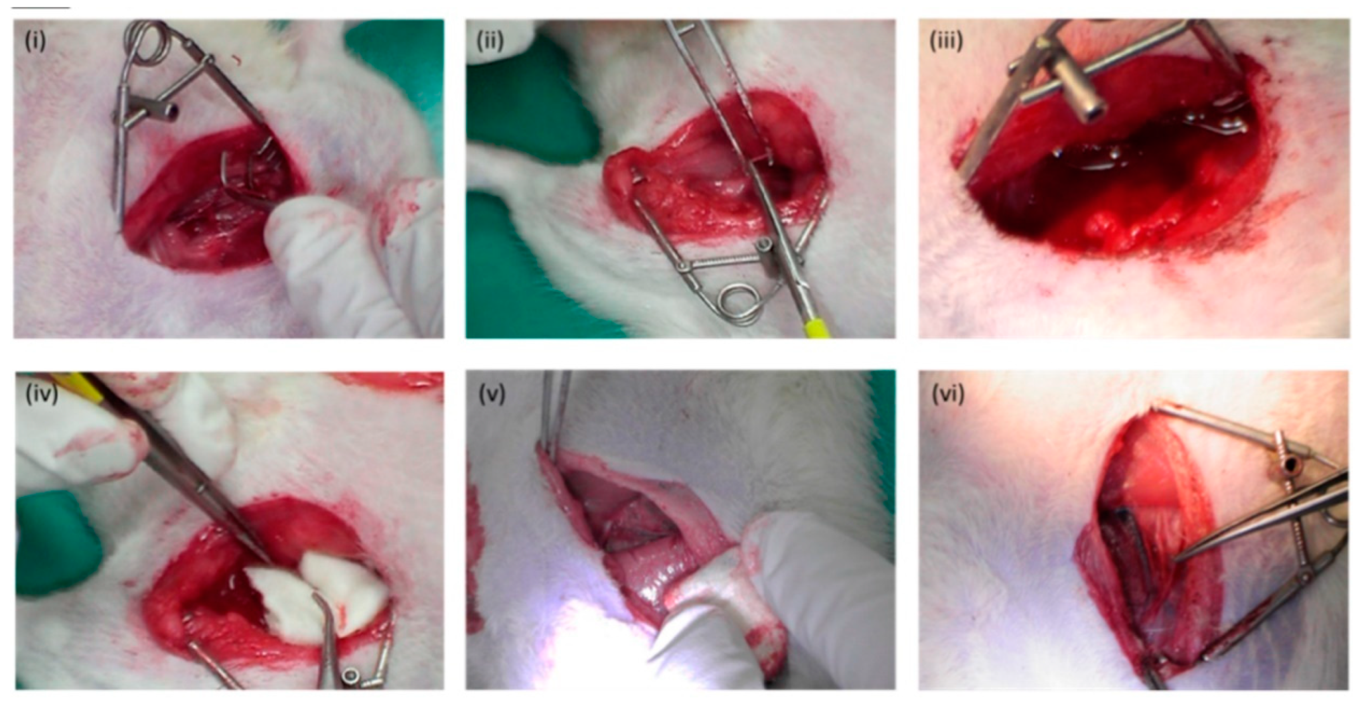
3.2. Application of CS Hydrogel as Hemostatic Materials
3.2.1. Prerequisites as Hemostatic Hydrogels
3.2.2. CS-Based Hemostatic Hydrogels
4. Conclusions and Future Perspectives
Author Contributions
Funding
Conflicts of Interest
References
- Watts, S.A.; Smith, J.E.; Woolley, T.; Rickard, R.F.; Gwyther, R.; Kirkman, E. Resuscitation with whole blood or blood components improves survival and lessens the pathophysiological burden of trauma and haemorrhagic shock in a pre-clinical porcine model. Eur. J. Trauma Emerg. Surg. 2022. [Google Scholar] [CrossRef] [PubMed]
- Corral, M.; Ferko, N.; Hollmann, S.; Broder, M.S.; Chang, E. Health and economic outcomes associated with uncontrolled surgical bleeding: A retrospective analysis of the premier perspectives database. Clin. Outcomes Res. 2015, 7, 409–421. [Google Scholar]
- Malik, A.; Rehman, F.U.; Shah, K.U.; Naz, S.S.; Qaisar, S. Hemostatic strategies for uncontrolled bleeding: A comprehensive update. J. Biomed. Mater. Res. Part B Appl. Biomater. 2021, 109, 1465–1477. [Google Scholar] [CrossRef] [PubMed]
- Lee, S.H.; Yun, S.J.; Ryu, S.; Choi, S.W.; Kim, H.J.; Kang, T.K.; Oh, S.C.; Cho, S.J. Massive bleeding from inferior mesenteric vein with hypovolemic shock: A rare complication of acute pancreatitis. J. Emerg. Med. 2018, 55, e5–e8. [Google Scholar] [CrossRef]
- Yu, Y.; Zheng, X.; Liu, X.; Zhao, J.; Wang, S. Injectable carboxymethyl chitosan-based hydrogel for simultaneous anti-tumor recurrence and anti-bacterial applications. Int. J. Biol. Macromol. 2023, 230, 123196. [Google Scholar] [CrossRef]
- Chen, Z.; Yao, J.; Zhao, J.; Wang, S. Injectable wound dressing based on carboxymethyl chitosan triple-network hydrogel for effective wound antibacterial and hemostasis. Int. J. Biol. Macromol. 2023, 225, 1235–1245. [Google Scholar] [CrossRef]
- Cai, J.; Guo, J.; Wang, S. Application of polymer hydrogels in the prevention of postoperative adhesion: A review. Gels 2023, 9, 98. [Google Scholar] [CrossRef]
- Pusateri, A.E.; Holcomb, J.B.; Kheirabadi, B.S.; Alam, H.B.; Wade, C.E.; Ryan, K.L. Making sense of the preclinical literature on advanced hemostatic products. J. Trauma. 2006, 60, 674–682. [Google Scholar] [CrossRef]
- Binnetoglu, K.; Kumandas, A.; Ekici, H.; Ozbaykus, A.C. Comparison of the algan hemostatic agent with floseal in rat liver laceration bleeding model. Eurasian J. Med. 2022, 54, 36–40. [Google Scholar] [CrossRef]
- An, S.; Jeon, E.J.; Jeon, J.; Cho, S.-W. A serotonin-modified hyaluronic acid hydrogel for multifunctional hemostatic adhesives inspired by a platelet coagulation mediator. Mater. Horiz. 2019, 6, 1169–1178. [Google Scholar] [CrossRef]
- Fonouni, H.; Kashfi, A.; Majlesara, A.; Stahlheber, O.; Konstantinidis, L.; Gharabaghi, N.; Kraus, T.W.; Mehrabi, A.; Oweira, H. Hemostatic efficiency of modern topical sealants: Comparative evaluation after liver resection and splenic laceration in a swine model. J. Biomed. Mater. Res. B Appl. Biomater. 2018, 106, 1307–1316. [Google Scholar] [CrossRef] [PubMed]
- Beudert, M.; Gutmann, M.; Luhmann, T.; Meinel, L. Fibrin sealants: Challenges and solutions. ACS Biomater. Sci. Eng. 2022, 8, 2220–2231. [Google Scholar] [CrossRef] [PubMed]
- Daud, A.; Kaur, B.; McClure, G.R.; Belley-Cote, E.P.; Harlock, J.; Crowther, M.; Whitlock, R.P. Fibrin and thrombin sealants in vascular and cardiac surgery: A systematic review and meta-analysis. Eur. J. Vasc Endovasc. Surg. 2020, 60, 469–478. [Google Scholar] [CrossRef] [PubMed]
- Valji, K. Cyanoacrylates for embolization in gastrointestinal bleeding: How super is glue? J. Vasc. Interv. Radiol. 2014, 25, 20–21. [Google Scholar] [CrossRef] [PubMed]
- Zhou, L.; Zhao, J.; Chen, Y.; Zheng, Y.; Li, J.; Zhao, J.; Zhang, J.; Liu, Y.; Liu, X.; Wang, S. MoS2-ALG-Fe/GOx hydrogel with Fenton catalytic activity for combined cancer photothermal, starvation, and chemodynamic therapy. Colloids Surf. B 2020, 195, 111243. [Google Scholar] [CrossRef] [PubMed]
- Wang, T.; Yi, W.; Zhang, Y.; Wu, H.; Fan, H.; Zhao, J.; Wang, S. Sodium alginate hydrogel containing platelet-rich plasma for wound healing. Colloids Surf. B Biointerfaces 2023, 222, 113096. [Google Scholar] [CrossRef]
- Xie, M.; Zeng, Y.; Wu, H.; Wang, S.; Zhao, J. Multifunctional carboxymethyl chitosan/oxidized dextran/sodium alginate hydrogels as dressing for hemostasis and closure of infected wounds. Int. J. Biol. Macromol. 2022, 219, 1337–1350. [Google Scholar] [CrossRef]
- Zhang, Y.; Zhu, C.; Zhang, Z.; Zhao, J.; Yuan, Y.; Wang, S. Oxidation triggered formation of polydopamine-modified carboxymethyl cellulose hydrogel for anti-recurrence of tumor. Colloids Surf. B 2021, 207, 112025. [Google Scholar] [CrossRef]
- Zhu, Y.; Zhang, Y.; Wu, H.; Wang, S.; Li, X. An oxidative polymerized carboxymethyl cellulose hydrogel for the combined anti-tumor recurrence. J. Mater. Sci. 2023, 58, 369–382. [Google Scholar] [CrossRef]
- Liu, X.; Zhang, Y.; Wu, H.; Tang, J.; Zhou, J.; Zhao, J.; Wang, S. A conductive gelatin methacrylamide hydrogel for synergistic therapy of osteosarcoma and potential bone regeneration. Int. J. Biol. Macromol. 2023, 228, 111–122. [Google Scholar] [CrossRef]
- Ouyang, Y.; Zhao, J.; Wang, S. Multifunctional hydrogels based on chitosan, hyaluronic acid and other biological macromolecules for the treatment of inflammatory bowel disease: A review. Int. J. Biol. Macromol. 2023, 227, 505–523. [Google Scholar] [CrossRef]
- Han, W.; Wang, S. Advances in hemostatic hydrogels that can adhere to wet surfaces. Gels 2023, 9, 2. [Google Scholar] [CrossRef]
- Jia, Z.; Zhu, F.; Li, X.; Liang, Q.; Zhuo, Z.; Huang, J.; Duan, L.; Xiong, J.; Wang, D. Repair of osteochondral defects using injectable chitosan-based hydrogel encapsulated synovial fluid-derived mesenchymal stem cells in a rabbit model. Mater. Sci. Eng. C Mater. Biol. Appl. 2019, 99, 541–551. [Google Scholar] [CrossRef]
- Jin, R.; Xu, J.; Duan, L.; Gao, G. Chitosan-driven skin-attachable hydrogel sensors toward human motion and physiological signal monitoring. Carbohydr. Polym. 2021, 268, 118240. [Google Scholar] [CrossRef]
- Ding, H.; Li, B.; Liu, Z.; Liu, G.; Pu, S.; Feng, Y.; Jia, D.; Zhou, Y. Nonswelling injectable chitosan hydrogel via UV crosslinking induced hydrophobic effect for minimally invasive tissue engineering. Carbohydr. Polym. 2021, 252, 117143. [Google Scholar] [CrossRef] [PubMed]
- Dong, X.; Li, C.; Zhang, M.; Zhao, Y.; Zhao, Z.; Li, W.; Zhang, X. Multifunctional injectable hydrogel for effective promotion of cartilage regeneration and protection against osteoarthritis: Combined chondroinductive, antioxidative and anti-in fl ammatory strategy. Sci. Technol. Adv. Mater. 2022, 23, 361–375. [Google Scholar] [CrossRef]
- Yan, X.; Fang, W.W.; Xue, J.; Sun, T.C.; Dong, L.; Zha, Z.; Qian, H.; Song, Y.H.; Zhang, M.; Gong, X.; et al. Thermoresponsive in situ forming hydrogel with sol-gel irreversibility for effective methicillin-resistant staphylococcus aureus infected wound healing. ACS Nano 2019, 13, 10074–10084. [Google Scholar] [CrossRef]
- Liang, Y.; Li, Z.; Huang, Y.; Yu, R.; Guo, B. Dual-dynamic-bond cross-linked antibacterial adhesive hydrogel sealants with on-demand removability for post-wound-closure and infected wound healing. ACS Nano 2021, 15, 7078–7093. [Google Scholar] [CrossRef]
- Jones, M.; Kujundzic, M.; John, S.; Bismarck, A. Crab vs. Mushroom: A review of crustacean and fungal chitin in wound treatment. Mar. Drugs 2020, 18, 18010064. [Google Scholar] [CrossRef]
- Negm, N.A.; Hefni, H.H.H.; Abd-Elaal, A.A.A.; Badr, E.A.; Abou Kana, M.T.H. Advancement on modification of chitosan biopolymer and its potential applications. Int. J. Biol. Macromol. 2020, 152, 681–702. [Google Scholar] [CrossRef]
- Bakshi, P.S.; Selvakumar, D.; Kadirvelu, K.; Kumar, N.S. Chitosan as an environment friendly biomaterial—A review on recent modifications and applications. Int. J. Biol. Macromol. 2020, 150, 1072–1083. [Google Scholar] [CrossRef] [PubMed]
- Cao, J.; Xiao, L.; Shi, X. Injectable drug-loaded polysaccharide hybrid hydrogels for hemostasis. RSC Adv. 2019, 9, 36858–36866. [Google Scholar] [CrossRef] [PubMed]
- Xia, Y.L.; Yang, R.H.; Wang, H.Y.; Li, Y.H.; Fu, C.F. Application of chitosan-based materials in surgical or postoperative hemostasis. Front. Mater. 2022, 9, 994265. [Google Scholar] [CrossRef]
- Hu, B.J.; Bao, G.C.; Xu, X.X.; Yang, K. Topical hemostatic materials for coagulopathy. J. Mater. Chem B 2022, 10, 1946–1959. [Google Scholar] [CrossRef]
- Simpson, A.; Shukla, A.; Brown, A.C. Biomaterials for hemostasis. Annu. Rev. Biomed. Eng. 2022, 24, 111–135. [Google Scholar] [CrossRef]
- Hu, Z.; Zhang, D.Y.; Lu, S.T.; Li, P.W.; Li, S.D. Chitosan-based composite materials for prospective hemostatic applications. Mar. Drugs 2018, 16, 273. [Google Scholar] [CrossRef]
- Lestari, W.; Yusry, W.; Haris, M.S.; Jaswir, I.; Idrus, E. A glimpse on the function of chitosan as a dental hemostatic agent. Jpn. Dent. Sci. Rev. 2020, 56, 147–154. [Google Scholar] [CrossRef]
- Ranjith, R.; Balraj, S.; Ganesh, J.; John Milton, M.C. Therapeutic agents loaded chitosan-based nanofibrous mats as potential wound dressings: A review. Mater. Today Chem. 2019, 12, 386–395. [Google Scholar] [CrossRef]
- Whang, H.S.; Kirsch, W.; Zhu, Y.H.; Yang, C.Z.; Hudson, S.M. Hemostatic agents derived from chitin and chitosan. J. Macromol. Sci. Polym. Rev. 2005, 45, 309–323. [Google Scholar] [CrossRef]
- Xie, Y.; Gao, P.; He, F.; Zhang, C. Application of alginate-based hydrogels in hemostasis. Gels 2022, 8, 109. [Google Scholar] [CrossRef]
- Wang, L.; Hao, F.; Tian, S.; Dong, H.; Nie, J.; Ma, G. Targeting polysaccharides such as chitosan, cellulose, alginate and starch for designing hemostatic dressings. Carbohydr. Polym. 2022, 291, 119574. [Google Scholar] [CrossRef] [PubMed]
- Chen, Y.; Wu, L.; Li, P.; Hao, X.; Yang, X.; Xi, G.; Liu, W.; Feng, Y.; He, H.; Shi, C. Polysaccharide based hemostatic strategy for ultrarapid hemostasis. Macromol. Biosci. 2020, 20, e1900370. [Google Scholar] [CrossRef]
- Wang, Y.; Liu, G.; Wu, L.; Qu, H.; Song, D.; Huang, H.; Wu, C.; Xu, M. Rational design of porous starch/hyaluronic acid composites for hemostasis. Int. J. Biol. Macromol. 2020, 1319–1329. [Google Scholar] [CrossRef] [PubMed]
- Weng, H.; Jia, W.; Li, M.; Chen, Z. New injectable chitosan-hyaluronic acid based hydrogels for hemostasis and wound healing. Carbohydr. Polym. 2022, 294, 119767. [Google Scholar] [CrossRef]
- Ndlovu, S.P.; Ngece, K.; Alven, S.; Aderibigbe, B.A. Gelatin-based hybrid scaffolds: Promising wound dressings. Polymers 2021, 13, 780187. [Google Scholar] [CrossRef]
- Lied, G.A.; Lund, K.B.; Storaas, T. Intraoperative anaphylaxis to gelatin-based hemostatic agents: A case report. J. Asthma Allergy 2019, 12, 163–167. [Google Scholar] [CrossRef]
- Kang, K.; Liu, Y.; Song, X.; Xu, L.; Zhang, W.; Jiao, Y.; Zhao, Y. Hemostatic performance of a-chitin/gelatin composite sponges with directional pore structure. Macromol. Biosci. 2022, 22, e2200020. [Google Scholar] [CrossRef]
- Biswas, S.; Bhunia, B.K.; Janani, G.; Mandal, B.B. Silk fibroin based formulations as potential hemostatic agents. ACS Biomater. Sci. Eng. 2022, 8, 2654–2663. [Google Scholar] [CrossRef]
- Shefa, A.A.; Taz, M.; Lee, S.Y.; Lee, B.T. Enhancement of hemostatic property of plant derived oxidized nanocellulose-silk fibroin based scaffolds by thrombin loading. Carbohydr. Polym. 2019, 208, 168–179. [Google Scholar] [CrossRef]
- Okata, S.; Hoshina, K.; Hanada, K.; Kamata, H.; Fujisawa, A.; Yoshikawa, Y.; Sakai, T. Hemostatic capability of a novel tetra-polyethylene glycol hydrogel. Ann. Vasc. Surg. 2022, 84, 398–404. [Google Scholar] [CrossRef]
- Boerman, M.A.; Roozen, E.; Sanchez-Fernandez, M.J.; Keereweer, A.R.; Felix Lanao, R.P.; Bender, J.; Hoogenboom, R.; Leeuwenburgh, S.C.; Jansen, J.A.; Van Goor, H.; et al. Next generation hemostatic materials based on NHS-ester functionalized poly(2-oxazoline)s. Biomacromolecules 2017, 18, 2529–2538. [Google Scholar] [CrossRef] [PubMed] [Green Version]
- Dhandapani, V.; Ringuette, V.; Desrochers, M.; Sirois, M.; Vermette, P. Composition, host responses and clinical applications of bioadhesives. J. Biomed. Mater. Res. B Appl. Biomater. 2022, 110, 2779–2797. [Google Scholar] [CrossRef] [PubMed]
- Kerris, E.W.J.; Hoptay, C.; Calderon, T.; Freishtat, R.J. Platelets and platelet extracellular vesicles in hemostasis and sepsis. J. Investig. Med. 2020, 68, 813–820. [Google Scholar] [CrossRef] [PubMed]
- Caspers, M.; Maegele, M.; Frohlich, M. Current strategies for hemostatic control in acute trauma hemorrhage and trauma-induced coagulopathy. Expert Rev. Hematol. 2018, 11, 987–995. [Google Scholar] [CrossRef] [PubMed]
- Leonardi, M.J. Laboratory evaluation of hemostasis disorders. Physician Assist. Clin. 2019, 4, 609–623. [Google Scholar] [CrossRef]
- Sundaram, M.N.; Mony, U.; Varma, P.K.; Rangasamy, J. Vasoconstrictor and coagulation activator entrapped chitosan based composite hydrogel for rapid bleeding control. Carbohydr. Polym. 2021, 258, 117634. [Google Scholar] [CrossRef]
- Lippi, G.; Favaloro, E.J. Laboratory hemostasis: From biology to the bench. Clin. Chem. Lab. Med. 2018, 56, 1035–1045. [Google Scholar] [CrossRef]
- Toonstra, C.; Hu, Y.; Zhang, H. Deciphering the roles of N-glycans on collagen-platelet interactions. J. Proteome Res. 2019, 18, 2467–2477. [Google Scholar] [CrossRef]
- Zhang, B.; Hu, X.; Wang, H.; Wang, R.; Sun, Z.; Tan, X.; Liu, S.; Wang, H. Effects of a dammarane-type saponin, ginsenoside Rd, in nicotine-induced vascular endothelial injury. Phytomedicine 2020, 79, 153325. [Google Scholar] [CrossRef]
- Yang, X.; Liu, W.; Li, N.; Wang, M.; Liang, B.; Ullah, I.; Neve, A.L.; Feng, Y.; Chen, H.; Shi, C. Design and development of polysaccharide hemostatic materials and their hemostatic mechanism. Biomater. Sci. 2017, 5, 2357–2368. [Google Scholar] [CrossRef]
- Wheeler, A.P.; Gailani, D. The intrinsic pathway of coagulation as a target for antithrombotic therapy. Hematol. Oncol. Clin. N. Am. 2016, 30, 1099–1114. [Google Scholar] [CrossRef] [Green Version]
- Zhong, Y.; Hu, H.; Min, N.; Wei, Y.; Li, X.; Li, X. Application and outlook of topical hemostatic materials: A narrative review. Ann. Transl. Med. 2021, 9, 577. [Google Scholar] [CrossRef]
- Versteeg, H.H.; Heemskerk, J.W.; Levi, M.; Reitsma, P.H. New fundamentals in hemostasis. Physiol. Rev. 2013, 93, 327–358. [Google Scholar] [CrossRef]
- Ebhodaghe, S.O. A short review on chitosan and gelatin-based hydrogel composite polymers for wound healing. J. Biomater. Sci. Polym. Ed. 2022, 33, 1595–1622. [Google Scholar] [CrossRef]
- Hao, R.; Peng, X.; Zhang, Y.; Chen, J.; Wang, T.; Wang, W.; Zhao, Y.; Fan, X.; Chen, C.; Xu, H. Rapid hemostasis resulting from the synergism of self-assembling short peptide and o-carboxymethyl chitosan. ACS Appl. Mater. Interfaces 2020, 12, 55574–55583. [Google Scholar] [CrossRef]
- Hao, Y.; Zhao, W.; Zhang, L.; Zeng, X.; Sun, Z.; Zhang, D.; Shen, P.; Li, Z.; Han, Y.; Li, P.; et al. Bio-multifunctional alginate/chitosan/fucoidan sponges with enhanced angiogenesis and hair follicle regeneration for promoting full-thickness wound healing. Mater. Des. 2020, 193, 108863. [Google Scholar] [CrossRef]
- Wang, Y.W.; Liu, C.C.; Cherng, J.H.; Lin, C.S.; Chang, S.J.; Hong, Z.J.; Liu, C.C.; Chiu, Y.K.; Hsu, S.D.; Chang, A.H. Biological effects of chitosan-based dressing on hemostasis mechanism. Polymers 2019, 11, 1906. [Google Scholar] [CrossRef]
- Mukhtar, M.; Csaba, N.; Robla, S.; Varela-Calvino, R.; Nagy, A.; Burian, K.; Kokai, D.; Ambrus, R. Dry powder comprised of isoniazid-loaded nanoparticles of hyaluronic acid in conjugation with mannose-anchored chitosan for macrophage-targeted pulmonary administration in tuberculosis. Pharmaceutics 2022, 14, 1543. [Google Scholar] [CrossRef]
- Liu, Y.; Niu, H.; Wang, C.; Yang, X.; Li, W.; Zhang, Y.; Ma, X.; Xu, Y.; Zheng, P.; Wang, J.; et al. Bio-inspired, bio-degradable adenosine 5’-diphosphate-modified hyaluronic acid coordinated hydrophobic undecanal-modified chitosan for hemostasis and wound healing. Bioact. Mater. 2022, 17, 162–177. [Google Scholar] [CrossRef]
- Huang, Y.; Zhang, Y.; Feng, L.; He, L.; Guo, R.; Xue, W. Synthesis of N-alkylated chitosan and its interactions with blood. Artif. Cells Nanomed. Biotechnol. 2018, 46, 544–550. [Google Scholar] [CrossRef]
- Chen, K.Y.; Lin, T.H.; Yang, C.Y.; Kuo, Y.W.; Lei, U. Mechanics for the adhesion and aggregation of red blood cells on chitosan. J. Mech. 2018, 34, 725–732. [Google Scholar] [CrossRef]
- Chou, T.-C.; Fu, E.; Wu, C.-J.; Yeh, J.-H. Chitosan enhances platelet adhesion and aggregation. Biochem. Biophys. Res. Commun. 2003, 302, 480–483. [Google Scholar] [CrossRef] [PubMed]
- Shen, E.; Chou, T.; Gau, C. Releasing growth factors from activated human platelets after chitosan stimulation: A possible bio-material for platelet-rich plasma preparation. Clin. Oral Implant. Res. 2006, 17, 572–578. [Google Scholar] [CrossRef] [PubMed]
- Patil, G.; Pawar, R.; Jadhav, S.; Ghormade, V. A chitosan based multimodal “soft” hydrogel for rapid hemostasis of non-compressible hemorrhages and its mode of action. Carbohydr. Polym. Technol. Appl. 2022, 4, 100237. [Google Scholar] [CrossRef]
- Chen, L.; Tianqing, L. Interaction behaviors between chitosan and hemoglobin. Int. J. Biol. Macromol. 2008, 42, 441–446. [Google Scholar] [CrossRef]
- Yin, M.; Wang, Y.; Zhang, Y.; Ren, X.; Qiu, Y.; Huang, T.S. Novel quaternarized N-halamine chitosan and polyvinyl alcohol nanofibrous membranes as hemostatic materials with excellent antibacterial properties. Carbohydr. Polym. 2020, 232, 115823. [Google Scholar] [CrossRef]
- Zhou, M.; Liao, J.; Li, G.; Yu, Z.; Xie, D.; Zhou, H.; Wang, F.; Ren, Y.; Xu, R.; Dai, Y.; et al. Expandable carboxymethyl chitosan/cellulose nanofiber composite sponge for traumatic hemostasis. Carbohydr. Polym. 2022, 294, 119805. [Google Scholar] [CrossRef]
- Thao, N.T.T.; Wijerathna, H.; Kumar, R.S.; Choi, D.; Dananjaya, S.H.S.; Attanayake, A.P. Preparation and characterization of succinyl chitosan and succinyl chitosan nanoparticle film: In vitro and in vivo evaluation of wound healing activity. Int. J. Biol. Macromol. 2021, 193, 1823–1834. [Google Scholar] [CrossRef]
- Sperling, C.; Maitz, M.F.; Grasso, S.; Werner, C.; Kanse, S.M. A positively charged surface triggers coagulation activation through factor vii activating protease (FSAP). ACS Appl. Mater. Interfaces 2017, 9, 40107–40116. [Google Scholar] [CrossRef]
- Kedzierska, M.; Blilid, S.; Milowska, K.; Kolodziejczyk-Czepas, J.; Katir, N.; Lahcini, M.; El Kadib, A.; Bryszewska, M. Insight into factors influencing wound healing using phosphorylated cellulose-filled-chitosan nanocomposite films. Int. J. Mol. Sci. 2021, 22, 11386. [Google Scholar] [CrossRef]
- Nie, W.; Yuan, X.; Zhao, J.; Zhou, Y.; Bao, H. Rapidly in situ forming chitosan/epsilon-polylysine hydrogels for adhesive sealants and hemostatic materials. Carbohydr. Polym. 2013, 96, 342–348. [Google Scholar] [CrossRef]
- Drozd, N.N.; Lunkov, A.P.; Il’ina, A.V.; Varlamov, V.P. Hemocompatibility of silver nanoparticles based on conjugate of quaternized chitosan with gallic acid in in vitro experiments. Bull. Exp. Biol. Med. 2020, 168, 507–511. [Google Scholar] [CrossRef]
- von Petersdorff-Campen, K.; Schmid Daners, M. Hemolysis testing in vitro: A review of challenges and potential improvements. ASAIO J. 2022, 68, 3–13. [Google Scholar] [CrossRef]
- Liu, X.L.; Yuan, L.; Li, D.; Tang, Z.C.; Wang, Y.W.; Chen, G.J.; Chen, H.; Brash, J.L. Blood compatible materials: State of the art. J. Mater. Chem. B 2014, 2, 5718–5738. [Google Scholar] [CrossRef]
- Sen, V.D.; Sokolova, E.M.; Neshev, N.I.; Kulikov, A.V.; Pliss, E.M. Low molecular chitosan–(poly)nitroxides: Synthesis and evaluation as antioxidants on free radical-induced erythrocyte hemolysis. React. Funct. Polym. 2017, 111, 53–59. [Google Scholar] [CrossRef]
- Chen, Y.; Zhang, Y.; Wang, F.; Meng, W.; Yang, X.; Li, P.; Jiang, J.; Tan, H.; Zheng, Y. Preparation of porous carboxymethyl chitosan grafted poly (acrylic acid) superabsorbent by solvent precipitation and its application as a hemostatic wound dressing. Mater. Sci. Eng. C Mater. Biol. Appl. 2016, 63, 18–29. [Google Scholar] [CrossRef]
- Hattori, H.; Ishihara, M. Changes in blood aggregation with differences in molecular weight and degree of deacetylation of chitosan. Biomed. Mater. 2015, 10, 015014. [Google Scholar] [CrossRef]
- Hu, Z.; Lu, S.; Cheng, Y.; Kong, S.; Li, S.; Li, C.; Yang, L. Investigation of the effects of molecular parameters on the hemostatic properties of chitosan. Molecules 2018, 23, 3147. [Google Scholar] [CrossRef]
- Wu, J.; Zhang, L. Dissolution behavior and conformation change of chitosan in concentrated chitosan hydrochloric acid solution and comparison with dilute and semidilute solutions. Int. J. Biol. Macromol. 2019, 121, 1101–1108. [Google Scholar] [CrossRef]
- Song, X.; Zhao, Y.; Liu, Y.; Zhang, W.; Yuan, X.; Xu, L.; Zhang, J. Effects of degree of deacetylation on hemostatic performance of partially deacetylated chitin sponges. Carbohydr. Polym. 2021, 273, 118615. [Google Scholar] [CrossRef]
- Markushin, S.G.; Akopova, I.I.; Blagodatskikh, I.V.; Kulikov, S.N.; Bezrodnykh, E.A.; Muranov, A.V.; Yamskov, I.A.; Tikhonov, V.E. Effect of molecular weight and degree of acetylation on adjuvantive properties of chitosan derivatives. Appl. Biochem. Microbiol. 2018, 54, 512–517. [Google Scholar] [CrossRef]
- Naveed, M.; Phil, L.; Sohail, M.; Hasnat, M.; Baig, M.; Ihsan, A.U.; Shumzaid, M.; Kakar, M.U.; Mehmood Khan, T.; Akabar, M.D.; et al. Chitosan oligosaccharide (COS): An overview. Int. J. Biol. Macromol. 2019, 129, 827–843. [Google Scholar] [CrossRef] [PubMed]
- Varlamov, V.P.; Il’ina, A.V.; Shagdarova, B.T.; Lunkov, A.P.; Mysyakina, I.S. Chitin/chitosan and its derivatives: Fundamental problems and practical approaches. Biochemistry 2020, 85, S154–S176. [Google Scholar] [CrossRef] [PubMed]
- Verma, C.; Quraishi, M.A.; Alfantazi, A.; Rhee, K.Y. Corrosion inhibition potential of chitosan based Schiff bases: Design, performance and applications. Int. J. Biol. Macromol. 2021, 184, 135–143. [Google Scholar] [CrossRef] [PubMed]
- Wang, X.; Guan, J.; Zhuang, X.; Li, Z.; Huang, S.; Yang, J.; Liu, C.; Li, F.; Tian, F.; Wu, J.; et al. Exploration of blood coagulation of N-alkyl chitosan nanofiber membrane in vitro. Biomacromolecules 2018, 19, 731–739. [Google Scholar] [CrossRef]
- Logun, M.T.; Dowling, M.B.; Raghavan, S.R.; Wallace, M.L.; Schmiedt, C.; Stice, S.; Karumbaiah, L. Expanding hydrophobically modified chitosan foam for internal surgical hemostasis: Safety evaluation in a murine model. J. Surg. Res. 2019, 239, 269–277. [Google Scholar] [CrossRef]
- Chaturvedi, A.; Dowling, M.B.; Gustin, J.P.; Scalea, T.M.; Raghavan, S.R.; Pasley, J.D.; Narayan, M. Hydrophobically modified chitosan gauze: A novel topical hemostat. J. Surg. Res. 2017, 207, 45–52. [Google Scholar] [CrossRef]
- Liu, L.; Lv, Q.; Zhang, Q.; Zhu, H.; Liu, W.; Deng, G.; Wu, Y.; Shi, C.; Li, H.; Li, L. Preparation of carboxymethyl chitosan microspheres and their application in hemostasis. Disaster Med. Public Health Prep. 2017, 11, 660–667. [Google Scholar] [CrossRef]
- Wang, Y.; Cao, H.; Wang, X. Synthesis and characterization of an injectable ε-polylysine/carboxymethyl chitosan hydrogel used in medical application. Mater. Chem. Phys. 2020, 248, 122902. [Google Scholar] [CrossRef]
- Shou, Y.; Zhang, J.; Yan, S.; Xia, P.; Xu, P.; Li, G.; Zhang, K.; Yin, J. Thermoresponsive chitosan/dopa-based hydrogel as an injectable therapy approach for tissue-adhesion and hemostasis. ACS Biomater. Sci. Eng. 2020, 6, 3619–3629. [Google Scholar] [CrossRef]
- Pandit, A.H.; Mazumdar, N.; Imtiyaz, K.; Alam Rizvi, M.M.; Ahmad, S. Self-healing and injectable hydrogels for anticancer drug delivery: A study with multialdehyde gum arabic and succinic anhydride chitosan. ACS Appl. Bio Mater. 2020, 3, 8460–8470. [Google Scholar] [CrossRef]
- Zhang, P.; Li, S.; Zhang, S.; Zhang, X.; Wan, L.; Yun, Z.; Ji, S.; Gong, F.; Huang, M.; Wang, L.; et al. GRGDS-functionalized chitosan nanoparticles as a potential intravenous hemostat for traumatic hemorrhage control in an animal model. Nanomedicine 2018, 14, 2531–2540. [Google Scholar] [CrossRef]
- Xu, Z.; Chen, T.; Zhang, K.Q.; Meng, K.; Zhao, H. Silk fibroin/chitosan hydrogel with antibacterial, hemostatic and sustained drug-release activities. Polym. Int. 2021, 70, 1741–1751. [Google Scholar] [CrossRef]
- Ju, J.; Jin, S.B.; Kim, S.; Choi, J.H.; Lee, H.A.; Son, D.; Lee, H.; Shin, M. Addressing the shortcomings of polyphenol-derived adhesives: Achievement of long shelf life for effective hemostasis. ACS Appl. Mater. Interfaces 2022, 14, 25115–25125. [Google Scholar] [CrossRef]
- Tang, L.L.; Dang, Y.; Wang, Y.; Zhang, Y.L.; Hu, T.S.; Ding, C.C.; Wu, H.; Ni, Y.H.; Chen, L.H.; Huang, L.L.; et al. Rapid fabrication of bionic pyrogallol-based self-adhesive hydrogel with mechanically tunable, self-healing, antibacterial, wound healing, and hemostatic properties. Biomater. Adv. 2022, 136, 212765. [Google Scholar] [CrossRef]
- Sanandiya, N.D.; Lee, S.; Rho, S.; Lee, H.; Kim, I.S.; Hwang, D.S. Tunichrome-inspired pyrogallol functionalized chitosan for tissue adhesion and hemostasis. Carbohydr Polym. 2019, 208, 77–85. [Google Scholar] [CrossRef]
- Shen, J.L.; Nada, A.A.; Abou-Zeid, N.Y.; Hudson, S.M. Synthesis of chitosan iodoacetamides via carbodiimide coupling reaction: Effect of degree of substitution on the hemostatic properties. Carbohydr. Polym. 2020, 229, 115522. [Google Scholar] [CrossRef]
- Ryu, J.H.; Lee, Y.; Kong, W.H.; Kim, T.G.; Park, T.G.; Lee, H. Catechol-functionalized chitosan/pluronic hydrogels for tissue adhesives and hemostatic materials. Biomacromolecules 2011, 12, 2653–2659. [Google Scholar] [CrossRef]
- Heimbuck, A.M.; Priddy-Arrington, T.R.; Padgett, M.L.; Llamas, C.B.; Barnett, H.H.; Bunnell, B.A.; Caldorera-Moore, M.E. Development of responsive chitosan-genipin hydrogels for the treatment of wounds. ACS Appl. Bio Mater. 2019, 2, 2879–2888. [Google Scholar] [CrossRef]
- Chandra Hembram, K.; Prabha, S.; Chandra, R.; Ahmed, B.; Nimesh, S. Advances in preparation and characterization of chitosan nanoparticles for therapeutics. Artif. Cells Nanomed. Biotechnol. 2016, 44, 305–314. [Google Scholar] [CrossRef]
- Parhi, R. Cross-linked hydrogel for pharmaceutical applications: A review. Adv. Pharm. Bull. 2017, 7, 515–530. [Google Scholar] [CrossRef] [PubMed]
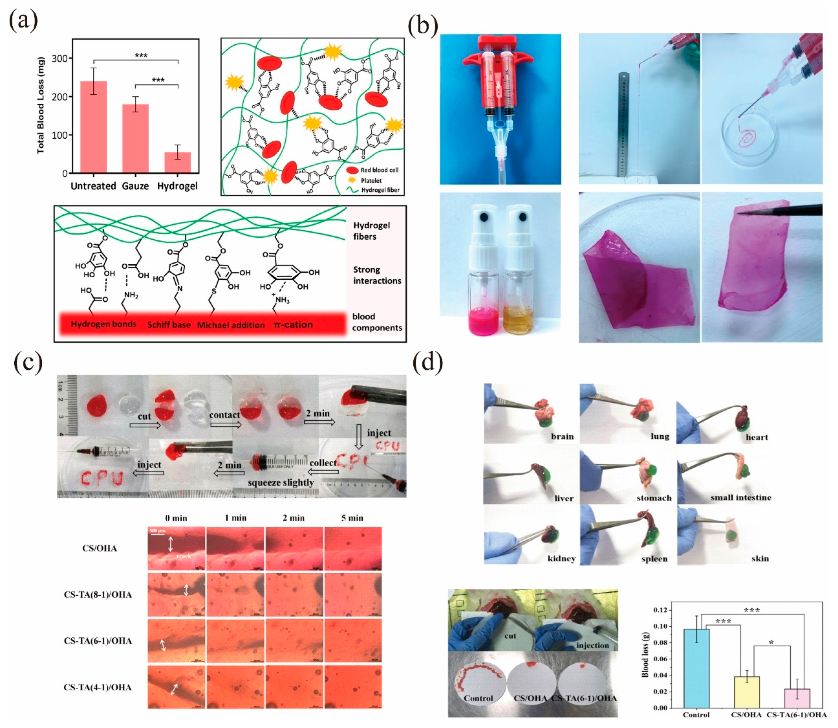
- Liu, S.; Jiang, N.; Chi, Y.; Peng, Q.; Dai, G.; Qian, L.; Xu, K.; Zhong, W.; Yue, W. Injectable and self-healing hydrogel based on chitosan-tannic acid and oxidized hyaluronic acid for wound healing. ACS Biomater. Sci. Eng. 2022, 8, 3754–3764. [Google Scholar] [CrossRef]
- Chen, G.; Yu, Y.; Wu, X.; Wang, G.; Ren, J.; Zhao, Y. Bioinspired multifunctional hybrid hydrogel promotes wound healing. Adv. Funct. Mater. 2018, 28, 1801386. [Google Scholar] [CrossRef]
- Song, F.; Kong, Y.; Shao, C.; Cheng, Y.; Lu, J.; Tao, Y.; Du, J.; Wang, H. Chitosan-based multifunctional flexible hemostatic bio-hydrogel. Acta Biomater. 2021, 136, 170–183. [Google Scholar] [CrossRef] [PubMed]
- Liu, J.; Li, J.; Yu, F.; Zhao, Y.X.; Mo, X.M.; Pan, J.F. In situ forming hydrogel of natural polysaccharides through Schiff base reaction for soft tissue adhesive and hemostasis. Int. J. Biol. Macromol. 2020, 147, 653–666. [Google Scholar] [CrossRef]
- Masood, N.; Ahmed, R.; Tariq, M.; Ahmed, Z.; Masoud, M.S.; Ali, I.; Asghar, R.; Andleeb, A.; Hasan, A. Silver nanoparticle impregnated chitosan-PEG hydrogel enhances wound healing in diabetes induced rabbits. Int. J. Pharm. 2019, 559, 23–36. [Google Scholar] [CrossRef]
- Risbud, M.V.; Bhat, S.V. Properties of polyvinyl pyrrolidone/beta-chitosan hydrogel membranes and their biocompatibility evaluation by haemorheological method. J. Mater. Sci. Mater. Med. 2001, 12, 75–79. [Google Scholar] [CrossRef]
- Sanchez-Cid, P.; Jimenez-Rosado, M.; Romero, A.; Perez-Puyana, V. Novel trends in hydrogel development for biomedical applications: A review. Polymers 2022, 14, 3023. [Google Scholar] [CrossRef]
- Li, C.; Obireddy, S.R.; Lai, W.F. Preparation and use of nanogels as carriers of drugs. Drug Deliv. 2021, 28, 1594–1602. [Google Scholar] [CrossRef]
- Zhang, Y.; Zhang, M.; Jiang, H.; Shi, J.; Li, F.; Xia, Y.; Zhang, G.; Li, H. Bio-inspired layered chitosan/graphene oxide nanocomposite hydrogels with high strength and pH-driven shape memory effect. Carbohydr. Polym. 2017, 177, 116–125. [Google Scholar] [CrossRef]
- Geng, L.; Hu, S.; Cui, M.; Wu, J.; Huang, A.; Shi, S.; Peng, X. Muscle-inspired double-network hydrogels with robust mechanical property, biocompatibility and ionic conductivity. Carbohydr. Polym. 2021, 262, 117936. [Google Scholar] [CrossRef] [PubMed]
- Fan, L.H.; Yang, H.; Yang, J.; Peng, M.; Hu, J. Preparation and characterization of chitosan/gelatin/PVA hydrogel for wound dressings. Carbohydr. Polym. 2016, 146, 427–434. [Google Scholar] [CrossRef]
- Do, N.H.N.; Truong, Q.T.; Le, P.K.; Ha, A.C. Recent developments in chitosan hydrogels carrying natural bioactive compounds. Carbohydr. Polym. 2022, 294, 119726. [Google Scholar] [CrossRef]
- Pourshahrestani, S.; Zeimaran, E.; Kadri, N.A.; Mutlu, N.; Boccaccini, A.R. Polymeric hydrogel systems as emerging biomaterial platforms to enable hemostasis and wound healing. Adv. Healthc. Mater. 2020, 9, e2000905. [Google Scholar] [CrossRef]
- Tu, Y.; Chen, N.; Li, C.; Liu, H.; Zhu, R.; Chen, S.; Xiao, Q.; Liu, J.; Ramakrishna, S.; He, L. Advances in injectable self-healing biomedical hydrogels. Acta Biomater. 2019, 90, 1–20. [Google Scholar] [CrossRef] [PubMed]
- Geng, H.; Dai, Q.; Sun, H.; Zhuang, L.; Song, A.; Caruso, F.; Hao, J.; Cui, J. Injectable and sprayable polyphenol-based hydrogels for controlling hemostasis. ACS Appl. Bio Mater. 2020, 3, 1258–1266. [Google Scholar] [CrossRef]
- Ou, Y.; Tian, M. Advances in multifunctional chitosan-based self-healing hydrogels for biomedical applications. J. Mater. Chem B 2021, 9, 7955–7971. [Google Scholar] [CrossRef] [PubMed]
- Tang, X.; Wang, X.; Sun, Y.; Zhao, L.; Li, D.; Zhang, J.; Sun, H.; Yang, B. Magnesium oxide-assisted dual-cross-linking bio-multifunctional hydrogels for wound repair during full-thickness skin injuries. Adv. Funct. Mater. 2021, 31, 2105718. [Google Scholar] [CrossRef]
- Feng, W.; Wang, Z. Shear-thinning and self-healing chitosan-graphene oxide hydrogel for hemostasis and wound healing. Carbohydr. Polym. 2022, 294, 119824. [Google Scholar] [CrossRef]
- Li, D.; Chen, J.; Wang, X.; Zhang, M.; Li, C.; Zhou, J. Recent advances on synthetic and polysaccharide adhesives for biological hemostatic applications. Front. Bioeng. Biotechnol. 2020, 8, 926. [Google Scholar] [CrossRef]
- Hamedi, H.; Moradi, S.; Hudson, S.M.; Tonelli, A.E.; King, M.W. Chitosan based bioadhesives for biomedical applications: A review. Carbohydr. Polym. 2022, 282, 119100. [Google Scholar] [CrossRef]
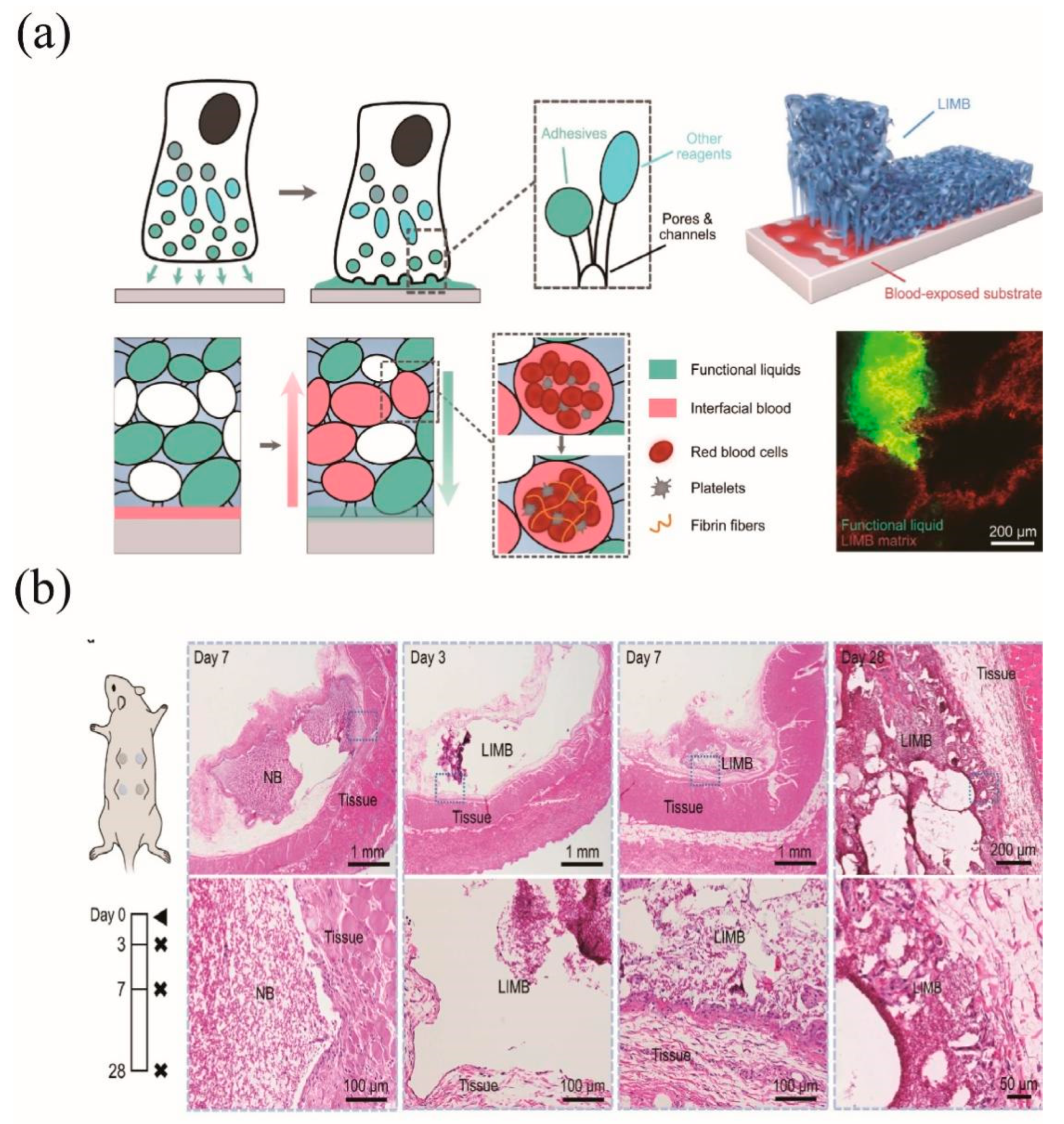
- Bao, G.; Gao, Q.; Cau, M.; Ali-Mohamad, N.; Strong, M.; Jiang, S.; Yang, Z.; Valiei, A.; Ma, Z.; Amabili, M.; et al. Liquid-infused microstructured bioadhesives halt non-compressible hemorrhage. Nat. Commun. 2022, 13, 5035. [Google Scholar] [CrossRef]
- Jung, H.Y.; Le Thi, P.; HwangBo, K.H.; Bae, J.W.; Park, K.D. Tunable and high tissue adhesive properties of injectable chitosan based hydrogels through polymer architecture modulation. Carbohydr. Polym. 2021, 261, 117810. [Google Scholar] [CrossRef]
- Zhang, Z.; Zhao, J.; Chen, Z.; Wu, H.; Wang, S. A molybdenum-based nanoplatform with multienzymes mimic capacity for oxidative stress-induced acute liver injury treatment. Inorg. Chem. Front. 2023. [Google Scholar] [CrossRef]
- Yang, X.; Wang, S.; Zhang, X.; Ye, C.; Wang, S.; An, X. Development of PVA-based microsphere as a potential embolization agent. Mater. Sci. Eng. C 2022, 135, 112677. [Google Scholar] [CrossRef]


| Classification | Materials | Advantages | Limitations | References |
|---|---|---|---|---|
| Natural polymers | CS | Cheap and easy production, renewable resources, biocompatibility, degradability | May produce allergic reactions | [36,37,38,39] |
| Alginate | Low cost, biocompatibility, low toxicity, adjustable gel properties | Poor stability under physiological conditions, low tissue adhesion, and poor mechanical properties | [40,41] | |
| Hyaluronic acid | Excellent glue formation, biocompatibility, biodegradability, inherent swelling properties | Limited treatment for hemorrhage, poor mechanical properties | [42,43,44] | |
| Gelatin | Biocompatibility, biodegradability, mild to moderate bleeding without suture fixation | Limited treatment for hemorrhage, possible allergic reactions | [45,46,47] | |
| Silk | High biocompatibility, biodegradability, and adjustable mechanical properties | Low adhesion properties in humid and dynamic environments | [45,48,49] | |
| Synthetic polymers | Polyethylene glycol (PEG) | High hydration capacity, biocompatibility, non-toxicity, high structural flexibility | High expansion ratio leading to compression of nerves and limited shelf life | [50,51,52] |
Disclaimer/Publisher’s Note: The statements, opinions and data contained in all publications are solely those of the individual author(s) and contributor(s) and not of MDPI and/or the editor(s). MDPI and/or the editor(s) disclaim responsibility for any injury to people or property resulting from any ideas, methods, instructions or products referred to in the content. |
© 2023 by the authors. Licensee MDPI, Basel, Switzerland. This article is an open access article distributed under the terms and conditions of the Creative Commons Attribution (CC BY) license (https://creativecommons.org/licenses/by/4.0/).
Share and Cite
Fan, P.; Zeng, Y.; Zaldivar-Silva, D.; Agüero, L.; Wang, S. Chitosan-Based Hemostatic Hydrogels: The Concept, Mechanism, Application, and Prospects. Molecules 2023, 28, 1473. https://doi.org/10.3390/molecules28031473
Fan P, Zeng Y, Zaldivar-Silva D, Agüero L, Wang S. Chitosan-Based Hemostatic Hydrogels: The Concept, Mechanism, Application, and Prospects. Molecules. 2023; 28(3):1473. https://doi.org/10.3390/molecules28031473
Chicago/Turabian StyleFan, Peng, Yanbo Zeng, Dionisio Zaldivar-Silva, Lissette Agüero, and Shige Wang. 2023. "Chitosan-Based Hemostatic Hydrogels: The Concept, Mechanism, Application, and Prospects" Molecules 28, no. 3: 1473. https://doi.org/10.3390/molecules28031473