
Marine Natural Products from Flora and Fauna of the Western Australian Coast: Taxonomy, Isolation and Biological Activity

1
School of Molecular Sciences, The University of Western Australia, Crawley, WA 6009, Australia
2
Australian National Phenome Centre and Centre for Computational and Systems Medicine, Health Futures Institute, Murdoch University, Harry Perkins Building, Perth, WA 6150, Australia
*
Author to whom correspondence should be addressed.
Molecules 2023, 28(3), 1452; https://doi.org/10.3390/molecules28031452
Submission received: 24 December 2022
/
Revised: 30 January 2023
/
Accepted: 30 January 2023
/
Published: 2 February 2023
(This article belongs to the Special Issue Biodiscovery Downunder: New Discoveries in Natural Products Chemistry)
Abstract
:Marine natural products occurring along the Western Australian coastline are the focus of this review. Western Australia covers one-third of the Australian coast, from tropical waters in the far north of the state to cooler temperate and Antarctic waters in the south. Over 40 years of research has resulted in the identification of a number of different types of secondary metabolites including terpenoids, alkaloids, polyketides, fatty acid derivatives, peptides and arsenic-containing natural products. Many of these compounds have been reported to display a variety of bioactivities. A description of the compound classes and their associated bioactivities from marine organisms found along the Western Australian coastline is presented.
1. Introduction
Natural products have long played an important role both as direct agents and as molecular scaffolds providing inspiration for novel pharmaceuticals [1]. Notably, over 60% of all agents used currently in the treatment of cancer can be traced back to a natural product source [1]. Similarly, nearly 50% of all anti-bacterial agents and all anti-parasitic small molecules are either natural products or natural-product-derived compounds [2], highlighting the importance of natural product discovery as a source of many different pharmaceutical agents.
Historically, marine natural product research has lagged behind its terrestrial counterpart due to the inaccessibility of samples, as well as the absence of ethnobotanical knowledge in guiding the selection of taxa for investigation [3]. The development of the field in the 1950s coincided with the mass natural product screening campaigns conducted by the National Cancer Institute (NCI) in the United States, as well as the development of SCUBA (Self-Contained Under-water Breathing Apparatus) technology, and later remotely operated vehicles that allowed natural product chemists unprecedented access to unfamiliar benthic biomes. More recently, significant advances in the field have been propelled by developments in tools for identifying small molecules such as high-resolution mass spectrometry (HR-MS) coupled to high-performance and ultra-high-performance liquid chromatography, as well as advances in high-resolution nuclear magnetic resonance (NMR) spectroscopy [3]. The past decades have also seen significant pharmaceutical interest in the discovery of novel drug entities from marine sources [2,4].
Western Australia covers one-third of Australia’s coast, from tropical waters in the far north of the state to cooler temperate and Antarctic waters in the state’s south [5]. The state has a topographically diverse continental margin, with features of the continental shelf including coarse sediments in the south of the state around Point Hillier and Bald Island with large rocky banks in the central western region around Houtman Abrolhos and a deep continental shelf in the north of the state. The central continental shelf features a number of deep submarine canyons off Perth, Two Rocks and Kalbarri [5]. The sponge gardens of Ningaloo Reef, Carnarvon shelf, have been denoted as biodiversity hotspots with an estimated 840 unique inhabitant sponge species [6].
Marine natural product research on taxa of the Western Australian coast was spear-headed in the late 1970s and early 1980s by then PhD student Robert J. Capon, under the tutelage of Prof. Emilio L. Ghisalberti and Prof. Phillip R. Jefferies at the University of Western Australia (UWA), investigating the secondary metabolite constituents of marine sponges and macro-algae of the South-Western Australian coast. In parallel to this was the work of Dr. Kevin Francesconi and Dr. John S. Edmonds at the Western Australian Marine Research Laboratories and later Prof Robert V. Stick and co-workers at UWA, investigating the sequestration and metabolism of elemental arsenic within the marine food web. Subsequent research efforts beginning in the 1990s were led by the research group of now Prof. Robert Capon, during his various affiliations with the University of Melbourne (UM) and the University of Queensland (UQ), analysing the secondary metabolomes of marine invertebrates recovered from scientific trawling expeditions conducted over the southwest of Western Australia and the Great Australian Bight. It was during this period that the research group of Prof. Tadeusz F. Molinski, at the University of California San Diego (UCSD), made significant inroads into the marine sponges of Ningaloo Reef and the Exmouth Gulf, culminating in the isolation of the phorboxazoles A and B (150, 151) from the marine sponge Phorbas sp. [7], at the time of isolation the second most-potent cytotoxic agents tested against the National Cancer Institute’s 60-cell-line screen. Also notable during this time was the isolation of the salicylihalamides A and B (163, 164) from a Haliclona sp. [8] by Michael R. Boyd and co-workers affiliated with the NCI, significant for their unprecedented mechanism of action via Vacuolar-ATPase inhibition [9]. Finally, prominent investigations during this period were also conducted in the laboratory of Prof. William Fenical at Scripps Institute of Oceanography, UCSD, investigating the secondary metabolites of tunicates and soft corals of the Indian Ocean and Western Australian Coast, notably leading to the isolation of eleutherobin (223), isolated from the Alcyonacaen soft coral Eleutherobia sp. [10].
The following review attempts to provide a comprehensive account of all natural products isolated from Western Australian waters as of December 2022, as well as an account of associated bioactivities and relevant taxonomic information. Articles relevant to the review were found using the MarinLit [11] database’s geographical search function and pursuit of any subsequent literature. Sections have been divided taxonomically. In the case of Porifera, necessitated by the extensive number of reported compounds, sub-sections have been further divided by presumed biosynthetic class: in the case of evident mixed biogenesis, compounds have been arbitrarily assigned to a relevant sub-section. Within subsections, an attempt has been made to detail the isolation of respective natural products chronologically. Moreover, for the sake of coherence, arsenic metabolites have been devoted their own section at the end of this review. Any work omitted from this review was unintentional on the part of the authors.
2. Discussion
2.1. Porifera
2.1.1. Terpenoids
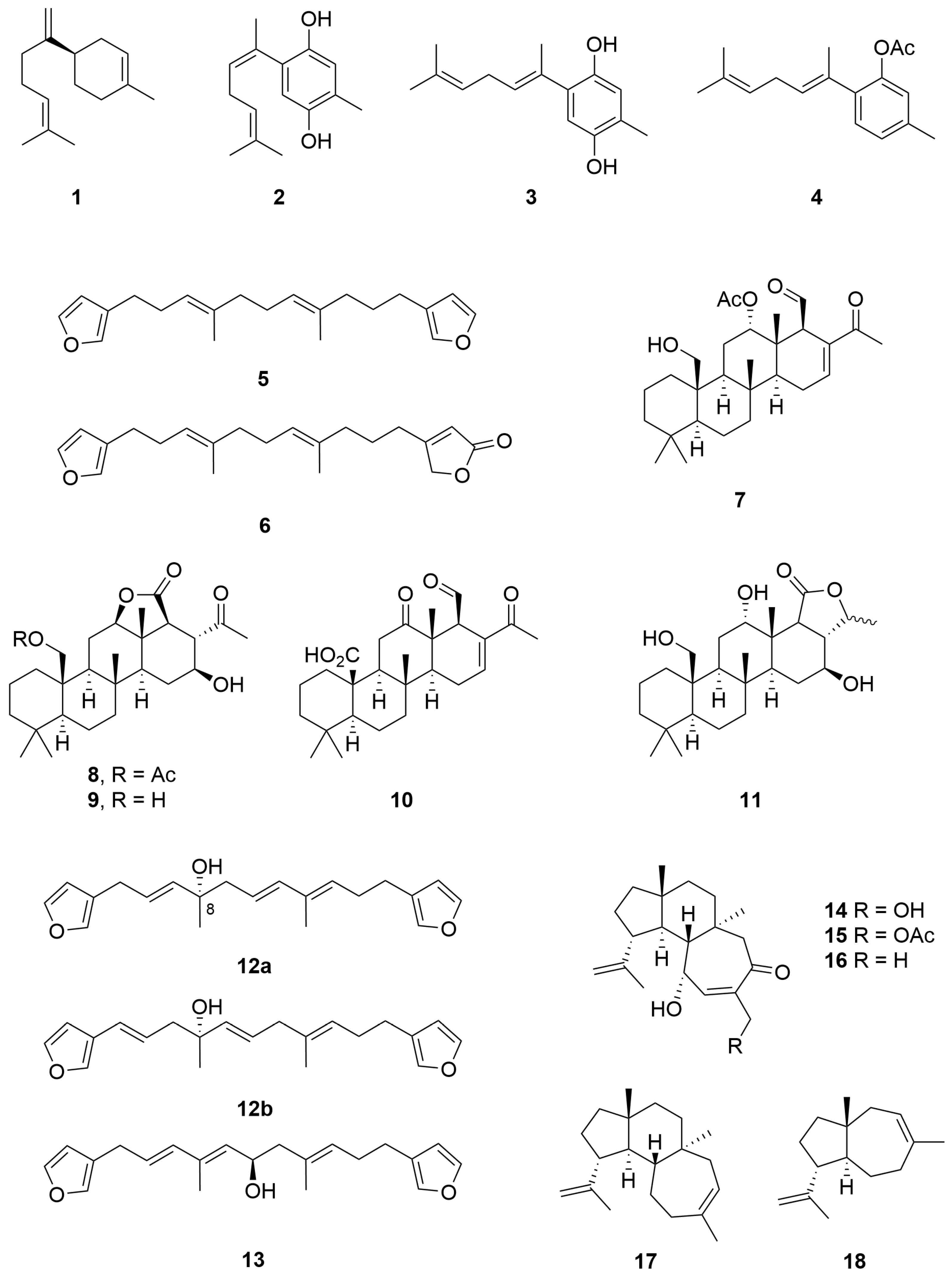
Three aromatic sesquiterpenes, as well as the known compound (−)-bisabolene (1), were isolated from the non-polar fractions of a Halichondria sp. (Order: Suberitida; Family: Halichondridae) collected off the coast of Lancellin. The unknown compounds were identified as (1′Z)- and (1′E)- 2-(1′,5′-dimethylhexa-4′-dienyl)-5-methylbenzene-1,4-diol (2, 3) and (1′E)-2-(1′,5′-dimethylhexa-1′,4′-dienyl)-5-methyl-phenyl acetate (4) (Figure 1) using 1H NMR and 13C NMR spectroscopy as well as chemical interconversion [12].
A Lendenfeldia sp. (Order: Dictyoceratida; Family: Thorectidae) specimen collected at Quobba Lagoon was the source of two known C-21 furanoterpenes (5, 6) as well as five new C-26 scalarene sesterterpenes (7–11) (Figure 1). The compounds were characterised spectroscopically as well as via chemical derivatisation and comparison to earlier reports. Two of the previously reported scalarenes and the novel compound 8 exhibited extremely potent inhibition of platelet aggregation, this providing a rationalisation for the anti-inflammatory activity of this group of compounds [13].
A Spongia sp. (Order: Dictyoceratida; Family: Spongiidae) collected east of Gun Island, South Abrolohos Group, was the source of a new C-21 bisfuranoterpene bearing a tertiary hydroxyl at position C-8 as an unstable oil (12a) [14]. The structure of the natural product was subsequently revised to 12b following two-dimensional NMR analysis [15]. The former publication also reports the revised stereochemistry via the Horeau method of another bisfuranoterpene 13 isolated from a Leiosella sp. collected by dredge off Rottnest Island (Figure 1) [14].
Three tricyclic diterpenes 14–16 were reported from a collection of Higginsia sp. (Order: Axinellida; Family: Stelligeridae) collected off Lancelin. The structure of 14 was verified via single-crystal X-ray diffraction [16]. The structures of the related monoacetate and monoalcohol were deduced in relation to that of 16. Subsequent reinvestigation of the non-polar fractions of the lipophilic sponge extract afforded the tricyclic diterpene hydrocarbon 17 and the daucadiene sesquiterpene 18 (Figure 1) [17]. A biosynthetic scheme arising from farnesyl and geranylgeranyl pyrophosphate was proposed. Furthermore, the authors suggest that compounds 14 to 16 derive via oxidation of 17, followed by intermolecular 4 + 2 cycloaddition of oxygen.
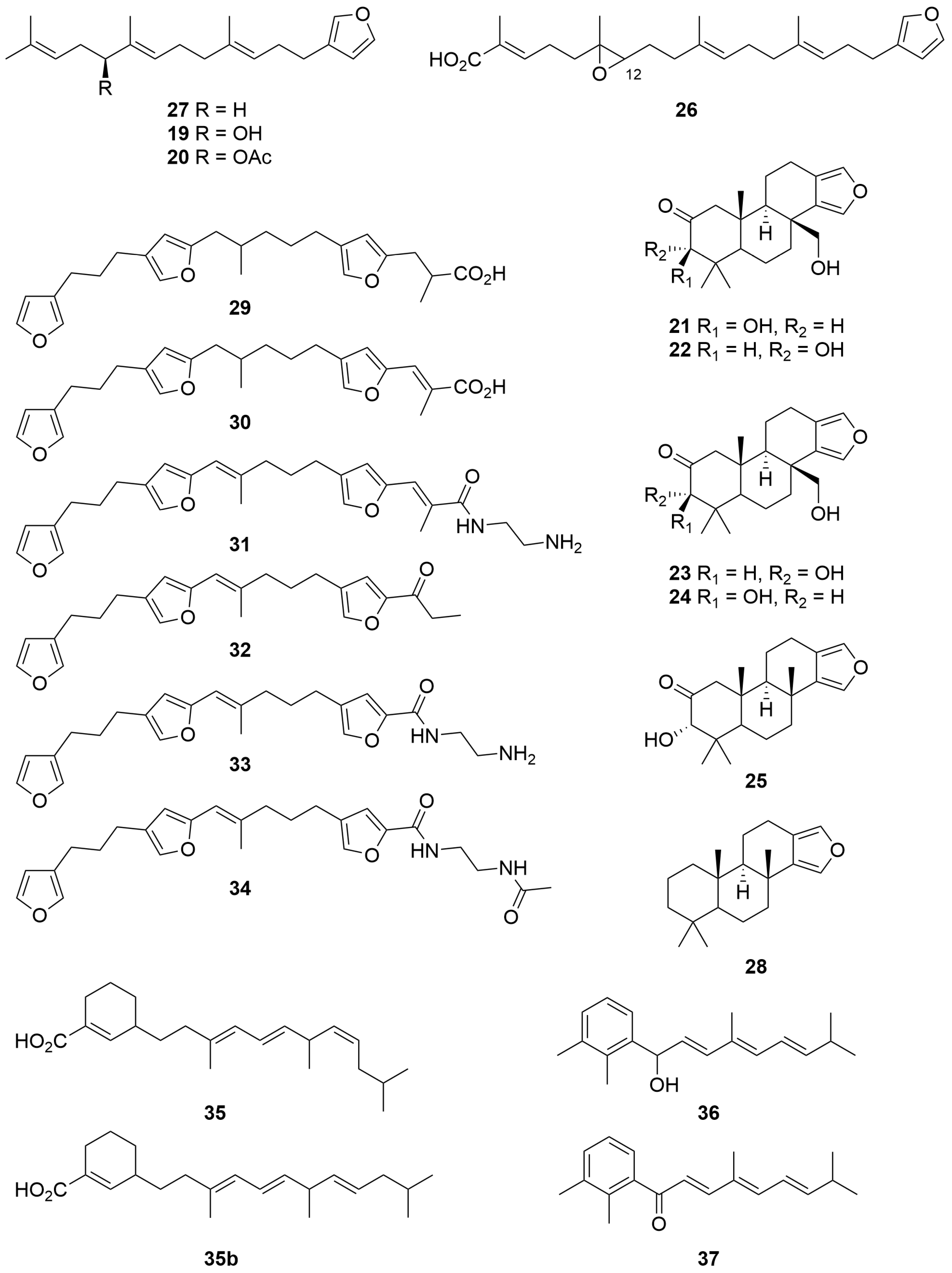
A Spongia sp. (Order: Dictyoceratida; Family: Spongiidae) collected from Exmouth gulf afforded the novel linear furanoditerpenes 12-hydroxy ambliofuran (19) and 12-acetoxyambliofuran (20) (Figure 2) [18]. Mosher’s ester analysis revealed the compounds to be a scalemic mixture of 3:1, predominantly S configured enantiomers: the authors note that the isolation of enantiomers in non-racemic proportions is unusual in the field of marine natural products. Additional investigations of the sponge extract unearthed the new tetracyclic furanoditerpenes 21–25 and the linear furanosesterpene 26 bearing an epoxide at C-12, as well as the known compounds 27 and 28 (Figure 2) [18].
The CH2Cl2 soluble fractions of a Hippospongia sp. (Order: Dictyoceratida; Family: Spongiidae) collected from south of the Great Australian Bight afforded six new C-25 derived linear furanoterpenes, given the trivial names hippospongins A–F (29–34) (Figure 2) [19]. The authors postulate a biosynthetic link between the commonly encountered C-25 tetronic acids and C-21 furanoditerpenes commonly encountered in marine sponges via the intermediacy of compounds 29–34. Hippospongin A (29) exhibited mild antibiotic activity, inhibiting the growth of Staphylococcus aureus at concentrations of circa 200 µg/disk in a standard agar plate assay [19].
An investigation into the chemistry of a Clathria sp. (Order: Poecilosclerida; Family: Microcionidae) collected off the Great Australian Bight yielded the novel compounds clathrins A, B and C (35–37) (Figure 2). Clathrin A (35) is postulated to provide support for the biosynthetic origins of other marine meroterpenoids derived via a mixed shikimate-terpenoid biosynthetic pathway. Attempts to elucidate the stereochemistry of clathrin B (36) were thwarted by the facile atmospheric oxidation of 36 to compound 37; furthermore, the absolute configuration of 35 remains unresolved [20].
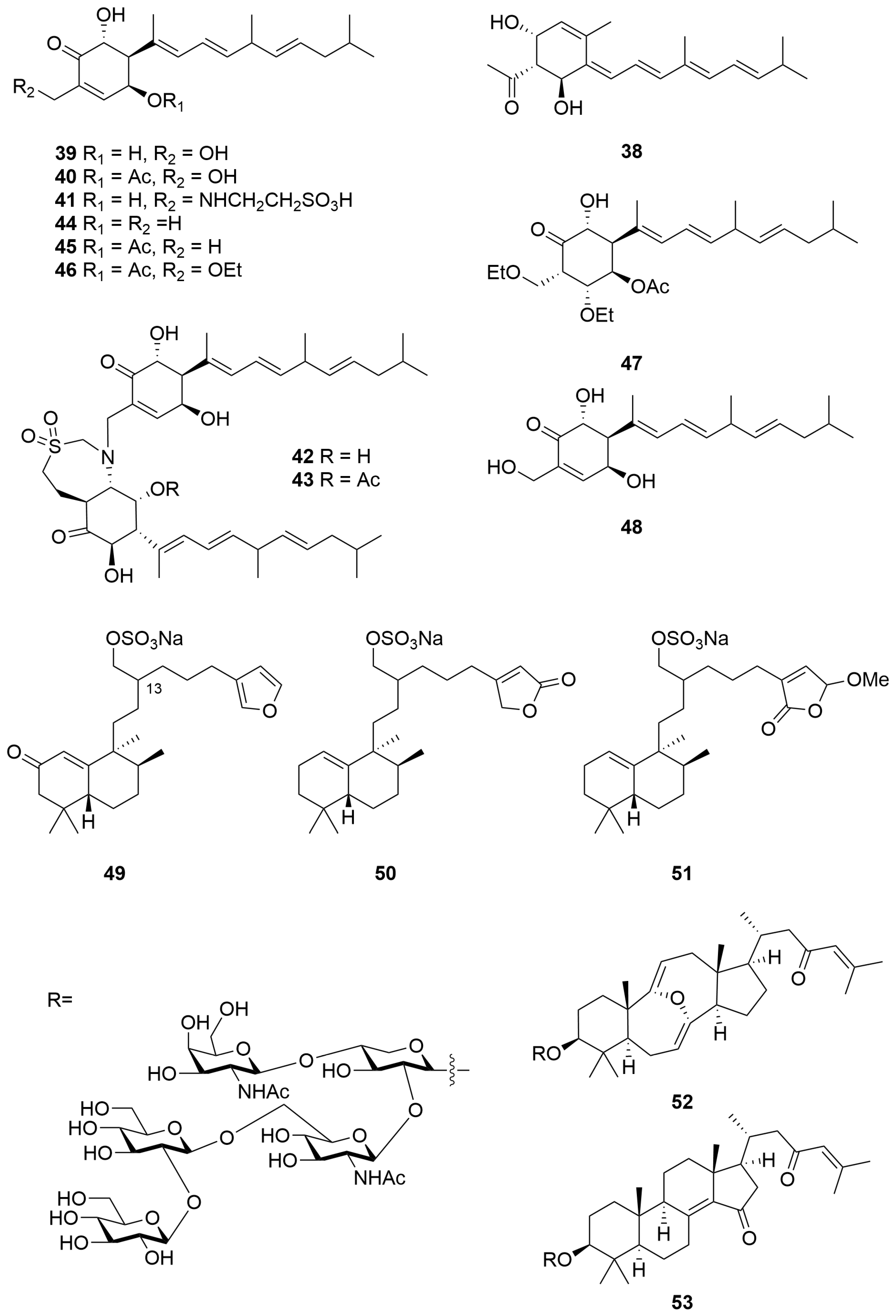
The ethanolic extracts of a Phorbas sp. (Order: Poecilosclerida; Family: Hymedismiidae) sourced from the Great Australian Bight afforded the rearranged diterpenoid phorbasin A (38) (Figure 3) as an unstable pale yellow solid; the unprecedented carbon skeleton of 38 was elucidated spectroscopically [21]. Analysis of a second Phorbas sp. unearthed phorbasins B and C [22], which were subsequently revised to structures 39 and 40 (Figure 3) with a terminal E, instead of Z, configured double bond [23]. (In light of the reassignment of phorbasins A and B to 39 and 40, we postulate that clathrin A (35) [20] may also require revision to structure 35b with all E configured olefins.) Further analysis of the sponge genus unearthed phorbasins D–F (41–43) (Figure 3), the dimeric compounds 42 and 43 bearing an unusual taurine-conjugated seven-membered heterocycle [23]. Research efforts into the phorbasin class culminated in the isolation of phorbasins G–K (44–48) [24]. The authors postulate a likely artefactual origin for phorbasins I (46) and J (47) via solvolysis [24]. The crude EtOH extract exhibited cytotoxic activity, as well as growth-inhibitory activity against the Gram-positive bacteria Staphylococcus aureus and Micrococcus luteus. Phorbasins B (39) and C (40) (Figure 3) were determined to be the principal antibacterial constituents. Compounds 39, 40, 44, 45 and 46 exhibited substantial potency and selective cytotoxicity against Neonatal Foreskin Fibroblasts (NFF) and human cancer (A549, HT29 and MM96L) cell lines. The structure–activity relationship of the metabolites was found to correlate with the presence of an α,β-unsaturated ketone functionality.
Chemical investigations of a Darwinella australensis (Order: Dendroceratida; Family: Darwinellidae) collected by SCUBA in the East Timor Sea afforded three new sesterpene sulfates, halisulfates 8–10 (49–51) (Figure 3) [25], isolated as their sodium salts. The relative configurations of the decalin moiety were elucidated using combined spectroscopic methods and via comparison to the known halisulfates 1–7. The relative configuration at C-13 remains unresolved. Halisulfates 9 (50) and 10 (51) exhibited inhibition of cell division of sea urchin eggs (Strongylocentrotus intermedius) in moderate concentration (IC50 = 50 µg/mL and 35 µg/mL, respectively) [25].
Sarasinosides A4 (52) and A5 (53) (Figure 3) were isolated from a marine sponge Melophlus sarasinorum (Order: Tetractinellida; Family: Geodiidae) collected near Scott Reef, along with five known sarasinosides [26]. The compounds were elucidated on the basis of extensive nuclear magnetic resonance experiments and density functional theory calculations, as well as MALDI-TOF-MS and GC-MS analysis. The compounds isolated bear the same oligosaccharide moiety and differ only in the composition of the aglycone. Compound 52 is unusual in the composition of its bis-enol ether moiety [26].
A sample of Stelletta sp. (Order: Tetractinellida; Family: Ancorinidae) collected by trawling operations in the Great Australian Bight was the source of the terpenyl-pyrrolizidine conjugates bistelletazines A–C (54–56) and the cyclic terpenyl-imidazole conjugate macrocycle bistelletazole A (57) (Figure 4) [27]. The authors note that despite extensive two-dimensional nuclear magnetic resonance experiments performed, the data acquired did not allow for the unambiguous assignment of stereochemistry for the pyrrolizidine portion of the molecule. The authors propose the compounds to share a convergent biosynthesis, the unique carbon scaffold arising from a presumed Diels–Alder reaction between two polyene sesquiterpene precursors [27].
Four meroterpenoid pigments, 18-aminoarenarone (58), 19-aminoarenarone (59), 18-methylaminoarenarone (60) and 19-methylaminoarenarone (61), and the new dimeric popolohuanone F (62) were isolated from a Dysidea sp. (Order: Dictyoceratida; Family: Dysideidae) collected from Scott Reef [28]. The sample also afforded the known compounds arenarol (63) and popolohuanone A (64) (Figure 4). Further investigation revealed that 62, 63 and 64 exhibited DPPH radical scavenging activity with IC50 values of 35 μM, 35 μM and 19 μM, respectively [28].
A new meroterpeneoid sulfate, fascioquinol A (65), was isolated from a deep-water Fasciospongia sp. (Order: Dictyoceratida; Family: Thorectidae) along with its desulfated counterpart fascioquinol B (66), and the acid-mediated cyclisation products fascioquinols C, D and strongylophorine-22 (67–69). The sponge also afforded the known meroterpene geranylgeranyl-1,4-hydroquinone (70) and its sulfated counterpart, to which was assigned the trivial name fascioquinol E (71), as well as the racemic chromenol fascioquinol F (72) (Figure 4) [29]. Further investigation revealed that 70 exhibited specific cytotoxic activity against gastric adenocarcinoma (AGS, IC50 = 8 µM) and neuroblastoma (SH-SY5Y, IC50 = 4 µM) cell lines; in addition to this, 65 and 66 exhibited promising Gram-positive activity towards Staphylococcus aureus (IC50 = 0.9–2.5 µM) and Bacillus subtilis (IC50 = 0.3–7.0 µM).
2.1.2. Alkaloids
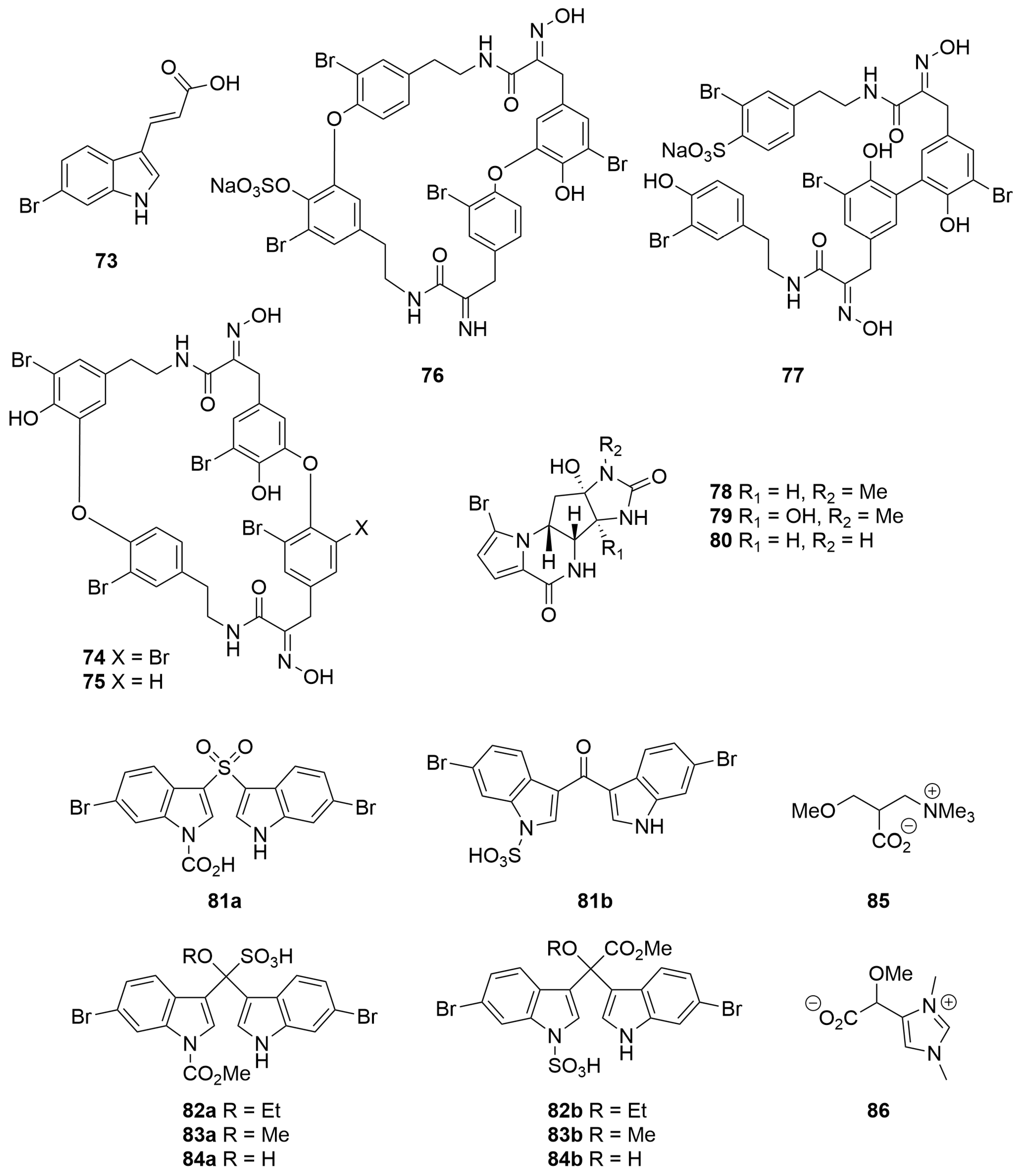
An Iotrochota sp. (Order: Poecilosclerida; Family: Iotrochotidae) collected from the Five Fathom Bank, off the coast of Fremantle, was reported to yield the novel metabolite (E)-3-(6-bromoindol-3-yl)prop-2-enoate (73) (Figure 5) [30]. The structure of the metabolite was proposed based on MS, IR and 1H NMR analysis and confirmed via total synthesis from 4-bromo-2-nitro-toluene [30].
Four novel bromo-tyrosine alkaloids of the bastadin class, bastadin 19 (9-debromobastadin 13, 74), bastadin 20 (75) and the sulfate half-esters 15,34-O-bis-sulfatobastadin 7 (76) and 10-O-sulfatobastadin 3 (77) (Figure 5), were isolated from the polar fractions of a Ianthella basta (Order: Verongiida; Family: Ianthellidae) collected from Stuarts Shoal, Exmouth Gulf [31]. The sponge also afforded a number of other known bastadins. The authors propose the use of MALDI-MS and a microscale derivatisation combined with 1H NMR fingerprinting of permethylated derivatives in order to rapidly dereplicate known bastadin and isobastarane isomers [31]. The compounds 15,34-O-disulfatobastadin 7 (76) and 10-O-sulfatobastadin 3 (77) exhibited moderate and specific activity as Sarcoplasmic Reticulum (SR) Ca2+ channel agonists (EC50 = 13.6 µM and 100 µM, respectively) of the Ry1R FKBP12 complex [31].
The methanolic extracts of two samples of Cymbastela sp. (Order: Axinellida; Family: Axinellidae) collected by SCUBA near Muiron Island afforded the known compound agelastin A (78) and the novel analogues agelastins C and D (79, 80) (Figure 5) [32]. The structures of the compounds were determined spectroscopically and via chemical derivatisation. Agelastatin A (78) exhibited potent activity against brine shrimp (LC50 = 5.0 µM) in addition to potent insecticidal activity against larvae of beet army worm, Spodoptera exigua, and corn rootworm, Diabrotica undecimpunctata [32].
The ethanolic extracts of an Echinodictyum sp. (Order: Axinellida; Family: Raspailiidae) collected in the Great Australian Bight afforded four novel compounds, echinosulfone A (81a) and the echinosulfonic acids A–C (82a–84a) (Figure 5) [33]. The proposed structures were assigned based on extensive two-dimensional NMR analysis. The compounds were found to account for the antibacterial activity of the crude extract but not the reported nematocidal activity. The structures of echinosulfone A and the echinosulfonic acids A–C were subsequently revised by three independent research groups contemporaneously to structures 81b–84b (Figure 5), respectively, on the basis of synthetic efforts, as well as single-crystal X-ray diffraction and density functional theory analysis [34,35,36]. Subsequent bioassay-guided fractionation of the sponge extract unearthed the novel betaine alkaloids (−)-echinobetaine A (85) [37] and (+)-echinobetaine B (86) (Figure 5) [38] as the principal nematocidal components responsible for the bioactivity of the sponge crude extract against the commercial livestock parasite Haemonchus contortus with echinobetaine B (86) exhibiting an LD99 of 8.3 µg/mL. The structures of racemic echinobetaines A and B have also been confirmed via total synthesis [38].
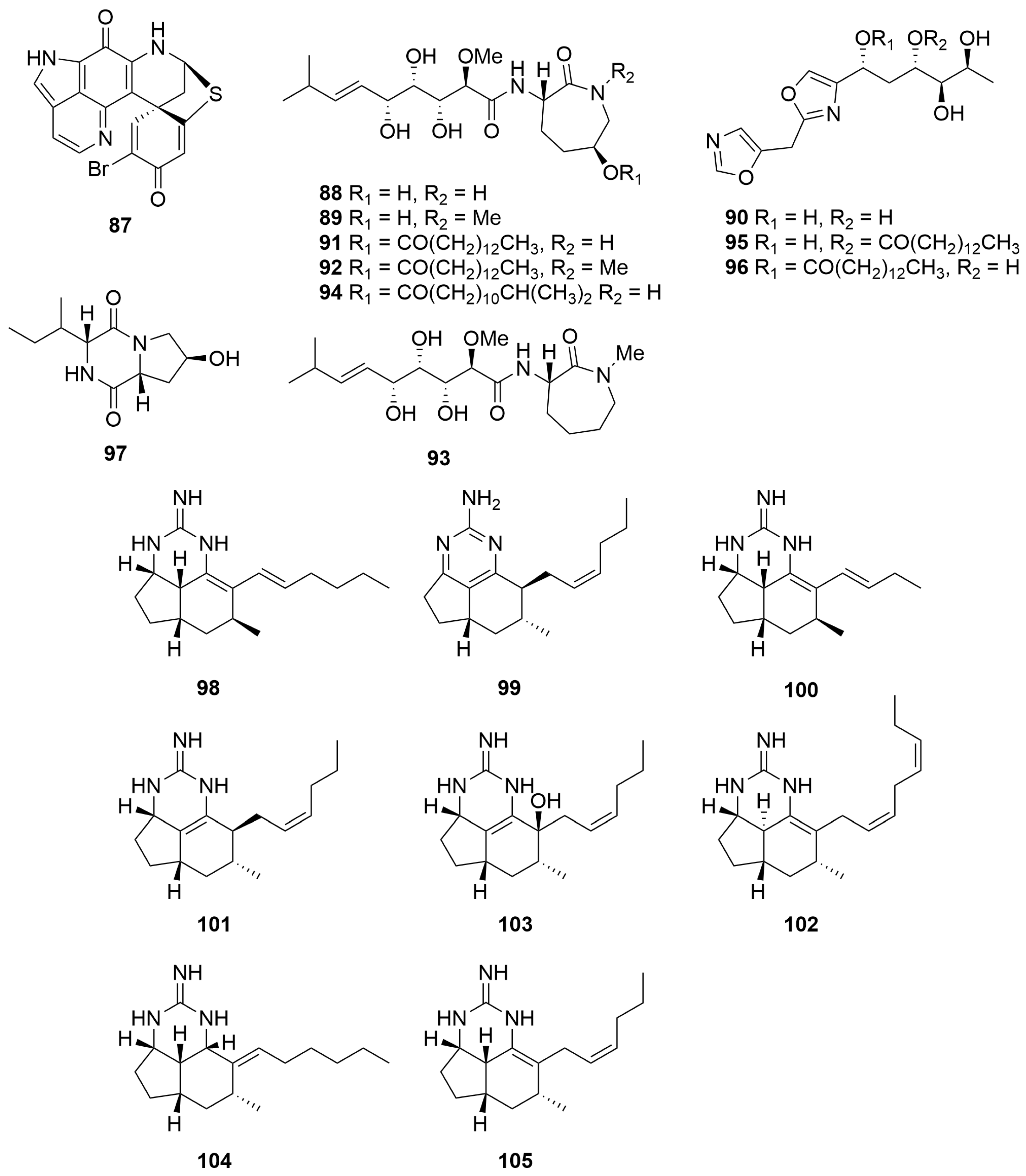
A Zyzzya sp. (Order: Poecilosclerida; Family: Acarnidae) collected at Assail Bank, between North Island and the Wallab Group, and a Latrunculia purpurea (Order: Poecilosclerida; Family: Latrunculiidae) collected on Horse Shoe Reef, west-northwest of Margaret Brock Lighthouse, were both reported to yield the novel pyrolloiminoquinone pigment discorhabdin Q (87) (Figure 6) [39]. The authors note that compound 87 was found not to be the principle cytotoxin in any of the extracts assayed; however, the metabolite exhibited moderate generalised cytotoxicity (mean panel GI50 = 0.5 µg/mL) in the NCI 60 cancer cell line panel [39].
A Jaspis sp. (Order: Tetractinellida; Family: Ancorinidae) collected off a low, uninhabited rocky island near the northwestern end of Serrurier Island, afforded the novel alkaloids bengamides Y (88) and Z (89) and bengazole Z (90) (Figure 6) [40]. In the same report, the authors detail that reinvestigation of the non-polar fractions yielded the known metabolites bengamides A (91) and B (92). Biological investigation revealed that metabolites 88 and 89 exhibited specific cytotoxicity against 10 cancer cell lines [40]. Bioassay-guided fractionation of a Stelleta sp. (Order: Tetractinellida; Family: Ancorinidae) collected off the western side of Jamieson Reef, Bonaparte Archipelago, afforded the known metabolites bengamides A (91), F (93), N (94) and Y (88) and bengazoles Z (90), C4 (95) and C6 (96) in addition to a novel diketopiperazine, cyclo-(4-S-hydroxy-R-proline-R-isoleucine) (97) [41]. The relative configuration of 97 was determined spectroscopically with the aid of molecular modelling software. Cyclo-(4-S-hydroxy-R-proline-R-isoleucine) (97) exhibited minimal activity towards MCF-7, H460 and HT-29 cells (GI50 > 200 µM) and no activity towards SF-268 or CHO-K1 cells. In contrast, the GI50 values for 88, 90, 91, 93, 94, 95 and 96 were comparable to those reported in previous studies [41].
A Clathria sp. (Order: Poecilosclerida; Family: Microcionidae) collected by trawl off the coast of Cape Arid yielded the new tricyclic guanidine alkaloid mirabilin G (98) (Figure 6) [42]. Reinvestigation of the same specimen afforded the known mirabilins C (99) and F (100) and the novel mirabilins H, I and J (101–103) [43]. Mirabilins C and F (99, 100) were characterised for the first time as underivatised natural products. The absolute stereochemistry of mirabilin F (100) was assigned for the first time (Figure 6). The authors propose a plausible biosynthetic route to the mirabilin, ptilocaulin and netamine alkaloids starting from polyketide precursors [43]. An Acanthella cavernosa (Order: Bubarida; Family: Dictyonellidae) collected in the southwest of the state was the source of the novel compound mirabilin K (104), the specimen also affording mirabilin G (98) and the related netamine M (105) (Figure 6) [44]. Mirabilin G (98) exhibited modest growth-inhibitory activity against the Gram-negative bacteria Escherichia coli and Serratia marcescens and the fungus Saccharomyces cerevisiae. Further investigations revealed that 98 and 105 inhibited cellular degradation of PDCD4 with EC50 values of 1.8 μg/mL and 2.8 μg/mL, respectively. It is noteworthy that 98 and 105 were the first reported marine natural products to stabilise PDCD4 under tumour-promoting conditions. Additional investigations revealed that 98–103 exhibited modest cytotoxic activity with LD50 values greater than 30 µM against neuroblastoma (SH-SY5Y), gastric (AGS), colorectal (HT29) and intestinal (Intestine-407) cancer cell lines.
The methanolic extracts of a Xestospongia sp. (Order: Haplosclerida; Family: Petrosiidae) collected at Benetts Shoal, Exmouth Gulf, afforded the dimeric 2,9-disubstituted-1-oxaquinolizidine alkaloids (+)-xestospongin A (106), (−)-xestospongin C (107) and (+)-xestospongin D (108), as well as arugospongine C (109), (+)-7S-hydroxyxestospongin A (110) and (+)-demethylxestospongin B (111) (Figure 7) [45]. The structure of (+)-7S-hydroxyxestospongin A (110) was solved using single-crystal X-ray diffraction, and the absolute configuration was secured using Mosher’s ester analysis. The absolute configuration of (+)-xestospongin D (108) was secured by analysis of anomalous dispersion in single-crystal X-ray diffraction experiments [45]. Compounds 106–109 and 111 exhibited modest antifungal activity (MIC 30–100 μg/mL) against various fluconazole-resistant Candida sp. [45].
Two novel pyrolloiminoquinone alkaloid pigments, isobatzelline E (112) and batzelline D (113), along with the known pigments batzelline C (114), isobatzelline C (115) and makaluvamine D (116) (Figure 7), were isolated from a Zyzzya fuliginosa (Order: Poecilosclerida; Family: Acarnidae) collected off Abrolohos Island [46]. Isobatzelline C (115) and to a lesser extent the known compounds makaluvamines A (117) and H (118) appear to inhibit HIV-1 envelope mediated cell fusion at concentrations less than 1.0 µg/mL [46].
Seven novel zwitterionic indole-2-carboxylic acids, trachycladindoles A–G (119–125) (Figure 7), were isolated from a Great Australian Bight sponge Trachycladus laevispirulifer (Order: Trachycladida; Family: Trachycladidae). Structures were elucidated based on comprehensive spectroscopic analysis. However, due to the paucity of material obtained, the relative configurations of trachycladindoles E (123) and F (124) remain unresolved; furthermore, the absolute configurations of 119–125 also remain unknown [47]. The authors postulate a biosynthetic scheme for the isolated trachycladindoles and related discodermindole family of alkaloids. Compounds 119–124 exhibited specific cytotoxicity against lung (A549), colorectal (HT29) and breast (MDAMB-231) cancer cell lines with GI50 and TGI values revealing sub µM potency. In addition to this, preliminary structure–activity relationship studies performed on compounds 119–125 highlighted an unusual bioactive molecular motif in favour of N-10 and N-12 dimethylation, as evidenced by the reported activity of compounds 120 and 122–124 [47].
A Western Australian Axinella sp. (Order: Axinellida; Family: Axinellidae) collected in the gulf of Exmouth was the source of the compounds herbindoles A (126), B (127) and C (128) (Figure 7) [48]. The structures of 126 to 128 were determined spectroscopically. The authors postulate that the biogenesis of the compounds is unlikely derived from tryptophan given the lack of substitution at position C-3 of the indole core. Compounds 126–128 exhibited cytotoxic activity against KB cells with an MIC of 5 µg/mL, >10 µg/mL and 10 µg/mL, respectively, and the combined extract also possessed significant fish feeding deterrent properties [48]. Two samples of Trikentrion flabelliforme (Order: Axinellida; Family: Raspailiidae) collected near Port Hedland yielded the new alkaloids trikentramides A–D (129–132) [49]. The planar structures and relative configurations of 129 to 132 were determined spectroscopically via comparison to prior literature reports [49]. Further evidence for the structures assigned was provided by quantum mechanical modelling and simulation of 13C NMR data as well as application of the DP4 algorithm pioneered by Goodman and co-workers [50]. Six new trikentrin-like natural products, (+)-trans-herbindole A (133) and trikentramides E–I (134–138) (Figure 7), have been recently reported from a sample of Trikentrion flabelliforme collected near Exmouth Gulf [51]. The relative and absolute configurations of 133 to 138 were determined by comparative analysis of optical rotation, computationally aided electronic circular dichroism spectroscopy (ECD) and chemical interconversion of the metabolites. The authors advance a plausible biosynthetic hypothesis for the formation of the trikentrin and herbindole classes of compounds beginning with the incorporation of a pyrrole-carboxylate thioester into a polyketide synthase. The authors also formulate an empirical mnemonic for the determination of the absolute stereochemistry of trikentrin and herbindole analogues dependant on the configuration of Me-C-8 [51].
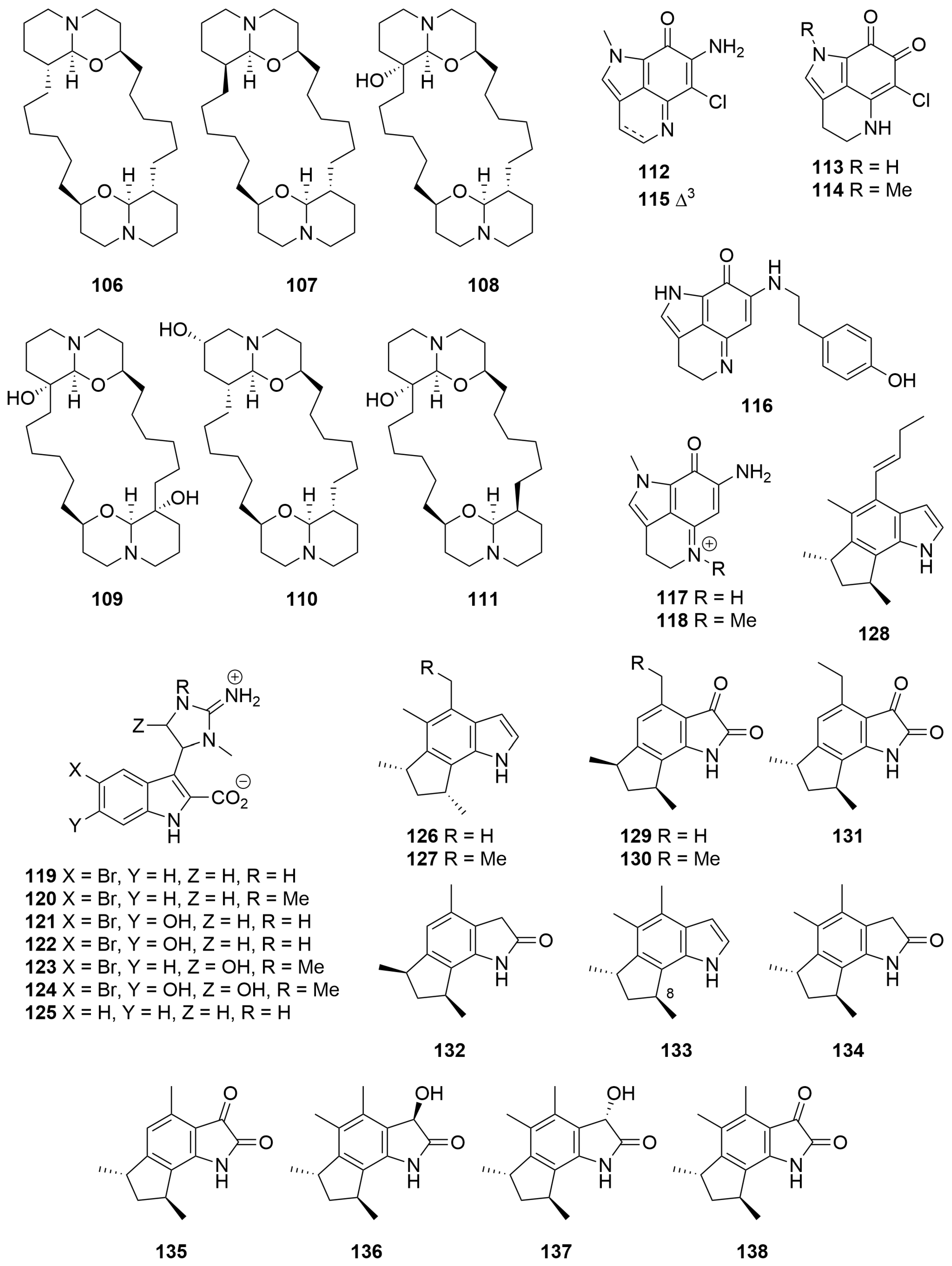
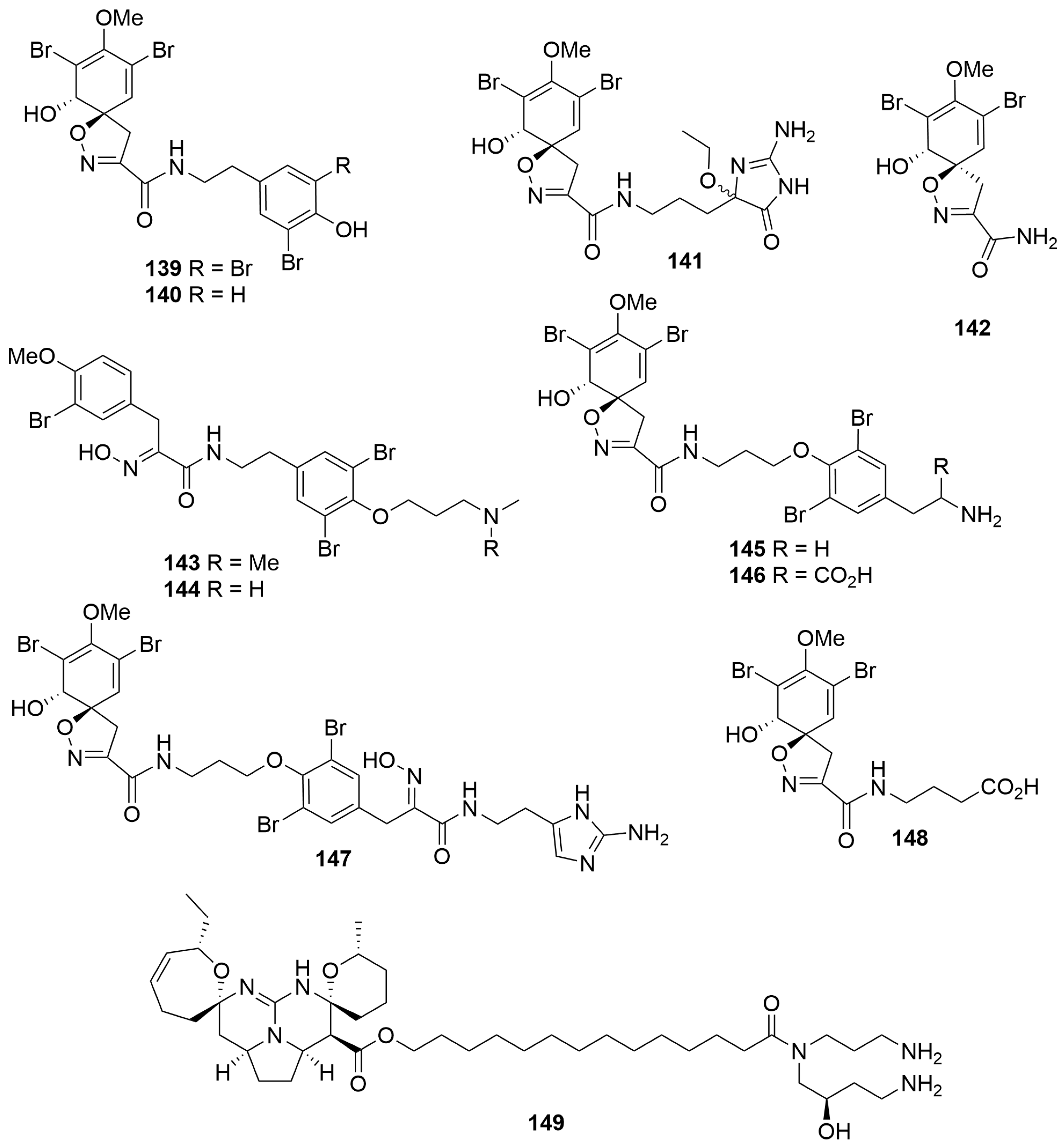
Two new bromotyrosine alkaloids, pseudoceratinamides A (139) and B (140), as well as an artefact of extraction (141) and the enantiomer of a known compound (148), were isolated from a Pseudoceratina cf. verrucosa (Order: Verongida; Family: Pseudoceratinidae) collected off the Dampier Peninsula [52]. The sponge specimen also afforded the known compounds 142 to 147 (Figure 8). The planar and relative configurations of the compounds were determined spectroscopically. Absolute configurations of all the compounds were determined using specific rotation and ECD measurements. The authors note that the original depiction of araplysin I (145) depicted the wrong absolute configuration, despite no work being conducted towards the absolute configuration of the molecule. Promulgation of this mistake throughout the literature means that at least some of the compounds assigned in relation to araplysin I will have to be revised. More importantly, the authors note that the isolation of enantiomers of previously isolated compounds highlights the possibility of enantiodivergence in the biosynthesis of the bromotrosine spirooxazoline alkaloids at the epoxidative dearomatisation step [52]. All compounds isolated exhibited moderate activity against Staphylococcus aureus strains. Biological testing revealed that pseudoceratinamide A (139) and pseudoceratinamide B (140) exhibited significant activity (MIQ = 0.31 μg) against methicillin-sensitive S. aureus. Compounds 140, 141, 143–145 and 147 exhibited comparable activity to vancomycin (MIQ = 0.63 μg) against methicillin-resistant S. aureus [52].
A sample of Monanchora viridis (Order: Poecilosclerida; Family: Crambeidae) collected off Cape Mentelle in the southwest of the state yielded the known compound crambescidin 800 (149) (Figure 8). Compound 149 exhibited cytotoxic activity in a panel of breast cancer cell lines, with triple-negative breast cancer (TNBC) cells showing more significant differences in cell viability than immortalised fibroblasts. Additionally, 149 was shown to cause cell cycle arrest at G2/M phase in T11 and SUM159PT cells, as well as inhibit the phosphorylation of the Akt/mTOR, MAPK and NF-κB pathways, which are responsible for tumour relapse and metastasis [53].
2.1.3. Polyketides
Bioassay-guided fractionation of a Phorbas sp. (Order: Poecilosclerida; Family: Hymedismiidae) collected by hand using SCUBA near Muiron Island afforded the potent cytotoxins phorboxazoles A (150) and B (151), epimeric at position C-13, as pale yellow amorphous solids [7]. The planar structures of 150 and 151 were determined based on extensive COSY and HMBC experiments, and the relative configurations of all stereocentres on the macrolide hemisphere of the molecule were assigned with the aid of ROESY spectroscopy [7]. The authors note that assignment of the macrolide ring was facilitated by the conformational restrictions imposed by the three oxane rings and one oxazole ring present on the scaffold. Subsequent work established the relative configuration of the hemiketal ring system via synthesis of a model compound and the assignment of absolute configuration via Mosher’s ester analysis [54]. Finally, the stereochemistry of methoxy C-43 was assigned by chemical conversion to dimethyl methoxysuccinate and comparison to an authentic sample of the R-enantiomer by chiral GC-MS [55] (Figure 9). Phorboxazoles A (150) and B (151) exhibited antifungal properties against Candida albicans, as well as inducing cell growth inhibition across a spectrum of cancer cells (leukemia, CCRF-CEM, GI50 = 0.25 nM; HCT-116, GI50 = 0.44 nM), and displayed extraordinary cytostatic activity (mean panel GI50 < 7.9 pM) in the NCI 60 cancer cell line panel [7].
Re-examination of the same Phorbas sp. extracts using highly sensitive cryo-probe NMR experiments yielded two new chlorocyclopropane macrolides, phorbasides A (152) and B (153) (Figure 9) [56]. The assignment of absolute configuration was achieved via empirical comparison of ECD data obtained to that of synthesised model systems, taking advantage of the vibronic fine structure associated with an asymmetrically perturbed ene-yne chromophore [56]. Subsequent work afforded phorbasides C–E (154–156) [57], the highly chlorinated muironolide A (157a) [58], differing in the absolute configuration of the chloro-cyclopropane ring, along with the nitrile-bearing hemi-phorboxazole A (158) [59], and most recently, phorbaside F (159) [60] and phorbasides G–I (160–162) (Figure 9) [61]. The structure of muironolide A was subsequently revised to 157b following total synthesis [62]. Biological evaluation of compounds 152–157 revealed modest cytotoxicity exhibited by the metabolites towards colon tumour cells (HCT-116; IC50 = 2–30 µM) with phorbaside C (154) exhibiting the most potent cytotoxic activity (IC50 = 2 µM).
Bioassay-guided fractionation of a Raspailia (raspalia) sp. (Order: Axinellida; Family: Raspailiidae) collected by trawl on the northern Rottnest Shelf afforded the known compounds phorboxazoles A and B (150, 151) (Figure 9) as the principal nematocidal agents, as well as the known synthetic compound esmodil (163), isolated for the first time as a natural product [63]. The structure of 163 (Figure 10) was confirmed spectroscopically and via total synthesis. Biological testing revealed that 150 and 151 exhibited nematocidal activity against Haemonchus contortus (LD99 = 0.5 mg/mL and 1.1 mg/mL, respectively) [63].
Two novel macrolide antibiotics, salicylihalamides A (164) and B (165) (Figure 10), were reported from a Haliclona sp. (Order: Haplosclerida; Family: Chalinidae) collected off the coast of Rottnest Island. The compounds contain an unusual highly saturated ene-amide side chain [8]. Additional work has analysed the spatiotemporal distribution of the metabolites across species of Haliclona collected across the southwest of the state [64]. Compound 164 exhibited highly potent and specific cytotoxicity (mean panel GI50 = 15 nM) in the NCI 60 cell line human tumour screen. COMPARE pattern-recognition analysis revealed no significant correlations to the profiles of other known antitumour compounds, suggesting that the salicylihalamides represented a potentially important new class of compounds for antitumour lead optimisation [8]. Subsequent work determined the unprecedented mechanism of action of 164 and 165 via Vacuolar-ATPase inhibition [9].
A collection of two Amphimedon species (Order: Haplosclerida; Family: Niphatidae) collected during trawling operations in the Great Australian Bight afforded the novel macro-bicyclic lactones/lactams amphilactams A–D (166–169) (Figure 10) [65]. The planar structures of 166 to 169 were elucidated on the basis of extensive spectroscopic evidence and comparison to synthetic model compounds. The relative and absolute configurations of 166 to 169 remain unknown. Compounds 166 to 169 were isolated in sufficient amounts to quantify their in vitro LD99 activities against Haemonchus contortus as 7.5 µg/mL, 47 µg/mL, 8.5 µg/mL and 0.39 µg/mL, respectively [65].
Bioassay-guided fractionation of a Geodia sp. (Order: Tetractinellida; Family: Geodiidae) collected in the Great Australian Bight yielded a new macrocyclic polyketide lactam tetramic acid, as a magnesium salt 170 (Figure 10), as the sole agent responsible for the in vitro nematocidal activity of the extract [66]. The structure of geodin A (170) was determined spectroscopically. The magnesium content of the sample was determined by energy-dispersive spectroscopy and atomic absorption spectroscopy allowing the authors to deduce the presence of one unit of magnesium for every two units of tetramic acid [66]. Geodin A (170) exhibited potent in vitro nematocidal activity (LD99 = 1.0 µg/mL) [66].
2.1.4. Fatty Acids
A collection of three Xestospongia sp. (Order: Haplosclerida; Family: Petrosiidae) collected from Bennett shoal in the Exmouth gulf afforded the new polybrominated unsaturated fatty acids (5E,11E,15E,19E)-20-bromoeicosa-5,11,15,19-tetraene-9,17-diynoic acid (172), (5Z,11E,15E,19E)-6,20-dibromoeicosa-5,11,15,19-tetraene-9,17-diynoic acid (173) and (Z,E)-14,14-dibromo-4,6,13-tetradecatrienoate (174). Compound 174 was characterised as its methyl ester (174a) (Figure 11), and additional fractionation afforded the known carboxylic acid 175 [68]. The authors note the unusual carbon chain lengths present on the metabolites. Additionally, ribosomal RNA analysis of the sponge specimens indicated that up to 46% of the RNA present in the extracts was eubacterial in origin [68].
Bioassay-guided fractionation of an Oceanapia sp. (Order: Haplosclerida; Family: Phloeodictyidae) collected off the northern Rottnest Shelf afforded the novel dithiocyanates thiocyanatins A, B and C (176–178) (Figure 11) [69]. The structures of 176 to 178 were elucidated spectroscopically and confirmed in a seven-to-eight-step total synthesis starting from 8-bromooctanoic acid [69]. Re-analysis of the ethanolic sponge extract afforded thiocyanatins D1 and D2 (177, 180) as an inseparable mixture and thiocyanatins E1 and E2 (181, 182), also as an inseparable mixture, as well as a number of analogues tentatively identified by 1H NMR and LC-ESIMS [70]. The structures of the novel metabolites were elucidated with respect to the prior compounds 176 to 178 and via comparison to synthetic model compounds. The thiocyanatins exhibited potent nematocidal activity, and preliminary structure–activity relationship investigations confirmed the key characteristics of the thiocyanatin pharmacophore. Thiocyanatin A (176) exhibited potent nematocidal activity (LD99 = 1.3 µg/mL) against Haemonchus contortus [69].
An Oceanapia sp. collected by dredge off Scott Reef afforded the hybrid α,ω-bifunctionalised sphingoid tetrahydroisoquinoline β-glycoside oceanalin A (183), as well as the known compound rhizochalin (184) (Figure 12) [71]. The structure of oceanalin A was elucidated on the basis of 1H NMR and 13C NMR spectroscopy as well as chemical derivatisation. The authors conclude that, given the absence of optical rotation for the cleaved eastern hemisphere of the molecule, as well as the propensity of tetrahydroisoquinoline compounds to epimerise, compound 183 is likely a 1:1 mixture of epimers at the C-26 position [71]. A number of new cerebrosides of which two representative examples are depicted (185, 186) were isolated from the same Oceanapia sp. collected off Scott Reef [72]. The cerebrosides were isolated as inseparable mixtures of compounds and assigned by NMR spectroscopy, MALDI-MS, chemical derivatisation and GC-MS [72]. Also from the same collection of Oceanapia sp. was isolated a ceramide fraction characterised by methanolysis and GC-MS analysis [73]. Most recently, the same collection of Oceania sp. afforded the new bolaampiphilic sphingoid bases rhizochalin B (187) and rhizochalinin B (188) (Figure 12) characterised by NMR spectroscopy as their peracetates [74]. The compounds contain an unusual butoxy group, which the authors note is uncommon in natural products. The ethanolic Oceanapia sp. extract exhibited antimicrobial activity against Staphylococcus aureus, Bacillus subtilis, Escherichia coli and Candida albicans and cytotoxic properties against the Erlich murine carcinoma. Metabolite 183 exhibited antifungal activity against Candida glabrata with an MIC of 30 µg/mL [71].
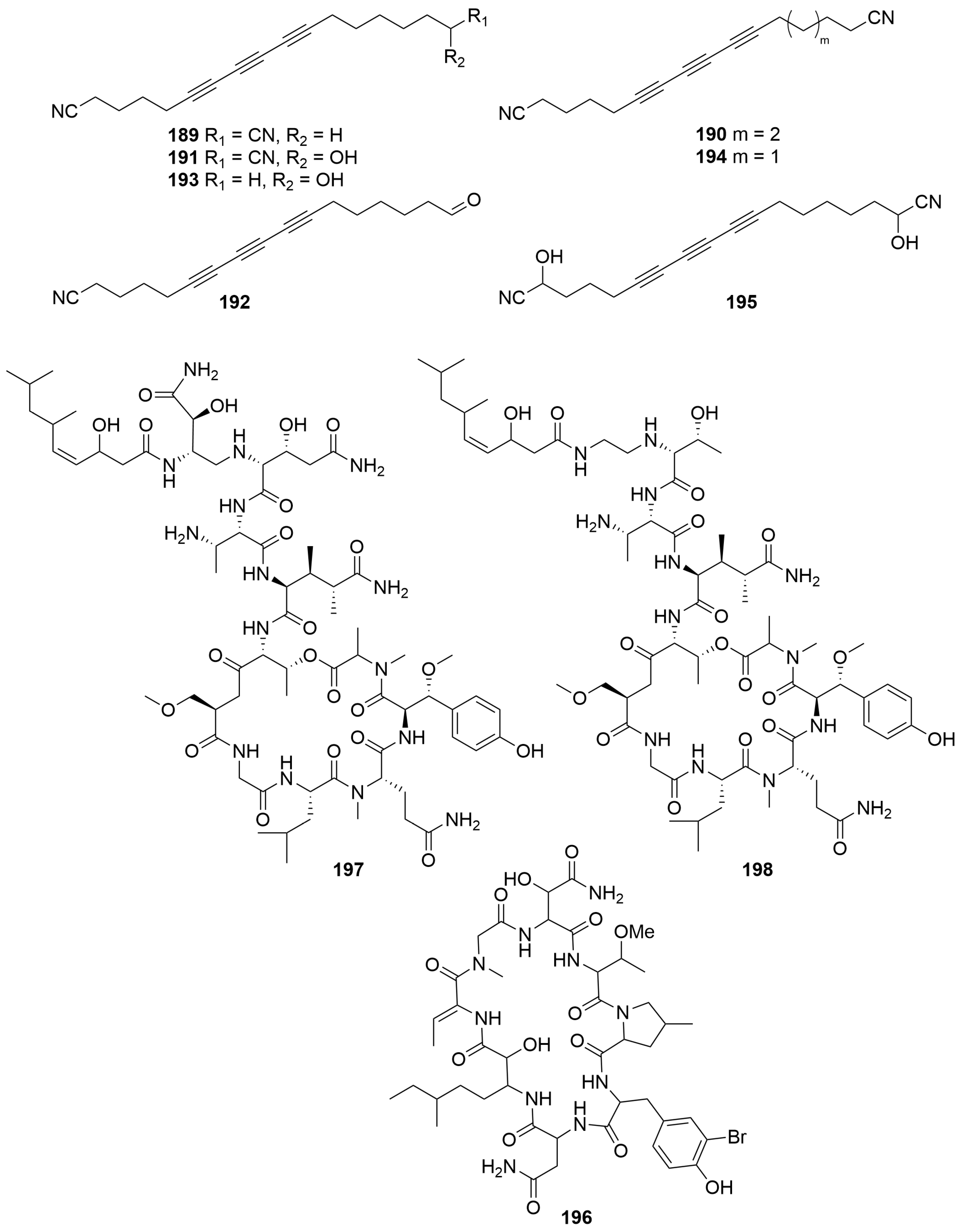
A Mycale sp. (Order: Poecilosclerida; Family: Mycalidae) collected by benthic sled off the coast of Albany afforded seven novel polyacetylene nitriles: albanitriles A–G (189–195) (Figure 13) [75]. The compounds were characterised spectroscopically. Compounds 191 and 195 were isolated as racemic mixtures. Albanitrile C (191) exhibited mild toxicity towards Bacillus subtilis at 90 μM. Additional investigations revealed that compounds 189–192 exhibited activity against Giardia duodenalis, with albanitrile A (189) exhibiting activity at a minimum concentration of 12 μM, which was comparable to metronidazole used as the positive control. Compounds 189 to 191 also exhibited weak inhibition against Tritrichomonas fetus, on the order of 200 μM [75].
2.1.5. Peptides
Bioassay-guided fractionation of a Theonella sp. (Order: Tetractinellida; Family: Theonellidae) collected by SCUBA near Perth, off Cape Vlamingh, afforded the cyclic octapeptide perthamide B (196) (Figure 13) [76]. The structure of compound 196 was elucidated spectroscopically; however, the relative and absolute configurations of the amino acid residues present remain unresolved. Compound 196 weakly inhibited binding of [125I]IL-1β to intact EL4.6.1 cells with an IC50 of 27.6 μM; however, the inhibition of binding could not be separated from the cytotoxic effects of 196 [76].
Two new HIV-inhibitory cyclic depsipetides, stellettapeptins A and B (197, 198) (Figure 13), were isolated form a sample of Stelletta sp. (Order: Tetractinellida; Family: Ancorinidae) collected in the states northwest [77]. The compounds contain a number of unusual non-proteinogenic amino acids. The structures of 197 and 198 were determined spectroscopically and using Marfey’s analysis. Biological investigations revealed that compounds 197 and 198 exhibited infection-inhibitory activity of human T-lymphoblastoid cells by HIV-1RF with EC50 values of 23 nM and 27 nM, respectively [77].
2.1.6. Miscellanea
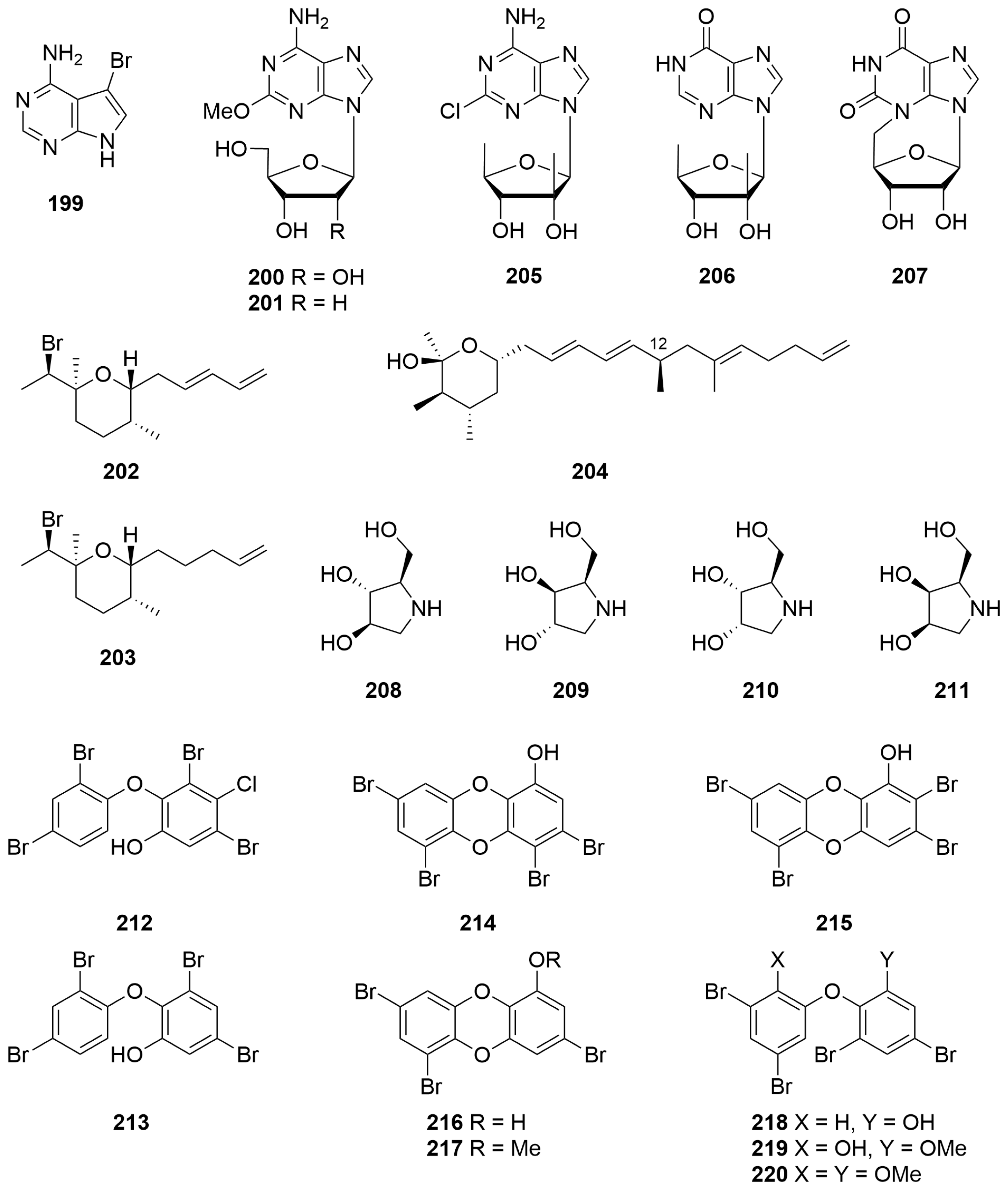
Bioassay-guided fractionation of an Echinodictyum sp. (Order: Axinellida; Family: Raspailiidae) afforded 4-amino-5-bromopyrrolo [2,3-d]pyrimidine (199) (Figure 14) for the first time as a natural product. The identity of the metabolite was verified spectroscopically and via total synthesis [78]. Metabolite 199 exhibited potential as a bronchodilator [78].
The known nucleoside spongosine (200), previously reported by Bergmann et al. from Cryptotethya crypta and 2′-deoxyspongosine (201) (Figure 14), previously only reported as a synthetic, was isolated from a sponge of the Order Hadromerida (Tethyidae) collected by hand from Exmouth Gulf [79]. 1H and 13C NMR spectral data for both compounds were reported for the first time.
Two new brominated tetrahydropyrans were obtained by preparative GC of a Haliclona sp. (Order: Haplosclerida; Family: Chalinidae) collected from Cosy Corner off the southwest coast of Western Australia [80]. The structures of (1′R,2S,2″E,5R,6R)-2-(1′-bromethyl)-2,5-dimethyl-6-(penta-2″,4″-dienyl)-tetrahydropyran (202) and (1′R,2S,5R,6R)-2-(1′-bromoethyl)-2,5-dimethyl-6-(pent-4″-enyl)-tetrahydropyran (203) (Figure 14) were determined spectroscopically and via chemical derivatisation. The absolute configuration of the compounds was determined using the Horeau method on an acyclic derivative [80]. Chemical investigations of a Haliclona sp. collected underneath an overhanging rocky substrate off Rottnest Island afforded 202 and 203, as well as a novel tetrahydropyran, rottnestol (203), in low milligram quantity. The structure of 204 was elucidated on the basis of 1H, 13C, COSY, HSQC, HMBC and NOE NMR studies [81]. The stereochemistry at C-12 was subsequently determined by total synthesis [82].
A Trachycladus laevispirulifer (Order: Trachycladida; Family: Trachycladidae) collected in Exmouth Gulf afforded the unprecedented 2′-C-methyl-5′-deoxyribofuranosyl nucleosides Trachycladines A and B (205, 206) (Figure 14) [83]. The structures of 205 and 206 were elucidated spectroscopically and by chemical derivatisation. Biological testing revealed that compound 205 exhibited cytotoxicity against several human cell lines, including leukaemia CCRF-CEM (IC50 = 0.4 µg/mL), colon tumour HCT-116 (IC50 = 0.9 µg/mL), breast tumour MCF-7 (IC50 = 0.2 µg/mL), MDAMB-435 (IC50 = 0.25 µg/mL) and MDA-N (IC50 0.1 µg/mL), but was inactive against yeasts (Candida albicans, Saccharomyces carlsbergensis) and bacteria (Staphylococcus aureus, Escherichia coli and Pseudomonas aeruginosa) in a disk diffusion assay at 200 µg/disk [83].
An Erylus sp. (Order: Geodiidae; Family: Erylinae; referred to in the original article as an Eryus sp.), collected west of Margaret River, yielded the first naturally occurring cyclo-nucleoside N-3,5′-cycloxanthosine (207) (Figure 14) [84]. The structure of the metabolite was elucidated spectroscopically and confirmed by total synthesis [84].
A sample of Haliclona sp. (Order: Haplosclerida; Family: Chalinidae) collected from Exmouth Gulf afforded 1,4-dideoxy-1,4-imino-D-arabinitol (207) (Figure 14), for the first time from a marine organism [85]. The identity of the compound was verified spectroscopically and by Marfey’s method. Investigation of a number of other sponges present in the same collection revealed the presence of 208 in a series of Raspalia sp. (Order: Axinellida; Family: Raspailiidae) as well as 1,4-dideoxy1,4-imino-D-xylitol (209), present both in a Haliclona sp. and Raspalia sp. Investigation of a Cymbastela sp. (Order: Axinellida; Family: Axinelllidae) revealed the presence of two additional isomers, identified tentatively as the diastereomers 1,4-dideoxy-1,4-imino-D-ribitol (210) and 1,4-dideoxy-1,4-imino-D-arabitol (211) (Figure 14) by Marfey’s analysis. Lack of authentic samples precluded the conclusive identification of 210 and 211 [85]. Compound 211 exhibited significant α-glycosidase inhibitory activity with an IC50 value of 0.16 μg/mL. Extracts containing compound 209 and the putative diastereomeric imino pentitols 210 and 211 exhibited significantly less α-glycosidase inhibitory activity [85].
Two polybrominated diphenol ethers, 3,5-dibromo-4-chloro-2-(2,4-dibromophenoxy)phenol (212) and 3,5-dibromo-2-(2,4-dibromophenoxy)phenol (213) (Figure 14), were isolated from an unidentified sponge collected by hand off the Rottnest Shelf. The structures of 212 and 213 were elucidated spectroscopically and confirmed by single-crystal X-ray diffraction [86].
A Dysidea dendyi (Order: Dictyoceratida; Family: Dysideidae) collected by hand from Scott Reef at a depth of 3 m afforded two new tetrabromodibenzo-p-dioxins: spongiadioxins A and B (214, 215) (Figure 14) [87]. The structures of 214 and 215 were elucidated using a combination of 1D and 2D NMR spectroscopy. Additionally, the structure of 214 was verified by single-crystal X-ray diffraction of a methyl ether obtained from 214. The structure of 215 was secured by chemical interconversion [87]. Re-investigation of the lipophilic sponge extracts afforded spongiadioxin C (216) and its methyl ether 217 as well as the related diphenyl ethers 218–220 [88]. The structures of the metabolites were determined spectroscopically and via semisynthesis. Biological evaluation revealed that compounds 214 and 215 exhibited cytotoxic activity against mouse Ehrlich carcinoma cells (ED50 = 29 and 15.5 µg/mL, respectively). In contrast, the LD50 for 214 and 215 in mice was determined to be more than 150 mg/kg [87]. Additional investigations revealed that 214–216 exhibited significant cell division inhibition for the fertilised eggs of the sea urchin Strongylocentrotus intermedius with an IC50 = 5.7 µM, 4.8 µM and 1.1 µM, respectively [88].
2.2. Cnidaria
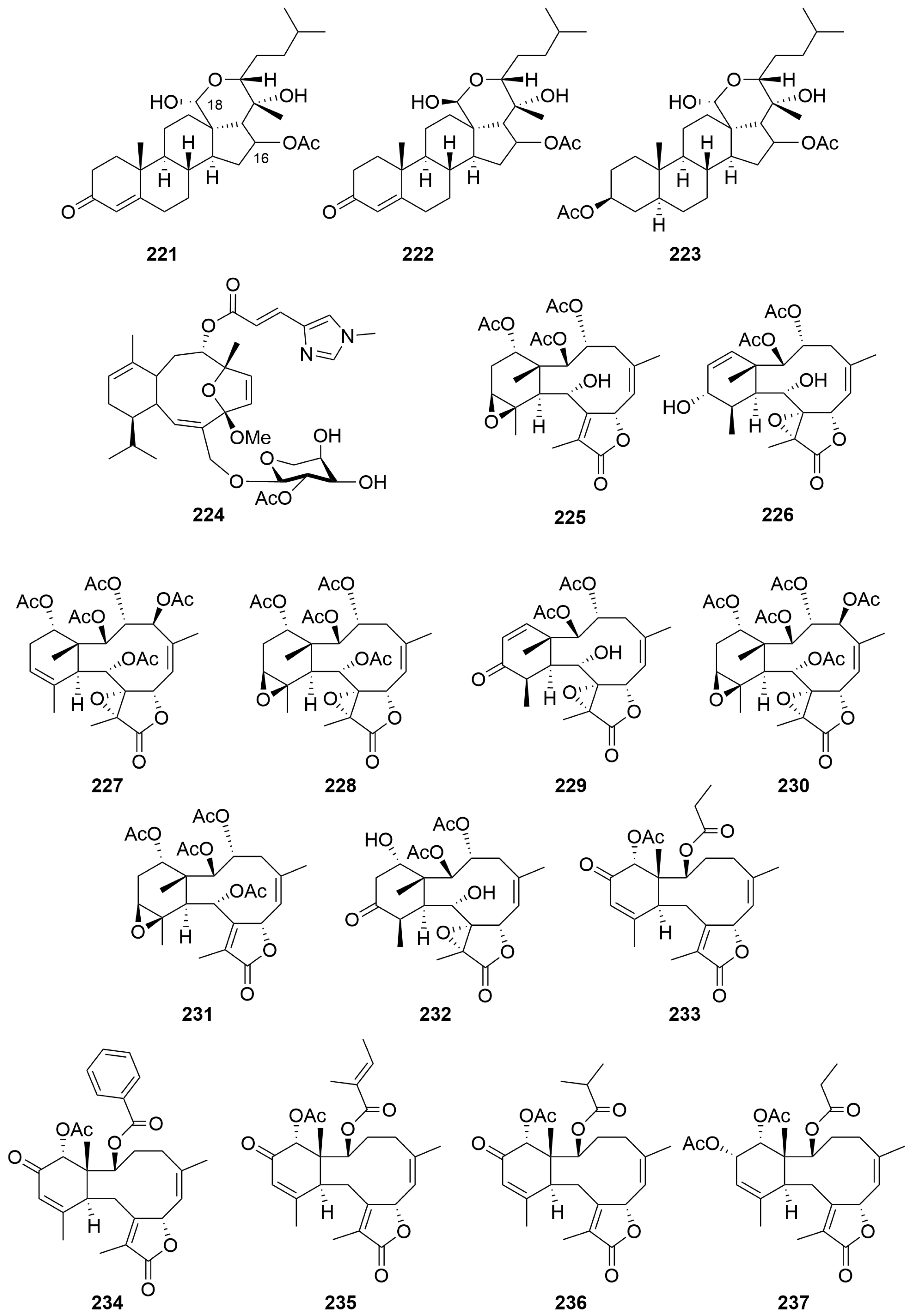
A sample of Ctenocella pectinata (Order: Alcyonacea; Family: Ellisellidae), collected in Exmouth Bay, afforded three new sterols, pectinoacetals A–C (221–223) (Figure 15), isolated as their monoacetylated derivatives [89]. The underivatised natural products were found to undergo rapid interconversion of the C-18 hemiacetal chiral centre. The structures of the natural products were elucidated spectroscopically and by chemical derivatisation. The relative configuration of the stereocentre at C-16 could not be assigned conclusively by NOE spectroscopy [89].
Bioassay-guided fractionation of a rare Alcyonacean soft coral Eleuthorobia sp. (Order: Alcyonacea; Family: Alcyoniidae), found near Bennett’s Shoal, yielded the new diterpene glycoside eleutherobin (224) (Figure 15) [10]. The structure of 224 was assigned spectroscopically. Eleutherobin (224) exhibited significant specific cytotoxicity against a diverse panel of breast, renal, ovarian and lung cancer cell lines with an IC50 range of 10–15 nM. Compound 224 was found to stabilize microtubules by competing for the paclitaxel binding site on the microtubule polymer [10].
Fractionation of a freeze-dried specimen of Briareum excavatum (Order: Alcyonacea; Family: Briareidae), collected at Rowley Shoals, afforded the known diterpene (1R*,2R*,3R*,5Z,7S*,8(17)Z,10R*,11R*,12S*,14S*)-2,3,14-triacetoxy-11,12-epoxybriara-5,8(17)-dien-18-one (225), as well as the new briarane diterpenes excavatolides N–T (226–232) (Figure 15) [90]. The structures of 225 to 232 were assigned spectroscopically and by comparison to literature. The authors explain the unusual spectroscopic features presented by excavatolide T (232) by geometry optimisation of the proposed structure using density functional theory. Compounds 226–229 exhibited various levels of cytotoxic activity against P388 murine leukaemia, A549 human lung carcinoma, HT29 human colon carcinoma and MEL28 human melanoma cells [90].
A sea pen Anthoptilum cf. kukenthali (Order: Pennatulacea; Family: Anthoptilidae), collected by dredge at a depth of 267 m, northwest of Port Hedland, afforded five new briarane diterpenoids: the anthoptilides A–E (233–237) (Figure 15). The structure of anthoptilide A (233) was solved by single-crystal X-ray diffraction. Compounds 223–237 inhibited [3H]CPDPX binding to rat brain adenosine A1 receptors with IC50 values of 420 µM, 45 µM, 3.1 µM, 500 µM and 490 µM, respectively [91].
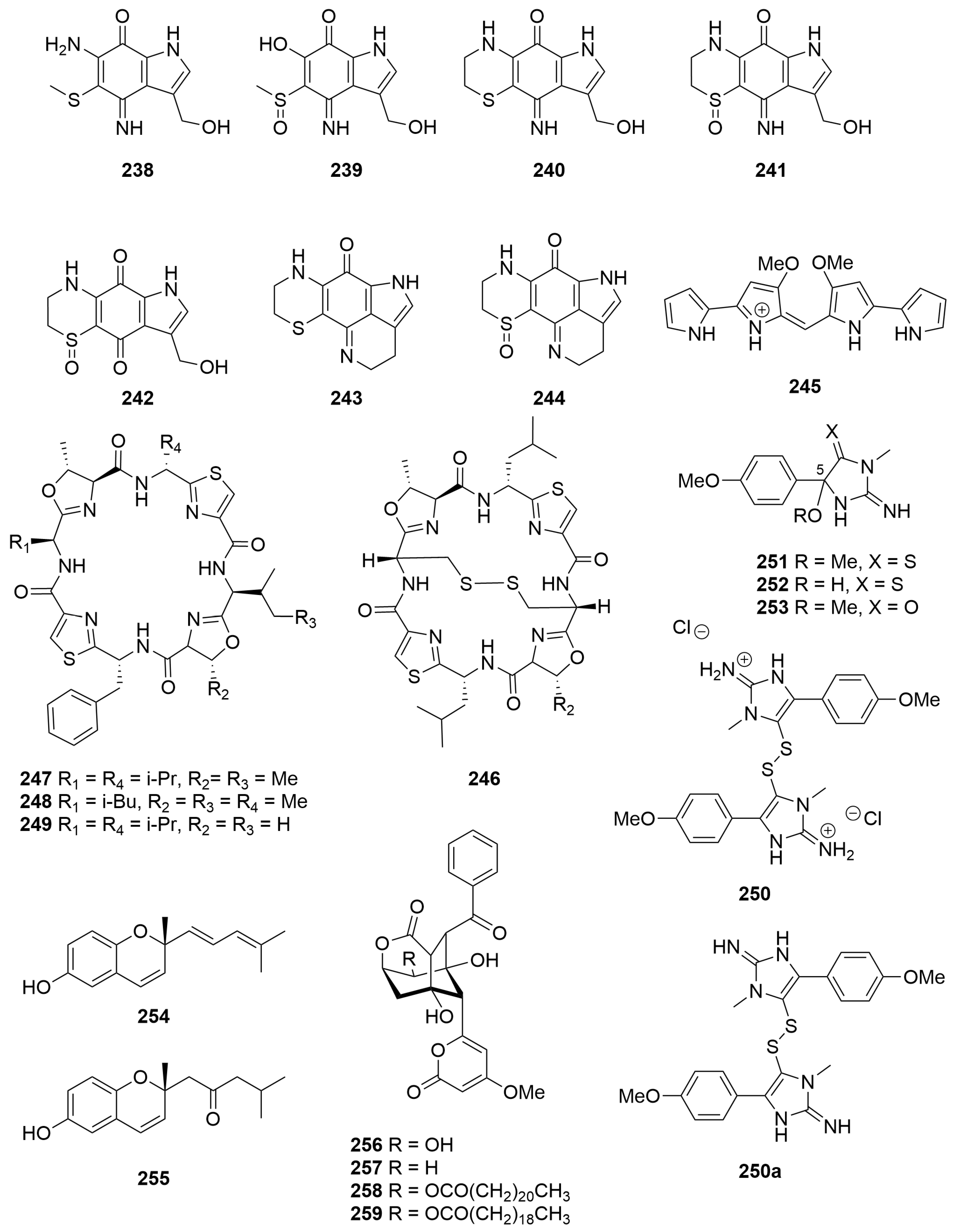
A sample of the stinging Hydroid Macrorhynchia philippina (Order: Leptothecata; Family: Aglaopheniidae), collected in the states northwest, afforded the novel pyrroloiminoquinone macrophilone A (238) (Figure 16) [92]. The structure of 238 was confirmed by total synthesis. Reinvestigation of the hydroid afforded the new metabolites, macrophilones B–G (239–244), the structures of which were elucidated using combined spectroscopic methods [93]. Compounds 238 to 244 are the first reported pyrroloiminoquinones from a marine Hydroid. The macrophilones 238–244 demonstrated inhibition of the enzymatic conjugation of SUMO to peptide substrates, and macrophilones A (238) and C (240) exhibited potent and specific cytotoxic activity in the NCI 60 cancer cell line panel [93]. Additionally, compound 238 showed sub-micromolar cytotoxicity towards lung adenocarcinoma cells [92].
2.3. Tunicata
Investigations of an ascidian collected in the Abrolhos Group afforded the deep blue tetra-pyrrole pigment 245 (Figure 16) [94]. The compound had previously been isolated from mutant strains of the Gram-negative bacterium Serratia marcescens, the structure of the pigment having been confirmed by total synthesis [95]. Microanalysis revealed that the ascidian pigment contained both chloride and bromide counter anions. Compound 245 exhibited an ability to increase the contractile force of guinea-pig ilea, with a dose-dependent increase evident [94].
Bioassay-guided fractionation of the colonial ascidian Lissoclinum patella (Order: Aplousobranchia; Family: Didemnidae), collected from the Montebello Archipelago, afforded three known cyclic peptides, ulithiacyclamide (246), lissoclinamide (247) and patellamide B (248), as well as the novel patellamide F (249) (Figure 16) [96]. The absolute configuration of pattelamide F (249) was confirmed by Marfey’s analysis. Patellamide B (248), patellamide F (249) and ulithiacyclamide (246) exhibited modest general cytotoxicity in the NCI 60 cell line human tumour screen with LC50 values of 48 µM, 13 µM and 3 µM, respectively [96].
A new dimeric disulfide alkaloid, polycarpine (250) (Figure 16), was isolated as its dihydrochloride salt from the extracts of the ascidian Polycarpa clavata (Order: Stolidobranchia; Family: Styelidae) [97]. Purification of the metabolite on silica afforded the free base 250a which readily decomposed to the monomeric products 251 to 253, arising from nucleophilic addition of water or methanol to position C-5 of the imidazole ring followed by cleavage of the disulfide bond [97]. Dissection of the organism into anatomical parts and fresh extraction in MeOH followed by immediate acquisition of NMR spectra demonstrated that compound 249 was the sole natural product and that it was located entirely in the organism’s branchial sac. Polycarpine dihydrochloride (250) exhibited cytotoxic activity against the human colon tumour cell line HCT-116 at 0.9 μg/mL [97].
Fresh specimens of a tunicate tentatively identified as Aplidium solidum (Order: Aplousobranchia; Family: Polyclinidae), collected in the Great Australian Bight, afforded the new chromenols (R)-2-methyl-2-(4-methylpenta-1,3-dienyl)-2H-chromen-6-ol (254) and 1-[(R)-6-hydroxy-2-methyl-2H-chromen-2-yl]-4-methylpentan-2-one (255) (Figure 16) [98]. The structures of 254 and 255 were elucidated spectroscopically. The absolute configuration of the metabolites was determined by hydrogenation and ozonolysis with acid work-up followed by comparison to the optical rotation of the known compound (R)-4,8-dimethylnonan-4-olide [98].
Chemical investigation of the brown encrusting ascidian Didemnum sp. (Order: Aplousobranchia; Family: Didemnidae), collected by hand using SCUBA, near Exmouth, afforded the known bacterial metabolite enterocin (256) as well as 5-deoxyenterocin (257), previously reported in a Japanese patent albeit with no spectroscopic data reported. Reversed-phase HPLC of the lipohilic fractions afforded the novel esters enterocin-5-behenate (258) and entorocin-5-arachidate (259) (Figure 16), assigned spectroscopically [99].
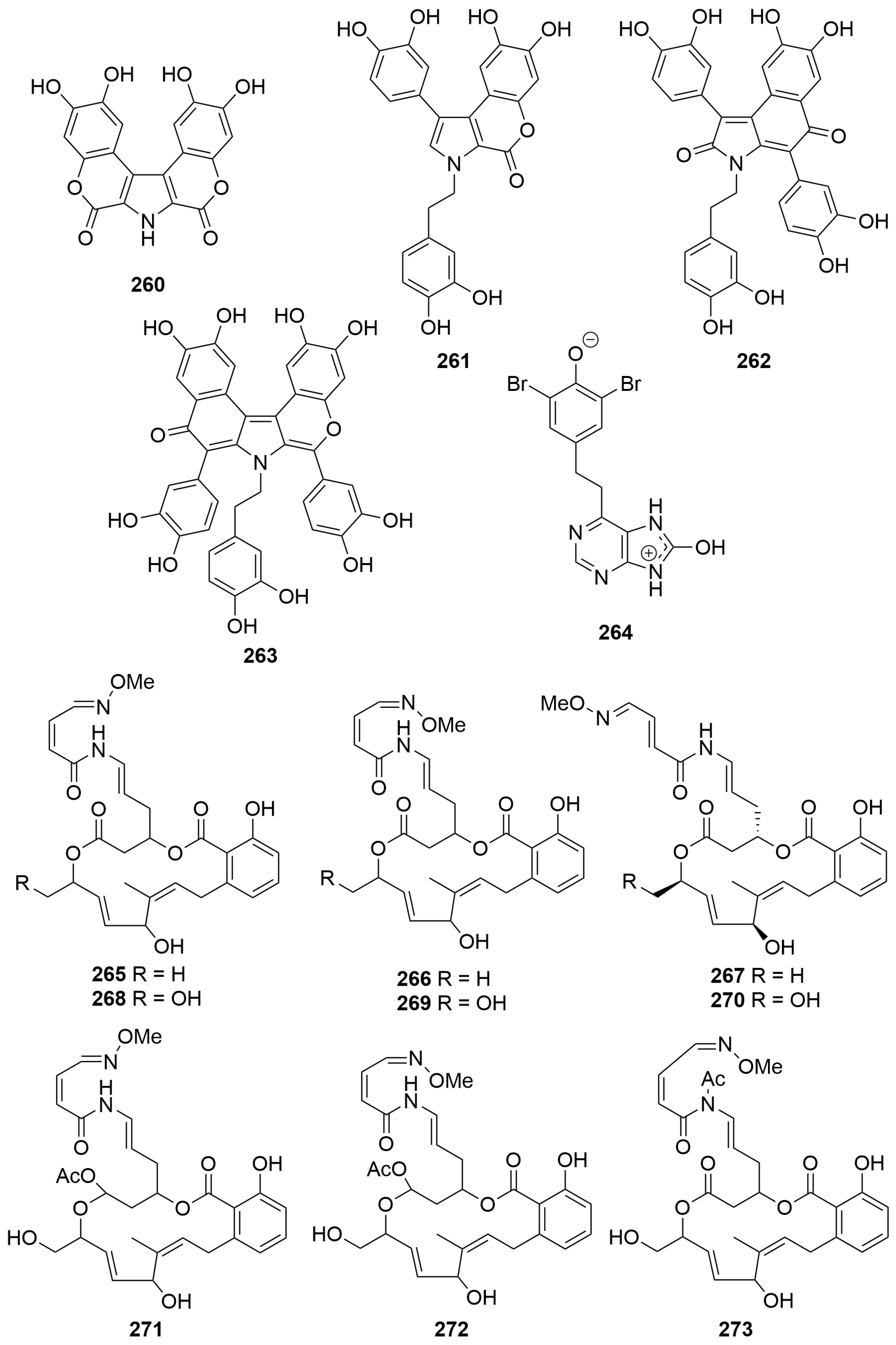
An undescribed ascidian Didemnum sp. (Order: Aplousobranchia; Family: Didemnidae), collected near Ningaloo Reef, yielded four new aromatic alkaloids ningalins A–D (260–263) (Figure 17), assigned structures 260 to 263 on the basis of spectroscopic analysis [100]. The authors propose a biosynthetic route to compounds 260 to 263 deriving from repeat condensation of DOPA. The absence of optical rotation in compounds 262 and 264 indicated that they are likely present as mixtures of racemates arising from rotamerism and helicity of the scaffolds [100].
Fractionation of an ascidian Aplidiopsis sp. (Order: Enterogona; Family: Polyclinidae) collected near Ningaloo Reef afforded the zwitterionic hydroxyadenine aplydiamine (264) (Figure 17). The structure of 264 was elucidated spectroscopically and by chemical derivatisation. The assignment of 264 as a zwitterion was based on HMBC correlations in d6-DMSO and the observed NOE correlation between all three exchangeable protons [101].
Two new cytotoxic macrolides, lobatamides A and B (265 and 266) (Figure 17), structurally related to the salicylihalamide class of macrolides, were isolated following bioassay-guided fractionation of an Aplidium lobatum (Order: Aplousobranchia; Family: Polyclinidae) [102]. Critical evidence for the structures of 265 and 266 was provided by analysis of the FAB-MS data. Re-investigation of the A. lobatum afforded the lobatamides C–F (267–270), the structures of which were elucidated spectroscopically [103]. The authors report the isolation of compounds 265 to 270 from three different shallow-water collections of Australian A. lobatum, an Aplidium sp. collected during a trawling expedition at the Great Australian Bight and finally from an unidentified, shallow-water collection of a Philippine tunicate. The authors note the spectral similarities between the lobatamides A–D (265–268) and the aplidites A–D, isolated from a Great Australian Bight Aplidum sp. [104], and propose revising the structures of the latter compounds to structures 265–268, respectively. The authors also propose revising the structures of the related aplidites E-G to structures 271 to 273 and renaming the natural products lobatamides G–I, respectively [103]. Given the reported isolation of lobatamide A from a species of terrestrial pseudomonad and the isolation of the related salicylihalamide macrolides from a marine sponge, the authors postulate a likely microbial origin for the family of compounds [103]. The relative and absolute configuration of lobatamide C (267) was subsequently confirmed following total synthesis of 267 by the Porco group [105]. Biological testing revealed that the lobatamides A–D (265–268) exhibited approximately equipotent specific cytotoxicity in the NCI 60 cell line human tumour screen (mean panel GI50′s ~1.6 nM). COMPARE pattern-recognition analysis revealed no significant correlations to the profiles of other known antitumour compounds, suggesting that compounds 265–268 may act by a novel mechanism of action. The differential cytotoxicity profiles of the compounds 265–268 did, however, show high (>0.7) COMPARE correlations among themselves, as well as with the salicylihalamides A and B (164, 165) isolated from the marine sponge Haliclona sp. The authors remark that the result is not surprising, given the structural similarities between the two compound families [103].
2.4. Echinodermata
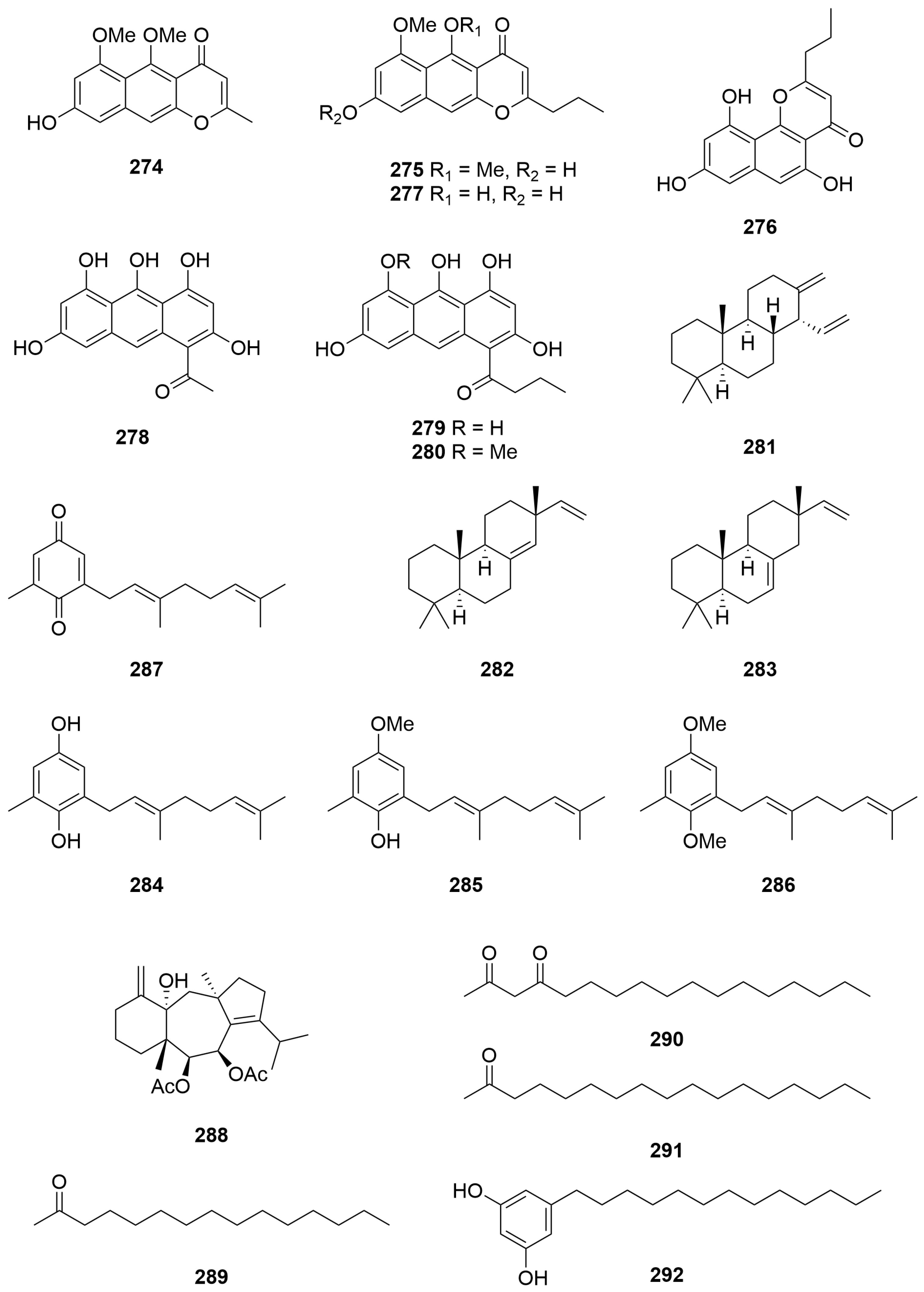
Investigation of the pigments of the crinoid Comantheria briareus (Bell) (Order: Comatulida; Family: Comatulidae) afforded the known naphthapyrone polyketides comantherin (273), neocomantherin (275) and comaparvin (276) as well as a novel pigment 5,8-dihydroxy-6-methoxy-2-propyl-4H-naphtho [2,3-b]pyran-4-one (277) (Figure 18). Isolation of the pigments present in Comatula solaris (Order: Comatulida; Family: Comatulidae) afforded the known compounds rhodolamprometrin (278) and rhodocomatulin-6,8-dimethyl ether (279). Chemical investigation of Comatula rotolaria afforded compounds 278 and 279 as well as rhodocomatulin-6-methyl ether (280) [106]. The echinoderm specimens analysed were all collected in the nets of prawn trawlers operating out of Carnarvon.
2.5. Plantae
A new cleistanthene diterpene hydrocarbon (281) was isolated from the leaves of Amphibolis antartica (Order: Alismatales; Family: Cymodoceaceae) collected from Shark Bay [107]. The structure of 281 was assigned spectroscopically. Chemical instability of the compound prevented degradative analysis. Samples of A. antartica collected near Perth contained 281, as well as the known derivatives sandaracopimaradiene (282) and isopunaradiene (283) (Figure 18), identified by GC-MS analysis. Analysis of individual specimens collected from Shark Bay by GC-MS revealed that the n-hydrocarbon content diminishes with maturity of the specimen, whereas concentration of 281 increases with leaf age [107].
2.6. Ochrophyta
Chromatography of the CH2Cl2 extracts of the brown algae Cystophora sp. (Order: Fucales; Family: Sargassaceae) collected from the wave-swept rock platforms of Cosy Corner, southwest WA, afforded three new isoprenoid dihydroquinones derived from geranyltoluquinol. The structures of the compounds were deduced as 284 to 286 (Figure 18) by 1H and 13C NMR spectroscopy and chemical interconversion [108]. Earlier isolation attempts had led to isolation of benzoquinone 287. The authors conclude that 287 is not a genuine natural product as acetylation of the Cystophora sp. crude extract led to the isolation of a diacetylated derivative of 284 and the observation that benzoquinone 287 was not present [108].
Isolation of the major lipophilic metabolite from a sample of Dictyota furcellata (Order: Dictyotales; Family: Dictyotaceae) collected from Cape Peron, Shark Bay, afforded the new dolastane diterpenoid (6S,7R,14S)-6,7-diacetoxydolasta-1(1S),8-dien-14-ol (288) [109]. The structure of 288 was deduced spectroscopically and confirmed by single-crystal X-ray diffraction.
Three acetogenin metabolites 289–291 and an alkyl resorcinol 292 (Figure 18) were isolated from the brown algae Caulocystis cephalornithos (Order: Fucales; Family: Cystoseiraceae) collected from Beacon Island, Wallabi Group. The structures of the metabolites were assigned as pentadecan-2-one (289), heptadecan-2,4-dione (290), heptadecan-2-one (291) and 5-tridecylresorcinol (292) on the basis of GC-MS and spectroscopic analysis [110].
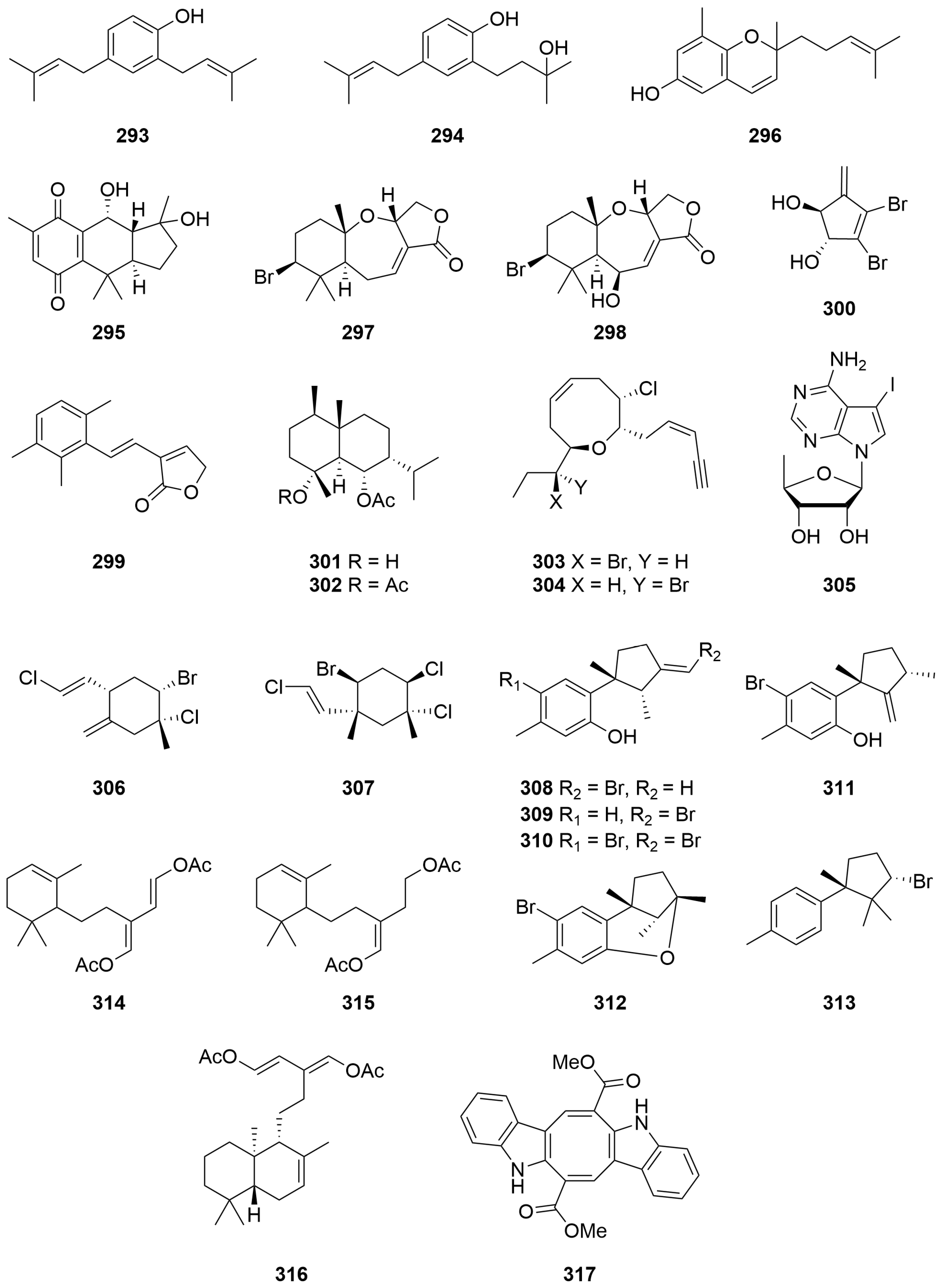
A sample of the brown alga Encyothalia cliftonii (Harvey) (Order: Sporochnales; Family: Sporochnaceae) afforded two bisprenylated phenols: 2,4-bis(3-methylbut-2-enyl)phenol (293), reported previously from Perithalia caudata and 2-(3-hydroxy-3-methylbutyl),4-(3-methylbut-2-enyl) phenol (294) (Figure 19) [111], the structures of which were solved spectroscopically and by chemical derivatisation. Biological testing indicated that 2,4-bis(3-methylbut-2-enyl)phenol (293) exhibited significant feeding deterrence towards the herbivorous sea urchin Tripneustes esculentus [111].
Chemical investigation of the brown alga Cystophora harveyi (Order: Fucales; Family: Sargassaceae), collected by SCUBA from the first bay to the east of the Cape Leeuwin lighthouse, yielded the new linearly fused tricyclic compound pycnanthaquinone C (295), the structure of which was elucidated spectroscopically [112]. Fractionation of the crude extract also afforded the known compounds atractylochromene (296), (2′E)-2-(3′,7′-dimethylocta-2′,6′-dienyl)-4-hydroxy-1-methoxy-6-methylbenzene (285), (2′E)-1,4-dimethoxy-(3′,7′-dimethylocta-2′,6′-dienyl)-6-methylbenzene (286) and (2′E)-2-(3′,7′-dimethylocta-2′,6′-dienyl)-6-methyl-2,5-cyclohexadiene-1,4-dione (287) (Figure 19). Atractylochromene (296) was reported for the first time from a marine organism. The authors provide a biosynthetic scheme for the formation of compounds 285 to 287 and 295 to 296. In addition, the authors note that compound 296 has previously been reported as an effective anti-inflammatory agent [112].
2.7. Rhodophyta
A sample of Laurencia filiformis (Order: Ceramiales; Family: Rhodomelaceae) collected from Point Peron yielded the sesquiterpene metabolites aplysisistatin (297), previously isolated from the sea hare, Aplysia angasi, as well as 6β-hydroxyaplysistatin (298) (Figure 19) [113]. The structures of 297 and 298 were assigned crystallographically. A chance observation led to the discovery that thermal rearrangement of 6β-hydroxyaplysistatin (298) afforded one major decomposition product 299 involving the formal loss of one unit of HBr and two units of water. The structure of the thermolysis product 299 was confirmed by Capon and Ghisalberti in a five-step total synthesis [114].
Chemical investigation of the red alga Vidalia spiralis (Order: Ceramiales; Family: Rhodomelaceae) collected at Yanchep yielded the new halogenated diol 3,4-dibromo-5-methylenecyclopent-3-ene-1,2-diol (300) (Figure 19) as a fine crystalline powder [115]. The structure of 300 was determined spectroscopically and by chemical derivatisation. Attempts to monoacetylate the diol failed, precluding the use of Horeau’s method to determine the absolute configuration of the natural product. The Vidalia spiralis crude dichloromethane extract exhibited hypotensive activity, and the crude methanol extract exhibited stimulant activity. Neither of these activities was evident, however, in the purified compound [115].
Fractionation of the lipophilic extracts of a sample of the red algae Laurencia filiformis (Order: Ceramiales; Family: Rhodomelaceae) yielded the novel brominated eudesmane sesquiterpenes austradiol acetatete (301) and austradiol diacetate (302) as well as the known cis-dihydrorhodophytin (303) and cis-epidihydrorhodophytin (304) (Figure 19) [116]. The structures of 301 and 302 were elucidated spectroscopically and by chemical derivatisation. Evidence for the proposed twist-boat conformation of austradiol acetate (301) was provided by complexation with a europium chemical shift reagent [116].
Bioassay-guided fractionation of a methanolic extract of the red alga Hypnea valendiae (Order: Gigartinales; Family: Cystocloniaceae), collected at Quobba Lagoon, returned 4-amino-7-(5′-deoxyribose-1′β-yl)-5-iodopyrrolo [2,3-d]pyrimidine (305) (Figure 19) as the principle active metabolite, the compound exhibited an ability to induce muscle relaxation and hypothermia [78]. A minor metabolite isolated from a subsequent extraction of Hypnea valendiae was tentatively assigned as the α-1′ isomer 305 by 1H NMR. Paucity of material and the requirement for bioassay prevented further structural validation [78].
Direct sublimation of the methylene chloride soluble extract of Plocamium mertensii (Order: Plocamiales; Family: Plocamiaceae), collected at Carnac Island, yielded (1R,2S,4S,1′E)-2-bromo-l-chloro-4-(2′-chloroetheny1)-1-methyl-5-methylenecyclohexane (306). The structure of 306 was solved by single-crystal X-ray diffraction [117]. An unidentified Plocamium species collected from the beach wash on Rottnest Island yielded a small quantity of crystalline (1R,2S,4R,5R,1′E)-4-bromo-l,2-dichloro-5-(2′-chloroetheny1)-l,5-dimethylcyclohexane (307) (Figure 19), the structure of which was also solved by single-crystal X-ray diffraction [117]. (1R,2S,4S,1′E)-2-Bromo-1-chloro-4-(2′-chloroethenyl)-1-methyl-5-methylenecyclohexane (306) exhibited unusual biological activity. Apart from weak cytostatic and antibacterial activity, the authors note that the compound produced a ‘spastic’ syndrome in mice which persisted for several days but was, however, reversible [117].
Samples of Laurencia filimformis f. heteroclada (Order: Ceramiales; Family: Rhodomelaceae) were collected from four sites along the West Australian coast. All of the samples were found to afford laurene sesquiterpene metabolites [118]. From the sample collected from Hamelin Bay was isolated allo-laurentirol (308). The sample from Lancelin afforded laurenisol (309), and the sample collected from Cottesloe Beach afforded bromolaurenisol (310). Fractionation of the sample collected at Shoalwater Bay, Rockingham, yielded laurenisol (309), bromolaurenisol (310), isolaurentirol (311), filiformin (312) and (−)-α-bromocuparene (313) (Figure 19) [118].
2.8. Chlorophyta
Two sesquiterpene metabolites were isolated from a specimen of Caulerpa flexilis var. muelleri (Order: Bryopsidales; Family: Caulerpaceae) collected from Cosy Corner. The structure of the metabolites was confirmed as (1E,3E)-2-[2′-(2″,6″,6″-trimethylcyclohex-2″-enyl)ethyl]buta-1,3-diene-I,4-diyl diacetate (314) and (2E)-3-formyl-5-(2′,6′,6′-trimethylcyclohex-2′-enyl)pent-2-enyl acetate (315) (Figure 19) using combined spectroscopic information [119]. The geometry of the trisubstituted double bond present on 314 was inferred from NOE experiments; however, the authors caution against conclusive assignment [119].
A sample of Caulerpa trifaria (Order: Bryopsidales; Family: Caulerpaceae) collected at Point Peron afforded the new sesquiterpene metabolite 316, the structure of which was deduced spectroscopically. The absolute configuration of the compound remains unknown. Samples of C. brownii, C. pexilis, C. peltata and C. racemosa also collected from Point Peron failed to yield 316. However C. peltata and C. racemosa afforded caulerpin (317) in low yield as red-plate crystals [120].
2.9. Cyanophyta
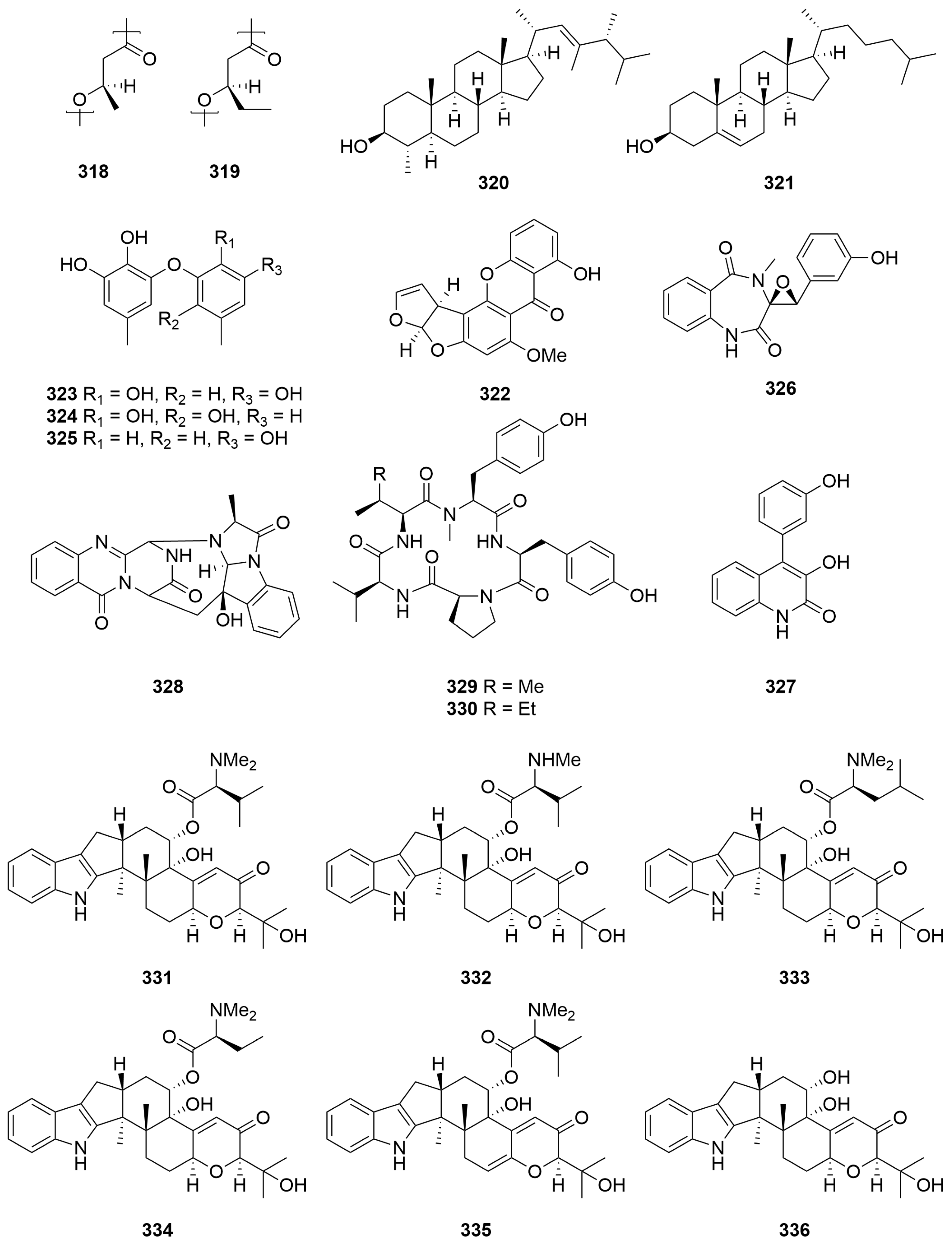
A sample of the freshwater cyanobacterium Aphanothece sp. (Order: Chroococcales; Family: Aphanothececeae) collected from Lake Joondalup afforded a polyester mixture composed of (R)-3-hydroxybutanoic acid (318) and (R)-3-hydroxypentanoic acid (319) in an approximate 2:1 ratio (Figure 20) [121]. The nature of the polymer was determined by hydrolysis, followed by spectral, chiroptical and GC-MS characterisation. Chemical analysis of Microcoleus sp. (Order: Oscillatoriales; Family: Microcoleaceae), Lyngbya aestuani (Order: Oscillatoriales; Family: Oscillatoriaceae), Murocoleur (Microcoleus) chthonoplastes and Entophysalis deusta (Order: Chroococcales; Family: Entophysalidaceae) stromatolite cyanobacterial mats collected from Shark Bay revealed the presence of similar polyesters to those found in Aphanothece sp. collected from Lake Joondalup [121].
2.10. Dinoflagelatta
Capillary GC-MS analysis of four closely related species of marine dinoflagellate identified dinosterol (320) (Figure 20) as the major sterol constituent of Prorocentrum balticum (Order: Prorocentrales; Family: Prorocentraceae) and Prorocentrum minimum [122]. Cholesterol (321) (Figure 20) was found to be the major constituent of Prorocentrum micans and Prorocentrum mexicanum [122]. Other steroid components were identified and annotated by GC-MS for all four species. The authors propose that the similarity of steroidal fractions from members of the same species grown in different laboratories suggests a strong genetic, rather than environmental, influence on the steroidal composition of such species and that the steroidal profiles reported may be used to delineate the species chemotaxonomically [122].
2.11. Fungi
A marine-derived Aspergillus versicolor (MST-MF495) (Order: Eurotiales; Family: Trichocomaceae) isolated from a sample of beach sand collected at Cottesloe afforded the known compounds sterigmatocystin (322), violaceol I (323), violaceol II (324), diorcinol (325), (−)-cyclopenol (326) and viridicatol (327), as well as the novel alkaloid cottoquinazoline A to which was assigned the partial relative stereostructure 328, and the two novel cyclic pentapeptides cotteslosins A and B (329, 330) (Figure 20) [123]. The structures of 328 to 330 were assigned spectroscopically and via the modified C3-Marfey’s Analysis. Violaceol I (323), violaceol II (324) and diorcinol (325) exhibited antibacterial properties. Additional biological testing indicated that the novel peptide 329 showed weak cytotoxic activity against human melanoma (MM418c5, EC50 = 66 µg/mL), prostate (DU145, EC50 = 90 µg/mL) and breast (T47D, EC50 = 94 µg/mL) cancer cell lines. Cotteslosin B (330) was reported to exhibit weak cytotoxic activity [123].
Recently, cultivation of a marine-derived Aspergillus noonimiae collected in waters near Perth afforded the indolic diterpenes noonindoles A–F (331–336) (Figure 20) as well as a number of minor metabolites putatively assigned via tandem MS analysis. Structures of the major compounds were assigned following detailed spectroscopic analysis and single-crystal X-ray diffraction. Testing of the metabolites against a panel of microorganisms revealed that the compounds were essentially devoid of biological activity, with the exception of mild antifungal activity displayed by 331 against Candida albicans [124].
2.12. Arsenic Metabolism in the Marine Food Web
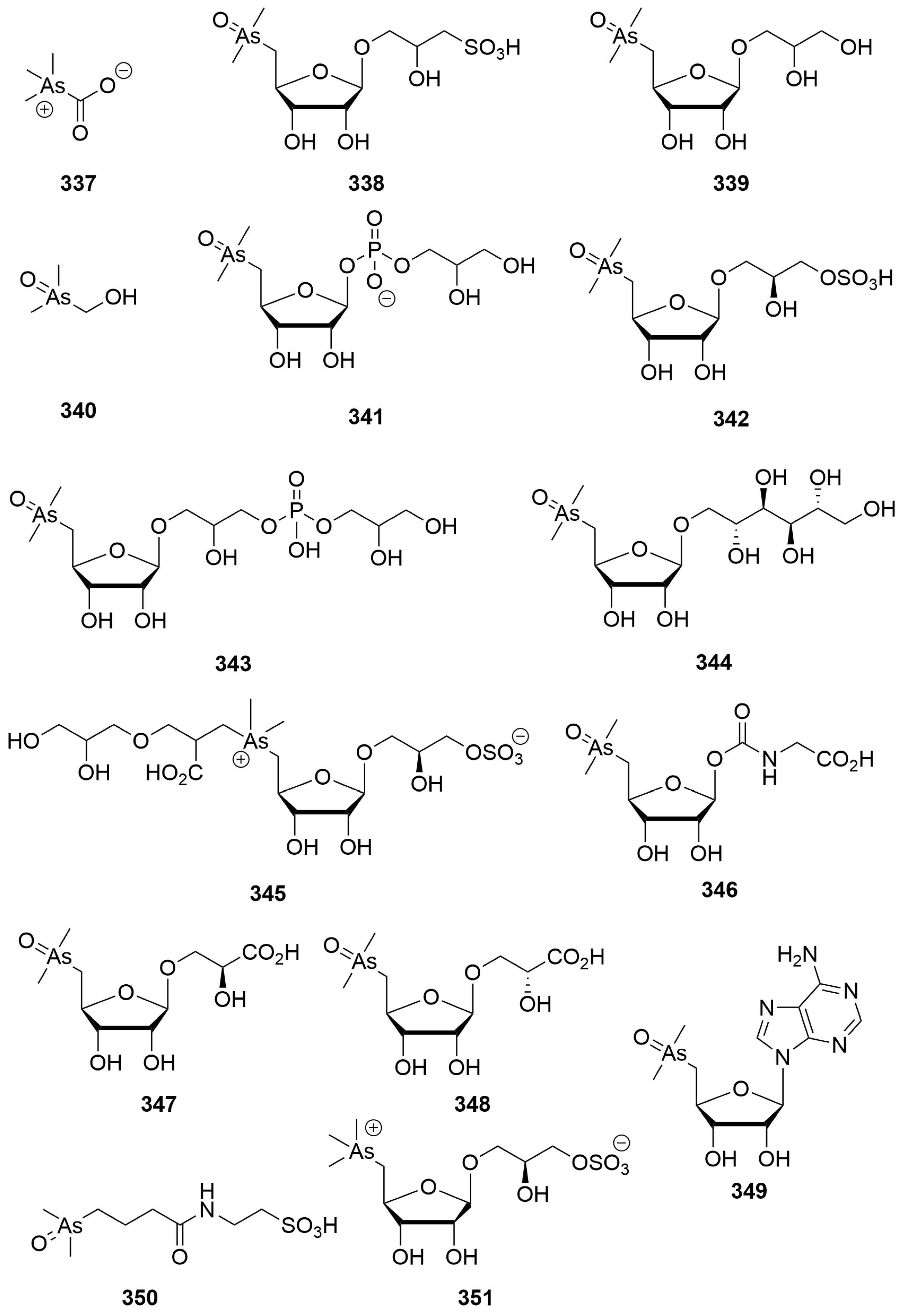
Vapour generation atomic absorption spectrometry guided fractionation of the commercially important western rock lobster, Panulirus cygnus (George) (Order: Decapoda; Family: Paluniridae), afforded arsenobetaine (337) (Figure 21) as the principal arsenic-containing metabolic constituent [125]. The structure of 337 was elucidated crystallographically and confirmed by total synthesis [125].
Investigation of the arsenical constituents of the brown kelp Ecklonia radiata (Order: Laminariales; Family: Lessoniaceae) afforded the metabolites 2-hydroxy-3-sulphopropyl-S-deoxy-5-(dimethylarsenoso)furanoside (338) and a 2,3-dihydroxypropyl-S-deoxy-S-(dimethylarsenoso)furanoside (339) (Figure 21) [126]. The authors propose that the compounds may act as intermediates between arsenate present in seawater and arsenobetaine present at higher trophic levels [126]. Key evidence for this proposal came from experiments investigating the anaerobic decomposition Ecklonia radiata, affording the new compound dimethyloxarsylethanol (340) [127]. Additional investigations of Ecklonia radiata yielded the minor metabolite 3-glycerophosphoryl-2-hydroxy-I-[5-deoxy-5-(dimethylarsinoyl)-β-ribofuranosyloxy]propane (341). The structure of this metabolite was elucidated principally by NMR spectroscopy [128].
Compound 342, as well as (2S)-3-[5-deoxy-5-(dimethylarsinoyl)-β-D-ribofuranosyloxy]-2-hydroxypropyl hydrogen sulfate (343), was isolated from the kidney of the giant clam, Tridacna maxima (Order: Cardiida; Family: Cardiidae), collected from Shark Bay [129]. The structure of 343 was solved using single-crystal X-ray diffraction. Targeted isolation of arsenical metabolites from the brown alga Sargassum lacerifolium (Order: Fucales; Family: Sargassaceae) afforded two new ribosides methyl 5-deoxy-5-(dimethylarsinoyl)-β-D-riboside (344) and 1-O-[5′-deoxy-5′-(dimethylarsinoyl)-β-D-ribosyl] mannitol (345), in addition to five known arsenic-containing ribosides [130].
Two batches of Tridacna maxima were re-analysed for additional arsenical metabolites, the first batch, collected from Exmouth in 1981, afforded three novel compounds, N-(5′-deoxy-5′-dimethylarsinoyl-β-D-ribosyloxycarbonyl) glycine (346), (2S)-3-(5′-deoxy-5′-dimethylarsinoyl-β-D-ribosyloxy)-2-hydroxypropanoic acid (347) and (2R)-3-(5′-deoxy-5′-dimethylarsinoyl-β-D-ribosyloxy)-2-hydroxypropanoic acid (348), as well as four dimethylarsinoylribosides reported previously (Figure 21) [131]. The second batch, collected from Exmouth in 1988, afforded two novel compounds: the arsenic-containing nucleoside 9-(5′-deoxy-5′-dimethylarsinoyl)-9H-adenosine (349), in addition to the taurine-conjugated N-4-dimethylarsinoyl)butanoyl]taurine (350). The second collection also afforded the previously reported (2S)-3-[(5′-deoxy-5′-trimethylarsonio-β-D-ribosyloxy)-2-hydroxypropyl] sulfate (351). The structures of 346 to 351 were validated by total synthesis [131]. The authors propose a biogenetic scheme for the formation of compounds 346 to 351 deriving from donation of all three alkyl groups present on S-adenosyl methionine to inorganic arsenate present in seawater [130].
3. Conclusions
Here we have reviewed the marine natural products that have been reported from the fauna and flora of Western Australian waters. This review describes the identification of over 350 metabolites representing a diverse array of chemical compounds that have been reported over the past 40 years. Most of the compounds have also been reported to display some biological activity in line with the high rates of bioactivity studies of marine natural products reported elsewhere [132,133].
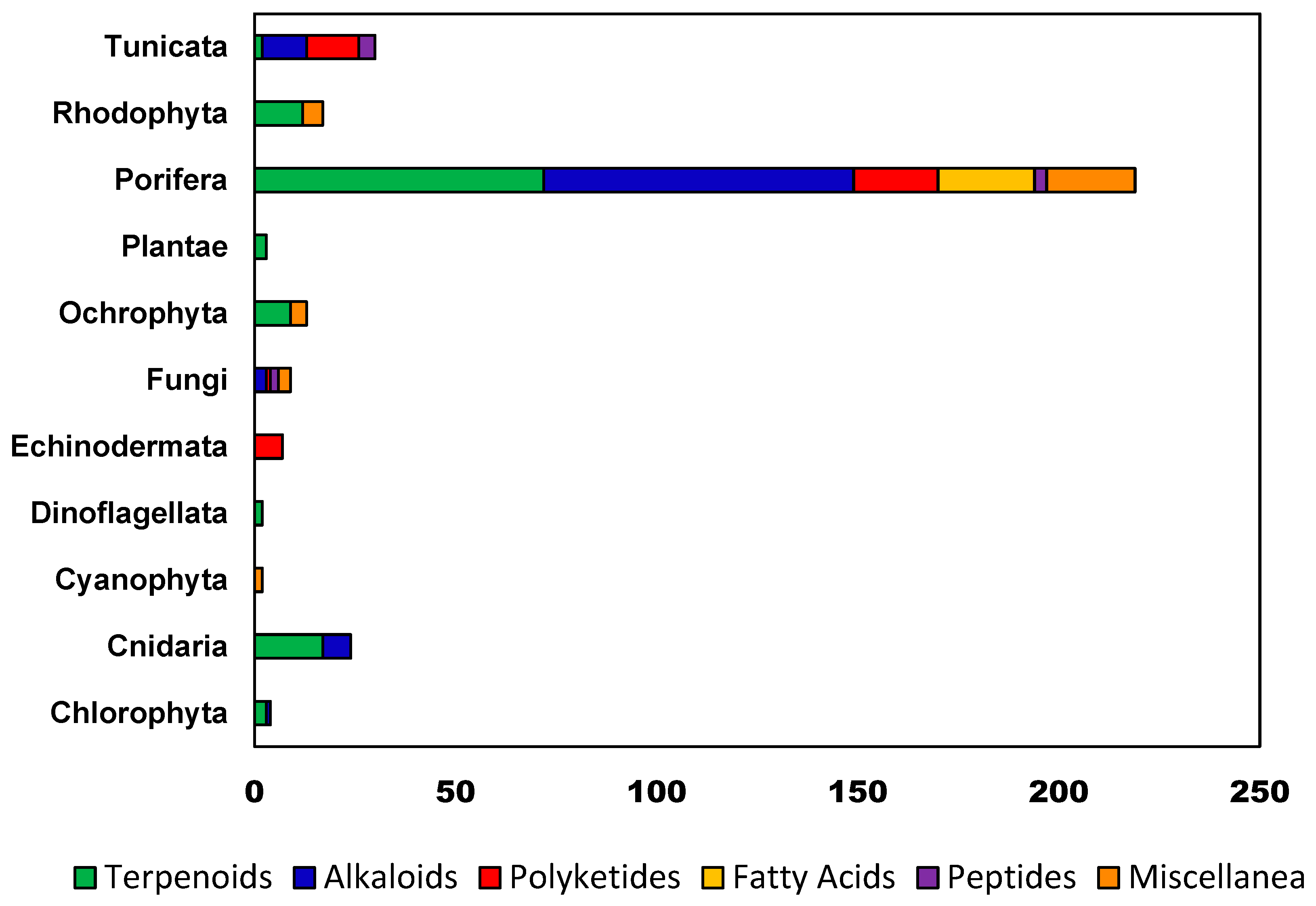
A statistical analysis of the distribution of marine natural products from various taxonomic sources and the percentage of each structural class among all the marine natural products reported from Western Australia (Figure 22) reveals that studies on Porifera have by far yielded the largest and most diverse number of compounds with 220 metabolites reported, from all six arbitrary biogenetic groupings. In general terms, it is likely that the quantity and relative percentage of compound classes isolated are, in part, a reflection of the biosynthetic potential of the organisms (and associated microbiota) under investigation and partly attributable to the interests of the lead researchers involved, as well as the chromatographic and analytical technology available at the time. The former observation is likely true of work conducted on Echinoderms, where all the metabolites reported from Western Australian species are polyketidic anthraquinones, a biogenic grouping known to be of chemotaxonomic relevance to the phylum [134]. The latter observation is afforded some support when analysing the extensive proportion of terpenoids isolated from Rhodophytes and Ochrophytes, work that was overwhelmingly conducted in the early 1980s by the Ghisalberti group, using predominately normal-phase column chromatography, compatible with the typically lipophilic metabolites reported. Subsequent research on alternative taxa, conducted from the 1990s to present, shows a trend towards compounds of increasingly varied biosynthetic provenance, including a higher proportion of alkaloids and polyketides, evident when analysing distributions of isolated metabolites from Porifera, Tunicata and Fungi. This trend can be explained when considering the proliferation of high- and ultra-high-performance liquid chromatography instruments, in analytical and preparative modes, as well as the propensity of researchers to operate under typically reversed-phase conditions, facilitating the purification and analysis of increasingly polar metabolites.
While there has been extensive research into marine natural products originating from other major marine biodiversity hotspots, such as the Americas, Southeast Asia, Japan, Eastern and Southern Australia and New Zealand, there have been relatively few major studies of marine natural products from Western Australia. This is the largest coastline of Australia, and biodiversity studies suggest that Western Australia marine areas are a source of significant biodiversity with many of the species remaining largely uncharacterised and underexplored [6,135,136,137]. In recent times, there have been a number of biodiversity expeditions to explore the species richness of the coastline, but chemical studies of these have so far been lacking. These recent expeditions have subsequently led to the advent of the Western Australian Marine Science Library (WAMBL), where collected specimens have been deposited for future genetic, biological and chemical analysis. The WAMBL provides researchers with easier access to specimens that were previously difficult to obtain, such as deep-sea marine sponges (>100 m). The variety of unclassified species within the WAMBL makes it of high interest for chemical and biological studies such as those we have started recently [34,53,67,75].
In an age of superbugs and viral pandemics, the need for discovering new anti-infective agents is paramount [138,139], and marine natural products are well known as a significant source of biologically active compounds [140]. To that end, the relatively underexplored chemical diversity of species occurring along the Western Australian coastline may offer many more opportunities in this area.
Author Contributions
Writing—original draft preparation, S.S. and S.K.M.; writing—review and editing, G.R.F.; visualisation, S.S.; supervision, G.R.F.; project administration, G.R.F. All authors have read and agreed to the published version of the manuscript.
Funding
This research received no external funding.
Institutional Review Board Statement
Not applicable.
Informed Consent Statement
Not applicable.
Data Availability Statement
Not applicable.
Acknowledgments
Samuele Sala and Scott K. Micke would like to acknowledge financial support from an Australian Government Research Training Program (RTP) scholarship.
Conflicts of Interest
The authors declare no conflict of interest.
References
- Newman, D.J.; Cragg, G.M. Natural Products as Sources of New Drugs over the Nearly Four Decades from 01/1981 to 09/2019. J. Nat. Prod. 2020, 83, 770–803. [Google Scholar] [CrossRef]
- Newman, D.J.; Cragg, G.M. Natural Products as Sources of New Drugs from 1981 to 2014. J. Nat. Prod. 2016, 79, 629–661. [Google Scholar] [CrossRef]
- Capon, R.J. Marine Natural Products Chemistry: Past, Present, and Future. Aust. J. Chem. 2010, 63, 851–854. [Google Scholar] [CrossRef]
- Molinski, T.F.; Dalisay, D.S.; Lievens, S.L.; Saludes, J.P. Drug development from marine natural products. Nat. Rev. Drug Discov. 2009, 8, 69–85. [Google Scholar] [CrossRef] [PubMed]
- Fromont, J.; Althaus, F.; McEnnulty, F.R.; Williams, A.; Salotti, M.; Gomez, O.; Gowlett-Holmes, K. Living on the edge: The sponge fauna of Australia’s southwestern and northwestern deep continental margin. Hydrobiologia 2012, 687, 127–142. [Google Scholar] [CrossRef]
- Schönberg, C.H.L.; Fromont, J. Sponge gardens of Ningaloo Reef (Carnarvon Shelf, Western Australia) are biodiversity hotspots. Hydrobiologia 2012, 687, 143–161. [Google Scholar] [CrossRef]
- Searle, P.A.; Molinski, T.F. Phorboxazoles A and B: Potent cytostatic macrolides from marine sponge Phorbas species. J. Am. Chem. Soc. 1995, 117, 8126–8131. [Google Scholar] [CrossRef]
- Erickson, K.L.; Beutler, J.A.; Cardellina, J.H.; Boyd, M.R. Salicylihalamides A and B, Novel Cytotoxic Macrolides from the Marine Sponge Haliclona sp. J. Org. Chem. 1997, 62, 8188–8192. [Google Scholar] [CrossRef]
- Boyd, M.R.; Farina, C.; Belfiore, P.; Gagliardi, S.; Kim, J.W.; Hayakawa, Y.; Beutler, J.A.; McKee, T.C.; Bowman, B.J.; Bowman, E.J. Discovery of a novel antitumor benzolactone enamide class that selectively inhibits mammalian vacuolar-type (H+)-ATPases. J. Pharmacol. Exp. Ther. 2001, 297, 114–120. [Google Scholar]
- Lindel, T.; Jensen, P.R.; Fenical, W.; Long, B.H.; Casazza, A.M.; Carboni, J.; Fairchild, C.R. Eleutherobin, a New Cytotoxin that Mimics Paclitaxel (Taxol) by Stabilizing Microtubules. J. Am. Chem. Soc. 1997, 119, 8744–8745. [Google Scholar] [CrossRef]
- MarineLit. Available online: http://pubs.rsc.org/marinlit (accessed on 20 December 2022).
- Capon, R.; Ghisalberti, E.; Jefferies, P. New aromatic sesquiterpenes from a Halichondria sp. Aust. J. Chem. 1982, 35, 2583–2587. [Google Scholar] [CrossRef]
- Kazlauskas, R.; Murphy, P.; Wells, R. Five new C26 tetracyclic terpenes from a sponge (Lendenfeldia sp.). Aust. J. Chem. 1982, 35, 51–59. [Google Scholar] [CrossRef]
- Capon, R.J.; Ghisalberti, E.L.; Jefferies, P.R. A new furanoterpene from a Spongia sp. Experientia 1982, 38, 1444–1445. [Google Scholar] [CrossRef]
- Capon, R.J.; Jenkins, A.; Rooney, F.; Ghisalberti, E.L. Structure Revision and Assignment of Absolute Stereochemistry of a Marine C21 Bisfuranoterpene. J. Nat. Prod. 2001, 64, 638–639. [Google Scholar] [CrossRef]
- Cassidy, M.; Ghisalberti, E.; Jefferies, P.; Skelton, B.; White, A. New Tricyclic Diterpenes from the Sponge Higginsia sp. Aust. J. Chem. 1985, 38, 1187–1195. [Google Scholar] [CrossRef]
- Cassidy, M.P.; Ghisalberti, E.L. New Terpene Hydrocarbons from the Sponge Higginsia sp. J. Nat. Prod. 1993, 56, 1190–1193. [Google Scholar] [CrossRef]
- Searle, P.A.; Molinski, T.F. Scalemic 12-hydroxyambliofuran and 12-acetoxy-ambliofuran, five tetracyclic furanoditerpenes and a furanosesterterpene from Spongia sp. Tetrahedron 1994, 50, 9893–9908. [Google Scholar] [CrossRef]
- Rochfort, S.J.; Atkin, D.; Hobbs, L.; Capon, R.J. Hippospongins A−F: New Furanoterpenes from a Southern Australian Marine Sponge Hippospongia sp. J. Nat. Prod. 1996, 59, 1024–1028. [Google Scholar] [CrossRef]
- Capon, R.J.; Miller, M.; Rooney, F. Clathrins A−C: Metabolites from a Southern Australian Marine Sponge, Clathria Species. J. Nat. Prod. 2000, 63, 821–824. [Google Scholar] [CrossRef]
- Vuong, D.; Capon, R.J. Phorbasin A: A Novel Diterpene from a Southern Australian Marine Sponge, Phorbas Species. J. Nat. Prod. 2000, 63, 1684–1685. [Google Scholar] [CrossRef]
- McNally, M.; Capon, R.J. Phorbasin B and C: Novel Diterpenes from a Southern Australian Marine Sponge, Phorbas Species. J. Nat. Prod. 2001, 64, 645–647. [Google Scholar] [CrossRef] [PubMed]
- Zhang, H.; Capon, R.J. Phorbasins D−F: Diterpenyl-taurines from a Southern Australian Marine Sponge, Phorbas sp. Org. Lett. 2008, 10, 1959–1962. [Google Scholar] [CrossRef] [PubMed]
- Zhang, H.; Major, J.M.; Lewis, R.J.; Capon, R.J. Phorbasins G–K: New cytotoxic diterpenes from a southern Australian marine sponge, Phorbas sp. Org. Biomol. Chem. 2008, 6, 3811–3815. [Google Scholar] [CrossRef] [PubMed]
- Makarieva, T.N.; Rho, J.-R.; Lee, H.-S.; Santalova, E.A.; Stonik, V.; Shin, J. New Sesterterpene Sulfates from the Sponge Darwinella australensis. J. Nat. Prod. 2003, 66, 1010–1012. [Google Scholar] [CrossRef]
- Santalova, E.A.; Denisenko, V.A.; Dmitrenok, P.S.; Berdyshev, D.V.; Stonik, V.A. Two New Sarasinosides from the Sponge Melophlus sarasinorum. Nat. Prod. Commun. 2006, 1, 265–271. [Google Scholar] [CrossRef]
- El-Naggar, M.; Piggott, A.M.; Capon, R.J. Bistellettazines A−C and Bistellettazole A: New Terpenyl−Pyrrolizidine and Terpenyl−Imidazole Alkaloids from a Southern Australian Marine Sponge, Stelletta sp. Org. Lett. 2008, 10, 4247–4250. [Google Scholar] [CrossRef]
- Utkina, N.K.; Denisenko, V.A.; Krasokhin, V.B. Sesquiterpenoid Aminoquinones from the Marine Sponge Dysidea sp. J. Nat. Prod. 2010, 73, 788–791. [Google Scholar] [CrossRef]
- Zhang, H.; Khalil, Z.G.; Capon, R.J. Fascioquinols A–F: Bioactive meroterpenes from a deep-water southern Australian marine sponge, Fasciospongia sp. Tetrahedron 2011, 67, 2591–2595. [Google Scholar] [CrossRef]
- Dellar, G.; Djura, P.; Sargent, M.V. Structure and synthesis of a new bromoindole from a marine sponge. J. Chem. Soc. Perkin Trans. 1 1981, 1679–1680. [Google Scholar] [CrossRef]
- Franklin, M.A.; Penn, S.G.; Lebrilla, C.B.; Lam, T.H.; Pessah, I.N.; Molinski, T.F. Bastadin 20 and Bastadin O-Sulfate Esters from Ianthella basta: Novel Modulators of the Ry1R FKBP12 Receptor Complex. J. Nat. Prod. 1996, 59, 1121–1127. [Google Scholar] [CrossRef]
- Hong, T.W.; Jímenez, D.R.; Molinski, T.F. Agelastatins C and D, New Pentacyclic Bromopyrroles from the Sponge Cymbastela sp., and Potent Arthropod Toxicity of (−)-Agelastatin A. J. Nat. Prod. 1998, 61, 158–161. [Google Scholar] [CrossRef] [PubMed]
- Ovenden, S.P.B.; Capon, R.J. Echinosulfonic Acids A−C and Echinosulfone A: Novel Bromoindole Sulfonic Acids and a Sulfone from a Southern Australian Marine Sponge, Echinodictyum. J. Nat. Prod. 1999, 62, 1246–1249. [Google Scholar] [CrossRef] [PubMed]
- Sala, S.; Nealon, G.L.; Sobolev, A.N.; Fromont, J.; Gomez, O.; Flematti, G.R. Structure Reassignment of Echinosulfone A and the Echinosulfonic Acids A–D Supported by Single-Crystal X-ray Diffraction and Density Functional Theory Analysis. J. Nat. Prod. 2020, 83, 105–110. [Google Scholar] [CrossRef] [PubMed]
- Holland, D.C.; Kiefel, M.J.; Carroll, A.R. Structure Revisions of the Sponge-Derived Dibrominated Bis-indole Alkaloids, Echinosulfone A and the Echinosulfonic Acids A to D. J. Org. Chem. 2020, 85, 3490–3496. [Google Scholar] [CrossRef] [PubMed]
- Neupane, P.; Salim, A.A.; Capon, R.J. Structure revision of the rare sponge metabolite echinosulfone A, and biosynthetically related echinosulfonic acids A–D. Tetrahedron Lett. 2020, 61, 151651. [Google Scholar] [CrossRef]
- Capon, R.J.; Vuong, D.; Lacey, E.; Gill, J.H. (−)-Echinobetaine A: Isolation, Structure Elucidation, Synthesis, and SAR Studies on a New Nematocide from a Southern Australian Marine Sponge, Echinodictyum sp. J. Nat. Prod. 2005, 68, 179–182. [Google Scholar] [CrossRef]
- Capon, R.J.; Vuong, D.; McNally, M.; Peterle, T.; Trotter, N.; Lacey, E.; Gill, J.H. (+)-Echinobetaine B: Isolation, structure elucidation, synthesis and preliminary SAR studies on a new nematocidal betaine from a southern Australian marine sponge, Echinodictyum sp. Org. Biomol. Chem. 2005, 3, 118–122. [Google Scholar] [CrossRef]
- Dijoux, M.-G.; Gamble, W.R.; Hallock, Y.F.; Cardellina, J.H.; van Soest, R.; Boyd, M.R. A New Discorhabdin from Two Sponge Genera. J. Nat. Prod. 1999, 62, 636–637. [Google Scholar] [CrossRef]
- Groweiss, A.; Newcomer, J.J.; O’Keefe, B.R.; Blackman, A.; Boyd, M.R. Cytotoxic Metabolites from an Australian Collection of the Sponge Jaspis Species. J. Nat. Prod. 1999, 62, 1691–1693. [Google Scholar] [CrossRef]
- Ovenden, S.P.B.; Nielson, J.L.; Liptrot, C.H.; Willis, R.H.; Tapiolas, D.M.; Wright, A.D.; Motti, C.A. A New Diketopiperazine, Cyclo-(4-S-hydroxy-R-proline-R-isoleucine), from an Australian Specimen of the Sponge Stelletta sp. Mar. Drugs 2011, 9, 2469–2478. [Google Scholar] [CrossRef]
- Capon, R.J.; Miller, M.; Rooney, F. Mirabilin G: A New Alkaloid from a Southern Australian Marine Sponge, Clathria Species. J. Nat. Prod. 2001, 64, 643–644. [Google Scholar] [CrossRef]
- El-Naggar, M.; Conte, M.; Capon, R.J. Mirabilins revisited: Polyketide alkaloids from a southern Australian marine sponge, Clathria sp. Org. Biomol. Chem. 2010, 8, 407–412. [Google Scholar] [CrossRef]
- Grkovic, T.; Blees, J.S.; Bayer, M.M.; Colburn, N.H.; Thomas, C.L.; Henrich, C.J.; Peach, M.L.; McMahon, J.B.; Schmid, T.; Gustafson, K.R. Tricyclic Guanidine Alkaloids from the Marine Sponge Acanthella cavernosa that Stabilize the Tumor Suppressor PDCD4. Mar. Drugs 2014, 12, 4593–4601. [Google Scholar] [CrossRef] [PubMed]
- Moon, S.-S.; MacMillan, J.B.; Olmstead, M.M.; Ta, T.A.; Pessah, I.N.; Molinski, T.F. (+)-7S-Hydroxyxestospongin A from the Marine Sponge Xestospongia sp. and Absolute Configuration of (+)-Xestospongin D. J. Nat. Prod. 2002, 65, 249–254. [Google Scholar] [CrossRef] [PubMed]
- Chang, L.C.; Otero-Quintero, S.; Hooper, J.N.A.; Bewley, C.A. Batzelline D and Isobatzelline E from the Indopacific Sponge Zyzzya fuliginosa. J. Nat. Prod. 2002, 65, 776–778. [Google Scholar] [CrossRef] [PubMed]
- Capon, R.J.; Peng, C.; Dooms, C. Trachycladindoles A–G: Cytotoxic heterocycles from an Australian marine sponge, Trachycladus laevispirulifer. Org. Biomol. Chem. 2008, 6, 2765–2771. [Google Scholar] [CrossRef] [PubMed]
- Herb, R.; Carroll, A.R.; Yoshida, W.Y.; Scheuer, P.J.; Paul, V.J. Polyalkylated cyclopentindoles: Cytotoxic fish antifeedants from a sponge, Axinella sp. Tetrahedron 1990, 46, 3089–3092. [Google Scholar] [CrossRef]
- Khokhar, S.; Feng, Y.; Campitelli, M.R.; Quinn, R.J.; Hooper, J.N.A.; Ekins, M.G.; Davis, R.A. Trikentramides A–D, Indole Alkaloids from the Australian Sponge Trikentrion flabelliforme. J. Nat. Prod. 2013, 76, 2100–2105. [Google Scholar] [CrossRef]
- Smith, S.G.; Goodman, J.M. Assigning Stereochemistry to Single Diastereoisomers by GIAO NMR Calculation: The DP4 Probability. J. Am. Chem. Soc. 2010, 132, 12946–12959. [Google Scholar] [CrossRef]
- Salib, M.N.; Molinski, T.F. Six Trikentrin-like Cyclopentanoindoles from Trikentrion flabelliforme. Absolute Structural Assignment by NMR and ECD. J. Org. Chem. 2018, 83, 1278–1286. [Google Scholar] [CrossRef]
- Ragini, K.; Fromont, J.; Piggott, A.M.; Karuso, P. Enantiodivergence in the Biosynthesis of Bromotyrosine Alkaloids from Sponges? J. Nat. Prod. 2017, 80, 215–219. [Google Scholar] [CrossRef]
- Shrestha, S.; Sorolla, A.; Fromont, J.; Blancafort, P.; Flematti, G.R. Crambescidin 800, Isolated from the Marine Sponge Monanchora viridis, Induces Cell Cycle Arrest and Apoptosis in Triple-Negative Breast Cancer Cells. Mar. Drugs 2018, 16, 53. [Google Scholar] [CrossRef] [PubMed]
- Searle, P.A.; Molinski, T.F.; Brzezinski, L.J.; Leahy, J.W. Absolute Configuration of Phorboxazoles A and B from the Marine Sponge Phorbas sp. 1. Macrolide and Hemiketal Rings. J. Am. Chem. Soc. 1996, 118, 9422–9423. [Google Scholar] [CrossRef]
- Molinski, T.F. Absolute configuration of phorboxazoles A and B from the marine sponge, Phorbas sp. 2. C43 and complete stereochemistry. Tetrahedron Lett. 1996, 37, 7879–7880. [Google Scholar] [CrossRef]
- Skepper, C.K.; MacMillan, J.B.; Zhou, G.-X.; Masuno, M.N.; Molinski, T.F. Chlorocyclopropane Macrolides from the Marine Sponge Phorbas sp. Assignment of the Configurations of Phorbasides A and B by Quantitative CD. J. Am. Chem. Soc. 2007, 129, 4150–4151. [Google Scholar] [CrossRef] [PubMed]
- MacMillan, J.B.; Xiong-Zhou, G.; Skepper, C.K.; Molinski, T.F. Phorbasides A−E, Cytotoxic Chlorocyclopropane Macrolide Glycosides from the Marine Sponge Phorbas sp. CD Determination of C-Methyl Sugar Configurations. J. Org. Chem. 2008, 73, 3699–3706. [Google Scholar] [CrossRef] [PubMed]
- Dalisay, D.S.; Morinaka, B.I.; Skepper, C.K.; Molinski, T.F. A Tetrachloro Polyketide Hexahydro-1H-isoindolone, Muironolide A, from the Marine Sponge Phorbas sp. Natural Products at the Nanomole Scale. J. Am. Chem. Soc. 2009, 131, 7552–7553. [Google Scholar] [CrossRef]
- Dalisay, D.S.; Molinski, T.F. Structure Elucidation at the Nanomole Scale. 2. Hemi-phorboxazole A from Phorbas sp. Org. Lett. 2009, 11, 1967–1970. [Google Scholar] [CrossRef]
- Dalisay, D.S.; Molinski, T.F. NMR Quantitation of Natural Products at the Nanomole Scale. J. Nat. Prod. 2009, 72, 739–744. [Google Scholar] [CrossRef]
- Dalisay, D.S.; Molinski, T.F. Structure Elucidation at the Nanomole Scale. 3. Phorbasides G−I from Phorbas sp. J. Nat. Prod. 2010, 73, 679–682. [Google Scholar] [CrossRef]
- Xiao, Q.; Young, K.; Zakarian, A. Total Synthesis and Structural Revision of (+)-Muironolide A. J. Am. Chem. Soc. 2015, 137, 5907–5910. [Google Scholar] [CrossRef] [PubMed]
- Capon, R.J.; Skene, C.; Liu, E.H.; Lacey, E.; Gill, J.H.; Heiland, K.; Friedel, T. Esmodil: An Acetylcholine Mimetic Resurfaces in a Southern Australian Marine Sponge Raspailia (Raspailia) sp. Nat. Prod. Res. 2004, 18, 305–309. [Google Scholar] [CrossRef] [PubMed]
- Abdo, D.A.; Motti, C.A.; Battershill, C.N.; Harvey, E.S. Temperature and Spatiotemporal Variability of Salicylihalamide A in the Sponge Haliclona sp. J. Chem. Ecol. 2007, 33, 1635–1645. [Google Scholar] [CrossRef]
- Ovenden, S.P.B.; Capon, R.J.; Lacey, E.; Gill, J.H.; Friedel, T.; Wadsworth, D. Amphilactams A−D: Novel Nematocides from Southern Australian Marine Sponges of the Genus Amphimedon. J. Org. Chem. 1999, 64, 1140–1144. [Google Scholar] [CrossRef]
- Capon, R.J.; Skene, C.; Lacey, E.; Gill, J.H.; Wadsworth, D.; Friedel, T. Geodin A Magnesium Salt: A Novel Nematocide from a Southern Australian Marine Sponge, Geodia. J. Nat. Prod. 1999, 62, 1256–1259. [Google Scholar] [CrossRef]
- Shrestha, S.; Sorolla, A.; Fromont, J.; Blancafort, P.; Flematti, G.R. Aurantoside C Targets and Induces Apoptosis in Triple Negative Breast Cancer Cells. Mar. Drugs 2018, 16, 361. [Google Scholar] [CrossRef]
- Brantley, S.E.; Molinski, T.F.; Preston, C.M.; DeLong, E.F. Brominated acetylenic fatty acids from Xestospongia sp., a marine sponge bacteria association. Tetrahedron 1995, 51, 7667–7672. [Google Scholar] [CrossRef]
- Capon, R.J.; Skene, C.; Liu, E.H.-T.; Lacey, E.; Gill, J.H.; Heiland, K.; Friedel, T. The Isolation and Synthesis of Novel Nematocidal Dithiocyanates from an Australian Marine Sponge, Oceanapia sp. J. Org. Chem. 2001, 66, 7765–7769. [Google Scholar] [CrossRef]
- Capon, R.J.; Skene, C.; Liu, E.H.-T.; Lacey, E.; Gill, J.H.; Heiland, K.; Friedel, T. Nematocidal Thiocyanatins from a Southern Australian Marine Sponge Oceanapia sp. J. Nat. Prod. 2004, 67, 1277–1282. [Google Scholar] [CrossRef]
- Makarieva, T.N.; Denisenko, V.A.; Dmitrenok, P.S.; Guzii, A.G.; Santalova, E.A.; Stonik, V.A.; MacMillan, J.B.; Molinski, T.F. Oceanalin A, a Hybrid α,ω-Bifunctionalized Sphingoid Tetrahydroisoquinoline β-Glycoside from the Marine Sponge Oceanapia sp. Org. Lett. 2005, 7, 2897–2900. [Google Scholar] [CrossRef]
- Guzii, A.G.; Makarieva, T.N.; Denisenko, V.A.; Svetashev, V.I.; Rodkina, S.A.; Dmitrenok, P.S.; Anastyuk, S.D.; Stonik, V.A. New cerebrosides from the marine sponge Oceanapia sp. Russ. Chem. Bull. 2006, 55, 928–933. [Google Scholar] [CrossRef]
- Guzii, A.G.; Makarieva, T.N.; Svetashev, V.I.; Denisenko, V.A.; Dmitrenok, P.S.; Pokanevich, E.V.; Santalova, E.A.; Krasokhin, V.B.; Stonik, V.A. New ceramides from sea sponge Oceanapia sp. Russ. J. Bioorganic Chem. 2006, 32, 288–294. [Google Scholar] [CrossRef]
- Makarieva, T.N.; Guzii, A.G.; Denisenko, V.A.; Dmitrenok, P.S.; Stonik, V.A. New two-headed sphingolipid-like compounds from the marine sponge Oceanapia sp. Russ. Chem. Bull. 2008, 57, 669–673. [Google Scholar] [CrossRef]
- Sala, S.; Fromont, J.; Gomez, O.; Vuong, D.; Lacey, E.; Flematti, G.R. Albanitriles A–G: Antiprotozoal Polyacetylene Nitriles from a Mycale Marine Sponge. J. Nat. Prod. 2019, 82, 3450–3455. [Google Scholar] [CrossRef]
- Gulavita, N.K.; Pomponi, S.A.; Wright, A.E.; Yarwood, D.; Sills, M.A. Isolation and structure elucidation of perthamide B, a novel peptide from the sponge Theonella sp. Tetrahedron Lett. 1994, 35, 6815–6818. [Google Scholar] [CrossRef]
- Shin, H.J.; Rashid, M.A.; Cartner, L.K.; Bokesch, H.R.; Wilson, J.A.; McMahon, J.B.; Gustafson, K.R. Stellettapeptins A and B, HIV-inhibitory cyclic depsipeptides from the marine sponge Stelletta sp. Tetrahedron Lett. 2015, 56, 4215–4219. [Google Scholar] [CrossRef] [PubMed]
- Kazlauskas, R.; Murphy, P.; Wells, R.; Jamieson, D. Halogenated pyrrolo [2,3-d]pyrimidine nucleosides from marine organisms. Aust. J. Chem. 1983, 36, 165–170. [Google Scholar] [CrossRef]
- Searle, P.A.; Molinski, T.F. Isolation of Spongosine and 2′-Deoxyspongosine from a Western Australian Sponge of the Order Hadromerida (Tethyidae). J. Nat. Prod. 1994, 57, 1452–1454. [Google Scholar] [CrossRef]
- Capon, R.J.; Ghisalberti, E.L.; Jefferies, P.R. New tetrahydropyrans from a marine sponge. Tetrahedron 1982, 38, 1699–1703. [Google Scholar] [CrossRef]
- Erickson, K.L.; Beutler, J.A.; Cardellina, J.H.; Boyd, M.R. Rottnestol, a new hemiketal from the sponge Haliclona sp. Tetrahedron 1995, 51, 11953–11958. [Google Scholar] [CrossRef]
- Czuba, I.R.; Rizzacasa, M.A. Total synthesis of (+)-rottnestol. Chem. Commun. 1999, 1419–1420. [Google Scholar] [CrossRef]
- Searle, P.A.; Molinski, T.F. Trachycladines A and B: 2′-C-methyl-5′-deoxyribofuranosyl nucleosides from the marine sponge Trachycladus laevispirulifer. J. Org. Chem. 1995, 60, 4296–4298. [Google Scholar] [CrossRef]
- Capon, R.J.; Trotter, N.S. N3,5′-Cycloxanthosine, the First Natural Occurrence of a Cyclonucleoside. J. Nat. Prod. 2005, 68, 1689–1691. [Google Scholar] [CrossRef] [PubMed]
- Saludes, J.P.; Lievens, S.C.; Molinski, T.F. Occurrence of the α-Glucosidase Inhibitor 1,4-Dideoxy-1,4-imino-D-arabinitol and Related Iminopentitols in Marine Sponges. J. Nat. Prod. 2007, 70, 436–438. [Google Scholar] [CrossRef] [PubMed]
- Capon, R.; Ghisalberti, E.L.; Jefferies, P.R.; Skelton, B.W.; White, A.H. Structural studies of halogenated diphenyl ethers from a marine sponge. J. Chem. Soc. Perkin Trans. 1 1981, 2464–2467. [Google Scholar] [CrossRef]
- Utkina, N.K.; Denisenko, V.A.; Scholokova, O.V.; Virovaya, M.V.; Gerasimenko, A.V.; Popov, D.Y.; Krasokhin, V.B.; Popov, A.M. Spongiadioxins A and B, Two New Polybrominated Dibenzo-p-dioxins from an Australian Marine Sponge Dysidea dendyi. J. Nat. Prod. 2001, 64, 151–153. [Google Scholar] [CrossRef]
- Utkina, N.K.; Denisenko, V.A.; Virovaya, M.V.; Scholokova, O.V.; Prokof’eva, N.G. Two New Minor Polybrominated Dibenzo-p-dioxins from the Marine Sponge Dysidea dendyi. J. Nat. Prod. 2002, 65, 1213–1215. [Google Scholar] [CrossRef] [PubMed]
- Roussis, V.; Fenical, W.; Harvala, C. Pectinoacetals A–C: Novel sterol hemiacetals from the gorgonian Ctenocella pectinata. Experientia 1993, 49, 265–267. [Google Scholar] [CrossRef]
- Neve, J.E.; McCool, B.J.; Bowden, B.F. Excavatolides N–T, New Briaran Diterpenes from the Western Australian Gorgonian Briareum excavatum. Aust. J. Chem. 1999, 52, 359–366. [Google Scholar] [CrossRef]
- Pham, N.B.; Butler, M.S.; Healy, P.C.; Quinn, R.J. Anthoptilides A–E, New Briarane Diterpenes from the Australian Sea Pen Anthoptilum cf. kukenthali. J. Nat. Prod. 2000, 63, 318–321. [Google Scholar] [CrossRef]
- Zlotkowski, K.; Hewitt, W.M.; Yan, P.; Bokesch, H.R.; Peach, M.L.; Nicklaus, M.C.; O’Keefe, B.R.; McMahon, J.B.; Gustafson, K.R.; Schneekloth, J.S. Macrophilone A: Structure Elucidation, Total Synthesis, and Functional Evaluation of a Biologically Active Iminoquinone from the Marine Hydroid Macrorhynchia philippina. Org. Lett. 2017, 19, 1726–1729. [Google Scholar] [CrossRef] [PubMed]
- Yan, P.; Ritt, D.A.; Zlotkowski, K.; Bokesch, H.R.; Reinhold, W.C.; Schneekloth, J.S.; Morrison, D.K.; Gustafson, K.R. Macrophilones from the Marine Hydroid Macrorhynchia philippina Can Inhibit ERK Cascade Signaling. J. Nat. Prod. 2018, 81, 1666–1672. [Google Scholar] [CrossRef] [PubMed]
- Kazlauskas, R.; Marwood, J.; Murphy, P.; Wells, R. A blue pigment from a compound ascidian. Aust. J. Chem. 1982, 35, 215–217. [Google Scholar] [CrossRef]
- Wasserman, H.H.; Friedland, D.J.; Morrison, D.A. A novel dipyrrolyldipyrromethene prodigiosin analog from Serratia marcescens. Tetrahedron Lett. 1968, 9, 641–644. [Google Scholar] [CrossRef]
- Rashid, M.A.; Gustafson, K.R.; Cardellina, J.H.; Boyd, M.R. Patellamide F, a New Cytotoxic Cyclic Peptide from the Colonial Ascidian Lissoclinum patella. J. Nat. Prod. 1995, 58, 594–597. [Google Scholar] [CrossRef] [PubMed]
- Kang, H.; Fenical, W. Polycarpine dihydrochloride: A cytotoxic dimeric disulfide alkaloid from the Indian ocean ascidian Polycarpa clavata. Tetrahedron Lett. 1996, 37, 2369–2372. [Google Scholar] [CrossRef]
- Rochfort, S.; Metzger, R.; Hobbs, L.; Capon, R. New Chromenols from a Southern Australian Tunicate, Aplidium solidum. Aust. J. Chem. 1996, 49, 1217–1219. [Google Scholar] [CrossRef]
- Kang, H.; Jensen, P.R.; Fenical, W. Isolation of Microbial Antibiotics from a Marine Ascidian of the Genus Didemnum. J. Org. Chem. 1996, 61, 1543–1546. [Google Scholar] [CrossRef]
- Kang, H.; Fenical, W. Ningalins A–D: Novel Aromatic Alkaloids from a Western Australian Ascidian of the Genus Didemnum. J. Org. Chem. 1997, 62, 3254–3262. [Google Scholar] [CrossRef]
- Kang, H.; Fenical, W. Aplidiamine, a unique zwitterionic benzyl hydroxyadenine from the Western Australian marine ascidian Aplidiopsis sp. Tetrahedron Lett. 1997, 38, 941–944. [Google Scholar] [CrossRef]
- Galinis, D.L.; McKee, T.C.; Pannell, L.K.; Cardellina, J.H.; Boyd, M.R. Lobatamides A and B, Novel Cytotoxic Macrolides from the Tunicate Aplidium lobatum. J. Org. Chem. 1997, 62, 8968–8969. [Google Scholar] [CrossRef]
- McKee, T.C.; Galinis, D.L.; Pannell, L.K.; Cardellina, J.H.; Laakso, J.; Ireland, C.M.; Murray, L.; Capon, R.J.; Boyd, M.R. The Lobatamides, Novel Cytotoxic Macrolides from Southwestern Pacific Tunicates. J. Org. Chem. 1998, 63, 7805–7810. [Google Scholar] [CrossRef]
- Murray, L.; Lim, T.; Currie, G.; Capon, R. Aplidites (A–G): Macrocyclic Orthonitrites from an Australian Tunicate, Aplidium sp. Aust. J. Chem. 1995, 48, 1253–1266. [Google Scholar] [CrossRef]
- Shen, R.; Lin, C.T.; Porco, J.A. Total Synthesis and Stereochemical Assignment of the Salicylate Antitumor Macrolide Lobatamide C. J. Am. Chem. Soc. 2002, 124, 5650–5651. [Google Scholar] [CrossRef]
- Francesconi, K. Pigments of some echinoderms collected from Western Australian waters. Aust. J. Chem. 1980, 33, 2781–2784. [Google Scholar] [CrossRef]
- Dunlop, R.W. Diterpenoid hydrocarbons in the sea grass Amphibolis antartica. Phytochemistry 1985, 24, 977–979. [Google Scholar] [CrossRef]
- Capon, R.J.; Ghisalberti, E.L.; Jefferies, P.R. Isoprenoid dihydroquinones from a brown alga, Cystophora sp. Phytochemistry 1981, 20, 2598–2600. [Google Scholar] [CrossRef]
- Dunlop, R.; Ghisalberti, E.; Jefferies, P.; Skelton, B.; White, A. Structure of a New Dolastane Diterpene from Dictyota furcellata. Aust. J. Chem. 1989, 42, 315–319. [Google Scholar] [CrossRef]
- Amico, V.; Biondi, D.; Cunsolo, F.; Ruberto, G. Three Acetogenins from the Brown Alba Caulocystis cephalornithos. J. Nat. Prod. 1990, 53, 1379–1382. [Google Scholar] [CrossRef]
- Roussis, V.; King, R.L.; Fenical, W. Secondary metabolite chemistry of the Australian brown alga Encyothalia cliftonii: Evidence for herbivore chemical defence. Phytochemistry 1993, 34, 107–111. [Google Scholar] [CrossRef]
- Laird, D.W.; Poole, R.; Wikström, M.; van Altena, I.A. Pycnanthuquinone C, an Unusual 6,6,5-Tricyclic Geranyltoluquinone from the Western Australian Brown Alga Cystophora harveyi. J. Nat. Prod. 2007, 70, 671–674. [Google Scholar] [CrossRef]
- Capon, R.; Ghisalberti, E.L.; Jefferies, P.R.; Skelton, B.W.; White, A.H. Sesquiterpene metabolites from Laurencia filiformis. Tetrahedron 1981, 37, 1613–1621. [Google Scholar] [CrossRef]
- Capon, R.; Ghisalberti, E.; Jefferies, P. Synthesis of the thermolysis product of 6β-hydroxyaplysistatin. J. Chem. Res. Synop. 1987, 4, 118–119. [Google Scholar]
- Kazlauskas, R.; Murphy, P.; Wells, R. A brominated metabolite from the red alga Vidalia spiralis. Aust. J. Chem. 1982, 35, 219–220. [Google Scholar] [CrossRef]
- Brennan, M.R.; Erickson, K.L. Austradiol acetate and austradiol diacetate, 4,6-dihydroxy-(+)-selinane derivatives from an Australian Laurencia sp. J. Org. Chem. 1982, 47, 3917–3921. [Google Scholar] [CrossRef]
- Capon, R.; Engelhardt, L.; Ghisalberti, E.; Jefferies, P.; Patrick, V.; White, A. Structural studies of polyhalogenated monoterpenes from Plocamium species. Aust. J. Chem. 1984, 37, 537–544. [Google Scholar] [CrossRef]
- Capon, R.J.; Ghisalberti, E.L.; Mori, T.A.; Jefferies, P.R. Sesquiterpenes from Laurencia spp. J. Nat. Prod. 1988, 51, 1302–1304. [Google Scholar] [CrossRef]
- Capon, R.; Ghisalberti, E.; Jefferies, P. New sesquiterpenes from Caulerpa flexilisvar. muelleri. Aust. J. Chem. 1981, 34, 1775–1778. [Google Scholar] [CrossRef]
- Capon, R.J.; Ghisalberti, E.L.; Jefferies, P.R. Metabolites of the green algae, Caulerpa species. Phytochemistry 1983, 22, 1465–1467. [Google Scholar] [CrossRef]
- Capon, R.J.; Dunlop, R.W.; Ghisalberti, E.L.; Jefferies, P.R. Poly-3-hydroxyalkanoates from marine and freshwater cyanobacteria. Phytochemistry 1983, 22, 1181–1184. [Google Scholar] [CrossRef]
- Volkman, J.K.; Rijpstra, W.I.C.; de Leeuw, J.W.; Mansour, M.P.; Jackson, A.E.; Blackburn, S.I. Sterols of four dinoflagellates from the genus Prorocentrum. Phytochemistry 1999, 52, 659–668. [Google Scholar] [CrossRef]
- Fremlin, L.J.; Piggott, A.M.; Lacey, E.; Capon, R.J. Cottoquinazoline A and Cotteslosins A and B, Metabolites from an Australian Marine-Derived Strain of Aspergillus versicolor. J. Nat. Prod. 2009, 72, 666–670. [Google Scholar] [CrossRef]
- Kankanamge, S.; Khalil, Z.G.; Bernhardt, P.V.; Capon, R.J. Noonindoles A–F: Rare Indole Diterpene Amino Acid Conjugates from a Marine-Derived Fungus, Aspergillus noonimiae CMB-M0339. Mar. Drugs 2022, 20, 698. [Google Scholar] [CrossRef]
- Edmonds, J.S.; Francesconi, K.A.; Cannon, J.R.; Raston, C.L.; Skelton, B.W.; White, A.H. Isolation, crystal structure and synthesis of arsenobetaine, the arsenical constituent of the western rock lobster Panulirus longipes cygnus George. Tetrahedron Lett. 1977, 18, 1543–1546. [Google Scholar] [CrossRef]
- Edmonds, J.S.; Francesconi, K.A. Arseno-sugars from brown kelp (Ecklonia radiata) as intermediates in cycling of arsenic in a marine ecosystem. Nature 1981, 289, 602–604. [Google Scholar] [CrossRef]
- Edmonds, J.S.; Francesconi, K.A.; Hansen, J.A. Dimethyloxarsylethanol from anaerobic decomposition of brown kelp (Ecklonia radiata): A likely precursor of arsenobetaine in marine fauna. Experientia 1982, 38, 643–644. [Google Scholar] [CrossRef]
- Edmonds, J.S.; Francesconi, K.A. Arsenic-containing ribofuranosides: Isolation from brown kelp Ecklonia radiata and nuclear magnetic resonance spectra. J. Chem. Soc. Perkin Trans. 1 1983, 2375–2382. [Google Scholar] [CrossRef]
- Edmonds, J.S.; Francesconi, K.A.; Healy, P.C.; White, A.H. Isolation and crystal structure of an arsenic-containing sugar sulphate from the kidney of the giant clam, Tridacna maxima. X-Ray crystal structure of (2S)-3-[5-deoxy-5-(dimethylarsinoyl)-β-D-ribofuranosyloxy]-2-hydroxypropyl hydrogen sulphate. J. Chem. Soc. Perkin Trans. 1 1982, 2989–2993. [Google Scholar] [CrossRef]
- Francesconi, K.A.; Edmonds, J.S.; Stick, R.V.; Skelton, B.W.; White, A.H. Arsenic-containing ribosides from the brown alga Sargassum lacerifolium: X-ray molecular structure of 2-amino-3-[5′-deoxy-5′-(dimethylarsinoyl)ribosyloxy]propane-1-sulphonic acid. J. Chem. Soc. Perkin Trans. 1 1991, 2707–2716. [Google Scholar] [CrossRef]
- Francesconi, K.A.; Edmonds, J.S.; Stick, R.V. Arsenic compounds from the kidney of the giant clam Tridacna maxima: Isolation and identification of an arsenic-containing nucleoside. J. Chem. Soc. Perkin Trans. 1 1992, 1349–1357. [Google Scholar] [CrossRef]
- Carroll, A.R.; Copp, B.R.; Davis, R.A.; Keyzers, R.A.; Prinsep, M.R. Marine natural products. Nat. Prod. Rep. 2021, 38, 362–413. [Google Scholar] [CrossRef] [PubMed]
- Carroll, A.R.; Copp, B.R.; Davis, R.A.; Keyzers, R.A.; Prinsep, M.R. Marine natural products. Nat. Prod. Rep. 2022, 39, 1122–1171. [Google Scholar] [CrossRef] [PubMed]
- Li, F.; Lin, Z.; Torres, J.P.; Hill, E.A.; Li, D.; Townsend, C.A.; Schmidt, E.W. Sea Urchin Polyketide Synthase SpPks1 Produces the Naphthalene Precursor to Echinoderm Pigments. J. Am. Chem. Soc. 2022, 144, 9363–9371. [Google Scholar] [CrossRef] [PubMed]
- Kirkendale, L.; Hosie, A.M.; Richards, Z. Defining biodiversity gaps for North West Shelf marine invertebrates. J. R. Soc. West. Aust. 2019, 102, 1–9. [Google Scholar]
- Fromont, J.; Abdul Wahab, M.A.; Gomez, O.; Ekins, M.; Grol, M.; Hooper, J.N. Patterns of Sponge Biodiversity in the Pilbara, Northwestern Australia. Diversity 2016, 8, 21. [Google Scholar] [CrossRef]
- Abdul Wahab, M.A.; Radford, B.; Fromont, J.; Hosie, A.M.; Miller, K.; Heyward, A. The diversity and distribution of mesophotic benthic invertebrates at Ningaloo Reef, Western Australia. Mar. Biodivers. 2019, 49, 2871–2886. [Google Scholar] [CrossRef]
- Liu, M.; El-Hossary, E.M.; Oelschlaeger, T.A.; Donia, M.S.; Quinn, R.J.; Abdelmohsen, U.R. Potential of marine natural products against drug-resistant bacterial infections. Lancet Infect. Dis. 2019, 19, e237–e245. [Google Scholar] [CrossRef]
- Sun, T.T.; Zhu, H.J.; Cao, F. Marine Natural Products as a Source of Drug Leads against Respiratory Viruses: Structural and Bioactive Diversity. Curr. Med. Chem. 2021, 28, 3568–3594. [Google Scholar] [CrossRef]
- Shinde, P.; Banerjee, P.; Mandhare, A. Marine natural products as source of new drugs: A patent review (2015–2018). Expert Opin. Ther. Pat. 2019, 29, 283–309. [Google Scholar] [CrossRef]
Figure 1.
Compounds 1 to 18.

Figure 2.
Compounds 19 to 37.

Figure 3.
Compounds 38 to 53.

Figure 4.
Compounds 54 to 72.

Figure 5.
Compounds 73 to 86.

Figure 6.
Compounds 87 to 105.

Figure 7.
Compounds 106 to 138.

Figure 8.
Compounds 139 to 149.

Figure 9.
Compounds 150 to 162.

Figure 10.
Compounds 163 to 171.

Figure 11.
Compounds 172 to 182.

Figure 12.
Compounds 183 to 188.

Figure 13.
Compounds 189 to 196.

Figure 14.
Compounds 197 to 220.

Figure 15.
Compounds 221 to 237.

Figure 16.
Compounds 238 to 259.

Figure 17.
Compounds 260 to 273.

Figure 18.
Compounds 274 to 292.

Figure 19.
Compounds 293 to 317.

Figure 20.
Compounds 318 to 336.

Figure 21.
Compounds 337 to 351.

Figure 22.
Number of metabolites isolated from Western Australian marine species, classified by taxonomy and putative biogenesis.
Figure 22.
Number of metabolites isolated from Western Australian marine species, classified by taxonomy and putative biogenesis.

Disclaimer/Publisher’s Note: The statements, opinions and data contained in all publications are solely those of the individual author(s) and contributor(s) and not of MDPI and/or the editor(s). MDPI and/or the editor(s) disclaim responsibility for any injury to people or property resulting from any ideas, methods, instructions or products referred to in the content. |
© 2023 by the authors. Licensee MDPI, Basel, Switzerland. This article is an open access article distributed under the terms and conditions of the Creative Commons Attribution (CC BY) license (https://creativecommons.org/licenses/by/4.0/).
Share and Cite
MDPI and ACS Style
Sala, S.; Micke, S.K.; Flematti, G.R. Marine Natural Products from Flora and Fauna of the Western Australian Coast: Taxonomy, Isolation and Biological Activity. Molecules 2023, 28, 1452. https://doi.org/10.3390/molecules28031452
AMA Style
Sala S, Micke SK, Flematti GR. Marine Natural Products from Flora and Fauna of the Western Australian Coast: Taxonomy, Isolation and Biological Activity. Molecules. 2023; 28(3):1452. https://doi.org/10.3390/molecules28031452
Chicago/Turabian StyleSala, Samuele, Scott K. Micke, and Gavin R. Flematti. 2023. "Marine Natural Products from Flora and Fauna of the Western Australian Coast: Taxonomy, Isolation and Biological Activity" Molecules 28, no. 3: 1452. https://doi.org/10.3390/molecules28031452