
Exploring the Co-Crystallization of Kojic Acid with Silver(I), Copper(II), Zinc(II), and Gallium(III) for Potential Antibacterial Applications


Abstract
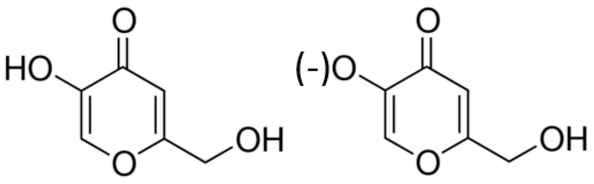
:1. Introduction
2. Results and Discussion
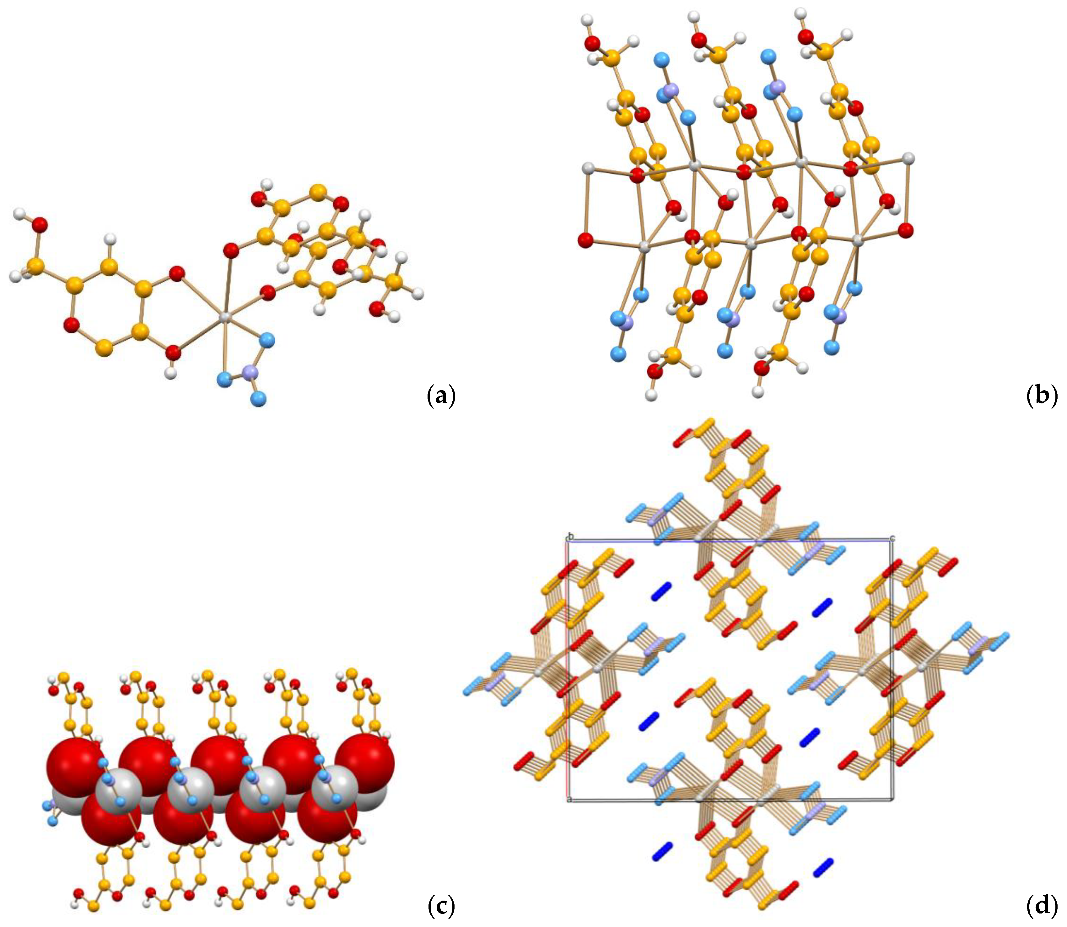
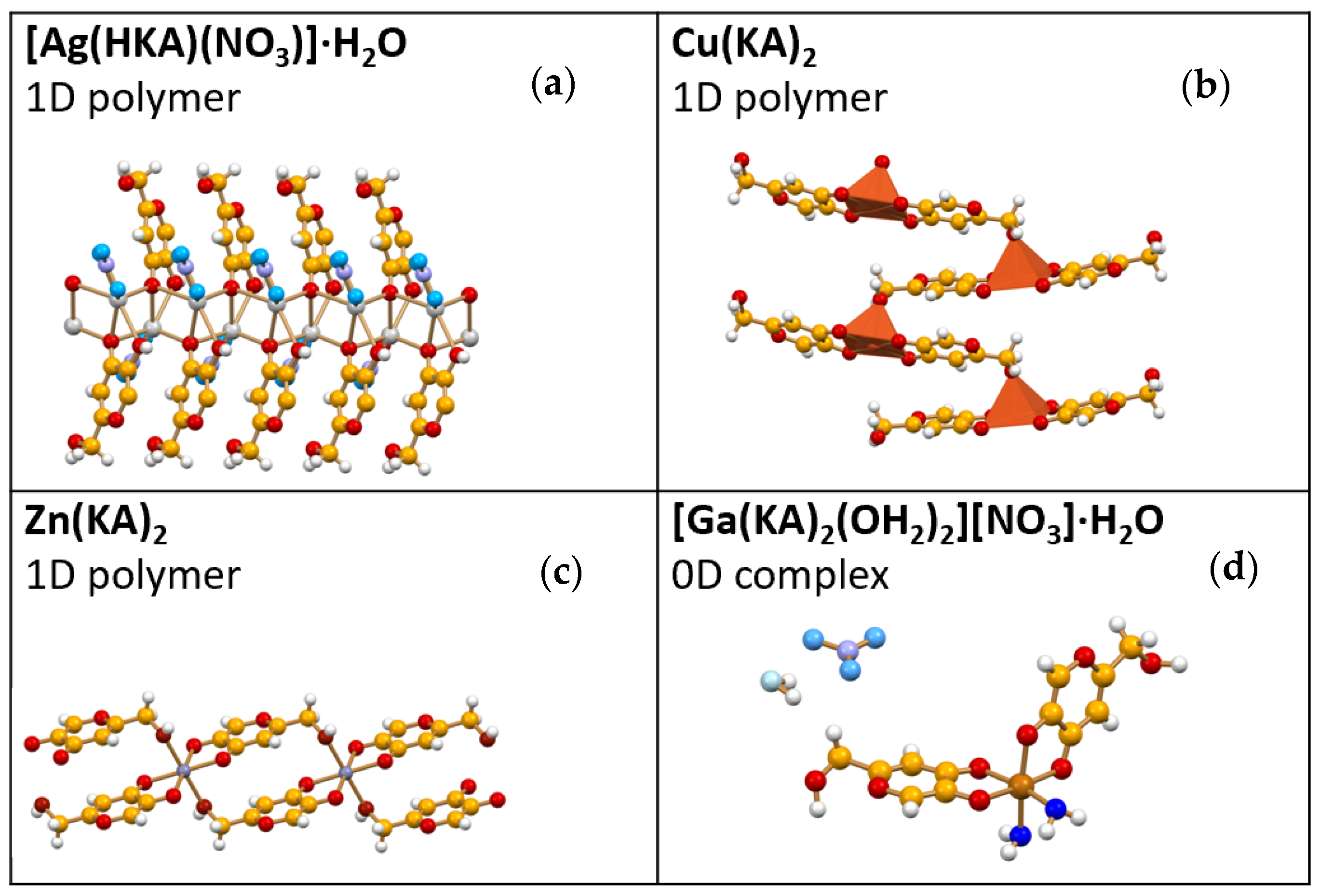
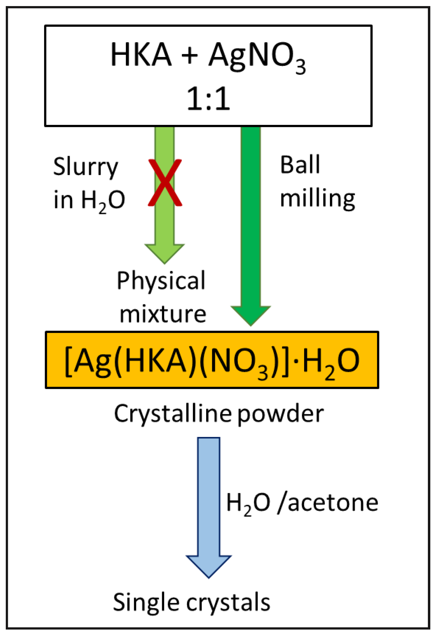
2.1. [Ag(HKA)(NO3)]∙H2O
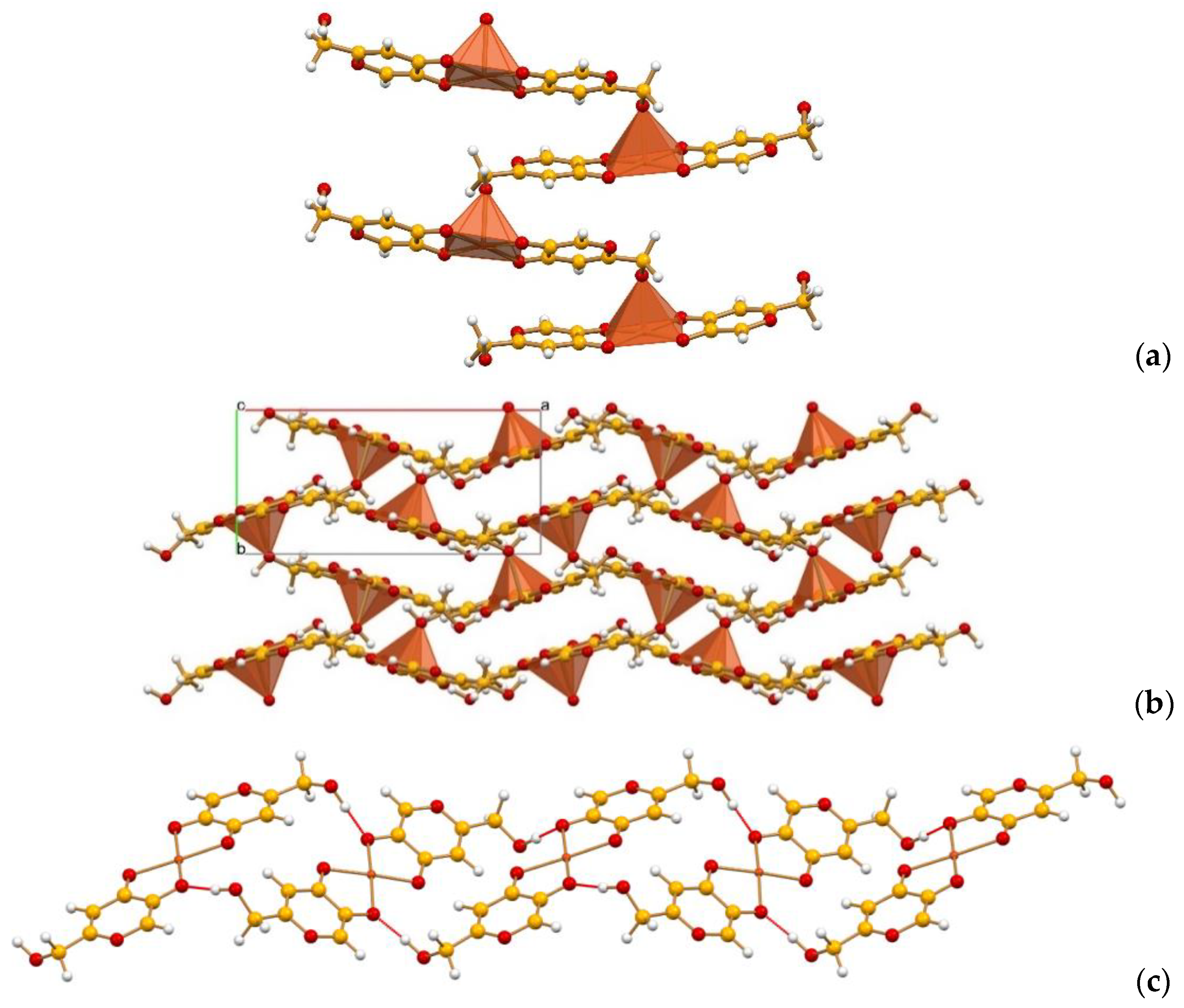
2.2. Cu(KA)2
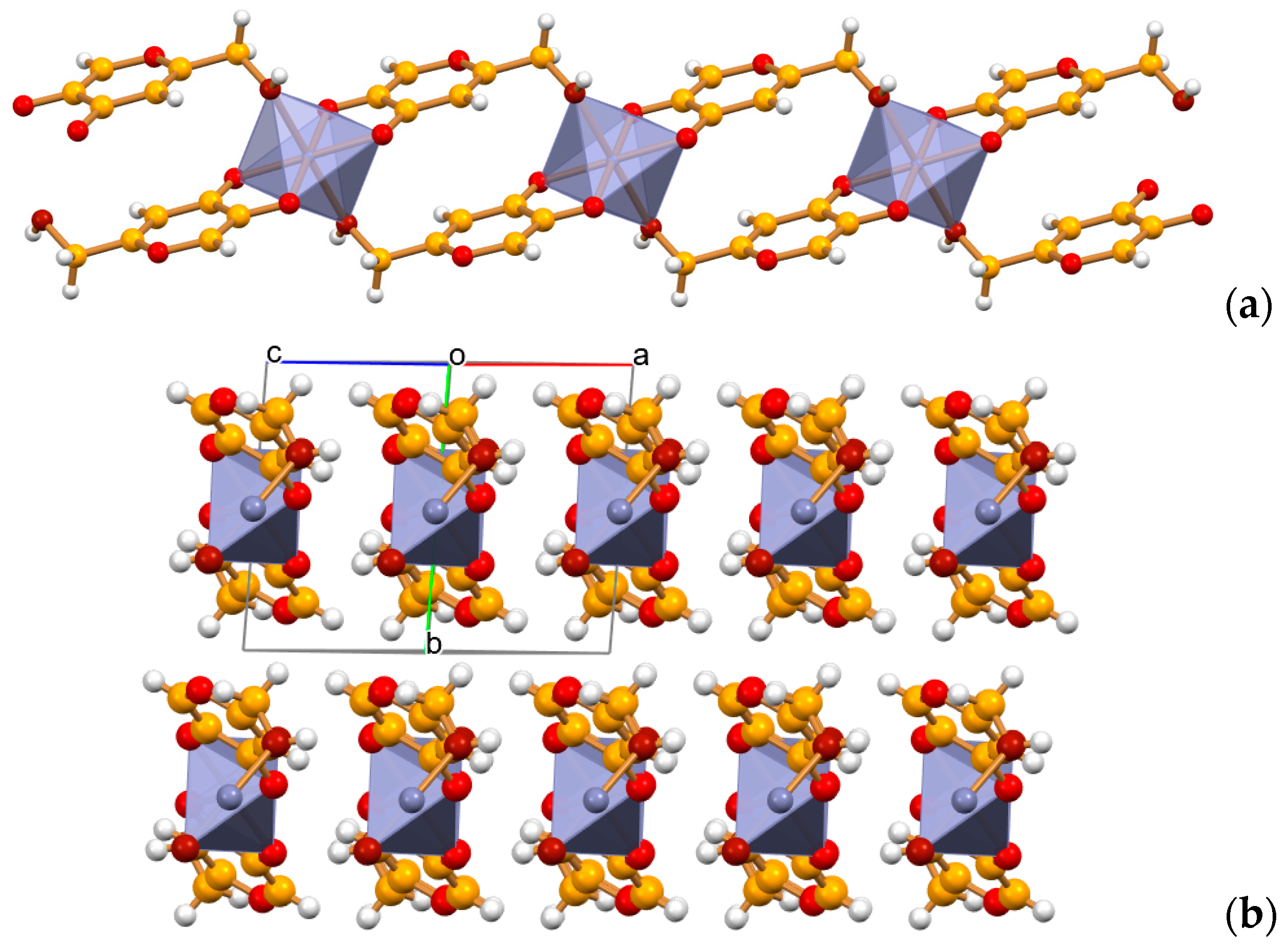
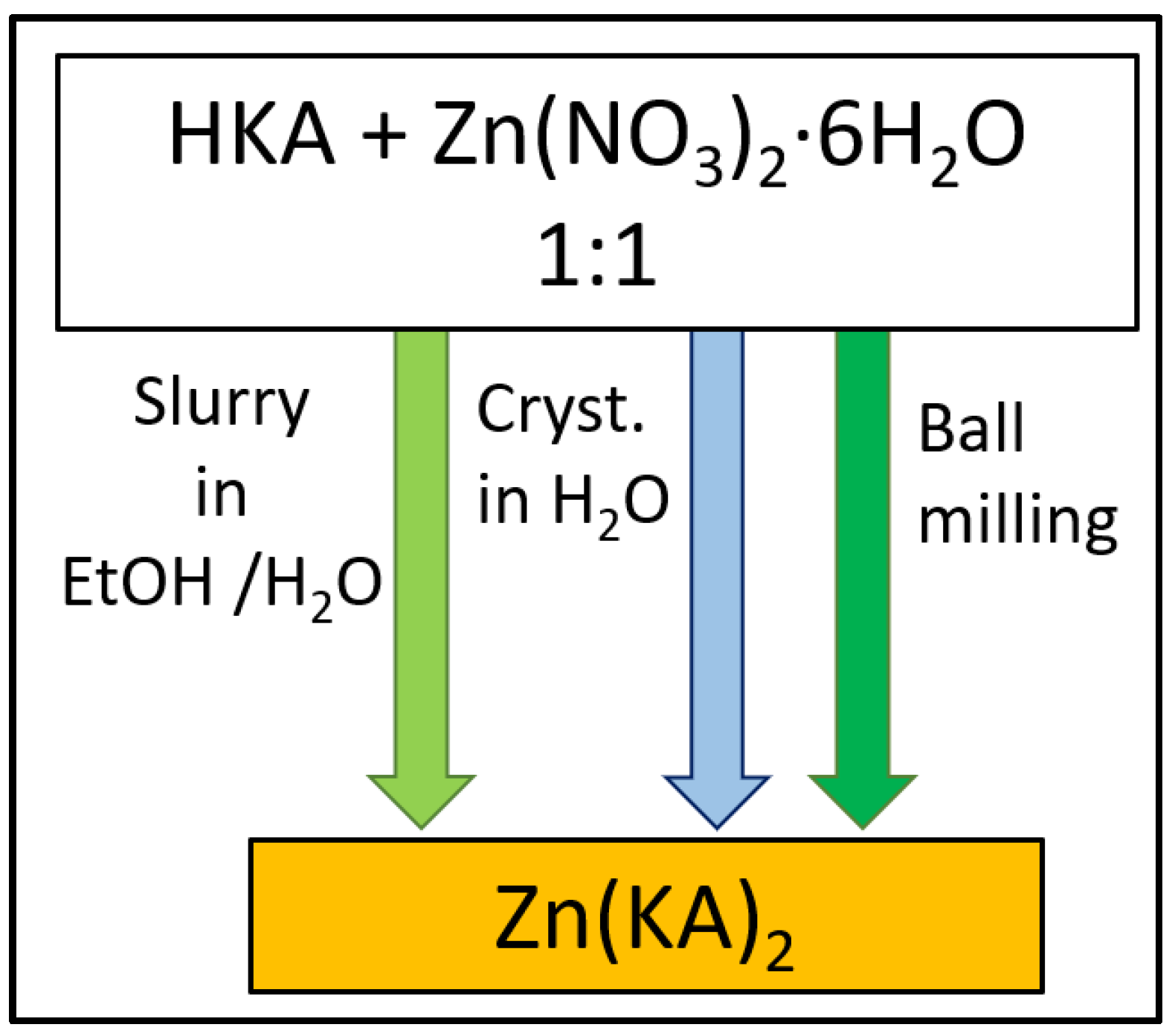
2.3. Zn(KA)2
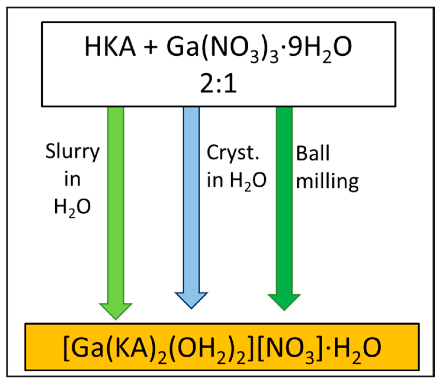
2.4. [Ga(KA)2(OH2)2][NO3]∙H2O
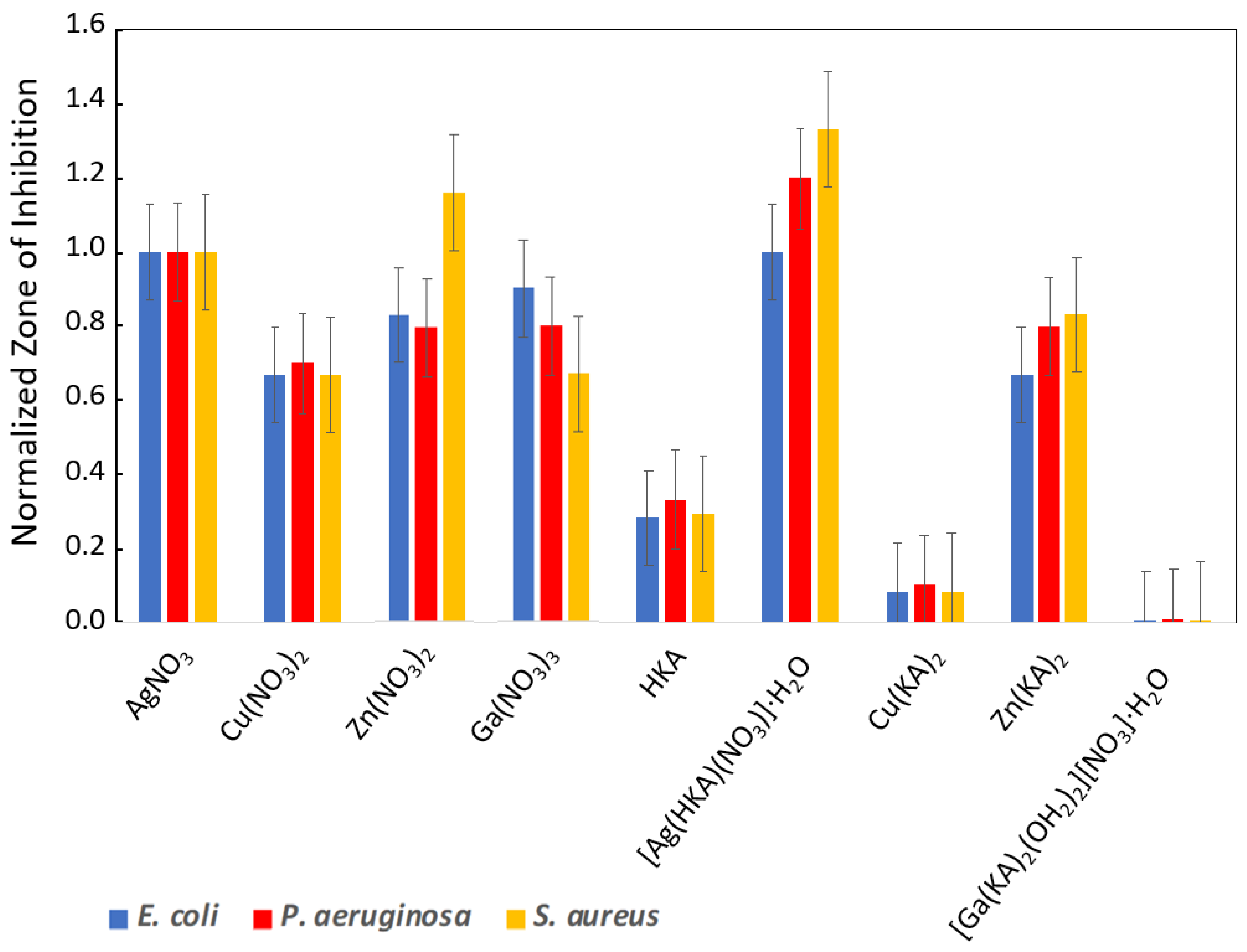
2.5. Antimicrobial Assays
3. Materials and Methods
3.1. Materials
3.2. Mechanochemical Synthesis
3.3. Slurry Syntheses
3.4. Solution Syntheses
3.5. Thermogravimetric Analysis
3.6. Single-Crystal X-ray Diffraction
3.7. X-ray Diffraction from Powder
3.8. Structural Characterization from Powder Data
3.9. Antimicrobial Analysis
4. Conclusions
Supplementary Materials
Author Contributions
Funding
Institutional Review Board Statement
Informed Consent Statement
Data Availability Statement
Acknowledgments
Conflicts of Interest
Sample Availability
References
- 2019 Antibacterial Agents in Clinical Development: An Analysis of the Antibacterial Clinical Development Pipeline; World Health Organization: Geneva, Switzerland, 2019.
- Lloyd, N.C.; Morgan, H.W.; Nicholson, B.K.; Ronimus, R.S. The composition of Ehrlich’s salvarsan: Resolution of a century-old debate. Angew. Chem. Int. Ed. 2005, 44, 941–944. [Google Scholar] [CrossRef] [PubMed] [Green Version]
- Ghosh, S. Cisplatin: The first metal based anticancer drug. Bioorg. Chem. 2019, 88, 102925. [Google Scholar] [CrossRef] [PubMed]
- Burdock, G.A.; Soni, M.G.; Carabin, I.G. Evaluation of health aspects of kojic acid in food. Regul. Toxicol. Pharmacol. 2001, 33, 80–101. [Google Scholar] [CrossRef] [PubMed]
- Saeedi, M.; Eslamifar, M.; Khezri, K. Kojic acid applications in cosmetic and pharmaceutical preparations. Biomed. Pharmacother. 2019, 110, 582–593. [Google Scholar] [CrossRef]
- Miyabe, C.; Dong, Y.; Wakamatsu, K.; Ito, S.; Kawakami, T. Kojic acid alters pheomelanin content in human induced pluripotent stem cell-derived melanocytes. J. Dermatol. 2020, 1, 2. [Google Scholar] [CrossRef]
- Saraei, M.; Zarrini, G.; Esmati, M.; Ahmadzadeh, L. Novel functionalized monomers based on kojic acid: Snythesis, characterization, polymerization and evalution of antimicrobial activity. Des. Monomers Polym. 2017, 20, 325–331. [Google Scholar] [CrossRef]
- Vishwanath, N.; Whitaker, C.; Allu, S.; Clippert, D.; Jouffroy, E.; Hong, J.; Stone, B.; Connolly, W.; Barrett, C.C.; Antoci, V. Silver as an antibiotic-independent antimicrobial: Review of current formulations and clinical relevance. Surg. Infect. 2022, 23, 769–780. [Google Scholar] [CrossRef]
- Alexander, J.W. History of the medical use of silver. Surg. Infect. 2009, 10, 289–292. [Google Scholar] [CrossRef] [Green Version]
- Ballin, J.C. Evaluation of a new topical agent for burn therapy: Silver sulfadiazine (Silvadene). JAMA 1974, 230, 1184–1185. [Google Scholar] [CrossRef]
- Fang, M.; Chen, J.-H.; Xu, X.-L.; Yang, P.-H.; Hildebrand, H.F. Antibacterial activities of inorganic agents on six bacteria associated with oral infections by two susceptibility tests. Int. J. Antimicrob. Agents 2006, 27, 513–517. [Google Scholar] [CrossRef]
- Yamgar, R.S.; Nivid, Y.; Nalawade, S.; Mandewale, M.; Atram, R.; Sawant, S.S. Novel Zinc(II) complexes of heterocyclic ligands as antimicrobial agents: Synthesis, characterisation, and antimicrobial studies. Bioinorg. Chem. Appl. 2014, 2014, 276598. [Google Scholar] [CrossRef] [PubMed] [Green Version]
- Levaditi, C.; Bardet, J.; Tchakirian, A.; Vaisman, A. Le gallium, propriétés thérapeutiques dans la syphilis et les trypanosomiases expérimentales. CR Hebd. Seances Acad. Sci. Ser. D Sci. Na 1931, 192, 1142–1143. [Google Scholar]
- Kaneko, Y.; Thoendel, M.; Olakanmi, O.; Britigan, B.E.; Singh, P.K. The transition metal gallium disrupts Pseudomonas aeruginosa iron metabolism and has antimicrobial and antibiofilm activity. J. Clin. Investig. 2007, 117, 877–888. [Google Scholar] [CrossRef]
- Sweetman, S.C. Martindale: The complete drug reference. 38th ed. Aust. Prescr. 2014, 38, 2147–2151. [Google Scholar]
- Kremer, E.; Facchin, G.; Estévez, E.; Alborés, P.; Baran, E.; Ellena, J.; Torre, M. Copper complexes with heterocyclic sulfonamides: Synthesis, spectroscopic characterization, microbiological and SOD-like activities: Crystal structure of [Cu(sulfisoxazole)2(H2O)4]·2H2O. J. Inorg. Biochem. 2006, 100, 1167–1175. [Google Scholar] [CrossRef] [PubMed]
- Nazirkar, B.; Mandewale, M.; Yamgar, R. Synthesis, characterization and antibacterial activity of Cu(II) and Zn(II) complexes of 5-aminobenzofuran-2-carboxylate Schiff base ligands. J. Taibah Univ. Sci. 2019, 13, 440–449. [Google Scholar] [CrossRef] [Green Version]
- Bolla, G.; Sarma, B.; Nangia, A.K. Crystal Engineering of Pharmaceutical Cocrystals in the Discovery and Development of Improved Drugs. Chem. Rev. 2022, 122, 11514–11603. [Google Scholar] [CrossRef]
- Shemchuk, O.; d’Agostino, S.; Fiore, C.; Sambri, V.; Zannoli, S.; Grepioni, F.; Braga, D. Natural antimicrobials meet a synthetic antibiotic: Carvacrol/thymol and ciprofloxacin cocrystals as a promising solid-state route to activity enhancement. Cryst. Growth Des. 2020, 20, 6796–6803. [Google Scholar] [CrossRef]
- Fiore, C.; Baraghini, A.; Shemchuk, O.; Sambri, V.; Morotti, M.; Grepioni, F.; Braga, D. Inhibition of the Antibiotic Activity of Cephalosporines by Co-Crystallization with Thymol. Cryst. Growth Des. 2022, 22, 1467–1475. [Google Scholar] [CrossRef]
- Ashooriha, M.; Khoshneviszadeh, M.; Khoshneviszadeh, M.; Moradi, S.E.; Rafiei, A.; Kardan, M.; Emami, S. 1,2,3-Triazole-based kojic acid analogs as potent tyrosinase inhibitors: Design, synthesis and biological evaluation. Bioorg. Chem. 2019, 82, 414–422. [Google Scholar] [CrossRef]
- Ashooriha, M.; Khoshneviszadeh, M.; Khoshneviszadeh, M.; Rafiei, A.; Kardan, M.; Yazdian-Robati, R.; Emami, S. Kojic acid–natural product conjugates as mushroom tyrosinase inhibitors. Eur. J. Med. Chem. 2020, 201, 112480. [Google Scholar] [CrossRef] [PubMed]
- Lachowicz, J.I.; Mateddu, A.; Coni, P.; Caltagirone, C.; Murgia, S.; Gibson, D.; Dalla Torre, G.; Lopez, X.; Meloni, F.; Pichiri, G. Study of the DNA binding mechanism and in vitro activity against cancer cells of Iron(III) and Aluminium(III) kojic acid derivative complexes. Dalton Trans. 2022, 51, 6254–6263. [Google Scholar] [CrossRef] [PubMed]
- Hosny, A.E.-D.M.; Rasmy, S.A.; Aboul-Magd, D.S.; Kashef, M.T.; El-Bazza, Z.E. The increasing threat of silver-resistance in clinical isolates from wounds and burns. Infect. Drug Resist. 2019, 12, 1985. [Google Scholar] [CrossRef] [PubMed] [Green Version]
- Panáček, A.; Kvítek, L.; Smékalová, M.; Večeřová, R.; Kolář, M.; Röderová, M.; Dyčka, F.; Šebela, M.; Prucek, R.; Tomanec, O. Bacterial resistance to silver nanoparticles and how to overcome it. Nat. Nanotechnol. 2018, 13, 65–71. [Google Scholar] [CrossRef]
- Muller, M.; Merrett, N.D. Pyocyanin production by Pseudomonas aeruginosa confers resistance to ionic silver. Antimicrob. Agents Chemother. 2014, 58, 5492–5499. [Google Scholar] [CrossRef] [Green Version]
- Guerrini, M.; d’Agostino, S.; Grepioni, F.; Braga, D.; Lekhan, A.; Turner, R.J. Antimicrobial activity of supramolecular salts of Gallium(III) and proflavine and the intriguing case of a trioxalate complex. Sci. Rep. 2022, 12, 3673. [Google Scholar] [CrossRef]
- Lekhan, A.; Fiore, C.; Shemchuk, O.; Grepioni, F.; Braga, D.; Turner, R.J. Comparison of Antimicrobial and Antibiofilm Activity of Proflavine Co-crystallized with Silver, Copper, Zinc, and Gallium Salts. ACS Appl. Bio. Mater. 2022, 5, 4203–4212. [Google Scholar] [CrossRef]
- Lemire, J.A.; Harrison, J.J.; Turner, R.J.J.N.R.M. Antimicrobial activity of metals: Mechanisms, molecular targets and applications. Nat. Rev. Microbiol. 2013, 11, 371–384. [Google Scholar] [CrossRef]
- Gugala, N.; Lemire, J.A.; Turner, R.J. The efficacy of different anti-microbial metals at preventing the formation of, and eradicating bacterial biofilms of pathogenic indicator strains. J. Antibiot. 2017, 70, 775–780. [Google Scholar] [CrossRef] [PubMed]
- Gugala, N.; Vu, D.; Parkins, M.D.; Turner, R.J. Specificity in the susceptibilities of Escherichia coli, Pseudomonas aeruginosa and Staphylococcus aureus clinical isolates to six metal antimicrobials. Antibiotics 2019, 8, 51. [Google Scholar] [CrossRef] [Green Version]
- Sheldrick, G.M. SHELXT–Integrated space-group and crystal-structure determination. Acta Crystallogr. Sect. A Found. Adv. 2015, 71, 3–8. [Google Scholar] [CrossRef] [PubMed] [Green Version]
- Sheldrick, G. XS, version 2013/1; Georg-August-Universität: Göttingen, Germany, 2013.
- Dolomanov, O.V.; Bourhis, L.J.; Gildea, R.J.; Howard, J.A.; Puschmann, H. OLEX2: A complete structure solution, refinement and analysis program. J. Appl. Crystallogr. 2009, 42, 339–341. [Google Scholar] [CrossRef]
- Macrae, C.F.; Edgington, P.R.; McCabe, P.; Pidcock, E.; Shields, G.P.; Taylor, R.; Towler, M.; Streek, J. Mercury: Visualization and analysis of crystal structures. J. Appl. Crystallogr. 2006, 39, 453–457. [Google Scholar] [CrossRef] [Green Version]
- Altomare, A.; Cuocci, C.; Giacovazzo, C.; Moliterni, A.; Rizzi, R.; Corriero, N.; Falcicchio, A. EXPO2013: A kit of tools for phasing crystal structures from powder data. J. Appl. Crystallogr. 2013, 46, 1231–1235. [Google Scholar] [CrossRef]
- Cohelo, A. TOPAS-Academic, Coelho Software: Brisbane, Australia, 2007.
- Fiore, C.; Shemchuk, O.; Grepioni, F.; Turner, R.J.; Braga, D. Proflavine and zinc chloride “team chemistry”: Combining antibacterial agents via solid-state interaction. CrystEngComm 2021, 23, 4494–4499. [Google Scholar] [CrossRef]
- Balouiri, M.; Sadiki, M.; Ibnsouda, S.K. Methods for in vitro evaluating antimicrobial activity: A review. J. Pharm. Anal. 2016, 6, 71–79. [Google Scholar] [CrossRef] [Green Version]


| E. coli | P. aeruginosa | S. aureus | |
|---|---|---|---|
| AgNO3 | <0.05 | <0.05 | <0.05 |
| Cu(NO3)2 | 0.78 | 1.50 | 1.50 |
| Zn(NO3)2 | 6.25 | 3.15 | 1.50 |
| Ga(NO3)3 | 1.50 | 0.40 | 0.78 |
| HKA | 6.25 | 3.10 | 1.50 |
| [Ag(HKA)·NO3]·H2O | <0.05 | <0.05 | <0.05 |
| Cu(KA)2 | 6.25 | 6.25 | 1.56 |
| Zn(KA)2 | 3.00 | 1.50 | 3.00 |
| [Ga(KA)2(OH2)2][NO3]·H2O | 3.00 | 3.10 | 3.00 |
| E. coli | P. aeruginosa | S. aureus | |
|---|---|---|---|
| AgNO3 | 0.10 | 0.05 | 0.10 |
| Cu(NO3)2 | 3.13 | 1.56 | 3.13 |
| Zn(NO3)2 | 3.13 | 3.13 | 6.25 |
| Ga(NO3)3 | 3.13 | 3.13 | 3.13 |
| HKA | 3.13 | 3.13 | >6.25 |
| [Ag(HKA)·NO3]·H2O | 0.10 | 0.10 | 0.98 |
| Cu(KA)2 | >6.25 | >6.25 | >6.25 |
| Zn(KA)2 | >6.25 | >6.25 | >6.25 |
| [Ga(KA)2(OH2)2][NO3]·H2O | >6.25 | 6.25 | >6.25 |
Disclaimer/Publisher’s Note: The statements, opinions and data contained in all publications are solely those of the individual author(s) and contributor(s) and not of MDPI and/or the editor(s). MDPI and/or the editor(s) disclaim responsibility for any injury to people or property resulting from any ideas, methods, instructions or products referred to in the content. |
© 2023 by the authors. Licensee MDPI, Basel, Switzerland. This article is an open access article distributed under the terms and conditions of the Creative Commons Attribution (CC BY) license (https://creativecommons.org/licenses/by/4.0/).
Share and Cite
Sun, R.; Casali, L.; Turner, R.J.; Braga, D.; Grepioni, F. Exploring the Co-Crystallization of Kojic Acid with Silver(I), Copper(II), Zinc(II), and Gallium(III) for Potential Antibacterial Applications. Molecules 2023, 28, 1244. https://doi.org/10.3390/molecules28031244
Sun R, Casali L, Turner RJ, Braga D, Grepioni F. Exploring the Co-Crystallization of Kojic Acid with Silver(I), Copper(II), Zinc(II), and Gallium(III) for Potential Antibacterial Applications. Molecules. 2023; 28(3):1244. https://doi.org/10.3390/molecules28031244
Chicago/Turabian StyleSun, Renren, Lucia Casali, Raymond J. Turner, Dario Braga, and Fabrizia Grepioni. 2023. "Exploring the Co-Crystallization of Kojic Acid with Silver(I), Copper(II), Zinc(II), and Gallium(III) for Potential Antibacterial Applications" Molecules 28, no. 3: 1244. https://doi.org/10.3390/molecules28031244