
New Supramolecular Hypoxia-Sensitive Complexes Based on Azo-Thiacalixarene
 , , ,
, , ,
Abstract
:1. Introduction
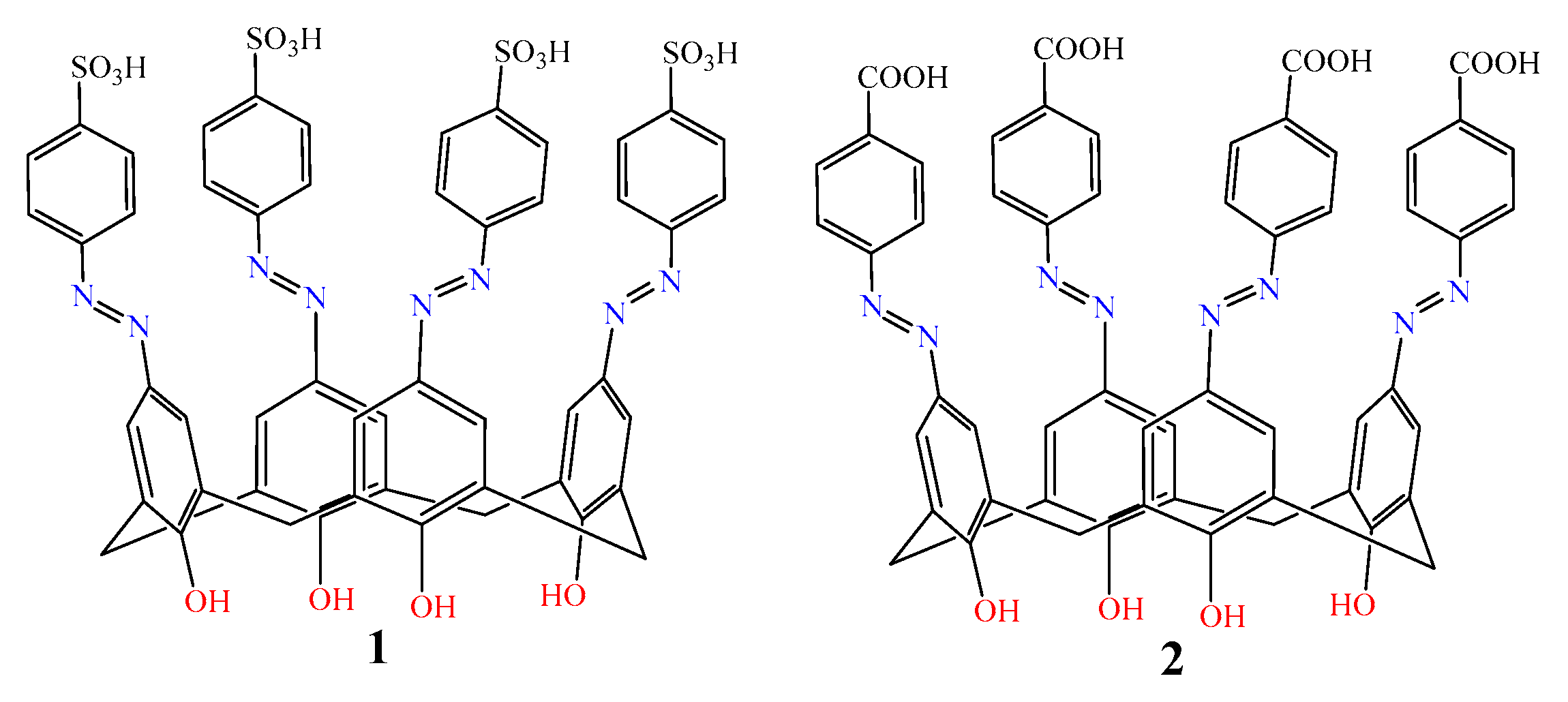
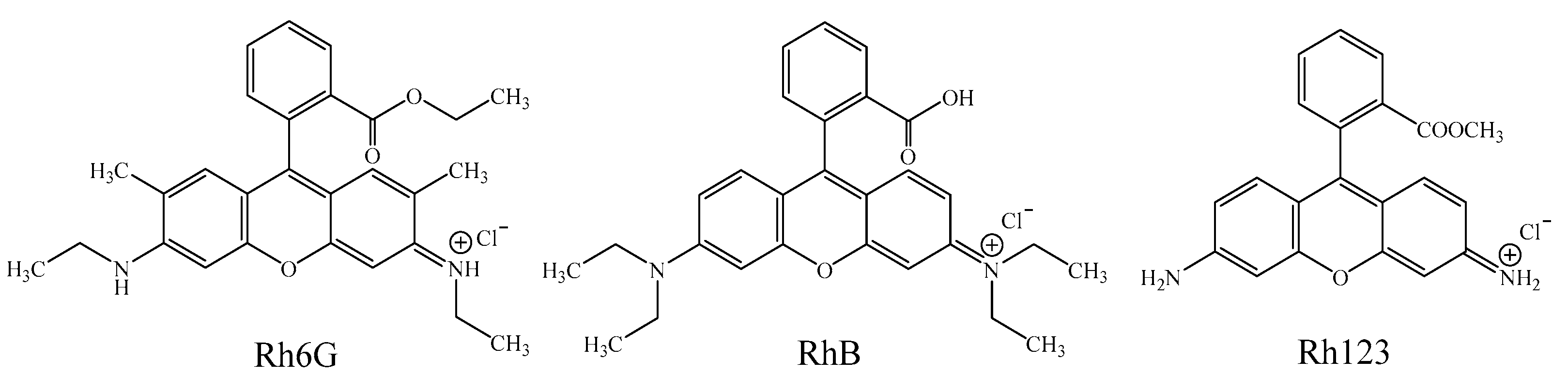
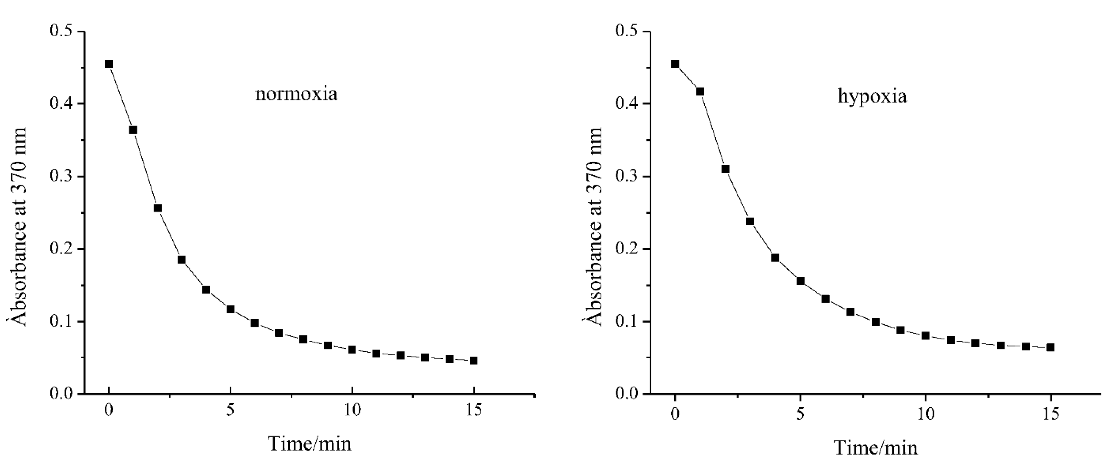
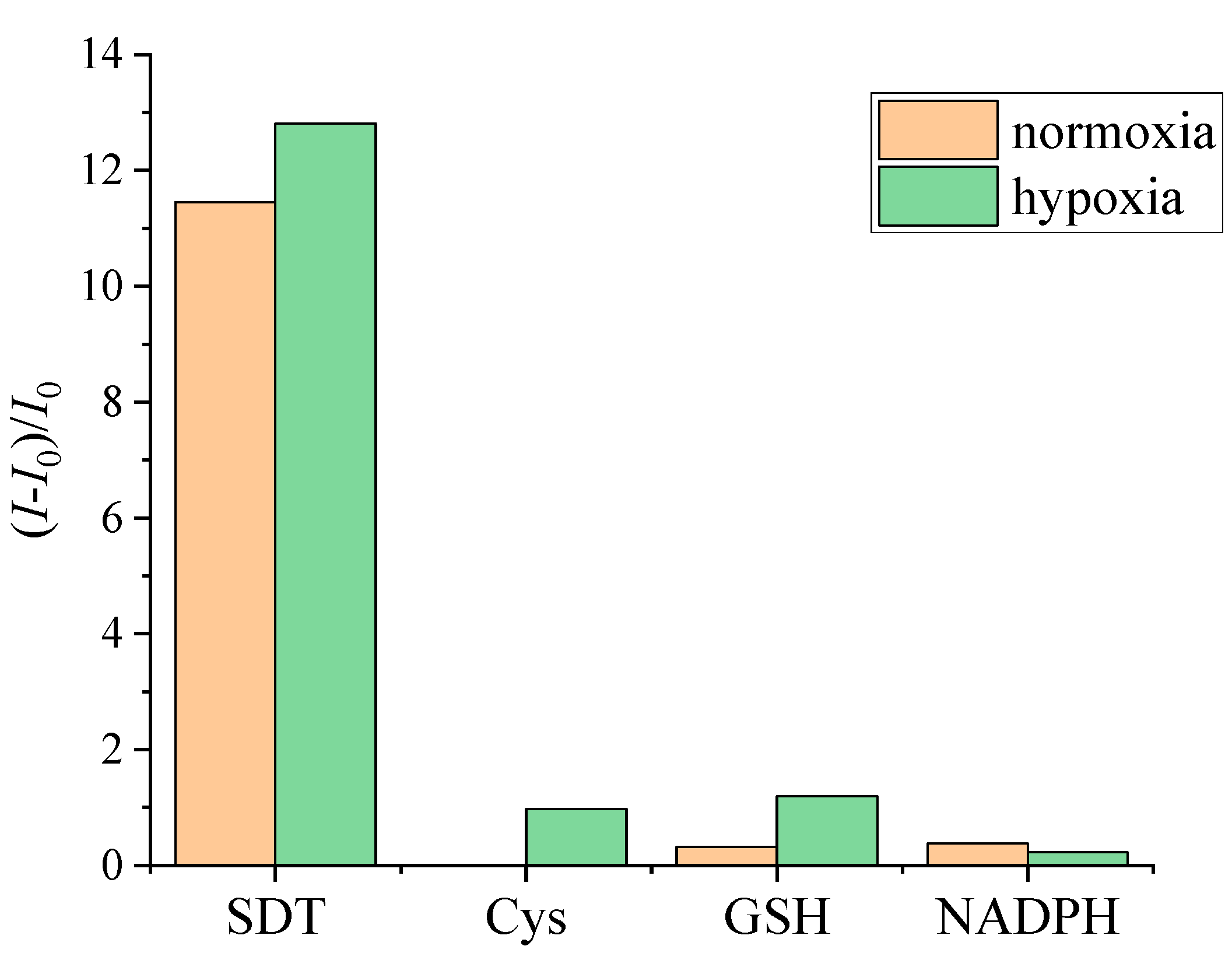
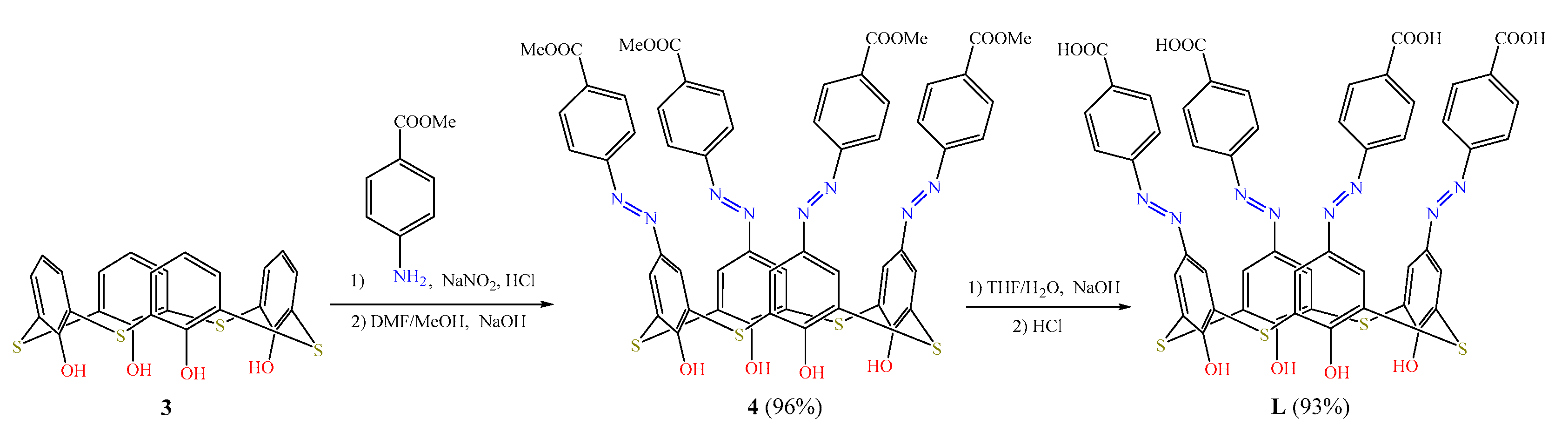
2. Results
3. Materials and Methods
3.1. Sample Preparation
3.2. Apparatus
3.3. Synthesis
4. Conclusions
Supplementary Materials
Author Contributions
Funding
Institutional Review Board Statement
Informed Consent Statement
Data Availability Statement
Acknowledgments
Conflicts of Interest
Sample Availability
References
- Sung, H.; Ferlay, J.; Siegel, R.L.; Laversanne, M.; Soerjomataram, I.; Jemal, A.; Bray, F. Global cancer statistics 2020: GLOBOCAN estimates of incidence and mortality worldwide for 36 cancers in 185 countries. CA A Cancer J. Clin. 2021, 71, 209–249. [Google Scholar] [CrossRef] [PubMed]
- Lee, J.W.; Ko, J.; Ju, C.; Eltzschig, H.K. Hypoxia signaling in human diseases and therapeutic targets. Exp. Mol. Med. 2019, 51, 1–13. [Google Scholar] [CrossRef] [PubMed] [Green Version]
- Bhandari, V.; Hoey, C.; Liu, L.Y.; Lalonde, E.; Ray, J.; Livingstone, J.; Lesurf, R.; Shiah, Y.J.; Vujcic, T.; Huang, X.Y.; et al. Molecular landmarks of tumor hypoxia across cancer types. Nat. Genet. 2019, 51, 308–318. [Google Scholar] [CrossRef] [PubMed]
- Semenza, G.L. Hypoxia, clonal selection, and the role of HIF-1 in tumor progression. Crit. Rev. Biochem. Mol. Biol. 2000, 35, 71–103. [Google Scholar] [CrossRef] [PubMed]
- Thomlinson, R.H.; Gray, L.H. The histological structure of some human lung cancers and the possible implications for radiotherapy. Br. J. Cancer 1955, 9, 539–549. [Google Scholar] [CrossRef] [PubMed] [Green Version]
- Hayashi, Y.; Yokota, A.; Harada, H.; Huang, G. Hypoxia/pseudohypoxia-mediated activation of hypoxia-inducible factor-1α in cancer. Cancer Sci. 2019, 110, 1510–1517. [Google Scholar] [CrossRef] [Green Version]
- Gilkes, D.M.; Semenza, G.L. Role of hypoxia-inducible factors in breast cancer metastasis. Future Oncol. 2013, 9, 1623–1636. [Google Scholar] [CrossRef] [PubMed] [Green Version]
- Brown, J.M.; William, W.R. Exploiting tumour hypoxia in cancer treatment. Nat. Rev. Cancer 2004, 4, 437–447. [Google Scholar] [CrossRef]
- Petrova, V.; Annicchiarico-Petruzzelli, M.; Melino, G.; Amelio, I. The hypoxic tumour microenvironment. Oncogenesis 2018, 7, 1–13. [Google Scholar] [CrossRef] [Green Version]
- Avagliano, A.; Granato, G.; Ruocco, M.R.; Romano, V.; Belviso, I.; Carfora, A.; Montagnani, S.; Arcucci, A. Metabolic reprogramming of cancer associated fibroblasts: The slavery of stromal fibroblasts. BioMed Res. Int. 2018, 111, 1–12. [Google Scholar] [CrossRef]
- Ammirante, M.; Shalapour, S.; Kang, Y.; Jamieson, C.A.M.; Karin, M. Tissue injury and hypoxia promote malignant progression of prostate cancer by inducing CXCL13 expression in tumor myofibroblasts. Proc. Natl. Acad. Sci. USA 2014, 111, 14776–14781. [Google Scholar] [CrossRef] [PubMed] [Green Version]
- Zhou, H.; Qin, F.; Chen, C. Designing hypoxia-responsive nanotheranostic agents for tumor imaging and therapy. Adv. Healthc. Mater. 2021, 10, 1–28. [Google Scholar] [CrossRef] [PubMed]
- Tanabe, K.; Hirata, N.; Harada, H.; Hiraoka, M.; Nishimoto, S.-I. Emission under hypoxia: One-electron reduction and fluorescence characteristics of an indolequinone-coumarin conjugate. Chembiochem 2008, 9, 426–432. [Google Scholar] [CrossRef] [PubMed]
- Liu, Y.; Xu, Y.F.; Qian, X.H.; Liu, J.W.; Shen, L.Y.; Li, J.H.; Zhang, Y.X. Novel fluorescent markers for hypoxic cells of naphthalimides with two heterocyclic side chains for bioreductive binding. Bioorg. Med. Chem. 2006, 14, 2935–2941. [Google Scholar] [CrossRef]
- Kumari, R.; Sunil, D.; Ningthoujam, R.S.; Kumar, N.V.A. Azodyes as markers for tumor hypoxia imaging and therapy: An up-to-date review. Chem.-Biol. Interact. 2019, 207, 91–104. [Google Scholar] [CrossRef]
- Chevalier, A.; Mercier, C.; Saurel, L.; Orenga, S.; Renard, P.-Y.; Romieu, A. The first latent green fluorophores for the detection of azoreductase activity in bacterial cultures. Chem. Commun. 2013, 49, 8815. [Google Scholar] [CrossRef]
- Leriche, G.; Budin, G.; Darwich, Z.; Weltin, D.; Mély, Y.; Klymchenko, A.S.; Wagner, A. A FRET-based probe with a chemically deactivatable quencher. Chem. Commun. 2012, 48, 3224–3226. [Google Scholar] [CrossRef]
- Verwilst, P.; Han, J.; Lee, J.; Mun, S.; Kang, H.-G.; Kim, J.S. Reconsidering azobenzene as a component of small-molecule hypoxia-mediated cancer drugs: A theranostic case study. Biomaterials 2017, 115, 104–114. [Google Scholar] [CrossRef]
- Yuan, P.; Zhang, H.; Qian, L.; Mao, X.; Du, S.; Yu, C.; Peng, B.; Yao, S.Q. Intracellular delivery of functional native antibodies under hypoxic conditions by using a biodegradable silica nanoquencher. Angew. Chem. Int. Ed. 2017, 56, 12481–12485. [Google Scholar] [CrossRef]
- Perche, F.; Biswas, S.; Wang, T.; Zhu, L.; Torchilin, V.P. Hypoxia-targeted siRNA delivery. Angew. Chem. Int. Ed. 2014, 53, 3362–3366. [Google Scholar] [CrossRef]
- Ma, X.; Zhao, Y.L. Biomedical applications of supramolecular systems based on host-guest interactions. Chem. Rev. 2015, 115, 7794–7839. [Google Scholar] [CrossRef]
- Chevalier, A.; Piao, W.; Hanaoka, K.; Nagano, T.; Renard, P.-Y.; Romieu, A. Azobenzene-caged sulforhodamine dyes: A novel class of ‘turn-on’ reactive probes for hypoxic tumor cell imaging. Methods Appl. Fluores 2015, 3, 044004. [Google Scholar] [CrossRef]
- Piao, W.; Tsuda, S.; Tanaka, Y.; Maeda, S.; Liu, F.; Takahashi, S.; Kushida, Y.; Komatsu, T.; Ueno, T.; Terai, T.; et al. Development of Azo-Based Fluorescent Probes to Detect Different Levels of Hypoxia. Angew. Chem. Int. Ed. 2013, 52, 13028–13032. [Google Scholar] [CrossRef]
- Tauran, Y.; Coleman, A.W.; Perret, F.; Kim, B. Cellular and in vivo biological activities of the calix[n]arenes. Curr. Org. Chem. 2015, 19, 2250–2270. [Google Scholar] [CrossRef]
- Nimse, S.B.; Kim, T. Biological applications of functionalized calixarenes. Chem. Soc. Rev. 2013, 42, 366–386. [Google Scholar] [CrossRef] [PubMed]
- Baldini, L.; Casnati, A.; Sansone, F. Multivalent and multifunctional calixarenes in bionanotechnology. Eur. J. Org. Chem. 2020, 2020, 5056–5069. [Google Scholar] [CrossRef]
- Tian, H.W.; Liu, Y.C.; Guo, D.S. Assembling features of calixarene-based amphiphiles and supra-amphiphiles. Mater. Chem. Front. 2020, 4, 46–98. [Google Scholar] [CrossRef]
- Mokhtari, B.; Pourabdollah, K. Applications of nano-baskets in drug development: High solubility and low toxicity. Drug Chem. Toxicol. 2013, 36, 119–132. [Google Scholar] [CrossRef]
- Ukhatskaya, E.V.; Kurkov, S.V.; Matthews, S.E.; Loftsson, T. Encapsulation of drug molecules into calix[n]arene nanobaskets. Role of aminocalix[n]arenes in biopharmaceutical field. J. Pharm. Sci. 2013, 102, 3485–3512. [Google Scholar] [CrossRef]
- Coleman, A.W.; Jebors, S.; Cecillon, S.; Perret, P.; Garin, D.; Marti-Battle, D.; Moulin, M. Toxicity and biodistribution of para-sulfonato-calix [4]arene in mice. New J. Chem. 2008, 32, 780–782. [Google Scholar] [CrossRef]
- Perret, F.; Coleman, A.W. Biochemistry of anionic calix[n]arenes. Chem. Commun. 2011, 47, 7303–7319. [Google Scholar] [CrossRef]
- Perret, F.; Lazar, A.N.; Coleman, A.W. Biochemistry of the para-sulfonato-calix[n]arenes. Chem. Commun. 2006, 23, 2425–2438. [Google Scholar] [CrossRef]
- Geng, W.-C.; Jia, S.; Zheng, Z.; Li, Z.; Ding, D.; Guo, D.-S. A noncovalent fluorescence turn-on strategy for hypoxia imaging. Angew. Chem. Int. Ed. 2019, 58, 2377–2381. [Google Scholar] [CrossRef]
- Mironova, D.; Burilov, B.; Galieva, F.; Khalifa, M.A.M.; Kleshnina, S.; Gazalieva, A.; Nugmanov, R.; Solovieva, S.; Antipin, I. Azocalix[4]arene-rhodamine supramolecular hypoxia-sensitive systems: A search for the best calixarene hosts and rhodamine guests. Molecules 2021, 26, 5451. [Google Scholar] [CrossRef]
- Chakrabarti, A.; Chawla, H.M.; Francis, T.; Pant, N.; Upreti, S. Synthesis and cation binding properties of new arylazo- and heteroarylazotetrathiacalix[4]arenes. Tetrahedron 2006, 62, 1150–1157. [Google Scholar] [CrossRef]
- Rajasekar, M. Recent Trends in Rhodamine derivatives as fluorescent probes for biomaterial applications. J. Mol. Struct. 2021, 1235, 130232. [Google Scholar] [CrossRef]
- Kennedy, A.R.; Conway, L.K.; Kirkhouse, J.B.A.; McCarney, K.M.; Puissegur, O.; Staunton, E.; Teat, S.J.; Warren, J.E. Monosulfonated Azo Dyes: A Crystallographic Study of the Molecular Structures of the Free Acid, Anionic and Dianionic Forms. Crystals 2020, 10, 662. [Google Scholar] [CrossRef]
- Zana, R.; Schmidt, J.; Talmon, Y. Tetrabutylammonium Alkyl Carboxylate Surfactants in Aqueous Solution: Self-Association Behavior, Solution Nanostructure, and Comparison with Tetrabutylammonium Alkyl Sulfate Surfactants. Langmuir 2005, 21, 11628–11636. [Google Scholar] [CrossRef] [PubMed]
- Reshetnyak, E.A.; Chernysheva, O.S.; Nikitina, N.A.; Loginova, L.P.; Mchedlov-Petrosyan, N.O. Activity coefficients of alkyl sulfate and alkylsulfonate ions in aqueous and water-salt premicellar solutions. Colloid J. 2014, 76, 358–365. [Google Scholar] [CrossRef]
- Burilov, V.A.; Mironova, D.A.; Ibragimova, R.R.; Nugmanov, R.I.; Solovieva, S.E.; Antipin, I.S. Detection of sulfate surface-active substances via fluorescent response using new amphiphilic thiacalix[4]arenes bearing cationic headgroups with Eosin Y dye. Colloids Surf. A Physicochem. Eng. Asp. 2017, 515, 41–49. [Google Scholar] [CrossRef]
- Solovieva, S.E.; Burilov, V.A.; Antipin, I.S. Thiacalix[4]arene’s Lower Rim Derivatives: Synthesis and Supramolecular Properties. Macroheterocycles 2017, 10, 134–146. [Google Scholar] [CrossRef]


| System | Kass, M−1 |
|---|---|
| L-Rh6G | 4.7 ± 0.1 × 10⁶ |
| L-RhB | 8.6 ± 0.2 × 10⁴ |
| L-Rh123 | 5.2 ± 0.4 × 10³ |
| 1-Rh6G 1 | 1.5 ± 0.1 × 10⁷ |
| 1-RhB 1 | 7.0 ± 0.1 × 10⁵ |
| 1-Rh123 1 | 5.0 ± 0.3 × 10⁵ |
| 2-Rh6G 1 | 1.0 ± 0.4 × 10⁵ |
| 2-RhB 1 | 2.6 ± 0.1 × 10⁴ |
| 2-Rh123 1 | 2.3 ± 0.6 × 10⁴ |
| Macrocycle | k, min−1 (Normoxia) | k, min−1 (Hypoxia) |
|---|---|---|
| L | 0.420 (Adj. R2 > 0.987) | 0.497 (Adj. R2 > 0.985) |
| 1 1 | 0.350 (Adj. R2 > 0.991) | - |
| 2 1 | 0.403 (Adj. R2 > 0.987) | - |
Disclaimer/Publisher’s Note: The statements, opinions and data contained in all publications are solely those of the individual author(s) and contributor(s) and not of MDPI and/or the editor(s). MDPI and/or the editor(s) disclaim responsibility for any injury to people or property resulting from any ideas, methods, instructions or products referred to in the content. |
© 2023 by the authors. Licensee MDPI, Basel, Switzerland. This article is an open access article distributed under the terms and conditions of the Creative Commons Attribution (CC BY) license (https://creativecommons.org/licenses/by/4.0/).
Share and Cite
Galieva, F.; Khalifa, M.; Akhmetzyanova, Z.; Mironova, D.; Burilov, V.; Solovieva, S.; Antipin, I. New Supramolecular Hypoxia-Sensitive Complexes Based on Azo-Thiacalixarene. Molecules 2023, 28, 466. https://doi.org/10.3390/molecules28020466
Galieva F, Khalifa M, Akhmetzyanova Z, Mironova D, Burilov V, Solovieva S, Antipin I. New Supramolecular Hypoxia-Sensitive Complexes Based on Azo-Thiacalixarene. Molecules. 2023; 28(2):466. https://doi.org/10.3390/molecules28020466
Chicago/Turabian StyleGalieva, Farida, Mohamed Khalifa, Zaliya Akhmetzyanova, Diana Mironova, Vladimir Burilov, Svetlana Solovieva, and Igor Antipin. 2023. "New Supramolecular Hypoxia-Sensitive Complexes Based on Azo-Thiacalixarene" Molecules 28, no. 2: 466. https://doi.org/10.3390/molecules28020466