
In Vitro Anti-Inflammatory Activity of Cotula anthemoides Essential Oil and In Silico Molecular Docking of Its Bioactives

 ,
,  , , ,
, , ,  and
and
Abstract
:1. Introduction
2. Results and Discussion
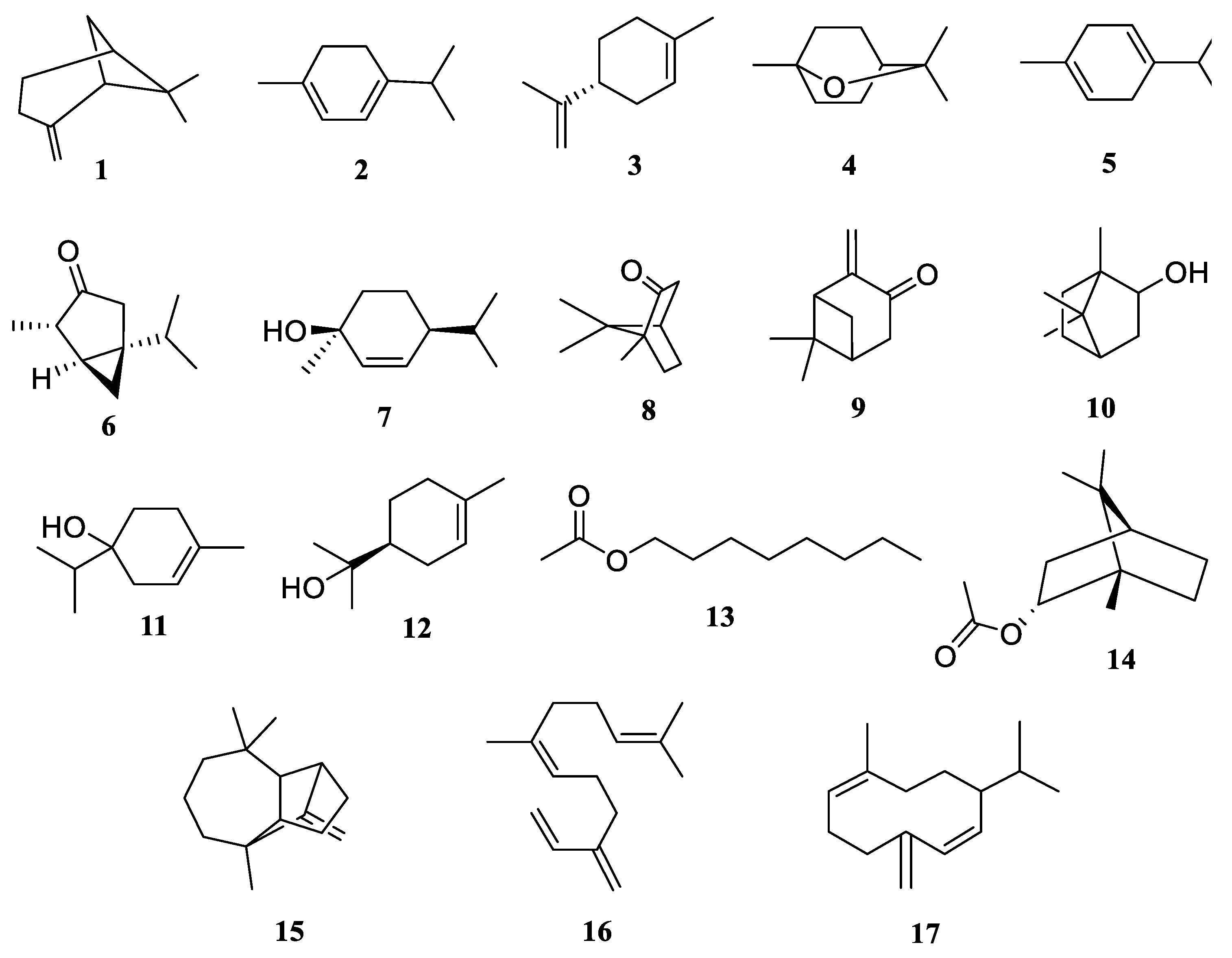
2.1. Essential Oil Composition
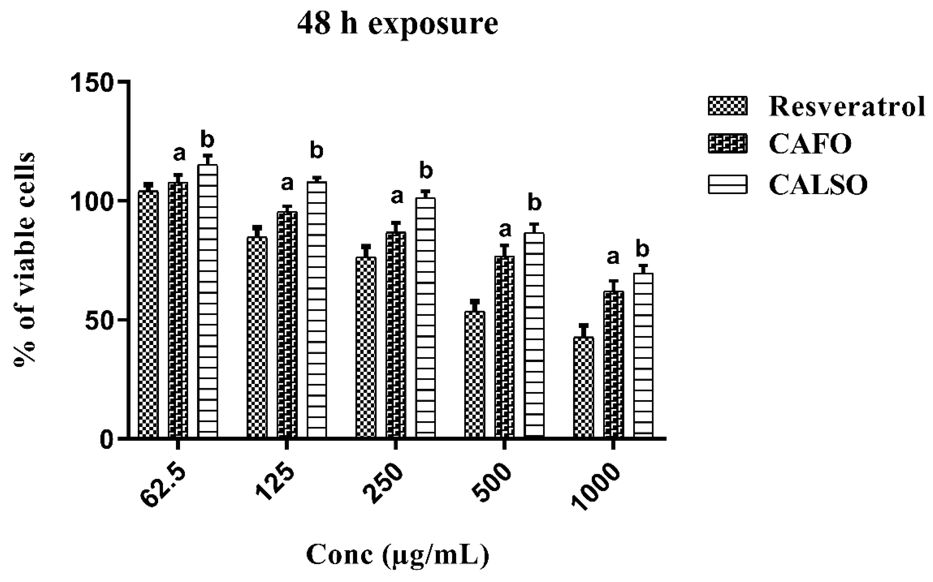
2.2. Cytotoxicity of C. anthemoides Essential Oils against RAW 264.7 Cells
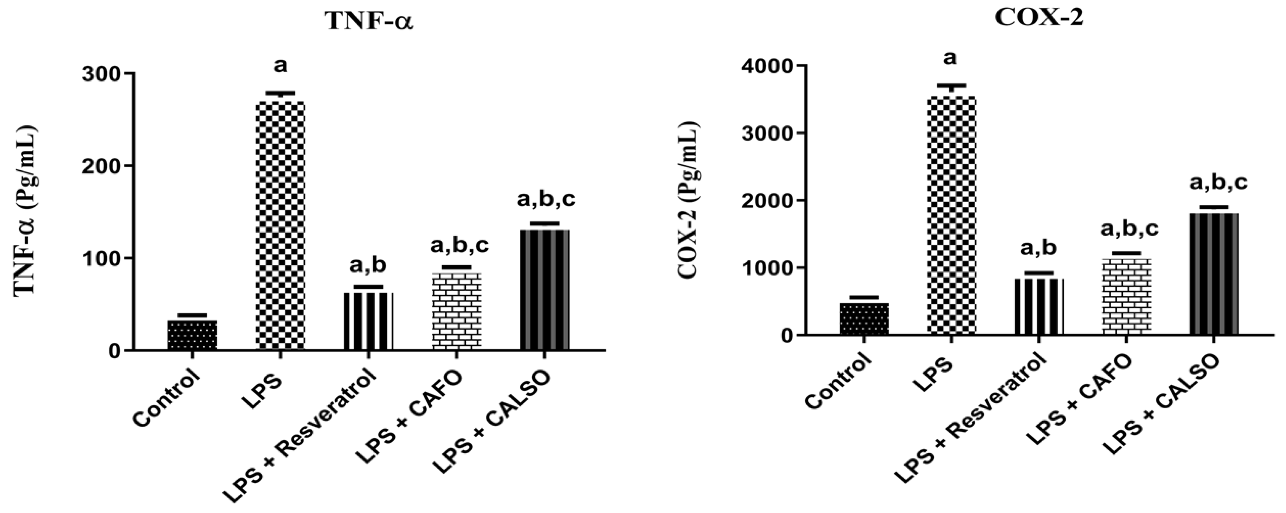
2.3. Effects of C. anthemoides Essential Oils on COX-2 Levels against LPS-Stimulated RAW 264.7 Cells
2.4. Effects of C. anthemoides Essential Oils on TNF-α Levels in LPS-Stimulated RAW 264.7 Cells
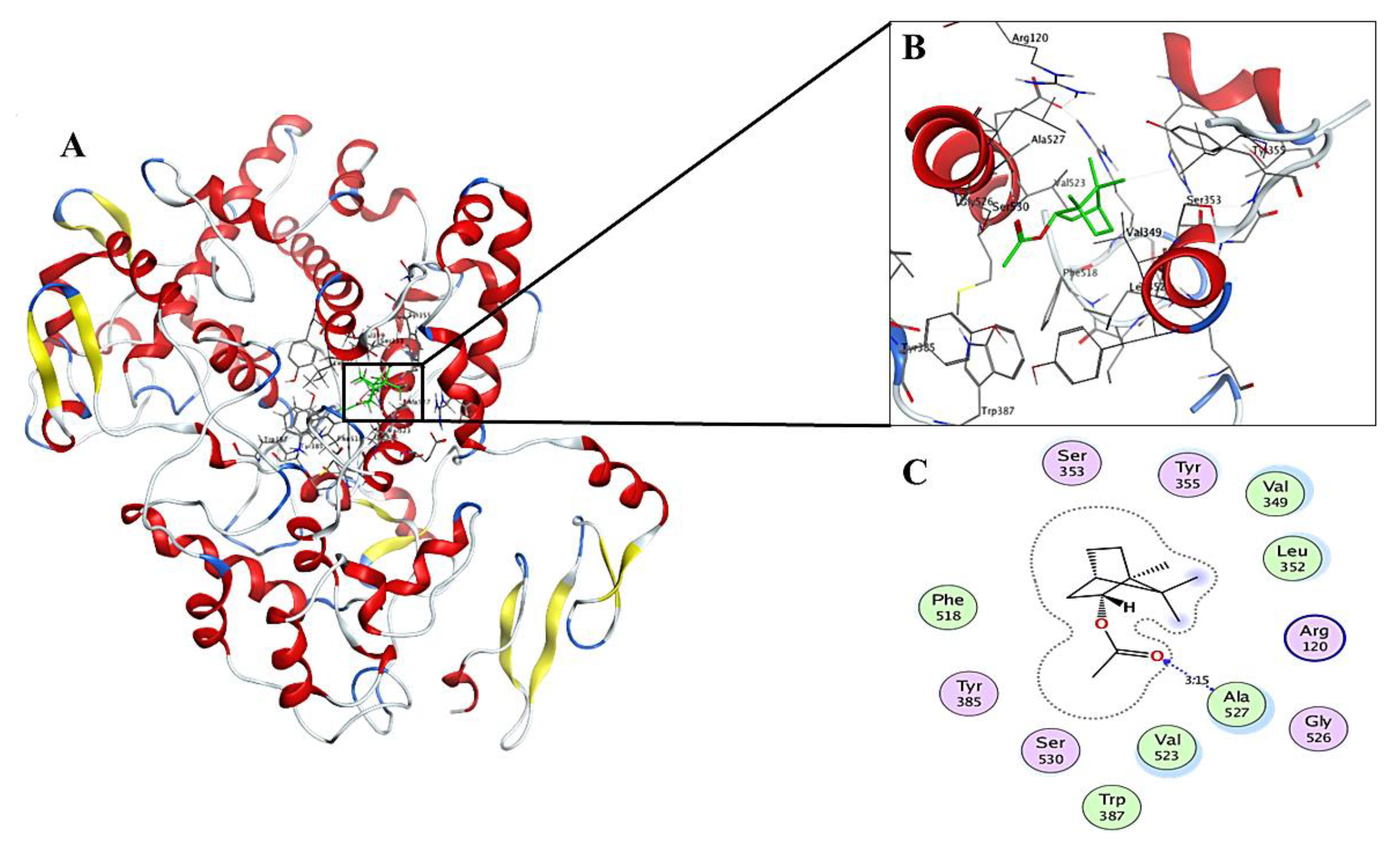
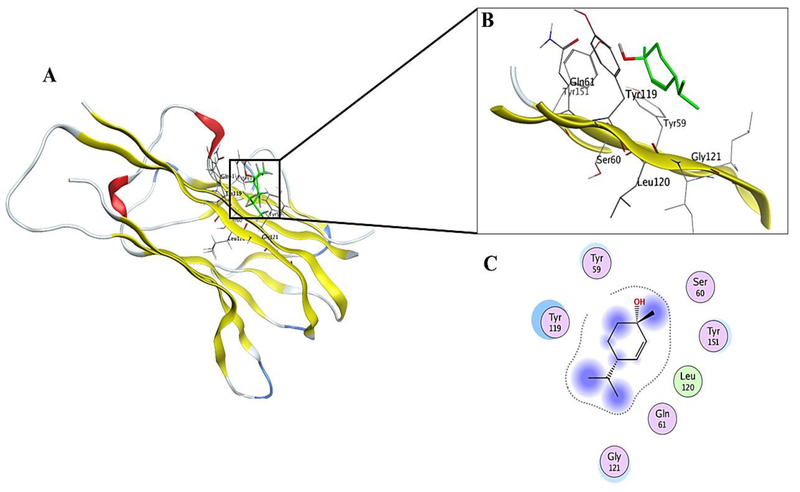
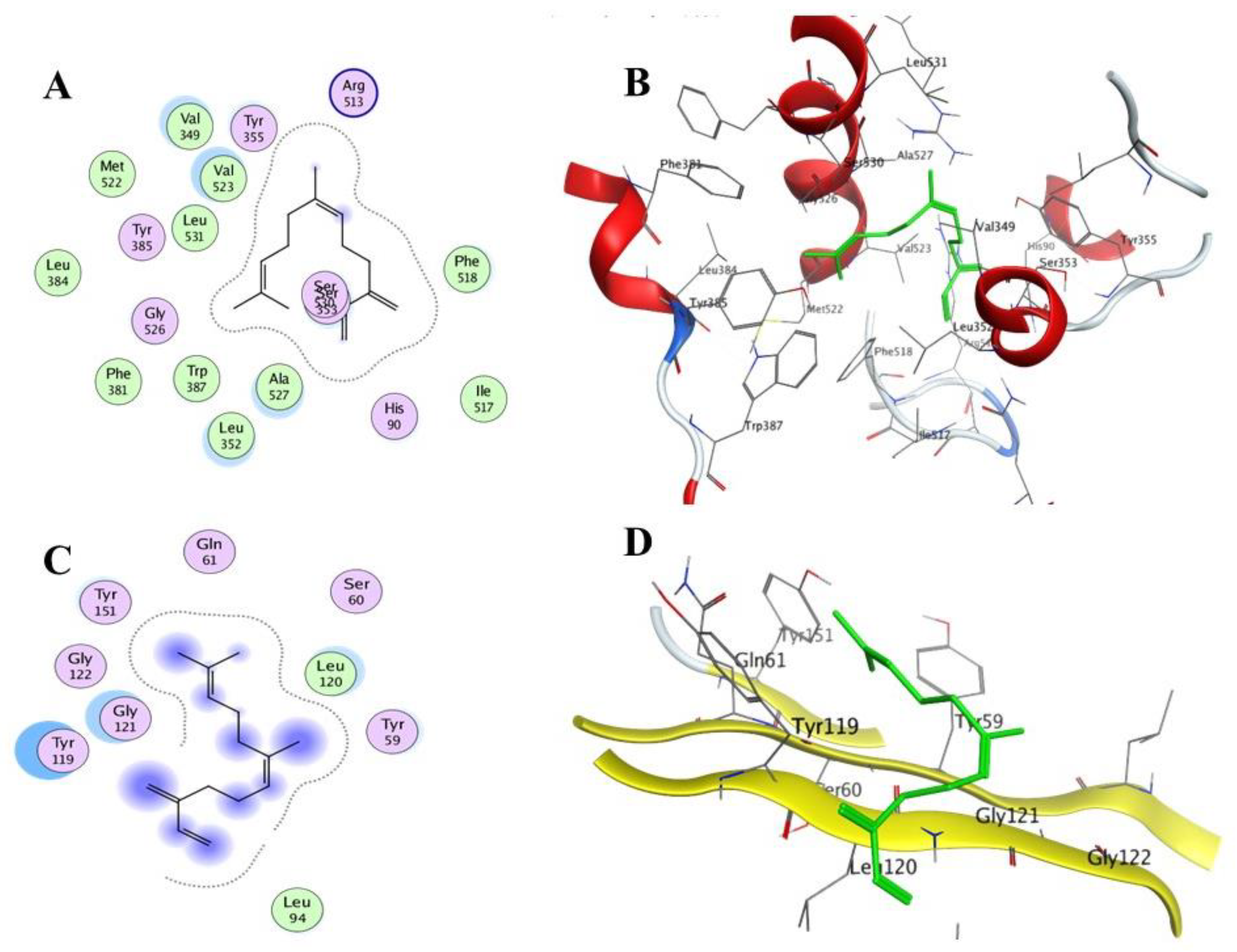
2.5. In Silico Molecular Docking Simulation
2.6. Structure Drug-like Properties
3. Materials and Methods
3.1. General
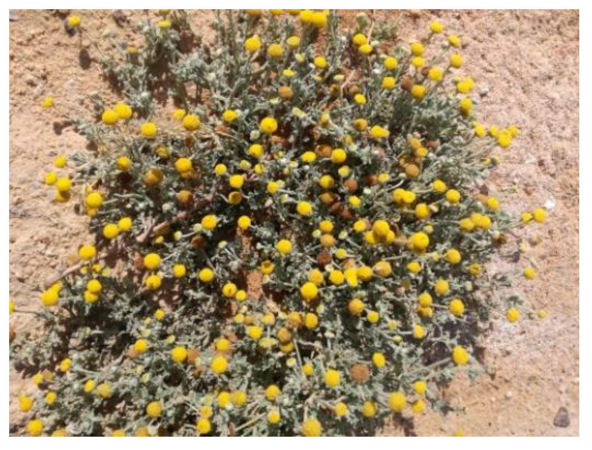
3.2. Plant Material
3.3. Extraction of Essential Oil
3.4. Gas Liquid Chromatography-Mass Spectrometry (GLC/MS)
3.5. Cell Culture, Treatments, and MTT Assay
3.6. Assesment of Proinflammatory Biomarkers in LPS-Induced RAW 264.7 Macrophages
3.7. In Silico Molecular Docking Simulation
3.8. Structure Drug-like Properties
3.9. Statistical Analysis
4. Conclusions
Author Contributions
Funding
Institutional Review Board Statement
Informed Consent Statement
Data Availability Statement
Acknowledgments
Conflicts of Interest
Sample Availability
References
- Xie, Z.; Wang, Y.; Huang, J.; Qian, N.; Shen, G.; Chen, L. Anti-inflammatory activity of polysaccharides from Phellinus linteus by regulating the NF-κB translocation in LPS-stimulated RAW264. 7 macrophages. Int. J. Biol. Macromol. 2019, 129, 61–67. [Google Scholar] [CrossRef] [PubMed]
- Nkadimeng, S.M.; Nabatanzi, A.; Steinmann, C.M.; Eloff, J.N. Phytochemical, Cytotoxicity, Antioxidant and Anti-Inflammatory Effects of Psilocybe Natalensis Magic Mushroom. Plants 2020, 9, 1127. [Google Scholar] [CrossRef] [PubMed]
- Calder, P.C.; Albers, R.; Antoine, J.-M.; Blum, S.; Bourdet-Sicard, R.; Ferns, G.; Folkerts, G.; Friedmann, P.; Frost, G.; Guarner, F. Inflammatory disease processes and interactions with nutrition. Br. J. Nutr. 2009, 101, 1–45. [Google Scholar] [CrossRef] [PubMed] [Green Version]
- Ji, K.-Y.; Kim, K.M.; Kim, Y.H.; Im, A.-R.; Lee, J.Y.; Park, B.; Na, M.; Chae, S. The enhancing immune response and anti-inflammatory effects of Anemarrhena asphodeloides extract in RAW 264.7 cells. Phytomedicine 2019, 59, 152789. [Google Scholar] [CrossRef] [PubMed]
- Nguyen, T.Q.; Duy Binh, T.; Pham, T.L.; Nguyen, Y.D.; Dai Trang, T.X.; Nguyen, T.T.; Kanaori, K.; Kamei, K. Anti-inflammatory effects of Lasia spinosa leaf extract in lipopolysaccharide-induced RAW 264.7 macrophages. Int. J. Mol. Sci. 2020, 21, 3439. [Google Scholar] [CrossRef] [PubMed]
- Zhang, M.; Hettiarachchy, N.S.; Horax, R.; Kannan, A.; Praisoody, A.; Muhundan, A. Phytochemicals, antioxidant and antimicrobial activity of Hibiscus sabdariffa, Centella asiatica, Moringa oleifera and Murraya koenigii leaves. J. Med. Plants Res. 2011, 5, 6672–6680. [Google Scholar]
- Dinarello, C.A. Anti-inflammatory agents: Present and future. Cell 2010, 140, 935–950. [Google Scholar] [CrossRef] [PubMed] [Green Version]
- Oboh, G. Effect of blanching on the antioxidant properties of some tropical green leafy vegetables. LWT-Food Sci. Technol. J. 2005, 38, 513–517. [Google Scholar] [CrossRef]
- Tiwari, M.; Dwivedi, U.; Kakkar, P. Suppression of oxidative stress and pro-inflammatory mediators by Cymbopogon citratus D. Stapf extract in lipopolysaccharide stimulated murine alveolar macrophages. Food Chem. Toxicol. 2010, 48, 2913–2919. [Google Scholar] [CrossRef] [PubMed]
- Lopes, D.C.D.X.P.; de Oliveira, T.B.; Viçosa, A.L.; Valverde, S.S.; Júnior, E.R. Anti-Inflammatory Activity of the Compositae Family and Its Therapeutic Potential. Planta Med. 2020, 87, 71–100. [Google Scholar] [CrossRef] [PubMed]
- Tadrent, W.; Alabdul Magid, A.; Kabouche, A.; Harakat, D.; Voutquenne-Nazabadioko, L.; Kabouche, Z. A new sulfonylated flavonoid and other bioactive compounds isolated from the aerial parts of Cotula anthemoides L. Nat. Prod. Res. 2017, 31, 1437–1445. [Google Scholar] [CrossRef] [PubMed]
- Bellakhdar, J. Pharmacopée Marocaine Traditionnelle; Ibis Press: Newburyport, MA, USA, 1997. [Google Scholar]
- Jana, M.; Lazrek, H.; Markouk, M. Effets bactériostatiques des extraits flavoniques de Cotula cinerea DEL. Al-Biruniya 1992, 8, 89–96. [Google Scholar]
- Larhsini, M.; Markouk, M.; Jaouhari, J.; Bekkouche, K.; Lazrek, H.; Jana, M. The antipyretic activity of some Moroccan medicinal plants. Phytother. Res. 2002, 16, 97–98. [Google Scholar] [CrossRef] [PubMed]
- Markouk, M.; Lazrek, H.; Jana, M. Analgesic effect of extracts from Cotula cinerea (L). Phytother. Res. 1999, 13, 229–230. [Google Scholar] [CrossRef]
- Pandey, A.K.; Tripathi, N. Aromatic plants of gorakhpur division: Their antimycotic properties and medicinal value. Int. J. Pharm. Sci. Rev. Res. 2011, 7, 142–147. [Google Scholar]
- Hassan, G.; Ahmad, T.; Mohi-ud-din, R. An ethnobotanical study in Budgam district of Kashmir valley: An attempt to explore and document traditional knowledge of the area. Int. Res. J. Pharm. 2013, 4, 201–204. [Google Scholar]
- Showkat, R.S.; Shah, S.W.A.; Bhat, B.A. Chemoprofiling of Medicinal Plants and Value Addition of Their Principle Constituents Through Synthetic Modifications. Master’s Thesis, University of Kashmir, Srinagar, India, 2013. [Google Scholar]
- Roma, F. Some Medicinal Forest Plants of Africa and Latin America; Food and Agriculture Organization of the United Nations: Rome, Italy, 1986. [Google Scholar]
- Tadrent, W.; Benteldjoune, M.; Laggoune, S.; Benmerache, A.; Kabouche, A.; Semra, Z.; Kabouche, Z. Composition and Antibacterial Activity of the Essential Oil of Cotula anthemoides. Chem. Nat. Compd. 2014, 50, 744–746. [Google Scholar] [CrossRef]
- Kether, F.B.H.; Mahjoub, M.A.; Mahjoub, S.A.; Salah, K.B.; Helal, A.N.; Mighri, Z. Chemical composition, in vitro antifungal and antioxidant activities of essential oil from Cotula coronopifolia L. growing in Tunisia. Afr. J. Microbiol. Res. 2012, 6, 4388–4395. [Google Scholar]
- Ruberto, G.; Baratta, M.T. Antioxidant activity of selected essential oil components in two lipid model systems. Food Chem. 2000, 69, 167–174. [Google Scholar] [CrossRef]
- Safaei-Ghomi, J.; Ebrahimabadi, A.H.; Djafari-Bidgoli, Z.; Batooli, H. GC/MS analysis and in vitro antioxidant activity of essential oil and methanol extracts of Thymus caramanicus Jalas and its main constituent carvacrol. Food Chem. 2009, 115, 1524–1528. [Google Scholar] [CrossRef]
- Yanishlieva, N.V.; Marinova, E.M.; Gordon, M.H.; Raneva, V.G. Antioxidant activity and mechanism of action of thymol and carvacrol in two lipid systems. Food Chem. 1999, 64, 59–66. [Google Scholar] [CrossRef]
- Yoon, W.-J.; Moon, J.; Song, G.; Lee, Y.; Han, M.; Lee, J.; Ihm, B.; Lee, W.; Lee, N.; Hyun, C. Artemisia fukudo essential oil attenuates LPS-induced inflammation by suppressing NF-κB and MAPK activation in RAW 264.7 macrophages. Food Chem. Toxicol. 2010, 48, 1222–1229. [Google Scholar] [CrossRef] [PubMed]
- Homnan, N.; Thongpraditchote, S.; Chomnawang, M. In vitroAnti-inflammatory effects of Thai herb essential oils. Pharm. Sci. Asia 2020, 47, 153–163. [Google Scholar] [CrossRef]
- Tosun, A.; Khan, S.; Kim, Y.S.; Calín-Sánchez, Á.; Hysenaj, X.; Carbonell-Barrachina, A. Essential oil composition and anti-inflammatory activity of Salvia officinalis L (Lamiaceae) in murin macrophages. Trop. J. Pharm. Res. 2014, 13, 937–942. [Google Scholar] [CrossRef] [Green Version]
- Pishgahzadeh, E.; Shafaroodi, H.; Asgarpanah, J. Analgesic and antiinflammatory activities of the essential oil from Artemisia sieberi Besser. Braz. J. Pharm. Sci. 2019, 55, e17011. [Google Scholar] [CrossRef] [Green Version]
- Ghouti, D.; Rached, W.; Abdallah, M.; Pires, T.C.; Calhelha, R.C.; Alves, M.J.; Abderrahmane, L.H.; Barros, L.; Ferreira, I.C. Phenolic profile and in vitro bioactive potential of Saharan Juniperus phoenicea L. and Cotula cinerea (Del) growing in Algeria. Food Funct. 2018, 9, 4664–4672. [Google Scholar] [CrossRef] [Green Version]
- Salman, S.; Shah, F.H.; Idrees, J.; Idrees, F.; Velagala, S.; Ali, J.; Khan, A.A. Virtual screening of immunomodulatory medicinal compounds as promising anti-SARS-COV-2 inhibitors. Future Virol. 2020, 15, 267–275. [Google Scholar] [CrossRef]
- Emon, N.U.; Alam, S.; Rudra, S.; Al Haidar, I.K.; Farhad, M.; Rana, M.E.H.; Ganguly, A. Antipyretic activity of the leaves extract of Caesalpinia digyna Rottl along with phytoconstituent’s binding affinity to COX-1, COX-2 and mPGES-1 receptors: An in vivo and in silico approaches (Antipyretic activity of Caesalpinia digyna Rottl). Saudi J. Biol. Sci. 2021, 28, 5302–5309. [Google Scholar] [CrossRef]
- Pistelli, L.; Najar, B.; Giovanelli, S.; Lorenzini, L.; Tavarini, S.; Angelini, L.G. Agronomic and phytochemical evaluation of lavandin and lavender cultivars cultivated in the Tyrrhenian area of Tuscany (Italy). Ind. Crops Prod. 2017, 109, 37–44. [Google Scholar] [CrossRef]
- Lipinski, C.A.; Lombardo, F.; Dominy, B.W.; Feeney, P.J. Experimental and computational approaches to estimate solubility and permeability in drug discovery and development settings. Adv. Drug Deliv. Rev. 1997, 23, 3–25. [Google Scholar] [CrossRef]
- Di Stefano, V.; Schillaci, D.; Cusimano, M.G.; Rishan, M.; Rashan, L. In vitro antimicrobial activity of frankincense oils from Boswellia sacra grown in different locations of the Dhofar region (Oman). Antibiotics 2020, 9, 195. [Google Scholar] [CrossRef] [PubMed] [Green Version]
- Hadidi, M.; Motamedzadegan, A.; Jelyani, A.Z.; Khashadeh, S. Nanoencapsulation of hyssop essential oil in chitosan-pea protein isolate nano-complex. LWT-Food Sci. Technol. J. 2021, 144, 111254. [Google Scholar] [CrossRef]
- Yu, M.; Chen, T.-T.; Zhang, T.; Jia, H.-M.; Li, J.-J.; Zhang, H.-W.; Zou, Z.-M. Anti-inflammatory constituents in the root and rhizome of Polygonum cuspidatum by UPLC-PDA-QTOF/MS and lipopolysaccharide-activated RAW264. 7 macrophages. J. Pharm. Biomed. Anal. 2021, 195, 113839. [Google Scholar] [CrossRef] [PubMed]
- Lee, H.H.; Ahn, E.K.; Hong, S.S.; Oh, J.S. Anti-inflammatory effect of tribulusamide D isolated from Tribulus terrestris in lipopolysaccharide-stimulated RAW264. 7 macrophages. Mol. Med. Rep. 2017, 16, 4421–4428. [Google Scholar] [CrossRef] [PubMed]
- Ho, C.-L.; Li, L.-H.; Weng, Y.-C.; Hua, K.-F.; Ju, T.-C. Eucalyptus essential oils inhibit the lipopolysaccharide-induced inflammatory response in RAW264. 7 macrophages through reducing MAPK and NF-κB pathways. BMC Complement. Altern. Med. 2020, 20, 200. [Google Scholar]
- Zia, K.; Ashraf, S.; Jabeen, A.; Saeed, M.; Nur-e-Alam, M.; Ahmed, S.; Al-Rehaily, A.J.; Ul-Haq, Z. Identification of potential TNF-α inhibitors: From in silico to in vitro studies. Sci. Rep. 2020, 10, 20974. [Google Scholar] [CrossRef] [PubMed]
- Abou Baker, D.H.; Amarowicz, R.; Kandeil, A.; Ali, M.A.; Ibrahim, E.A. Antiviral activity of Lavandula angustifolia L. and Salvia officinalis L. essential oils against avian influenza H5N1 virus. J. Sci. Food Agric. 2021, 4, 100135. [Google Scholar] [CrossRef]
- Allam, A.E.; Abouelela, M.E.; Assaf, H.K.; Sayed, A.M.; Nafady, A.M.; El-Shanawany, M.A.; Takano, F.; Ohta, T. Phytochemical and in silico studies for potential constituents from Centaurium spicatum as candidates against the SARS-CoV-2 main protease and RNA-dependent RNA polymerase. Nat. Prod. Res. 2021, 1–8. [Google Scholar] [CrossRef] [PubMed]


| No | Rt, min | Compound Name | Retention Index | Composition (%) ** | ||
|---|---|---|---|---|---|---|
| (Cal.) | (Rep.) * | Leaves and Stems | Flowers | |||
| 1 | 6.6 | β-Pinene | 976 | 974 | 0.31 ± 0.01 | 1.02 ± 0.12 |
| 2 | 7.9 | α-Terpinene | 1014 | 1014 | 0.26 ± 0.02 | 0.10 ± 0.01 |
| 3 | 8.3 | D-Limonene | 1026 | 1024 | 0.15 ± 0.09 | 0.26 ± 0.09 |
| 4 | 8.4 | Eucalyptol | 1032 | 1031 | 1.14 ± 0.06 | 0.42 ± 0.07 |
| 5 | 9.3 | γ-Terpinene | 1060 | 1059 | 0.52 ± 0.23 | 0.16 ± 0.03 |
| 6 | 9.8 | trans-Thujone | 1114 | 1112 | 5.14 ± 0.36 | 10.40 ± 0.57 |
| 7 | 11.4 | cis-para-Menth-2-ene-1-ol | 1120 | 1118 | 0.34 ± 0.03 | 0.17 ± 0.01 |
| 8 | 12.5 | Camphor | 1145 | 1141 | 88.79 ± 1.17 | 86.45 ± 1.01 |
| 9 | 12.9 | Pinocarvone | 1162 | 1160 | 0.25 ± 0.11 | 0.09 ± 0.02 |
| 10 | 13.1 | Borneol | 1168 | 1165 | *** ND | 0.11 ± 0.08 |
| 11 | 13.5 | Terpinen-4-ol | 1178 | 1177 | 1.48 ± 0.49 | 0.40 ± 0.01 |
| 12 | 14.0 | α-Terpineol | 1186 | 1186 | 0.22 ± 0.02 | *** ND |
| 13 | 15.3 | Octanol acetate | 1214 | 1211 | 0.13 ± 0.01 | 0.24 ± 0.01 |
| 14 | 17.0 | Bornyl acetate | 1284 | 1284 | 0.48 ± 0.16 | 0.18 ± 0.07 |
| 15 | 21.0 | Longifolene | 1403 | 1407 | 0.12 ± 0.08 | *** ND |
| 16 | 22.4 | cis-β-Farnesene | 1448 | 1454 | 0.58 ± 0.01 | *** ND |
| 17 | 23.0 | Germacrene D | 1480 | 1484 | 0.09 ± 0.04 | *** ND |
| No. | Compound | COX-2 (PDB ID: 5KIR) | TNF-α (PDB ID: 2AZ5) | ||
|---|---|---|---|---|---|
| Pose Score (kcal/mol) | RMSD Refine (Å) | Pose Score (kcal/mol) | RMSD Refine (Å) | ||
| 1 | β-Pinene | −7.7550 | 0.98 | −4.4942 | 1.44 |
| 2 | α-Terpinene | −7.6562 | 1.09 | −4.9016 | 1.29 |
| 3 | D-Limonene | −7.7438 | 1.24 | −4.9351 | 1.72 |
| 4 | Eucalyptol | −8.4517 | 0.78 | −4.6986 | 1.76 |
| 5 | γ-Terpinene | −7.5690 | 1.03 | −5.1749 | 1.64 |
| 6 | trans-Thujone | −7.5807 | 0.71 | −5.1960 | 2.07 |
| 7 | cis-para-Menth-2-ene-1-ol | −8.7486 | 0.99 | −6.7740 | 1.25 |
| 8 | Camphor | −7.6977 | 0.89 | −4.8496 | 1.08 |
| 9 | Pinocarvone | −7.2938 | 0.88 | −4.8235 | 1.77 |
| 10 | Borneol | −7.8185 | 0.67 | −4.7328 | 1.41 |
| 11 | Terpinen-4-ol | −9.3323 | 0.70 | −6.2579 | 1.18 |
| 12 | α-Terpineol | −7.6466 | 0.58 | −4.7338 | 1.48 |
| 13 | Octanol acetate | −8.6976 | 0.84 | −5.5871 | 0.97 |
| 14 | Bornyl acetate | −9.6206 | 0.91 | −5.1830 | 1.68 |
| 15 | Longifolene | −9.4917 | 0.59 | −5.8426 | 1.48 |
| 16 | cis-β-Farnesene | −9.4392 | 1.09 | −5.9222 | 1.35 |
| 17 | Germacrene D | −8.9982 | 1.02 | −5.0475 | 1.38 |
| 18 | Resveratrol | −11.2915 | 0.69 | −6.9306 | 1.17 |
| No. | Name | * logP | TPSA | n atoms | MW | nHBA | nHBD | Number of Violations | nrotb | MVol |
|---|---|---|---|---|---|---|---|---|---|---|
| 1 | β-Pinene | 3.33 | 0 | 10 | 136.24 | 0 | 0 | 0 | 0 | 152.37 |
| 2 | α-Terpinene | 3.36 | 0 | 10 | 136.24 | 0 | 0 | 0 | 1 | 156.74 |
| 3 | D-Limonene | 3.62 | 0 | 10 | 136.24 | 0 | 0 | 0 | 1 | 157.3 |
| 4 | Eucalyptol | 2.72 | 9.23 | 11 | 154.25 | 1 | 0 | 0 | 0 | 166.66 |
| 5 | γ-Terpinene | 3.36 | 0 | 10 | 136.24 | 0 | 0 | 0 | 1 | 156.74 |
| 6 | trans-Thujone | 2.16 | 17.07 | 11 | 152.24 | 1 | 0 | 0 | 1 | 160.21 |
| 7 | cis-para-Menth-2-ene-1-ol | 2.8 | 20.23 | 11 | 154.25 | 1 | 1 | 0 | 1 | 170.67 |
| 8 | Camphor | 2.16 | 17.07 | 11 | 152.24 | 1 | 0 | 0 | 0 | 159.86 |
| 9 | Pinocarvone | 2.23 | 17.07 | 11 | 150.22 | 1 | 0 | 0 | 0 | 154.55 |
| 10 | Borneol | 2.48 | 20.23 | 11 | 154.25 | 1 | 1 | 0 | 1 | 166.28 |
| 11 | Terpinen-4-ol | 2.6 | 20.23 | 11 | 154.25 | 1 | 1 | 0 | 1 | 170.65 |
| 12 | α-Terpineol | 2.57 | 0 | 10 | 136.24 | 0 | 0 | 0 | 1 | 157.32 |
| 13 | Octanol acetate | 3.47 | 26.3 | 13 | 184.28 | 2 | 0 | 0 | 7 | 201.74 |
| 14 | Bornyl acetate | 3.05 | 26.3 | 14 | 196.29 | 2 | 0 | 0 | 2 | 202.23 |
| 15 | Longifolene | 5.82 | 0 | 15 | 204.36 | 0 | 0 | 1 | 6 | 239.27 |
| 16 | cis-β-Farnesene | 5.84 | 0 | 15 | 204.36 | 0 | 0 | 1 | 7 | 239.82 |
| 17 | Germacrene D | 5.43 | 0 | 15 | 204.36 | 0 | 0 | 1 | 1 | 234.9 |
Publisher’s Note: MDPI stays neutral with regard to jurisdictional claims in published maps and institutional affiliations. |
© 2022 by the authors. Licensee MDPI, Basel, Switzerland. This article is an open access article distributed under the terms and conditions of the Creative Commons Attribution (CC BY) license (https://creativecommons.org/licenses/by/4.0/).
Share and Cite
Refaey, M.S.; Abouelela, M.E.; El-Shoura, E.A.M.; Alkhalidi, H.M.; Fadil, S.A.; Elhady, S.S.; Abdelhameed, R.F.A. In Vitro Anti-Inflammatory Activity of Cotula anthemoides Essential Oil and In Silico Molecular Docking of Its Bioactives. Molecules 2022, 27, 1994. https://doi.org/10.3390/molecules27061994
Refaey MS, Abouelela ME, El-Shoura EAM, Alkhalidi HM, Fadil SA, Elhady SS, Abdelhameed RFA. In Vitro Anti-Inflammatory Activity of Cotula anthemoides Essential Oil and In Silico Molecular Docking of Its Bioactives. Molecules. 2022; 27(6):1994. https://doi.org/10.3390/molecules27061994
Chicago/Turabian StyleRefaey, Mohamed S., Mohamed E. Abouelela, Ehab A. M. El-Shoura, Hala M. Alkhalidi, Sana A. Fadil, Sameh S. Elhady, and Reda F. A. Abdelhameed. 2022. "In Vitro Anti-Inflammatory Activity of Cotula anthemoides Essential Oil and In Silico Molecular Docking of Its Bioactives" Molecules 27, no. 6: 1994. https://doi.org/10.3390/molecules27061994