
Phytochemical Insights into Ficus sur Extracts and Their Biological Activity

 ,
,  ,
, 
 , , ,
, , ,
Abstract
:1. Introduction
2. Results and Discussion
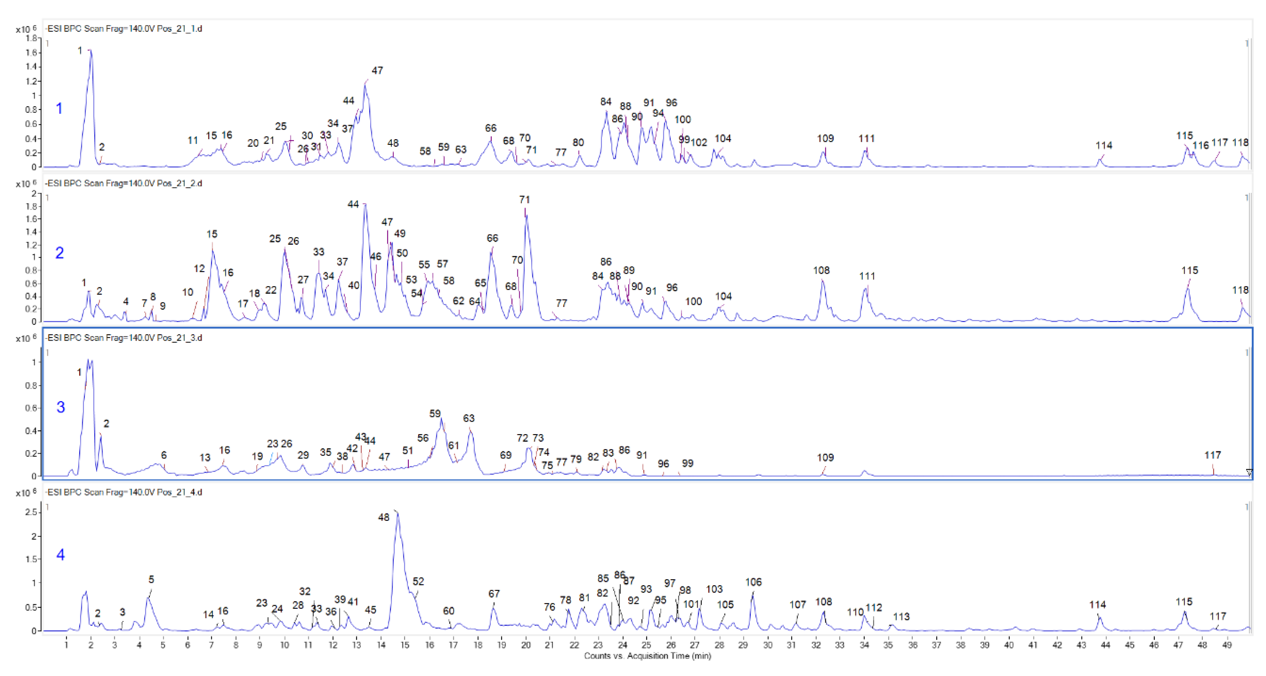
2.1. Bioactive Compounds
2.2. Antioxidant Effects
2.3. Enzymatic Inhibitory Activities
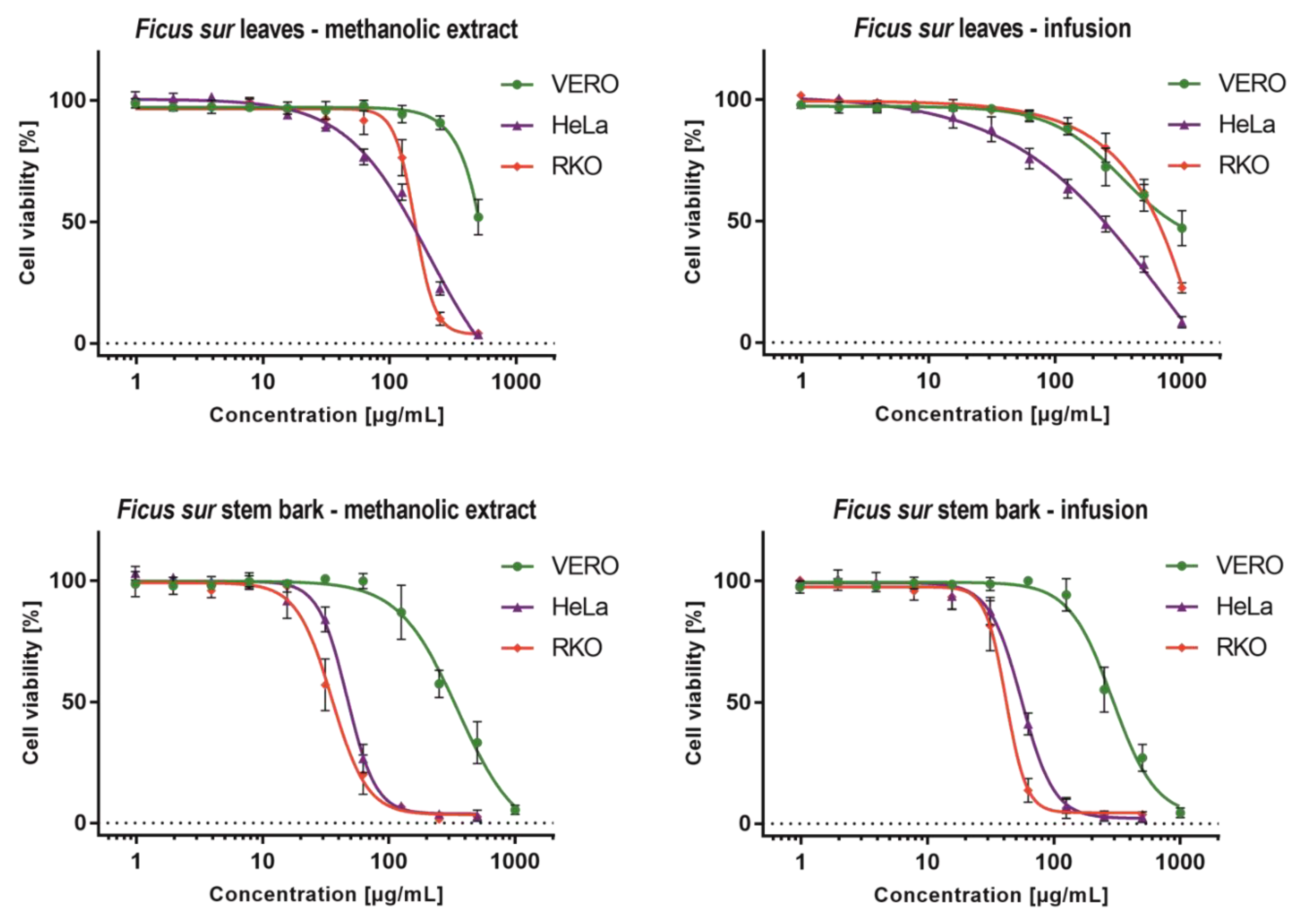
2.4. Cytotoxicity Evaluation
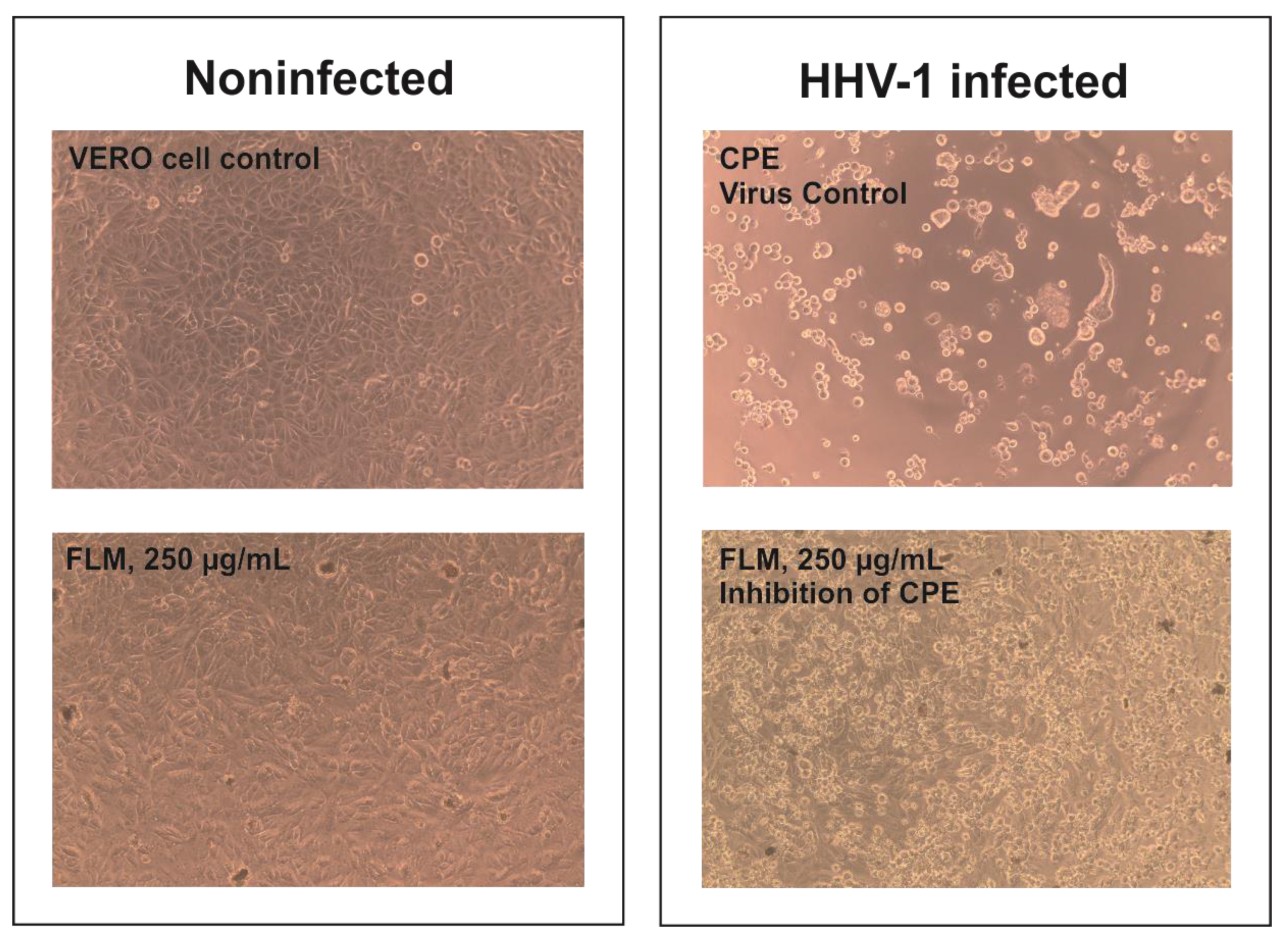
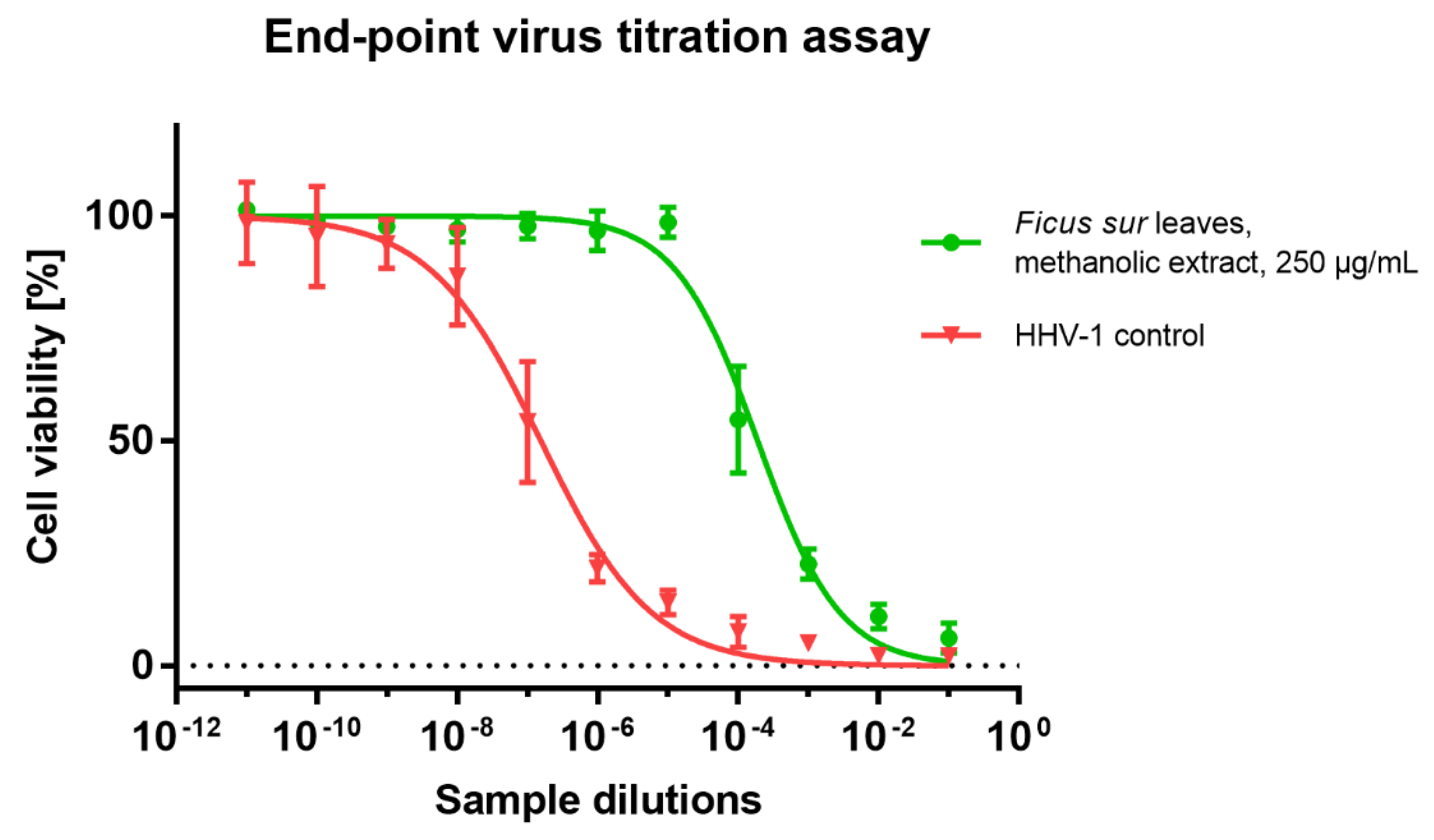
2.5. Antiviral Potential
3. Materials and Methods
3.1. Plant Materials and Preparation of Extracts
3.2. Chromatographic Conditions
3.3. Detection Conditions
3.4. Total Phenolic and Flavonoid Content
3.5. Antioxidant and Enzyme Inhibitory Assays
3.6. Cytotoxicity Testing
3.7. Evaluation of Antiviral Potential
4. Conclusions
Supplementary Materials
Author Contributions
Funding
Institutional Review Board Statement
Informed Consent Statement
Data Availability Statement
Conflicts of Interest
References
- Petrovska, B.B. Historical review of medicinal plants’ usage. Pharmacogn. Rev. 2012, 6, 1–5. [Google Scholar] [CrossRef] [PubMed] [Green Version]
- Yoo, S.; Nam, H.; Lee, D. Phenotype-oriented network analysis for discovering pharmacological effects of natural compounds. Sci. Rep. 2018, 8, 11667. [Google Scholar] [CrossRef] [PubMed] [Green Version]
- Stierle, A.; Strobel, G.; Stierle, D. Taxol and taxane production by Taxomyces andreanae, an endophytic fungus of Pacific yew. Science 1993, 260, 214–216. [Google Scholar] [CrossRef] [PubMed]
- Murugesu, S.; Selamat, J.; Perumal, V. Phytochemistry, Pharmacological Properties, and Recent Applications of Ficus benghalensis and Ficus religiosa. Plants 2021, 10, 2749. [Google Scholar] [CrossRef] [PubMed]
- Available online: http://www.worldfloraonline.org/ (accessed on 12 December 2021).
- Clark, A.M. Natural products as a resource for new drugs. Pharm Res. 1996, 13, 1133–1144. [Google Scholar] [CrossRef] [PubMed]
- Esievo, K.B.; Anthony, S.O.; Fatokun, O.T.; Kunle, O.F. Ficus capensis Thumb. (Moraceae): Review of its ethnomedicinal uses, pharmacological activities and phytochemical constituents. Arch. Curr. Res. Int. 2018, 12, 1–7. [Google Scholar] [CrossRef]
- Lansky, E.P.; Paavilainen, H.M. Figs: The Genus Ficus; CRC Press: Boca Raton, FL, USA, 2010. [Google Scholar]
- Sabiu, S.; O'Neill, F.H.; Ashafa, A.O.T. The Purview of Phytotherapy in the Management of Kidney Disorders: A Systematic Review on Nigeria and South Africa. Afr J. Tradit. Complement. Altern. Med. 2016, 13, 38–47. [Google Scholar] [PubMed]
- Beyi, M. Ethnobotanical investigation of traditional medicinal plants in dugda district, Oromia Regio. SM J. Med. Plant Stud. 2018, 2, 1007. [Google Scholar]
- Regassa, R. Diversity and conservation status of some economically valued indigenous medicinal plants in Hawassa College of Teacher Education Campus, Southern Ethiopia. Int. J. Adv. Res. 2013, 1, 308–328. [Google Scholar]
- Ayele, M.; Makonnen, E.; Ayele, A.G.; Tolcha, Y. Evaluation of the Diuretic Activity of the Aqueous and 80% Methanol Extracts of Ficus sur Forssk (Moraceae) Leaves in Saline-loaded Rats. J. Exp. Pharmacol. 2020, 12, 619. [Google Scholar] [CrossRef] [PubMed]
- Ishola, I.O.; Olayemi, S.O.; Yemitan, O.K.; Ekpemandudiri, N.K. Mechanisms of anticonvulsant and sedative actions of the ethanolic stem-bark extract of Ficus sur Forssk (Moraceae) in rodents. Pak. J. Biol. Sci. 2013, 16, 1287–1294. [Google Scholar] [CrossRef] [PubMed]
- Diba, D.; Mekasha, Y.; Urge, M.; Tolera, A. Feed intake, digestibility, growth performance, and blood profile of pigs fed mixtures of dried and ground fig (Ficus sur) fruits and graded levels of maize. Trop. Anim. Health Prod. 2015, 47, 339–346. [Google Scholar] [CrossRef] [PubMed]
- Yarmolinsky, L.; Zaccai, M.; Ben-Shabat, S.; Mills, D.; Huleihel, M. Antiviral activity of ethanol extracts of Ficus binjamina and Lilium candidum in vitro. New Biotechnol. 2009, 26, 307–313. [Google Scholar] [CrossRef] [PubMed]
- Camero, M.; Marinaro, M.; Lovero, A.; Elia, G.; Losurdo, M.; Buonavoglia, C.; Tempesta, M. In vitro antiviral activity of Ficus carica latex against caprine herpesvirus-1. Nat. Prod. Res. 2014, 28, 2031–2035. [Google Scholar] [CrossRef] [PubMed]
- Lazreg Aref, H.; Gaaliche, B.; Fekih, A.; Mars, M.; Aouni, M.; Pierre Chaumon, J.; Said, K. In vitro cytotoxic and antiviral activities of Ficus carica latex extracts. Nat. Prod. Res. 2011, 25, 310–319. [Google Scholar] [CrossRef] [PubMed]
- Huang, N.-C.; Hung, W.-T.; Tsai, W.-L.; Lai, F.-Y.; Lin, Y.-S.; Huang, M.-S.; Chen, J.-J.; Lin, W.-Y.; Weng, J.-R.; Chang, T.-H. Ficus septica plant extracts for treating Dengue virus in vitro. PeerJ 2017, 5, e3448. [Google Scholar] [CrossRef] [PubMed]
- Cagno, V.; Civra, A.; Kumar, R.; Pradhan, S.; Donalisio, M.; Sinha, B.N.; Ghosh, M.; Lembo, D. Ficus religiosa L. bark extracts inhibit human rhinovirus and respiratory syncytial virus infection in vitro. J. Ethnopharmacol. 2015, 176, 252–257. [Google Scholar] [CrossRef] [PubMed]
- Ghosh, M.; Civra, A.; Rittà, M.; Cagno, V.; Mavuduru, S.G.; Awasthi, P.; Lembo, D.; Donalisio, M. Ficus religiosa L. bark extracts inhibit infection by herpes simplex virus type 2 in vitro. Arch. Virol. 2016, 161, 3509–3514. [Google Scholar] [CrossRef] [PubMed]
- Khairunisa, S.Q.; Indriati, D.W.; Tumewu, L.; Widyawaruyanti, A.; Nasronudin, N. Screening of anti-HIV activities in ethanol extract and fractions from Ficus fistulosa leaves. J. Basic Clin. Physiol. Pharmacol. 2021, 32, 737–742. [Google Scholar] [CrossRef]
- Altemimi, A.; Lakhssassi, N.; Baharlouei, A.; Watson, D.G.; Lightfoot, D.A. Phytochemicals: Extraction, Isolation, and Identification of Bioactive Compounds from Plant Extracts. Plants 2017, 6, 42. [Google Scholar] [CrossRef]
- Zhang, Q.-W.; Lin, L.-G.; Ye, W.-C. Techniques for extraction and isolation of natural products: A comprehensive review. Chin. Med. 2018, 13, 20. [Google Scholar] [CrossRef] [PubMed] [Green Version]
- Elhawary, S.S.; Younis, I.Y.; El Bishbishy, M.H.; Khattab, A.R. LC–MS/MS-Based chemometric analysis of phytochemical diversity in 13 Ficus spp. (Moraceae): Correlation to their in vitro antimicrobial and in silico quorum sensing inhibitory activities. Ind. Crops Prod. 2018, 126, 261–271. [Google Scholar] [CrossRef]
- Farag, M.A.; Abdelfattah, M.S.; Badr, S.E.; Wessjohann, L.A. Profiling the chemical content of Ficus lyrata extracts via UPLC-PDA-qTOF-MS and chemometrics. Nat. Prod. Res. 2014, 28, 1549–1556. [Google Scholar] [CrossRef] [PubMed]
- Clifford, M.N.; Wu, W.; Kuhnert, N. The chlorogenic acids of Hemerocallis. Food Chem. 2006, 95, 574–578. [Google Scholar] [CrossRef]
- Fotirić Akšić, M.; Dabić Zagorac, D.; Sredojević, M.; Milivojević, J.; Gašić, U.; Meland, M.; Natić, M. Chemometric characterization of strawberries and blueberries according to their phenolic profile: Combined effect of cultivar and cultivation system. Molecules 2019, 24, 4310. [Google Scholar] [CrossRef] [PubMed] [Green Version]
- Gevrenova, R.; Zengin, G.; Sinan, K.I.; Yıldıztugay, E.; Zheleva-Dimitrova, D.; Picot-Allain, C.; Mahomoodally, M.F.; Imran, M.; Dall’Acqua, S. UHPLC-MS Characterization and biological insights of different solvent extracts of two Achillea species (A. aleppica and A. santolinoides) from Turkey. Antioxidants 2021, 10, 1180. [Google Scholar] [CrossRef] [PubMed]
- Guimarães, R.; Barros, L.; Dueñas, M.; Carvalho, A.M.; Queiroz, M.J.R.; Santos-Buelga, C.; Ferreira, I.C. Characterisation of phenolic compounds in wild fruits from Northeastern Portugal. Food Chem. 2013, 141, 3721–3730. [Google Scholar] [CrossRef] [PubMed] [Green Version]
- Human Metabolome Database (HMDB). 2021. Available online: https://hmdb.ca/ (accessed on 31 July 2021).
- MassBank of North America (MoNA) Database. 2021. Available online: https://mona.fiehnlab.ucdavis.edu/ (accessed on 31 July 2021).
- Metlin Database. 2021. Available online: https://metlin.scripps.edu/ (accessed on 31 July 2021).
- PubChem. 2021. Available online: https://pubchem.ncbi.nlm.nih.gov/ (accessed on 31 July 2021).
- Simirgiotis, M.J. Antioxidant capacity and HPLC-DAD-MS profiling of Chilean Peumo (Cryptocarya alba) fruits and comparison with German Peumo (Crataegus monogyna) from Southern Chile. Molecules 2013, 18, 2061–2080. [Google Scholar] [CrossRef] [Green Version]
- Singh, A.; Kumar, S.; Kumar, B. LC-MS Identification of Proanthocyanidins in Bark and Fruit of six Terminalia species. Nat. Prod. Commun. 2018, 13, 1934578X1801300511. [Google Scholar] [CrossRef] [Green Version]
- Świątek, Ł.; Sieniawska, E.; Sinan, K.I.; Maciejewska-Turska, M.; Boguszewska, A.; Polz-Dacewicz, M.; Senkardes, I.; Guler, G.O.; Bibi Sadeer, N.; Mahomoodally, M.F. LC-ESI-QTOF-MS/MS Analysis, Cytotoxic, Antiviral, Antioxidant, and Enzyme Inhibitory Properties of Four Extracts of Geranium pyrenaicum Burm. f.: A Good Gift from the Natural Treasure. Int. J. Mol. Sci. 2021, 22, 7621. [Google Scholar] [CrossRef]
- Zengin, G.; Mahomoodally, M.F.; Picot-Allain, M.C.N.; Sinan, K.I.; Ak, G.; Etienne, O.K.; Sieniawska, E.; Maciejewska-Turska, M.; Świątek, Ł.; Rajtar, B. Chemical composition, biological properties and bioinformatics analysis of two Caesalpina species: A new light in the road from nature to pharmacy shelf. J. Pharm. Biomed. Anal. 2021, 198, 114018. [Google Scholar] [CrossRef] [PubMed]
- Bibi Sadeer, N.; Montesano, D.; Albrizio, S.; Zengin, G.; Mahomoodally, M.F. The Versatility of Antioxidant Assays in Food Science and Safety—Chemistry, Applications, Strengths, and Limitations. Antioxidants 2020, 9, 709. [Google Scholar] [CrossRef] [PubMed]
- Kim, D.O.; Lee, K.W.; Lee, H.J.; Lee, C.Y. Vitamin C equivalent antioxidant capacity (VCEAC) of phenolic phytochemicals. J. Agric. Food Chem. 2002, 50, 3713–3717. [Google Scholar] [CrossRef] [PubMed]
- Cheng, Z.; Li, Y. Reducing power: The measure of antioxidant activities of reductant compounds? Redox Rep. 2004, 9, 213–217. [Google Scholar] [CrossRef] [PubMed]
- Segura Campos, M.R.; Ruiz Ruiz, J.; Chel-Guerrero, L.; Betancur Ancona, D. Coccoloba uvifera (L.) (Polygonaceae) Fruit: Phytochemical Screening and Potential Antioxidant Activity. J. Chem. 2015, 2015, 534954. [Google Scholar] [CrossRef] [Green Version]
- Abdel-Hameed, E.-S.S.; Bazaid, S.A.; Salman, M.S. Characterization of the Phytochemical Constituents of Taif Rose and Its Antioxidant and Anticancer Activities. BioMed Res. Int. 2013, 2013, 345465. [Google Scholar] [CrossRef] [PubMed] [Green Version]
- Colović, M.B.; Krstić, D.Z.; Lazarević-Pašti, T.D.; Bondžić, A.M.; Vasić, V.M. Acetylcholinesterase inhibitors: Pharmacology and toxicology. Curr. Neuropharmacol. 2013, 11, 315–335. [Google Scholar] [CrossRef] [PubMed] [Green Version]
- Tundis, R.; Loizzo, M.R.; Menichini, F. Natural products as alpha-amylase and alpha-glucosidase inhibitors and their hypoglycaemic potential in the treatment of diabetes: An update. Mini Rev. Med. Chem. 2010, 10, 315–331. [Google Scholar] [CrossRef] [PubMed]
- Deri, B.; Kanteev, M.; Goldfeder, M.; Lecina, D.; Guallar, V.; Adir, N.; Fishman, A. The unravelling of the complex pattern of tyrosinase inhibition. Sci. Rep. 2016, 6, 34993. [Google Scholar] [CrossRef] [PubMed] [Green Version]
- AlGhalban, F.M.; Khan, A.A.; Khattak, M.N.K. Comparative anticancer activities of Ficus carica and Ficus salicifolia latex in MDA-MB-231 cells. Saudi J. Biol. Sci. 2021, 28, 3225–3234. [Google Scholar] [CrossRef] [PubMed]
- Purnamasari, R.; Winarni, D.; Permanasari, A.A.; Agustina, E.; Hayaza, S.; Darmanto, W. Anticancer activity of methanol extract of Ficus carica leaves and fruits against proliferation, apoptosis, and necrosis in Huh7it cells. Cancer Inform. 2019, 18, 1176935119842576. [Google Scholar] [CrossRef] [PubMed] [Green Version]
- Remadevi, V.; Mohan Lathika, L.; Sasikumar Sujatha, A.; Sreeharshan, S. Ficus extract—A promising agent for antimammary tumorigenesis: A review on current status and future possibilities. Phytother. Res. 2019, 33, 1597–1603. [Google Scholar] [CrossRef] [PubMed]
- Yen, G.-C.; Chen, C.-S.; Chang, W.-T.; Wu, M.-F.; Cheng, F.-T.; Shiau, D.-K.; Hsu, C.-L. Antioxidant activity and anticancer effect of ethanolic and aqueous extracts of the roots of Ficus beecheyana and their phenolic components. J. Food Drug Anal. 2018, 26, 182–192. [Google Scholar] [CrossRef] [PubMed] [Green Version]
- Lv, H.; Hu, C.; Xie, Z.; Wang, P.; Chen, X.; Wen, C. Purification, characterization and anti-tumor activity of a pectic-type polysaccharide isolated from Ficus pandurata H. Int. J. Biol. Macromol. 2020, 153, 201–206. [Google Scholar] [CrossRef] [PubMed]
- Bafor, E.E.; McKenna, J.; Rowan, E.G.; Edrada-Ebel, R. Characterisation of the antiproliferative constituents and activity of Ficus exasperata (Vahl) on ovarian cancer cells–a preliminary investigation. Nat. Prod. Res. 2017, 31, 2164–2168. [Google Scholar] [CrossRef] [PubMed] [Green Version]
- Świątek, Ł.; Sieniawska, E.; Mahomoodally, M.F.; Sadeer, N.B.; Wojtanowski, K.K.; Rajtar, B.; Polz-Dacewicz, M.; Paksoy, M.Y.; Zengin, G. Phytochemical Profile and Biological Activities of the Extracts from Two Oenanthe Species (O. aquatica and O. silaifolia). Pharmaceuticals 2022, 15, 50. [Google Scholar] [CrossRef] [PubMed]
- Kane, C.J.; Menna, J.H.; Sung, C.-C.; Yeh, Y.-C. Methyl gallate, methyl-3, 4, 5-trihydroxybenzoate, is a potent and highly specific inhibitor of herpes simplex virus in vitro. II. Antiviral activity of methyl gallate and its derivatives. Biosci. Rep. 1988, 8, 95–102. [Google Scholar] [CrossRef] [PubMed]
- Robin, V.; Boustie, J.; Amoros, M.; Girre, L. In-vitro antiviral activity of seven psiadia species, Asteraceae: Isolation of two antipoliovirus flavonoids from Psiadia dentata. Pharm. Pharmacol. Commun. 1998, 4, 61–64. [Google Scholar]
- Nagai, T.; Suzuki, Y.; Tomimori, T.; Yamada, H. Antiviral activity of plant flavonoid, 5, 7, 4′-trihydroxy-8-methoxyflavone, from the roots of Scutellaria baicalensis against influenza A (H3N2) and B viruses. Biol. Pharm. Bull. 1995, 18, 295–299. [Google Scholar] [CrossRef] [PubMed] [Green Version]
- Sookkongwaree, K.; Geitmann, M.; Roengsumran, S.; Petsom, A.; Danielson, U.H. Inhibition of viral proteases by Zingiberaceae extracts and flavones isolated from Kaempferia parviflora. Die Pharmazie—Int. J. Pharm. Sci. 2006, 61, 717–721. [Google Scholar]
- Behbahani, M.; Shanehsazzadeh, M.; Shokoohinia, Y.; Soltani, M. Evaluation of anti-herpetic activity of methanol seed extract and fractions of Securigera securidaca in vitro. J. Antivir. Antiretrovir. 2013, 5, 72–76. [Google Scholar]
- Behbahani, M.; Sayedipour, S.; Pourazar, A.; Shanehsazzadeh, M. In vitro anti-HIV-1 activities of kaempferol and kaempferol-7-O-glucoside isolated from Securigera securidaca. Res. Pharm. Sci. 2014, 9, 463. [Google Scholar] [PubMed]
- Yarmolinsky, L.; Huleihel, M.; Zaccai, M.; Ben-Shabat, S. Potent antiviral flavone glycosides from Ficus benjamina leaves. Fitoterapia 2012, 83, 362–367. [Google Scholar] [CrossRef] [PubMed]
- Cheng, H.-Y.; Yang, C.-M.; Lin, T.-C.; Shieh, D.-E.; Lin, C.-C. ent-Epiafzelechin-(4α→ 8)-epiafzelechin extracted from Cassia javanica inhibits herpes simplex virus type 2 replication. J. Med. Microbiol. 2006, 55, 201–206. [Google Scholar] [CrossRef] [PubMed]
- Grochowski, D.M.; Uysal, S.; Aktumsek, A.; Granica, S.; Zengin, G.; Ceylan, R.; Locatelli, M.; Tomczyk, M. In vitro enzyme inhibitory properties, antioxidant activities, and phytochemical profile of Potentilla thuringiaca. Phytochem. Lett. 2017, 20, 365–372. [Google Scholar] [CrossRef]
- Uysal, S.; Zengin, G.; Locatelli, M.; Bahadori, M.B.; Mocan, A.; Bellagamba, G.; De Luca, E.; Mollica, A.; Aktumsek, A. Cytotoxic and enzyme inhibitory potential of two Potentilla species (P. speciosa L. and P. reptans Willd.) and their chemical composition. Front. Pharmacol. 2017, 8, 290. [Google Scholar] [CrossRef] [PubMed]


| Parts | Solvents | TPC (mg GAE/g) | TFC (mg RE/g) |
|---|---|---|---|
| Leaves | MeOH | 58.46 ± 0.28 c | 27.47 ± 0.28 a |
| Infusion | 51.77 ± 0.77 d | 16.65 ± 0.18 b | |
| Stem barks | MeOH | 109.79 ± 2.19 b | 2.54 ± 0.10 c |
| Infusion | 115.51 ± 1.60 a | 1.13 ± 0.11 d |
| Comp. No | Tentative Identification | R Time | Molecular Mass | [M − H]− | Fragment Ions (m/z) | Extracts |
|---|---|---|---|---|---|---|
| 1 | Quinic acid | 1.91 | 192.0507 | 191.0507 | 173.0464; 111.0437; 93.0318; 85.0262 | 1,2,3 |
| 2 | Citric acid | 2.33 | 192.0180 | 191.0180 | 111.0035; 87.0052 | 1,2,3,4 |
| 3 | Caffeic acid derivative | 3.40 | 242.0302 | 241.0302 | 179.0273; 153.0497; 135.0406; 123.0407; 109.0230 | 4 |
| 4 | Quinic acid derivative | 3.46 | 534.1621 | 533.1621 | 337.0845; 191.0508 | 2 |
| 5 | Quinic acid derivative | 4.04 | 406.0968 | 405.0968 | 213.0351; 191.0511 | 4 |
| 6 | 4-Hydroxy-2-(hydroxymethyl)benzoic acid | 4.09 | 168.0307 | 167.0307 | 149.0234; 123.0429 | 3 |
| 7 | Dihydroxybenzoic acid glucoside derivative | 4.13 | 532.0929 | 531.0929 | 353.0781; 315.0642; 153.0155; 96.9570 | 2 |
| 8 | Quinic acid derivative | 4.22 | 470.0574 | 469.0574 | 435.1422; 371.0962; 191.0423 | 2 |
| 9 | Hydroxycaffeoyl-quinic acid | 4.76 | 372.0900 | 371.0900 | 353.0840; 197.0360; 191.0533; 179.0312; 173.0431;135.0409 | 2 |
| 10 | Glucogallic acid/Glucosyl gallate | 6.32 | 332.0607 | 331.0607 | 169.0124; 151.0003; 125.0211 | 2 |
| 11 | Dihydroxybenzoic acid glucoside derivative | 6.49 | 436.0075 | 435.0075 | 315.0710; 153.0056 | 1 |
| 12 | Dihydro-caffeoyl-quinic acid | 6.56 | 356.0959 | 355.0959 | 191.0522; 181.0167; 173.0451; 137.0164; 111.0044 | 2 |
| 13 | Hydroxybenzoic acid derivative | 6.77 | 432.1224 | 431.1124 | 137.0243; 93.0383 | 3 |
| 14 | Caffeoyl-hydroxybenzoic acid | 7.23 | 300.0717 | 299.07117 | 239.0562; 179.0356; 137.0228 | 4 |
| 15 | Dihydroxybenzoic acid glucoside isomer 1 | 7.487 | 316.0628 | 315.0628 | 153.0134; 152.0105; 108.0213 | 1,2 |
| 16 | Dihydroxybenzoic acid | 7.59 | 154.0143 | 153.0143 | 109.0302; 108.0225 | 1,2,3,4 |
| 17 | Coumaric acid-hexoside-pentoside | 8.48 | 458.0892 | 457.0892 | 325.0865; 163.0347 | 2 |
| 18 | Dihydroxybenzoic acid glucoside isomer 2 | 8.51 | 316.0660 | 315.0660 | 153.0147; 109.0258 | 2 |
| 19 | 3-Hydroxy-4-methoxymandelate glucoside | 8.86 | 360.0924 | 359.0924 | 197.0449; 182.0215; 153.0557; 138.0321; 123.0129 | 3 |
| 20 | Caffeic acid derivative hexoside | 9.06 | 376.0601 | 375.0601; | 341.1069; 213.0650; 201.0144; 179.0316; 135.0409 | 1 |
| 21 | Quinic acid derivative | 9.08 | 372.0868 | 371.0868 | 251.0544; 191.052; 167.0327 | 1 |
| 22 | 2-Isopropylmalic acid | 9.19 | 176.0571 | 175.0571 | 157.0486; 115.0371; 85.0638 | 2 |
| 23 | Hydroxybenzoic acid | 9.29 | 138.0208 | 137.0208 | 108.0231 | 3, 4 |
| 24 | Unidentified | 9.39 | 376.1236 | 375.1236 | 312.0725; 169.0827; 151.0726 | 4 |
| 25 | Hydroxybenzoic acid 4-O-glucoside | 10.17 | 300.0669 | 299.0669 | 137.0203; 93.0315 | 1,2 |
| 26 | 3-O-Caffeoylquinic acid | 10.36 | 354.0782 | 353.0782 | 191.0578; 179.0370; 161.0243; 135.0461 | 1,2,3 |
| 27 | Dihydroxybenzoic acid glucoside isomer 3 | 10.69 | 316.0660 | 315.0660 | 153.0144; 109.0258 | 2 |
| 28 | Caffeic acid glucoside | 10.76 | 342.0825 | 341.0825 | 179.0357; 161.0246; 135.0457 | 4 |
| 29 | Dihydroxybenzoic acid O-glucoside-pentoside | 10.84 | 448.1382 | 447.1382 | 315.1063; 153.0548; 109.0301; 108.0249 | 3 |
| 30 | Hydroxycoumarin | 10.93 | 340.0715 | 339.0715 | 177.0178 | 1 |
| 31 | Methyl gallate | 11.02 | 184.0234 | 183.0234 | 168.0071; 124.0155; 78.0128 | 1 |
| 32 | Hydroxybenzoic acid | 11.13 | 138.0210 | 137.0210 | 109.0209; 108.0308; 93.0345 | 4 |
| 33 | Glucogallic acid/Glucosyl gallate | 11.50 | 332.0607 | 331.0607 | 169.0113;168.0047;125.0225 | 1,2,4 |
| 34 | 3-O-p-Coumaroylquinic acid | 11.30 | 338.0840 | 337.0840 | 191.0540; 163.0384 | 1,2 |
| 35 | Procyanidin B (dimer of (epi)catechin) | 11.62 | 578.1270 | 577.1270 | 451.1080; 425.0892; 407.0834; 289.0734; 245.0811; 125.0239 | 3 |
| 36 | Caffeic and coumaric acid derivative | 12.20 | 542.1494 | 541.1494 | 523.1466; 475.1519; 361.0900; 235.0490; 215.0901; 179.0358; 163.0398; 137.0249 | 4 |
| 37 | 1-O-Coumaroylquinic acid | 12.28 | 338.0840 | 337.0840 | 191.0509; 173.0417; 163.0356 | 1,2 |
| 38 | Glucogallic acid tartaric ester | 12.33 | 464.1040 | 463.1040 | 331.0608; 169.0147; 168.0037; 149.9937; 125.0212 | 3 |
| 39 | Caffeic acid glucoside | 12.34 | 342.0825 | 341.0825 | 179.0344; 161.0230; 135.0440 | 4 |
| 40 | Gallic acid glucoside derivative | 12.55 | 412.0173 | 411.0173 | 367.355; 331.0632; 240.9965; 169.0091; 125.0157 | 2 |
| 41 | Caffeic acid derivative hexoside | 12.63 | 542.1494 | 541.1494 | 379.0983; 179.0323; 161.0220; 135.0425 | 4 |
| 42 | Methyl gallate hexoside-pentoside | 12.70 | 448.1542 | 447.1542 | 345.1151; 183.0660; 168.0382; 161.0437 | 3 |
| 43 | (Epi)gallocatechin | 13.18 | 306.0612 | 305.0612 | 287.0562; 269.0489; 219.0678; 195.0310; 179.0321; 161.0246; 137.0237; 125. 0235 | 3 |
| 44 | 5-O-Caffeoylquinic acid | 13.27 | 354.0811 | 353.0811 | 191.0521; 161.0214; 135.0406 | 1,2,3 |
| 45 | Dihydroxycoumarin | 13.47 | 178.0152 | 177.0152 | 161.0960; 133.0279; 105.0338 | 4 |
| 46 | 3-O-Feruloylquinic acid | 13.66 | 368.0964 | 367.0964 | 193.0481; 173.0430; 134.0355 | 2 |
| 47 | 4-O-Caffeoylquinic acid | 14.32 | 354.0811 | 353.0811 | 191.0516; 179.0306; 173.0412; 135.0414 | 2 |
| 48 | Caffeic acid | 14.41 | 180.0293 | 179.0293 | 135.0451 | 1,4 |
| 49 | Coumaroyltartaric acid isomer 1 | 14.68 | 296.0404 | 295.0404 | 163.0357; 149.0051; 130.9957; 119.0460 | 2 |
| 50 | Tartaric acid | 14.94 | 150.0055 | 149.0055 | 130.9997; 102.9993; 87.0065 | 2 |
| 51 | 4-O-Methylgallocatechin | 15.25 | 320.0763 | 319.0763 | 287.0497; 243.0251; 197.0451; 161.0242; 125.0233 | 3 |
| 52 | Caffeic and hydroxycinnamic acid derivative | 15.39 | 380.0995 | 379.0995 | 369.0971; 251.0590; 217.0658; 179.0345; 161.0261; 135.0426 | 4 |
| 53 | Coumaric acid hexoside derivative | 15.49 | 442.0969 | 441.0969 | 325.0912; 163.0378; 119.0507 | 2 |
| 54 | Coumaric acid | 15.76 | 164.0361 | 163.0361 | 119.0483 | 2 |
| 55 | Coumaroyltartaric acid isomer 2 | 15.82 | 296.0404 | 295.0404 | 163.0363; 149.0056; 130.9944; 119.0472 | 2 |
| 56 | B-type procyanidin trimer | 16.03 | 866.1873 | 865.1873 | 739.1731; 713.1525; 577.1396; 425.0996; 287.0524; 245.0479; 125.0216 | 3 |
| 57 | Caffeic acid pentoside | 16.15 | 312.0340 | 311.0340 | 179.0036; 135.0380 | 2 |
| 58 | 4-O-p-Coumaroylquinic acid | 16.33 | 338.0871 | 337.0871 | 191.0520; 173.0423; 163.0363 | 1,2 |
| 59 | Procyanidin B (dimer of (epi)catechin) | 16.54 | 578.1226 | 577.1226 | 451.0947; 425.0842; 407.0791; 289.0774; 245.0500 | 1,3 |
| 60 | Caffeic acid and hydroxycoumarin derivative | 16.93 | 458.1282 | 457.1282 | 383.0989; 221.0481; 179.0362; 161.0255; 135.0433; 133.0275; 117.0335 | 4 |
| 61 | (Epi)catechin derivative | 17.11 | 326.0425 | 325.0425 | 289.0667; 245.0790; 205.0449; 125.0275 | 3 |
| 62 | Sinapoyl-ferulate | 17.22 | 400.0876 | 399.0876 | 223.0469; 205.0374; 193.0429; 129.0150; 111.0050; 85.0271 | 2 |
| 63 | (Epi)catechin | 17.26 | 290.0637 | 289.0637 | 245.0785; 205.0476; 187.0356; 179.0295; 125.0258 | 1,3 |
| 64 | Ferulic acid pentoside | 18.03 | 326.0500 | 325.0500 | 193.0474; 178.0238; 134.0347 | 2 |
| 65 | Caffeoylshikimic acid | 18.03 | 336.0713 | 335.0713 | 179.0325; 173. 0429; 161.0208; 135.0420 | 2 |
| 66 | 5-O-p-Coumaroylquinic acid | 18.50 | 338.0871 | 337.0871 | 191.0514; 173.0417; 163.0356 | 1,2 |
| 67 | Unidentified | 18.67 | 578.1268 | 577.1268 | 541.1482; 379.0918; 179.0298; 161.0208; 135.0406 | 4 |
| 68 | Caffeoylshikimic acid isomer | 19.17 | 336.0713 | 335.0713 | 179.0302; 173. 0411; 161.0200; 155.0291; 135.0415 | 1, 2 |
| 69 | B-type procyanidin trimer | 19.20 | 866.1873 | 865.1873 | 739.1553; 713.1344; 577.1240; 425.0756; 287.0531; 245.0383; 125.0183 | 3 |
| 70 | 4-O-Feruloylquinic acid | 19.75 | 368.0972 | 367.0972 | 191.0531; 173.0423 | 1,2 |
| 71 | Caffeoylmalic acid | 20.07 | 296.0416 | 295.0416 | 179.0315; 133.115; 115.0010 | 1,2 |
| 72 | Procyanidin B (dimer of (epi)catechin) derivative | 20.07 | 880.1656 | 879.1656 | 727.1275; 577.1073; 439.0638; 407.0538; 287.0468; 245.0405; 125.0207 | 3 |
| 73 | B-type procyanidin trimer | 20.38 | 866.1873 | 865.1873 | 739.1553; 713.1344; 577.1292; 451.0938; 425.0813; 407.0716; 287.0522; 245.0383; 125.0207 | 3 |
| 74 | (Epi)catechin-(epi)gallocatechin | 20.55 | 594.1023 | 593.1223 | 575.1061; 467.1034; 441.0792; 423.0659; 305.0549; 287.0417; 245.0385; 125.0204 | 3 |
| 75 | Procyanidin B (dimer of (epi)catechin) derivative | 20.76 | 721.6486 | 720.6486 | 644.1217; 577.1295; 289.0645; 245.0399; 125.0202 | 3 |
| 76 | Quercetin-O-di-glucoside | 21.27 | 626.1342 | 625.1342 | 463.0867; 301.0301; 300.0238; 178.9961; 151.0005 | 4 |
| 77 | Quercetin-O-di-rhamnosyl-glucoside/galcactoside | 21.28 | 756.1880 | 755.1880 | 609.1432; 300.0275; 271.0254; 178.9947; 151.0029 | 1,2,3 |
| 78 | Unidentified | 21.74 | 536.1387 | 535.1387 | 491.1498; 323.0715; 281.0600; 179.0314; 161.0215 | 4 |
| 79 | Procyanidin dimer monoglycoside | 22.18 | 740.1601 | 739.1601 | 587.1146; 459.0606; 449.0773; 435.0625; 289.062; 245.0766; 125.0214 | 3 |
| 80 | 5,8-Dihydroxy-7-methoxyflavone-O-glucoside-rhamnoside | 22.22 | 592.1811 | 591.1811; | 445.1129 325.0735; 297.0404; 293.0638; 282.0504 | 1 |
| 81 | Quercetin-3-O-arabino-glucoside | 22.24 | 596.1230 | 595.1230 | 301.0279; 300.0229; 271.0219; 255.0251; 178.9902 | 4 |
| 82 | Rutin | 23.06 | 610.1377 | 609.1377 | 301.0285; 300.0216; 271.0215; 178.9902; 150.9959 | 3,4 |
| 83 | Procyanidin B (dimer of (epi)catechin) | 23.25 | 578.1270 | 577.1270 | 451.0997; 425.0849; 407.0722; 289.0671; 125.0228 | 3 |
| 84 | Rutin-O-(p-coumaroyl) malate | 23.32 | 890.1862 | 889.1862 | 609.1328; 300.0192; 271.0182; 178.9951; 163.0345; 150.9965; 133.0093 | 1, 2 |
| 85 | Quercetin derivative hexoside | 23.85 | 566.1131 | 565.1131 | 403.1489; 301.0262; 300.0204; 271.0208; 255.0250; 178.9927; 150.9985 | 4 |
| 86 | Quercetin-O-glucoside | 23.86 | 464.0778 | 463.0778 | 300.0028; 271.0183; 178.9940; 150.9981 | 1,2,3,4 |
| 87 | Kaempferol-O-pentoside-hexoside | 24.08 | 580.1286 | 579.1286 | 447.0887; 285.0334; 284.0273; 255.0245; 227.0320; 150.9995; 133.0083 | 4 |
| 88 | Kaempferol-3-O-rhamnosyl galactoside | 24.21 | 594.1381 | 593.1381 | 285.0311; 257.0382; 255.0197; 229.0456; 187.0403 | 1, 2 |
| 89 | Ferulic acid | 24.26 | 194.0463 | 193.0463 | 178.0225; 149.0556; 134.0330; 117.0299 | 2 |
| 90 | Quercetin-O-(glucosyl-feruloylmalate) | 24.36 | 774.1473 | 773.1473 | 463.0811; 309.0561; 193.0466; 134.0333 | 1,2 |
| 91 | Quercetin-O-pentoside (arabinoside) | 24.75 | 434.0683 | 433.0686 | 301.0267; 300.0213; 271.0193; 255.0248; 227.0281; 150.9994 | 1,2,3 |
| 92 | Kaempferol-C-di-hexoside-O-hexoside | 24.85 | 772.1694 | 771.1694 | 609.1382; 485.1224; 429.0806; 383.0925; 323.0716; 285.0338; 255.0239; 227.0213; 161.0214; 133.0241 | 4 |
| 93 | Luteolin-O-glucuronide | 25.13 | 462.0664 | 461.0664 | 285.0343; 175.0245; 133.0246; | 4 |
| 94 | Kaempferol-O-glucoside | 25.18 | 448.0832 | 447.0832 | 284.0261; 255.0237; 227.0277; 150.9974 | 1 |
| 95 | Quercetin-O-(caffeoyl-di-glucoside) | 25.44 | 788.1650 | 787.1650 | 625.1372; 461.0677; 387.1494; 301.0286; 300.0232; 179.0375; 161.0161 | 4 |
| 96 | Quercetin-O-glucuronide | 25.71 | 478.0832 | 477.0832 | 301.0285; 271.0556; 178.9914; 150.9982 | 1,2,3 |
| 97 | Quercetin-O-arabinoside-di-glucoside | 25.92 | 758.1530 | 757.1530 | 595.1207; 463.0830; 301.0292; 300.0215; 178.9928; 150.9968 | 4 |
| 98 | Quercetin-O-glucoside-arabinoside-glucuronide | 26.21 | 772.1694 | 771.1694 | 595.1243; 301.0275; 300.0228; 271.0201; 178.9963; 150.9999 | 4 |
| 99 | Kaempferol-O-di-pentoside | 26.40 | 550.1142 | 549.1142 | 417.0841; 285.0453 | 1,3 |
| 100 | Kaempferol-O-pentoside | 26.42 | 418.0743 | 417.0743 | 284.0376; 285.0421 | 1,2 |
| 101 | Di-caffeoyl-dihydroxybenzoic acid | 26.60 | 478.0983 | 477.0983 | 433.1033; 315.0667; 179.0321; 161.0204; 153.0140; 152.0098; 135.0443; 109.0295; 108.0158 | 4 |
| 102 | (Epi)-afzelechin-7-O-glucoside | 26.82 | 436.1199 | 435.1199 | 345.0936; 273.0730; 167.0336 | 1 |
| 103 | Kaempferol-O-(caffeoyl-arabinoside-glucoside | 27.09 | 742.1608 | 741.1608 | 579.1326; 455.1050; 429.0733; 285.0349; 284.0270; 255.0312; 227.0223; 179.0308; 161.0221; 135.0376 | 4 |
| 104 | Kaempferol-O-rhamnoside | 27.88 | 432.0906 | 431.0906 | 285.0431; 284.0317; 255.0287; 227.0334 | 1,2 |
| 105 | Quercetin-O-glucoside-arabinoside-glucuronide | 27.99 | 772.1694 | 771.1694 | 595.1228; 301.0258; 300.0216; 271.0204; 255.0185; 178.9945; 150.9824 | 4 |
| 106 | Kaempferol-O-arabinosisde-glucoside-rhamnoside | 29.28 | 726.1658 | 725.1658 | 579.1227; 285.0346; 284.0272; 255.0222; 227.0298 | 4 |
| 107 | Luteolin | 31.09 | 286.0364 | 285.0364 | 175.0368; 133.0300 | 4 |
| 108 | Trihydroxy-octadecadienoic acid | 32.36 | 328.2116 | 327.2116 | 291.1989; 229.1460; 211.1336; 171.1031 | 2,4 |
| 109 | Unidentified | 32.36 | 396.1960 | 395.1960 | 349.2045; 327.2269; 251.1307; 233.1170; 193.0888; 171.1032 | 1, 3 |
| 110 | Trihydroxy-octadecenoic acid | 33.91 | 330.2292 | 329.2292 | 311.2203; 293.1239; 229.1450; 211.1334; 171.1011 | 4 |
| 111 | 12-oxo-10E-dodecenoic acid | 34.01 | 228.1222 | 227.1222 | 209.1179; 183.1395; 165.1298 | 1,2 |
| 112 | Apigenin | 34.28 | 270.0417 | 269.0417 | 227.0349; 151.0027; 117.0349; 107.0126 | 4 |
| 113 | Trihydroxy-octadecenoic acid derivative | 35.261 | 444.1493 | 443.1493 | 329.2280; 309.1244; 293.1891 | 4 |
| 114 | 12-oxo-10E-dodecenoic acid derivative | 43.89 | 722.3519 | 721.3519 | 675.3594; 397.1348; 227.2159 | 1,4 |
| 115 | Hydroxy octadecatrienoic acid | 47.40 | 294.2048 | 293.2048 | 275.2015; 224.1403; 195.1388 | 1,2,4 |
| 116 | Palmitic acid derivative | 47.50 | 700.3671 | 699.3671 | 653.7889; 397.1369; 255.2329 | 1 |
| 117 | Palmitic acid derivative | 48.36 | 541.3192 | 540.3192 | 480.3059; 255.2310 | 1,3,4 |
| 118 | Hydroxy octadecadienoic acid derivative | 49.62 | 366.2055 | 365.2055 | 317.2080; 295.2254; 277.2110 | 1,2 |
| Parts | Solvents | DPPH (mg TE/g) | ABTS (mg TE/g) | CUPRAC (mg TE/g) | FRAP (mg TE/g) | PBD (mmol TE/g) | MCA (mg EDTAE/g) |
|---|---|---|---|---|---|---|---|
| Leaves | MeOH | 48.66 ± 0.04 c | 81.41 ± 0.05 b | 160.80 ± 1.55 c | 108.50 ± 2.04 c | 1.65 ± 0.11 b | 7.88 ± 0.99 c |
| Infusion | 44.22 ± 0.06 c | 72.32 ± 1.69 b | 147.58 ± 1.59 c | 77.28 ± 0.25 d | 1.65 ± 0.08 b | 22.95 ± 0.20 a | |
| Stem barks | MeOH | 475.79 ± 6.83 a | 804.31 ± 4.52 a | 937.86 ± 14.44 a | 523.17 ± 2.92 b | 5.00 ± 0.30 a | 4.62 ± 0.64 d |
| Infusion | 463.58 ± 1.17 b | 804.91 ± 5.45 a | 910.68 ± 12.14 b | 614.33 ± 2.79 a | 5.05 ± 0.05 a | 13.22 ± 0.18 b |
| Parts | Solvents | AChE (mg GALAE/g) | BChE (mg GALAE/g) | Tyrosinase (mg KAE/g) | Amylase (mmol ACAE/g) | Glucosidase (mmol ACAE/g) |
|---|---|---|---|---|---|---|
| Leaves | MeOH | 2.11 ± 0.24 b | 2.31 ± 0.21 b | 68.12 ± 0.47 b | 0.61 ± 0.01 c | 3.98 ± 0.03 a |
| Infusion | na | na | 0.35 ± 0.08 d | 0.13 ± 0.01 d | 3.90 ± 0.01 b | |
| Stem barks | MeOH | 2.91 ± 0.07 a | 6.56 ± 0.34 a | 69.84 ± 0.35 a | 0.77 ± 0.01 a | na |
| Infusion | 1.88 ± 0.11 b | na | 51.55 ± 0.24 c | 0.74 ± 0.02 b | na |
| Ficus sur | Solvent | CC50 ± SD (µg/mL) | |||
|---|---|---|---|---|---|
| VERO | HeLa | RKO | |||
| Leaves | MeOH | FLM | >500 | 138.3 ± 3.78 | 159.97 ± 12.3 |
| Infusion | FLI | >500 | 212.0 ± 19.45 | 594.23 ± 41.0 | |
| Stem bark | MeOH | FSBM | 340.1 ± 22.72 | 47.89 ± 0.27 | 36.8 ± 4.92 |
| Infusion | FSBI | 301.12 ± 30.31 | 56.12 ± 1.89 | 42.96 ± 4.94 | |
| Ficus sur | Solvent | Concentration (µg/mL) | Decrease of HHV-1 Infectious Titer (Δlog *) |
|---|---|---|---|
| Leaves | MeOH | 250 | 2.86 ± 0.17 |
| 125 | 1.03 ± 0.23 | ||
| Infusion | 100 | 0.52 ± 0.19 | |
| 50 | 0.09 ± 0.16 | ||
| Stem bark | MeOH | 125 | 0.12 ± 0.06 |
| 62.5 | 0.07 ± 0.25 | ||
| Infusion | 125 | 0.92 ± 0.52 | |
| 62.5 | 0.35 ± 0.13 |
Publisher’s Note: MDPI stays neutral with regard to jurisdictional claims in published maps and institutional affiliations. |
© 2022 by the authors. Licensee MDPI, Basel, Switzerland. This article is an open access article distributed under the terms and conditions of the Creative Commons Attribution (CC BY) license (https://creativecommons.org/licenses/by/4.0/).
Share and Cite
Sieniawska, E.; Świątek, Ł.; Sinan, K.I.; Zengin, G.; Boguszewska, A.; Polz-Dacewicz, M.; Bibi Sadeer, N.; Etienne, O.K.; Mahomoodally, M.F. Phytochemical Insights into Ficus sur Extracts and Their Biological Activity. Molecules 2022, 27, 1863. https://doi.org/10.3390/molecules27061863
Sieniawska E, Świątek Ł, Sinan KI, Zengin G, Boguszewska A, Polz-Dacewicz M, Bibi Sadeer N, Etienne OK, Mahomoodally MF. Phytochemical Insights into Ficus sur Extracts and Their Biological Activity. Molecules. 2022; 27(6):1863. https://doi.org/10.3390/molecules27061863
Chicago/Turabian StyleSieniawska, Elwira, Łukasz Świątek, Kouadio Ibrahime Sinan, Gokhan Zengin, Anastazja Boguszewska, Małgorzata Polz-Dacewicz, Nabeelah Bibi Sadeer, Ouattara Katinan Etienne, and Mohamad Fawzi Mahomoodally. 2022. "Phytochemical Insights into Ficus sur Extracts and Their Biological Activity" Molecules 27, no. 6: 1863. https://doi.org/10.3390/molecules27061863