Article

Isolation, Structure Elucidation and Antimicrobial Evaluation of Natural Pentacyclic Triterpenoids and Phytochemical Investigation of Different Fractions of Ziziphus spina-christi (L.) Stem Bark Using LCHRMS Analysis

1
Department of Chemistry, Faculty of Science, Zagazig University, Zagazig 44519, Egypt
2
Department of Chemistry, College of Science, Sultan Qaboos University, P.O. Box 50, Muscat P.C. 123, Oman
3
Department of Chemistry, College of Sciences, University of Ha’il, Ha’il 55476, Saudi Arabia
4
Department of Pharmacognosy, Faculty of Pharmacy, Fayoum University, Fayoum 63514, Egypt
5
Department of Pharmacognosy, Faculty of Pharmacy, Badr University in Cairo (BUC), Badr City 11829, Egypt
6
School of Biotechnology, Badr University in Cairo (BUC), Badr City 11829, Egypt
*
Authors to whom correspondence should be addressed.
Molecules 2022, 27(6), 1805; https://doi.org/10.3390/molecules27061805
Submission received: 28 December 2021 / Revised: 12 February 2022 / Accepted: 7 March 2022 / Published: 10 March 2022

Abstract

:
Ziziphus spina-christi L. (ZSC-L) is a tree with thorny branches, belongs to the family Rhamnaceae and grows in the sub-tropics. The purpose of this research is to isolate and partially purify bioactive components from the crude ethanol extract of the stem bark of ZSC-L. Besides, bioassay-guided fractionation of ZSC-L stem bark was conducted using different solvents. The solvents were reutilized to minimize the production cost and environmental harm. In addition, the antimicrobial activities of the fractions were analyzed, followed by metabolic profiling using LC-HRMS. The n-butanol fraction showed the highest antimicrobial efficacy, so it was subjected to further purification. For the first time, two major compounds were isolated from the stem bark of ZSC-L and identified as lupane-type pentacyclic triterpenoids betulinic acid and betulin. Both compounds were used as antibacterial and anticancer agents and considered as a green product as the extraction procedure reduced the use of hazardous chemicals. Metabolic characterization of ZSC-L and its bioactive fractions were performed using LC-HR-ESI-MS and the results revealed the dereplication of 36 compounds belonging to different chemical classes. Flavonoids and triterpenes were the most prominent metabolite classes in the different fractions. The molecular docking results were obtained by studying the interaction of betulin and betulinic acid with antimicrobial receptors (4UYM, 1IYL, 1AJ2, 6J7L, 1AD4, 2VEG) to support the in vitro results. Our study highlights that Ziziphus spina-christi and its phytoconstituents, especially triterpenoids, act as a promising antimicrobial candidate in pharmaceutical and clinical applications.

1. Introduction

Zizyphus spina-christi L. (ZSC-L) is known as Nabka, Christ’s thorn, and Jujube plant and is distributed throughout Upper Egypt and Sinai [1]. Traditionally, it is used as a demulcent, emollient, astringent, and natural remedy for toothaches [2]. The leaves and roots are traditionally used to treat local wounds and skin conditions, respectively [3]. Furthermore, a decoction of the bark and fresh fruits is used as a body wash to enhance wound healing, while the fruits are used to alleviate dysentery [4,5]. Previous phytochemical investigations of ZSC-L stem bark revealed the existence of alkaloids, flavonoids, sterols, tannins and triterpenoids, saponins and ZSC-L stem bark reported for its antimicrobial and cytotoxic activities [6,7].
Mahran et al. reported the isolation of novel saponin glycosides from the leaves of n-butanol fraction of ZSC-L, namely christinin A, B, C, and D [8]. A recent study regarding the phytochemical characterization of leaves of ZSC-L resulted in the identification of 10 dammarane-type saponins and 12 phenolic compounds [9]. Flavonoids, such as quercetin, kaempferol, and phloretin derivatives, were also identified in the methanol extract of ZSC-L fruits using HPLC/ESI-MS analysis [10]. Another study reported the identification of phenolic compounds such as coumaric acid, rutin, apigenin, quercetin, chlorogenic acid and syringic acid in the methanol extract of ZSC-L stem, whereas ferulic acid, rutin, p-hydroxybenzoic acid and chlorogenic acid were identified in the fruits [11].
Solvent recovery and re-use in bioprocesses have the potential to decrease the pollution and waste formation substantially. However, the solvent recovery technique is typically energy-intensive and expensive [12]. In this research, an integrated bioprocess was designed for the fermentative synthesis of protopanaxadiol (PPD) from ethanol waste recycled in the downstream extraction process, which was resulted in solvent recovery and reutilization at a low cost [12].
Betulinic acid (Figure 1), identified as 3β-hydroxy-lup-20(29)-en-28-oic acid, is a plant-derived pentacyclic lupane-type triterpene that is widespread among different plants: for example, Quisqualis fructus [13], leaves of Vitex negundo [14], roots of Anemone raddeana [15], leaves and wood of Doliocarpus schottianus [16], and stem bark of Zizyphus joazeiro [17]. A closely related compound, betulin (lup-20(29)-ene-3β,28-diol) (Figure 1), a natural pentacyclic triterpenoid alcohol triterpene, was isolated from the white-barked birch trees (Betula species) with the yield of 22% (dry weight) [18]. It has wide biological activities. Kim et al. reported that betulin is used as a synthetic intermediate and easily converted to betulinic acid with high yield synthetically [19]. Betulin is characterized by poor water solubility that restricts its biological activity. A recent report by Myszka et al. overcome this problem through synthesis and by testing three different d-glycosaminoside derivatives of betulin in vitro. The structure of betulin was modified by the addition of 2-amino-2-deoxy-d-gluco- and -d-galactopyranosyl groups to the C-3 position. The three new derivatives revealed potent antimicrobial activity and cytotoxicity with IC50 values range from 1.74 to 89.44 μM against MCF-7 breast cancer cells [20].
Previous reports revealed the biological importance of betulin as it showed adaptogenic, antioxidant, cytotoxic, anti-inflammatory, immune-modulator and hypolipemic activities [21,22,23]. Also, a combination of betulinic acid with anticancer drugs showed induction of apoptosis, caspases and inhibition of the survival of clonogenic tumor cells [24]. Betulinic acid exerts a plethora of pharmacological properties, especially as anti-inflammatory, antibacterial, antiviral agents, in addition to its antidiabetic, antimalarial, anti-HIV and antitumor properties [25].
The purpose of this study was to explore the antimicrobial activity of different fractions of Z. spina-christi (L.) stem bark, followed by bioassay-guided fractionation and isolation of the major bioactive compounds. Moreover, this study explores the metabolic pattern of different fractions of ZSC-L stem bark using LCHRMS.

2. Results and Discussion

2.1. Antimicrobial Activity of Different Extracts

Mean zone of inhibition in mm ± standard deviation beyond well diameter (6 mm) produced on a range of environmental and clinically pathogenic microorganisms using different crude extracts. Results are depicted in the following (Table 1) that showed n-butanol as the most active extract against all the microorganisms, while the diethyl extract showed no activity against any microorganism. Previous study by Haque et al. revealed that semisynthetic betulin derivatives were screened against five bacterial strains, Enterobacter aerogenes, Escherichia coli, Enterococcus faecalis, Pseudomonas aeruginosa, Staphylococcus aureus and a fungal strain Candida albicans, using broth microdilution assays. Primary antimicrobial screening at 50 µM concentration led to the identification of five compounds showing antimicrobial properties (inhibition of growth by >70% against one or more microbial strains). According to the dose-response results, 28-O-(N-acetylanthraniloyl) betulin was the most active, showing MIC values 90 of 6.25 µM against two Gram-positive bacteria, E. faecalis and S. aureus [26]. A review article by Yogeeswari et al. reported a survey of the literature dealing with betulinic acid-related biological properties that has appeared from the 1990s to the beginning of 2003. A broad range of medical and pharmaceutical disciplines are covered, including a brief introduction about discovery, phytochemical aspects, organic synthesis, anti-HIV and cytotoxic mechanisms of action. Various structural modifications were carried out and their biological and pharmacokinetic profiles are also incorporated. Betulinic acid has been shown to exhibit a variety of biological activities including inhibition of human immunodeficiency virus (HIV), antibacterial, antimalarial, anti-inflammatory, anthelmintic and antioxidant properties [22].
The n-Butanol fraction showed the highest antimicrobial efficacy, so it was subjected to separation using silica column chromatography. The extract was fractionated by column silica chromatography using gradient elution to obtain forty-one fractions (F1–F41). Fractions 2 and 3 eluted with chloroform-ethyl acetate (80:2 v/v) to yield two lupane-type pentacyclic triterpenoids betulinic acid (16, 20.4 mg) and betulin (23, 16.7 mg).

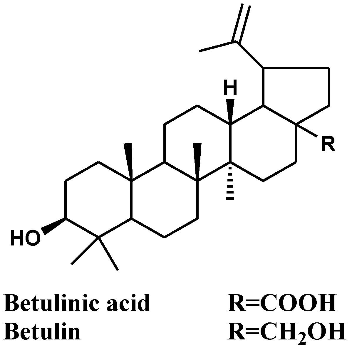
2.2. Characterization of the Isolated Compounds

The isolated pure compounds were subjected to structural characterization using melting point range, TLC and spectroscopic techniques (UV, IR, Mass and NMR). The results revealed that the structure of the isolated compounds are betulinic acid (16) and betulin (23) as compared with previous reports [28,29,30,31]. Fractions eluted from ethyl acetate-n-hexane (1:2) showed single spots on TLC. The Rf value for this fraction was found to be 0.470, which is like the Rf value of betulin in the same solvent system (0.471). Similarly, betulinic acid (16) was also eluted using the same solvent system and the Rf value (0.531) was as compared with the reported data [29,32]. Results are presented in the following (Table 2). The spectral data are available in the Supplementary Material (Figures S1–S6).
Betulinic acid (16): IR (neat, cm−1) 3464 (br), 2943, 2870, 1643, 1685, 1188, 883, 1454. MS: m/z M+1: 457.3305, 407(22), 353(15), 339(12), 325(100), 240(13). 1H NMR (CDCl3, 500 MHz): δ (ppm): 4.76 (1H, br s, H-29a), 4.63 (1H, br s, H-29b), 3.2 (1H, m, H-3), 1.7 (3H, s, H-30) 0.99 (3H, s), 0.98 (3H, s), 0.95 (3H, s), 0.84 (3H, s) and 0.77 (3H, s). 13C NMR (CDCl3, 125 MHz): δ (ppm): 38.9 (C-1), 29.7 (C-2), 79.0 (C-3), 38.7 (C-4), 55.4 (C-5), 18.3 (C-6), 34.3 (C-7), 40.7 (C-8), 50.5 (C-9), 37.2 (C-10), 20.9 (C-11), 25.7 (C-12), 38.4 (C-13), 42.4 (C-14), 30.6 (C-15), 32.2 (C-16), 56.3 (C-17), 46.9 (C-18), 49.3 (C-19), 142.6 (C-20), 29.7 (C-21), 34.3 (C-22), 27.9 (C-23), 15.3 (C-24), 16.1 (C-25), 16.0 (C-26), 14.7 (C-27), 178.6 (C-28), 109.7 (C-29), 19.4 (C-30).
Betulin (23): IR (neat, cm−1) 3421 (br), 2927, 2820, 1648, 1454, 1377, 1184, 1045, 886, 1454. MS: m/z M+: 442.3799, 393(12), 279(23), 203(100), 189 (93), 1H NMR (CDCl3, 500 MHz): δ (ppm): 4.70 (1H, br s, H-29a), 4.58 (1H, br s, H-29b), 3.79 (1H, d, J = 10.8 Hz, H-28b), 3.33 (1H, d, J = 10.8 Hz, H-28a), 3.18 (1H, dd, J = 9.8, 5.3 Hz, H-3α), 1.67 (3H, s, H- 30), 0.99 (3H, s, H-27), 0.97 (3H, s, H-26), 0.96 (3H, s, H-23), 0.80 (3H, s, H-25), 0.75 (3H, s, H-24). 13C NMR (CDCl3, 125 MHz): δ (ppm): 38.9 (C-1), 27.5 (C-2), 79.3 (C-3), 38.8 (C-4), 55.4 (C-5), 18.4 (C-6), 34.1 (C-7), 41.0 (C-8), 50.5 (C-9), 37.4 (C-10), 20.9 (C-11), 25.3 (C-12), 37.2 (C-13), 42.8 (C-14), 27.1 (C-15), 29.2 (C-16), 47.9 (C-17), 49.9 (C-18), 48.8 (C-19), 150.9 (C-20), 29.8 (C-21), 34.1 (C-22), 28.1 (C-23), 15.4 (C-24), 16.2 (C-25), 16.1 (C-26), 14.8 (C-27), 60.6 (C-28), 109.8 (C-29), 19.2 (C-30).

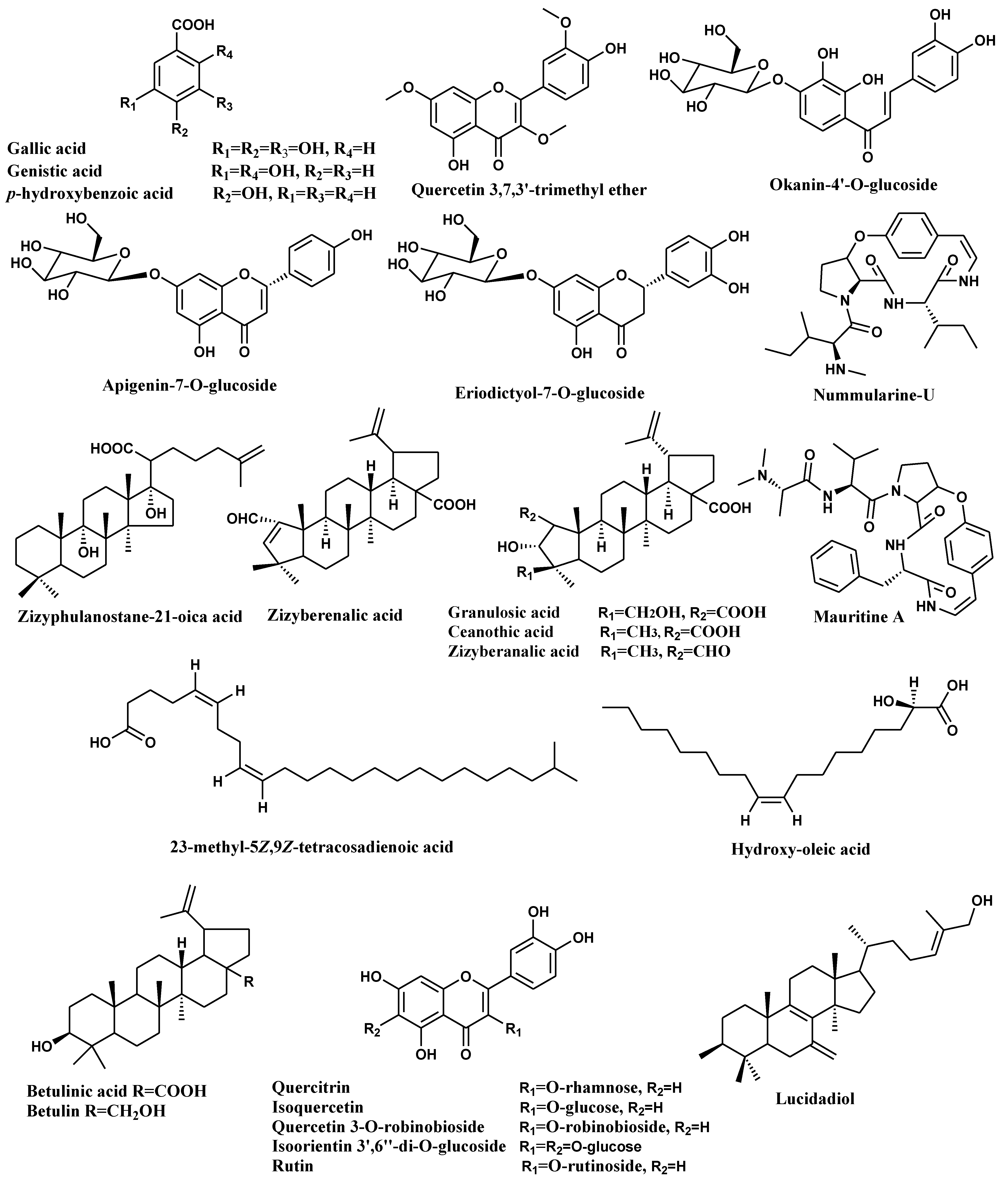
2.3. HPLC-ESI-MS/MS Analysis of Different Extracts of Zizyphus spina-christi L. Stem Bark

Previous research reported the identification of various bioactive metabolites from Z. spina-christi L. [7,10,33,34]. In this study, LC-HR-MS analysis was used to identify metabolites from various fractions of ZSC-L. The results revealed the presence and identification of 36 compounds from different fractions belonging to different phytochemical classes as organic acids, alkaloids hydrocarbons, triterpenes, and fatty acids, where flavonoids and triterpenes were recognized as the most prevalent class of compounds in different fractions. The tentatively identified secondary metabolites are listed in Table 3. Compounds are illustrated in Figure 2. This is the first comprehensive metabolites profiling of ZSC-L. using HPLC-ESI-MS. The metabolite profile of ZSC-L. indicates the presence of 11 flavonoids tentatively identified as quercetin 3,7,3′-trimethyl ether (4), eriodictyol-7-O-glucoside (7), apigenin-7-O-glucoside (8), okanin-4′-O-glucoside (15), quercitrin (17), isoquercetin (19), kaempferol-3-O-α-l-arabinoside (29), luteolin 7,3′-diglucoside (30), isoorientin 3′,6′′-di-O-glucoside (34), quercetin 3-O-robinobioside (35), rutin (36). Among these flavonoids, (17), (19), (34), (35) and (36) were previously isolated from ZSC-L [10,34]. Besides, eight triterpenes were suggested with molecular formula C30H46O6, C30 H50 O3, C30H46O5, C30H46O4, C30H48O3, C32H52O2, C30H50O2 and C30H44O3; identified as granulosic acid (9), zizyphulanostane-21-oica acid (12), ceanothic acid (13), zizyberanalic acid (14), betulinic acid (16), lupeol acetate (20), betulin (23), and zizyberenalic acid (26).
Additionally, fatty acid compounds identified and characterized as hydroxy-oleic acid (5), 23-methyl-5Z,9Z-tetracosadienoic acid (11), trihydroxy-oleic acid (25), n-hexadecanoic acid (27), hexadecanoic acid, ethyl ester (28), docosanedioic acid (31), octadecanoic acid, ethyl ester (33) based on their m/z 298.036, 378.3493, 330.2762, 256.2388, 284.2933, 370.307, 312.5, respectively, and in accordance with the molecular formulas C18H34O3, C25H46O2,C18H34O5, C16 H32O2, C18H36O2,C22H42O4, C20H40O2, respectively. A recent study conducted on the lipophilic fraction of different parts of Z. lotus revealed the identification of 99 compounds by GC/MS analysis, where the root bark showed the highest percentage of pentacyclic triterpenoids, especially betulinic acid, while the leaves and seeds were rich in unsaturated fatty acids [35].
The peak with m/z 470.3367, with a molecular formula C26H39N4O4, was identified as the cyclopeptide alkaloid, nummularine-U (10), which was previously isolated from stem bark of Z. spina-christi [33]. Also, another cyclopeptide alkaloid was characterized as mauritine A (6) and teleocidin B-1 (21) that were reported previously in Zizyphus mauritiana [36]. Four phenolic acids were characterized as gallic acid (1), genistic acid (2), p-hydroxybenzoic acid (3) and sinapoyl malate (24). Moreover, two sterol and one hydrocarbon were identified as stigmast-7-en-3-ol (18), lucidadiol (22), and tetracosane (32), respectively. Cadi et al. reported the polyphenolic components of Z. lotus fruits through PLC-DAD-ESI/MS analysis, where the most abundant compound in the ethyl acetate extract was p-hydroxybenzoic acid and in the methanol extract quercetin 3-O-rhamnoside-7-O-glucoside was the major component [37]. By comparing our results with the previously mentioned literature regarding the different species and different parts of Zizyphus; the different fractions of Z. spina-chritis L. stem bark are a rich source of pentacyclic triterpenoids, fatty acids and polyphenolic compounds.

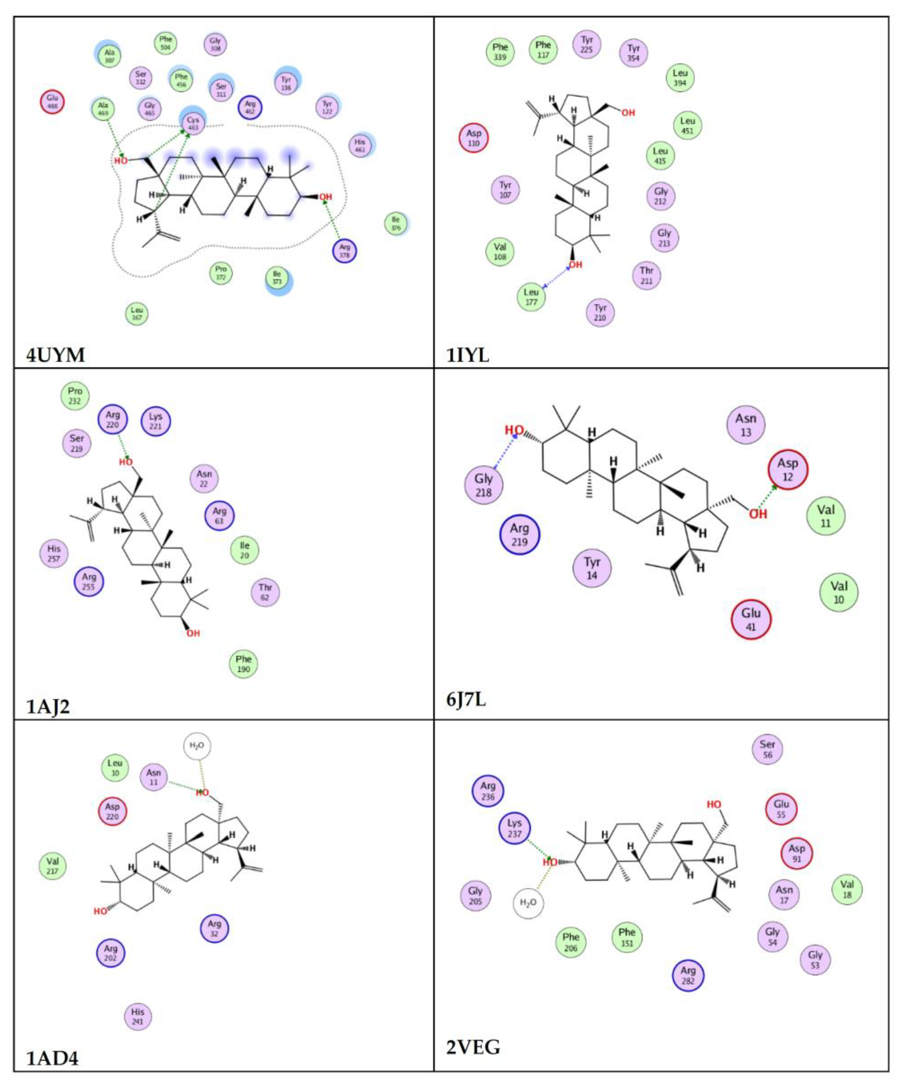
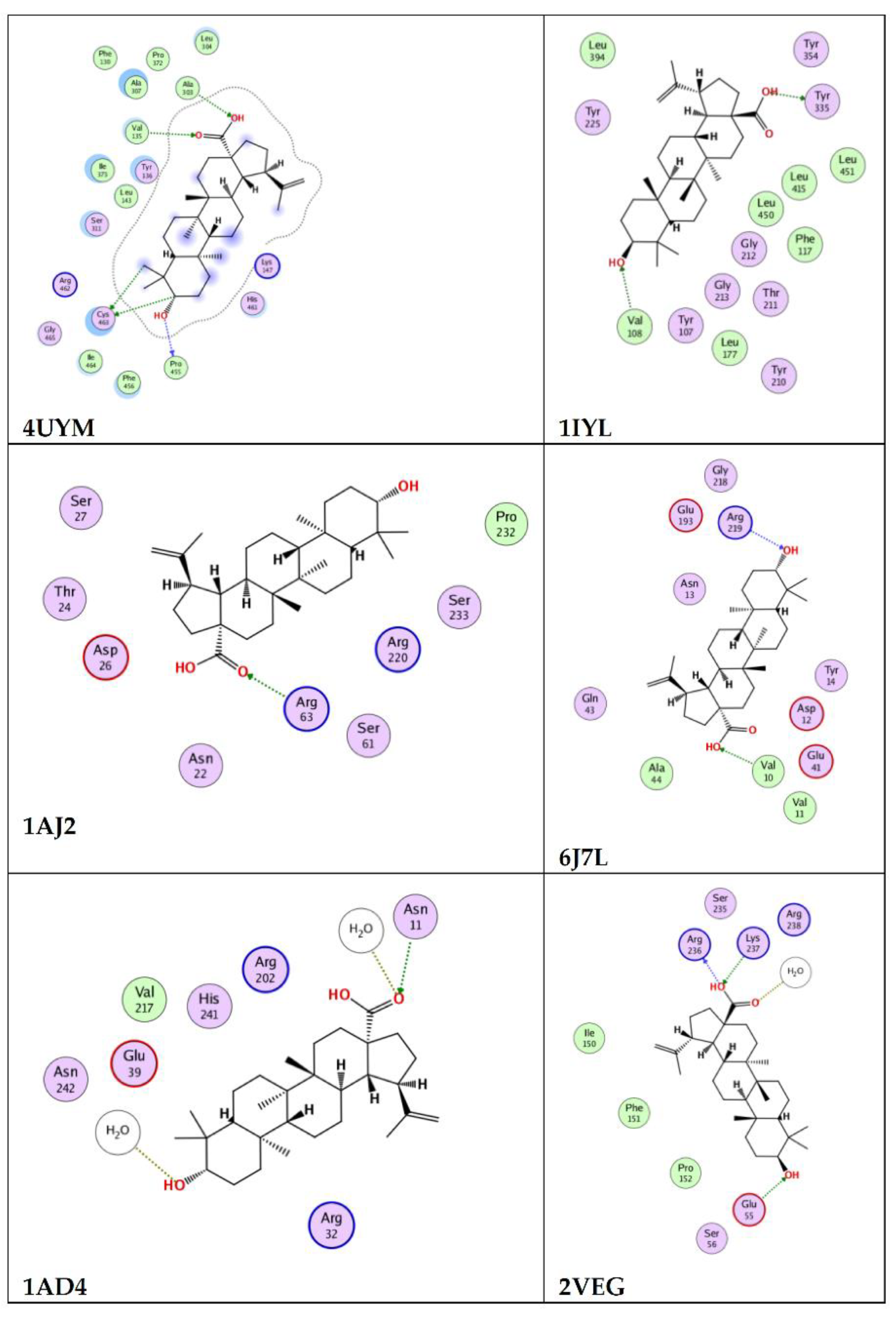
2.4. In Silico Molecular Docking

Giving account to the retrieved antimicrobial activity, an in silico molecular docking study was conducted on betulin and betulinic acid to study their potential mechanism (Table 4). In this context, among the six potential microbial enzymes used in the docking analysis, betulinic acid showed higher antifungal activity against A. Fumigatus by interacting with the 4UYM protein (sterol 14-alpha demethylase) with docking score of −12.3. and betulin showed strong binding affinity with 1IYL protein (C. albicans N-myristoyltransferase), achieving a docking score of −13.5. Betulin and betulinic acid interact with 1AD4 protein (dihydropteroate synthetase) via hydrogen bonding to exhibit the antibacterial activity against S. aureus, achieving docking scores of −7.4 and −8.8, respectively. With the 2VEG protein (dihydropteroate synthase), betulinic acid and betulin interacted via hydrogen bonding with binding score of −9.4 and −10.8, respectively, to exhibit antibacterial activity against S. pneumonia. Betulin and betulinic acid showed good binding affinity with 1AJ2 protein (dihydropteroate synthase) with docking scores of −11.5 and −9.2, respectively, to exhibit antibacterial activity against E. coli. The ligand–protein interaction behaviors were estimated based on the docking score function. In general, both compounds achieved acceptable binding affinities with all the targets as well as good interaction pattern. The results suggest that betulin and betulinic acid may have different mechanisms as antimicrobial agents. Figure 3 and Figure 4 show the feasible binding geometries of betulin and betulinic acid with the target proteins.
Betulin has been reported for its molecular interaction with the target DNA gyrase A of S. aureus by computational docking tools. Results revealed its strong affinity toward the DNA Gyrase A with docking score −9.23 [59]. Another report by Rajkumari et al. revealed the efficacy of betulin and betulinic acid in inhibition of quorum sensing (QS)-mediated virulence factors in P. aeruginosa, and they serve as potent competitive inhibitors through restricting the binding of the natural ligands to the QS receptors, LasR and RhlR [60].

3. Materials and Methods

3.1. Chemicals

All solvents used for this study obtained from Sigma-Aldrich, Fisher Scientific, Scharlau Spain and VWR BDH Prolabo chemical (analytical grade).

3.2. Plant Material

Fresh bark of the Z. spina-christi was collected from the Hail area of Saudi Arabia using GPS coordinates (27.48472222, 41.69555556), (27.51416667, 41.70027778), (27.53833333, 41.69500000) and (26.00583333, 40.47222222). The taxonomic authentication was performed kindly by Dr. Sherif Sayed Sharawy, professor of taxonomy, University of Hail. A voucher specimen of the plant material was deposited in the Herbarium of the Biology department, University of Hail. The bark was air-dried, split into pieces, and stored in a dry area for future investigation.

3.3. Preparation of the Plant Extract

The stem bark of Z. spina-christi L (5.2 kg) was air-dried and extracted with absolute ethanol three times (3 × 20 L). The ethanol extract was filtered and concentrated under reduced pressure to provide 119.59 g of total extract. It was then fractionated using multiple solvents, including diethyl ether (2.5 L × 2), chloroform (2.5 L × 2), ethyl acetate (2.5 L × 2), and n-butanol (2.5 L × 2). Separately, the solvents were evaporated under reduced pressure to obtain 41.20 g of diethyl ether residue, 8.45 g of chloroform extract, 7.20 g of ethyl acetate extract, and 4.18 g of n-butanol extract, individually. The antibacterial activity of these fractions was investigated.

3.4. Antimicrobial Activity

The antimicrobial activity was performed against fungi (Aspergillus fumigatus: RCMB 02568, Candida albicans: RCMB 05036), Gram-positive (Streptococcus pneumoniae: RCMB 010010, Staphylococcus aureus RCMB 010028), and Gram-negative bacteria (Pseudomonas aeruginosa: RCMB 010043, Escherichia coli: RCMB 010052) by using agar well diffusion technique, as previously described in [6,27]. Test organisms were obtained from the Regional Center for Mycology and Biotechnology (RCMB), Cairo, Egypt. Data are expressed in the form of mean ± SD.

3.5. Separation and Purification of the Plant Metabolites from n-Butanol Fraction

The n-butanol extract was subjected to column chromatography on silica gel and eluted using n-hexane (100%), chloroform (100%), followed by gradient elution of chloroform: ethyl acetate from 100% to 0%, followed by ethyl acetate: methanol from 100% to 0%, followed by methanol-acetonitrile (75:25 v/v; 50:50 v/v and 25:75 v/v), ended by 3 volumes of methanol-acetonitrile-formic acid (5:4:1 v/v/v), which ultimately yielded forty-one fractions (F1–F41). Fractions collected thereof were then analyzed using TLC with different mobile phases, and similar fractions were combined together and concentrated under reduced pressure. The CAMMAG® LINOMAT 5 application system was used to automatically spot fractions on TLC plates, and the development operations were carried out using two solvent systems: chloroform: methanol (9:1; v/v) or toluene: ethyl acetate: formic acid (TEF, 7: 5: 1; v/v/v). After drying at room temperature, the purity of fractions was determined using a CAMMAG® TLC scanning device at Al-Azhar University’s Regional Center for Mycology and Biotechnology (RCMB). Fractions 2 and 3 eluted with chloroform-ethyl acetate (80:2 v/v) were then purified by preparative TLC plates using chloroform-ethyl acetate (90:10) as developer to afford two lupane-type pentacyclic triterpenoids betulinic acid (16, 20.4 mg) and betulin (23, 16.7 mg). The pure compounds were recognized based on their UV absorbance at 254 and 365 nm or visible light appearance and Rf value. Also, the TLC plates were visualized by vanillin-sulphuric acid spray reagent and heating them for 10 min at 120 °C.

3.6. LC-HR/ESI-MS Analysis of Different Extracts of Z. spina-christi L. Stem Bark

Tentative metabolite assignments were obtained by comparing mass spectral data of the identified compounds in both negative and positive ionization modes with previously reported data, as well as data from online public databases to which references were added Table 3.
The Q-TOF-LC/MS system, 6530 (Agilent Technologies) equipped with an autosampler (G7129A), a Quat. Pump (G7104C) and a column comp (G7116A) were used for chromatographic separation. The injection volume was 8 µL. The analytes were separated on a Zorbax RP-18 column from Agilent Technologies (dimensions: 150 mm × 3 mm, dp = 2.7 µm) in a flow rate of 0.3 mL/min. The mobile phase consisted of a combination of solvent A Water (0.1 formic acid) and solvent B (acetonitrile + 0.1% formic acid). The gradient elution was as follows: t = 0 min, 3% B; t = 15 min, 10% B; t = 40 min, 20% B; t = 70 min, 40% B; t = 90 min, 60% B; t = 110 min, 80% B; t = 120 min, 90% B and t = 135 min, 100% B. Mass spectra were simultaneously acquired using ESI in positive ionization mode with a capillary voltage of 4000 V. The mass spectra were recorded in the m/z range of 40 to 1500 m/z. The gas temperature and drying gas flow were 350 °C and 10 L/min, respectively.

3.7. In Silico Molecular Docking Studies

Six potential targets for betulin and betulinic acid were downloaded from the protein data bank (www.pdb.org) (accessed on 29 January 2022). The following IDs were used, 4UYM, 1IYL, 1AJ2, 6J7L, 1AD4, 2VEG [61,62,63,64,65,66] for sterol 14-alpha demethylase (CYP51B) from a pathogenic Aspergillus fumigatus; Candida albicans N-myristoyltransferase, E. coli dihydropteroate synthase, Pseudomonas aeruginosa Earp, Staphylococcus aureus, dihydropteroate synthase and Dihydropteroate synthase from Streptococcus pneumonia, respectively. All docking simulations were conducted using MOE software [67]. The receptor and the ligand were prepared using the standard structure optimization protocol of the software. The active site was set as where the co-crystalized ligand was bound in each corresponding target. The docking was performed using a molecular database of betulin and betulinic acid and following the induced fit protocol of MOE software [68]. The London dG and triangular matcher algorithms were used as scoring and placement methods, respectively. Each co-crystalized ligand was redocked in the vicinity of each target and the RMSD between the re-docked pose and co-crystalized ligand was calculated and used to confirm the docking validity. MOE 2019 interface was used to visualize and analyze the docking results as well as produce 2D interaction images. The docking validation data are available in the Supplementary Material (Table S1).

4. Conclusions

In the present work, different fractions of Z. spina-christi L. exhibited a varying degree of antimicrobial activity. Besides, LC-HR-MS analysis was used to identify metabolites of different fractions of Z. spina-christi L. The results revealed the presence and identification of 36 phytochemical compounds and biological studies carried out on the stem bark of Z. spina-christi L. Phytochemical investigations led to the isolation of two pure compounds, betulinic acid (C30H48O3) and betulin (C30H50O2). The structure of these compounds was determined by IR spectroscopy, mass spectroscopy, 1H and 13C NMR and confirmed by comparing with the previously reported values. The molecular docking studies on betulinic acid and betulin against enzymes in various microorganism revealed the potential binding affinity to the site of the appropriate targets. The n-butanol fractions of Z. spina-christi L. have potent antimicrobial activity. Further investigation of the isolated metabolites is required to identify the bioactive compounds responsible for antimicrobial, antioxidant and cytotoxic effects that may have potentials in pharmaceutical and clinical applications.

Supplementary Materials

The following supporting information can be downloaded at: https://www.mdpi.com/article/10.3390/molecules27061805/s1, Figures S1–S6: Spectral data of the isolated compounds; Table S1. The docking validation data.

Author Contributions

E.N.A., writing—original draft, writing—review and editing, formal analysis, investigation. S.I.H., formal analysis, investigation. S.R., formal analysis, investigation. M.H.H., formal analysis, investigation. S.H.A., formal analysis and investigation. M.A.A., formal analysis. All authors have read and agreed to the published version of the manuscript.

Funding

This research received no external funding.

Institutional Review Board Statement

Not applicable.

Informed Consent Statement

Not applicable.

Data Availability Statement

Data are available upon request from the first author.

Acknowledgments

The authors extend their appreciation and thanks to Shaker Abass Khalil, Faculty of Science, Zagazig University, Egypt, for criticizing the reading of and supporting this research work. The authors also would like to express their gratitude to Mahmoud A. El Hassab, Department of Pharmaceutical Chemistry, Faculty of Pharmacy, King Salman International University (KSIU), South Sinai, Egypt, for molecular docking studies.

Conflicts of Interest

The authors declare no conflict of interest.

Sample Availability

Samples of the plant material and different fractions may be available from the authors.

References

  1. Adzu, B.; Amos, S.; Dzarma, S.; Wambebe, C.; Gamaniel, K. Effect of Zizyphus spina-christi Willd aqueous extract on the central nervous system in mice. J. Ethnopharmacol. 2002, 79, 13–16. [Google Scholar] [CrossRef]
  2. Shahat, A.A.; Pieters, L.; Apers, S.; Nazeif, N.M.; Abdel-Azim, N.S.; Berghe, D.V.; Vlietinck, A.J. Chemical and biological investigations on Zizyphus spina-christi L. Phyther. Res. 2001, 15, 593–597. [Google Scholar] [CrossRef] [PubMed]
  3. Adzu, B.; Amos, S.; Wambebe, C.; Gamaniel, K. Antinociceptive activity of Zizyphus spina-christi root bark extract. Fitoterapia 2001, 72, 344–350. [Google Scholar] [CrossRef]
  4. Waggas, A.M.; Al-hasani, R.H. Effect of Sidr (Zizyphus spina-christi) fruit extract on the central nervous system in male albino rats. Am. J. Sustain. Agric. 2009, 4, 263–267. [Google Scholar]
  5. Adzu, B.; Haruna, A.K. Studies on the use of Zizyphus spina-christi against pain in rats and mice. Afr. J. Biotechnol. 2007, 6, 1317–1324. [Google Scholar] [CrossRef]
  6. Ads, E.N.; Rajendrasozhan, S.; Hassan, S.I.; Sharawy, S.M.S.; Humaidi, J.R. Phytochemical, antimicrobial and cytotoxic evaluation of Ziziphus spina-christi (L.) stem bark. Biomed. Res. 2017, 28, 6646–6653. [Google Scholar]
  7. Ads, E.N.; Rajendrasozhan, S.; Hassan, S.I.; Sharawy, S.M.S.; Humaidi, J.R. Phytochemical screening of different organic crude extracts from the stem bark of Ziziphus spina-christi (L.). Biomed. Res. 2018, 29, 1645–1652. [Google Scholar] [CrossRef] [Green Version]
  8. Mahran, G.E.H.; Glombitza, K.; Mirhom, Y.; Hartmann, R.; Michei, C.G. Novel Saponins from Zizyphus spina-christi Growing in Egypt. Planta Med. 1995, 62, 163–165. [Google Scholar] [CrossRef]
  9. Bozicevic, A.; De Mieri, M.; Di Benedetto, A.; Gafner, F.; Hamburger, M. Dammarane-type saponins from leaves of Ziziphus spina-christi. Phytochemistry 2017, 138, 134–144. [Google Scholar] [CrossRef]
  10. Pawlowska, A.M.; Camangi, F.; Bader, A.; Braca, A. Flavonoids of Zizyphus jujuba L. and Zizyphus spina-christi (L.) Willd (Rhamnaceae) fruits. Food Chem. 2009, 112, 858–862. [Google Scholar] [CrossRef]
  11. Ghafoor, A.O.; Qadir, H.K.; Fakhri, N.A. Analysis of phenolic compounds in extracts of Ziziphus spina-christi using RPHPLC method. J. Chem. Pharm. Res. 2012, 4, 3158–3163. [Google Scholar]
  12. Zhao, F.; Zhang, C.; Nan, W.; Lu, W. An integrated bioprocess for fermentative production of protopanaxadiol by recycling ethanol waste during down-stream extraction process. J. Clean. Prod. 2019, 234, 1–8. [Google Scholar] [CrossRef]
  13. Hak, C.K.; Young, D.M.; Kyung, R.K.; Eun, J.B.; Chong, S.L.; Kang, R.L. A new acylglycosyl sterol from Quisqualis Fructus. Arch. Pharm. Res. 2003, 26, 275–278. [Google Scholar] [CrossRef]
  14. Chandramu, C.; Manohar, R.D.; Krupadanam, D.G.L.; Dashavantha, R.V. Isolation, characterization and biological activity of betulinic acid and ursolic acid from Vitex negundo L. Phyther. Res. 2003, 17, 129–134. [Google Scholar] [CrossRef]
  15. Yamashita, K.; Lu, H.; Lu, J.; Chen, G.; Yokoyama, T.; Sagara, Y.; Manabe, M.; Kodama, H. Effect of three triterpenoids, lupeol, betulin, and betulinic acid on the stimulus-induced superoxide generation and tyrosyl phosphorylation of proteins in human neutrophils. Clin. Chim. Acta 2002, 325, 91–96. [Google Scholar] [CrossRef]
  16. De Oliveira, B.H.; Santos, C.A.M.; Espíndola, A.P.D.M. Determination of the triterpenoid, betulinic acid, in Doliocarpus schottianus by HPLC. Phytochem. Anal. 2002, 13, 95–98. [Google Scholar] [CrossRef]
  17. Schühly, W.; Heilmann, J.; Çalis, I.; Sticher, O. New triterpenoids with antibacterial activity from Zizyphus joazeiro. Planta Med. 1999, 65, 740–743. [Google Scholar] [CrossRef]
  18. O’Connell, M.M.; Bentley, M.D.; Campbell, C.S.; Cole, B.J.W. Betulin and lupeol in bark from four white-barked birches. Phytochemistry 1988, 27, 2175–2176. [Google Scholar] [CrossRef]
  19. Kim, D.S.H.L.; Chen, Z.; Van Nguyen, T.; Pezzuto, J.M.; Qiu, S.; Lu, Z.Z. A concise semi-synthetic approach to betulinic acid from betulin. Synth. Commun. 1997, 27, 1607–1612. [Google Scholar] [CrossRef]
  20. Myszka, H.; Grzywacz, D.; Zdrowowicz, M.; Spisz, P.; Butowska, K.; Rak, J.; Piosik, J.; Jaśkiewicz, M.; Kamysz, W.; Liberek, B. Design, synthesis and biological evaluation of betulin-3-yl 2-amino-2-deoxy-β-d-glycopyranosides. Bioorg. Chem. 2020, 96, 103568. [Google Scholar] [CrossRef]
  21. Cichewicz, R.H.; Kouzi, S.A. Chemistry, Biological Activity, and Chemotherapeutic Potential of Betulinic Acid for the Prevention and Treatment of Cancer and HIV Infection. Med. Res. Rev. 2004, 24, 90–114. [Google Scholar] [CrossRef] [PubMed]
  22. Yogeeswari, P.; Sriram, D. Betulinic Acid and Its Derivatives: A Review on their Biological Properties. Curr. Med. Chem. 2010, 12, 657–666. [Google Scholar] [CrossRef] [PubMed]
  23. El Deeb, K.S.; Al-Haidari, R.A.; Mossa, J.S.; Ateya, A.M. Phytochemical and pharmacological studies of Maytenus forsskaoliana. Saudi Pharm. J. 2003, 11, 184–191. [Google Scholar]
  24. Fulda, S.; Debatin, K.M. Sensitization for anticancer drug-induced apoptosis by betulinic acid. Neoplasia 2005, 7, 162–170. [Google Scholar] [CrossRef] [PubMed] [Green Version]
  25. Lou, H.; Li, H.; Zhang, S.; Lu, H.; Chen, Q. A Review on Preparation of Betulinic Acid and Its Biological Activities. Molecules 2021, 26, 5583. [Google Scholar] [CrossRef]
  26. Haque, S.; Nawrot, D.A.; Alakurtti, S.; Ghemtio, L.; Yli-Kauhaluoma, J.; Tammela, P. Screening and characterisation of antimicrobial properties of semisynthetic betulin derivatives. PLoS ONE 2014, 9, e102696. [Google Scholar] [CrossRef] [Green Version]
  27. Aly, S.H.; Elissawy, A.; Eldahshan, O.; Elshanawany, M.; Singab, A.N. Phytochemical investigation using GC/MS analysis and evaluation of antimicrobial and cytotoxic activities of the lipoidal matter of leaves of Sophora secundiflora and Sophora tomentosa. Arch. Pharm. Sci. Ain. Shams Univ. 2020, 4, 207–214. [Google Scholar]
  28. Jamila, N.; Khairuddean, M.; Khan, S.N.; Khan, N.; Osman, H. Phytochemicals from the bark of Garcinia hombroniana and their biological activities. Rec. Nat. Prod 2014, 8, 312–316. [Google Scholar]
  29. Joshi, H.; Saxena, G.K.; Singh, V.; Arya, E.; Singh, R.P. Phytochemical investigation, isolation and characterization of betulin from bark of Betula utilis. J. Pharmacogn. Phytochem. 2013, 2, 145–151. [Google Scholar]
  30. Chrobak, E.; Jastrzębska, M.; Bębenek, E.; Kadela-Tomanek, M.; Marciniec, K.; Latocha, M.; Wrzalik, R.; Kusz, J.; Boryczka, S. Molecular Structure, In Vitro Anticancer Study and Molecular Docking of New Phosphate Derivatives of Betulin. Molecules 2021, 26, 737. [Google Scholar] [CrossRef]
  31. Tijjani, A.; Ndukwe, I.G.; Ayo, R.G. Isolation and characterization of lup-20 (29)-ene-3, 28-diol (Betulin) from the stem-bark of Adenium obesum (Apocynaceae). Trop. J. Pharm. Res. 2012, 11, 259–262. [Google Scholar] [CrossRef] [Green Version]
  32. Wani, M.S.; Gupta, R.C.; Pradhan, S.K.; Munshi, A.H. Estimation of four triterpenoids, betulin, lupeol, oleanolic acid, and betulinic acid, from bark, leaves, and roots of Betula utilis D. Don using a validated high-performance thin-layer chromatographic method. JPC-J. Planar Chromatogr. TLC 2018, 31, 220–229. [Google Scholar] [CrossRef]
  33. Tuenter, E.; Foubert, K.; Staerk, D.; Apers, S.; Pieters, L. Isolation and structure elucidation of cyclopeptide alkaloids from Ziziphus nummularia and Ziziphus spina-christi by HPLC-DAD-MS and HPLC-PDA-(HRMS)-SPE-NMR. Phytochemistry 2017, 138, 163–169. [Google Scholar] [CrossRef] [PubMed]
  34. Nawwar, M.A.M.; Ishak, M.S.; Michael, H.N.; Buddrust, J. Leaf flavonoids of Ziziphus spina-christi. Phytochemistry 1984, 23, 2110–2111. [Google Scholar] [CrossRef]
  35. Zazouli, S.; Chigr, M.; Ramos, P.A.B.; Rosa, D.; Castro, M.M.; Jouaiti, A.; Duarte, M.F.; Santos, S.A.O.; Silvestre, A.J.D. Chemical Profile of Lipophilic Fractions of Different Parts of Zizyphus lotus L. by GC-MS and Evaluation of Their Antiproliferative and Antibacterial Activities. Molecules 2022, 27, 483. [Google Scholar] [CrossRef]
  36. Tschesche, R.; Wilhelm, H.; Fehlhaber, H. Alkaloids from Rhamnaceae. xIV. mauritin-A and mauritin-B, two peptide alkaloids from Zizyphus mauritiana Lam. Tetrahedron Lett. 1972, 13, 2609–2612. [Google Scholar] [CrossRef]
  37. Cadi, H.E.; Bouzidi, H.E.L.; Selama, G.; Cadi, A.E.; Ramdan, B.; Oulad, Y.; Majdoub, E.; Alibrando, F.; Dugo, P.; Mondello, L.; et al. Physico-Chemical and Phytochemical Characterization of Moroccan Wild Jujube “Zizyphus lotus (L.)” Fruit Crude Extract and Fractions. Molecules 2020, 25, 5237. [Google Scholar] [CrossRef]
  38. Benslama, A.; Harrar, A.; Gul, F.; Demirtas, I. Phenolic Compounds, Antioxidant and Antibacterial Activities of Zizyphus lotus L. Leaves Extracts. Nat. Prod. J. 2017, 7, 316–322. [Google Scholar] [CrossRef]
  39. Iwashina, T.; Saito, Y.; Peng, C.; Yokota, M.; Kokubugata, G. Foliar Flavonoids from Two Begonia Species in Japan Tsukasa. Bull. Natl. Museum Nat. Sci. Ser. B 2008, 34, 175–181. [Google Scholar]
  40. Ayoub, I.M.; Korinek, M.; El-shazly, M.; Wetterauer, B.; El-beshbishy, H.A. Anti-Allergic, Anti-Inflammatory and Anti-Hyperglycemic Activity of Chasmanthe aethiopica Leaf Extract and Its Profiling Using LC/MS and GLC/MS. Plants 2021, 10, 1118. [Google Scholar] [CrossRef]
  41. Álvarez-Fernández, M.A.; Cerezo, A.B.; Canete-Rodriguez, A.M.; Troncoso, A.M.; García-Parrilla, M.C. Composition of nonanthocyanin polyphenols in alcoholic-fermented strawberry products using LC–MS (QTRAP), high-resolution MS (UHPLC-Orbitrap-MS), LC-DAD, and antioxidant activity. J. Agric. Food Chem. 2015, 63, 2041–2051. [Google Scholar] [CrossRef] [PubMed]
  42. Švehlíková, V.; Bennett, R.N.; Mellon, F.A.; Needs, P.W.; Piacente, S.; Kroon, P.A.; Bao, Y. Isolation, identification and stability of acylated derivatives of apigenin 7-O-glucoside from chamomile (Chamomilla recutita [L.] Rauschert). Phytochemistry 2004, 65, 2323–2332. [Google Scholar] [CrossRef] [PubMed]
  43. Ganapaty, S.; Thomas, P.S.; Ramana, K.V.; Karagianis, G.; Waterman, P.G. Dammarane and ceanothane triterpenes from Zizyphus glabrata. Zeitschrift Fur Naturforsch.-Sect. B J. Chem. Sci. 2006, 61, 87–92. [Google Scholar] [CrossRef] [Green Version]
  44. Ck, S.; Ps, U. GC-MS and HR-LCMS fingerprinting of various parts of Oroxylum indicum (L.) Vent. A comparative phytochemical study based on plant part substitution approach. J. Pharmacogn. Phytochem. 2020, 9, 1817–1824. [Google Scholar]
  45. Mukhtar, H.M.; Ansari, S.H.; Ali, M.; Naved, T. New Compounds from Zizyphus vulgaris. Pharm. Biol. 2004, 42, 508–511. [Google Scholar] [CrossRef]
  46. Kundu, A.B.; Barik, B.R.; Mondal, D.N.; Dey, A.K.; Banerji, A. Zizyberanalic acid, A pentacyclic triterpenoid of Zizyphus jujuba. Phytochemistry 1989, 28, 3155–3158. [Google Scholar] [CrossRef]
  47. Abdel Maboud, T. Cytotoxic Potentials and Phytoconstituents Profiling of Blepharis Edulis (Forssk.) Pers. Using Uhplc/Q-Tof-Ms-Ms. Al-Azhar J. Pharm. Sci. 2021, 63, 37–56. [Google Scholar] [CrossRef]
  48. Ayatollahi, A.M.; Ghanadian, M.; Afsharypour, S.; Abdella, O.M.; Mirzai, M.; Askari, G. Pentacyclic triterpenes in Euphorbia microsciadia with their T-cell proliferation activity. Iran. J. Pharm. Res. 2011, 10, 287–294. [Google Scholar] [CrossRef]
  49. Abuzaid, H.; Amin, E.; Moawad, A.; Abdelmohsen, U.R.; Hetta, M.; Mohammed, R. Liquid Chromatography High-Resolution Mass Spectrometry Analysis, Phytochemical and Biological Study of Two Aizoaceae Plants: A New Kaempferol Derivative from Trianthema portulacastrum L. Pharmacogn. Res. 2020, 12, 212–218. [Google Scholar] [CrossRef]
  50. Hitotsuyanagi, Y.; Fujiki, H.; Suganuma, M.; Aimi, N.; Sakai, S.; Endo, Y.; Shudo, K.; Sugimura, T. Isolation and structure elucidation of teleocidin B-1, B-2, B-3, and B-4. Chem. Pharm. Bull. 1948, 32, 4233–4236. [Google Scholar] [CrossRef] [Green Version]
  51. Ha, D.T.; Loan, L.T.; Hung, T.M.; Han, L.V.N.; Khoi, N.M.; Dung, L.V.; Min, B.S.; Nguyen, N.P.D. An improved HPLC-DAD method for quantitative comparisons of triterpenes in Ganoderma lucidum and its five related species originating from Vietnam. Molecules 2015, 20, 1059–1077. [Google Scholar] [CrossRef] [PubMed] [Green Version]
  52. Kosyakov, D.S.; Ul’yanovskii, N.V.; Falev, D.I. Determination of triterpenoids from birch bark by liquid chromatography-tandem mass spectrometry. J. Anal. Chem. 2014, 69, 1264–1269. [Google Scholar] [CrossRef]
  53. Stobiecki, M.; Skirycz, A.; Kerhoas, L.; Kachlicki, P.; Muth, D.; Einhorn, J.; Mueller-Roeber, B. Profiling of phenolic glycosidic conjugates in leaves of Arabidopsis thaliana using LC/MS. Metabolomics 2006, 2, 197–219. [Google Scholar] [CrossRef]
  54. Lee, S.M.; Min, B.S.; Lee, C.; Kim, K.; Kho, Y.H. Cytotoxic Triterpenoids from the Fruits of Zizyphus jujuba. Planta Med. 2003, 69, 1051–1054. [Google Scholar]
  55. Liu, M.-H.; Zhang, Q.; Zhang, Y.-H.; Lu, X.-Y.; Fu, W.-M.; He, J.-Y. Chemical Analysis of Dietary Constituents in Rosa roxburghii and Rosa sterilis Fruits. Mol. Artic. 2016, 21, 1204. [Google Scholar] [CrossRef]
  56. El Sayed, A.M.; Basam, S.M.; El-Naggar, E.M.B.A.; Marzouk, H.S.; El-Hawary, S. LC–MS/MS and GC–MS profiling as well as the antimicrobial effect of leaves of selected Yucca species introduced to Egypt. Sci. Rep. 2020, 10, 17778. [Google Scholar] [CrossRef]
  57. Brito, A.; Ramirez, J.E.; Areche, C.; Sepúlveda, B.; Simirgiotis, M.J. HPLC-UV-MS profiles of phenolic compounds and antioxidant activity of fruits from three citrus species consumed in Northern Chile. Molecules 2014, 19, 17400–17421. [Google Scholar] [CrossRef]
  58. Patil, S.H.; Kurlapkar, D.D.; Gaikwad, D.K. Phytochemical Characterization of Natural Dye Extracted from Senna siamea Pods. Open Access Libr. J. 2020, 7, 1–11. [Google Scholar] [CrossRef]
  59. Oyedemi, B.O.M.; Oyedemi, S.O.; Swain, S.S.; Prieto, J.M.; Stapleton, P. Bactericidal and antibiotic-modulation activities of methanol crude extracts of Ligustrum lucidum and Lobelia inflata against MRSA phenotypes: Molecular docking studies of some isolated compounds from both plants against DNA gyrase A. S. Afr. J. Bot. 2020, 130, 54–63. [Google Scholar] [CrossRef]
  60. Rajkumari, J.; Borkotoky, S.; Murali, A.; Suchiang, K.; Mohanty, S.K.; Busi, S. Attenuation of quorum sensing controlled virulence factors and biofilm formation in Pseudomonas aeruginosa by pentacyclic triterpenes, betulin and betulinic acid. Microb. Pathog. 2018, 118, 48–60. [Google Scholar] [CrossRef]
  61. Hargrove, T.Y.; Wawrzak, Z.; Lamb, D.C.; Guengerich, F.P.; Lepesheva, G.I. Structure-functional characterization of cytochrome P450 sterol 14α-demethylase (CYP51B) from Aspergillus fumigatus and molecular basis for the development of antifungal drugs. J. Biol. Chem. 2015, 290, 23916–23934. [Google Scholar] [CrossRef] [PubMed] [Green Version]
  62. Sogabe, S.; Masubuchi, M.; Sakata, K.; Fukami, T.A.; Morikami, K.; Shiratori, Y.; Ebiike, H.; Kawasaki, K.; Aoki, Y.; Shimma, N. Crystal structures of Candida albicans N-myristoyltransferase with two distinct inhibitors. Chem. Biol. 2002, 9, 1119–1128. [Google Scholar] [CrossRef] [Green Version]
  63. Achari, A.; Somers, D.O.; Champness, J.N.; Bryant, P.K.; Rosemond, J.; Stammers, D.K. Crystal structure of the anti-bacterial sulfonamide drug target dihydropteroate synthase. Nat. Struct. Biol. 1997, 4, 490–497. [Google Scholar] [CrossRef] [PubMed]
  64. He, C.; Liu, N.; Li, F.; Jia, X.; Peng, H.; Liu, Y.; Xiao, Y. Complex structure of Pseudomonas aeruginosa arginine rhamnosyltransferase EarP with its acceptor elongation factor P. J. Bacteriol. 2019, 201, e00128-19. [Google Scholar] [CrossRef] [Green Version]
  65. Hampele, I.C.; D’Arcy, A.; Dale, G.E.; Kostrewa, D.; Nielsen, J.; Oefner, C.; Page, M.G.P.; Schönfeld, H.-J.; Stüber, D.; Then, R.L. Structure and function of the dihydropteroate synthase from Staphylococcus aureus. J. Mol. Biol. 1997, 268, 21–30. [Google Scholar] [CrossRef]
  66. Levy, C.; Minnis, D.; Derrick, J.P. Dihydropteroate synthase from Streptococcus pneumoniae: Structure, ligand recognition and mechanism of sulfonamide resistance. Biochem. J. 2008, 412, 379–388. [Google Scholar] [CrossRef] [Green Version]
  67. Vilar, S.; Cozza, G.; Moro, S. Medicinal chemistry and the molecular operating environment (MOE): Application of QSAR and molecular docking to drug discovery. Curr. Top. Med. Chem. 2008, 8, 1555–1572. [Google Scholar] [CrossRef]
  68. El Hassab, M.A.; Fares, M.; Amin, M.K.; Al-Rashood, S.T.; Alharbi, A.; Eskandrani, R.O.; Alkahtani, H.M.; Eldehna, W.M. Toward the Identification of Potential α-Ketoamide Covalent Inhibitors for SARS-CoV-2 Main Protease: Fragment-Based Drug Design and MM-PBSA Calculations. Processes 2021, 9, 1004. [Google Scholar] [CrossRef]
Figure 1. Structure of the two major compounds in the stem bark of Z. spina-christi L.
Molecules 27 01805 g001
Figure 2. Major compounds identified in Z. spina-christi L. stem bark different fractions (total ethanol extract, diethyl ether, n-butanol) using HPLC-ESI-MS.
Molecules 27 01805 g002
Figure 3. Interaction of 4UYM, 1IYL, 1AJ2, 6J7L, 1AD4, and 2VEG proteins with ligand betulin.
Molecules 27 01805 g003
Figure 4. Interaction of 4UYM, 1IYL, 1AJ2, 6J7L, 1AD4, and 2VEG proteins with ligand betulinic acid.
Molecules 27 01805 g004
Table 1. Antimicrobial effects of various stem bark fractions of Z. spina-christi L.
Tested MicroorganismsChloroformn-ButanolDiethyl EtherPositive Control
FungiZone of InhibitionAmphotericin B
Aspergillus fumigatus (RCMB 02568)NA18.6 ± 0.58NA23.7 ± 1.20
Candida albicans (RCMB 05036)NA20.6 ± 1.20NA25.4 ± 0.58
Gram-positive Bacteria Ampicillin
Streptococcus pneumoniae (RCMB 010010)13.2 ± 0.6318.2 ± 0.58NA23.8 ± 1.20
Staphylococcus aureus (RCMB 010028)16.4 ± 1.2020.1 ± 0.63NA27.4 ± 0.72
Gram-negative Bacteria Ciprofloxacin
Pseudomonas aeruginosa (RCMB 010043)15.3 ± 1.5016.2 ± 0.58NA20.6 ± 1.20
Escherichia coli (RCMB 010052)15.9 ± 0.6318.4 ± 0.72NA23.4 ± 0.63
The test was performed using the diffusion agar technique described in [6,27]. Data are expressed in the form of mean ± SD, NA: no activity; RCMB: Regional Center for Mycology and Biotechnology Antimicrobial unit test organisms.
Table 2. Characterization of the Isolated Compound.
Physical PropertiesCompound 16Compound 23
ColorWhiteYellowish white needles (in CHCl3-MeOH)
StateCrystalline SolidCrystalline Solid
Melting Point296–297 °C244–246 °C
SolubilitySoluble in chloroform, ethyl acetate and isobutanolSoluble in chloroform and ethyl acetate and isobutanol
Rf Value0.531 in ethyl acetate-n-hexane (1:2) & 0.92 in toluene: ethyl acetate: formic acid (T:E:F, 7:5:1; v/v/v)0.47 in ethyl acetate-n-hexane (1:2) & 0.81 in toluene: ethyl acetate: formic acid (T:E:F, 7:5:1; v/v/v)
Table 3. The LC-HRESIMS metabolite profiling of Zizyphus spina-chritis L. stem bark different fractions (Total ethanol extract, diethyl ether, n-butanol).
No.tR (min.)Compound NameMolecular Formulam/zTotal ExtractDiethyl Ethern-ButanolReferences
16.753Gallic acidC7H6O5168.0130++-[38]
212.493Genistic acidC7H6O4154.0332++-[38]
317.543p-hydroxybenzoic acidC7H6O3136.0232++-[38]
432.314Quercetin 3,7,3′-trimethyl etherC18H16O7345.1512+--[39]
540.872Hydroxy-oleic acidC18H34O3298.0363-+-[40]
651.079Mauritine AC32H41N5O5576.3172+--[36]
753.425Eriodictyol-7-O-glucosideC21H22O11450.2016+-+[41]
865.849Apigenin-7-O-glucosideC21H20O10432.1981--+[42]
974.425Granulosic acidC30H46O6504.3203+--[43]
1084.917Nummularine-UC26H39N4O4470.3367+--[33]
1187.87523-methyl-5Z,9Z-tetracosadienoic acidC25H46O2378.3493-+-[44]
1290.720Zizyphulanostane-21-oica acidC30H50O3458.3386+--[45]
1391.314Ceanothic acidC30H46O5487.3407++-[46]
1492.362Zizyberanalic acidC30H46O4470.3292++-[46]
1595.412Okanin-4′-O-glucosideC21H22O11451.3191+--[47]
1697.288Betulinic acidC30H48O3457.3305+-+[48]
17100.690QuercitrinC28H48O4448.3563+--[34]
18100.826Stigmast-7-en-3-olC29H50 O414.3121++-[49]
19102.449IsoquercetinC21H20O12464.3511+--[10]
20102.533Lupeol acetateC32H52O2468.3028-+-[40]
21109.603Teleocidin B-1C28H41N3O2451.3204+--[50]
22111.252LucidadiolC30H48O3456.3594+-+[51]
23113.057BetulinC30H50O2442.3799+-+[52]
24116.061Sinapoyl malateC15H16O9340.2317++-[53]
25117.696Trihydroxy-oleic acid C18H34O5330.2762+++[50]
26122.270Zizyberenalic acidC30H44O3452.3086+--[54]
27123.091n-Hexadecanoic acidC16H32O2256.2388+--[55]
28125.489Hexadecanoic acid, ethyl esterC18H36O2284.2933+++[7]
29126.36Kaempferol-3-O-α-l-arabinosideC20H18O10418.3809+++[56]
30128.231Luteolin 7,3′-diglucosideC27H30O16610.1543+-+[57]
31130.029Docosanedioic acidC22H42O4370.3070+--[58]
32133.177TetracosaneC24H50338.3404+++[55]
33134.497Octadecanoic acid, ethyl esterC20H40O2312.3246+++[7]
34139.627Isoorientin 3′,6′′-di-O-glucosideC32H38O21758.1921+-+[10]
35150.535Quercetin 3-O-robinobiosideC27H30O16610.1823+--[10]
36154.874RutinC27H30O16610.1823+++[10]
(+): present, (-): absent.
Table 4. The predicted docking scores of betulin and betulinic acid for inhibitor binding with tested proteins.
Targets ProteinsCompoundDocking Scores
A. Fumigatus (4UYM)Betulin−11.7
Betulinic acid−12.3
C. Albicans (1IYL)Betulin−13.5
Betulinic acid−12.1
E. coli (1AJ2)Betulin−11.5
Betulinic acid−9.2
P. aeruginosa (6J7L)Betulin−7.6
Betulinic acid−7.7
S. aureus (1AD4)Betulin−7.4
Betulinic acid−8.8
S. pneumonia (2VEG)Betulin−10.8
Betulinic acid−9.4
Publisher’s Note: MDPI stays neutral with regard to jurisdictional claims in published maps and institutional affiliations.

