
Traditional Medicinal Plants as a Source of Inspiration for Osteosarcoma Therapy


Abstract
:1. Introduction
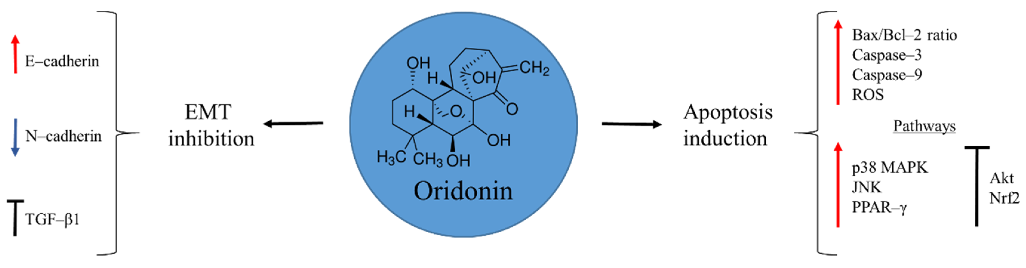
2. Oridonin
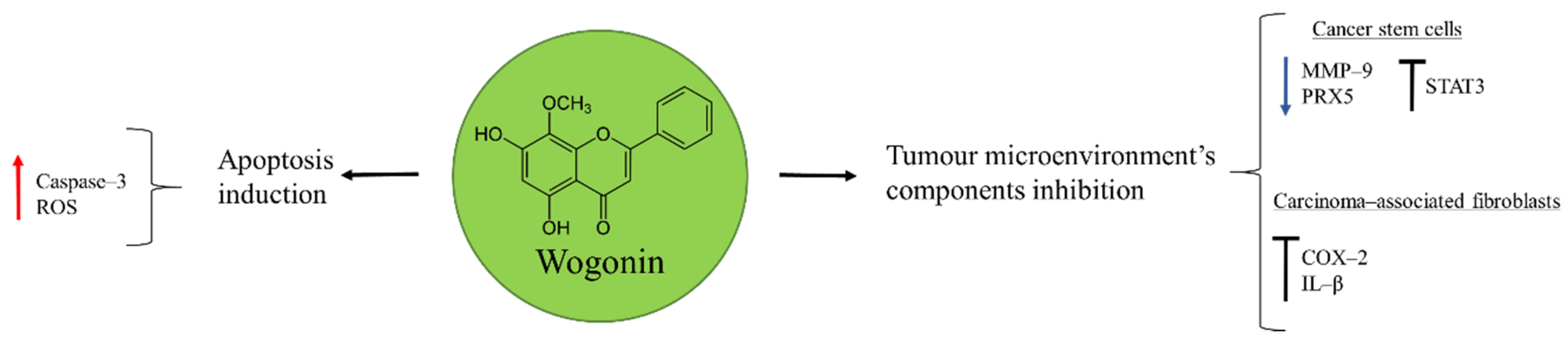
3. Wogonin
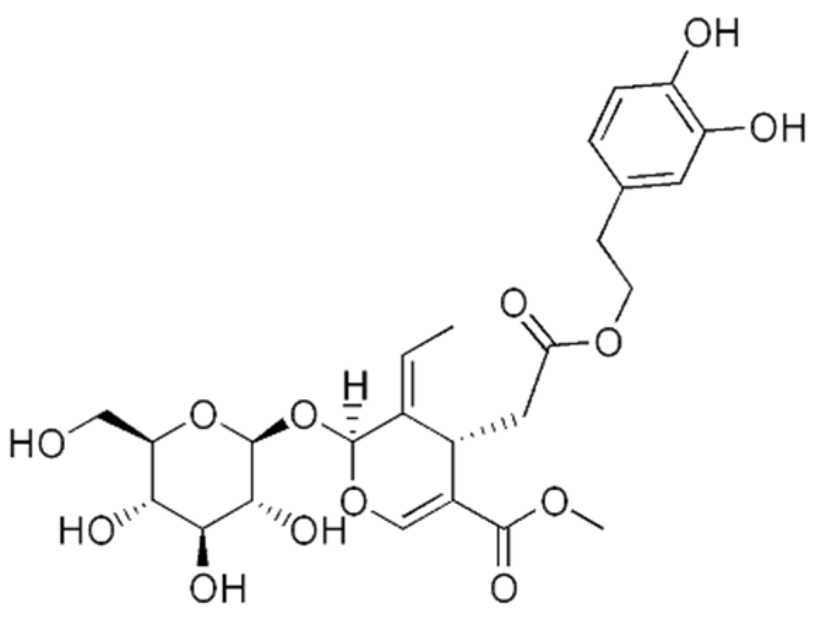
4. Oleuropein
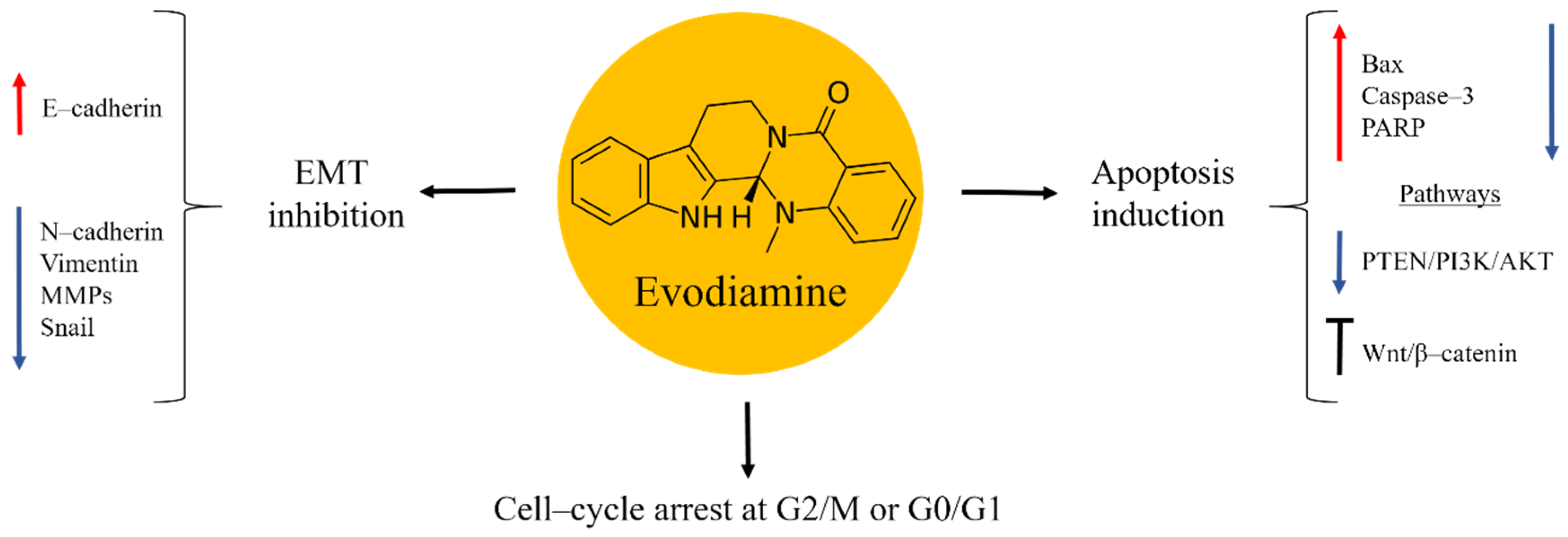
5. Evodiamine
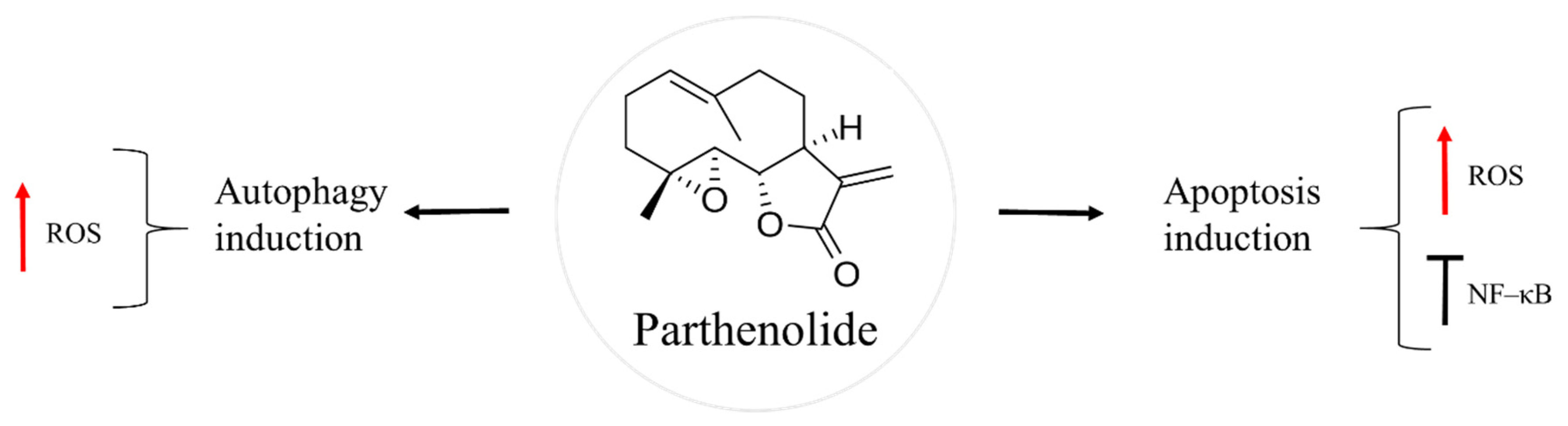
6. Parthenolide
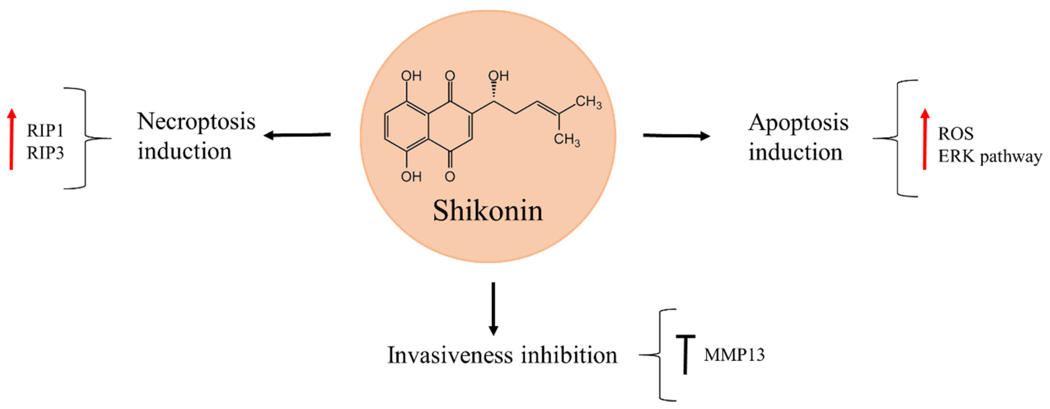
7. Shikonin
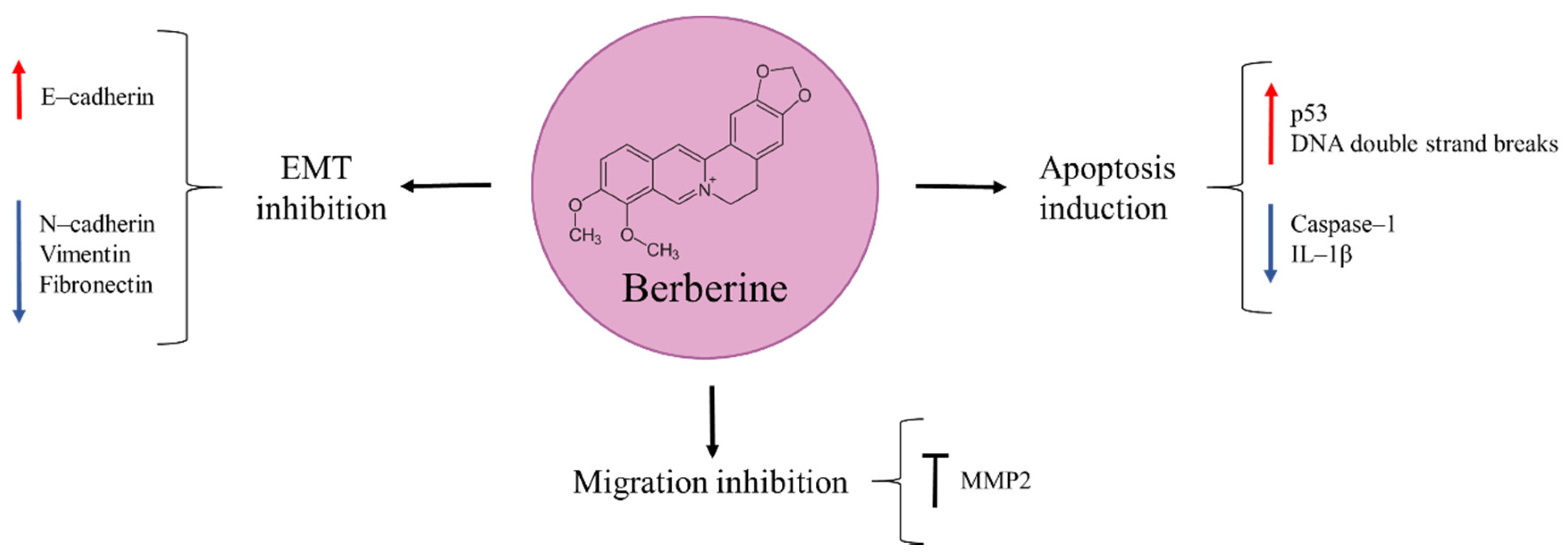
8. Berberine
9. Triptolide
10. Novel Natural Products
11. Discussion and Future Perspectives
12. Conclusions
Author Contributions
Funding
Conflicts of Interest
Abbreviations
References
- Ottaviani, G.; Jaffe, N. The Epidemiology of Osteosarcoma. Cancer Treat Res. 2009, 152, 3–13. [Google Scholar] [PubMed]
- Mirabello, L.; Troisi, R.J.; Savage, S.A. International osteosarcoma incidence patterns in children and adolescents, middle ages and elderly persons. Int. J. Cancer 2009, 125, 229–234. [Google Scholar] [CrossRef] [PubMed] [Green Version]
- Misaghi, A.; Goldin, A.; Awad, M.; Kulidjian, A.A. Osteosarcoma: A comprehensive review. SICOT-J. 2018, 4, 12. [Google Scholar] [CrossRef] [Green Version]
- Durfee, R.A.; Mohammed, M.; Luu, H.H. Review of Osteosarcoma and Current Management. Rheumatol. Ther. 2016, 3, 221–243. [Google Scholar] [CrossRef] [PubMed] [Green Version]
- Kansara, M.; Teng, M.; Smyth, M.; Thomas, D. Translational biology of osteosarcoma. Nat. Cancer 2014, 14, 722–735. [Google Scholar] [CrossRef] [PubMed]
- Hansen, M.F.; Seton, M.; Merchant, A. Osteosarcoma in Paget’s Disease of Bone. J. Bone Miner. Res. 2006, 21, 58–63. [Google Scholar] [CrossRef]
- Deyrup, A.T.; Montag, A.G.; Inwards, C.Y.; Xu, M.Z.; Swee, R.G.; Unni, K.K. Sarcomas Arising in Paget Disease of Bone: A Clinicopathologic Analysis of 70 Cases. Arch. Pathol. Lab. Med. 2007, 131, 942–946. [Google Scholar] [CrossRef]
- Huang, M.; Lu, J.-J.; Ding, J. Natural Products in Cancer Therapy: Past, Present and Future. Nat. Prod. Bioprospecting 2021, 11, 5–13. [Google Scholar] [CrossRef]
- Eder, J.; Sedrani, R.; Wiesmann, C. The discovery of first-in-class drugs: Origins and evolution. Nat. Rev. Drug Discov. 2014, 13, 577–587. [Google Scholar] [CrossRef]
- Madariaga-Mazón, A.; Naveja, J.J.; Medina-Franco, J.L.; Noriega-Colima, K.O.; Martinez-Mayorga, K. DiaNat-DB: A molecular database of antidiabetic compounds from medicinal plants. RSC Adv. 2021, 11, 5172–5178. [Google Scholar] [CrossRef]
- Paul, D.; Sanap, G.; Shenoy, S.; Kalyane, D.; Kalia, K.; Tekade, R.K. Artificial intelligence in drug discovery and development. Drug Discov. Today 2021, 26, 80–93. [Google Scholar] [CrossRef] [PubMed]
- Koźmiński, P.; Halik, P.K.; Chesori, R.; Gniazdowska, E. Overview of Dual-Acting Drug Methotrexate in Different Neurological Diseases, Autoimmune Pathologies and Cancers. Int. J. Mol. Sci. 2020, 21, 3483. [Google Scholar] [CrossRef] [PubMed]
- González-Fernández, Y.; Imbuluzqueta, E.; Zalacain, M.; Mollinedo, F.; Patiño-García, A.; Blanco-Prieto, M.J. Doxorubicin and edelfosine lipid nanoparticles are effective acting synergistically against drug-resistant osteosarcoma cancer cells. Cancer Lett. 2017, 388, 262–268. [Google Scholar] [CrossRef]
- Yang, F.; Teves, S.S.; Kemp, C.J.; Henikoff, S. Doxorubicin, DNA torsion, and chromatin dynamics. Biochim. Biophys. Acta 2014, 1845, 84–89. [Google Scholar] [CrossRef] [PubMed] [Green Version]
- Dasari, S.; Tchounwou, P.B. Cisplatin in cancer therapy: Molecular mechanisms of action. Eur. J. Pharmacol. 2014, 740, 364–378. [Google Scholar] [CrossRef] [Green Version]
- Chen, B.; Yang, J.-Z.; Wang, L.-F.; Zhang, Y.-J.; Lin, X.-J. Ifosfamide-loaded poly (lactic-co-glycolic acid) PLGA-dextran polymeric nanoparticles to improve the antitumor efficacy in Osteosarcoma. BMC Cancer 2015, 15, 752. [Google Scholar] [CrossRef] [Green Version]
- Mulder, R.L.; Paulides, M.; Langer, T.; Kremer, L.C.; van Dalen, E.C. Cyclophosphamide versus ifosfamide for paediatric and young adult bone and soft tissue sarcoma patients. Cochrane Database Syst. Rev. 2015, 2015, CD006300. [Google Scholar] [CrossRef]
- Chamberlin, S.R.; Blucher, A.; Wu, G.; Shinto, L.; Choonoo, G.; Kulesz-Martin, M.; McWeeney, S. Natural Product Target Network Reveals Potential for Cancer Combination Therapies. Front. Pharmacol. 2019, 10, 557. [Google Scholar] [CrossRef]
- Frank, J.D.; Barton, S.J.; Angelo, J.M. Antineoplastic Drugs. In Pharmacology and Therapeutics for Dentistry; Kwok, I.K.K., Vincent, E.C., Gibson, J.N., Eds.; Elsevier Inc.: Mosby, Norway, 2017; pp. 530–562. [Google Scholar]
- Thirumaran, R.; Prendergast, G.C.; Gilman, P.B. Cytotoxic Chemotherapy in Clinical Treatment of Cancer. In Cancer Immunotherapy: Immune Suppression and Tumor Growth; Prendergast, G.C., Jaffee, E.M., Eds.; Elsevier Inc.: Burlington, VT, USA; San Diego, CA, USA; London, UK, 2007; pp. 101–116. [Google Scholar]
- Birjees, M.; Ahmad, M.; Zafar, M.; Nawaz, S.; Jehanzeb, S.; Ullah, F.; Zaman, W. Traditional knowledge of wild medicinal plants used by the inhabitants of Garam Chashma valley, district Chitral, Pakistan. Acta Ecol. Sin. 2022, 42, 19–33. [Google Scholar] [CrossRef]
- Ekor, M. The growing use of herbal medicines: Issues relating to adverse reactions and challenges in monitoring safety. Front. Pharmacol. 2014, 4, 177. [Google Scholar] [CrossRef] [Green Version]
- Haque, I.; Chowdhury, A.B.M.; Shahjahan, M.; Harun, M.; Dostogir, G. Traditional healing practices in rural Bangladesh: A qualitative investigation. BMC Complement. Altern. Med. 2018, 18, 62. [Google Scholar] [CrossRef] [PubMed] [Green Version]
- Omara, T.; Kiprop, A.K.; Ramkat, R.C.; Cherutoi, J.; Kagoya, S.; Nyangena, D.M.; Tebo, T.A.; Nteziyaremye, P.; Karanja, L.N.; Jepchirchir, A.; et al. Medicinal Plants Used in Traditional Management of Cancer in Uganda: A Review of Ethnobotanical Surveys, Phytochemistry, and Anticancer Studies. Evid.-Based Complement. Altern. Med. 2020, 2020, 3529081. [Google Scholar] [CrossRef] [PubMed] [Green Version]
- Kuruppu, A.I.; Paranagama, P.; Goonasekara, C.L. Medicinal plants commonly used against cancer in traditional medicine formulae in Sri Lanka. Saudi Pharm. J. 2019, 27, 565–573. [Google Scholar] [CrossRef] [PubMed]
- Cole, C.; Burgoyne, T.W.; Lee, A.; Stehno-Bittel, L.; Zaid, G.H. Arum Palaestinum with isovanillin, linolenic acid and β-sitosterol inhibits prostate cancer spheroids and reduces the growth rate of prostate tumors in mice. BMC Complement. Altern. Med. 2015, 15, 264. [Google Scholar] [CrossRef] [Green Version]
- Abu-Darwish, M.S.; Efferth, T. Medicinal Plants from Near East for Cancer Therapy. Front. Pharmacol. 2018, 9, 56. [Google Scholar] [CrossRef] [Green Version]
- Wang, X.-H.; Zhang, S.-F.; Bao, J.-T.; Liu, F.-Y. Oridonin synergizes with Nutlin-3 in osteosarcoma cells by modulating the levels of multiple Bcl-2 family proteins. Tumor Biol. 2017, 39, 1010428317701638. [Google Scholar] [CrossRef] [Green Version]
- Li, X.; Zhang, C.-T.; Ma, W.; Xie, X.; Huang, Q. Oridonin: A Review of Its Pharmacology, Pharmacokinetics and Toxicity. Front. Pharmacol. 2021, 12, 645824. [Google Scholar] [CrossRef]
- Li, J.; Wu, Y.; Wang, D.; Zou, L.; Fu, C.; Zhang, J.; Leung, G.P.-H. Oridonin synergistically enhances the anti-tumor efficacy of doxorubicin against aggressive breast cancer via pro-apoptotic and anti-angiogenic effects. Pharmacol. Res. 2019, 146, 104313. [Google Scholar] [CrossRef]
- Wang, Y.; Zhu, Z. Oridonin inhibits metastasis of human ovarian cancer cells by suppressing the mTOR pathway. Arch. Med Sci. 2019, 15, 1017–1027. [Google Scholar] [CrossRef]
- Yao, Z.; Xie, F.; Li, M.; Liang, Z.; Xu, W.; Yang, J.; Liu, C.; Li, H.; Zhou, H.; Qu, L.-H. Oridonin induces autophagy via inhibition of glucose metabolism in p53-mutated colorectal cancer cells. Cell Death Dis. 2017, 8, e2633. [Google Scholar] [CrossRef]
- Luo, D.; Yi, Y.; Peng, K.; Liu, T.; Yang, J.; Liu, S.; Zhao, W.; Qu, X.; Yu, W.; Gu, Y.; et al. Oridonin derivatives as potential anticancer drug candidates triggering apoptosis through mitochondrial pathway in the liver cancer cells. Eur. J. Med. Chem. 2019, 178, 365–379. [Google Scholar] [CrossRef]
- Tian, L.; Xie, K.; Sheng, D.; Wan, X.; Zhu, G. Antiangiogenic effects of oridonin. BMC Complement. Altern. Med. 2017, 17, 192. [Google Scholar] [CrossRef] [PubMed] [Green Version]
- Du, Y.; Zhang, J.; Yan, S.; Tao, Z.; Wang, C.; Huang, M.; Zhang, X. Oridonin inhibits the proliferation, migration and invasion of human osteosarcoma cells via suppression of matrix metalloproteinase expression and STAT3 signalling pathway. J. BUON 2019, 24, 1175–1180. [Google Scholar]
- Lu, Y.; Sun, Y.; Zhu, J.; Yu, L.; Jiang, X.; Zhang, J.; Dong, X.; Ma, B.; Zhang, Q. Oridonin exerts anticancer effect on osteosarcoma by activating PPAR-γ and inhibiting Nrf2 pathway. Cell Death Dis. 2018, 9, 15. [Google Scholar] [CrossRef] [PubMed] [Green Version]
- Jin, S.; Shen, J.-N.; Wang, J.; Huang, G.; Zhou, J.-G. Oridonin induced apoptosis through Akt and MAPKs signaling pathways in human osteosarcoma cells. Cancer Biol. Ther. 2007, 6, 261–268. [Google Scholar] [CrossRef] [Green Version]
- Sun, Y.; Jiang, X.; Lu, Y.; Zhu, J.; Yu, L.; Ma, B.; Zhang, Q. Oridonin prevents epithelial-mesenchymal transition and TGF-β1-induced epithelial-mesenchymal transition by inhibiting TGF-β1/Smad2/3 in osteosarcoma. Chem.-Biol. Interact. 2018, 296, 57–64. [Google Scholar] [CrossRef]
- Kazantseva, L.; Becerra, J.; Santos-Ruiz, L. Oridonin enhances antitumor effects of doxorubicin in human osteosarcoma cells. Pharmacol. Rep. 2022, 74, 248–256. [Google Scholar] [CrossRef] [PubMed]
- Zhang, Y.; Wang, S.; Dai, M.; Nai, J.; Zhu, L.; Sheng, H. Solubility and Bioavailability Enhancement of Oridonin: A Review. Molecules 2020, 25, 332. [Google Scholar] [CrossRef] [PubMed] [Green Version]
- Wang, S.-Q.; Wang, C.; Chang, L.-M.; Zhou, K.-R.; Wang, J.-W.; Ke, Y.; Yang, D.-X.; Shi, H.-G.; Wang, R.; Shi, X.-L.; et al. Geridonin and paclitaxel act synergistically to inhibit the proliferation of gastric cancer cells through ROS-mediated regulation of the PTEN/PI3K/Akt pathway. Oncotarget 2016, 7, 72990–73002. [Google Scholar] [CrossRef] [Green Version]
- Liu, X.; Xu, J.; Zhou, J.; Shen, Q. Oridonin and its derivatives for cancer treatment and overcoming therapeutic resistance. Genes Dis. 2020, 8, 448–462. [Google Scholar] [CrossRef]
- Kimura, Y.; Sumiyoshi, M. Anti-tumor and anti-metastatic actions of wogonin isolated from Scutellaria baicalensis roots through anti-lymphangiogenesis. Phytomedicine 2013, 20, 328–336. [Google Scholar] [CrossRef] [PubMed]
- Chung, J.-G.; Lin, C.-C.; Kuo, C.-L.; Lee, M.-H.; Lai, K.-C.; Lin, J.-P.; Yang, J.-S.; Yu, C.-S.; Lu, C.-C.; Chiang, J.-H.; et al. Wogonin triggers apoptosis in human osteosarcoma U-2 OS cells through the endoplasmic reticulum stress, mitochondrial dysfunction and caspase-3-dependent signaling pathways. Int. J. Oncol. 2011, 39, 217–224. [Google Scholar] [CrossRef] [Green Version]
- Ge, Z.; Ding, S. The Crosstalk Between Tumor-Associated Macrophages (TAMs) and Tumor Cells and the Corresponding Targeted Therapy. Front. Oncol. 2020, 10, 590941. [Google Scholar] [CrossRef]
- Koh, H.; Sun, H.-N.; Xing, Z.; Liu, R.; Chandimali, N.; Kwon, T.; Lee, D.-S. Wogonin Influences Osteosarcoma Stem Cell Stemness Through ROS-dependent Signaling. In Vivo 2020, 34, 1077–1084. [Google Scholar] [CrossRef]
- Huynh, D.L.; Kwon, T.; Zhang, J.J.; Sharma, N.; Gera, M.; Ghosh, M.; Kim, N.; Cho, S.K.; Lee, D.S.; Park, Y.H.; et al. Wogonin suppresses stem cell-like traits of CD133 positive osteosarcoma cell via inhibiting matrix metallopeptidase-9 expression. BMC Complement. Altern. Med. 2017, 17, 304. [Google Scholar] [CrossRef] [Green Version]
- Gioti, K.; Papachristodoulou, A.; Benaki, D.; Aligiannis, N.; Skaltsounis, A.-L.; Mikros, E.; Tenta, R. Assessment of the Nutraceutical Effects of Oleuropein and the Cytotoxic Effects of Adriamycin, When Administered Alone and in Combination, in MG-63 Human Osteosarcoma Cells. Nutrients 2021, 13, 354. [Google Scholar] [CrossRef] [PubMed]
- Flemmig, J.; Kuchta, K.; Arnhold, J.; Rauwald, H. Olea europaea leaf (Ph.Eur.) extract as well as several of its isolated phenolics inhibit the gout-related enzyme xanthine oxidase. Phytomedicine 2011, 18, 561–566. [Google Scholar] [CrossRef] [PubMed]
- Haloui, E.; Marzouk, Z.; Marzouk, B.; Bouftira, I.; Bouraoui, A.; Fenina, N. Pharmacological activities and chemical composition of the Olea europaea L. leaf essential oils from Tunisia. J. Food Agric. Environ. 2010, 8, 204–208. [Google Scholar]
- Esmaeili-Mahani, S.; Rezaeezadeh-Roukerd, M.; Esmaeilpour, K.; Abbasnejad, M.; Rasoulian, B.; Sheibani, V.; Kaeidi, A.; Hajializadeh, Z. Olive (Olea europaea L.) leaf extract elicits antinociceptive activity, potentiates morphine analgesia and suppresses morphine hyperalgesia in rats. J. Ethnopharmacol. 2010, 132, 200–205. [Google Scholar] [CrossRef]
- Goldsmith, C.D.; Bond, D.R.; Jankowski, H.; Weidenhofer, J.; Stathopoulos, C.E.; Roach, P.D.; Scarlett, C.J. The Olive Biophenols Oleuropein and Hydroxytyrosol Selectively Reduce Proliferation, Influence the Cell Cycle, and Induce Apoptosis in Pancreatic Cancer Cells. Int. J. Mol. Sci. 2018, 19, 1937. [Google Scholar] [CrossRef] [Green Version]
- Vanella, L.; Acquaviva, R.; Di Giacomo, C.; Sorrenti, V.; Galvano, F.; Santangelo, R.; Cardile, V.; Gangia, S.; D’Orazio, N.; Abraham, N.G. Antiproliferative effect of oleuropein in prostate cell lines. Int. J. Oncol. 2012, 41, 31–38. [Google Scholar] [CrossRef] [PubMed] [Green Version]
- Liu, L.; Ahn, K.S.; Shanmugam, M.K.; Wang, H.; Shen, H.; Arfuso, F.; Chinnathambi, A.; Alharbi, S.A.; Chang, Y.; Sethi, G.; et al. Oleuropein induces apoptosis via abrogating NF-κB activation cascade in estrogen receptor–negative breast cancer cells. J. Cell. Biochem. 2019, 120, 4504–4513. [Google Scholar] [CrossRef] [PubMed]
- Moran, J.M.; Leal-Hernandez, O.; Canal-Macías, M.L.; Roncero-Martin, R.; Guerrero-Bonmatty, R.; Aliaga, I.; Zamorano, J.D.P. Antiproliferative Properties of Oleuropein in Human Osteosarcoma Cells. Nat. Prod. Commun. 2016, 11, 491–492. [Google Scholar]
- Przychodzen, P.; Wyszkowska, R.; Gorzynik-Debicka, M.; Kostrzewa, T.; Kuban-Jankowska, A.; Gorska-Ponikowska, M. Anticancer Potential of Oleuropein, the Polyphenol of Olive Oil, With 2-Methoxyestradiol, Separately or in Combination, in Human Osteosarcoma Cells. Anticancer Res. 2019, 39, 1243–1251. [Google Scholar] [CrossRef]
- Tian, K.-M.; Li, J.-J.; Xu, S.-W. Rutaecarpine: A promising cardiovascular protective alkaloid from Evodia rutaecarpa (Wu Zhu Yu). Pharmacol. Res. 2019, 141, 541–550. [Google Scholar] [CrossRef]
- Jiang, J.; Hu, C. Evodiamine: A Novel Anti-Cancer Alkaloid from Evodia rutaecarpa. Molecules 2009, 14, 1852–1859. [Google Scholar] [CrossRef]
- Yang, S.; Chen, J.; Tan, T.; Wang, N.; Huang, Y.; Wang, Y.; Yuan, X.; Zhang, P.; Luo, J.; Luo, X. Evodiamine Exerts Anticancer Effects Against 143B and MG63 Cells Through the Wnt/β-Catenin Signaling Pathway. Cancer Manag. Res. 2020, 12, 2875–2888. [Google Scholar] [CrossRef]
- Bai, X.; Meng, H.; Ma, L.; Guo, A. Inhibitory effects of evodiamine on human osteosarcoma cell proliferation and apoptosis. Oncol. Lett. 2015, 9, 801–805. [Google Scholar] [CrossRef] [Green Version]
- Meng, Z.-J.; Wu, N.; Liu, Y.; Shu, K.-J.; Zou, X.; Zhang, R.-X.; Pi, C.-J.; He, B.-C.; Ke, Z.-Y.; Chen, L.; et al. Evodiamine inhibits the proliferation of human osteosarcoma cells by blocking PI3K/Akt signaling. Oncol. Rep. 2015, 34, 1388–1396. [Google Scholar] [CrossRef] [Green Version]
- Sun, G.; Zhang, C.; Song, H.; Guo, J.; Li, M.; Cao, Y. WZY-321, a novel evodiamine analog, inhibits glioma cell growth in an autophagy-associated manner. Oncol. Lett. 2019, 17, 2465–2472. [Google Scholar] [CrossRef] [Green Version]
- Kan, S.-F.; Yu, C.-H.; Pu, H.-F.; Hsu, J.-M.; Chen, M.-J.; Wang, P.S. Anti-proliferative effects of evodiamine on human prostate cancer cell lines DU145 and PC3. J. Cell. Biochem. 2007, 101, 44–56. [Google Scholar] [CrossRef] [PubMed]
- Pareek, A.; Suthar, M.; Rathore, G.S.; Bansal, V. Feverfew (Tanacetum parthenium L.): A systematic review. Pharmacogn. Rev. 2011, 5, 103–110. [Google Scholar] [CrossRef] [PubMed] [Green Version]
- Knight, D.W. Feverfew: Chemistry and biological activity. Nat. Prod. Rep. 1995, 12, 271–276. [Google Scholar] [CrossRef] [PubMed]
- Nakshatri, H.; Rice, S.E.; Bhat-Nakshatri, P. Antitumor agent parthenolide reverses resistance of breast cancer cells to tumor necrosis factor-related apoptosis-inducing ligand through sustained activation of c-Jun N-terminal kinase. Oncogene 2004, 23, 7330–7344. [Google Scholar] [CrossRef] [PubMed] [Green Version]
- Kim, S.L.; Liu, Y.C.; Seo, S.Y.; Kim, S.H.; Kim, I.H.; Lee, S.O.; Lee, S.T.; Kim, D.-G.; Kim, S.W. Parthenolide induces apoptosis in colitis-associated colon cancer, inhibiting NF-κB signaling. Oncol. Lett. 2015, 9, 2135–2142. [Google Scholar] [CrossRef] [Green Version]
- Sun, Y.; Clair, D.K.S.; Xu, Y.; Crooks, P.A.; Clair, W.H.S. A NADPH Oxidase–Dependent Redox Signaling Pathway Mediates the Selective Radiosensitization Effect of Parthenolide in Prostate Cancer Cells. Cancer Res. 2010, 70, 2880–2890. [Google Scholar] [CrossRef] [Green Version]
- Vazhappilly, C.G.; Kumar, D.; Kumar, R. Relative In Vitro Potentials of Parthenolide to Induce Apoptosis and Cell Cycle Arrest in Skin Cancer Cells. Curr. Drug Discov. Technol. 2016, 13, 34–40. [Google Scholar] [CrossRef]
- Zuch, D.; Giang, A.-H.; Shapovalov, Y.; Schwarz, E.; Rosier, R.; O’Keefe, R.; Eliseev, R.A. Targeting Radioresistant Osteosarcoma Cells With Parthenolide. J. Cell. Biochem. 2012, 113, 1282–1291. [Google Scholar] [CrossRef] [Green Version]
- Gong, T.; Su, X.; Xia, Q.; Wang, J.; Kan, S. Expression of NF-κB and PTEN in osteosarcoma and its clinical significance. Oncol. Lett. 2017, 14, 6744–6748. [Google Scholar] [CrossRef] [Green Version]
- Kishida, Y.; Yoshikawa, H.; Myoui, A.; Seliger, B.; Fedorushchenko, A.; Brenner, W.; Ackermann, A.; Atkins, D.; Hanash, S.; Lichtenfels, R. Parthenolide, a Natural Inhibitor of Nuclear Factor-κB, Inhibits Lung Colonization of Murine Osteosarcoma Cells. Clin. Cancer Res. 2007, 13, 59–67. [Google Scholar] [CrossRef] [Green Version]
- Dhanasekaran, D.N.; Reddy, E.P. JNK-signaling: A multiplexing hub in programmed cell death. Genes Cancer 2017, 8, 682–694. [Google Scholar] [CrossRef] [PubMed] [Green Version]
- Yang, C.; Yang, Q.O.; Kong, Q.-J.; Yuan, W.; Yang, Y.-P.O. Parthenolide Induces Reactive Oxygen Species-Mediated Autophagic Cell Death in Human Osteosarcoma Cells. Cell. Physiol. Biochem. 2016, 40, 146–154. [Google Scholar] [CrossRef]
- D’Anneo, A.; Carlisi, D.; Lauricella, M.; Emanuele, S.; Di Fiore, R.; Vento, R.; Tesoriere, G. Parthenolide induces caspase-independent and AIF-mediated cell death in human osteosarcoma and melanoma cells. J. Cell. Physiol. 2013, 228, 952–967. [Google Scholar] [CrossRef] [PubMed] [Green Version]
- Schwarz, R.; Bruland, O.; Cassoni, A.; Schomberg, P.; Bielack, S. The Role of Radiotherapy in Oseosarcoma. Cancer Treat. Res. 2009, 152, 147–164. [Google Scholar] [CrossRef] [PubMed]
- Mahajan, A.; Woo, S.Y.; Kornguth, D.G.; Hughes, D.; Huh, W.; Chang, E.L.; Herzog, C.E.; Pelloski, C.E.; Anderson, P. Multimodality treatment of osteosarcoma: Radiation in a high-risk cohort. Pediatr. Blood Cancer 2008, 50, 976–982. [Google Scholar] [CrossRef]
- Sugiyasu, K.; Nanno, K.; Tamai, N.; Hashimoto, N.; Kishida, Y.; Yoshikawa, H.; Myoui, A. Radio-sensitization of the murine osteosarcoma cell line LM8 with parthenolide, a natural inhibitor of NF-κB. Oncol. Lett. 2011, 2, 407–412. [Google Scholar] [CrossRef] [Green Version]
- Chang, I.-C.; Huang, Y.-J.; Chiang, T.-I.; Yeh, C.-W.; Hsu, L.-S. Shikonin Induces Apoptosis through Reactive Oxygen Species/Extracellular Signal-Regulated Kinase Pathway in Osteosarcoma Cells. Biol. Pharm. Bull. 2010, 33, 816–824. [Google Scholar] [CrossRef] [Green Version]
- Kong, W.-Y.; Chen, X.-F.; Shi, J.; Baloch, S.K.; Qi, J.-L.; Zhu, H.-L.; Wang, X.-M.; Yang, Y.-H. Design and Synthesis of Fluoroacylshikonin as an Anticancer Agent. Chirality 2013, 25, 757–762. [Google Scholar] [CrossRef]
- Fu, Z.; Deng, B.; Liao, Y.; Shan, L.; Yin, F.; Wang, Z.; Zeng, H.; Zuo, D.; Hua, Y.; Cai, Z. The anti-tumor effect of shikonin on osteosarcoma by inducing RIP1 and RIP3 dependent necroptosis. BMC Cancer 2013, 13, 580. [Google Scholar] [CrossRef] [Green Version]
- Deng, B.; Feng, Y.; Deng, B. TIPE2 Mediates the Suppressive Effects of Shikonin on MMP13 in Osteosarcoma Cells. Cell. Physiol. Biochem. 2015, 37, 2434–2443. [Google Scholar] [CrossRef]
- Deng, B.; Qiu, B. Shikonin inhibits invasiveness of osteosarcoma through MMP13 suppression. Tumor Biol. 2015, 36, 9311–9317. [Google Scholar] [CrossRef] [PubMed]
- Yang, Q.; Li, S.; Fu, Z.; Lin, B.; Zhou, Z.; Wang, Z.; Hua, Y.; Cai, Z. Shikonin promotes adriamycin-induced apoptosis by upregulating caspase-3 and caspase-8 in osteosarcoma. Mol. Med. Rep. 2017, 16, 1347–1352. [Google Scholar] [CrossRef] [PubMed] [Green Version]
- Janeway, K.A.; Grier, H.E. Sequelae of osteosarcoma medical therapy: A review of rare acute toxicities and late effects. Lancet Oncol. 2010, 11, 670–678. [Google Scholar] [CrossRef]
- Liu, Y.; Zhang, L.; Song, H.; Ji, G. Update on Berberine in Nonalcoholic Fatty Liver Disease. Evid.-Based Complement. Altern. Med. 2013, 2013, 1–8. [Google Scholar] [CrossRef] [Green Version]
- Mishra, R.; Nathani, S.; Varshney, R.; Sircar, D.; Roy, P. Berberine reverses epithelial-mesenchymal transition and modulates histone methylation in osteosarcoma cells. Mol. Biol. Rep. 2020, 47, 8499–8511. [Google Scholar] [CrossRef]
- Liu, J.; Liu, P.; Xu, T.; Chen, Z.; Kong, H.; Chu, W.; Wang, Y.; Liu, Y. Berberine Induces Autophagic Cell Death in Acute Lymphoblastic Leukemia by Inactivating AKT/mTORC1 Signaling. Drug Des. Dev. Ther. 2020, 14, 1813–1823. [Google Scholar] [CrossRef]
- Shen, Z.-Q.; Wang, J.; Tan, W.-F.; Huang, T.-M. Berberine inhibits colorectal tumor growth by suppressing SHH secretion. Acta Pharmacol. Sin. 2021, 42, 1190–1194. [Google Scholar] [CrossRef]
- Lu, W.; DU, S.; Wang, J. Berberine inhibits the proliferation of prostate cancer cells and induces G0/G1 or G2/M phase arrest at different concentrations. Mol. Med. Rep. 2015, 11, 3920–3924. [Google Scholar] [CrossRef] [Green Version]
- Sun, Y.; Wang, W.; Tong, Y. Berberine Inhibits Proliferative Ability of Breast Cancer Cells by Reducing Metadherin. Med Sci. Monit. 2019, 25, 9058–9066. [Google Scholar] [CrossRef]
- Jiang, S.-X.; Qi, B.; Yao, W.-J.; Gu, C.-W.; Wei, X.-F.; Zhao, Y.; Liu, Y.-Z.; Zhao, B.-S. Berberine displays antitumor activity in esophageal cancer cells in vitro. World J. Gastroenterol. 2017, 23, 2511–2518. [Google Scholar] [CrossRef]
- Liu, Z.; Liu, Q.; Xu, B.; Wu, J.; Guo, C.; Zhu, F.; Yang, Q.; Gao, G.; Gong, Y.; Shao, C. Berberine induces p53-dependent cell cycle arrest and apoptosis of human osteosarcoma cells by inflicting DNA damage. Mutat. Res. Mol. Mech. Mutagen. 2009, 662, 75–83. [Google Scholar] [CrossRef] [PubMed]
- Zhu, Y.; Ma, N.; Li, H.-X.; Tian, L.; Ba, Y.-F.; Hao, B. Berberine induces apoptosis and DNA damage in MG-63 human osteosarcoma cells. Mol. Med. Rep. 2014, 10, 1734–1738. [Google Scholar] [CrossRef] [PubMed] [Green Version]
- Jin, H.; Jin, X.; Cao, B.; Wang, W. Berberine affects osteosarcoma via downregulating the caspase-1/IL-1β signaling axis. Oncol. Rep. 2017, 37, 729–736. [Google Scholar] [CrossRef] [PubMed] [Green Version]
- Gao, X.; Zhang, C.; Wang, Y.; Zhang, P.; Zhang, J.; Hong, T. Berberine and Cisplatin Exhibit Synergistic Anticancer Effects on Osteosarcoma MG-63 Cells by Inhibiting the MAPK Pathway. Molecules 2021, 26, 1666. [Google Scholar] [CrossRef]
- Zhao, X.; Zhang, Q.; Chen, L. Triptolide induces the cell apoptosis of osteosarcoma cells through the TRAIL pathway. Oncol. Rep. 2016, 36, 1499–1505. [Google Scholar] [CrossRef] [Green Version]
- He, M.-F.; Liu, L.; Ge, W.; Shaw, P.-C.; Jiang, R.; Wu, L.-W.; But, P.P.-H. Antiangiogenic activity of Tripterygium wilfordii and its terpenoids. J. Ethnopharmacol. 2009, 121, 61–68. [Google Scholar] [CrossRef]
- Phillips, P.A.; Dudeja, V.; McCarroll, J.A.; Borja-Cacho, D.; Dawra, R.K.; Grizzle, W.E.; Vickers, S.M.; Saluja, A.K. Triptolide Induces Pancreatic Cancer Cell Death via Inhibition of Heat Shock Protein 70. Cancer Res. 2007, 67, 9407–9416. [Google Scholar] [CrossRef] [Green Version]
- Li, X.; Zang, A.; Jia, Y.; Zhang, J.; Fan, W.; Feng, J.; Duan, M.; Zhang, L.; Huo, R.; Jiao, J.; et al. Triptolide reduces proliferation and enhances apoptosis of human non-small cell lung cancer cells through PTEN by targeting miR-21. Mol. Med. Rep. 2016, 13, 2763–2768. [Google Scholar] [CrossRef] [Green Version]
- Wang, H.; Ma, D.; Wang, C.; Zhao, S.; Liu, C. Triptolide Inhibits Invasion and Tumorigenesis of Hepatocellular Carcinoma MHCC-97H Cells Through NF-κB Signaling. Med Sci. Monit. 2016, 22, 1827–1836. [Google Scholar] [CrossRef] [Green Version]
- Kwon, H.-Y.; Kim, K.-S.; An, H.-K.; Moon, H.-I.; Kim, H.-J.; Lee, Y.-C. Triptolide induces apoptosis through extrinsic and intrinsic pathways in human osteosarcoma U2OS cells. Indian J. Biochem. Biophys. 2013, 50, 485–491. [Google Scholar]
- Li, X.; Lu, Q.; Xie, W.; Wang, Y.; Wang, G. Anti-tumor effects of triptolide on angiogenesis and cell apoptosis in osteosarcoma cells by inducing autophagy via repressing Wnt/β-Catenin signaling. Biochem. Biophys. Res. Commun. 2018, 496, 443–449. [Google Scholar] [CrossRef] [PubMed]
- Qin, W.; Li, S.; Miao, Y.; Shi, Q.; Wang, Y.; Li, J.; Chen, Z.; Wang, J.; Ling, C. Triptolide induces mitochondrial apoptosis through modulating dual specificity phosphatase 1/mitogen-activated protein kinases cascade in osteosarcoma cells. Neoplasma 2018, 65, 21–33. [Google Scholar] [CrossRef] [PubMed] [Green Version]
- Zhao, L.; Jiang, B.; Wang, D.; Liu, W.; Zhang, H.; Liu, W.; Qiu, Z. Triptolide reduces the viability of osteosarcoma cells by reducing MKP-1 and Hsp70 expression. Exp. Ther. Med. 2016, 11, 2005–2010. [Google Scholar] [CrossRef] [PubMed] [Green Version]
- Wang, Z.; Zhou, J.-Y.; Ma, D.K.; Buck, S.; Wu, G.S.; Mbbs, Y.R. High level of mitogen-activated protein kinase phosphatase-1 expression is associated with cisplatin resistance in osteosarcoma. Pediatr. Blood Cancer 2008, 51, 754–759. [Google Scholar] [CrossRef] [PubMed] [Green Version]
- Jiang, C.; Fang, X.; Zhang, H.; Wang, X.; Li, M.; Jiang, W.; Tian, F.; Zhu, L.; Bian, Z. AMD3100 combined with triptolide inhibit proliferation, invasion and metastasis and induce apoptosis of human U2OS osteosarcoma cells. Biomed. Pharmacother. 2017, 86, 677–685. [Google Scholar] [CrossRef]
- Banerjee, S.; Thayanithy, V.; Sangwan, V.; Mackenzie, T.N.; Saluja, A.K.; Subramanian, S. Minnelide reduces tumor burden in preclinical models of osteosarcoma. Cancer Lett. 2013, 335, 412–420. [Google Scholar] [CrossRef] [Green Version]
- Hu, N.; Wang, C.; Dai, X.; Zhou, M.; Gong, L.; Yu, L.; Peng, C.; Li, Y. Phillygenin inhibits LPS-induced activation and inflammation of LX2 cells by TLR4/MyD88/NF-κB signaling pathway. J. Ethnopharmacol. 2020, 248, 112361. [Google Scholar] [CrossRef]
- Wu, S.; Zhang, Y.; Zhang, Y.; Chen, L.; Xu, X.; Dang, Y.; Ti, X. Phillygenin regulates proliferation and apoptosis of non-small cell lung cancer through by AMPK/ERK/NF-κB axis. Die Pharm.-Int. J. Pharm. Sci. 2020, 75, 512–515. [Google Scholar] [CrossRef]
- Ding, X.; Lu, D.; Fan, J. A natural product phillygenin suppresses osteosarcoma growth and metastasis by regulating the SHP-1/JAK2/STAT3 signaling. Biosci. Biotechnol. Biochem. 2021, 85, 307–314. [Google Scholar] [CrossRef]
- Wei, J.; Chen, J.-R.; Pais, E.M.A.; Wang, T.-Y.; Miao, L.; Li, L.; Li, L.-Y.; Qiu, F.; Hu, L.-M.; Gao, X.-M.; et al. Oxyresveratrol Is a Phytoestrogen Exerting Anti-inflammatory Effects Through NF-κB and Estrogen Receptor Signaling. Inflammation 2017, 40, 1285–1296. [Google Scholar] [CrossRef]
- Rahman, A.; Bishayee, K.; Sadra, A.; Huh, S.-O. Oxyresveratrol activates parallel apoptotic and autophagic cell death pathways in neuroblastoma cells. Biochim. Biophys. Acta Gen. Subj. 2017, 1861, 23–36. [Google Scholar] [CrossRef] [PubMed]
- Sunilkumar, D.; Drishya, G.; Chandrasekharan, A.; Shaji, S.K.; Bose, C.; Jossart, J.; Perry, J.J.P.; Mishra, N.; Kumar, G.B.; Nair, B.G. Oxyresveratrol drives caspase-independent apoptosis-like cell death in MDA-MB-231 breast cancer cells through the induction of ROS. Biochem. Pharmacol. 2020, 173, 113724. [Google Scholar] [CrossRef] [PubMed]
- Liu, Y.; Ren, W.; Bai, Y.; Wan, L.; Sun, X.; Liu, Y.; Xiong, W.; Zhang, Y.-Y.; Zhou, L. Oxyresveratrol prevents murine H22 hepatocellular carcinoma growth and lymph node metastasis via inhibiting tumor angiogenesis and lymphangiogenesis. J. Nat. Med. 2018, 72, 481–492. [Google Scholar] [CrossRef] [PubMed]
- Lv, T.; Jian, Z.; Li, D.; Ao, R.; Zhang, X.; Yu, B. Oxyresveratrol induces apoptosis and inhibits cell viability via inhibition of the STAT3 signaling pathway in Saos-2 cells. Mol. Med. Rep. 2020, 22, 5191–5198. [Google Scholar] [CrossRef] [PubMed]
- Calonghi, N.; Farruggia, G.; Boga, C.; Micheletti, G.; Fini, E.; Romani, L.; Telese, D.; Faraci, E.; Bergamini, C.; Cerini, S.; et al. Root Extracts of Two Cultivars of Paeonia Species: Lipid Composition and Biological Effects on Different Cell Lines: Preliminary Results. Molecules 2021, 26, 655. [Google Scholar] [CrossRef] [PubMed]
- De Azevedo, J.W.V.; Fernandes, T.A.A.D.M.; Fernandes, J.V.; de Azevedo, J.C.V.; Lanza, D.C.F.; Bezerra, C.M.; Andrade, V.S.; de Araujo, J.M.G. Biology and pathogenesis of human osteosarcoma. Oncol. Lett. 2020, 19, 1099–1116. [Google Scholar] [CrossRef] [Green Version]
- Park, T.S.; Donnenberg, V.S.; Donnenberg, A.D.; Zambidis, E.T.; Zimmerlin, L. Dynamic Interactions Between Cancer Stem Cells and Their Stromal Partners. Curr. Pathobiol. Rep. 2014, 2, 41–52. [Google Scholar] [CrossRef] [Green Version]
- Carvalho, C.; Santos, R.X.; Cardoso, S.; Correia, S.; Oliveira, P.J.; Santos, M.S.; Moreira, P.I. Doxorubicin: The Good, the Bad and the Ugly Effect. Curr. Med. Chem. 2009, 16, 3267–3285. [Google Scholar] [CrossRef]
- Pugazhendhi, A.; Edison, T.N.J.I.; Velmurugan, B.K.; Jacob, J.A.; Karuppusamy, I. Toxicity of Doxorubicin (Dox) to different experimental organ systems. Life Sci. 2018, 200, 26–30. [Google Scholar] [CrossRef]
- De Luca, A.; Raimondi, L.; Salamanna, F.; Carina, V.; Costa, V.; Bellavia, D.; Alessandro, R.; Fini, M.; Giavaresi, G. Relevance of 3d culture systems to study osteosarcoma environment. J. Exp. Clin. Cancer Res. 2018, 37, 2. [Google Scholar] [CrossRef] [Green Version]
- Monteiro, C.F.; Custódio, C.A.; Mano, J.F. Bioengineering a humanized 3D tri-culture osteosarcoma model to assess tumor invasiveness and therapy response. Acta Biomater. 2021, 134, 204–214. [Google Scholar] [CrossRef] [PubMed]
- Dehelean, C.; Marcovici, I.; Soica, C.; Mioc, M.; Coricovac, D.; Iurciuc, S.; Cretu, O.; Pinzaru, I. Plant-Derived Anticancer Compounds as New Perspectives in Drug Discovery and Alternative Therapy. Molecules 2021, 26, 1109. [Google Scholar] [CrossRef] [PubMed]
- Calonghi, N.; Boga, C.; Nitti, P.; Telese, D.; Bordoni, S.; Farruggia, G.; Asaro, F.; Grandi, M.; Zalambani, C.; Micheletti, G. Effects of Regioisomerism on the Antiproliferative Activity of Hydroxystearic Acids on Human Cancer Cell Lines. Molecules 2022, 27, 2396. [Google Scholar] [CrossRef] [PubMed]
- Puglia, C.; Lauro, M.R.; Tirendi, G.G.; Fassari, G.E.; Carbone, C.; Bonina, F.; Puglisi, G. Modern drug delivery strategies applied to natural active compounds. Expert Opin. Drug Deliv. 2017, 14, 755–768. [Google Scholar] [CrossRef]
- Khazei, K.; Mohajeri, N.; Bonabi, E.; Turk, Z.; Zarghami, N. New Insights Toward Nanostructured Drug Delivery of Plant-Derived Polyphenol Compounds: Cancer Treatment and Gene Expression Profiles. Curr. Cancer Drug Targets 2021, 21, 689–701. [Google Scholar] [CrossRef]
- Yin, Q.; Tang, L.; Cai, K.; Tong, R.; Sternberg, R.; Yang, X.; Dobrucki, L.W.; Borst, L.B.; Kamstock, D.; Song, Z.; et al. Pamidronate functionalized nanoconjugates for targeted therapy of focal skeletal malignant osteolysis. Proc. Natl. Acad. Sci. USA 2016, 113, E4601–E4609. [Google Scholar] [CrossRef] [Green Version]


| Chemotherapeutic Drug | Description | Effect | Reference |
|---|---|---|---|
| High-dose methotrexate | Folic acid analogue | Induced apoptosis and inhibited DNA synthesis, through blockage of dyhidrofolate reductase (DHFR) | [12] |
| Doxorubicin | Anthracycline | Induced cell death through intercalation between DNA strands, Topoisomerase II complex stabilization, and induction of oxidative stress | [13,14] |
| Cisplatin | Platinum-based compound | Induced apoptosis and inhibited genetic material replication and repair through DNA adduct formation | [15] |
| Ifosfamide | Alkylating a gent | Induced apoptosis and inhibited genetic material replication through DNA intra- and inter-strand crosslinks | [4,16,17] |
| Natural Product | Dose | Target | Effect | Model | Cell Line | Reference |
|---|---|---|---|---|---|---|
| Oridonin | 0–200 μM | MMP–2, 3, 9 and STAT3 pathway | Induced apoptosis, inhibited proliferation, migration, and invasion | In vitro | U2OS | [35] |
| Oridonin | 0–100 μM | PPAR–γ and Nrf2 pathways | Induced apoptosis and inhibited proliferation | In vitro and in vivo | MG63 and HOS | [36] |
| Oridonin | 0–100 μM | Akt, ERK, p38 MAPK and JNK pathways | Induced apoptosis and suppressed proliferation | In vitro | MG63, U2OS and Saos–2 | [37] |
| Oridonin | 0–4 μM; 0, 10 and 15 mg/kg | TGF-β1/Smad2/3 | Inhibited EMT, migration, invasion, and lung metastasis | In vitro and in vivo | MG63, U2OS and 143B | [38] |
| Wogonin | 0–100 μM; 25 and 50 mg/kg | - | Reduced tumour growth, metastasis, angiogenesis, lymphangiogenesis, and TAM number | In vitro and in vivo | LM8 | [43] |
| Wogonin | 0–150 μM | ROS and caspase–3 | Induced apoptosis | In vitro | U2OS | [44] |
| Wogonin | 0–80 μM | ROS | Reduced cell viability, proliferation, stemness, migration, and self-renewal capacities | In vitro | CD133+ Cal72 | [46] |
| Wogonin | 0–80 μM | MMP–9 | Induced apoptosis, inhibited migration invasion, and reduced renewal capacities | In vitro | CD133+ Cal72 | [47] |
| Oleuropein | 50–400 μM | - | Reduced proliferation | In vitro | MG63 and Saos–2 | [55] |
| Evodiamine | 0–32 μM | Wnt/β–catenin pathway | Induced apoptosis, inhibited proliferation, migration, and invasion; suppressed EMT and caused cell-cycle arrest | In vitro | MG63 and 143B | [59] |
| Evodiamine | 0–12.5 μg/mL | Bcl–2, Bax, caspase–3, and survivin | Inhibited proliferation and induced apoptosis | In vitro | U2OS | [60] |
| Evodiamine | 0–4 μM | PTEN/PI3K/Akt pathway | Inhibited proliferation, induced apoptosis and caused cell-cycle arrest | In vitro and in vivo | 143B | [61] |
| Parthenolide | 0–25 μM | ROS | Induced cell death, autophagy, and mitophagy | In vitro | MG63 and Saos–2 | [74] |
| Parthenolide | 0–100 μM | AIF | Induced cell death | In vitro | MG63 | [75] |
| Parthenolide | 0–20 μM | NF–κB pathway | Induced cell death and radiosensitivity | In vitro | LM7 | [70] |
| Parthenolide | 0 and 1 μg/mL; 1 and 2 mg/kg | NF–κB | Enhanced radiosensitivity and inhibited tumour growth | In vitro and in vivo | LM8 | [78] |
| Shikonin | 0–8 μM | ROS, ERK, and Bcl–2 | Induced apoptosis | In vitro | 143B | [79] |
| Shikonin | 0–15 μM; 2 mg/kg | RIP1 and RIP3 | Induced cell death, necroptosis, and increased the survival time in metastatic disease | In vitro and in vivo | K7, K12, K7M3, U2OS and 143B | [81] |
| Berberine | 0–80 μM | MMP–2, H3K27me3, and EZH2 | Inhibited proliferation, migration and EMT | In vitro | MG63 | [87] |
| Berberine | 0–50 μg/mL | p53, p21, p27, and cyclin E | Induced apoptosis, inhibited proliferation, and caused cell-cycle arrest | In vitro | U2OS, Saos–2 and HOS | [93] |
| Berberine | 0–80 μM | DNA | Induced DNA damage and apoptosis | In vitro | MG63 | [94] |
| Berberine | 0–120 μg/mL; 20 mg/kg | Caspase–1/IL–1β pathway | Induced apoptosis, inhibited tumour growth, and modulated inflammation in tumour microenvironment | In vitro and in vivo | MG63 and Saos–2 | [95] |
| Triptolide | 0–200 nM | DR–5/p53/Bax/caspase–9/–3 and DR–5/FADD/caspase– 8/lysosomal/cathepsin B/caspase–3 pathways | Suppressed cell viability and induced apoptosis | In vitro | MG63 | [97] |
| Triptolide | 0–500 nM | procaspase–8,–9, Bcl–2, Bid, Fas, FasL, Bax, caspase–3, PARP, mitochondrial and cytosolic cytochrome c | Inhibited cell growth, induced cell-cycle arrest, and apoptosis | In vitro | U2OS | [102] |
| Triptolide | 0–200 nM | HIF–1alpha, VEGF, and Wnt/β–catenin pathway | Inhibited angiogenesis, induced apoptosis through autophagy activation | In vitro | MG63 | [103] |
| Triptolide | 0–400 nM; 0.2 mg/kg | DUSP1 | Inhibited cell viability, migration and invasion; induced apoptosis and caused cell-cycle arrest | In vitro and in vivo | MG63, U2OS and UMR–106 | [104] |
| Phillygenin | 0–200 μM | SHP–1/JAK2/STAT3 pathway | Inhibited cell growth and motility | In vitro | 143B, HOS, SJSA | [111] |
| Oxyresveratrol | 0–45 μM | STAT3 pathway | Inhibited cell viability and induced apoptosis | In vitro | Saos–2 | [116] |
Publisher’s Note: MDPI stays neutral with regard to jurisdictional claims in published maps and institutional affiliations. |
© 2022 by the authors. Licensee MDPI, Basel, Switzerland. This article is an open access article distributed under the terms and conditions of the Creative Commons Attribution (CC BY) license (https://creativecommons.org/licenses/by/4.0/).
Share and Cite
Kazantseva, L.; Becerra, J.; Santos-Ruiz, L. Traditional Medicinal Plants as a Source of Inspiration for Osteosarcoma Therapy. Molecules 2022, 27, 5008. https://doi.org/10.3390/molecules27155008
Kazantseva L, Becerra J, Santos-Ruiz L. Traditional Medicinal Plants as a Source of Inspiration for Osteosarcoma Therapy. Molecules. 2022; 27(15):5008. https://doi.org/10.3390/molecules27155008
Chicago/Turabian StyleKazantseva, Liliya, José Becerra, and Leonor Santos-Ruiz. 2022. "Traditional Medicinal Plants as a Source of Inspiration for Osteosarcoma Therapy" Molecules 27, no. 15: 5008. https://doi.org/10.3390/molecules27155008