
Structural Characterization of Unusual Fatty Acid Methyl Esters with Double and Triple Bonds Using HPLC/APCI-MS2 with Acetonitrile In-Source Derivatization

 ,
,
Abstract
:1. Introduction
2. Results and Discussion
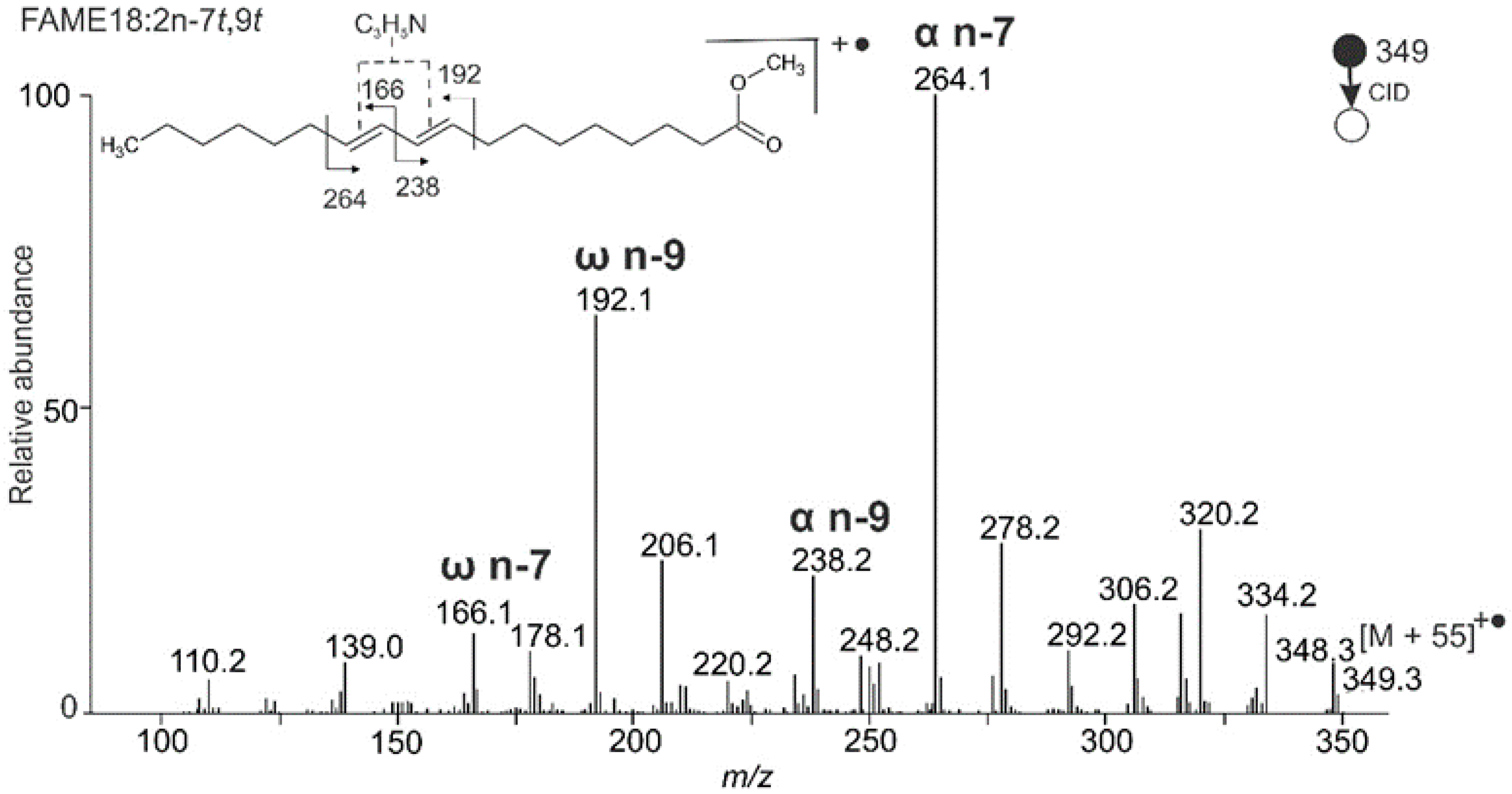
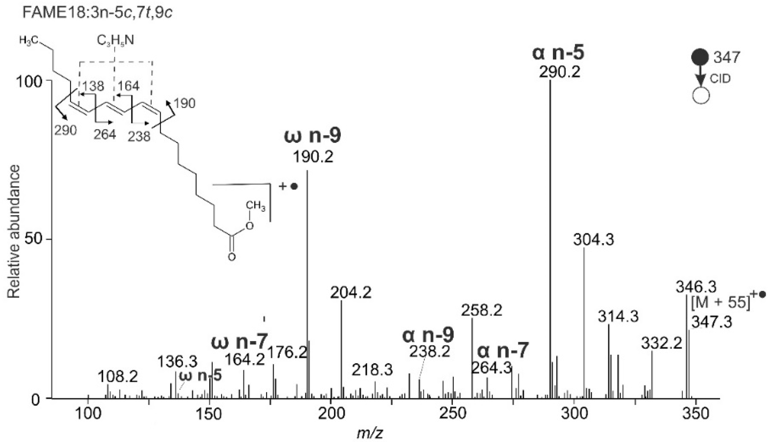
2.1. Mass Spectra of Standards with Conjugated Double Bonds
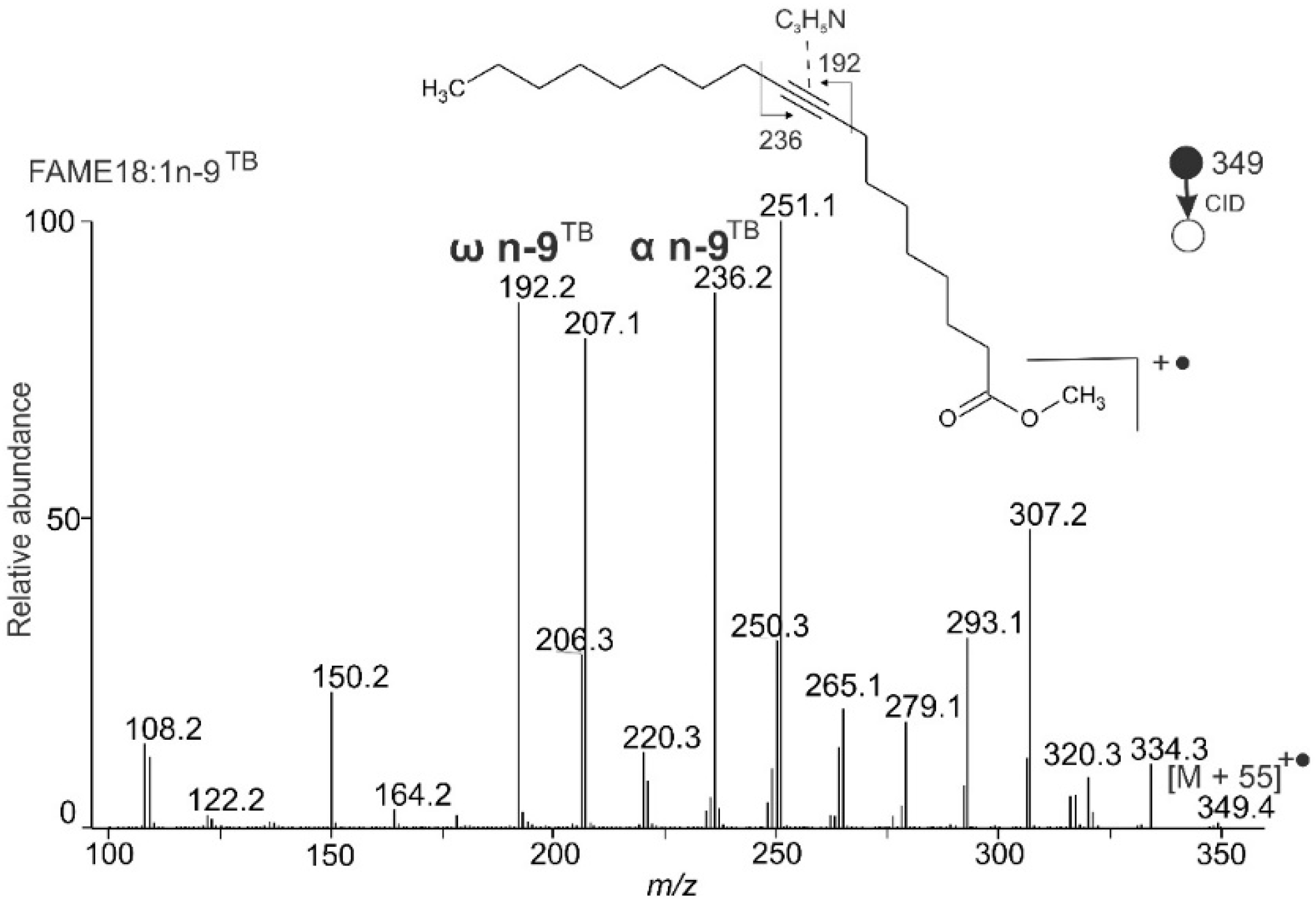
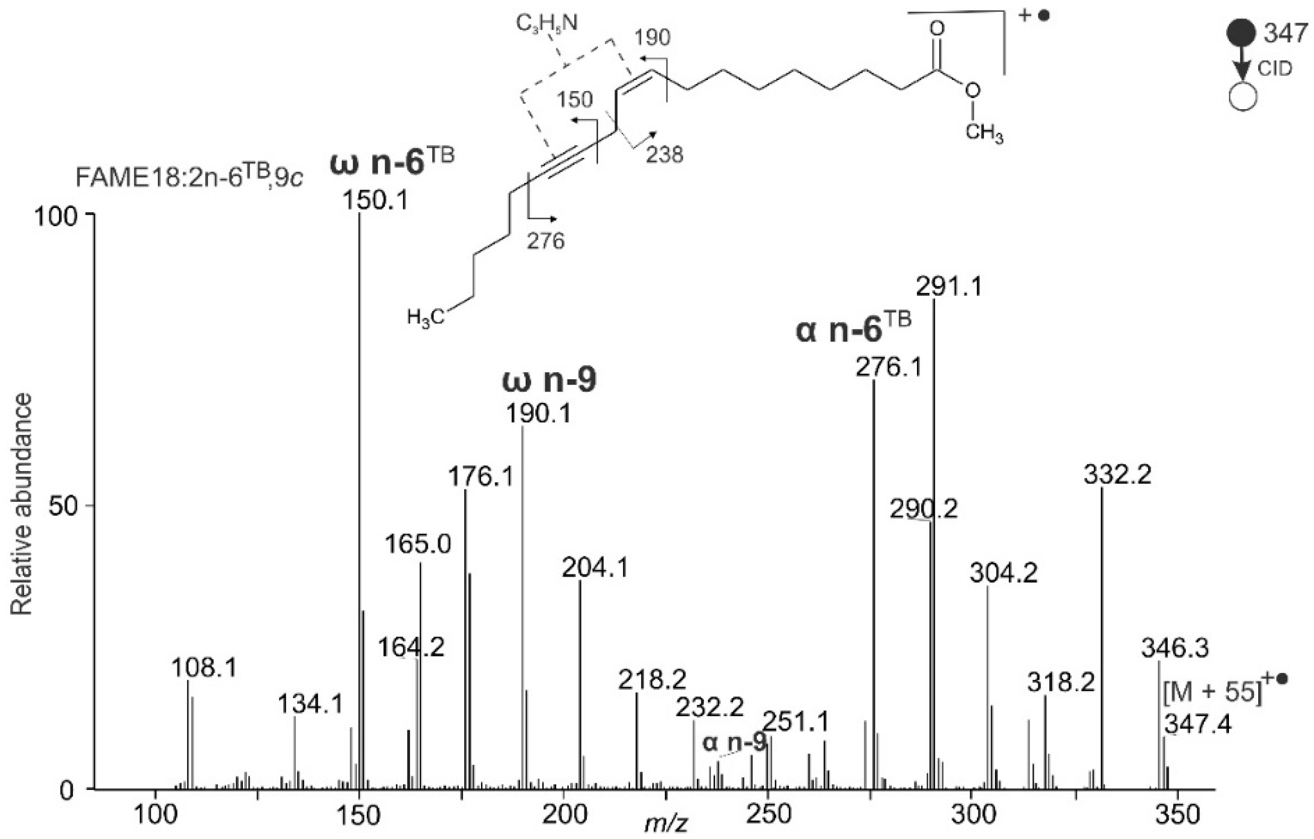
2.2. Mass Spectra of Standards with a Triple Bond
2.3. Analysis of Natural Samples
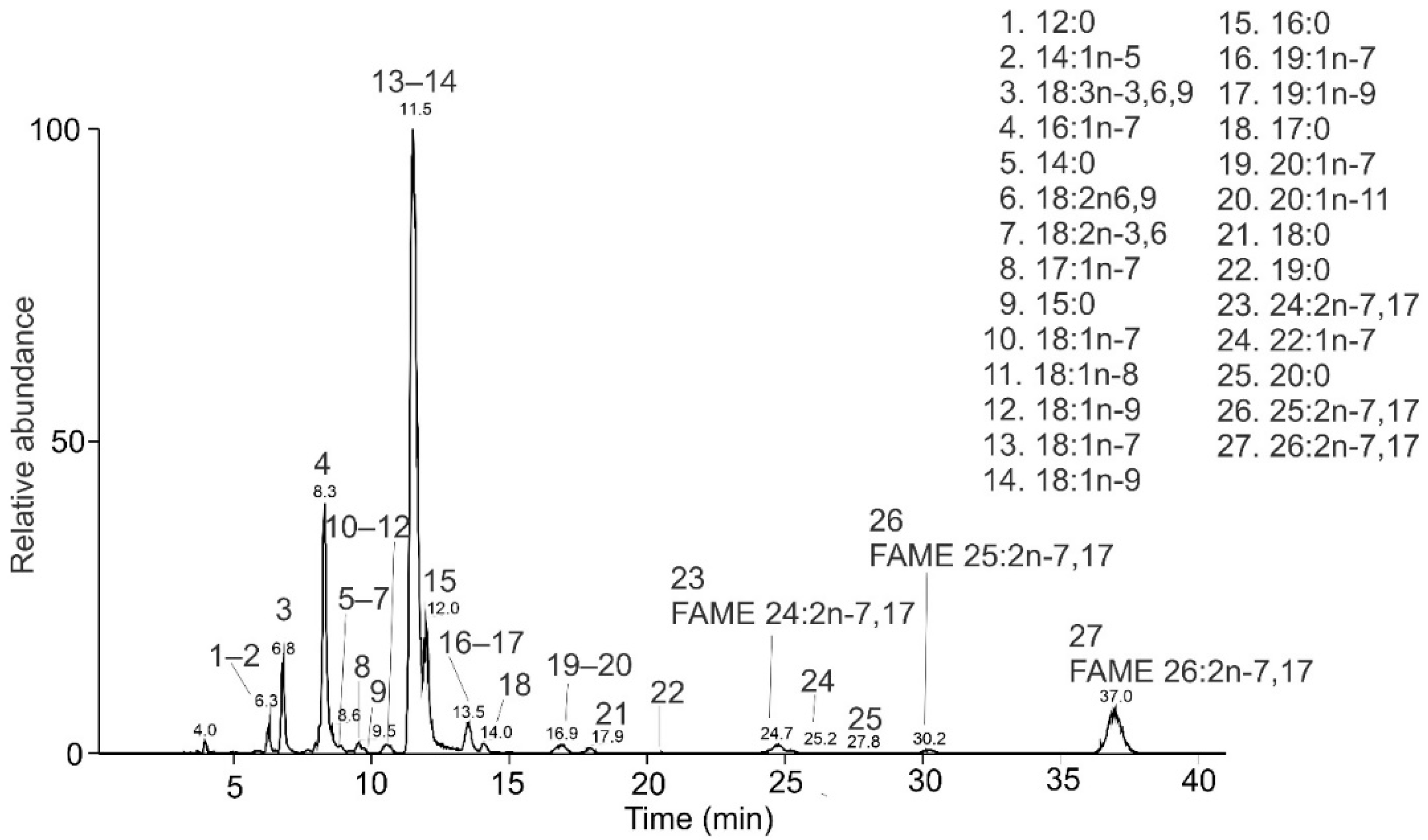
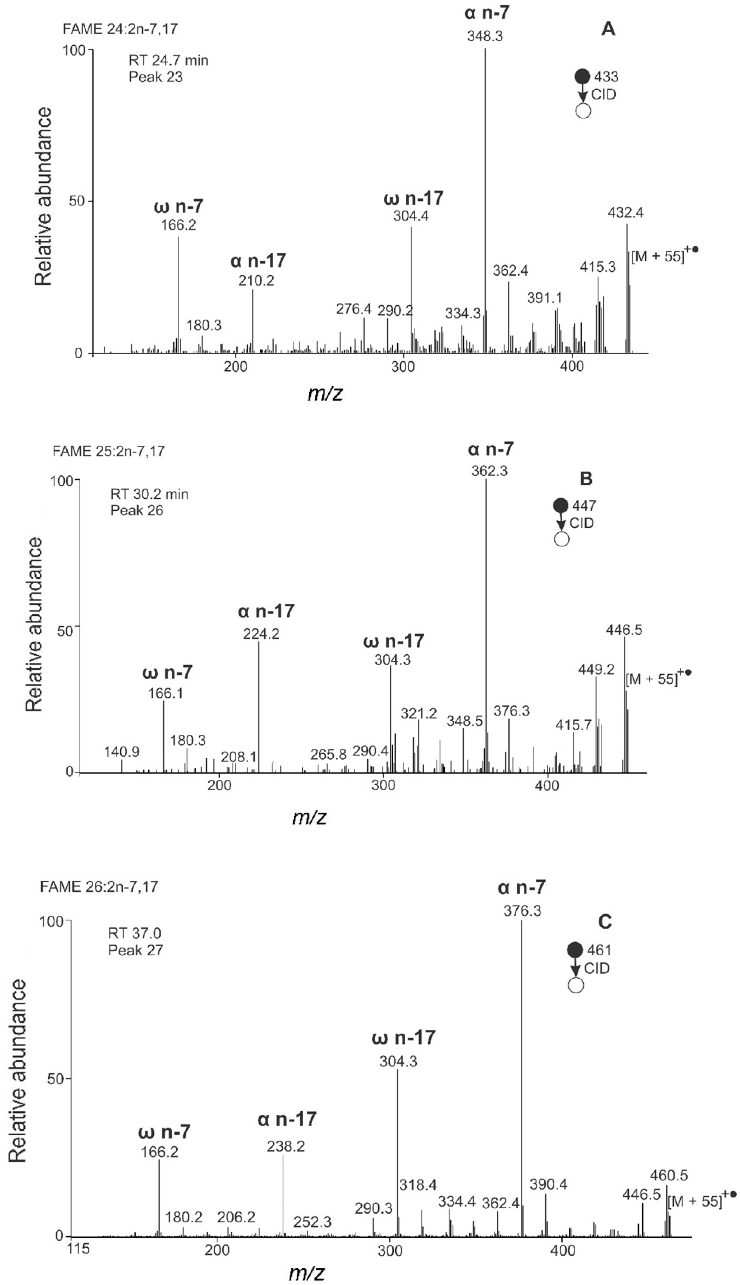
2.3.1. FAMEs from the Fat Body of Bombus pratorum
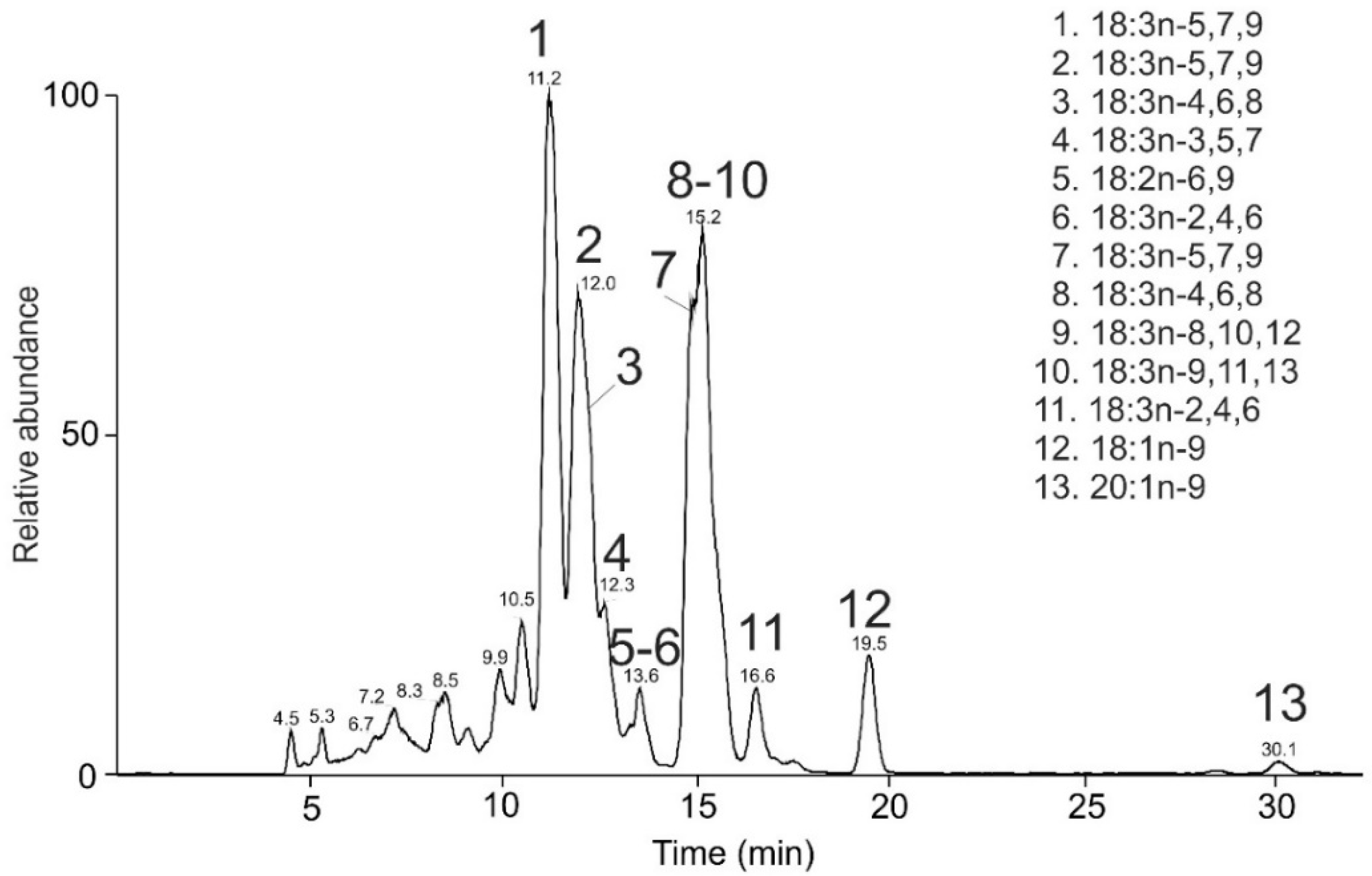
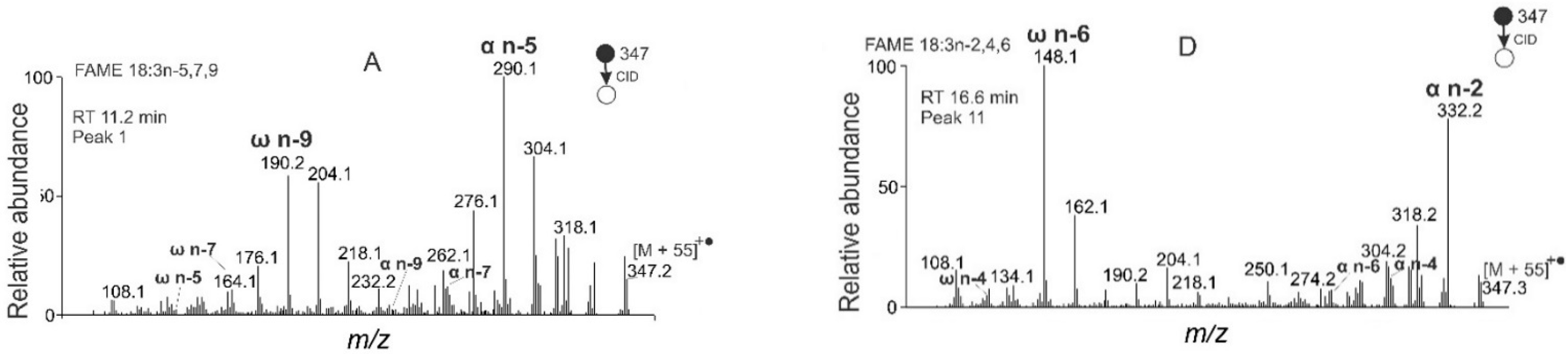
2.3.2. FAMEs from Pomegranate Seed Oil
2.3.3. FAMEs from Marrubium vulgare Seeds
2.3.4. FAMEs from Santalum album Seeds
3. Experimental
3.1. Chemicals and Materials
3.2. Extraction and Transesterification of Lipids
3.3. RP-HPLC/APCI-MS and APCI-MS
3.4. Fragment Ion Abbreviations and Nomenclature
4. Conclusions
Supplementary Materials
Author Contributions
Funding
Institutional Review Board Statement
Data Availability Statement
Acknowledgments
Conflicts of Interest
Sample Availability
References
- Dembitsky, V.M. Anticancer activity of natural and synthetic acetylenic lipids. Lipids 2006, 41, 883–924. [Google Scholar] [CrossRef] [PubMed]
- Li, X.-C.; Jacob, M.R.; Khan, S.I.; Ashfaq, M.K.; Babu, K.S.; Agarwal, A.K.; El Sohly, H.N.; Manly, S.P.; Clark, A.M. Potent In Vitro Antifungal Activities of Naturally Occurring Acetylenic Acids. Antimicrob. Agents Chemother. 2008, 52, 2442–2448. [Google Scholar] [CrossRef] [PubMed] [Green Version]
- Vetter, W.; Walther, W.; Vecchi, M. Pyrrolidide als Derivate für die Strukturaufklärung aliphatischer und alicyclischer Carbonsäuren mittels Massenspektrometrie. Helv. Chim. Acta 1971, 54, 1599–1605. [Google Scholar] [CrossRef]
- Yu, Q.T.; Liu, B.N.; Zhang, J.Y.; Huang, Z.H. Location of methyl branchings in fatty acids: Fatty acids in uropygial secretion of shanghai duck by GC-MS of 4,4-dimethyloxazoline derivatives. Lipids 1989, 24, 160. [Google Scholar] [CrossRef] [Green Version]
- Francis, G.W.; Veland, K. Alkylthiolation for the determination of double-bond positions in linear alkenes. J. Chromatogr. A 1987, 219, 379–384. [Google Scholar] [CrossRef]
- Ma, X.; Chong, L.; Tian, R.; Shi, R.; Hu, T.; Ouyang, Z.; Xia, Y. Identification and quantitation of lipid C=C location isomers: A shotgun lipidomics approach enabled by photochemical reaction. Proc. Natl. Acad. Sci. USA 2016, 113, 2573–2578. [Google Scholar] [CrossRef] [Green Version]
- Murphy, R.C.; Okuno, T.; Johnson, C.A.; Barkley, R.M. Determination of Double Bond Positions in Polyunsaturated Fatty Acids Using the Photochemical Paternò-Büchi Reaction with Acetone and Tandem Mass Spectrometry. Anal. Chem. 2017, 89, 8545–8553. [Google Scholar] [CrossRef] [PubMed]
- Xie, X.; Xia, Y. Analysis of Conjugated Fatty Acid Isomers by the Paternò-Büchi Reaction and Trapped Ion Mobility Mass Spectrometry. Anal. Chem. 2019, 91, 7173–7180. [Google Scholar] [CrossRef] [PubMed]
- Zhao, Y.; Zhao, H.; Zhao, X.; Jia, J.; Ma, Q.; Zhang, S.; Zhang, X.; Chiba, H.; Hui, S.-P.; Ma, X. Identification and Quantitation of C=C Location Isomers of Unsaturated Fatty Acids by Epoxidation Reaction and Tandem Mass Spectrometry. Anal. Chem. 2017, 89, 10270–10278. [Google Scholar] [CrossRef] [PubMed]
- Song, C.; Gao, D.; Li, S.; Liu, L.; Chen, X.; Jiang, Y. Determination and quantification of fatty acid C=C isomers by epoxidation reaction and liquid chromatography-mass spectrometry. Anal. Chim. Acta 2019, 1086, 82–89. [Google Scholar] [CrossRef] [PubMed]
- Wan, L.; Gong, G.; Liang, H.; Huang, G. In situ analysis of unsaturated fatty acids in human serum by negative-ion paper spray mass spectrometry. Anal. Chim. Acta 2019, 1075, 120–127. [Google Scholar] [CrossRef] [PubMed]
- Takashima, S.; Toyoshi, K.; Yamamoto, T.; Shimozawa, N. Positional determination of the carbon–carbon double bonds in unsaturated fatty acids mediated by solvent plasmatization using LC–MS. Sci. Rep. 2020, 10, 12988. [Google Scholar] [CrossRef] [PubMed]
- Yang, W.-C.; Adamec, A.J.; Regnier, F.E. Enhancement of the LC/MS Analysis of Fatty Acids through Derivatization and Stable Isotope Coding. Anal. Chem. 2007, 79, 5150–5157. [Google Scholar] [CrossRef] [PubMed]
- Yang, K.; Dilthey, B.G.; Gross, R.W. Identification and Quantitation of Fatty Acid Double Bond Positional Isomers: A Shotgun Lipidomics Approach Using Charge-Switch Derivatization. Anal. Chem. 2013, 85, 9742–9750. [Google Scholar] [CrossRef] [Green Version]
- Thomas, M.C.; Mitchell, T.W.; Harman, D.G.; Deeley, J.M.; Murphy, R.C.; Blanksby, S.J. Elucidation of Double Bond Position in Unsaturated Lipids by Ozone Electrospray Ionization Mass Spectrometry. Anal. Chem. 2007, 79, 5013–5022. [Google Scholar] [CrossRef] [PubMed] [Green Version]
- Mitchell, T.W.; Pham, H.; Thomas, M.C.; Blanksby, S.J. Identification of double bond position in lipids: From GC to OzID. J. Chromatogr. B 2009, 877, 2722–2735. [Google Scholar] [CrossRef]
- Poad, B.L.J.; Marshall, D.L.; Harazim, E.; Gupta, R.; Narreddula, V.R.; Young, R.S.E.; Duchoslav, E.; Campbell, J.L.; Broadbent, J.A.; Cvačka, J.; et al. Combining Charge-Switch Derivatization with Ozone-Induced Dissociation for Fatty Acid Analysis. J. Am. Soc. Mass Spectrom. 2019, 30, 2135–2143. [Google Scholar] [CrossRef]
- Xu, Y.; Brenna, J.T. Atmospheric Pressure Covalent Adduct Chemical Ionization Tandem Mass Spectrometry for Double Bond Localization in Monoene- and Diene-Containing Triacylglycerols. Anal. Chem. 2007, 79, 2525–2536. [Google Scholar] [CrossRef] [Green Version]
- Vrkoslav, V.; Háková, M.; Pecková, K.; Urbanová, K.; Cvačka, J. Localization of Double Bonds in Wax Esters by High-Performance Liquid Chromatography/Atmospheric Pressure Chemical Ionization Mass Spectrometry Utilizing the Fragmentation of Acetonitrile-Related Adducts. Anal. Chem. 2011, 83, 2978–2986. [Google Scholar] [CrossRef]
- Vrkoslav, V.; Cvačka, J. Identification of the double-bond position in fatty acid methyl esters by liquid chromatography/atmospheric pressure chemical ionisation mass spectrometry. J. Chromatogr. A 2012, 1259, 244–250. [Google Scholar] [CrossRef]
- Háková, E.; Vrkoslav, V.; Mikova, R.; Schwarzová, K.; Bosakova, Z.; Cvačka, J. Localization of double bonds in triacylglycerols using high-performance liquid chromatography/atmospheric pressure chemical ionization ion-trap mass spectrometry. Anal. Bioanal. Chem. 2015, 407, 5175–5188. [Google Scholar] [CrossRef] [PubMed]
- Šubčíková, L.; Hoskovec, M.; Vrkoslav, V.; Čmelíková, T.; Háková, E.; Míková, R.; Coufal, P.; Doležal, A.; Plavka, R.; Cvačka, J. Analysis of 1,2-diol diesters in vernix caseosa by high-performance liquid chromatography—Atmospheric pressure chemical ionization mass spectrometry. J. Chromatogr. A 2015, 1378, 8–18. [Google Scholar] [CrossRef] [PubMed]
- Kalužíková, A.; Vrkoslav, V.; Harazim, E.; Hoskovec, M.; Plavka, R.; Buděšínský, M.; Bosáková, Z.; Cvačka, J. Cholesteryl esters of ω-(O-acyl)-hydroxy fatty acids in vernix caseosa. J. Lipid Res. 2017, 58, 1579–1590. [Google Scholar] [CrossRef] [PubMed] [Green Version]
- Van Pelt, C.K.; Carpenter, B.K.; Brenna, J.T. Studies of structure and mechanism in acetonitrile chemical ionization tandem mass spectrometry of polyunsaturated fatty acid methyl esters. J. Am. Soc. Mass Spectrom. 1999, 10, 1253–1262. [Google Scholar] [CrossRef] [Green Version]
- Michaud, A.L.; Diau, G.-Y.; Abril, R.; Brenna, J. Double bond localization in minor homoallylic fatty acid methyl esters using acetonitrile chemical ionization tandem mass spectrometry. Anal. Biochem. 2002, 307, 348–360. [Google Scholar] [CrossRef]
- Michaud, A.L.; Yurawecz, M.P.; Delmonte, P.; Corl, B.A.; Bauman, D.E.; Brenna, J.T. Identification and Characterization of Conjugated Fatty Acid Methyl Esters of Mixed Double Bond Geometry by Acetonitrile Chemical Ionization Tandem Mass Spectrometry. Anal. Chem. 2003, 75, 4925–4930. [Google Scholar] [CrossRef] [PubMed]
- Lawrence, P.; Brenna, J.T. Acetonitrile Covalent Adduct Chemical Ionization Mass Spectrometry for Double Bond Localization in Non-Methylene-Interrupted Polyene Fatty Acid Methyl Esters. Anal. Chem. 2006, 78, 1312–1317. [Google Scholar] [CrossRef] [PubMed]
- Barthélemy, M.; Elie, N.; Pellissier, L.; Wolfender, J.-L.; Stien, D.; Touboul, D.; Eparvier, V. Structural Identification of Antibacterial Lipids from Amazonian Palm Tree Endophytes through the Molecular Network Approach. Int. J. Mol. Sci. 2019, 20, 2006. [Google Scholar] [CrossRef] [Green Version]
- Spitzer, V.; Marx, F.; Maia, J.G.; Pfeilsticker, K. Curupira tefeensis II: Occurrence of Acetylenic Fatty Acids. Fette Seifen Anstrichm. 1991, 93, 169–174. [Google Scholar] [CrossRef]
- Spitzer, V.; Bordignon, S.A.D.L.; Schenkel, E.P.; Marx, F. Identification of nine acetylenic fatty acids, 9-hydroxystearic acid and 9,10-epoxystearic acid in the seed oil of Jodina rhombifolia Hook et Arn. (Santalaceae). J. Am. Oil Chem. Soc. 1994, 71, 1343–1348. [Google Scholar] [CrossRef]
- Spitzer, V. The mass spectra of the 4,4-dimethyloxazoline derivatives of some conjugated hydroxy ene-yne C17 and C18 fatty acids. J. Am. Oil Chem. Soc. 1996, 73, 489–492. [Google Scholar] [CrossRef]
- Spitzer, V.; Tomberg, W.; Hartmann, R.; Aichholz, R. Analysis of the seed oil of Heisteria silvanii (Olacaceae)—A rich source of a novel C18 acetylenic fatty acid. Lipids 1997, 32, 1189–1200. [Google Scholar] [CrossRef] [PubMed]
- Gurr, M.I.; Harwood, J.L.; Frayn, K.N.; Murphy, D.J.; Michell, R.H. Lipids: Biochemistry, Biotechnology and Health, 6th ed.; Wiley-Blackwell: Hoboken, NJ, USA, 2016. [Google Scholar]
- Litchfield, C.; Greenberg, A.J.; Noto, G.; Morales, R.W. Unusually high levels of C24−C30 fatty acids in sponges of the class Demospongiae. Lipids 1976, 11, 567–570. [Google Scholar] [CrossRef] [PubMed]
- Morales, R.W.; Litchfield, C. Incorporation of 1-14C-Acetate into C26 fatty acids of the marine sponge Microciona prolifera. Lipids 1977, 12, 570–576. [Google Scholar] [CrossRef] [PubMed]
- Christie, W.W.; Brechany, E.Y.; Stefanov, K.; Popov, S. The fatty acids of the sponge Dysidea fragilis from the black sea. Lipids 1992, 27, 640–644. [Google Scholar] [CrossRef]
- Nechev, J.; Christie, W.W.; Robaina, R.; De Diego, F.; Popov, S.; Stefanov, K. Chemical composition of the sponge Hymeniacidon sanguinea from the Canary Islands. Comp. Biochem. Physiol. Part A Mol. Integr. Physiol. 2004, 137, 365–374. [Google Scholar] [CrossRef] [PubMed] [Green Version]
- Kawashima, H. Unusual minor nonmethylene-interrupted di-, tri-, and tetraenoic fatty acids in limpet gonads. Lipids 2005, 40, 627–630. [Google Scholar] [CrossRef]
- Zhukova, N.V. Lipid Classes and Fatty Acid Composition of the Tropical Nudibranch Mollusks Chromodoris sp. and Phyllidia coelestis. Lipids 2007, 42, 1169–1175. [Google Scholar] [CrossRef]
- Carballeira, N. New advances in fatty acids as antimalarial, antimycobacterial and antifungal agents. Prog. Lipid Res. 2008, 47, 50–61. [Google Scholar] [CrossRef] [PubMed] [Green Version]
- Cvačka, J.; Kofroňová, E.; Vašíčková, S.; Stránský, K.; Jiroš, P.; Hovorka, O.; Kindl, J.; Valterová, I. Unusual Fatty Acids in the Fat Body of the Early Nesting Bumblebee, Bombus pratorum. Lipids 2008, 43, 441–450. [Google Scholar] [CrossRef] [PubMed]
- Sehat, N.; Kramer, J.K.G.; Mossoba, M.M.; Yurawecz, M.P.; Roach, J.A.G.; Eulitz, K.; Morehouse, K.M.; Ku, Y. Identification of conjugated linoleic acid isomers in cheese by gas chromatography, silver ion high performance liquid chromatography and mass spectral reconstructed ion profiles. Comparison of chromatographic elution sequences. Lipids 1998, 33, 963–971. [Google Scholar] [CrossRef] [PubMed]
- Yurawecz, M.P.; Roach, J.A.G.; Sehat, N.; Mossoba, M.M.; Kramer, J.K.G.; Fritsche, J.; Steinhart, H.; Ku, Y. A new conjugated linoleic acid isomer, 7 trans, 9 cis-octadecadienoic acid, in cow milk, cheese, beef and human milk and adipose tissue. Lipids 1998, 33, 803–809. [Google Scholar] [CrossRef] [PubMed]
- Ip, C.; Chin, S.F.; Scimeca, J.A.; Pariza, M.W. Mammary cancer prevention by conjugated dienoic derivative of linoleic acid. Cancer Res. 1991, 51, 6118–6124. [Google Scholar]
- Pariza, M.W.; Park, Y.; Cook, M.E. The biologically active isomers of conjugated linoleic acid. Prog. Lipid Res. 2001, 40, 283–298. [Google Scholar] [CrossRef]
- O’Connor, R.; Heinzelman, D.; Freeman, A.; Pack, F. Spectrophotometric Determination of Alpha-Eleostearic Acid in Freshly Extracted Tung Oil Determination of Extinction Coefficients in Oil Solvents. Ind. Eng. Chem. Anal. Ed. 1945, 17, 467–470. [Google Scholar] [CrossRef]
- Özgül-Yücel, S. Determination of conjugated linolenic acid content of selected oil seeds grown in Turkey. J. Am. Oil Chem. Soc. 2005, 82, 893–897. [Google Scholar] [CrossRef]
- Hopkins, C.Y.; Chisholm, M.J. A survey of the conjugated fatty acids of seed oils. J. Am. Oil Chem. Soc. 1968, 45, 176–182. [Google Scholar] [CrossRef]
- Chisholm, M.J.; Hopkins, C.Y. Conjugated fatty acids of tragopogon and calendula seed oils. Can. J. Chem. 1960, 38, 2500–2507. [Google Scholar] [CrossRef] [Green Version]
- Toyama, Y.; Tsuchiya, T. A new stereoisomer of eleostearic acid in pomegranate seed oil. J. Soc. Chem. Ind. Jpn. B 1935, 38, 182–185. [Google Scholar]
- Cao, Y.; Gao, H.-L.; Chen, J.-N.; Chen, Z.-Y.; Yang, L. Identification and Characterization of Conjugated Linolenic Acid Isomers by Ag+-HPLC and NMR. J. Agric. Food Chem. 2006, 54, 9004–9009. [Google Scholar] [CrossRef]
- Saha, S.S.; Patra, M.; Ghosh, M. In vitro antioxidant study of vegetable oils containing conjugated linolenic acid isomers. LWT 2012, 46, 10–15. [Google Scholar] [CrossRef]
- Aruna, P.; Venkataramanamma, D.; Singh, A.K.; Singh, R. Health Benefits of Punicic Acid: A Review. Compr. Rev. Food Sci. Food Saf. 2015, 15, 16–27. [Google Scholar] [CrossRef] [PubMed]
- De Melo, I.L.P.; de Carvalho, E.B.T.; de Oliveira e Silva, A.M.; Yoshime, L.T.; Sattler, J.A.G.; Pavan, R.T.; Mancini-Filho, J. Characterization of constituents, quality and stability of pomegranate seed oil (Punica granatum L.). Food Sci. Technol. 2016, 36, 132–139. [Google Scholar] [CrossRef] [Green Version]
- Costa, A.; Silva, L.; Torres, A. Chemical composition of commercial cold-pressed pomegranate (Punica granatum) seed oil from Turkey and Israel, and the use of bioactive compounds for samples’ origin preliminary discrimination. J. Food Compos. Anal. 2019, 75, 8–16. [Google Scholar] [CrossRef]
- Benjamin, S.; Spener, F. Conjugated linoleic acids as functional food: An insight into their health benefits. Nutr. Metab. 2009, 6, 36. [Google Scholar] [CrossRef] [Green Version]
- Dubey, K.K.D.; Sharma, G.; Kumar, A. Conjugated Linolenic Acids: Implication in Cancer. J. Agric. Food Chem. 2019, 67, 6091–6101. [Google Scholar] [CrossRef] [PubMed]
- Badami, R.; Patil, K. Structure and occurrence of unusual fatty acids in minor seed oils. Prog. Lipid Res. 1980, 19, 119–153. [Google Scholar] [CrossRef]
- Dembitsky, V.M.; Maoka, T. Allenic and cumulenic lipids. Prog. Lipid Res. 2007, 46, 328–375. [Google Scholar] [CrossRef]
- Bagby, M.O.; Smith, C.R.; Wolff, I.A. Laballenic Acid. A New Allenic Acid from Leonotis nepetaefolia Seed Oil1. J. Org. Chem. 1965, 30, 4227–4229. [Google Scholar] [CrossRef]
- Aitzetmüller, D.U.P.D.K.; Tsevegsüren, N.; Vosmann, K. A New Allenic Fatty Acid in Phlomis (Lamiaceae) Seed Oil. Fette Seifen Anstrichm. 1997, 99, 74–78. [Google Scholar] [CrossRef]
- Mikolajczak, K.L.; Rogers, M.F.; Smith, C.R.; Wolff, I.A. An octadecatrienoic acid from Lamium purpureum L. seed oil containing 5,6-allenic and trans-16-olefinic unsaturation. Biochem. J. 1967, 105, 1245–1249. [Google Scholar] [CrossRef] [Green Version]
- Smith, C. Occurrence of unusual fatty acids in plants. Prog. Chem. Fats Other Lipids 1971, 11, 137–177. [Google Scholar] [CrossRef]
- Bohlmann, F.; Burkhardt, T.; Zdero, C. Naturally Occurring Acetylenes; Academic Press: London, UK, 1973; pp. 1–222. [Google Scholar]
- Huang, Y.; Zhang, S.-B.; Chen, H.-P.; Zhao, Z.-Z.; Li, Z.-H.; Feng, T.; Liu, J.-K. New acetylenic acids and derivatives from the Basidiomycete Craterellus lutescens (Cantharellaceae). Fitoterapia 2016, 115, 177–181. [Google Scholar] [CrossRef] [PubMed]
- Fatope, M.O.; Adoum, O.A.; Takeda, Y. C18 Acetylenic Fatty Acids of Ximenia americana with Potential Pesticidal Activity. J. Agric. Food Chem. 2000, 48, 1872–1874. [Google Scholar] [CrossRef] [PubMed]
- Li, X.-C.; Jacob, M.R.; ElSohly, H.N.; Nagle, D.G.; Smillie, T.J.; Walker, L.A.; Clark, A.M. Acetylenic Acids Inhibiting Azole-Resistant Candida albicans from Pentagonia gigantifolia. J. Nat. Prod. 2003, 66, 1132–1135. [Google Scholar] [CrossRef]
- Carballeira, N.M.; Sanabria, D.; Cruz, C.; Parang, K.; Wan, B.; Franzblau, S. 2,6-hexadecadiynoic acid and 2,6-nonadecadiynoic acid: Novel synthesized acetylenic fatty acids as potent antifungal agents. Lipids 2006, 41, 507–511. [Google Scholar] [CrossRef] [Green Version]
- Xu, T.; Tripathi, S.K.; Feng, Q.; Lorenz, M.; Wright, M.A.; Jacob, M.R.; Mask, M.M.; Baerson, S.R.; Li, X.-C.; Clark, A.M.; et al. A Potent Plant-Derived Antifungal Acetylenic Acid Mediates Its Activity by Interfering with Fatty Acid Homeostasis. Antimicrob. Agents Chemother. 2012, 56, 2894–2907. [Google Scholar] [CrossRef] [PubMed] [Green Version]
- Kilimnik, A.; Kuklev, D.V.; Dembitsky, V.M. Antitumor Acetylenic Lipids. Mathews J. Pharm. Sci. 2016, 1, 5. [Google Scholar]
- Aitzetmüller, K.; Matthäus, B.; Friedrich, H. A new database for seed oil fatty acids—The database SOFA. Eur. J. Lipid Sci. Technol. 2003, 105, 92–103. [Google Scholar] [CrossRef]
- Aitzetmüller, K. Santalbic acid in the plant kingdom. Plant Syst. Evol. 2012, 298, 1609–1617. [Google Scholar] [CrossRef]
- Neff, W.E.; Adlof, R.O.; Konishi, H.; Weisleder, D. High-performance liquid chromatography of the triacylglycerols of Vernonia galamensis and Crepis alpina seed oils. J. Am. Oil Chem. Soc. 1993, 70, 449–455. [Google Scholar] [CrossRef]
- Neff, W.E.; Adlof, R.O.; El-Agaimy, M. Silver ion high-performance liquid chromatography of the triacylglycerols of Crepis alpina seed oil. J. Am. Oil Chem. Soc. 1994, 71, 853–855. [Google Scholar] [CrossRef]
- Sun, J.-Y.; Guo, X.; Smith, M.A. Identification of Crepenynic Acid in the Seed Oil of Atractylodes lancea and A. macrocephala. J. Am. Oil Chem. Soc. 2017, 94, 655–660. [Google Scholar] [CrossRef] [Green Version]
- Anderson, W.H.; Gellerman, J.L. Acetylenic acids from mosses. Lipids 1975, 10, 501–502. [Google Scholar] [CrossRef] [PubMed]
- Dembitsky, V.M.; Řezanka, T. Distribution of acetylenic acids and polar lipids in some aquatic bryophytes. Phytochemistry 1995, 40, 93–97. [Google Scholar] [CrossRef]
- Kalacheva, G.S.; Sushchik, N.N.; Gladyshev, M.I.; Makhutova, O.N. Seasonal dynamics of fatty acids in the lipids of water moss Fontinalis antipyretica from the Yenisei River. Russ. J. Plant Physiol. 2009, 56, 795–807. [Google Scholar] [CrossRef]
- Pejin, B.; Bianco, A.; Newmaster, S.; Sabovljevic, M.; Vujisić, L.; Tešević, V.; Vajs, V.; De Rosa, S. Fatty acids of Rhodobryum ontariense (Bryaceae). Nat. Prod. Res. 2011, 26, 696–702. [Google Scholar] [CrossRef] [PubMed]
- Aveldano, M.I.; VanRollins, M.; Horrocks, L.A. Separation and Quantitation of Free Fatty Acids and Fatty Acid Methyl Esters by Reverse Phase High Pressure Liquid Chromatograph. J. Lipid Res. 1983, 24, 83–93. [Google Scholar] [CrossRef]
- Rao, M.S.; Hidajat, K.; Ching, C.B. Reversed-Phase HPLC: The Separation Method for the Characterization and Purification of Long Chain Polyunsaturated Fatty Acids--A Review. J. Chromatogr. Sci. 1995, 33, 9–21. [Google Scholar] [CrossRef]
- Carballeira, N.; Shalabi, F.; Cruz, C.; Rodriguez, J.; Rodríguez, E. Comparative study of the fatty acid composition of sponges of the genus Ircinia. Identification of the new 23-methyl-5,9-tetracosadienoic acid. Comp. Biochem. Physiol. Part B Comp. Biochem. 1991, 100, 489–492. [Google Scholar] [CrossRef]
- Christie, W.W.; Brechany, E.Y.; Marekov, I.N.; Stefanov, K.L.; Andreev, S.N. The fatty acids of the sponge Hymeniacidon sanguinea from the Black Sea. Comp. Biochem. Physiol. Part B Comp. Biochem. 1994, 109, 245–252. [Google Scholar] [CrossRef]
- Makarieva, T.N.; Santalova, E.A.; Gorshkova, I.A.; Dmitrenok, A.S.; Guzii, A.G.; Gorbach, V.I.; Svetashev, V.I.; Stonik, V.A. A new cytotoxic fatty acid (5Z,9Z)-22-methyl-5,9-tetracosadienoic acid and the sterols from the far Eastern sponge Geodinella robusta. Lipids 2002, 37, 75–80. [Google Scholar] [CrossRef]
- Kullenberg, B.; Bergström, G.; Ställberg-Stenhagen, S. Volatile components of the marking secretion of male bumblebees. Acta Chim. Scand. 1970, 24, 1481–1483. [Google Scholar] [CrossRef] [PubMed] [Green Version]
- Sassano, G.; Sanderson, P.; Franx, J.; Groot, P.; van Straalen, J.; Bassaganya-Riera, J. Analysis of pomegranate seed oil for the presence of jacaric acid. J. Sci. Food Agric. 2009, 89, 1046–1052. [Google Scholar] [CrossRef]
- Elfalleh, W.; Tlili, N.; Nasri, N.; Yahia, Y.; Hannachi, H.; Chaira, N.; Ying, M.; Ferchichi, A. Antioxidant Capacities of Phenolic Compounds and Tocopherols from Tunisian Pomegranate (Punica granatum) Fruits. J. Food Sci. 2011, 76, C707–C713. [Google Scholar] [CrossRef]
- Yoshime, L.T.; De Melo, I.L.P.; Sattler, J.A.G.; Torres, R.P.; Mancini-Filho, J. Bioactive compounds and the antioxidant capacities of seed oils from pomegranate (Punica granatum L.) and bitter gourd (Momordica charantia L.). Food Sci. Technol. 2019, 39, 571–580. [Google Scholar] [CrossRef] [Green Version]
- Hajib, A.; Nounah, I.; Harhar, H.; Gharby, S.; Kartah, B.; Matthäus, B.; Bougrin, K.; Charrouf, Z. Oil content, lipid profiling and oxidative stability of “Sefri” Moroccan pomegranate (Punica granatum L.) seed oil. OCL 2021, 28, 5. [Google Scholar] [CrossRef]
- Topkafa, M.; Kara, H.; Sherazi, S.T.H. Evaluation of the Triglyceride Composition of Pomegranate Seed Oil by RP-HPLC Followed by GC-MS. J. Am. Oil Chem. Soc. 2015, 92, 791–800. [Google Scholar] [CrossRef]
- Alcaraz-Mármol, F.; Nuncio-Jáuregui, N.; Calín-Sánchez, Á.; Carbonell-Barrachina, Á.A.; Martínez, J.J.; Hernández, F. Determination of fatty acid composition in arils of 20 pomegranates cultivars grown in Spain. Sci. Hortic. 2015, 197, 712–718. [Google Scholar] [CrossRef]
- Van Nieuwenhove, C.P.; Moyano, A.; Castro-Gómez, P.; Fontecha, J.; Sáez, G.; Zárate, G.; Pizarro, P.L. Comparative study of pomegranate and jacaranda seeds as functional components for the conjugated linolenic acid enrichment of yogurt. LWT 2019, 111, 401–407. [Google Scholar] [CrossRef]
- Fadavi, A.; Barzegar, M.; Azizi, M.H. Determination of fatty acids and total lipid content in oilseed of 25 pomegranates varieties grown in Iran. J. Food Compos. Anal. 2006, 19, 676–680. [Google Scholar] [CrossRef]
- Hernández, F.; Melgarejo, P.; Martínez, R.; Legua, P. Fatty acid composition of seed oils from important Spanish pomegranate cultivars. Ital. J. Food Sci. 2011, 23, 188–193. [Google Scholar]
- Jing, P.; Ye, T.; Shi, H.; Sheng, Y.; Slavin, M.; Gao, B.; Liu, L.; Yu, L. Antioxidant properties and phytochemical composition of China-grown pomegranate seeds. Food Chem. 2012, 132, 1457–1464. [Google Scholar] [CrossRef] [PubMed]
- Hopkins, C.Y.; Chisholm, M.J.; Orgodnik, J.A. Identity and configuration of conjugated fatty acids in certain seed oils. Lipids 1969, 4, 89–92. [Google Scholar] [CrossRef]
- Montañés, F.; Tallon, S.; Catchpole, O. Isolation of Non-methylene Interrupted or Acetylenic Fatty Acids from Seed Oils Using Semi-preparative Supercritical Chromatography. J. Am. Oil Chem. Soc. 2017, 94, 981–991. [Google Scholar] [CrossRef]
- Stránský, K.; Jursík, T. Simple quantitative transesterification of lipids 1. Introduction. Fette Seifen Anstrichm. 1996, 98, 65–71. [Google Scholar] [CrossRef]
- Christie, W.W.; Han, X. Lipid Analysis—Isolation, Separation, Identification and Structural Analysis of Lipids; The Oily Press: Bridgwater, UK, 2003; p. 212. [Google Scholar]


| MBR | Arrangement of Multiple Bonds |
|---|---|
| 79 | One triple bond –C≡C– |
| 81 | One double bond –CH=CH– |
| 93 | Two cumulated double bonds –CH=C=CH– |
| 103 | Two conjugated triple bonds –C≡C–C≡C– |
| 105 | One double bond and one triple bond, conjugated –CH=CH–C≡C– |
| 107 | Two conjugated double bonds –CH=CH–CH=CH– |
| 119 | One double bond and one triple bond, methylene-interrupted –CH=CH–CH2–C≡CH– |
| 121 | Two methylene-interrupted double bonds –CH=CH–CH2–CH=CH– |
| 133 | Three conjugated double bonds –CH=CH–CH=CH–CH=CH– |
| 161 | Three methylene-interrupted double bonds –CH=CH–CH2–CH=CH–CH2–CH=CH– |
| 14n + 107 | Two double bonds interrupted by several methylenes (–CH2–)n |
| FAME | tR (min) | Rel. Peak Area (%) | Literature Data (%) * |
|---|---|---|---|
| 12:0 | 6.2 | 1.3 | 3.7 ± 4.2 |
| 14:1n-5 | 6.5 | ˂0.1 | 0.2 ± 0.3 |
| 18:3n-3,6,9 | 6.8 | 4.3 | 2.7 ± 0.3 |
| 16:1n-7 | 8.3 | 13.7 | 7.7 ± 1.9 |
| 16:1n-5 | - | - | 0.8 ± 0.4 |
| 14:0 | 8.4 | 1.7 | 5.4 ± 2.1 |
| 18:2n-6,9 | 8.5 | 0.7 | 0.8 ± 0.6 |
| 18:2n-3,6 | 8.5 | 0.3 | - |
| 17:1n-7 | 9.4 | 0.4 | - |
| 15:0 | 9.7 | 0.2 | ˂0.1 |
| 18:1n-7 | 10.5 | 0.6 | - |
| 18:1n-8 | 10.6 | ˂0.1 | - |
| 18:1n-9 | 10.6 | 0.3 | - |
| 18:1n-7 | 11.5 | 35.2 | 17.3 ± 3.6 |
| 18:1n-9 | 11.5 | 19.3 | 35.1 ± 3.2 |
| 16:0 | 12.0 | 5.5 | 18.5 ± 2.4 |
| 19:1n-9 | 13.5 | 2.6 | - |
| 19:1n-7 | 13.5 | 0.6 | - |
| 17:0 | 14.0 | 0.5 | - |
| 20:1n-7 | 16.9 | 0.8 | ˂0.1 |
| 20:1n-11 | 16.9 | 0.6 | ˂0.1 |
| 18:0 | 17.9 | 0.1 | 0.7 ± 0.3 |
| 19:0 | 21.4 | ˂0.1 | - |
| 24:2n-7,17 | 24.7 | 1.4 | - |
| 22:1n-7 | 25.2 | 0.3 | - |
| 20:0 | 28.0 | ˂0.1 | 0.2 ± 0.1 |
| 22:0 | - | - | 0.3 ± 0.1 |
| 25:2n-7,17 | 30.2 | 0.5 | - |
| 26:2n7,17 | 37.0 | 9.2 | 5.0 ± 2.4 |
| 23:0 | 38.5 | ˂0.1 | - |
| 24:1n-15 | 39.3 | ˂0.1 | ˂0.1 |
| 24:0 | - | - | ˂0.1 |
| 26:1n-17 | - | - | 0.2 ± 0.1 |
| FAME | tR (min) | Rel. Peak Area (%) | References |
|---|---|---|---|
| 18:3n-5,7,9 | 11.2 | 36.0 | [51,86,87,89,93,94,95] |
| 18:3n-5,7,9 | 12.0 | 24.2 | [51,86,87,89] |
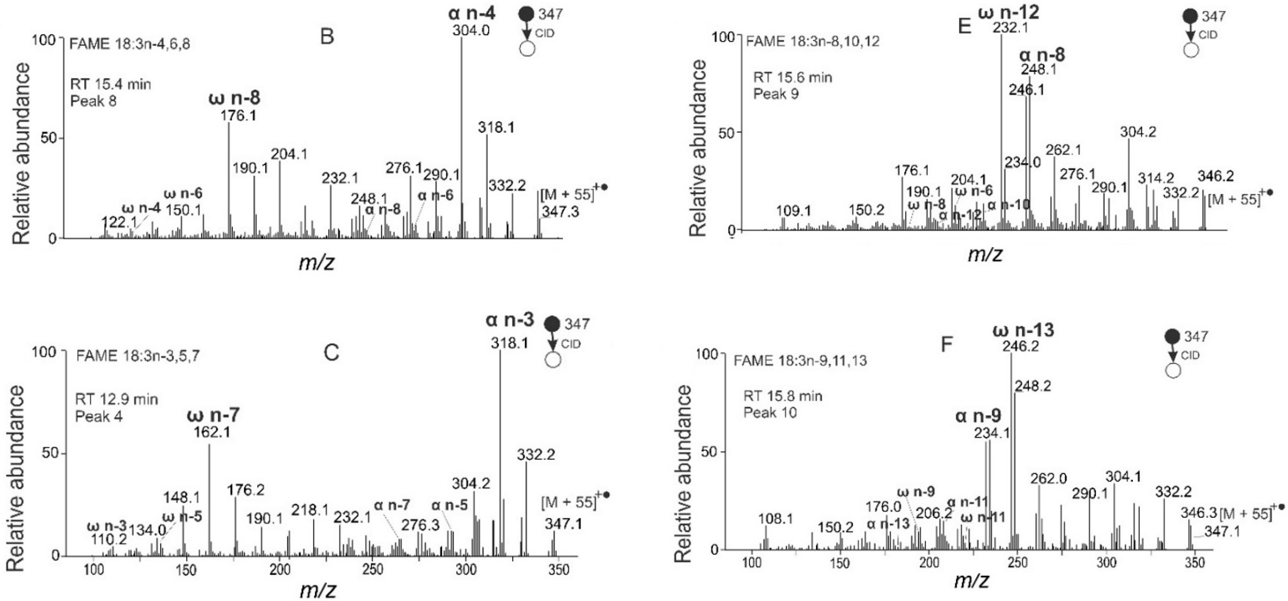
| 18:3n-4,6,8 | 12.3 | 2.5 | [51] |
| 18:3n-3,5,7 | 12.9 | 1.7 | [51] |
| 18:2n-6,9 | 13.6 | 1.5 | [86,87,89,93,94,95] |
| 18:3n-2,4,6 | 13.7 | 1.0 | - |
| 18:3n-5,7,9 | 14.8 | 9.4 | [51,86,87,89] |
| 18:3n-4,6,8 | 15.4 | 10.1 | [51] |
| 18:3n-8,10,12 | 15.6 | 3.3 | - |
| 18:3n-9,11,13 | 15.8 | 2.7 | - |
| 18:3n-2,4,6 | 16.6 | 2.3 | - |
| 18:1n-9 | 19.5 | 4.5 | [86,87,89,93,94,95] |
| 20:1n-9 | 30.1 | 0.8 | [86,89] |
| 14:0 | - | - | [87,93] |
| 16:0 | - | - | [86,87,89,93,94,95] |
| 18:0 | - | - | [86,87,89,93,94,95] |
| 18:3n-3,6,9 | - | - | [95] |
| 20:0 | - | - | [86,87,89,95] |
| 22:0 | - | - | [93] |
| 24:0 | - | - | [86,87] |
| 24:1 | - | - | [86] |
| FAME | tR (min) | Rel. Peak Area (%) | References |
|---|---|---|---|
| 18:3n-3,6,9 | 10.1 | 6.6 | - |
| 18:2n-6,9 | 12.8 | 25.4 | - |
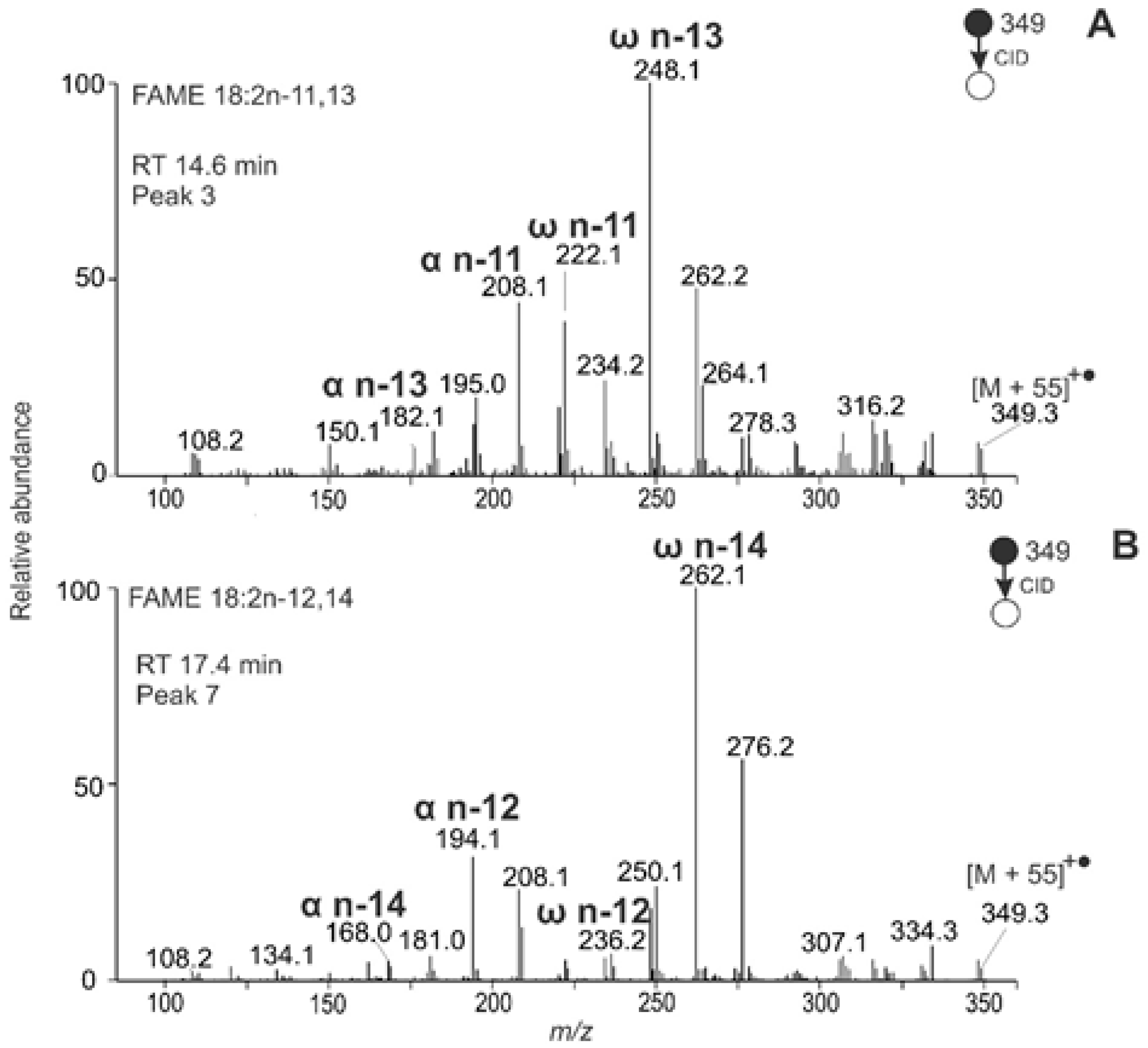
| 18:2n-11,13 | 14.6 | 0.3 | - |
| 19:2n-6,9 | 14.9 | 1.2 | - |
| 18:2n-11,14 | 15.1 | 2.7 | - |
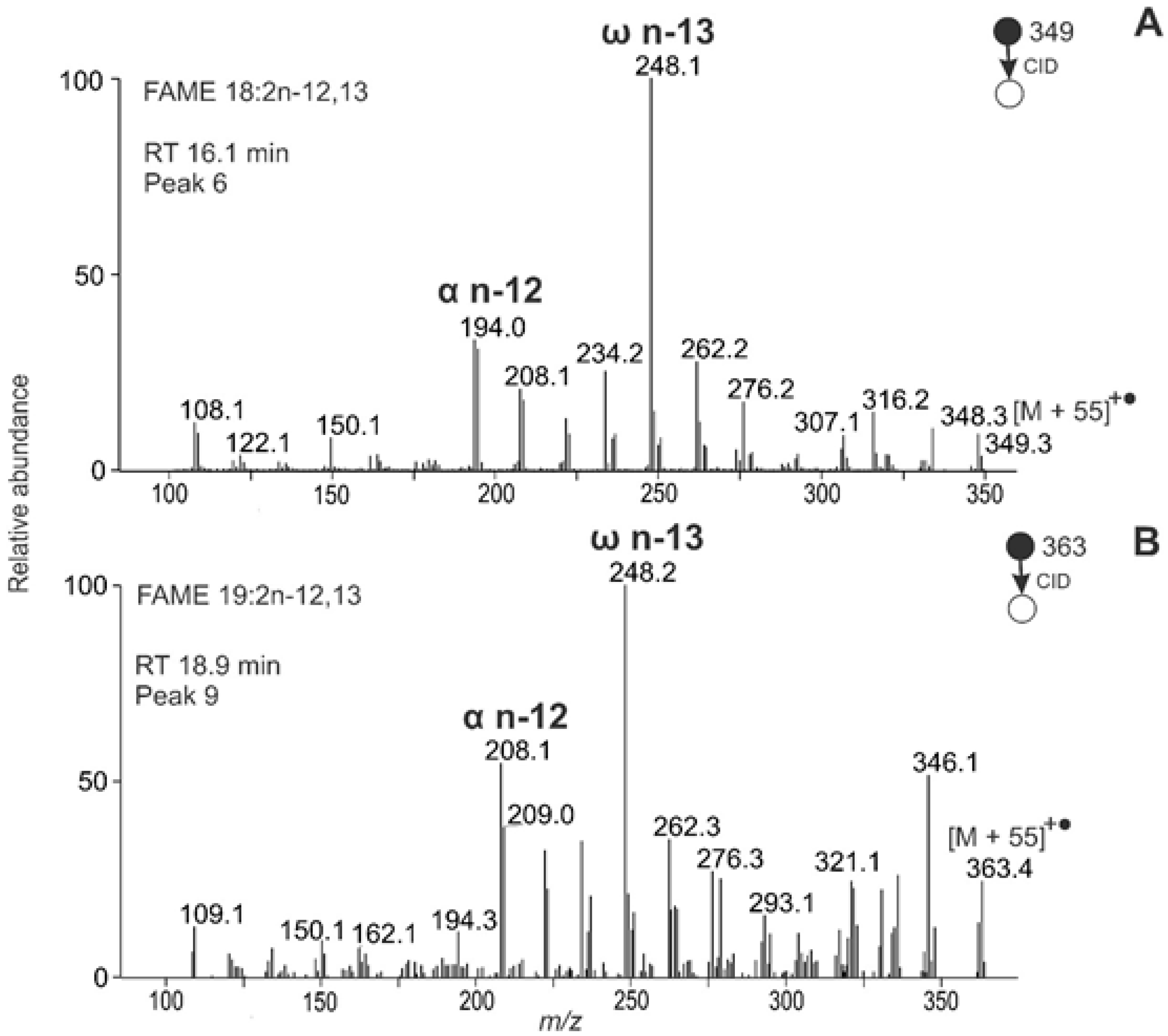
| 18:2n-12,13 | 16.1 | 31.4 | [61] |
| 18:2n-12,14 | 17.4 | 1.6 | - |
| 18:1n-9 | 18.2 | 26.5 | - |
| 19:2n-12,13 | 18.9 | 0.7 | - |
| 18:1n-12 | 19.2 | 0.6 | - |
| 19:1n-10 | 21.5 | 1.5 | - |
| 20:2n-12,13 * | 22.6 | 0.4 | [61] |
| 20:2n-12,14 * | 25.1 | 0.1 | - |
| 20:1n-9 | 27.9 | 0.2 | - |
| 20:1n-12 | 28.8 | 0.9 | - |
| 21:1 * | 35.2 | ˂0.1 | - |
| 20:1n-11 | - | - | [61] |
| FAME | tR (min) | Rel. Peak Area (%) | Literature Data (%) * |
|---|---|---|---|
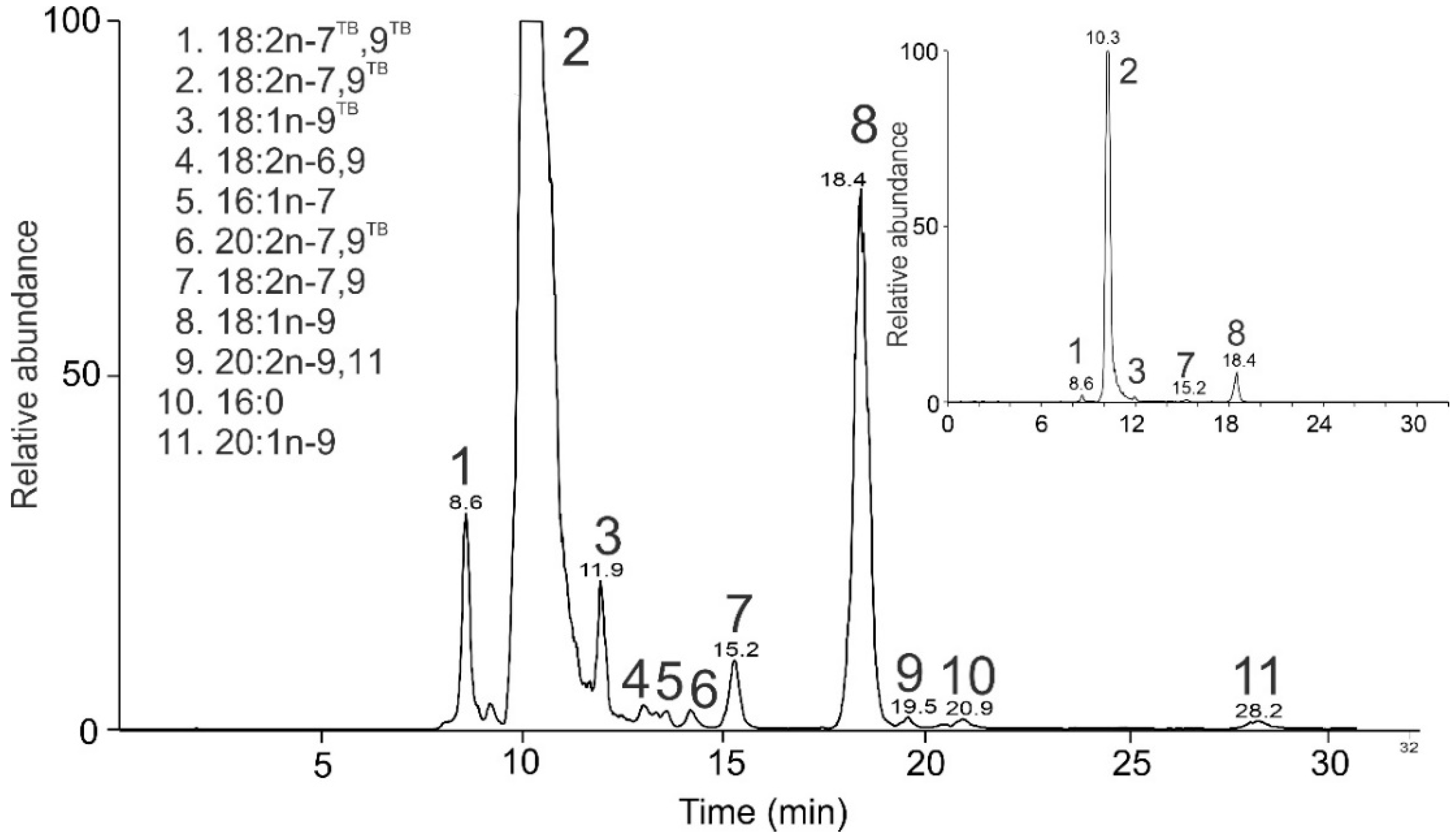
| 18:2 n-7TB,9TB | 8.6 | 1.6 | - |
| 18:2n-7,9TB | 10.3 | 89.0 | 33.5 |
| 18:1n-9TB | 11.9 | 0.7 | - |
| 18:2n-6,9 | 13.0 | 0.2 | 1.5 |
| 16:1n-7 | 13.6 | 0.2 | 0.8 |
| 20:2n-7,9TB | 14.2 | 0.2 | - |
| 18:2n-7,9 | 15.2 | 0.7 | - |
| 18:1n-9 | 18.4 | 7.0 | 52.1 |
| 20:2n-9,11 | 19.5 | 0.1 | - |
| 16:0 | 20.9 | 0.1 | 3.7 |
| 20:1n-9 | 28.2 | 0.2 | - |
| 16:1n-9 | - | - | 0.1 |
| 17:1 | - | - | 0.3 |
| 17:2 | - | - | 0.5 |
| 18:0 | - | - | 1.7 |
| 18:1n-7 | - | - | 1.4 |
| 18:3n-3,6,9 | - | - | 3.1 |
| 18:4n-3,6,9,12 | - | - | 1.3 |
Publisher’s Note: MDPI stays neutral with regard to jurisdictional claims in published maps and institutional affiliations. |
© 2021 by the authors. Licensee MDPI, Basel, Switzerland. This article is an open access article distributed under the terms and conditions of the Creative Commons Attribution (CC BY) license (https://creativecommons.org/licenses/by/4.0/).
Share and Cite
Horká, P.; Vrkoslav, V.; Kindl, J.; Schwarzová-Pecková, K.; Cvačka, J. Structural Characterization of Unusual Fatty Acid Methyl Esters with Double and Triple Bonds Using HPLC/APCI-MS2 with Acetonitrile In-Source Derivatization. Molecules 2021, 26, 6468. https://doi.org/10.3390/molecules26216468
Horká P, Vrkoslav V, Kindl J, Schwarzová-Pecková K, Cvačka J. Structural Characterization of Unusual Fatty Acid Methyl Esters with Double and Triple Bonds Using HPLC/APCI-MS2 with Acetonitrile In-Source Derivatization. Molecules. 2021; 26(21):6468. https://doi.org/10.3390/molecules26216468
Chicago/Turabian StyleHorká, Petra, Vladimír Vrkoslav, Jiří Kindl, Karolina Schwarzová-Pecková, and Josef Cvačka. 2021. "Structural Characterization of Unusual Fatty Acid Methyl Esters with Double and Triple Bonds Using HPLC/APCI-MS2 with Acetonitrile In-Source Derivatization" Molecules 26, no. 21: 6468. https://doi.org/10.3390/molecules26216468